Review

Recent Advances in pH and Redox Responsive Polymer Nanocomposites for Cancer Therapy

by
Shivalingayya Gaddimath
1,
Shivanand Payamalle
2,
Keshavananada Prabhu Channabasavana Hundi Puttaningaiah
3,* and
Jaehyun Hur
3,*
1
Department of Chemistry, Vijayanagara Sri Krishnadevaraya University, Bellary 583105, India
2
Department of Botany, KLE Society’s Gudleppa Hallikeri Arts, Science and Commerce College, Haveri 581110, India
3
Department of Chemical and Biological Engineering, Gachon University, Seongnam-si 13120, Gyeonggi-do, Republic of Korea
*
Authors to whom correspondence should be addressed.
J. Compos. Sci. 2024, 8(1), 28; https://doi.org/10.3390/jcs8010028
Submission received: 29 November 2023 / Revised: 1 January 2024 / Accepted: 8 January 2024 / Published: 11 January 2024
(This article belongs to the Special Issue Progress in Polymer Composites, Volume III)

Abstract

:
Cancer therapy currently focuses on personalized targeted treatments. A promising approach uses stimuli-responsive biomaterials for site-specific drug release, such as pH- and redox-triggered polymer nanocomposites. These materials respond to the tumor microenvironment, enhance efficacy, and reduce off-target effects. Cancer cells with anomalous properties such as acidic cytosolic pH and elevated redox potential are targeted by these biomaterials. An imbalance in ions and biological thiols in the cytoplasm contributes to tumor growth. Functionalized polymer nanocomposites with large surface areas and specific targeting outperform conventional small-molecule materials. To overcome problems such as low bioavailability, uncontrolled drug release, and poor cell penetration, multifunctional nanomaterials make it easier for drugs to enter certain cellular or subcellular systems. High therapeutic efficacy is achieved through surface functionalization, site-specific targeting, and the use of stimuli-responsive components. In particular, pH and redox dual-stimuli-based polymeric nanocomposites for cancer therapeutics have scarcely been reported. This article provides recent progress in pH- and redox-responsive polymer nanocomposites for site-specific drug delivery in cancer therapy. It explores the design principles, fabrication methods, mechanisms of action, and prospects of these dual-stimuli-responsive biomaterials.

Graphical Abstract

1. Introduction

Cancer continues to pose a formidable global health challenge, necessitating constant advancements in treatment modalities [1]. However, despite the efforts invested in the efficient development of cancer mortality [2], the mortality rate has decreased in the past half decade owing to positive outcomes related to killing cancer cells in early cancer diagnosis and a better understanding of improved diagnostic techniques, treatments, and advanced tumor biology [3,4]. Although traditional cancer treatments have worked in some cases, they have some problems, such as suboptimal drug performance and their uneven distribution. Surgery, intense radiation, and chemo/hormonal drugs are often used to treat cancer, which can kill healthy cells and cause toxicity [5,6]. Therefore, the development of effective therapeutic methods is required. Cytotoxic drugs have limited efficacy because they do not stick to cancer cells, and many anticancer drugs are hydrophobic, which makes it hard for them to reach cell surfaces [7,8]. In this context, cancer chemotherapy and detection technologies offer promising tools for reducing side effects, increasing biocompatibility, and improving therapeutic efficacy [9,10]. Smart nanomaterials such as liposomes, micelles, dendrimers, and polymeric nanoparticles (NPs) can mitigate these problems by enabling the creation of targeted drug delivery systems or active intracellular delivery into cancer cells [11]. This method of drug delivery is supported by evidence that NPs interact with the surface of cells, causing the cytosolic pH to drop (acidity) and the redox potential to rise [12,13].
Polymeric nanocarriers have garnered considerable attention owing to their efficient delivery of bioactive compounds for biomedical applications [14]. These carriers can be further modified to incorporate stimuli-responsive signals triggered by endogenous factors such as pH, redox potential, glucose levels, or exogenous factors such as magnetism, light, or ultrasound, enabling precise control of drug release under specific conditions [15,16,17,18,19,20]. Various strategies have been developed to generate stimulus-responsive signals in polymeric nanomaterials, including synthetic and bio-based polymers. This is especially important for overcoming the limitations associated with the release of hydrophobic drugs, including poor absorption, limited bioavailability, and formulation challenges [21,22]. Polymer NPs play a significant role in therapeutics by safeguarding delicate drugs until they reach their intended delivery sites. However, there are numerous physicochemical and biological obstacles that hinder the targeted delivery of cargos, drug solubilization, biocompatibility, and site-specific delivery to cells and tissues, making therapeutic delivery challenging [23,24]. To address these challenges, engineered or smart nanopolymer systems have been developed that possess physicochemical properties that respond to dual stimuli, such as pH/magnetic fields, pH/redox potential, pH/temperature, double pH changes, temperature/reduction, pH and diols, and temperature/enzymes [25,26]. NPs are designed to undergo chemical alterations in response to various biological stimuli. One of the key challenges in cancer treatment and diagnosis is the development of engineered gene and drug delivery systems that specifically target diseased cells without harming healthy tissues or cells, particularly within the tumor microenvironment (TME). Achieving this requires the precise and controlled delivery of anticancer agents [27,28]. Polymer NPs with stimuli-responsive properties can be used in emulsion polymerization, layer-by-layer assembly, and self-assembly [29] to address these problems and improve targeted drug delivery.
Stimuli-responsive biomaterials have emerged as a promising solution for exploring controlled and targeted drug delivery stimuli, offering the potential for personalized and site-specific cancer therapy. Stimuli-responsive biomaterials are carefully designed to detect and react to certain signals in the tumor microenvironment. This allows therapeutic agents to be released precisely where needed. This targeted drug delivery approach aims to maximize therapeutic effects on cancer cells while minimizing damage to healthy tissues, ultimately reducing overall toxicity and enhancing treatment efficacy. By harnessing the unique characteristics of cancer cells and their surrounding microenvironments, among the diverse range of stimuli-responsive biomaterials, pH- and redox-responsive polymer nanocomposites have garnered considerable attention for their dual-stimuli responsiveness and potential as future generation biomaterials. Hence, these novel polymer nanocomposites have been engineered to respond to changes in pH and redox conditions, which are characteristic features of the tumor microenvironment [30]. The slightly acidic pH of tumor tissues, resulting from an increased metabolic rate and inefficient vascularization, serves as a distinctive trigger for pH responsiveness. At the same time, cancer cells have high amounts of reducing agents, such as glutathione (GSH), which makes redox responsiveness possible. The design principles of pH- and redox-responsive polymer nanocomposites involve the incorporation of pH- and redox-sensitive elements into a polymer matrix. These parts allow biomaterials to change their structure and physicochemical properties in a controlled manner when the pH and redox conditions change. This makes it easier for drugs to be released when required. Consequently, pH- and redox-responsive polymer nanocomposites offer the unique advantage of dual-stimuli responsiveness, further enhancing their precision and effectiveness for drug delivery in cancer therapy. For example, the TME displays a coordinated pH reduction with lactic acid, carbonic acid, and metabolic byproducts. This acidity promotes invasiveness and disrupts the delivery of therapeutic agents. Simultaneously, reactive oxygen species (ROS) and reactive nitrogen species (RNS) contribute to the complex oxidative nature, and glutathione depletion creates a tumor-permissive microenvironment. Understanding the acidic and redox species produced at cancer sites provides insights into target-overcoming resistance mechanisms.
This review aims to provide a comprehensive overview of recent advancements in pH- and redox-triggered polymer nanocomposites for site-specific drug release in cancer therapy. This review examines the design ideas and methods used to prepare these biomaterials. It also explains how they work in response to pH and redox stimuli and discusses in vitro and in vivo studies that show how they could be used to treat cancer. Furthermore, this review analyzes the challenges and future perspectives of innovative biomaterials, underscoring the importance of continued research and development in this exciting field. The emergence of pH- and redox-responsive polymer nanocomposites has marked a significant stride toward personalized and targeted cancer therapy, offering hope for improved patient outcomes and a brighter future for cancer treatment.

1.1. Polymeric Nanomaterials

Polymer NPs include organic and inorganic NPs [31]. They are widely used as therapeutic agents because of their significant efficacy and plethora of benefits in cancer treatment [32]. Several subtypes of polymeric NPs have been developed for specific drug-triggered delivery to tumors. NPs are classified into polymeric micelles, dendritic polymers, polymeric nanospheres, and polymeric conjugate complexes [33,34]. The structural and typological diversity of polymeric NPs employed in different applications is shown in Figure 1. The general structure of polymeric NPs is shown in Section (a) [31]. This provides a crucial understanding of the fundamental architectural components of these NPs. This covers the polymer matrix as well as other elements or alterations that support its functional and responsive qualities. The wide variety of polymeric NPs used in various scientific and medical applications is discussed in detail in Section (b) [34]. The structural forms of NPs include brush polymers, micelles, dendrimers, nanofibers, nanoparticles, polymerases, and nanogels. Owing to their distinct qualities, each form can be customized for a particular need. The advantages of polymeric composite materials are that they have properties as drug carriers imposed by low-solubility drug solubilization, provide potential to tumor targets, control drug release, and can achieve specific active targeting in tumor diagnosis [35,36]. Considering that the polymeric micelles are spherical, their sizes range from 10 to 100 nm. The simplicity of polymeric micelle formation is maintained by the spontaneous self-assembly of the hydrophobic and hydrophilic copolymer molecules [37]. Drug-encapsulated nanocarriers provide physical mixing rather than chemical conjugation. Hydrophobic shells and micelles act as drug reservoirs, while hydrophilic shells ensure the solubilization of micelles in aqueous solutions [38,39]. Furthermore, nanocomposites enhance the properties of materials such as polymers, metals, and ceramics through the incorporation of NPs. Various nanocomposites have been developed, such as polymer matrix nanocomposites (e.g., polyethylene glycol (PEG), polyvinyl alcohol (PVA), chitosan), metal matrix nanocomposites (Cu, Mg, Ti), and ceramic matrix nanocomposites (Si3N4/SiC, Al2O3/SiC, MgO/SiC). Polymer matrix nanocomposites are materials in which polymers are reinforced with NPs to enhance their mechanical properties. Metal matrix nanocomposites incorporate NPs into metal matrices to improve their strength. Ceramic matrix nanocomposites use NPs to enhance the performance of ceramics. In addition, polymer-coated magnetic nanocomposites, in which polymers encapsulate magnetic NPs, combine the properties of polymers and magnetism for various applications such as drug delivery and sensing in biomedical applications. These nanocomposites influence nanoscale reinforcements to enhance the overall functionality of the materials that they are composed of, leading to improved performance in different contexts.
The polymer composite nanocarriers must possess the following properties.
(1)
Remain stable in blood until they reach the TME.
(2)
Improve hydrophilic properties and delay recognition in the immune system, allowing it to enhance targets of desired cells/tissues after the reticuloendothelial system (RES) and mononuclear phagocyte system (MPS) surface activity.
(3)
These are gathered in the TMS while allowing them to pass through an irregular vasculature tumor condition.
(4)
Respond to stimuli-controlled drug release of loaded therapeutic contents.
(5)
The ability to modify surface functionalization.
(6)
Tumor interstitial fluid penetration occurs in the TMS.
(7)
Reach specific active targeting sites for drug phenomena [40,41,42].
The NPs that could be placed in the tumor interstitial space allow for prolonged fluid circulation through a passive mechanism, and their accumulation enhances surface permeability and retention effects. In cases of active targeting, polymeric NPs must be modified by targeting agents on the surface area to enhance tumor efficiency and minimize side effects [43]. Polymeric micelles are developed from synthetic polymers and biopolymers. Polyamines, polyether, polyamides, and polyesters are synthetic polymers, polypeptides, polysaccharides, and polynucleotides, respectively, which are classified as biopolymers. Engineered polymeric micelles can be designed as synthetic polymers depending on their structure, variability, and biocompatibility. Biopolymers have a highly defined structure and are considered more biocompatible than synthetic materials because of reduced contamination of their side products [44,45].

1.2. Biopolymer-Coated Nanocomposites

In recent years, biopolymer-coated nanocomposites have shown promise in cancer therapy because of their unique properties [46]. These nanocomposites are composed of biocompatible materials such as chitosan, alginate, and albumin, which are versatile in drug delivery [47]. Biopolymer coatings enhance the stability, biocompatibility, and targeted delivery of therapeutic agents into cancer cells. Moreover, the nanoscale dimensions of these composites enable passive targeting through the enhanced permeability and retention (EPR) effect, concentrating the therapeutic payload at the tumor site [48]. Controlled release of anticancer drugs from nanocomposites ensures sustained therapeutic levels, improved efficacy, and minimal side effects. Additionally, the surface modification of biopolymer-coated nanocomposites with targeting ligands facilitates active targets and enhances their specificity for cancer cells [47]. This approach maximizes the therapeutic effect while minimizing damage to healthy tissues. Furthermore, the integration of imaging agents into these nanocomposites allows for the real-time monitoring of drug release and distribution and provides valuable insights into treatment efficacy [47].
The field of biopolymer-coated nanocomposites for cancer therapy continues to evolve with the development of more effective and targeted cancer treatments. Shariatinia et al. [49] designed biopolymer nanocomposite films for cancer treatment, using blended chitosan incorporated with mesoporous nanoparticles and metformin (MET) drug. This approach yielded novel drug delivery systems designed for controlled drug release. The designed nanoparticles demonstrated enhanced hydrophilicity, hydrolytic stability, biocompatibility, and notable improvements in the mechanical and drug release properties, thereby presenting a promising avenue for an advanced drug delivery system. Luo et al. [50] revealed that drug release adheres to a non-Fickian diffusion mechanism. This mechanism effectively prevents premature drug release from the target cells, as evidenced by in vitro drug release studies. Graphene-based nanocomposites uniformly embed graphene oxide (GO) into a three-dimensional porous network of bacterial cellulose (BC), culminating in a distinctive BC/GO nanocomposite drug nanocarrier for ibuprofen (IBU). Hence, biopolymers are poised as pharmaceutical treatment troves of ingredients for drug delivery because they provide a balance between efficient release and minimal cytotoxicity. This insight into the functionality of biopolymers is promising for advancing safe drug delivery strategies.

2. Role of Tumor Microenvironment

NPs for physicochemical and biological roadblocks impose requirements of size, biocompatibility, penetration, and surface activity to prevent non-specific targets and introduce specific binding materials to targets. Recently, polymeric dual-stimuli-responsive NPs have attracted increasing attention for drug delivery because of their physicochemical properties, which significantly improve the bioactivity of specific delivery agents for certain diseases and tumor treatment [51]. The physicochemical properties of surface ligands, based on drug delivery systems at the molecular and cellular levels, enhance the versatility of multifunctional nanoparticles [52]. Including these surfer ligands constructed with various internal and external stimuli factors such as temperature, magnetic field, ultrasound, and light [53,54], these studies can be demonstrated both in vivo and in vitro. Physicochemical properties occur simultaneously at the pathological site in the intercellular compartment [55]. This type of compartment system was developed to study specific signals from the solid TME [56]. In the subcellular system, tumor targeting, diagnosis, and imaging undergo multifunctional activities, with all three workloads performed using a single move-enhancing multimodal approach toward tumor disease [57]. In addition, the extracellular environment is more acidic (pH 6.5) than blood circulation [58,59,60].
Figure 2 summarizes the main components of the complex and dynamic milieu that constitute the TME within the tumor tissue [61,62,63,64]. The diversity of the TME can be attributed to the varied genetic and phenotypic traits exhibited by tumor cells. Stromal components such as fibroblasts, extracellular matrix proteins, and other supporting tissues are also part of the TME. These components are essential for providing structural and nutritional support to tumors. Because immune cells can perform both pro- and antitumor functions in the TME, their presence is crucial. The invasion of immune cells into the tumor tissue is depicted graphically in Figure 2. The microenvironment surrounding tumors frequently displays aberrant collagen networks and vasculature. Disorders in the collagen and blood artery architecture can affect the effectiveness of drug delivery, metastasis, and tumor growth. Understanding these elements and their interactions is essential for appreciating the potential and difficulties of the TME in our experimental setting. This offers important information about the possible interactions between these components and the overall tumor response that may result from our experimental interventions.
Figure 3 [65] shows an overview of the genetic TME and how it plays a key role in making oncolytic viruses (OVs) more effective in treating cancer. This clarifies the key tactics and elements for maximizing the efficacy of OVs in cancer treatment. Section (A) discusses methods to better target tumors, such as serotype switching, tumor-targeting peptides, glycoprotein integration, single-chain antibodies, and tumor-specific promoters, so that treatment is more likely to work on cancer cells. The importance of virulence gene deletion in improving the safety of OV treatment is emphasized in Section (B). Strategies to increase antitumor efficacy are discussed in Section (C). These strategies include the use of immunostimulatory molecules, suicide genes, extracellular matrix-degrading enzymes, and antiviral compounds. Section (D) explains how to use markers, such as GFP, Rluc, NIS, and NET, to track the dynamics of OV replication.

2.1. Tumor Redox Microenvironment

There are differences in the redox potential between intracellular and extracellular levels. Synthetic polymers or biopolymers are linked to or formulated with redox-sensitive bonds, which allow for the formation of redox-sensitive materials in intracellular compartments such as mitochondria, cytosol, and cell nuclei. The glutathione (GSH) level inside the TME is about 0.5 × 10⁻³ M due to the elevated concentration of GSH and reductive moieties inside the tumor cell. In addition, the GSH levels in tumor tissues are four times higher than those in normal tissues. It has also been demonstrated that tumor tissues show significantly greater reduction conditions (reduction of the thiol group) and hypoxia than normal or healthy cells. Therefore, various redox-responsive micelle nanomaterials with the ability to enhance the release of therapeutic agents from their respective surfaces have been developed. These are usually located between the hydrophobic and hydrophilic segments, and responsive agents such as disulfide bond (-S-S-), thioether bond (-S-), di selenide bond (-se-ss-), and the thiol group (-SH-) also provide the redox-responsive site-specific drug release [66,67,68]. Further developments have marked the difference between the GSH level of the TME and that of normal tissues, which provides a promising platform for designing prodrugs (polymeric micelle NPs). Hence, disulfide forms self-assembled NPs with the development of glutathione (GHS) in the extracellular and intracellular compartments. The oxidized form of the thiol group (-SH-) can generate disulfide linkages between peptide molecules in peptide synthesis; this disulfide occurs as a side chain or middle chain of polymeric molecules. It is cleaved in the presence of GHS, which leads to drug release and the degradation of polymeric micelles [69,70]. Additionally, tumor tissues exhibit variations in elevated levels of glutathione (GSH) compared to normal tissues, with the extent of the increase differing among tumor types owing to factors such as tumor heterogeneity, individual patient differences, and the tumor microenvironment. In cancer therapy, diverse approaches such as chemotherapy, radiation, immunotherapy, and targeted therapies influence GSH levels in refined ways. Chemotherapy induces both increased GSH production and depletion, whereas radiation induces elevated GSH levels to counteract oxidative stress. Immunotherapy affects the immune response, and targeted therapies indirectly modify GSH metabolism. Figure 4 provides a schematic of the synthesis and subcutaneous colorectal cancer growth in mouse models. It describes how a glutathione inhibitor and composite hydrogel-mediated sonodynamic therapy affect the progression of subcutaneous colorectal cancer in mouse models, as well as intracellular GSH synthesis.
Sun et al. reported the use of redox-responsive micelles to selectively trigger drug delivery in the TME for the treatment of laryngopharyngeal carcinoma. To enhance the antitumor efficacy of NPs, a redox-responsive amphiphilic polymer was developed by formulating heparosan and deoxycholic acid, which were fabricated through a disulfide bond. The polymer micelles self-assembled with favorable cargo-loading capacity and contained doxorubicin (DOX). In addition, self-assembled NPs can be disassembled by the reductive cleavage of disulfide, which triggers drug release into the intracellular compartment. Heparosan@deoxycholic acid micelles (HSDMs) showed GSH-triggered drug release with a nearly 100% release rate. Here, a 100% release rate means that in a high glutathione (GSH) environment (10 mM), all drugs encapsulated in the HSD micelles are released. FaDu cancer cells internalize HSDMs via clathrin-mediated endocytosis. Hence, the DOX@HSD of FaDu cancer cells achieves the triggered drug delivery system over that of normal cells [71].
Peng et al. reported that the protection of tumor cells is mainly due to the reduction and oxidation states of glutathione (GSH) and NADPH/NADP+, both of which have different reducing capacities and environments. At the molecular level, GHS exhibits high concentrations as compared to NADPH/NADP+, which shows subsequent disulfide bonds and excess ROS reactions, reaching an intracellular high GHS concentration of about 10 mM, while the extracellular environment ranges from 2 to 20 μM in the drug delivery system [72]. Recent developments in pH-and redox dual-responsive nanohydrogels have been carried out in vitro. The DOX drug release rate was up to 95.7%, whereas the disulfide linkage acted as a nanohydrogel redox agent and was degraded using PPT and GSH (reducing agents), further leading to degradation-triggered drug release. Hence, it exhibits non-toxicity to HEK 293 cells, helping to kill cancerous cells or glioma tumors during cancer therapy [73].

2.2. Tumor pH Microenvironment

pH variation plays a significant role in internalizing NPs into cells, allowing them to enter acidified cells via vessels. Tumor cells containing the pH response signal have a lower acidified pH than normal tissues. For effective controlled drug delivery, intentionally designed pH-responsive polymeric nanomaterials, which change the cellular level charge and hydrophilicity depending on the pH microenvironment system [74], are used. Two types of polymers are commonly used: cationic and anionic polymers for specific drug delivery. For anionic polymers, more pH-responsive polymers are used, such as poly (glutamic acid) (PGA), poly (ethyl acrylic acid) (PEAA), poly (methacrylic acid) (PMAA), and poly (acrylic acid) (PAA) [75]. If anionic polymeric tumor cells show a transition from hydrophilic to hydrophobic, then the pH decreases, which leads to the destabilization or deformation of polymers, swelling, or changes in solubility, leading to drug release and polymer disassembly; hence, cationic polymers change from hydrophobic to hydrophilic [76]. For example, at a lower pH, DOX (an anticancer drug) is loaded with conjugated polymer micelles and a cross-linked polymer ion core. Protonation formulated with carboxylic groups as nanocarriers at the tissue level is accelerated and releases DOX owing to lower electrostatic attractions in both the protonated acid and DOX formulation groups [77].
Cationic pH-responsive polymers exhibit positive charges in the cellular compartment, which are advantageous for cellular uptake. These include poly (β-amino ester), poly (β- amino ester), poly(L-histidine) (PH is), etc., which are commonly de-protonated at the basic pH level and protonated at the acidic pH level. Min et al. developed MPEG-poly (β-amino ester) polymeric characterized biodegradable and pH-responsive polymers, which are useful in cancer treatment; hydrophilic PEG with PbAE (biodegradable) results in PEG-PbAE copolymers formed by self-assembly. It triggers drug delivery and shows sharp pH-dependent biomaterials at the tumor level, with a pH of 6.4 [78]. Considerable pH variation is repeatedly used for the design of suitable stimuli-responsive nanomaterials to account for abnormally rapid metabolism and cell proliferation. A great amount of end products is developed by tumor tissues and causes cytotoxic effects in neighboring tissues, while an acidic pH commonly ranges between 5.7 and 6.9 [79]. Thus, a variety of products have been designed to deliver drugs or genes into tumors and obtain controlled drug release facilities at targeted sites in cancer diagnosis [80,81,82]. Figure 5 illustrates the mechanisms of pH activation and micellar self-assembly. The mechanism by which DOX is released by micelles when they self-assemble in response to GSH in a biological system is depicted in Figure 5A. A schematic representation of the pH activation of NPs in the TME is shown in Figure 5B. Chang et al. developed polymeric micelles consisting of a designed copolymer and N-boc-histidine [83]. The capped N-Boc-histidine improved micelle biodegradability and biocompatibility. Doxorubicin loaded into the micelles acts as an anticancer drug. The micelles significantly release the drug at the acidic pH of the cancer microenvironment (pH 6.2) while remaining stable at the pH of normal tissues (pH 7.4). Normal tissues contain pH 7.4, as in the cancer microenvironment, and an acidic pH significantly triggers doxorubicin drug release at pH 6.2. The acidic pH polymer nanocarriers release doxorubicin with lower circulatory toxicity than the free drugs. The anticancer drugs are released into tumor cells in an acidic pH microenvironment, resulting in good intercellular drug release and minimizing extracellular action in tumor diagnosis. Hu et al. reported on the reduced cytotoxicity of pH-triggered doxorubicin drug release polymeric micelles, owing to the high internalization of NPs into tumors [84]. Yu et al. developed polymeric micelles containing PbAE with altered size, surface charge, and drug-resistant antitumor sites [85].

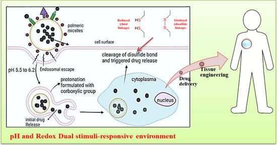
2.3. pH–Redox Tumor Microenvironment

The TME obtained with combined pH and redox dual-stimuli response (1) for the tumor extracellular environment can depend on its size, surface properties, and morphology (response to endosomal pH); thus, barriers include tumor accumulation, tumor penetration, blood circulation, and cellular uptake. (2) The intracellular environment response to pH, GSH level, and ROS cleavable moiety in tumor vasculature induces rapid drug release inside tumor cells. Polymeric nanocomposites have been developed to overcome both extracellular and intracellular barriers and enhance the antitumor effect. In this context, internal stimuli-responsive micelle nanomaterials are induced by the pathophysiological properties of normal and cancer cells while undergoing dynamic changes with various factors in vivo. Therefore, it is difficult to control the precision and response speed of nanomaterials in the TME. Regarding external stimuli, the key objective is to achieve high-level, deeper penetration without harming normal tissues, with maximum specificity, efficacy, and selectivity [86,87,88,89,90,91,92]. As discussed above, the stimulus–response combination and intracellular environments are characterized by different pH values, while pH is maintained at nearly 7.4 in the normal extracellular compartment. Once the surface is internalized or enters into endocytosis, the polymeric drug carriers first encounter the early endosome with a pH of nearly 6.2 and then enter the late endosome with a pH of approximately 5.5. Thus, tumor cells maintain a lower pH environment because of the generated ROS and free radicals (OH, H2O2, and O2) present in the TME. ROS levels sharply increase by three-fold compared with those in normal cells. These high-reactivity molecules construct a combined dual-sensitive pH–redox polymer prodrug and NPs which address the stability dilemma, reduced size, and surface charge reversal to enhance triggered drug release in tumor cells. Titratable groups like carboxylic acids and amine groups have been introduced into prodrugs, inducing improved extracellular stability led by prolonged circulation, stealth surface, and improved tumor accumulation (usually cytotoxic drugs). After entering the cancer cells, ROS trigger initial drug release and protonation, formulated with carboxylic groups. In addition, disulfide has been employed in ROS, which associates with core crosslink polymeric micelles to prevent drug leakage during blood circulation, followed by therapeutic drugs loaded into NP materials, with either covalent or non-covalent binding, leading to the development of polymeric drug carriers in GSH reduction conditions and thereby enhancing the surrounding hydrophilicity and swelling of the micellar shell. The disulfide bond is cleaved when exposed to an abundance of GSH, thereby causing cellular disassembly of the polymeric micelle structure. Thus, core–shell micelles furnish better DOX drug release in the TME via pH-triggered swelling and GHS-triggered disassembly. While various cancer cells exhibit elevated ROS compared to normal cells due to altered metabolism and mutations, the degree of increase varies significantly depending on the tumor type. Gliomas and leukemia show substantial ROS rises due to mitochondrial dysfunction. Prostate and breast cancers show complex patterns with both high and low ROS zones within the tumor microenvironment. Additionally, factors like tumor stage, hypoxia, and treatment can all influence ROS levels further. Therefore, ROS levels are context-dependent and do not always follow a threefold increase across all tumor types.
Chen et al. reported that pH/redox-responsive NPs can regulate tumor hypoxia. NPs modified with DMMA are usually prepared by stacking NPs that are sensitive to GSH and tumor acidity. DMMA undergoes surface charge reversal, resulting in a thick negatively charged shell and a positively charged disulfide cross-linked core. The NPs enter the tumor with a negative surface charge and reach prolonged blood circulation under acidic conditions with a large size of ~145 nm. In contrast, addressing the stability dilemma, surface charge reversal, and size reduction (~40 nm) enhances deeper penetration and cellular uptake. Therefore, after entering the cancer cells, drug carriers induce cytoplasmic GSH and rapid DOX release by cross-linking with the polypeptide core. The core–shell causes DOX to be released into tumor cells when pH and GSH cause it to break apart, which has antitumor effects [93]. Figure 6 shows how the pH-redox cascade and polypeptide core self-assembly deliver drugs to specific areas of the tumor. The pH–redox TME-mediated cascade, which is essential for improving drug delivery, is shown in Figure 6a. The focus of the study was the design of a smart nanoparticle (SNP) with a double-changeable center shell structure for improved drug delivery in cancerous conditions. SNP could lead to a perfect cascade of drug delivery. (I) Long circulation. (II) Enhanced accumulation. (III) Deep penetration. (IV) Promote internalization. (V) Accelerated drug release. This demonstrated the benefits of the SNP for drug delivery, including long-course, improved gathering, deep infiltration, advanced assimilation, and accelerated drug release. The shell, composed of polypeptide that has been slightly modified by dimethylmaleic anhydride, underwent size reduction and charge inversion in the acidic cancer microenvironment due to electrostatic interactions. The interconnected center maintained a small size, and its degradation in intracellular environments correlated with drug release, inducing increased antitumor motility. This designed SNP provided a potential solution to the deep cancer infiltration of nanomedicine by achieving a fundamental charge inversion and a sharp size decrease. A disulfide cross-linked polypeptide core and a PEGylated shell are involved in this cascade. PEGylated shells help stabilize NPs and prolong bloodstream circulation, which helps to precisely target tumors. The disulfide cross-linked polypeptide core responds to the redox environment of the tumor and allows the drug to be released in a controlled manner.
Xiong et al. demonstrated the synthesis of pH- and redox-sensitive micelles for better delivery of DOX and GNPs (gold NPs). The polymeric micelles consist of an amphiphilic copolymer of PCL-SS-PDMAEMA. The conjugate PDMAEMA is protonated and utilizes acidic conditions, thereby intensifying the hydrophilicity, followed by the micellar shell, which leads to swelling and disulfide bond cleavage when exposed to GSH, causing disassembly in the cellular or subcellular compartment. The conjugated NPs loaded with DOX@(PCL-SS-PDMAEMA) @GNPs, that is, core–shell micelles, provide better drug delivery to tumor cells by triggering pH-triggered swelling and GSH-triggered disassembly in the intercellular region of tumor cells [94]. The self-assembly of PCL-SS-PDMAEMA, the polymer at the center of our drug delivery system, is shown in detail in Figure 6b. This core plays a key role in encapsulating therapeutic components necessary for cancer treatment and computed tomography (CT) imaging, such as gold NPs and the anticancer medication DOX [94]. Shi et al. reported a four-arm PCL-PEG copolymer with hydrophobic PCL, which was conjugated with hydrophilic PEG via a disulfide bond. This disulfide bond is degraded in response to or when tumors are exposed to high levels of GSH, resulting in rapid DOX release [95]. In drug delivery strategies, these conjugated disulfide bonds are reduced in the biological milieu due to the unique reversible nature of the covalent bond present in the cellular compartment and possess controlled cleavage drug release, providing an intracellular redox potential drug delivery tool [96]. John et al. reported the synthesis of polymeric micelles such as the p(His)n–SS–PU–SS–p(His)n triblock, induced by polymerization, thereby triggering drug release in tumor cells [97]. A thorough summary of pH- and redox-responsive polymeric NPs and their use in drug administration is provided in Table 1. Numerous polymeric NPs, their loaded cargo or drugs, the targeted drug release or delivery methods that they use, the intended therapy or application, the biological evaluation context, and relevant references are compiled in this table.
In Figure 7, we describe the design and synthesis of pH-responsive triblock copolymers indicated as p(L–histidine) n–SS–polyurethane–SS–p(L–histidine) n, where ‘n’ denotes the number of repeating units (25, 35, 50, and 75). This copolymer is developed for the goal of achieving intracellular drug release. The picture is comprised of two panels, (a) and (b), both giving essential insights into the structure and behavior of the copolymer system. The first panel demonstrates the stepwise production of the triblock copolymers. It begins with the pH-responsive poly(L–histidine) blocks (p(His)), with variable lengths represented by ‘n’. These blocks are joined to polyurethane segments by disulfide (SS) connections on both sides, generating a triblock structure. The changes in ‘n’ (25, 35, 50, and 75) imply varied lengths of the poly(L–histidine) blocks, allowing for tunability in the pH responsiveness of the copolymer. The second panel shows an interesting perspective of the self-assembled polyurethane nanodaisies generated by the copolymer in response to variations in pH. The pH-responsive character of the poly(L–histidine) blocks plays a critical role in triggering the self-assembly of polyurethane nanodaisies. In acidic pH circumstances, the poly(L–histidine) blocks undergo protonation, leading to alterations in their structure and subsequently inducing the self-assembly of the polyurethane segments. Moreover, the presence of disulfide (SS) links in the copolymer enables for stimuli-responsive drug release. The breakage of these disulfide bonds is initiated by the intracellular reducing agent glutathione (GSH), permitting regulated release of the encapsulated pharmacological payload. This design method permits the construction of a dynamic drug delivery system that reacts to both pH fluctuations and intracellular circumstances, giving a possible platform for targeted and controlled drug release inside the cellular environment. In Table 2, we give a summary of in vivo investigations carried out to assess the efficacy of several pH/redox-responsive polymeric nanoparticles as drug delivery platforms. The table contains multiple medication delivery techniques together with the basic principles of action. Each item in the table gives essential knowledge into the in vivo behavior of these polymeric nanoparticles. Similarly, in Table 3, we offer a complete summary of the diverse pH- and redox-causing species that are created under distinct experimental settings, together with the related fold changes observed. This information is essential for understanding the dynamic changes in the chemical environment and how these variations impact the responsiveness of the experimental system.

3. Design Principles and Fabrication Methods

This section discusses the design principles behind pH- and redox-responsive polymer nanocomposites. It explores how these materials respond to changes in the pH and redox conditions typically found in the TME. Various fabrication methods, including nanoprecipitation, emulsion polymerization, and layer-by-layer assembly, are discussed in detail, highlighting their advantages and limitations in creating drug-loaded polymer nanocomposites with specific responsive properties.

3.1. Design Principles of pH- and Redox-Responsive Polymer Nanocomposites

The design of pH-responsive polymer nanocomposites exploits the acidic nature of the TME. The extracellular pH of solid tumors is generally lower (6.5–7.2) than that of healthy tissues (approximately 7.4) [12,112]. To achieve pH responsiveness, researchers have incorporated pH-sensitive moieties into polymer matrices. Common pH-sensitive groups include weakly acidic functional groups, such as carboxylic acids (-COOH) and sulfonic acids (-SO3H), which undergo ionization in response to pH changes [59,62]. As the pH decreases in the TME, these acidic groups become ionized, leading to a charge repulsion effect and subsequent swelling or disintegration of the polymer nanocomposite, triggering drug release. Redox responsiveness relies on altered redox potential in cancer cells due to elevated levels of reducing agents such as glutathione (GSH) [113]. Cancer cells maintain high concentrations of GSH that serves as a reducing agent in intracellular redox reactions. Researchers have designed redox-responsive polymer nanocomposites by incorporating disulfide (–SS–) linkages or other redox-sensitive motifs. In the reducing environment of cancer cells, disulfide bonds are cleaved, causing the polymer nanocomposite to destabilize and release the drug payload [59]. To achieve dual responsiveness, pH- and redox-sensitive elements are combined in the polymer nanocomposites. This design enables the fine-tuning of drug release based on the combined effects of pH and redox conditions in the TME. By strategically incorporating pH- and redox-sensitive functionalities, polymer nanocomposites can respond to two different stimuli, thereby enhancing the precision and control of drug release.

3.2. Fabrication Methods

Nanoprecipitation is a commonly employed technique for fabricating polymer nanocomposites with high drug-loading capacity. Using this technique, a drug and polymer solution are prepared independently, with the polymer containing groups that are sensitive to pH and/or redox reactions. Following the combination of these solutions, usually with strong shearing or sonication, the organic solvent rapidly diffuses into the aqueous phase, causing the spontaneous production of NPs. Numerous medication delivery applications can benefit from precise control over particle size and drug loading offered by the nanoprecipitation technique. The simplicity and scalability of this method allow for precise control over the release profile and encapsulation of both hydrophilic and hydrophobic medicines. There are a few restrictions to consider. Medication instability is possibly caused by contact with organic solvents during manufacturing, restricted ability to regulate the surface properties of NPs, and difficulties in attaining consistent drug dispersion [114].
Emulsion polymerization is another flexible technique for creating pH- and redox-responsive polymer nanocomposites. This procedure creates a stable emulsion by dispersing the monomers, medications, and emulsifying agents in the aqueous phase. Subsequently, the emulsion is polymerized using appropriate techniques or by introducing free radicals. As a result, the polymer nanocomposites that are produced are usually composed of core–shell structures, where the drug payload is contained within the core. The ability to encapsulate a broad variety of medications and control the method of drug loading, surface properties, and particle size are among its benefits. The use of emulsifying chemicals that may affect biocompatibility, the requirement for strict purification procedures, and the possibility of drug degradation during polymerization are limitations. Layer-by-layer assembly is a versatile technique that involves the sequential deposition of alternating layers of oppositely charged polymers or polyelectrolytes onto substrates. By incorporating pH- and redox-sensitive polymers into a multilayer assembly, researchers can design polymer nanocomposites with tunable responsiveness. Drug molecules can be loaded into the interlayer spaces or encapsulated within a single layer. The advantages include precise control over film thickness and drug loading, the ability to tailor responsiveness through layer selection, and compatibility with a variety of drugs [29].
Limitations include a time-consuming process that may result in relatively thick films and pose potential challenges in maintaining film stability in vivo. The design principles and fabrication methods for pH- and redox-responsive polymer nanocomposites play pivotal roles in their application as site-specific drug delivery systems in cancer therapy [115]. By ingeniously engineering these biomaterials to respond to the unique microenvironment of tumors, researchers can enhance their therapeutic efficacy while minimizing off-target effects. Nanoprecipitation, emulsion polymerization, and layer-by-layer assembly are promising techniques for creating drug-loaded polymer nanocomposites with specific responsive properties. Ongoing research in this field is expected to lead to the development of more sophisticated and effective dual-stimuli-responsive biomaterials for personalized cancer treatment [29,112].

4. Biocompatibility and Safety

Biocompatibility and safety are critical aspects in the development and clinical translation of biomaterials, particularly for applications in drug delivery systems. In this context, pH- and redox-responsive polymer nanocomposites have attracted significant interest owing to their potential to respond to specific physiological conditions, making them suitable candidates for targeted drug delivery. However, before these materials can be safely used in clinical settings, thorough examination and evaluation of their biocompatibility and potential long-term effects on healthy tissues are essential.
Biocompatibility refers to the ability of a material to interact favorably with biological systems without causing adverse reactions or toxicity. In the case of pH- and redox-responsive polymer nanocomposites, their interaction with the surrounding biological environment is of the utmost importance. The pH is a crucial parameter that varies in different tissues and cellular compartments. Target sites can be precisely controlled for drug release because of the conformational changes that pH-responsive polymers in nanocomposites undergo in response to local pH levels. In vitro tests employing cell cultures are performed to evaluate biocompatibility by monitoring cell survival, growth, and any possible cytotoxicity caused by these materials [88]. In vivo studies have also been performed using animal models to evaluate the tissue response and systemic effects of these polymer nanocomposites. Redox-responsive biomaterials can respond to changes in the cellular redox state, which is often altered in disease states [94]. These materials can be designed to release drugs in response to intracellular redox conditions. Biocompatibility evaluations of redox-responsive polymer nanocomposites involve in vitro and in vivo studies like those of pH responsiveness [86,94]. The long-term effects of biomaterials on healthy tissues are crucial for ensuring patient safety during extended drug delivery. However, these evaluations require long-term in vivo studies using animal models. To assess the biocompatibility of these materials over time, researchers examine tissue responses, possible inflammatory reactions, and systemic impacts over prolonged periods of time. An important factor in determining the safety of pH- and redox-responsive polymer nanocomposites is preclinical toxicology research. To identify possible harmful effects, establish safe dosage ranges for therapeutic usage, and ascertain dose–response correlations, these investigations entail extensive testing in animals. Adherence to the regulatory criteria for conducting these studies is essential to guarantee a thorough evaluation of the safety of the materials prior to human trials. The development of intelligent biodegradable materials is crucial to ensure safe medication administration. The ability of a substance to decompose into non-toxic metabolites and finally leave the body is known as its biodegradability. This feature is especially crucial for long-term use because it prevents materials from building up in tissues. To verify the safety profiles of these materials, biocompatibility assessments should consider the breakdown products. A comprehensive assessment of the biocompatibility and safety of pH- and redox-responsive polymer nanocomposites is necessary for their effective clinical applications as drug delivery systems. Through preclinical toxicity investigations and a thorough assessment of their interactions with healthy tissues, scientists can ensure the creation of secure biomaterials for precise and regulated medication administration across a range of medical uses.

5. Future Perspectives and Challenges

This section provides an overview of the potential applications of pH- and redox-triggered polymer nanocomposites in cancer therapy as they move closer to clinical trials. It discusses how novel approaches are required to increase drug-loading capability, response specificity, and in vivo stability. In addition, possible synergies with other systems that respond to stimuli and combination therapies are investigated. The ability of pH- and redox-responsive polymer nanocomposites to load drugs is one of the main challenges. To maximize the number of therapeutic chemicals that can be loaded onto these biomaterials without sacrificing their stability and responsiveness, researchers should concentrate on developing novel techniques. Enhanced drug-loading efficiencies can be attained by investigating new methods for drug encapsulation and surface modification. Although site-specific drug release is possible with pH- and redox-responsive polymer nanocomposites, obtaining an even greater response specificity is essential. Future research should aim to design biomaterials that respond only to the precise TME and avoid premature drug release from non-targeted tissues. This could involve the incorporation of additional stimuli-responsive elements or the use of advanced targeting ligands. The stability and longevity of polymer nanocomposites in complex in vivo environments are vital for successful clinical translation. Researchers need to address the challenges related to biodegradation, immune responses, and potential clearance from the body. Developing stable and long-lasting polymer nanocomposites will ensure sustained drug release and optimize therapeutic outcomes. The future of cancer therapy lies in personalized medicine. pH- and redox-responsive polymer nanocomposites can be customized according to individual patient characteristics and specific tumor types. Tailoring drug payloads, stimulus responsiveness, and targeting ligands to match each patient’s unique profile could significantly enhance treatment efficacy and minimize side effects.
Despite promising preclinical results, translating pH- and redox-triggered polymer nanocomposites from the laboratory to clinical practice presents challenges. Rigorous testing in large animal models and addressing potential toxicity concerns are essential steps for human trials. Cancer cells exhibit substantial heterogeneity even within the same tumor. The adaptation of pH- and redox-responsive biomaterials to accommodate this diversity poses significant challenges. Strategies to address intertumoral variability and ensure efficient drug delivery to all tumor regions need to be explored. The approval process for novel biomaterials in the clinical setting involves stringent regulatory guidelines. Demonstrating the safety, efficacy, and long-term effects of pH- and redox-responsive polymer nanocomposites is critical for obtaining regulatory approval and market acceptance. Although the combination of different stimuli-responsive systems or therapies holds promise for enhancing treatment outcomes, it introduces complexities in terms of drug interactions, dosage optimization, and potential adverse effects. The development of effective combination strategies that maximize synergies while minimizing drawbacks remains challenging. For widespread clinical adoption, pH- and redox-responsive polymer nanocomposites must be scalable and cost-effective. Identifying suitable, readily available raw materials and streamlining manufacturing processes are essential for reducing production costs and ensuring affordability for patients.

6. Conclusions

pH- and redox-triggered polymer nanocomposites hold significant promise as future generation, dual-stimuli-responsive biomaterials for site-specific drug release during cancer therapy. Their ability to precisely target tumor cells while sparing healthy tissues has the potential to revolutionize cancer treatment, offering improved therapeutic outcomes and enhanced patient quality of life. Nevertheless, further research and clinical investigations are essential to overcome existing challenges and unlock the full potential of these innovative biomaterials in cancer therapeutics.
The development of pH- and redox-responsive polymer nanocomposites as dual-stimuli-responsive biomaterials has shown great promise for revolutionizing cancer therapy. However, to fully harness their potential, researchers must address the challenges of drug-loading capacity, response specificity, in vivo stability, and regulatory approval. Collaborative efforts among researchers, clinicians, and industry stakeholders are crucial to overcome these obstacles and pave the way for the successful clinical translation of these innovative biomaterials. As advancements continue, pH- and redox-responsive polymer nanocomposites hold immense potential for improving cancer treatments and ultimately contributing to better patient outcomes.

Author Contributions

S.G., S.P. and K.P.C.H.P. contributed to writing and revising the manuscript. K.P.C.H.P. and J.H. equally supervised the study. All authors have read and agreed to the published version of the manuscript.

Funding

This research was supported by the Basic Science Research Capacity Enhancement Project through the Korea Basic Science Institute (National Research Facilities and Equipment Center) grant funded by the Ministry of Education (2019R1A6C1010016) and the Basic Science Research Program through the National Research Foundation of Korea (NRF) funded by the Ministry of Education (NRF-2021R1F1A1050130).

Institutional Review Board Statement

Not available.

Informed Consent Statement

Not available.

Data Availability Statement

Not available.

Conflicts of Interest

The authors declare no conflicts of interest.

References

  1. Puyol, M.; Seoane, J.; Aguilar, E.; Vozza, L.B.; Orbe, I.; Crawford, K.H.; Fernández, A.; Bray, F.; Johnson, S.E.; Gopal, S. WORLD CANCER RESEARCH DAY: A Call to Action for a Coordinated International Research Effort to Prevent, Diagnose, and Treat Cancer. Clin. Cancer Res. 2021, 27, 963–966. [Google Scholar] [CrossRef]
  2. Smith, L.; Stiller, C.A.; Aitken, J.F.; Hjalgrim, L.L.; Johannesen, T.; Lahteenmaki, P.; McCabe, M.G.; Phillips, R.; Pritchard-Jones, K.; Steliarova-Foucher, E.; et al. International variation in childhood cancer mortality rates from 2001 to 2015: Comparison of trends in the International Cancer Benchmarking Partnership countries. Int. J. Cancer 2021, 150, 28–37. [Google Scholar] [CrossRef]
  3. Parvanian, S.; Mostafavi, S.M.; Aghashiri, M. Multifunctional nanoparticle developments in cancer diagnosis and treatment. Sens. Bio-Sens. Res. 2017, 13, 81–87. [Google Scholar] [CrossRef]
  4. ReFaey, K.; Tripathi, S.; Grewal, S.S.; Bhargav, A.G.; Quinones, D.J.; Chaichana, K.L.; Antwi, S.O.; Cooper, L.T.; Meyer, F.B.; Dronca, R.S.; et al. Cancer Mortality Rates Increasing vs Cardiovascular Disease Mortality Decreasing in the World: Future Implications. Mayo Clin. Proc. Innov. Qual. Outcomes 2021, 5, 645–653. [Google Scholar] [CrossRef] [PubMed]
  5. Moodley, T.; Singh, M. Current Stimuli-Responsive Mesoporous Silica Nanoparticles for Cancer. Pharmaceutics 2021, 13, 71. [Google Scholar] [CrossRef]
  6. Zubair, M.; Wang, S.; Ali, N. Advanced Approaches to Breast Cancer Classification and Diagnosis. Front. Pharmacol. 2021, 11, 632079. [Google Scholar] [CrossRef]
  7. Li, W.Q.; Guo, H.F.; Li, L.Y.; Zhang, Y.F.; Cui, J.W. The promising role of antibody drug conjugate in cancer therapy: Combining targeting ability with cytotoxicity effectively. Cancer Med. 2021, 10, 4677–4696. [Google Scholar] [CrossRef] [PubMed]
  8. Yang, H.Y.; Jang, M.S.; Gao, G.H.; Lee, J.H.; Lee, D.S. Construction of redox/pH dual stimuli-responsive PEGylated polymeric micelles for intracellular doxorubicin delivery in liver cancer. Polym. Chem. 2016, 7, 1813–1825. [Google Scholar] [CrossRef]
  9. Alsaab, H.O.; Al-Hibs, A.S.; Alzhrani, R.; Alrabighi, K.K.; Alqathama, A.; Alwithenani, A.; Almalki, A.H.; Althobaiti, Y.S. Nanomaterials for Antiangiogenic Therapies for Cancer: A Promising Tool for Personalized Medicine. Int. J. Mol. Sci. 2021, 22, 1631. [Google Scholar] [CrossRef]
  10. Das Kurmi, B.; Patel, P.; Paliwal, R.; Paliwal, S.R. Molecular approaches for targeted drug delivery towards cancer: A concise review with respect to nanotechnology. J. Drug Deliv. Sci. Technol. 2020, 57, 101682. [Google Scholar] [CrossRef]
  11. Mukhtar, M.; Bilal, M.; Rahdar, A.; Barani, M.; Arshad, R.; Behl, T.; Brisc, C.; Banica, F.; Bungau, S. Nanomaterials for Diagnosis and Treatment of Brain Cancer: Recent Updates. Chemosensors 2020, 8, 117. [Google Scholar] [CrossRef]
  12. Solanki, R.; Rostamabadi, H.; Patel, S.; Jafari, S.M. Anticancer nano-delivery systems based on bovine serum albumin nanoparticles: A critical review. Int. J. Biol. Macromol. 2021, 193, 528–540. [Google Scholar] [CrossRef] [PubMed]
  13. Huang, W.-Y.; Lai, C.-H.; Peng, S.-L.; Hsu, C.-Y.; Hsu, P.-H.; Chu, P.-Y.; Feng, C.-L.; Lin, Y.-H. Targeting Tumor Cells with Nanoparticles for Enhanced Co-Drug Delivery in Cancer Treatment. Pharmaceutics 2021, 13, 1327. [Google Scholar] [CrossRef]
  14. Chang, D.; Ma, Y.; Xu, X.; Xie, J.; Ju, S. Stimuli-Responsive Polymeric Nanoplatforms for Cancer Therapy. Stimuli-Responsive Polym. Nanoplatforms 2021, 9, 707319. [Google Scholar] [CrossRef] [PubMed]
  15. Shivalingayya; Preeti, R.K.; Ganiger, S.K.; Shashidhar, A.C.; Lagashetty, A. Multifunctional Nanoparticles for Biomedical Applications. J. Chem. Biol. Phys. Sci. 2022, 12, 410–422. [Google Scholar]
  16. Gunathilake, T.M.S.U.; Ching, Y.C.; Chuah, C.H.; Rahman, N.A.; Liou, N.-S. Recent advances in celluloses and their hybrids for stimuli-responsive drug delivery. Int. J. Biol. Macromol. 2020, 158, 670–688. [Google Scholar] [CrossRef]
  17. Liu, D.; Yang, F.; Xiong, F.; Gu, N. The Smart Drug Delivery System and Its Clinical Potential. Theranostics 2016, 6, 1306–1323. [Google Scholar] [CrossRef]
  18. Majumder, J.; Minko, T. Multifunctional and stimuli-responsive nanocarriers for targeted therapeutic delivery. Expert Opin. Drug Deliv. 2021, 18, 205–227. [Google Scholar] [CrossRef]
  19. Sun, Q.; Wang, Z.; Liu, B.; He, F.; Gai, S.; Yang, P.; Yang, D.; Li, C.; Lin, J. Recent advances on endogenous/exogenous stimuli-triggered nanoplatforms for enhanced chemodynamic therapy. Coord. Chem. Rev. 2021, 451, 214267. [Google Scholar] [CrossRef]
  20. Abdo, G.G.; Zagho, M.M.; Khalil, A. Recent advances in stimuli-responsive drug release and targeting concepts using mesoporous silica nanoparticles. Emergent Mater. 2020, 3, 407–425. [Google Scholar] [CrossRef]
  21. Chivere, V.T.; Kondiah, P.P.D.; Choonara, Y.E.; Pillay, V. Nanotechnology-Based Biopolymeric Oral Delivery Platforms for Advanced Cancer Treatment. Cancers 2020, 12, 522. [Google Scholar] [CrossRef]
  22. Patra, J.K.; Das, G.; Fraceto, L.F.; Campos, E.V.R.; del Pilar Rodriguez-Torres, M.; Acosta-Torres, L.S.; Diaz-Torres, L.A.; Grillo, R.; Swamy, M.K.; Sharma, S.; et al. Nano based drug delivery systems: Recent developments and future prospects. J. Nanobiotechnol. 2018, 16, 71. [Google Scholar] [CrossRef]
  23. Zhou, Q.; Zhang, L.; Yang, T.; Wu, H. Stimuli-responsive polymeric micelles for drug delivery and cancer therapy. Int. J. Nanomed. 2018, 13, 2921–2942. [Google Scholar] [CrossRef]
  24. Moghimi, S.M.; Hunter, A.C.; Murray, J.C. Nanomedicine: Current status and future prospects. FASEB J. 2005, 19, 311–330. [Google Scholar] [CrossRef] [PubMed]
  25. Aflori, M. Smart Nanomaterials for Biomedical Applications—A Review. Nanomaterials 2021, 11, 396. [Google Scholar] [CrossRef]
  26. Zhang, P.; Gao, Z.; Cui, J.; Hao, J. Dual-Stimuli-Responsive Polypeptide Nanoparticles for Photothermal and Photodynamic Therapy. ACS Appl. Bio Mater. 2019, 3, 561–569. [Google Scholar] [CrossRef]
  27. Jeyamogan, S.; Khan, N.A.; Siddiqui, R. Application and Importance of Theranostics in the Diagnosis and Treatment of Cancer. Arch. Med. Res. 2021, 52, 131–142. [Google Scholar] [CrossRef]
  28. Deng, C.; Jiang, Y.; Cheng, R.; Meng, F.; Zhong, Z. Biodegradable polymeric micelles for targeted and controlled anticancer drug delivery: Promises, progress and prospects. Nano Today 2012, 7, 467–480. [Google Scholar] [CrossRef]
  29. Singh, A.; Talekar, M.; Tran, T.-H.; Samanta, A.; Sundaram, R.; Amiji, M. Combinatorial Approach in the Design of Multifunctional Polymeric Nano-Delivery Systems for Cancer Therapy. J. Mater. Chem. B 2014, 2, 8069–8084. [Google Scholar] [CrossRef]
  30. Roma-Rodrigues, C.; Raposo, L.R.; Valente, R.; Fernandes, A.R.; Baptista, P.V. Combined cancer therapeutics—Tackling the complexity of the tumor microenvironment. Wiley Interdiscip. Rev. Nanomed. Nanobiotechnol. 2021, 13, e1704. [Google Scholar] [CrossRef]
  31. Carreiró, F.; Oliveira, A.M.; Neves, A.; Pires, B.; Venkatesh, D.N.; Durazzo, A.; Lucarini, M.; Eder, P.; Silva, A.M.; Santini, A.; et al. Polymeric Nanoparticles: Production, Characterization, Toxicology and Ecotoxicology. Molecules 2020, 25, 3731. [Google Scholar]
  32. Katifelis, H.; Gazouli, M. Cancer-Targeted Nanotheranostics: Recent Advances and Future Perspectives. Cancer Nanotheranostics 2021, 2, 97–115. [Google Scholar]
  33. Luo, C.; Sun, J.; Sun, B.; He, Z. Prodrug-based nanoparticulate drug delivery strategies for cancer therapy. Trends Pharmacol. Sci. 2014, 35, 556–566. [Google Scholar] [CrossRef] [PubMed]
  34. Tang, Z.; He, C.; Tian, H.; Ding, J.; Hsiao, B.S.; Chu, B.; Chen, X. Polymeric Nanostructured Materials for Biomedical Applications. Prog. Polym. Sci. 2016, 60, 86–128. [Google Scholar] [CrossRef]
  35. Tyrrell, Z.L.; Shen, Y.; Radosz, M. Fabrication of micellar nanoparticles for drug delivery through the self-assembly of block copolymers. Prog. Polym. Sci. 2010, 35, 1128–1143. [Google Scholar] [CrossRef]
  36. Steichen, S.D.; Caldorera-Moore, M.; Peppas, N.A. A review of current nanoparticle and targeting moieties for the delivery of cancer therapeutics. Eur. J. Pharm. Sci. 2013, 48, 416–427. [Google Scholar] [CrossRef] [PubMed]
  37. Choi, J.-W.; An, J.; Son, S.-R.; Kim, S.; Park, J.; Park, C.B.; Lee, J.H. Rational design of surface-confined nanostructured self-assemblies based on functional comb-shaped copolymers for tunable molecular orientation. React. Funct. Polym. 2021, 168, 105042. [Google Scholar] [CrossRef]
  38. Bae, Y.; Kataoka, K. Intelligent polymeric micelles from functional poly(ethylene glycol)-poly(amino acid) block copolymers. Adv. Drug Deliv. Rev. 2009, 61, 768–784. [Google Scholar] [CrossRef]
  39. Lallana, E.; Sousa-Herves, A.; Fernandez-Trillo, F.; Riguera, R.; Fernandez-Megia, E. Click chemistry for drug delivery nanosystems. Pharm. Res. 2012, 29, 1–34. [Google Scholar] [CrossRef]
  40. Deng, B.; Ma, P.; Xie, Y. Reduction-Sensitive Polymeric Nanocarriers in Cancer Therapy: A Comprehensive Review. Nanoscale 2015, 7, 12773–12795. [Google Scholar] [CrossRef]
  41. Date, T.; Nimbalkar, V.; Kamat, J.; Mittal, A.; Mahato, R.I.; Chitkara, D. Lipid-polymer hybrid nanocarriers for delivering cancer therapeutics. J. Control. Release 2017, 271, 60–73. [Google Scholar] [CrossRef]
  42. Xia, W.; Tao, Z.; Zhu, B.; Zhang, W.; Liu, C.; Chen, S.; Song, M. Targeted Delivery of Drugs and Genes Using Polymer Nanocarriers for Cancer Therapy. Int. J. Mol. Sci. 2021, 22, 9118. [Google Scholar] [CrossRef]
  43. Attia, M.F.; Anton, N.; Wallyn, J.; Omran, Z.; Vandamme, T.F. An overview of active and passive targeting strategies to improve the nanocarriers efficiency to tumour sites. J. Pharm. Pharmacol. 2019, 71, 1185–1198. [Google Scholar] [CrossRef]
  44. Lv, S.; Sylvestre, M.; Prossnitz, A.N.; Yang, L.F.; Pun, S.H. Design of Polymeric Carriers for Intracellular Peptide Delivery in Oncology Applications. Chem. Rev. 2021, 121, 11653–11698. [Google Scholar] [CrossRef]
  45. Tan, H.; Marra, K.G. Injectable, Biodegradable Hydrogels for Tissue Engineering Applications. Materials 2010, 3, 1746–1767. [Google Scholar] [CrossRef]
  46. Feldman, D. Review Polymers and Polymer Nanocomposites for Cancer Therapy. Appl. Sci. 2019, 9, 3899. [Google Scholar] [CrossRef]
  47. Gopi, S.; Amalraj, A.; Sukumaran, N.P.; Haponiuk, J.T.; Thomas, S. Biopolymers and Their Composites for Drug Delivery: A Brief Review. Macromol. Symp. 2018, 380, 1800114. [Google Scholar] [CrossRef]
  48. Wu, J. The Enhanced Permeability and Retention (EPR) Effect: The Significance of the Concept and Methods to Enhance Its Application. J. Pers. Med. 2021, 11, 771. [Google Scholar] [CrossRef]
  49. Shariatinia, Z.; Zahraee, Z. Controlled release of metformin from chitosan-based nanocomposite films containing mesoporous MCM-41 nanoparticles as novel drug delivery systems. J. Colloid Interface Sci. 2017, 501, 60–76. [Google Scholar] [CrossRef]
  50. Luo, H.; Ao, H.; Li, G.; Li, W.; Xiong, G.; Zhu, Y.; Wan, Y. Bacterial cellulose/graphene oxide nanocomposite as a novel drug delivery system. Appl. Phys. 2017, 17, 249. [Google Scholar] [CrossRef]
  51. Park, J.H.; Lee, S.; Kim, J.H.; Park, K.; Kim, K.; Kwon, I.C. Polymeric nanomedicine for cancer therapy. Prog. Polym. Sci. 2008, 33, 113–137. [Google Scholar] [CrossRef]
  52. Yang, H.Y.; Li, Y.; Lee, D.S. Multifunctional and Stimuli-Responsive Magnetic Nanoparticle-Based Delivery Systems for Biomedical Applications. Adv. Therap. 2018, 1, 1800011. [Google Scholar] [CrossRef]
  53. Mi, P. Stimuli-responsive nanocarriers for drug delivery, tumor imaging, therapy and theranostics. Theranostics 2020, 10, 4557–4588. [Google Scholar] [CrossRef] [PubMed]
  54. Qian, X.L.; Li, J.; Wei, R.; Lin, H.; Xiong, L.X. Internal and external triggering mechanism of “smart” nanoparticle-based DDSs in targeted tumor therapy. Curr. Pharm. Des. 2018, 24, 1639–1651. [Google Scholar] [CrossRef] [PubMed]
  55. Meng, F.; Cheng, R.; Deng, C.; Zhong, Z. Intracellular drug release nanosystems. Mater. Today 2012, 15, 436–442. [Google Scholar] [CrossRef]
  56. Du, J.; Lane, L.A.; Nie, S. Stimuli-Responsive Nanoparticles for Targeting the Tumor Microenvironment. J. Control. Release 2015, 219, 205–214. [Google Scholar] [CrossRef]
  57. Van Vlerken, L.E.; Amiji, M.M. Multi-functional polymeric nanoparticles for tumour-targeted drug delivery. Expert Opin. Drug Deliv. 2006, 3, 205–216. [Google Scholar] [CrossRef]
  58. Vaupel, P. Tumor Microenvironmental Physiology and Its Implications for Radiation Oncology. Semin. Radiat. Oncol. 2004, 14, 198–206. [Google Scholar] [CrossRef]
  59. Kimura, N.; Maeki, M.; Sato, Y.; Note, Y.; Ishida, A.; Tani, H.; Harashima, H.; Tokeshi, M. Development of the iLiNP Device: Fine Tuning the Lipid Nanoparticle Size within 10 nm for Drug Delivery. ACS Omega 2018, 3, 5044–5051. [Google Scholar] [CrossRef]
  60. Haley, B.; Frenkel, E. Nanoparticles for drug delivery in cancer treatment. Urol. Oncol. Semin. Orig. Investig. 2008, 26, 57–64. [Google Scholar] [CrossRef]
  61. Salatin, S.; Maleki Dizaj, S.; Yari Khosroushahi, A. Effect of the surface modification, size, and shape on cellular uptake of nanoparticles. Cell Biol. Int. 2015, 39, 881–890. [Google Scholar] [CrossRef]
  62. Aghebati-Maleki, A.; Dolati, S.; Ahmadi, M.; Baghbanzhadeh, A.; Asadi, M.; Fotouhi, A.; Yousefi, M.; Aghebati-Maleki, L. Nanoparticles and cancer therapy: Perspectives for application of nanoparticles in the treatment of cancers. J. Cell. Physiol. 2020, 235, 1962–1972. [Google Scholar] [CrossRef] [PubMed]
  63. Fernández, J.P.; Luddy, K.A.; Harmon, C.; O’farrelly, C. Hepatic tumor microenvironments and effects on NK cell phenotype and function. Int. J. Mol. Sci. 2019, 20, 4131. [Google Scholar] [CrossRef]
  64. Gu, F.X.; Karnik, R.; Wang, A.Z.; Alexis, F.; Levy-Nissenbaum, E.; Hong, S.; Langer, R.S.; Farokhzad, O.C. Targeted nanoparticles for cancer therapy. Nano Today 2007, 2, 14–21. [Google Scholar] [CrossRef]
  65. Lan, Q.; Xia, S.; Wang, Q.; Xu, W.; Huang, H.; Jiang, S.; Lu, L. Development of oncolytic virotherapy: From genetic modification to combination therapy. Front. Med. 2020, 14, 160–184. [Google Scholar] [CrossRef] [PubMed]
  66. Mollazadeh, S.; Mackiewicz, M.; Yazdimamaghani, M. Recent advances in the redox-responsive drug delivery nanoplatforms: A chemical structure and physical property perspective. Mater. Sci. Eng. C 2021, 118, 111536. [Google Scholar] [CrossRef] [PubMed]
  67. Tomasetti, L.; Breunig, M. Preventing obstructions of nanosized drug delivery systems by the extracellular matrix. Adv. Healthc. Mater. 2018, 7, 1700739. [Google Scholar] [CrossRef]
  68. Zhang, X.; Han, L.; Liu, M.; Wang, K.; Tao, L.; Wan, Q.; Wei, Y. Recent progress and advances in redox-responsive polymers as controlled delivery nanoplatforms. Mater. Chem. Front. 2017, 1, 807–822. [Google Scholar] [CrossRef]
  69. Chibh, S.; Kour, A.; Yadav, N.; Kumar, P.; Yadav, P.; Chauhan, V.S.; Panda, J.J. Redox-Responsive Dipeptide Nanostructures toward Targeted Cancer Therapy. ACS Omega 2020, 5, 3365–3375. [Google Scholar] [CrossRef]
  70. Chen, W.; Zhang, C.; Chen, D.; Li, Y.; Wu, S.; Xu, C.; Su, L.; Zhang, Q. Tumor redox microenvironment modulating composite hydrogels for enhanced sonodynamic therapy of colorectal cancer. J. Mater. Chem. B 2022, 10, 1960–1968. [Google Scholar] [CrossRef]
  71. Sun, C.; Li, X.; Du, X.; Wang, T. Redox-responsive micelles for triggered drug delivery and effective laryngopharyngeal cancer therapy. Int. J. Biol. Macromol. 2018, 112, 65–73. [Google Scholar] [CrossRef] [PubMed]
  72. Guo, X.; Cheng, Y.; Zhao, X.; Luo, Y.; Chen, J.; Yuan, W.-E. Advances in redox-responsive drug delivery systems of tumor microenvironment. J. Nanobiotechnol. 2018, 16, 74. [Google Scholar] [CrossRef] [PubMed]
  73. Pan, Y.-J.; Chen, Y.-Y.; Wang, D.-R.; Wei, C.; Guo, J.; Lu, D.-R.; Chu, C.-C.; Wang, C.-C. Redox/pH dual stimuli-responsive biodegradable nanohydrogels with varying responses to dithiothreitol and glutathione for controlled drug release. Biomaterials 2012, 33, 6570–6579. [Google Scholar] [CrossRef] [PubMed]
  74. Demirci, S.; Celebioglu, A.; Aytac, Z.; Uyar, T. pH-responsive nanofibers with controlled drug release properties. Polym. Chem. 2014, 5, 2050–2056. [Google Scholar] [CrossRef]
  75. Ratemi, E. pH-responsive polymers for drug delivery applications. In Stimuli Responsive Polymeric Nanocarriers for Drug Delivery Applications; Elsevier: Amsterdam, The Netherlands, 2018; Volume 1, pp. 121–141. [Google Scholar]
  76. Deirram, N.; Zhang, C.; Kermaniyan, S.S.; Johnston, A.P.R.; Such, G.K. pH-Responsive Polymer Nanoparticles for Drug Delivery. Macromol. Rapid Commun. 2019, 40, 18009107. [Google Scholar] [CrossRef] [PubMed]
  77. Kim, J.O.; Kabanov, A.V.; Bronich, T.K. Polymer Micelles with Cross-Linked Polyanion Core for Delivery of a Cationic Drug Doxorubicin. J. Control. Release 2009, 15, 197–204. [Google Scholar] [CrossRef] [PubMed]
  78. Min, K.H.; Kim, J.H.; Bae, S.M.; Shin, H.; Kim, M.S.; Park, S.; Lee, H.; Park, R.W.; Kim, I.S.; Kim, K.; et al. Tumoral acidic pH-responsive MPEG-poly (β-amino ester) polymeric micelles for cancer targeting therapy. J. Control. Release 2010, 144, 259–266. [Google Scholar] [CrossRef]
  79. Liu, J.; Huang, Y.; Kumar, A.; Tan, A.; Jin, S.; Mozhi, A.; Liang, X.J. pH-sensitive nano-systems for drug delivery in cancer therapy. Biotechnol. Adv. 2014, 32, 693–710. [Google Scholar] [CrossRef]
  80. Danhier, F.; Feron, O.; Préat, V. To exploit the tumor microenvironment: Passive and active tumor targeting of nanocarriers for anti-cancer drug delivery. J. Control. Release 2010, 148, 135–146. [Google Scholar] [CrossRef]
  81. Uthaman, S.; Huh, K.M.; Park, I.-K. Tumor microenvironment-responsive nanoparticles for cancer theragnostic applications. Biomater. Res. 2018, 22, 22. [Google Scholar] [CrossRef]
  82. Li, Y.; Yang, H.Y.; Lee, D.S. Polymer-based and pH-sensitive nanobiosensors for imaging and therapy of acidic pathological areas. Pharm. Res. 2016, 33, 2358–2372. [Google Scholar] [CrossRef] [PubMed]
  83. Chang, G.; Li, C.; Lu, W.; Ding, J. N-Boc-histidine-capped PLGA-PEG-PLGA as a smart polymer for drug delivery sensitive to tumor extracellular pH. Macromol. Biosci. 2010, 10, 1248–1256. [Google Scholar] [CrossRef]
  84. Hu, F.Q.; Zhang, Y.Y.; You, J.; Yuan, H.; Du, Y.Z. pH triggered doxorubicin delivery of PEGylated glycolipid conjugate micelles for tumor targeting therapy. Mol. Pharm. 2012, 9, 2469–2478. [Google Scholar] [CrossRef] [PubMed]
  85. Yu, Y.; Zhang, X.; Qiu, L. The anti-tumor efficacy of curcumin when delivered by size/charge-changing multistage polymeric micelles based on amphiphilic poly(beta-amino ester) derivates. Biomaterials 2014, 35, 467–3479. [Google Scholar] [CrossRef]
  86. Shi, J.; Ren, Y.; Ma, J.; Luo, X.; Li, J.; Wu, Y.; Gu, H.; Fu, C.; Cao, Z.; Zhang, J. Novel CD44-targeting and pH/redox-dual-stimuli-responsive core–shell nanoparticles loading triptolide combats breast cancer growth and lung metastasis. J. Nanobiotechnol. 2021, 19, 188. [Google Scholar] [CrossRef]
  87. Hettiarachchi, S.D.; Cilingir, E.K.; Maklouf, H.; Seven, E.S.; Paudyal, S.; Vanni, S.; Graham, R.M.; Leblanc, R.M. pH and redox triggered doxorubicin release from covalently linked carbon dots conjugates. Nanoscale 2021, 13, 5507–5518. [Google Scholar] [CrossRef] [PubMed]
  88. Sun, H.; Meng, F.; Cheng, R.; Deng, C.; Zhong, Z. Reduction and pH dual-bioresponsive crosslinked polymersomes for efficient intracellular delivery of proteins and potent induction of cancer cell apoptosis. Acta Biomater. 2014, 10, 2159–2168. [Google Scholar] [CrossRef] [PubMed]
  89. Hu, X.; Li, H.; Luo, S.; Liu, T.; Jiang, Y.; Liu, S. Thiol and pH dual-responsive dynamic covalent shell cross-linked micelles for triggered release of chemotherapeutic drugs. Polym. Chem. 2013, 4, 695–706. [Google Scholar] [CrossRef]
  90. Chiang, W.H.; Ho, V.T.; Huang, W.C.; Huang, Y.F.; Chern, C.S.; Chiu, H.C. Dual Stimuli-Responsive Polymeric Hollow Nanogels Designed as Carriers for Intracellular Triggered Drug Release. Langmuir 2012, 28, 15056–15064. [Google Scholar] [CrossRef]
  91. Luo, Y.; Yin, X.; Yin, X.; Chen, A.; Zhao, L.; Zhang, G.; Liao, W.; Huang, X.; Li, J.; Zhang, C.Y. Dual pH/Redox-Responsive Mixed Polymeric Micelles for Anticancer Drug Delivery and Controlled Release. Pharmaceutics 2019, 11, 176. [Google Scholar] [CrossRef]
  92. Liang, K.; Such, G.K.; Zhu, Z.; Yan, Y.; Lomas, H.; Caruso, F. Charge-Shifting Click Capsules with Dual-Responsive Cargo Release Mechanisms. Adv. Mater. 2011, 23, H273–H277. [Google Scholar] [CrossRef] [PubMed]
  93. Bahadur, K.C.R.; Thapa, B.; Xu, P. pH and Redox Dual Responsive Nanoparticle for Nuclear Targeted Drug Delivery. Mol. Pharm. 2012, 9, 2719–2729. [Google Scholar]
  94. Jing, X.; Zhi, Z.; Jin, L.; Wang, F.; Wu, Y.; Wang, D.; Yan, K.; Shao, Y.; Meng, L. pH/redox dual-stimuli responsive cross-linked polyphosphazene nanoparticles for multimodal imaging guided chemo-photodynamic therapy. Nanoscale 2012, 11, 9457–9467. [Google Scholar] [CrossRef]
  95. Curcio, M.; Paolì, A.; Cirillo, G.; Di Pietro, S.; Forestiero, M.; Giordano, F.; Mauro, L.; Amantea, D.; Di Bussolo, V.; Nicoletta, F.P.; et al. Combining Dextran Conjugates with Stimuli-Responsive and Folate-Targeting Activity: A New Class of Multifunctional Nanoparticles for Cancer Therapy. Nanomaterials 2021, 11, 1108. [Google Scholar] [CrossRef]
  96. Chen, J.; Qiu, X.; Ouyang, J.; Kong, J.; Zhong, W.; Xing, M.M. pH and Reduction Dual-Sensitive Copolymeric Micelles for Intracellular Doxorubicin Delivery. Biomacromolecules 2011, 12, 3601–3611. [Google Scholar] [CrossRef] [PubMed]
  97. Liu, J.; Liu, X.; Yuan, Y.; Li, Q.; Chang, B.; Xu, L.; Cai, B.; Qi, C.; Li, C.; Jiang, X.; et al. Supramolecular Modular Approach towards Conveniently Constructing and Multifunctioning a pH/Redox Dual Responsive Drug Delivery Nanoplatform for Improved Cancer Chemotherapy. ACS Appl. Mater. Interfaces 2018, 10, 26473–26484. [Google Scholar] [CrossRef]
  98. Yu, K.; Yang, X.; He, L.; Zheng, R.; Min, J.; Su, H.; Shan, S.; Jia, Q. Facile preparation of pH/reduction dual-stimuli responsive dextran nanogel as environment-sensitive carrier of doxorubicin. Polymer 2020, 200, 122585. [Google Scholar] [CrossRef]
  99. Cheng, R.; Meng, F.; Deng, C.; Klok, H.A.; Zhong, Z. Dual and Multi-Stimuli Responsive Polymeric Nanoparticles for Programmed Site-Specific Drug Delivery. Biomaterials 2013, 34, 3647–3657. [Google Scholar] [CrossRef]
  100. Fu, X.; Hosta-Rigau, L.; Chandrawati, R.; Cui, J. Multi-Stimuli-Responsive Polymer Particles, Films, and Hydrogels for Drug Delivery. Chem 2018, 4, 2084–2107. [Google Scholar] [CrossRef]
  101. Guo, X.; Wei, X.; Jing, Y.; Zhou, S. Size Changeable Nanocarriers with Nuclear Targeting for Effectively Overcoming Multidrug Resistance in Cancer Therapy. Adv. Mater. 2015, 27, 6450–6456. [Google Scholar] [CrossRef]
  102. Du, J.Z.; Du, X.J.; Mao, C.Q.; Wang, J. Tailor made Dual pH-Sensitive Polymer-Doxorubicin Nanoparticles for Efficient Anticancer Drug Delivery. J. Am. Chem. Soc. 2011, 133, 17560–17563. [Google Scholar] [CrossRef]
  103. Dai, L.; Li, X.; Duan, X.; Li, M.; Niu, P.; Xu, H.; Cai, K.; Yang, H. A pH/ROS Cascade-Responsive Charge-Reversal Nanosystem with Self-Amplified Drug Release for Synergistic Oxidation-Chemo-therapy. Adv. Sci. 2019, 6, 1801807. [Google Scholar] [CrossRef]
  104. Chen, J.; Ding, J.; Wang, Y.; Cheng, J.; Ji, S.; Zhuang, X.; Chen, X. Sequentially Responsive Shell-Stacked Nanoparticles for Deep Penetration into Solid Tumors. Adv. Mater. 2017, 29, 1701170. [Google Scholar] [CrossRef] [PubMed]
  105. Xiong, D.; Zhang, X.; Peng, S.; Gu, H.; Zhang, L.J. Smart pH-sensitive micelles based on redox degradable polymers as DOX/GNPs carriers for controlled drug release and CT imaging. Colloid Surf. B 2018, 163, 29–40. [Google Scholar] [CrossRef] [PubMed]
  106. Shi, H.; Xu, M.; Zhu, J.; He, Z.; Zhang, Y.; Xu, Q.; Niu, Y.; Liu, Y. Programmed co-delivery of platinum nanodrugs and gemcitabine by a clustered nanocarrier for precision chemotherapy for NSCLC tumors. J. Mater. Chem. B 2020, 8, 332–342. [Google Scholar] [CrossRef] [PubMed]
  107. Saito, G.; Swanson, J.A.; Lee, K.-D. Drug delivery strategy utilizing conjugation via reversible disulfide linkages: Role and site of cellular reducing activities. Adv. Drug Deliv. Rev. 2003, 55, 199–215. [Google Scholar] [CrossRef] [PubMed]
  108. John, J.V.; Uthaman, S.; Augustine, R.; Chen, H.; Park, I.-K.; Kim, I. pH/Redox Dual Stimuli-Responsive Sheddable Nanodaisies for Efficient Intracellular Tumour-Triggered Drug Delivery. J. Mater. Chem. B 2013, 5, 5027–5036. [Google Scholar] [CrossRef] [PubMed]
  109. Malaki, M.; Xu, W.; Kasar, A.K.; Menezes, P.L.; Dieringa, H.; Varma, R.S.; Gupta, M. Advanced Metal Matrix Nanocomposites. Metals 2019, 9, 330. [Google Scholar] [CrossRef]
  110. Konopka, K. Particle-Reinforced Ceramic Matrix Composites—Selected Examples. J. Compos. Sci. 2022, 6, 178. [Google Scholar] [CrossRef]
  111. Popova, V.; Dmitrienko, E.; Chubarov, A. Magnetic Nanocomposites and Imprinted Polymers for Biomedical Applications of Nucleic Acids. Magnetochemistry 2023, 9, 12. [Google Scholar] [CrossRef]
  112. Ramesh, M.; Kumar, L.R.; Khan, A.; Asiri, A.M. 22-Self-healing polymer composites and its chemistry. In Self Healing Composite Marker; Woodhead Publishing: Sawston, UK, 2020; pp. 415–427. [Google Scholar]
  113. Chauhan, N.; Singh, Y. Engineered polymeric materials/nanomaterials for growth factor/drug delivery in bone tissue engineering applications. In Nanoscale Engineering of Biomaterials: Properties and Applications; Springer Nature: Singapore, 2022; pp. 349–396. [Google Scholar]
  114. Roy, A.; Manna, K.; Pal, S. Recent advances in various stimuli-responsive hydrogels: From synthetic designs to emerging healthcare applications. Mater. Chem. Front. 2022, 6, 2338–2385. [Google Scholar] [CrossRef]
  115. Okamoto, M. Polymer Nanocomposites. Eng 2023, 4, 457–479. [Google Scholar] [CrossRef]
Figure 1. (a) Polymeric nanoparticle structure [31]. (b) Various polymeric nanoparticles include nanofibers, dendrimers, brush polymers, nanoparticles, polymerases, micelles, monocausal, and nanogels [34].
Jcs 08 00028 g001
Figure 2. Tumor microenvironment components: heterogeneous tumor cells, stromal elements, and immune cells in a dysregulated vasculature and collagen network [63].
Jcs 08 00028 g002
Figure 3. Schematic overview of the genetic tumor microenvironment for enhancing oncolytic virus (OV) performance in cancer therapy. (A) Strategies for improved tumor targeting such as serotype switching, tumor-targeting peptides, glycoproteins from other viruses, single-chain antibodies (scAb), tumor-specific promoters, and miRNA target sequences. (B) Enhancing safety through virulence gene deletion. (C) Amplifying antitumor efficacy such as insertion of immunostimulatory molecules/cytokines, suicide genes (proapoptotic proteins and prodrug-activating enzymes), ECM-degrading enzymes, and antivasculature molecules. (D) Monitoring OV replication dynamics (GFP, Rluc, NIS, NET) [65].
Jcs 08 00028 g003
Figure 4. Impact of GSH inhibitor and composite hydrogel-mediated SDT on intracellular GSH synthesis and subcutaneous colorectal cancer growth in mouse models [70].
Jcs 08 00028 g004
Figure 5. Mechanism of micelle self-assembly and pH activation. (A) Diagram showing micelles coming together on their own and releasing DOX in response to GSH. (B) Schematic view of pH activation of nanoparticles in tumor microenvironment [81].
Jcs 08 00028 g005
Figure 6. pH–redox tumor microenvironment-mediated cascade and polypeptide core self-assembly for targeted drug delivery. (a) pH–redox tumor microenvironment-mediated cascade for optimized drug delivery with PEGylated shell and disulfide cross-linked polypeptide core [93]. (b) polymer PCL-SS-PDMAEMA self-assembly, GNPs, and DOX loading and release for cancer chemotherapy and CT imaging [94].
Jcs 08 00028 g006
Figure 7. (a) Design and synthesis of pH-responsive p(L–histidine) n–SS–polyurethane–SS–p(L–histidine) n triblock copolymers (n = 25, 35, 50, and 75) for intracellular drug release. (b) View of self-assembled polyurethane nanodaisies triggered by pH-responsive p(His) blocks and disulfide bond cleavage in response to GSH [92].
Jcs 08 00028 g007
Table 1. In vitro studies of the different pH/redox-responsive polymeric nanoparticles, drug delivery strategies, and their mechanisms.
Polymeric NanoparticlesCargo/DrugDrug Release/TargetingTherapyBiological EvolutionReferences
poly (β-amino esters)CD44Controlled drug releaseBreast cancer; lung metastasisIn vitro[98]
Conjugated (C-dots-HBA-dox) and (C-dots-S–S-dox)DoxorubicinControlled drug releaseCancer chemotherapyIn vitro[99]
PEG–PAA(SH)–PDEAFITC-BSA/CCControlled drug releaseCancer therapyIn vitro[100]
PCL-b-P(OEGMA-co-MAEBA)Camptothecin or DoxorubicinAccelerated drug releaseCancer chemotherapyIn vitro[101]
acrylic acid (AAc) and 2-methacryloylethyl acrylate (MEA)DoxorubicinRapid drug releaseAnticancer treatmentIn vitro[102]
(PAE-ss-mPEG)DoxorubicinControlled drug releaseAnticancer treatmentIn vitro[103]
(PDPA) capsulesRhodamine B isothiocyanate- labeled OVACargo release--In vitro[104]
Poly (2-(pyridin-2-yldisulfanyl) ethyl acrylate)RPDSG/DOXControlled drug releaseCancer therapyIn vitro[105]
cross-linked polyphosphazeneCurcumin and Ce6Controlled drug releaseCancer therapyIn vivo and in vitro[106]
DEXssPEGCOOHDoxorubicinTargeted releaseCancer therapyIn vitro[107]
RPAE-PEGDoxorubicinControlled drug releaseCancer therapyIn vitro[108]
MSNs (DOX@PRMSNs)DoxorubicinTargeting ligandsCancer therapyIn vitro[86]
DOX@Dex-SS nanogelDoxorubicinCumulative amount of drug releaseCancer chemotherapyIn vitro[87]
Table 2. In vivo studies of the different pH/redox-responsive polymeric nanoparticles, drug delivery strategies, and their mechanisms.
Polymeric NanoparticlesCargo/DrugTherapyBiological EvolutionReferences
FHCPCe NPsCurcumin and Ce6Chemotherapy/
photodynamic therapy
In vivo[106]
Triblock copolymerDoxorubinAnticancer treatmentIn vivo[97]
P(CPT-MAA) nanogelCamptothecinChemotherapyIn vivo[109]
mPEG-SS-PNLGDoxorubicinAnticancer treatmentIn vivo[110]
mPEG-b-PAE-ss-DOXDoxorubinChemotherapyIn vivo[111]
Table 3. This table shows which pH- and redox-causing species are differently produced and at what fold.
Therapeutic AgentsBefore TherapyDuring TherapyFold ChangesReferences
pH-causing species (H+, OH, H3O+)Slightly acidic (6.5–7.0)More acidic (5.0–6.0)Increased twice[93,96]
Redox-causing species (ROS, GSH, etc.)High ROS: damage cellsLow ROS: reduce cell damageReduced to half[86,93,94,95,96,97]
Disclaimer/Publisher’s Note: The statements, opinions and data contained in all publications are solely those of the individual author(s) and contributor(s) and not of MDPI and/or the editor(s). MDPI and/or the editor(s) disclaim responsibility for any injury to people or property resulting from any ideas, methods, instructions or products referred to in the content.

