


Toxins 2015, 7(11), 4730-4744; https://doi.org/10.3390/toxins7114730 - 13 Nov 2015
Cited by 15 | Viewed by 6145
Abstract
►
Show Figures
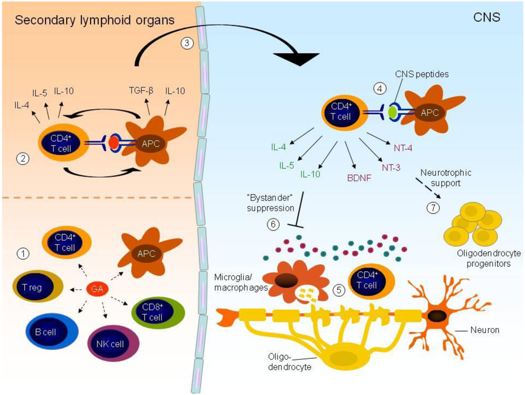
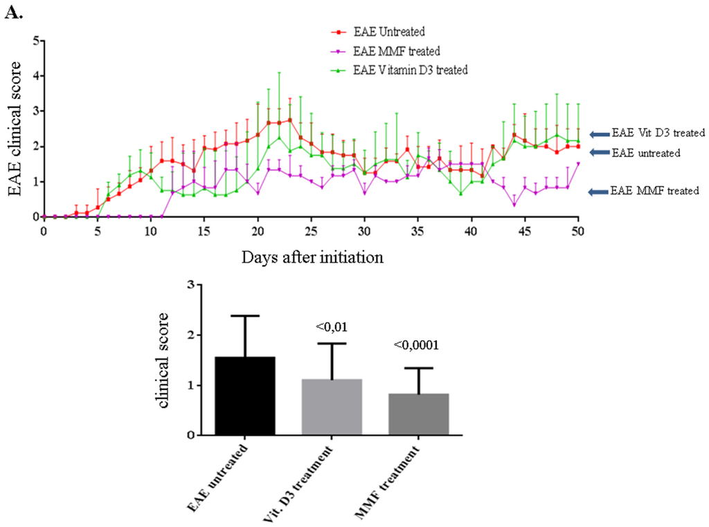
Experimental autoimmune encephalomyelitis (EAE) is a CD4+ T cell mediated inflammatory demyelinating disease that is induced in mice by administration of peptides derived from myelin proteins. We developed EAE in SJL mice by administration of PLP139–151 peptide. The effect of treating
[...] Read more.
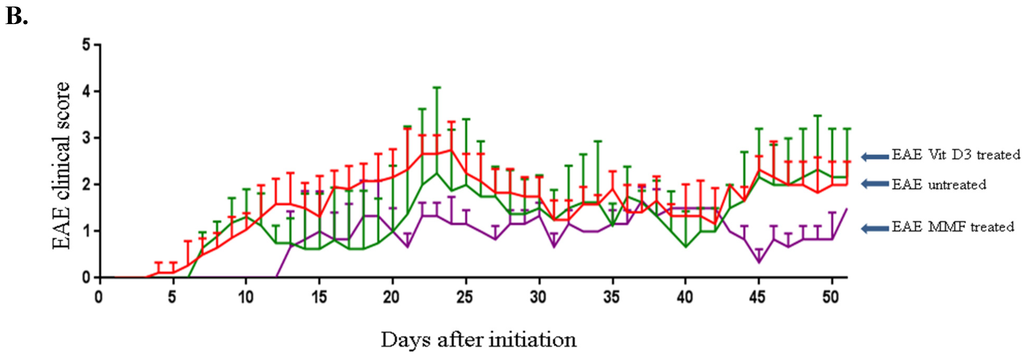
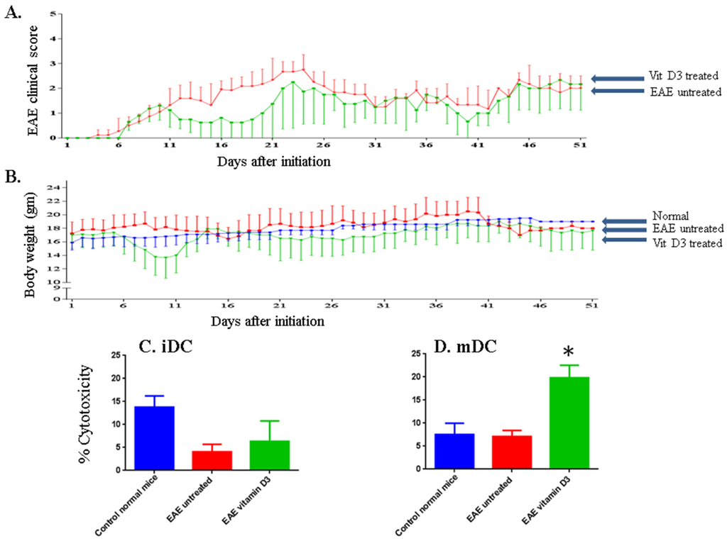
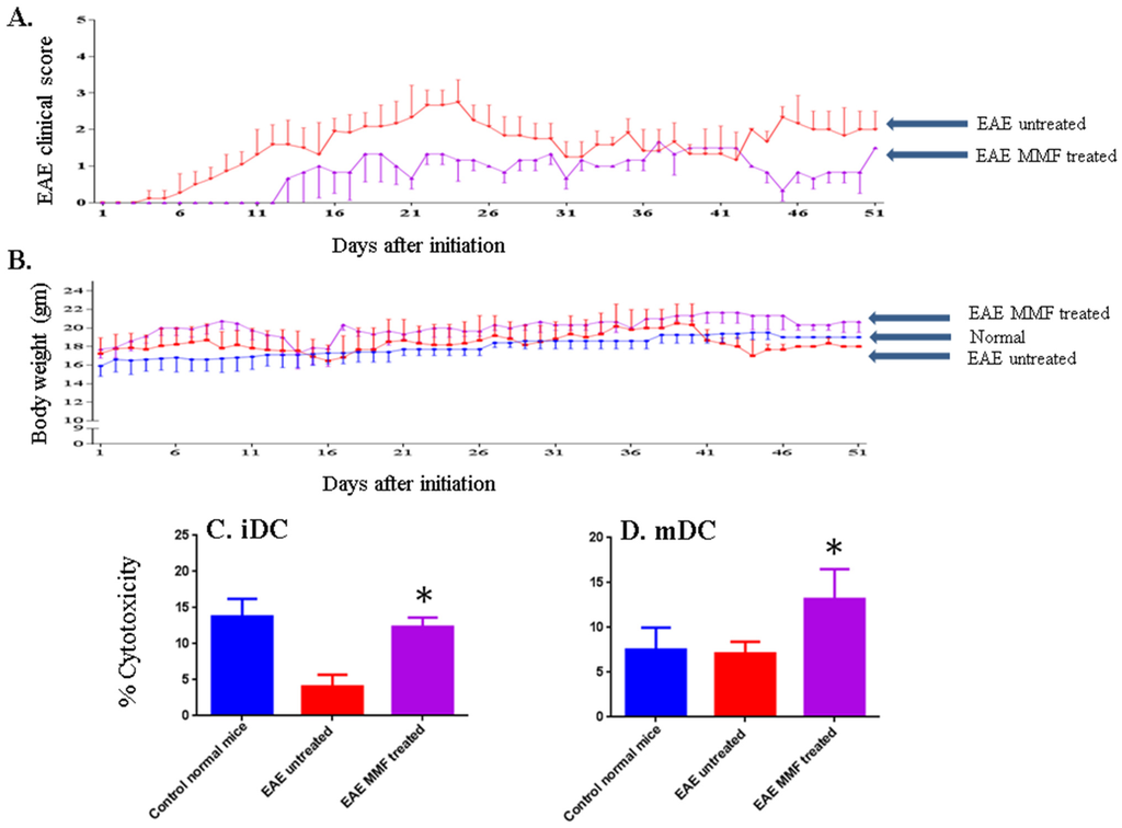
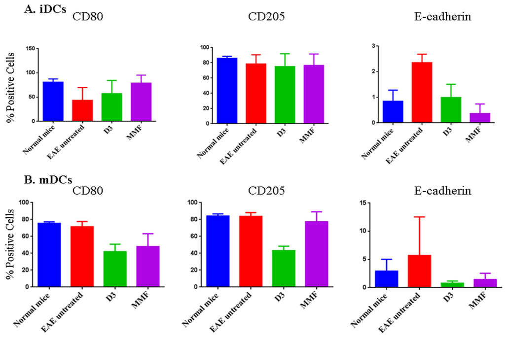
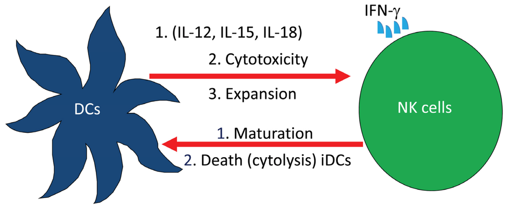
Experimental autoimmune encephalomyelitis (EAE) is a CD4+ T cell mediated inflammatory demyelinating disease that is induced in mice by administration of peptides derived from myelin proteins. We developed EAE in SJL mice by administration of PLP139–151 peptide. The effect of treating these mice with 1α,25-Dihydroxyvitamin D3 (vitamin D3), or with monomethyl fumarate (MMF) was then examined. We observed that both vitamin D3 and MMF inhibited and/or prevented EAE in these mice. These findings were corroborated with isolating natural killer (NK) cells from vitamin D3-treated or MMF-treated EAE mice that lysed immature or mature dendritic cells. The results support and extend other findings indicating that an important mechanism of action for drugs used to treat multiple sclerosis (MS) is to enhance NK cell lysis of dendritic cells.
Full article
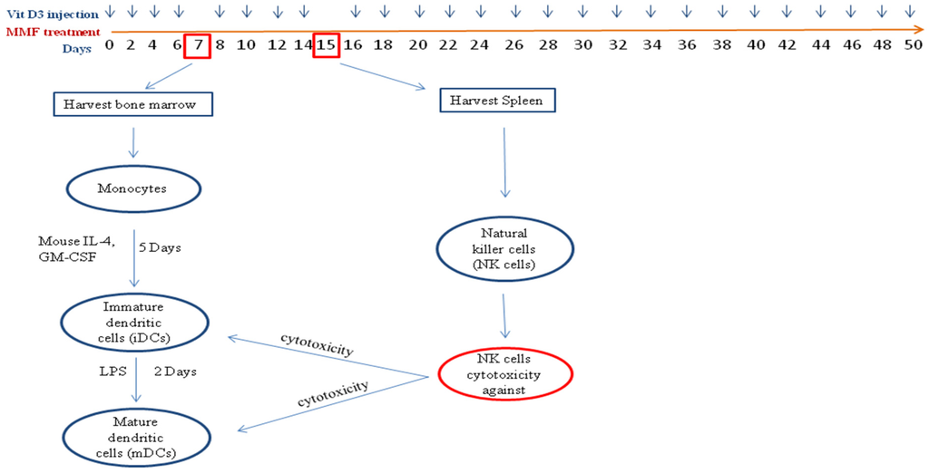
Figure 1


