Topic Editors

Department of Pathology, Faculty of Medicine, Tokai University School of Medicine, 143 Shimokasuya, Isehara 259-1193, Japan
ICM—Paris Brain Institute, Pitié Salpêtrière Hospital, 47, bd de l'Hôpital, 75013 Paris, France

The Tumor Microenvironment, Immuno-Oncology, and Immune Checkpoint: Implications for Current and Emergent Immunotherapies, 2nd Edition

Abstract submission deadline
30 November 2026
Manuscript submission deadline
31 December 2026
Viewed by
13161

Topic Information

Dear Colleagues,

Immunotherapy uses the body’s natural defenses (immune system) to fight disease. For example, our immune system can help to destroy cancer cells. Immuno-oncology is a type of immunotherapy that is directed against cancer.

There are two main parts of the immune system:

(1) Innate immunity (also known as built-in immune protection) has mechanisms that are prepared to act immediately, such as the skin, inner lining of the gut and lungs, stomach acid, intestinal bacteria, and leukocytes (neutrophils) that can find and kill bacteria.

In innate immunity, pathogens are recognized by receptors encoded in the germline. The receptors, known as pattern recognition receptors (PPRs), have a broad specificity for pathogen-associated molecular patterns (PAMPs).

Innate immune cells are granulocytes (neutrophils, eosinophils, basophils, mast cells), monocytes, macrophages, dendritic cells, and innate lymphoid cells (cytotoxic Natural Killer (NK) cells, and several noncytotoxic cells (IL1, 2, etc.)).

(2) Acquired immunity is immune protection that is “learned.”

After being exposed to certain diseases and a type of bacteria, fungus, or virus for the first time, the immune system develops memory and responds quicker and better the second time.

Lymphocytes are involved in the acquired immune response.

B cells/lymphocytes make antibodies that lock onto the surface of bacteria or viruses. B lymphocytes are part of the memory of the immune system.

There are different kinds of T lymphocytes: helper T cells (Th) and killer/cytotoxic T cells (Tc). Th cells stimulate B cells to make antibodies and help Tc cells to develop. Tc cells kill the body’s own cells infected by viruses or bacteria.

In adaptive immunity, pathogens are recognized by receptors that are generated randomly. These receptors recognize epitopes (polypeptides). The receptors are B-cell (BCR) and T-cell (TCR) receptors. The response is slow (3–5 days) and has memory.

The tumor microenvironment (TME) is comprised of the tumor, the extracellular matrix (ECM), and variable infiltration by nontransformed cells, including vascular vessels, fibroblasts, and immune system infiltrates. The TME plays a major role in tumor initiation, progression, infiltration and metastasis, and resistance to treatment. The tumoral immune microenvironment comprises variable percentages of CD8+Tc, naïve CD4+T cells, FOXP3+ regulatory T lymphocytes (Tregs), B cells, neutrophils, macrophages, NK cells, and dendritic cells. Macrophages, known as tumor-associated macrophages (TAMs), are a key component of the microenvironment. These M2-like TAMs are associated with tumor migration and invasion, metastasis, growth, angiogenesis, tissue remodeling, and inhibiting the host immune response (Treg induction, inactivation of T cells, etc.). Relevant markers associated with TAMs are PD-L1/L2, MMPs, M-CSF, CSF1R, IL-10, TGFB, prostaglandins, IDO1/2, etc.

The immune checkpoint refers to the mechanisms the immune system uses to avoid collateral damage during the physiological immune response and maintain self-tolerance. Ligand–receptor interactions are mediated by co-stimulatory molecules such as CD80, CD86, GITRL, and ICOSLG expressed by antigen-presenting cells (APCs), and CD28, GITR, and ICOS expressed by T cells. Conversely, coinhibitory signaling suppresses T cell activation using PD-1, CTLA-4, and LAG3 expressed by T cells.

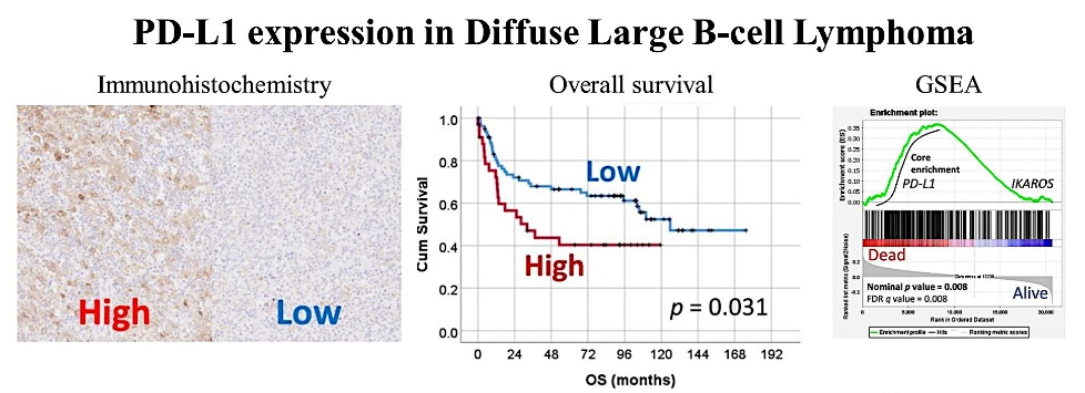
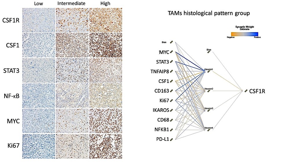
In the tumor microenvironment, cancer cells take advantage of the coinhibitory pathway, expressing markers such as PD-L1 (Figure 1). In addition, tumor-associated macrophages (TAMs) can also express PD-L1 and CSF1R (Figure 2).
Jisc logo

Figure 1. PD-L1 expression in diffuse large B-cell lymphoma (DLBCL) and the correlation with the poor prognosis of patients.
AI 2021, 2(1), 106-134; https://doi.org/10.3390/ai2010008
Jisc logo

Figure 2. CSF1R expression by tumor-associated macrophages (TAMs) in diffuse large B-cell lymphoma (DLBCL) and the correlation with other markers using a neural network.
Hemato 2021, 2(2), 182-206; https://doi.org/10.3390/hemato2020011
Jisc logo

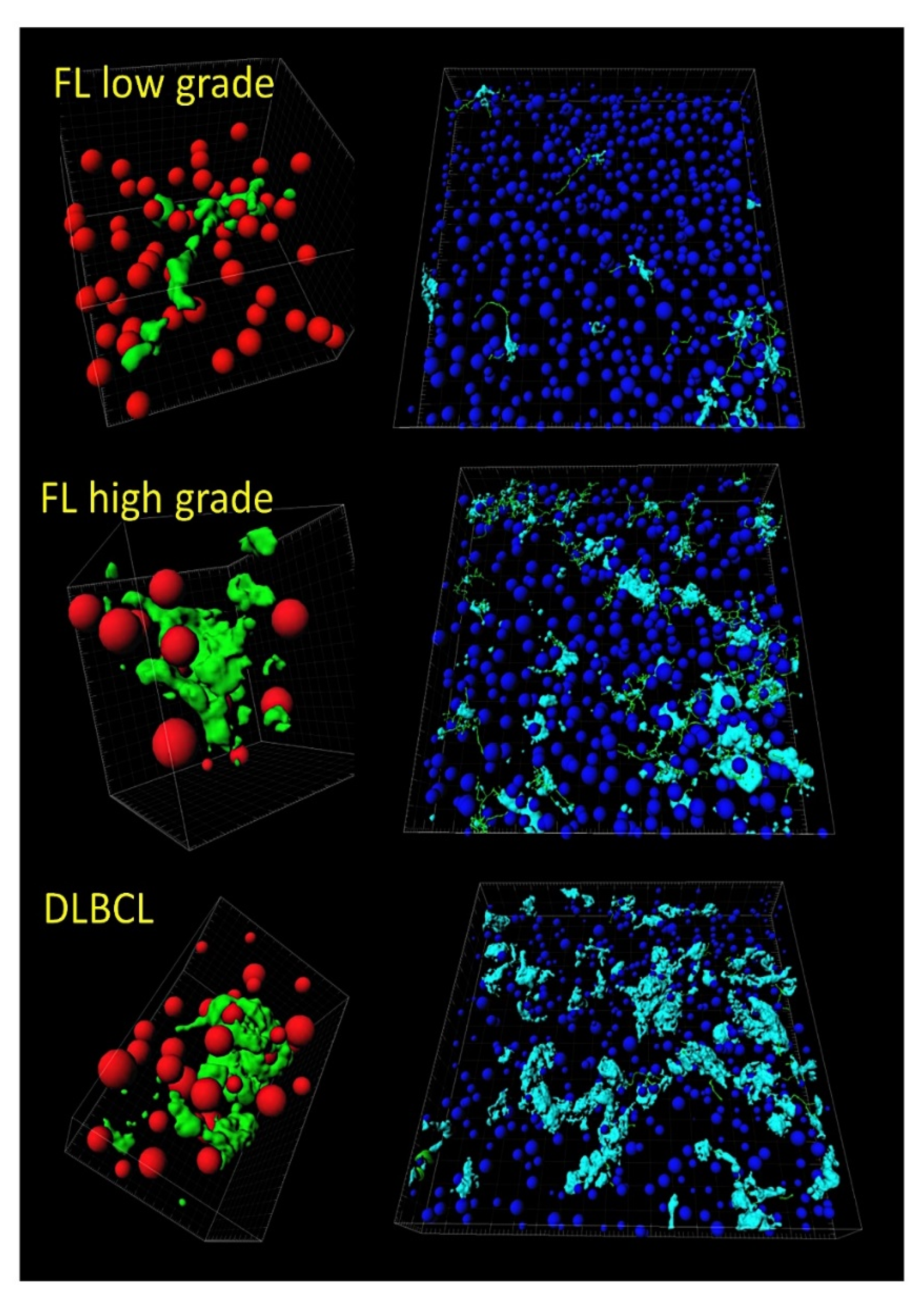
Figure 3. Tridimensional analysis of tumor-associated macrophages (TAMs) in follicular lymphoma and transformation to diffuse large B-cell lymphoma (DLBCL). Both the quantity and the shape of TAMs increase in disease progression and transformation to high-grade B-cell lymphoma.
Cancers 2022, 14(21), 5318; https://doi.org/10.3390/cancers14215318

Other factors are immunostimulatory signals for turning up cold tumors, which are not restricted to neoantigens derived from mutations. For instance, these include stress signals (DAMPs), the DNA damage response, and metabolic changes in the TME. Finally, the key role of immunosenescence and/or epitranscriptomic changes in immune cells that challenge tumor immunity must also be considered.

Some kinds of cancer are susceptible to treatment with immunotherapy because cancer cells are different from normal cells, so the immune system recognizes them and kills these abnormal cells. There are different types of immunotherapies:

  • Monoclonal antibodies (MABs), which recognize and attach certain proteins on cell surfaces;
  • Checkpoint inhibitors (immune checkpoint blockade (ICB) therapies);
  • Cytokines, which help boost the immune system;
  • Vaccines, which help recognize and attack cancer;
  • CAR T-cell therapy (also known as adoptive cell transfer).

Since standard chemotherapeutic agents and radiation, as well as targeted therapies, elicit/instigate an antitumor immune response, the combination of therapeutic modalities is the best treatment approach. Nevertheless, the current ICB therapies have a limitation in that they acquire resistance mechanisms.

This Topic accepts manuscripts that deal with all aspects of the immune response in hematological neoplasia and cancer, from basic to clinical research and integrative analyses (including mice models and artificial intelligence analyses).

Dr. Joaquim Carreras
Dr. Luis J. Castro-Vega
Topic Editors

Keywords

  • hematological neoplasia
  • cancer
  • immune microenvironment
  • immune checkpoint
  • immuno-oncology
  • artificial intelligence
  • tumor-associated macrophages

Participating Journals

Journal Name Impact Factor CiteScore Launched Year First Decision (median) APC
Biomedicines
biomedicines
3.9 5.2 2013 15.3 Days CHF 2600 Submit
Cancers
cancers
4.5 8.0 2009 16.3 Days CHF 2900 Submit
Cells
cells
5.1 9.9 2012 17.5 Days CHF 2700 Submit
Journal of Clinical Medicine
jcm
3.0 5.7 2012 17.3 Days CHF 2600 Submit
Pharmaceutics
pharmaceutics
4.9 7.9 2009 14.9 Days CHF 2900 Submit
Reports
reports
0.8 - 2018 18.4 Days CHF 1400 Submit

Preprints.org is a multidiscipline platform providing preprint service that is dedicated to sharing your research from the start and empowering your research journey.

MDPI Topics is cooperating with Preprints.org and has built a direct connection between MDPI journals and Preprints.org. Authors are encouraged to enjoy the benefits by posting a preprint at Preprints.org prior to publication:

  1. Immediately share your ideas ahead of publication and establish your research priority;
  2. Protect your idea from being stolen with this time-stamped preprint article;
  3. Enhance the exposure and impact of your research;
  4. Receive feedback from your peers in advance;
  5. Have it indexed in Web of Science (Preprint Citation Index), Google Scholar, Crossref, SHARE, PrePubMed, Scilit and Europe PMC.

Published Papers (8 papers)

Order results
Result details
Journals
Select all
Export citation of selected articles as:
7 pages, 480 KiB  
Case Report
Diaphragmatic Palsy Due to a Paraneoplastic Autoimmune Syndrome Revealed by Checkpoint Inhibitors
by Jean-Baptiste Destival, Jean-Marie Michot, Cécile Cauquil, Nicolas Noël, Salima Hacein-Bey-Abina, Pascale Chrétien and Olivier Lambotte
Reports 2024, 7(4), 84; https://doi.org/10.3390/reports7040084 - 11 Oct 2024
Viewed by 481
Abstract
Background and Clinical Significance: Immune checkpoint inhibitors (ICIs) have revolutionized cancer treatment but may underlie diverse and potentially life-threatening immune-related adverse events (irAEs). They may cause various conditions leading to respiratory failure, including myasthenic syndromes and myositis. However, diaphragmatic paralysis (DP) has rarely been [...] Read more.
Background and Clinical Significance: Immune checkpoint inhibitors (ICIs) have revolutionized cancer treatment but may underlie diverse and potentially life-threatening immune-related adverse events (irAEs). They may cause various conditions leading to respiratory failure, including myasthenic syndromes and myositis. However, diaphragmatic paralysis (DP) has rarely been reported. To describe patients with diaphragmatic paralysis in a pharmacovigilance registry, we searched the prospective REISAMIC registry at the Gustave Roussy Cancer Center (Villejuif, France) for cases of diaphragmatic palsy (DP) occurring from September 2014 to December 2021. Case Presentation: We identified three patients, in whom DP was confirmed by diaphragmatic ultrasonography, pulmonary function tests, and/or diaphragmatic electroneuromyogram. Diaphragmatic palsy was life-threatening in all patients, as it caused respiratory failure requiring mechanical ventilation. In all cases, a pre-existing subclinical paraneoplastic syndrome was detected. Onconeural antibodies (anti-titin and anti-VGCC) were detected in these patients before and after the initiation of ICI therapy, suggesting a mixed paraneoplastic syndrome with features overlapping those of myasthenic syndrome (myasthenia gravis in one patient and Lambert–Eaton syndrome in another) and myositis. Conclusions: Diaphragmatic palsy is a severe irAE potentially resulting from different mechanisms, including myositis and neuromuscular junction involvement (myasthenia gravis, Lambert–Eaton). Antineuronal antibodies associated with such conditions were already present in our patients prior to immunotherapy initiation, suggesting ICIs could trigger flare-ups of pre-existing silent paraneoplastic autoimmune conditions. Full article
Show Figures

Figure 1

15 pages, 1491 KiB  
Article
Real-World Comparison of Health-Related Quality of Life Associated with Use of Immune-Checkpoint Inhibitors in Oncology Patients
by Abdulrahman Alwhaibi, Miteb A. Alenazi, Saad D. Alnofaie, Abdullah M. Aldekhail, Rakan J. Alanazi, Sultan Alghadeer, Abdulrhman A. Alghamdi and Saleh A. Alanazi
J. Clin. Med. 2024, 13(16), 4918; https://doi.org/10.3390/jcm13164918 - 20 Aug 2024
Viewed by 701
Abstract
Background: Immune checkpoint inhibitors (ICIs) offer a new treatment approach for cancer, with an improvement in patient survival. However, it remains unclear whether their use impacts the quality of life of treated patients. This study aims to compare the health-related quality of life [...] Read more.
Background: Immune checkpoint inhibitors (ICIs) offer a new treatment approach for cancer, with an improvement in patient survival. However, it remains unclear whether their use impacts the quality of life of treated patients. This study aims to compare the health-related quality of life (HRQoL) of patients treated with different anti-PD-1 and anti-PD-L1 drugs, including several single or combination therapies. Methods: This is a prospective observational study conducted with adult cancer patients who received at least one dose of anti-PD-1 or anti-PD-L1. The HRQoL of all adult patients was assessed using the European Organization for Research and Treatment of Cancer (EORTC) Quality of Life Questionnaire-Core 30 module (QLQ-C30), version 3, Arabic version. Results: A total of 199 patients were found to be eligible for this study. Of these, 93 patients (82 on a single medication and 11 on multiple ICIs) completed the questionnaire, with a response rate of 46.7%. The majority of patients were treated with pembrolizumab (39.8%), followed by a smaller number treated with nivolumab (35.5%). Most of the patients were diagnosed with solid and advanced malignancies—88.2% (p = 0.023) and 87.1% (p = 0.021), respectively—with a significant difference between treatment groups. The median functioning score was 84.7%, with no significant difference between treatment groups (p = 0.752). Fatigue and pain were noted in >50% of patients, influencing the overall cohort’s score related to these symptoms, with scores of 88.8% and 83.3%, respectively. Although a non-significant variation was found in the scores of all combined symptoms among all groups, ranging from 82.1% to 90.4% (p = 0.931), patients receiving anti-PD-1 + anti-PD-L1 tended to more frequently complain about fatigue, pain, dyspnea, and constipation and hence, exhibited the worst, yet non-significant, scores compared to those of the other groups, with p = 0.234, p = 0.79, p = 0.704, and p = 0.86, respectively. All combined groups scored 83.3% on the global health scale. Nevertheless, the nivolumab-treated patients scored 75%, which was the worst global health score compared with those of the other groups, but this score was not statistically significant (p = 0.809). Conclusions: Our findings revealed no significant difference in the impact of different ICIs on the HRQoL of cancer patients. However, a larger number of cases would be necessary to provide a robust analysis and to yield conclusive results. Full article
Show Figures

Figure 1

14 pages, 2090 KiB  
Article
Peripheral Soluble Immune Checkpoint-Related Proteins Were Associated with Survival and Treatment Efficacy of Osteosarcoma Patients, a Cohort Study
by Binghao Li, Qinchuan Wang, Yihong Luo, Sicong Wang, Sai Pan, Wenting Zhao, Zhaoming Ye and Xifeng Wu
Cancers 2024, 16(9), 1628; https://doi.org/10.3390/cancers16091628 - 24 Apr 2024
Viewed by 1341
Abstract
Background: The immune checkpoint blockade remains obscure in osteosarcoma (OS). We aim to explore the clinical significance of soluble immune checkpoint (ICK)-related proteins in OS. Methods: We profiled 14 soluble ICK-related proteins (BTLA, GITR, HVEM, IDO, LAG-3, PD-1, PD-L1, PD-L2, TIM-3, CD28, CD80, [...] Read more.
Background: The immune checkpoint blockade remains obscure in osteosarcoma (OS). We aim to explore the clinical significance of soluble immune checkpoint (ICK)-related proteins in OS. Methods: We profiled 14 soluble ICK-related proteins (BTLA, GITR, HVEM, IDO, LAG-3, PD-1, PD-L1, PD-L2, TIM-3, CD28, CD80, CD137, CD27, and CTLA-4) in the plasma of 76 OS patients and matched controls. We evaluated the associations between the biomarkers and the risk of OS using unconditional multivariate logistic regression. The multivariate Cox model was utilized to develop the prediction model of OS. Immune subtypes were established from the identified biomarkers. Transcriptional data from GEO were analyzed to elucidate potential mechanisms. Results: We found that sTIM3, sCD137, sIDO, and sCTLA4 were significantly correlated with OS risk (all p < 0.05). sBTLA, sPDL2, and sCD27 were significantly associated with the risk of lung metastasis, whereas sBTLA and sTIM3 were associated with the risk of disease progression. We also established an immune subtype based on sBTLA, sPD1, sTIM3, and sPDL2. Patients in the sICK-type2 subtype had significantly decreased progression-free survival (PFS) and lung metastasis-free survival (LMFS) than those in the sICK-type1 subtype (log-rank p = 2.8 × 10⁻², 1.7 × 10⁻², respectively). Interestingly, we found that the trend of LMFS and PFS in the subtypes of corresponding ICK genes’ expression was opposite to the results in the blood (log-rank p = 2.6 × 10⁻⁴, 9.5 × 10⁻⁴, respectively). Conclusion: Four soluble ICK-related proteins were associated with the survival of OS patients. Soluble ICK-related proteins could be promising biomarkers for the outcomes and immunotherapy of OS patients, though more research is warranted. Full article
Show Figures

Figure 1

19 pages, 2894 KiB  
Review
Structure–Activity Relationship of PAD4 Inhibitors and Their Role in Tumor Immunotherapy
by Yijiang Jia, Renbo Jia, Ayijiang Taledaohan, Yanming Wang and Yuji Wang
Pharmaceutics 2024, 16(3), 335; https://doi.org/10.3390/pharmaceutics16030335 - 28 Feb 2024
Viewed by 2608
Abstract
Protein arginine deiminase 4 (PAD4) plays an important role in cancer progression by participating in gene regulation, protein modification, and neutrophil extracellular trap (NET) formation. Many reversible and irreversible PAD4 inhibitors have been reported recently. In this review, we summarize the structure–activity relationships [...] Read more.
Protein arginine deiminase 4 (PAD4) plays an important role in cancer progression by participating in gene regulation, protein modification, and neutrophil extracellular trap (NET) formation. Many reversible and irreversible PAD4 inhibitors have been reported recently. In this review, we summarize the structure–activity relationships of newly investigated PAD4 inhibitors to bring researchers up to speed by guiding and describing new scaffolds as optimization and development leads for new effective, safe, and selective cancer treatments. In addition, some recent reports have shown evidence that PAD4 inhibitors are expected to trigger antitumor immune responses, regulate immune cells and related immune factors, enhance the effects of immune checkpoint inhibitors, and enhance their antitumor efficacy. Therefore, PAD4 inhibitors may potentially change tumor immunotherapy and provide an excellent direction for the development and clinical application of immunotherapy strategies for related diseases. Full article
Show Figures

Figure 1

16 pages, 2360 KiB  
Systematic Review
A Systematic Review of the Advances in the Study of T Lymphocyte Suppressor Receptors in HBV Infection: Potential Therapeutic Targets
by Daqiong Zhou, Lili Liu, Jiangyu Liu, Hong Li, Jing Zhang and Zhenhuan Cao
J. Clin. Med. 2024, 13(5), 1210; https://doi.org/10.3390/jcm13051210 - 21 Feb 2024
Viewed by 1566
Abstract
Background: HBV-specific T lymphocytes are pivotal in eliminating the hepatitis B virus (HBV) and regulating intrahepatic inflammatory reactions. Effective T cell responses curtail HBV infection; however, compromised immunity can result in persistent infection. Beyond the acute phase, the continued presence of antigens and [...] Read more.
Background: HBV-specific T lymphocytes are pivotal in eliminating the hepatitis B virus (HBV) and regulating intrahepatic inflammatory reactions. Effective T cell responses curtail HBV infection; however, compromised immunity can result in persistent infection. Beyond the acute phase, the continued presence of antigens and inflammation leads to the increased expression of various inhibitory receptors, such as PD-1, CTLA-4, Tim-3, LAG3, 2B4, CD160, BTLA, and TIGIT. This escalates the dysfunction of and diminishes the immune and proliferative abilities of T cells. Methods: In this study, we reviewed English-language literature from PubMed, Web of Science, and Scopus up to 9 July 2023. This paper aims to elucidate the inhibitory effects of these receptors on HBV-specific T lymphocytes and how immune function can be rejuvenated by obstructing the inhibitory receptor signaling pathway in chronic HBV patients. We also summarize the latest insights into related anti-HBV immunotherapy. Result: From 66 reviewed reports, we deduced that immunotherapy targeting inhibitory receptors on T cells is a reliable method to rejuvenate T cell immune responses in chronic HBV patients. However, comprehensive combination therapy strategies are essential for a functional cure. Conclusions: Targeting T cell suppressor receptors and combining immunotherapy with antiviral treatments may offer a promising approach towards achieving a functional cure, urging future research to prioritize effective combination therapeutic strategies for chronic HBV infection. Full article
Show Figures

Figure 1

11 pages, 231 KiB  
Systematic Review
The Use of Fecal Microbiota Transplant in Overcoming and Modulating Resistance to Anti-PD-1 Therapy in Patients with Skin Cancer
by Tahne Vongsavath, Rodd Rahmani, Kyaw Min Tun and Vignan Manne
Cancers 2024, 16(3), 499; https://doi.org/10.3390/cancers16030499 - 24 Jan 2024
Cited by 3 | Viewed by 1801
Abstract
While immune checkpoint inhibitors have evolved into the standard of care for advanced melanoma, 40–50% of melanoma cases progress while on therapies. The relationship between bacterium and carcinogenesis is well founded, such as in H. pylori in gastric cancers, and Fusobacterium in colorectal [...] Read more.
While immune checkpoint inhibitors have evolved into the standard of care for advanced melanoma, 40–50% of melanoma cases progress while on therapies. The relationship between bacterium and carcinogenesis is well founded, such as in H. pylori in gastric cancers, and Fusobacterium in colorectal cancers. This interplay between dysbiosis and carcinogenesis questions whether changes in the microbiome could affect treatment. Thus, FMT may find utility in modifying the efficacy of anti-PD-1. This review aims to examine the use of FMT in treatment-resistant melanoma. A literature search was performed using the keywords “fecal microbiota transplant” and “skin cancer”. Studies were reviewed for inclusion criteria and quality and in the final stage, and three studies were included. Overall objective responses were reported in 65% of patients who were able to achieve CR, and 45% who achieved PR. Clinical benefit rate of combined CR/PR with stable disease greater or equal to 6 months was 75%. Reported objective responses found durable stable disease lasting 12 months. Overall survival was 7 months, and overall PRS was 3 months. As for the evaluation of safety, many patients reported grade 1–2 FMT related AE. Only following the administration of anti-PD-1 therapy were there a grade 3 or higher AE. Full article
10 pages, 825 KiB  
Article
Adverse Effect of the Duration of Antibiotic Use Prior to Immune Checkpoint Inhibitors on the Overall Survival of Patients with Recurrent Gynecologic Malignancies
by Hye-Ji Jung, Jong-Ho Park, Jina Oh, Sae-Mi Lee, Il-Yeo Jang, Jung-Yong Hong, Yoo-Young Lee and Hyun Jin Choi
Cancers 2023, 15(24), 5745; https://doi.org/10.3390/cancers15245745 - 7 Dec 2023
Viewed by 1200
Abstract
Purpose: Antibiotic use preceding immune checkpoint inhibitor (ICI) treatment has been associated with a decreased efficacy of ICI in solid tumors. In this study, we evaluated the effect of antibiotic use before ICI therapy on oncological outcomes. Methods: We examined patients with recurrent [...] Read more.
Purpose: Antibiotic use preceding immune checkpoint inhibitor (ICI) treatment has been associated with a decreased efficacy of ICI in solid tumors. In this study, we evaluated the effect of antibiotic use before ICI therapy on oncological outcomes. Methods: We examined patients with recurrent gynecologic malignancies at two academic institutions. The clinical data, including antibiotic use within 60 days of ICI initiation, type of antibiotics, reasons for antibiotic use, body mass index, tumor site, chemotherapy-free interval, prior history of radiotherapy, disease control rate (DCR), and overall survival (OS), were assessed. Results: Of 215 patients, 22.9% (n = 47) received antibiotics before ICI treatment. The most common cancer was ovarian (52.1%, n = 112), followed by cervical (24.7%, n = 53) and endometrial (16.7%, n = 36). When we divided the cohort based on antibiotic use before ICIs, there were no significant differences in the DCR and baseline characteristics between the two groups. On multivariate analyses, the variables associated with poor OS were previous use of antibiotics for a cumulative duration of >14 days (HR 2.286, 95% CI 1.210–4.318; p = 0.011); Eastern Cooperative Oncology Group 2 or 3 (HR 4.677, 95% CI 2.497–8.762; p < 0.001); and chemotherapy-free interval of <6 months (HR 2.007, 95% CI 1.055–3.819; p = 0.034). Conclusion: Prior use of antibiotics for a cumulative duration of >14 days was associated with reduced survival in recurrent gynecologic malignancies. Full article
Show Figures

Figure 1

15 pages, 1894 KiB  
Article
CX3CR1-Expressing Immune Cells Infiltrate the Tumor Microenvironment and Promote Radiation Resistance in a Mouse Model of Lung Cancer
by Tamar Ben-Mordechai, Yaacov R. Lawrence, Zvi Symon, Ariel Shimoni-Sebag and Uri Amit
Cancers 2023, 15(22), 5472; https://doi.org/10.3390/cancers15225472 - 19 Nov 2023
Viewed by 2218
Abstract
Introduction: Chemokine (C-X3-C Motif) Receptor 1 (CX3CR1) is present in a subset of the immune cells in the tumor microenvironment (TME) and plays an essential and diverse role in cancer progression. However, its potential function in the irradiated TME remains unknown. Materials and [...] Read more.
Introduction: Chemokine (C-X3-C Motif) Receptor 1 (CX3CR1) is present in a subset of the immune cells in the tumor microenvironment (TME) and plays an essential and diverse role in cancer progression. However, its potential function in the irradiated TME remains unknown. Materials and Methods: A mouse lung cancer model was performed by subcutaneously inoculating Lewis Lung Carcinoma (LLC) cells expressing luciferase (Luc-2) and mCherry cells in CX3CR1GFP/GFP, CX3CR1DTR/+, and wild–type (WT) mice. Bioluminescence imaging, clonogenic assay, and flow cytometry were used to assess tumor progression, proliferation, and cell composition after radiation. Results: Radiation provoked a significant influx of CX3CR1-expressing immune cells, notably monocytes and macrophages, into the TME. Co-culturing irradiated LLC cells with CX3CR1-deficient monocytes, and macrophages resulted in reduced clonogenic survival and increased apoptosis of the cancer cells. Interestingly, deficiency of CX3CR1 in macrophages led to a redistribution of the irradiated LLC cells in the S-phase, parallel to increased expression of cyclin E1, required for cell cycle G1/S transition. In addition, the deficiency of CX3CR1 expression in macrophages altered the cytokine secretion with a decrease in interleukin 6, a crucial mediator of cancer cell survival and proliferation. Next, LLC cells were injected subcutaneously into CX3CR1DTR/+ mice, sensitive to diphtheria toxin (DT), and WT mice. After injection, tumors were irradiated with 8 Gy, and mice were treated with DT, leading to conditional ablation of CX3CR1-expressing cells. After three weeks, CX3CR1-depleted mice displayed reduced tumor progression. Furthermore, combining the S-phase-specific chemotherapeutic gemcitabine with CX3CR1 cell ablation resulted in additional attenuation of tumor progression. Conclusions: CX3CR1-expressing mononuclear cells invade the TME after radiation therapy in a mouse lung cancer model. CX3CR1 cell depletion attenuates tumor progression following radiation and sensitizes the tumor to S–phase-specific chemotherapy. Thus, we propose a novel strategy to improve radiation sensitivity by targeting the CX3CR1-expressing immune cells. Full article
Show Figures

Figure 1

Back to TopTop