Article

Transcriptome Analysis Reveals Cross-Talk between the Flagellar Transcriptional Hierarchy and Secretion System in Plesiomonas shigelloides

1
TEDA Institute of Biological Sciences and Biotechnology, Nankai University, Tianjin 300457, China
2
Key Laboratory of Molecular Microbiology and Technology of the Ministry of Education, Nankai University, Tianjin 300457, China
3
Tianjin Key Laboratory of Microbial Functional Genomics, TEDA College, Nankai University, Tianjin 300457, China
4
State Key Laboratory of Medicinal Chemical Biology, College of Pharmacy, Tianjin Key Laboratory of Molecular Drug Research, Nankai University, Tianjin 300353, China
*
Authors to whom correspondence should be addressed.
Int. J. Mol. Sci. 2024, 25(13), 7375; https://doi.org/10.3390/ijms25137375
Submission received: 6 June 2024 / Revised: 2 July 2024 / Accepted: 3 July 2024 / Published: 5 July 2024
(This article belongs to the Collection Microbial Virulence Factors)

Abstract

:
Plesiomonas shigelloides, a Gram-negative bacillus, is the only member of the Enterobacteriaceae family able to produce polar and lateral flagella and cause gastrointestinal and extraintestinal illnesses in humans. The flagellar transcriptional hierarchy of P. shigelloides is currently unknown. In this study, we identified FlaK, FlaM, FliA, and FliAL as the four regulators responsible for polar and lateral flagellar regulation in P. shigelloides. To determine the flagellar transcription hierarchy of P. shigelloides, the transcriptomes of the WT and ΔflaK, ΔflaM, ΔfliA, and ΔfliAL were carried out for comparison in this study. Quantitative Real-Time Polymerase Chain Reaction (qRT-PCR) and luminescence screening assays were used to validate the RNA-seq results, and the Electrophoretic Mobility Shift Assay (EMSA) results revealed that FlaK can directly bind to the promoters of fliK, fliE, flhA, and cheY, while the FlaM protein can bind directly to the promoters of flgO, flgT, and flgA. Meanwhile, we also observed type VI secretion system (T6SS) and type II secretion system 2 (T2SS-2) genes downregulated in the transcriptome profiles, and the killing assay revealed lower killing abilities for ΔflaK, ΔflaM, ΔfliA, and ΔfliAL compared to the WT, indicating that there was a cross-talk between the flagellar hierarchy system and bacterial secretion system. Invasion assays also showed that ΔflaK, ΔflaM, ΔfliA, and ΔfliAL were less effective in infecting Caco-2 cells than the WT. Additionally, we also found that the loss of flagellar regulators causes the differential expression of some of the physiological metabolic genes of P. shigelloides. Overall, this study aims to reveal the transcriptional hierarchy that controls flagellar gene expression in P. shigelloides, as well as the cross-talk between motility, virulence, and physiological and metabolic activity, laying the groundwork for future research into P. shigelloides’ coordinated survival in the natural environment and the mechanisms that infect the host.

1. Introduction

The genus Plesiomonas, represented by a single species, Plesiomonas shigelloides, is a Gram-negative bacillus associated with gastrointestinal and extraintestinal diseases in humans [1], such as an invasive shigellosis-like disease, cholera-like illness, pseudoappendicitis, meningitis, acute secretory gastroenteritis, and bacteremia [2,3,4,5,6,7]. Many pathogenic bacterial species express flagella on their surfaces, which are their primary means of locomotion or motility, and flagellar motility is necessary for them to reach the site of infection, which is the initial step in the establishment of a bacterial infection [8]. Meanwhile, flagellum motility provides a significant advantage for bacteria to move toward favorable conditions or away from detrimental environments and to effectively compete with other microorganisms [9].
The flagellum consists of three parts: the basal body, hook, and filament [10]. Flagellar motility, an energetically expensive process, requires the coordinated expression of more than 50 genes encoding regulatory proteins, structural components of the secretion and assembly apparatus, and chemo-sensor machinery [8]. The coordinated expression of these promoters clusters gene transcription at three or four levels of hierarchy: classes I to III or I to IV [11]. Peritrichous flagellated bacteria, such as Salmonella and Escherichia coli, have three levels of hierarchy, and transcription of class I and II genes requires the housekeeping sigma factor 70 (σ70) [12]. However, some bacteria, such as Vibrio cholerae [13,14,15], Vibrio parahaemolyticus [16], and Aeromonas hydrophila [11,17], have four levels of flagella regulatory hierarchy. Furthermore, several of the aforementioned bacteria contain both polar and lateral flagella. Bacteria with polar flagellation exhibit four transcriptional levels: class I promoters encode a σ54-dependent activator (FlrA) and activate class II σ54-dependent promoters (FlrBC), which encode a two-component signal-transducing system whose regulator activates class III σ54-dependent promoters [13,14,15,16,17]. Moreover, class II promoters also encode the σ28 factor (FliA), which activates the transcription of class IV genes. The lateral flagellar system, such as that of V. parahaemolyticus and A. hydrophila, which have three levels of flagella regulatory hierarchy, is encoded by more than 30 lateral flagella genes and regulated by the LafK master regulator [18]. Furthermore, LafK may compensate for FlaK’s role in enabling swimming in some bacteria [19]. In the expression of the lateral flagellar system, the master regulator LafK activates the class II genes, and then the expression of class III genes is activated by σ28, which is encoded by fliAL [16,20,21]. In a word, in a spatiotemporal-dependent manner, the expression of flagellar genes is strictly regulated by the respective regulators and sigma factors.
The primary function of flagella is to provide motility to bacteria; nevertheless, they are also related to bacterial pathogenicity. Flagellum-mediated motility plays a critical role in interactions between Vibrio species and their hosts, and these bacteria colonization and lethality in their respective hosts decrease when motility is restricted [22]. Vibrios have therefore been shown to be valuable models for researching flagellar control and its role in colonization. Motility has been identified as a virulence determinant of V. cholerae. Additionally, non-motile mutants in competition assays using an infant mouse model system show no significant defect in their capacity to colonize the small intestine [23]. A Na+ gradient across the membrane drives V. cholerae flagellar rotation while also regulating transcription of the toxT gene, which is necessary for CT and TCP expression, revealing a possible mechanism for connecting virulence factor expression to motility [24,25]. Coster and Kenner found that the human reactogenicity of live attenuated V. cholerae vaccines is also lowered in non-motile mutants [26]. In addition to being more frequently documented in V. cholerae, the correlation between flagella and virulence has also been investigated in other bacteria. Flagella motility, like that of V. cholerae, is critical in the pathogenesis of V. campbellii, and inhibiting motility greatly reduces host mortality during infection [27]. Previous studies have also reported the synergistic action of SPI-1 gene expression in Salmonella enterica serovar typhimurium through transcriptional cross-talk with the flagellar system [28]. Zhao et al. found that the type III secretion system intersects with the lateral flagellar system in A. hydrophila [29]. In a burned mouse model of infection, non-flagellated mutants of P. aeruginosa have been demonstrated to exhibit reduced penetration of cultured corneal epithelial cells and to be deficient in pathogenicity [30,31]. As a result, the significance of flagella in pathogenicity is obvious.
In addition to granting motility to bacteria and influencing adherence and virulence, flagellar regulators and flagellar genes are also involved in the physiological metabolism of bacteria. The relationship between the ability to survive periods of acid shock and the fine-tuned expression of motility genes and fitness is currently unknown; however, Schumacher et al. found an inverse correlation between the expression of FlhDC, the master regulator of flagellation, and acid shock survival in E. coli [32]. According to Hoque et al., the ∆flrA mutation in V. cholerae increases iron utilization and oxidative stress tolerance since it significantly upregulates most of the genes involved in heme absorption, siderophore uptake, and vibriobactin uptake [33]. Sinha-Ray et al. found that a mutation in V. cholerae’s flrA gene is inversely involved in the vps-independent biofilm driving bacteria toward nutrients in lake water [34]. Rodríguez-Herva et al. suggested that in addition to a number of motility and chemotaxis genes, the fliA gene product is also necessary for the expression of some genes potentially involved in amino acid utilization or stress responses [35]. Li et al. reported that flagellum hook-associated protein (FlgK) increases the resilience of C. sakazakii to dehydration and regulates formate dehydrogenase and nitrate reductase [36]. It has also been reported in studies that FliZ plays a key role in controlling lipase and hemolysin activities [37,38].
P. shigelloides is a special member of the Enterobacteriaceae family that can produce both polar and lateral flagella, and Merino et al. showed that P. shigelloides has two distinct gene clusters: one for lateral flagella biosynthesis and another for biosynthetic polar flagella [39]. Merino and co-workers reported that although P. shigelloides lacks a FlrB ortholog, P. shigelloides FlrC (FlaM) contains the PAS and His Kinase A domains found in the FlrC proteins of Vibrio species and A. hydrophila, indicating that P. shigelloides FlaM may activate transcription from σ54-dependent promoters of class III genes [39]. Furthermore, the P. shigelloides polar flagella gene regions show greater similarity to those reported in Vibrio or Aeromonas than the regions in Enterobacteriaceae [40]. Though a number of studies have implicated motility as being important for bacteria, the transcriptional regulation network of polar and lateral flagella in P. shigelloides has yet to be revealed. In this work, we identified FlaK, FlaM, FliA, and FliAL as the four regulators responsible for polar and lateral flagellar regulation in P. shigelloides. Through transcriptome sequencing analysis, we confirmed the polar and lateral flagellar regulatory hierarchy and further revealed the cross-talk between the flagellar hierarchy system and P. shigelloides’ virulence and physiological metabolism.

2. Results

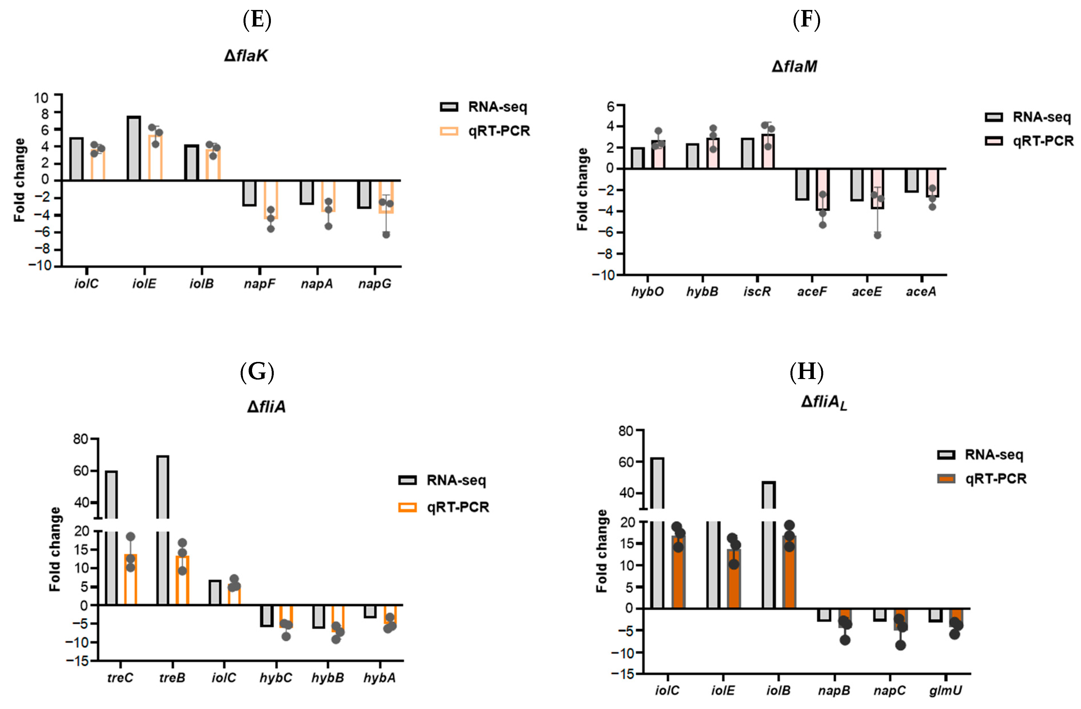
2.1. RNA Sequencing of Flagellar Regulatory Mutants

Given the basis of the previous research, we aligned the P. shigelloides flagellar gene clusters for orthologous alignment. The results indicated that the orthologous similarity of the flagellar gene in P. shigelloides with V. cholerae, V. parahaemolyticus, and A. hydrophila is relatively similar (Tables S3 and S4). Moreover, the highest overall homologous similarity is between the polar and lateral flagella gene clusters of P. shigelloides and V. parahaemolyticus, followed by A. hydrophila and V. cholerae. Previous reports corroborate our observations: The polar flagella gene cluster in the P. shigelloides genome contains the regulatory factors FlaK, FlaM, and FliA. Similar to FlrBC in Vibrio sp. or A. hydrophila, FlaM serves a similar function. Furthermore, the major regulator LafK is absent from the P. shigelloides lateral flagella gene cluster, which only contains the gene FliAL. To determine which P. shigelloides flagella genes are controlled by the flagellar transcription hierarchy, RNA-Seq of the WT and flaK, flaM, fliA, and fliAL deletion strains was carried out for comparison. The volcano plot revealed that FlaK, FlaM, FliA, and FliAL regulate the expression of 152, 341, 515, and 37 differentially expressed genes (DEGs) in their respective transcriptomes (Figure 1A–D). Moreover, the DEGs’ functionalities and enriched pathways in the ΔflaK, ΔflaM, ΔfliA, and ΔfliAL mutants were analyzed using the Gene Ontology (GO) and Kyoto Encyclopedia of Genes and Genomes (KEGG) pathway databases (Figure S1A–H). Additionally, three upregulated and three downregulated DEGs in the ΔflaK, ΔflaM, ΔfliA, and ΔfliAL transcriptome profiles were selected, respectively, for validation using qRT-PCR in the WT and mutant strains. The results of qRT-PCR were consistent with RNA-seq analysis (Figure 1E–H), indicating the reliability of the RNA-seq.

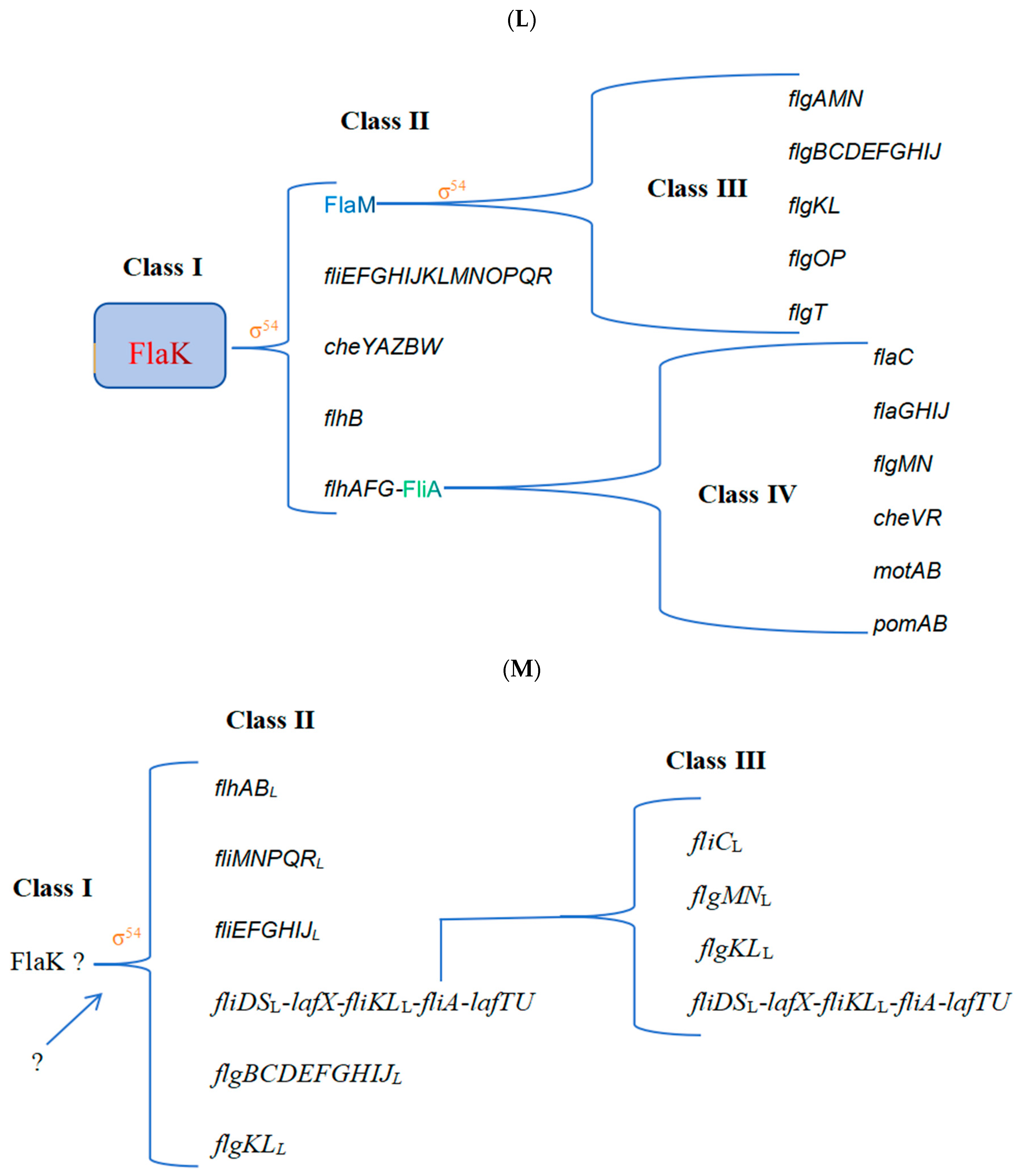
2.2. Refinement of the Flagellar Transcription Hierarchy

P. shigelloides contains over 50 polar flagellar genes and up to 36 lateral flagellar genes (Figure S2). The heat map of polar flagellar gene expression in the ΔflaK, ΔflaM, ΔfliA, and ΔfliAL transcriptome profiles revealed that FlaK positively regulated nearly all of the polar flagellar gene clusters (Figure 2A and Table S5), which also indicates that FlaK, as a primary regulator of the polar flagellar hierarchy, is important for the regulation of P. shigelloides’ motility. On various promoters of the polar flagellar gene cluster, FlaM and FliA, however, have distinct regulatory functions. The transcriptome data indicate that FlaM positively regulates the expression of polar flagellar gene clusters flgAMN, flgB-J, flgKL, and flgOP and flagellar gene flgT; however, the expression of fliE-R, a polar flagellar gene cluster, was upregulated following the loss of FlaM (Figure 2A and Table S6). Furthermore, the results also revealed that FliA positively regulated the expression of the flagellar gene flaC, flagellar gene cluster flaGHIIJ, and chemotaxis genes cheVR, motAB, and pomAB, while FliA had a negative regulatory effect on the flagellar gene cluster that FlaM positively regulated (Figure 2A and Table S7). To confirm the authenticity of the RNA-seq results, the flagellar genes positively regulated by FlaK, FlaM, and FliA were verified, respectively, by using qRT-PCR and construct promoter–lux fusions; the results of qRT-PCR and lux assays were consistent with RNA-seq (Figure 2C–E). Furthermore, consistent with previous reports, we also found that P. shigelloides’ genome lacked the master regulator LafK in the lateral flagellar system; yet, LafK in V. parahaemolyticus and A. hydrophila exhibited high homologous similarity to FlaK in P. shigelloides (Figures S3 and S4). Moreover, we found that the downregulation of lateral flagella genes coincided with the loss of Flak (Table S5), suggesting that FlaK may be involved in the regulation of the lateral flagellar system. For this reason, we performed a heatmap analysis of the lateral flagella gene expression in the transcriptome profiles of ΔflaK and ΔfliAL (Figure 2B). The results revealed that FliAL positively regulated the expression of the lateral flagellar gene fliCL and the lateral flagellar gene clusters flgMNL, flgKLL, and fliDSL-lafX-fliKLL-fliA-lafTU (Figure 2B and Table S8). The differential expression of the lateral flagellar gene in the transcriptome profiles of ΔflaK and ΔfliAL was then validated by qRT-PCR and construct promoter–lux fusions (Figure 2F,G), which demonstrated that the experimental data matched.
Furthermore, the EMSAs results showed that FlaK can directly bind to the promoters of fliK, fliE, flhA, and cheY, but not to the promoters of flhB and flaM (Figure 2H). The FlaM protein can bind directly to the promoters of flgO, flgT, and flgA but not those of flgB, flgK, and flgL (Figure 2I). Unfortunately, no flagellar gene promoter was screened for binding to FliA or FliAL. In addition, we observed the migration of the WT, ΔflaK, ΔflaM, ΔfliA, ΔfliAL, and complementation strains grown in swimming agar plates and the flagella produced by the aforementioned strains, which indicated that ΔflaK, ΔfliA, and ΔfliAL have reduced motility compared with the WT, while ΔflaM completely loses motility, and the complementation strains recovered the motility level of the WT (Figure 2J). The TEM results showed that the creation of flagella decreased and grew shorter in ΔflaK, ΔflaM, and ΔfliA, while ΔflaM was generated without any flagella, and the flagella production ability of corresponding complementation strains was greatly increased (Figure 2K). Together with our earlier research on the regulation of P. shigelloides flagellar genes by RpoN, we also demonstrated that FlaK and FlaM in P. shigelloides are σ54-dependent activators, which is similar to V. parahaemolyticus and A. hydrophila. Finally, we characterized and mapped the polar and lateral flagellar gene transcriptional hierarchy of P. shigelloides (Figure 2L,M), drawing from a series of observed experimental results. Taken together, the revelation of the lateral and polar flagella transcriptional hierarchy of P. shigelloides, a pathogenic bacterium, not only completes the flagella gene transcriptional regulation network but also paves the way for its processes of motility and chemotaxis.

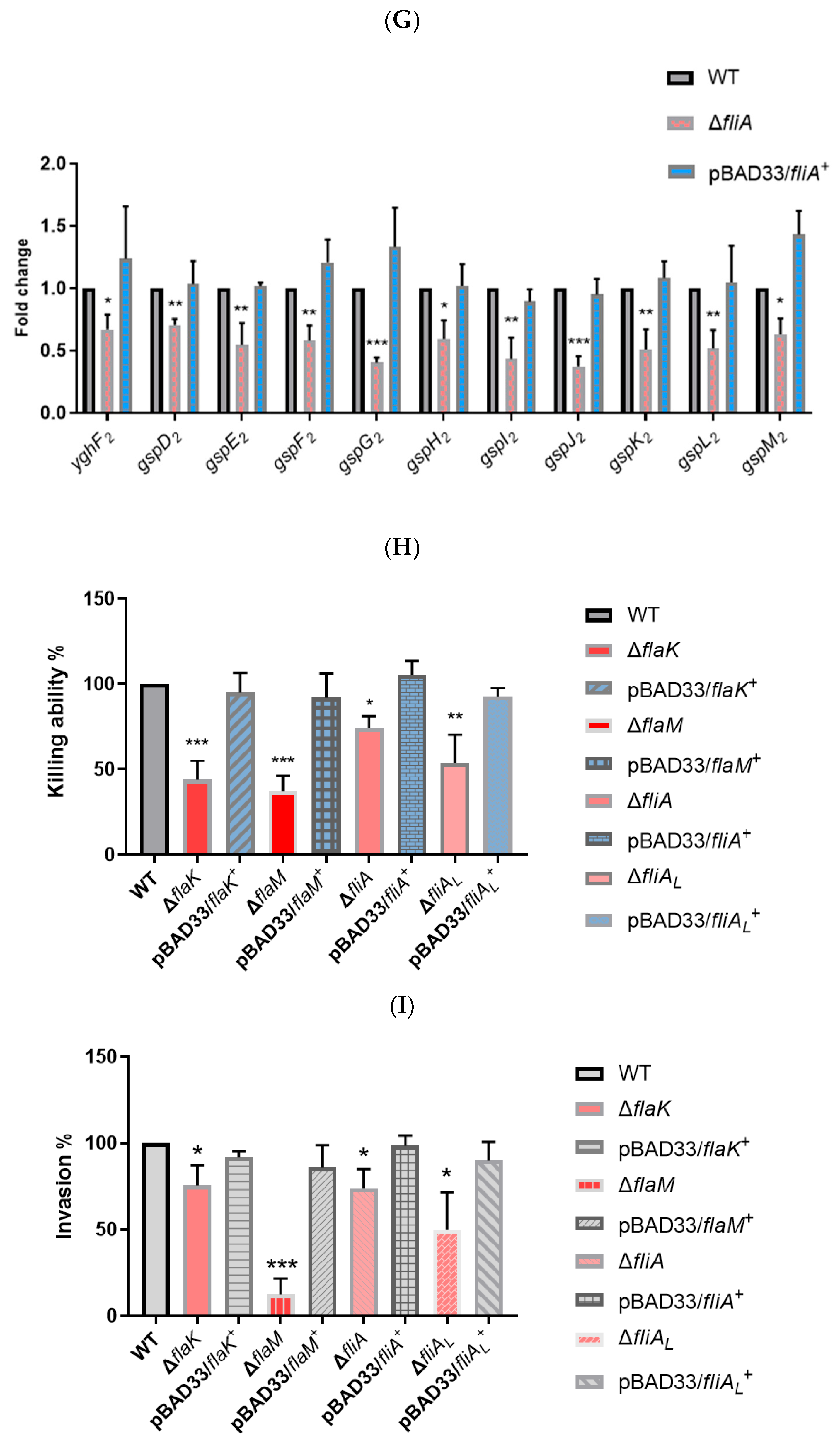
2.3. Flagellar Regulatory Mutants Reduce the Killing Ability and Virulence of P. shigelloides to Varying Degrees

In this study, the differentially expressed genes are also involved in the bacterial secretion system, including the observed T6SS significantly downregulated in the ΔflaK, ΔflaM, and ΔfliAL transcriptome profiles (Figure 3A) and T2SS-2 downregulated in the ΔflaM and ΔfliA transcriptome profiles (Figure 3B). Meanwhile, we validated the differential expression of T6SS or T2SS-2 genes in each transcriptome using qRT-PCR in the WT, ΔflaK, ΔflaM, ΔfliA, ΔfliAL, and complementation strains (Figure 3C–G). The qRT-PCR results were consistent with RNA-seq, which also suggests a cross-regulatory relationship between the flagellar system and bacterial secretion system. T6SS is a well-known contact-dependent bacterial weapon that can kill competitors directly by translocating proteinaceous toxins. Furthermore, we confirmed in a prior publication that T2SS-2 is also associated with P. shigelloides’ killing ability. Subsequently, the killing assay was used to compare the ability of the WT, ΔflaK, ΔflaM, ΔfliA, ΔfliAL, and complementation strains to kill E. coli MG1655. The results showed that the killing abilities of ΔflaK, ΔflaM, ΔfliA, and ΔfliAL were all decreased compared to the WT, while the killing abilities of the relative complement strains were restored to the level of the WT (Figure 3H). However, the killing ability of ΔflaM was significantly decreased, which may be because the absence of FlaM affected the expression of two gene clusters, T6SS and T2SS-2. Furthermore, invasion assays were performed to verify whether FlaK, FlaM, FliA, and FliAL affect the virulence of P. shigelloides. Subsequently, invasion experiments revealed that, in comparison with the WT, ΔflaK, ΔflaM, ΔfliA, and ΔfliAL were all less able to infect Caco-2 cells, whereas ΔflaM was significantly reduced, and the relative complement strains’ ability to infect Caco-2 cells was restored to WT levels (Figure 3I). However, we did not find any transcript-level changes in the T3SS gene in the transcriptome. We speculated that FlaK, FlaM, FliA, and FliAL may also affect the ability of P. shigelloides to infect Caco-2 cells by upregulating the expression of T6SS or T2SS-2, or their absence may cause P. shigelloides’ motility to decrease and thus affect the infectability of P. shigelloides. Taken together, our findings unveil a connection between the flagellar and secretory systems in P. shigelloides and provide new insights into the pathogenesis of this bacterium.

2.4. Cross-Talk between the Flagellar System and P. shigelloides’ Physiological Metabolism

In this study, we also found that the loss of flagellar regulators causes the differential expression of some of the physiological metabolic genes of P. shigelloides, and our proposal for the cross-talk between the flagellar system and P. shigelloides’ physiological metabolism is presented in Figure 4.

3. Discussion

Plesiomonas shigelloides, a Gram-negative foodborne pathogen of the Enterobacteriaceae family, has been known to exist for almost 80 years since Ferguson’s 1947 discovery [41]. P. shigelloides is extensively present in freshwater lakes, rivers, and streams and can be found in the environment, in animals, and in humans [42,43]. Acute gastroenteritis is the primary sign of infection; it can also lead to sepsis and neurological conditions [42]. P. shigelloides has received comparatively less attention and reports than other foodborne pathogens such as Salmonella, V. parahemolyticus, and diarrheal E. coli [44]. For the majority of pathogenic bacteria, the flagellum is a necessary component of their motility as well as their pathogenicity, which allows them to adhere to, colonize, or infect eukaryotic cells.
Currently, the flagellar hierarchical regulatory systems of most pathogens are reported, either E. coli and Salmonella, which have tertiary flagellar hierarchical regulation, or V. cholerae [13,14,15], V. parahaemolyticus [16], A. hydrophila [11,17], and P. aeruginosa [8] with fourth flagellar regulation. Two different P. shigelloides flagella gene clusters were described previously [39], and combined with the previously reported [40] and orthologous similarity alignment to the flagellar gene clusters, we found that the polar and lateral flagella gene clusters of P. shigelloides and V. parahaemolyticus exhibit the greatest overall homologous similarity, trailed by A. hydrophila and V. cholerae. In this study, RNA-Seq, qRT-PCR, lux assay, and EMSAs were used to confirm the flagellar transcription hierarchy. The EMSA results revealed that FlaK can directly bind to the promoters of fliK, fliE, flhA, and cheY, while the FlaM protein can bind directly to the promoters of flgO, flgT, and flgA, but no flagellar gene promoter was tested for binding to FliA or FliAL. The polar flagellate hierarchy system regulated by FlaK, FlaM, and FliA was finally confirmed. And we also demonstrated that FlaK and FlaM in P. shigelloides are σ54-dependent activators in earlier research on the regulation of P. shigelloides’ flagellar genes by RpoN [45]. In the P. shigelloides lateral gene cluster, only FliAL was present, but no LafK ortholog was found. All lateral gene clusters, including the non-functional ones in the Enterobacteriaceae, have reported this gene [46]. On the other hand, LafK in V. parahaemolyticus and A. hydrophila showed high homologous similarity to FlaK in P. shigelloides, and the deletion of flaK results in the downregulation of lateral flagella gene clusters in RNA-seq, qRT-PCR, and lux assays, indicating that it is possible that FlaK regulates the lateral flagellar system. On this premise, we defined the lateral flagellar gene transcriptional hierarchy in P. shigelloides under the regulation of FlaK and FliAL; nevertheless, FlaK may replace LafK’s function in regulating the lateral flagellar system, which requires additional validation. Furthermore, we also observed that the upregulation of flagellar genes in the RNA-seq data, which we hypothesized was caused by the complex flagellar regulation mechanism of P. shigelloides, and it is possible that there is a negative regulation of flagellar genes, for which we have further investigation. Additionally, we will further confirm the binding sites of FlaK and FlaM in the promoter region of the flagellar gene using DNA footprinting.
It was previously reported that there was cross-talk between the flagellar hierarchy system and the bacterial secretion system. Speare L et al. demonstrated that flagella-dependent aggregation factors and TasL (T6SS2) may act in concert to facilitate cell–cell contact, thus mediating the killing ability of bacteria [47]. In the early stages of infection, Huang Z et al. demonstrated that the lateral flagellar-associated flhA gene was crucial for the adhesion and colonization of V. metschnikovii. Additionally, they also discovered that V. metschnikovii promoted a high level of cytotoxicity through the synergistic interaction of T6SS and the lateral flagella [48]. Furthermore, Bouteiller M et al. demonstrated that there was a cross-talk between Pseudomonas fluorescens’s class IV flagellar gene expression and the type VI secretion system [49]. In this study, we observed T6SS significantly downregulated in the ΔflaK, ΔflaM, and ΔfliAL transcriptome profiles and T2SS-2 downregulated in the ΔflaM and ΔfliA transcriptome profiles, which were then validated by qRT-PCR. Previous work has indicated a correlation between T2SS-2 and the killing ability of P. shigelloides [45]. In the meantime, the killing assay revealed lower killing abilities for ΔflaK, ΔflaM, ΔfliA, and ΔfliAL compared to the WT, indicating a cross-regulatory relationship between the flagellar and bacterial secretion systems in P. shigelloides. Furthermore, a number of investigations also showed a connection between the flagellar system and T3SS, which influences the pathogenicity of bacteria. In P. plecoglossicida, Qin et al. found cross-talk between the type III secretion system and flagella assembly using comparative secretome analysis [50]. Soria-Bustos J et al. suggested that the presence of an intact T3SS is required for the assembly of flagella, highlighting the existence in the EPEC of a cross-talk between these two virulence-associated T3SSs [51]. Nevertheless, while our invasion experiments revealed that ΔflaK, ΔflaM, ΔfliA, and ΔfliAL were less capable of infecting Caco-2 cells than the WT, we were unable to detect any transcript-level alterations of the T3SS gene in the transcriptome. And the growth assays in the lag and log phases also showed no significant difference between the WT and ΔflaK, ΔflaM, ΔfliA, and ΔfliAL strains (Figure S5). In this case, we hypothesized that the downregulation of T6SS and T2SS-2, or the expression of some virulence genes caused by the loss of flagella regulators in this study, or the decreased motility of P. shigelloides affected the adhesion of P. shigelloides, resulting in a decrease in P. shigelloides infection to Caco-2 cells, which, of course, requires further confirmation.
In addition to the flagellar system and the bacterial secretion system, we have also previously described the correlation between the flagellar system and bacterial physiological metabolism [32,33,34,35,36,37,38]. Similarly, in this study, we also found that the loss of flagellar regulators causes the differential expression of some of the physiological metabolic genes of P. shigelloides. Among the genes downregulated in the ΔflaK mutant were genes mainly responsible for periplasmic nitrate reductase, pyrimidine metabolism, and purine metabolism (Table S5). Upregulated genes in the ΔflaK mutant included those involved in myo-inositol catabolism and propanoate metabolism (Table S5). FlaM regulates slightly more physiological and metabolic genes than FlaK does. Among the genes downregulated in the ΔflaM mutant were those responsible for the biosynthesis of amino acids, glycerophospholipid metabolism, the citrate cycle, the ribosome, purine metabolism, and thiamine metabolism (Table S6). Among the genes upregulated in the ΔflaM mutant were those responsible for hydrogenase (Table S6). The physiological metabolic genes regulated by FliA are the most numerous of the four flagellar hierarchy regulators. Among the genes downregulated in the ΔfliA mutant were those responsible for the citrate cycle, fatty acid degradation and metabolism, glyoxylate and dicarboxylate metabolism, periplasmic nitrate reductase, NADH-quinone oxidoreductase, pyrimidine metabolism, hydrogenase, glycerophospholipid metabolism, and curli production assembly (Table S7). Among the genes upregulated in the ΔfliA mutant were those responsible for glycolysis/gluconeogenesis, ribosome, myo-inositol catabolism, pyruvate metabolism, starch and sucrose metabolism, and porphyrin metabolism (Table S7). Both FliA in the polar flagellar gene cluster and FliAL in the lateral flagellar gene cluster are σ factors; however, the number of DEGs regulated by both differs greatly. Among the genes downregulated in the ΔfliAL mutant were genes responsible for periplasmic nitrate reductase (Table S8). Among the genes upregulated in the ΔfliAL mutant were those responsible for myo-inositol catabolism (Table S8). Although we revealed a relationship between the flagellar system and P. shigelloides’ physiological metabolic activity, the mechanism requires more investigation (Figure 4).
Overall, our study not only revealed the transcriptional hierarchy of polar and lateral flagellar genes but also demonstrated the cross-talk between the flagellar system and the secretory system in P. shigelloides, which influences P. shigelloides’ killing ability. Additionally, we also identified the effects of the flagellar system on the virulence and partial physiological metabolism of P. shigelloides. These findings will provide a foundation for understanding the relationship and mechanism between motility, virulence, and physiological and metabolic activity in P. shigelloides.

4. Materials and Methods

4.1. Bacterial Strains, Plasmids, and Growth Conditions

Table S1 shows the plasmids and bacterial strains used in this study. According to the experimental requirements, bacteria were cultured at 30 °C or 37 °C using Luria–Bertani (LB) liquid, semi-solid media, and solid, as well as Dulbecco’s Modified Eagle’s Media (DMEM) supplemented with 20% fetal bovine serum (FBS). Meanwhile, supplements containing 25 μg/mL of ampicillin, 50 μg/mL of kanamycin, or 25 μg/mL of chloramphenicol were added to the media as needed.

4.2. Construction of Gene Deletion Strains and Complementation

The gene deletion methods in this study were performed as previously reported [52]. To put it briefly, the target genes’ (flaK, flaM, fliA, and fliAL) upstream and downstream regions were amplified using the genome of the P. shigelloides WT as the PCR amplification template. These upstream and downstream regions were then connected by PCR amplification, and the successfully connected DNA fragments and the pRE112 plasmid were digested by an endonuclease and linked with DNA ligase to form a new recombinant vector. The recombinant vector was electrotransformed into E. coli S17-1 λpir and subsequently mixed with the P. shigelloides WT equally in antibiotic-free solid plates at 37 °C for 24 h for homologous recombination. Mixed-grown colonies were subsequently diluted and plated onto a medium containing 25 μg/mL ampicyl and chloramphenicol, and agarose gel electrophoresis and PCR were used to identify the positive single colonies. Ultimately, the positively identified clones were transferred to solid plates with 20% sucrose and a 25 μg/mL ampicyl antibiotic concentration. The pRE112 plasmid’s susceptibility to sucrose-induced lethality and the inability of the P. shigelloides WT to grow in a medium containing chloramphenicol were used to screen for gene deletion mutant strains. The complementation strains, pBAD33/flaK+, pBAD33/flaM+, pBAD33/fliA+, and pBAD33/fliAL+, were constructed by introducing the recombinant vector, pBAD33 plasmid with target genes, into the relative gene-deleted strains via electroporation. Agarose gel electrophoresis and PCR product sequencing were used to verify the accuracy of the deletion and complementation strains. All primers used in this study are listed in Table S2.

4.3. RNA Isolation and Transcriptome Sequencing

The WT and deletion mutant strains (ΔflaK, ΔflaM, ΔfliA, and ΔfliAL) were cultured overnight at 37 °C after diluting 1:100 into 20 mL of LB liquid medium, and then the bacteria were transferred to a fresh medium the next day and grew to OD600 = 0.8 (each strain was transduced to three culture media simultaneously). TRIzol® Reagent (Invitrogen, Waltham, MA, USA) was used to separate total RNA from the WT and deletion mutant strains (three total RNA samples were extracted from each strain), which was subsequently processed with RNase-free DNase and dissolved in RNase-free water. Meanwhile, RNA degradation and contamination were assessed using 1% agarose gel, and a NanoDrop-2000 spectrophotometer (Thermo Fisher, Waltham, MA, USA) measured RNA concentration at OD260 and determined RNA purity via the ratio of OD260/OD230 and OD260/OD280. Following testing and qualifying, the cDNA library was sequenced on an Illumina NovaSeq 6000 platform (Illumina, San Diego, CA, USA) to produce 150 bp paired-end reads. Gene expression levels were measured using HTSeq, and the length and number of reads mapped to each gene were used to calculate the Fragments Per Kilobase of transcript sequence per Million base pairs sequenced (FPKM) value of each gene. The criteria for differentially expressed genes (DEGs) were set as |log2 fold change| ≥ 1 and adjusted p-value (padj) ≤ 0.05.

4.4. Quantitative Real-Time Polymerase Chain Reaction (qRT-PCR)

To further validate the RNA-Seq data and reveal the flagellar hierarchy regulatory network, we performed qRT-PCR for the WT, ΔflaK, ΔflaM, ΔfliA, ΔfliAL, pBAD33/flaK+, pBAD33/flaM+, pBAD33/fliA+, and pBAD33/fliAL+. The isolation, quantification, and detection of total RNA from the WT, deletion mutant strains, and complementation strains were performed as described above. cDNA was generated using the PrimeScript™ RT reagent kit (Takara Bio, Shiga, Japan), and qRT-PCR analysis was performed using an Applied Biosystems ABI 7500 sequence detection system (Applied Biosystems, Foster City, CA, USA). A 96-well optical reaction plate (Applied Biosystems) was used for each qRT-PCR experiment, which included 1 μL of cDNA, 10 μL of FastStart Universal SYBR Green Master (ROX) mix, and two gene-specific primers with a final concentration of 0.3 mM each. The cycle threshold approach (2−∆∆CT) was used to compute relative target gene expression levels as fold changes [53], with the gyrB gene in P. shigelloides serving as a reference control [45]. Furthermore, several steps must be taken to reduce qRT-PCR error during the experiment, such as preventing RNA degradation and DNA contamination, accurately quantifying RNA, using deionized water as a negative control template to avoid non-specific amplification, and selecting appropriate internal reference genes. Each experiment was conducted three times.

4.5. Motility Assay and Transmission Electron Microscopy (TEM) of Flagella

The motility assay was carried out as previously described [54]. In short, the WT, ΔflaK, ΔflaM, ΔfliA, ΔfliAL, pBAD33/flaK+, pBAD33/flaM+, pBAD33/fliA+, and pBAD33/fliAL+ strains were inoculated into 20 mL of LB liquid medium and cultured overnight at 37 °C, and then transferred to fresh medium the next day and cultured to OD600 = 0.8. Subsequently, 1 μL of the fresh bacterial solution was absorbed into the center of the swimming agar plates and static cultured for 12 h at 30 °C, and then the bacteria’s swimming distance was measured and photographed. The trials were conducted at three different time points, each with six repetitions. Moreover, TEM and negative staining were used to visualize the flagella of the WT, ΔflaK, ΔflaM, ΔfliA, ΔfliAL, pBAD33/flaK+, pBAD33/flaM+, pBAD33/fliA+, and pBAD33/fliAL+ strains, as also previously described [55]. Briefly, all strains were inoculated into 20 mL of LB liquid medium and cultured overnight at 37 °C, and then transferred to fresh medium the next day and cultured to OD600 = 0.4. A 20 μL aliquot of each sample was separately placed on a Formvar/carbon grid (400 mesh), which was glow-discharged prior to use to increase the hydrophilicity. The grids were washed with 0.1 M sodium acetate (pH 6.6), negatively stained with 2% phosphotungstic acid, and air dried for 6 min before being viewed at 120 kV with a Tecnai transmission electron microscope (Thermo Fisher, Waltham, MA, USA).

4.6. Luminescence Screening Assay

Luminescence screening assays were carried out as previously described [56]. In brief, PCR was used to amplify the target gene’s promoter region, which was then digested by two restriction enzymes, XhoI and BamHI, along with plasmid pMS402, to generate a new recombinant plasmid using DNA ligase. Subsequently, the fusion reporter plasmid used in this study (Table S1) was transformed into the WT and deletion mutant strains, and then the positive clones, screened and identified, were grown in the LB medium to the mid-log phase. The promoter activity was evaluated at OD600 with a Synergy 2 plate reader (Agilent BioTek, Santa Clara, CA, USA). Each experiment was conducted three times, with six replicates each.

4.7. Expression and Purification of Proteins and Electrophoretic Mobility Shift Assays (EMSAs)

The proteins used in this study’s EMSAs, FlaK, FlaM, FliA, and FliAL, were cloned into pMAL-c5X and expressed in E. coli BL21 (DE3) and purified using amylose resin (New England Bio Labs, Ipswich, MA, USA) affinity chromatography. EMSAs were performed using a mixture of each probe, 60 ng, and escalating doses of the fusion protein in a 20 μL reaction volume with 20 mM Tris-HCl (pH 7.5), 120 mM KCl, 5 mM MgCl2, 1 mM DTT, 5% glycerol, and 1 μg Poly (dI.dC) incubated at 30 °C for 45 min. Moreover, the DNA–protein complexes were separated using a 6% PAGE in a 0.5 × TBE buffer for 2 h at 140 V. After being stained with GelRed for 5 min, gels were scanned using a gel imaging system (GE Healthcare, Chicago, IL, USA).

4.8. Killing Assay

The E. coli MG1655 killing assay, with some modifications, was carried out as described previously [57]. Overnight cultures from P. shigelloides and E. coli were diluted in the LB medium and grown at 37 °C until the bacteria reached OD600 = 1.5, and then the cells were harvested and concentrated. The ratio of a 1:1 mixture of the predator and prey bacteria was prepared, and 20 µL of this mixture was added to antibiotic-free LB agar plates. The two bacteria were then co-cultured for 3 h, washed with phosphate buffer, and serial dilutions were spotted onto antibiotic-containing LB solid plate medium and incubated overnight at 37 °C. The killing ability of the ΔflaK, ΔflaM, ΔfliA, ΔfliAL, pBAD33/flaK+, pBAD33/flaM+, pBAD33/fliA+, and pBAD33/fliAL+ strains was reported as a percentage relative to that of the WT. The killing assay was performed three times, with six repetitions each time.

4.9. Growth Assay

Growth assays were performed as described previously [58]. In brief, the WT, ΔflaK, ΔflaM, ΔfliA, and ΔfliAL strains were cultured at 37 °C in an LB medium overnight, and the cultured bacterial solution, at a ratio of 1:200 per well, was added to a 96-well cell plate containing 200 μL of LB or DMEM. Meanwhile, the sterile LB and DMEM served as a control group. A Molecular Devices Spectramax 190 full-wavelength microplate reader (Molecular Devices LLC., San Jose, CA, USA) was used for the dynamic growth experiment. The trials were conducted at three different time points, each with six repetitions.

4.10. Invasion Assay

The invasion assay was performed with minor modifications to the previously described method [59]. Briefly, the WT, ΔflaK, ΔflaM, ΔfliA, ΔfliAL, pBAD33/flaK+, pBAD33/flaM+, pBAD33/fliA+, and pBAD33/fliAL+ strains were cultured overnight in LB, and then transferred to new LB the next day, at a 1:100 inoculation ratio, until the bacteria reached OD600 = 0.6. Confluent monolayers of roughly 1 × 10⁵ Caco-2 cells per well, in 24-well plates, were overlaid with approximately 5 × 10⁷ WT, ΔflaK, ΔflaM, ΔfliA, ΔfliAL, pBAD33/flaK+, pBAD33/flaM+, pBAD33/fliA+, and pBAD33/fliAL+ bacterial cells. Subsequently, Caco-2 and bacterial cells, at 37 °C in 5% CO2, were co-cultured for 1 h to induce invasion, and then 100 μg/mL of gentamicin was added to the cell culture medium to kill extracellular bacteria. Finally, the cells, after the monolayer had been twice washed with PBS, were lysed for 10 min using 0.1% Triton X-100. The invasion rate was calculated as the ratio of the number of recovered bacteria to the total number of bacterial cells used for infection. Furthermore, the invasion assay was performed three times, with six repetitions each time.

4.11. Statistical Analysis

GraphPad Prism v7.0 software (GraphPad Inc., La Jolla, CA, USA) was used to statistically analyze all data, which are expressed as means ± standard deviation (SD) [60]. The independent samples t-test and Mann–Whitney U test were used to determine differences between the groups. Furthermore, a probability value (p) ≤ 0.05 was considered statistically significant (*** p ≤ 0.001; ** p ≤ 0.01; * p ≤ 0.05; ns indicates not significant).

Supplementary Materials

The following supporting information can be downloaded at: https://www.mdpi.com/article/10.3390/ijms25137375/s1.

Author Contributions

J.Y.: investigation, conceptualization, project administration, methodology, writing—original draft. Z.Z.: project administration, methodology. H.S.: data curation, formal analysis. X.X.: project administration, methodology. A.L.: methodology, formal analysis. P.D.: software, visualization. X.G.: software, visualization. J.W.: software, visualization. Y.W.: investigation, conceptualization, writing—original draft, writing—review and editing. B.C.: investigation, conceptualization, writing—original draft, funding acquisition, supervision, writing—review and editing. All authors have read and agreed to the published version of the manuscript.

Funding

This work was funded by the National Key Programs for Infectious Diseases of China (funding numbers 2017ZX10303405-001, 2017ZX10104002-001-006, and 2018ZX1 0712001-017).

Institutional Review Board Statement

Not applicable.

Informed Consent Statement

Not applicable.

Data Availability Statement

The RNA sequencing data generated in this study are available in the NCBI SRA database (accession numbers: SRR22678961, SRR22679947, SRR22696781, SRR22686293, and SRR22686286). Other data are presented within the manuscript and the Supplementary Materials.

Conflicts of Interest

The authors declare no conflicts of interest.

References

  1. Puah, S.M.; Puthucheary, S.D.A.; Chua, K.H. Virulence Profiles among Gastrointestinal and Extraintestinal Clinical Isolates of Plesiomonas shigelloides. Jpn. J. Infect. Dis. 2022, 75, 407–410. [Google Scholar] [CrossRef] [PubMed]
  2. Mandal, B.K.; Whale, K.; Morson, B.C. Acute colitis due to Plesiomonas shigelloides. Br. Med. J. (Clin. Res. Ed.) 1982, 285, 1539–1540. [Google Scholar] [CrossRef] [PubMed]
  3. McNeeley, D.; Ivy, P.; Craft, J.C.; Cohen, I. Plesiomonas: Biology of the organism and diseases in children. Pediatr. Infect. Dis. 1984, 3, 176–181. [Google Scholar] [CrossRef] [PubMed]
  4. Tsukamoto, T.; Kinoshita, Y.; Shimada, T.; Sakazaki, R. Two epidemics of diarrhoeal disease possibly caused by Plesiomonas shigelloides. J. Hyg. 1978, 80, 275–280. [Google Scholar] [CrossRef] [PubMed]
  5. Billiet, J.; Kuypers, S.; Van Lierde, S.; Verhaegen, J. Plesiomonas shigelloides meningitis and septicaemia in a neonate: Report of a case and review of the literature. J. Infect. 1989, 19, 267–271. [Google Scholar] [CrossRef] [PubMed]
  6. Fischer, K.; Chakraborty, T.; Hof, H.; Kirchner, T.; Wamsler, O. Pseudoappendicitis caused by Plesiomonas shigelloides. J. Clin. Microbiol. 1988, 26, 2675–2677. [Google Scholar] [CrossRef] [PubMed]
  7. Pennycook, K.M.; Pennycook, K.B.; McCready, T.A.; Kazanowski, D. Severe cellulitis and bacteremia caused by Plesiomonas shigelloides following a traumatic freshwater injury. IDCases 2019, 19, e00637. [Google Scholar] [CrossRef] [PubMed]
  8. Dasgupta, N.; Wolfgang, M.C.; Goodman, A.L.; Arora, S.K.; Jyot, J.; Lory, S.; Ramphal, R. A four-tiered transcriptional regulatory circuit controls flagellar biogenesis in Pseudomonas aeruginosa. Mol. Microbiol. 2003, 50, 809–824. [Google Scholar] [CrossRef] [PubMed]
  9. Fenchel, T. Microbial behavior in a heterogeneous world. Science 2002, 296, 1068–1071. [Google Scholar] [CrossRef]
  10. Tsang, J.; Hoover, T.R. Basal Body Structures Differentially Affect Transcription of RpoN- and FliA-Dependent Flagellar Genes in Helicobacter pylori. J. Bacteriol. 2015, 197, 1921–1930. [Google Scholar] [CrossRef]
  11. Wilhelms, M.; Molero, R.; Shaw, J.G.; Tomás, J.M.; Merino, S. Transcriptional hierarchy of Aeromonas hydrophila polar-flagellum genes. J. Bacteriol. 2011, 193, 5179–5190. [Google Scholar] [CrossRef] [PubMed]
  12. Kutsukake, K. Autogenous and global control of the flagellar master operon, flhD, in Salmonella typhimurium. Mol. Genet. Genom. 1997, 254, 440–448. [Google Scholar] [CrossRef] [PubMed]
  13. Correa, N.E.; Peng, F.; Klose, K.E. Roles of the regulatory proteins FlhF and FlhG in the Vibrio cholerae flagellar transcription hierarchy. J. Bacteriol. 2005, 187, 6324–6332. [Google Scholar] [CrossRef] [PubMed]
  14. Klose, K.E.; Mekalanos, J.J. Distinct roles of an alternative sigma factor during both free-swimming and colonizing phases of the Vibrio cholerae pathogenic cycle. Mol. Microbiol. 1998, 28, 501–520. [Google Scholar] [CrossRef] [PubMed]
  15. Prouty, M.G.; Correa, N.E.; Klose, K.E. The novel sigma54- and sigma28-dependent flagellar gene transcription hierarchy of Vibrio cholerae. Mol. Microbiol. 2001, 39, 1595–1609. [Google Scholar] [CrossRef] [PubMed]
  16. Stewart, B.J.; McCarter, L.L. Lateral flagellar gene system of Vibrio parahaemolyticus. J. Bacteriol. 2003, 185, 4508–4518. [Google Scholar] [CrossRef] [PubMed]
  17. Wilhelms, M.; Gonzalez, V.; Tomás, J.M.; Merino, S. Aeromonas hydrophila lateral flagellar gene transcriptional hierarchy. J. Bacteriol. 2013, 195, 1436–1445. [Google Scholar] [CrossRef] [PubMed]
  18. Gu, D.; Meng, H.; Li, Y.; Ge, H.; Jiao, X. A GntR Family Transcription Factor (VPA1701) for Swarming Motility and Colonization of Vibrio parahaemolyticus. Pathogens 2019, 8, 235. [Google Scholar] [CrossRef]
  19. Kim, Y.K.; McCarter, L.L. Cross-regulation in Vibrio parahaemolyticus: Compensatory activation of polar flagellar genes by the lateral flagellar regulator LafK. J. Bacteriol. 2004, 186, 4014–4018. [Google Scholar] [CrossRef]
  20. Merino, S.; Shaw, J.G.; Tomás, J.M. Bacterial lateral flagella: An inducible flagella system. FEMS Microbiol. Lett. 2006, 263, 127–135. [Google Scholar] [CrossRef]
  21. Shinoda, S.; Okamoto, K. Formation and function of Vibrio parahaemolyticus lateral flagella. J. Bacteriol. 1977, 129, 1266–1271. [Google Scholar] [CrossRef]
  22. Petersen, B.D.; Liu, M.S.; Podicheti, R.; Yang, A.Y.; Simpson, C.A.; Hemmerich, C.; Rusch, D.B.; van Kessel, J.C. The Polar Flagellar Transcriptional Regulatory Network in Vibrio campbellii Deviates from Canonical Vibrio Species. J. Bacteriol. 2021, 203, e0027621. [Google Scholar] [CrossRef] [PubMed]
  23. Gardel, C.L.; Mekalanos, J.J. Alterations in Vibrio cholerae motility phenotypes correlate with changes in virulence factor expression. Infect. Immun. 1996, 64, 2246–2255. [Google Scholar] [CrossRef] [PubMed]
  24. Kojima, S.; Yamamoto, K.; Kawagishi, I.; Homma, M. The polar flagellar motor of Vibrio cholerae is driven by an Na+ motive force. J. Bacteriol. 1999, 181, 1927–1930. [Google Scholar] [CrossRef] [PubMed]
  25. Häse, C.C.; Mekalanos, J.J. Effects of changes in membrane sodium flux on virulence gene expression in Vibrio cholerae. Proc. Natl. Acad. Sci. USA 1999, 96, 3183–3187. [Google Scholar] [CrossRef] [PubMed]
  26. Coster, T.S.; Killeen, K.P.; Waldor, M.K.; Beattie, D.T.; Spriggs, D.R.; Kenner, J.R.; Trofa, A.; Sadoff, J.C.; Mekalanos, J.J.; Taylor, D.N. Safety, immunogenicity, and efficacy of live attenuated Vibrio cholerae O139 vaccine prototype. Lancet 1995, 345, 949–952. [Google Scholar] [PubMed]
  27. Yang, Q.; Defoirdt, T. Quorum sensing positively regulates flagellar motility in pathogenic Vibrio harveyi. Environ. Microbiol. 2015, 17, 960–968. [Google Scholar] [CrossRef] [PubMed]
  28. Hamed, S.; Wang, X.; Shawky, R.M.; Emara, M.; Aldridge, P.D.; Rao, C.V. Synergistic action of SPI-1 gene expression in Salmonella enterica serovar typhimurium through transcriptional crosstalk with the flagellar system. BMC Microbiol. 2019, 19, 211. [Google Scholar] [CrossRef] [PubMed]
  29. Zhao, Y.H.; Shaw, J.G. Cross-Talk between the Aeromonas hydrophila Type III Secretion System and Lateral Flagella System. Front. Microbiol. 2016, 7, 1434. [Google Scholar] [CrossRef]
  30. Montie, T.C.; Doyle-Huntzinger, D.; Craven, R.C.; Holder, I.A. Loss of virulence associated with absence of flagellum in an isogenic mutant of Pseudomonas aeruginosa in the burned-mouse model. Infect. Immun. 1982, 38, 1296–1298. [Google Scholar] [CrossRef]
  31. Fleiszig, S.M.; Arora, S.K.; Van, R.; Ramphal, R. FlhA, a component of the flagellum assembly apparatus of Pseudomonas aeruginosa, plays a role in internalization by corneal epithelial cells. Infect. Immun. 2001, 69, 4931–4937. [Google Scholar] [CrossRef] [PubMed]
  32. Schumacher, K.; Braun, D.; Kleigrewe, K.; Jung, K. Motility-activating mutations upstream of flhDC reduce acid shock survival of Escherichia coli. Microbiol. Spectr. 2024, 12, e0054424. [Google Scholar] [CrossRef] [PubMed]
  33. Hoque, M.M.; Noorian, P.; Espinoza-Vergara, G.; Adhikary, S.; To, J.; Rice, S.A.; McDougald, D. Increased iron utilization and oxidative stress tolerance in a Vibrio cholerae flrA mutant confers resistance to amoeba predation. Appl. Environ. Microbiol. 2023, 89, e0109523. [Google Scholar] [CrossRef] [PubMed]
  34. Sinha-Ray, S.; Ali, A. Mutation in flrA and mshA Genes of Vibrio cholerae Inversely Involved in vps-Independent Biofilm Driving Bacterium Toward Nutrients in Lake Water. Front. Microbiol. 2017, 8, 1770. [Google Scholar] [CrossRef]
  35. Rodríguez-Herva, J.J.; Duque, E.; Molina-Henares, M.A.; Navarro-Avilés, G.; Van Dillewijn, P.; De La Torre, J.; Molina-Henares, A.J.; La Campa, A.S.; Ran, F.A.; Segura, A.; et al. Physiological and transcriptomic characterization of a fliA mutant of Pseudomonas putida KT2440. Environ. Microbiol. Rep. 2010, 2, 373–380. [Google Scholar] [CrossRef] [PubMed]
  36. Li, P.; Zong, W.; Zhang, Z.; Lv, W.; Ji, X.; Zhu, D.; Du, X.; Wang, S. Effects and molecular mechanism of flagellar gene flgK on the motility, adhesion/invasion, and desiccation resistance of Cronobacter sakazakii. Food Res. Int. 2023, 164, 112418. [Google Scholar] [CrossRef] [PubMed]
  37. Jubelin, G.; Lanois, A.; Severac, D.; Rialle, S.; Longin, C.; Gaudriault, S.; Givaudan, A. FliZ is a global regulatory protein affecting the expression of flagellar and virulence genes in individual Xenorhabdus nematophila bacterial cells. PLoS Genet. 2013, 9, e1003915. [Google Scholar] [CrossRef] [PubMed]
  38. Lanois, A.; Jubelin, G.; Givaudan, A. FliZ, a flagellar regulator, is at the crossroads between motility, haemolysin expression and virulence in the insect pathogenic bacterium Xenorhabdus. Mol. Microbiol. 2008, 68, 516–533. [Google Scholar] [CrossRef]
  39. Merino, S.; Aquilini, E.; Fulton, K.M.; Twine, S.M.; Tomás, J.M. The polar and lateral flagella from Plesiomonas shigelloides are glycosylated with legionaminic acid. Front. Microbiol. 2015, 6, 649. [Google Scholar] [CrossRef]
  40. Chilcott, G.S.; Hughes, K.T. Coupling of flagellar gene expression to flagellar assembly in Salmonella enterica serovar typhimurium and Escherichia coli. Microbiol. Mol. Biol. Rev. 2000, 64, 694–708. [Google Scholar] [CrossRef]
  41. Ferguson, W.W.; Henderson, N.D. Description of Strain C27: A Motile Organism with the Major Antigen of Shigella sonnei Phase I. J. Bacteriol. 1947, 54, 179–181. [Google Scholar] [CrossRef] [PubMed]
  42. Janda, J.M.; Abbott, S.L.; McIver, C.J. Plesiomonas shigelloides Revisited. Clin. Microbiol. Rev. 2016, 29, 349–374. [Google Scholar] [CrossRef] [PubMed]
  43. Edwards, M.S.; McLaughlin, R.W.; Li, J.; Wan, X.; Liu, Y.; Xie, H.; Hao, Y.; Zheng, J. Putative virulence factors of Plesiomonas shigelloides. Antonie Van Leeuwenhoek 2019, 112, 1815–1826. [Google Scholar] [CrossRef] [PubMed]
  44. Ekundayo, T.C.; Okoh, A.I. A global bibliometric analysis of Plesiomonas-related research (1990–2017). PLoS ONE 2018, 13, e0207655. [Google Scholar] [CrossRef] [PubMed]
  45. Yan, J.; Guo, X.; Li, J.; Li, Y.; Sun, H.; Li, A.; Cao, B. RpoN is required for the motility and contributes to the killing ability of Plesiomonas shigelloides. BMC Microbiol. 2022, 22, 299. [Google Scholar] [CrossRef]
  46. Canals, R.; Altarriba, M.; Vilches, S.; Horsburgh, G.; Shaw, J.G.; Tomás, J.M.; Merino, S. Analysis of the lateral flagellar gene system of Aeromonas hydrophila AH-3. J. Bacteriol. 2006, 188, 852–862. [Google Scholar] [CrossRef]
  47. Speare, L.; Zhao, L.; Pavelsky, M.N.; Jackson, A.; Smith, S.; Tyagi, B.; Sharpe, G.C.; Woo, M.; Satkowiak, L.; Bolton, T.; et al. Flagella are required to coordinately activate competition and host colonization factors in response to a mechanical signal. bioRxiv 2024. [Google Scholar] [CrossRef]
  48. Huang, Z.; Yu, K.; Lan, R.; Glenn Morris, J.; Xiao, Y.; Ye, J.; Zhang, L.; Luo, L.; Gao, H.; Bai, X.; et al. Vibrio metschnikovii as an emergent pathogen: Analyses of phylogeny and O-antigen and identification of possible virulence characteristics. Emerg. Microbes Infect. 2023, 12, 2252522. [Google Scholar] [CrossRef]
  49. Bouteiller, M.; Gallique, M.; Bourigault, Y.; Kosta, A.; Hardouin, J.; Massier, S.; Konto-Ghiorghi, Y.; Barbey, C.; Latour, X.; Chane, A.; et al. Crosstalk between the Type VI Secretion System and the Expression of Class IV Flagellar Genes in the Pseudomonas fluorescens MFE01 Strain. Microorganisms 2020, 8, 622. [Google Scholar] [CrossRef] [PubMed]
  50. Qin, P.; Luan, Y.; Yang, J.; Chen, X.; Wu, T.; Li, Y.; Munang’andu, H.M.; Shao, G.; Chen, X. Comparative secretome analysis reveals cross-talk between type III secretion system and flagella assembly in Pseudomonas plecoglossicida. Heliyon 2023, 9, e22669. [Google Scholar] [CrossRef]
  51. Soria-Bustos, J.; Saldaña-Ahuactzi, Z.; Samadder, P.; Yañez-Santos, J.A.; Laguna, Y.M.; Cedillo-Ramírez, M.L.; Girón, J.A. The Assembly of Flagella in Enteropathogenic Escherichia coli Requires the Presence of a Functional Type III Secretion System. Int. J. Mol. Sci. 2022, 23, 13705. [Google Scholar] [CrossRef] [PubMed]
  52. Xi, D.; Jing, F.; Liu, Q.; Cao, B. Plesiomonas shigelloides sipD mutant, generated by an efficient gene transfer system, is less invasive. J. Microbiol. Methods 2019, 159, 75–80. [Google Scholar] [CrossRef] [PubMed]
  53. Jiang, L.; Feng, L.; Yang, B.; Zhang, W.; Wang, P.; Jiang, X.; Wang, L. Signal transduction pathway mediated by the novel regulator LoiA for low oxygen tension induced Salmonella Typhimurium invasion. PLoS Pathog. 2017, 13, e1006429. [Google Scholar] [CrossRef]
  54. Wilhelms, M.; Fulton, K.M.; Twine, S.M.; Tomás, J.M.; Merino, S. Differential glycosylation of polar and lateral flagellins in Aeromonas hydrophila AH-3. J. Biol. Chem. 2012, 287, 27851–27862. [Google Scholar] [CrossRef]
  55. Evans, M.R.; Fink, R.C.; Vazquez-Torres, A.; Porwollik, S.; Jones-Carson, J.; McClelland, M.; Hassan, H.M. Analysis of the ArcA regulon in anaerobically grown Salmonella enterica sv. Typhimurium. BMC Microbiol. 2011, 11, 58. [Google Scholar] [CrossRef]
  56. Li, Y.; Yan, J.; Guo, X.; Wang, X.; Liu, F.; Cao, B. The global regulators ArcA and CytR collaboratively modulate Vibrio cholerae motility. BMC Microbiol. 2022, 22, 22. [Google Scholar] [CrossRef]
  57. Borgeaud, S.; Metzger, L.C.; Scrignari, T.; Blokesch, M. The type VI secretion system of Vibrio cholerae fosters horizontal gene transfer. Science 2015, 347, 63–67. [Google Scholar] [CrossRef] [PubMed]
  58. Yan, J.; Li, Y.; Guo, X.; Wang, X.; Liu, F.; Li, A.; Cao, B. The effect of ArcA on the growth, motility, biofilm formation, and virulence of Plesiomonas shigelloides. BMC Microbiol. 2021, 21, 266. [Google Scholar] [CrossRef] [PubMed]
  59. Schubert, R.H.; Holz-Bremer, A. Cell adhesion of Plesiomonas shigelloides. Zentralblatt Hyg. Umweltmed. 1999, 202, 383–388. [Google Scholar] [CrossRef]
  60. Yang, S.; Xi, D.; Wang, X.; Li, Y.; Li, Y.; Yan, J.; Cao, B. Vibrio cholerae VC1741 (PsrA) enhances the colonization of the pathogen in infant mice intestines in the presence of the long-chain fatty acid, oleic acid. Microb. Pathog. 2020, 147, 104443. [Google Scholar] [CrossRef]
Figure 1. Transcriptomic analysis of the P. shigelloides between the WT and ΔflaK, ΔflaM, ΔfliA, and ΔfliAL strains. The volcano plot of differentially expressed genes (DEGs) in the ΔflaK (A), ΔflaM (B), ΔfliA (C), and ΔfliAL (D) transcriptome profiles. The red circle was upregulated genes, the green circle was downregulated genes, and the blue circle had no DEGs. Three upregulated and three downregulated DEGs were selected for validation using qRT-PCR in the ΔflaK (E), ΔflaM (F), ΔfliA (G), and ΔfliAL (H) transcriptome profiles, respectively.
Ijms 25 07375 g001aIjms 25 07375 g001b
Figure 2. Flak, FlaM, FliA, and FliAL regulated the motility and flagellar synthesis of P. shigelloides. (A) A heat map of polar flagellar gene expression in the ΔflaK, ΔflaM, and ΔfliA transcriptome profiles. (B) A heat map of lateral flagellar gene expression in the ΔflaK and ΔfliAL transcriptome profiles. (C) Transcription verification of the polar flagellar gene clusters II in the ΔflaK transcriptome profiles by qRT-PCR and lux assay. (D) Transcription verification of the polar flagellar gene clusters III in the ΔflaM transcriptome profiles by qRT-PCR and lux assay. (E) Transcription verification of the polar flagellar gene clusters IV in the ΔfliA transcriptome profiles by qRT-PCR and lux assay. (F) Transcription verification of the lateral flagellar gene clusters II in the ΔflaK transcriptome profiles by qRT-PCR and lux assay. (G) Transcription verification of the lateral flagellar gene clusters III in the ΔfliAL transcriptome profiles by qRT-PCR and lux assay. (H) The EMSAs of the Flak protein with fliK and fliE, flhA and cheY, and flhB and flaM promoters. (I) The EMSAs of the FlaM protein with flgO and flgT, flgA and flgB, and flgK and flgL promoters. (J) The motility of the WT, ΔflaK, ΔflaM, ΔfliA, ΔfliAL, and complementation strains grown in swimming agar plate. (K) A TEM visualization of the flagella produced by the WT, ΔflaK, pBAD33/flaK+, ΔflaM, pBAD33/flaM+, ΔfliA, pBAD33/fliA+, ΔfliAL, and pBAD33/fliAL+. The hollow bacterial flagella were pointed to by the colored arrows. (L) The polar flagellar gene transcriptional hierarchy of P. shigelloides. (M) The putative lateral flagellar gene transcriptional hierarchy of P. shigelloides. Significant differences were indicated by asterisks (*** p ≤ 0.001; ** p ≤ 0.01; * p ≤ 0.05).
Ijms 25 07375 g002aIjms 25 07375 g002bIjms 25 07375 g002cIjms 25 07375 g002d
Figure 3. (A) A heat map of the T6SS gene cluster expression in the ΔflaK, ΔflaM, and ΔfliAL transcriptome profiles. (B) A heat map of the T2SS-2 gene cluster expression in the ΔflaM and ΔfliA transcriptome profiles. (C) Transcription verification of the T6SS gene clusters in the ΔflaK transcriptome profiles by qRT-PCR. (D) Transcription verification of the T6SS gene clusters in the ΔflaM transcriptome profiles by qRT-PCR. (E) Transcription verification of the T2SS-2 gene clusters in the ΔflaM transcriptome profiles by qRT-PCR. (F) Transcription verification of the T6SS gene clusters in the ΔfliAL transcriptome profiles by qRT-PCR. (G) Transcription verification of the T2SS-2 gene clusters in the ΔfliA transcriptome profiles by qRT-PCR. (H) The killing assay of the WT, ΔflaK, ΔflaM, ΔfliA, ΔfliAL, and the complementation strains. (I) The invasion assay of the WT, ΔflaK, ΔflaM, ΔfliA, ΔfliAL, and the complementation strains. Significant differences were indicated by asterisks (*** p ≤ 0.001; ** p ≤ 0.01; * p ≤ 0.05; ns indicates not significant).
Ijms 25 07375 g003aIjms 25 07375 g003bIjms 25 07375 g003c
Figure 4. A schematic of the proposed cross-talk between the flagellar system and P. shigelloides’ physiological metabolism. The light blue, yellow, purple, and light green regions in the venn diagram represent transcriptomic profiles affected by Flak, FliA, FliAL, and FlaM, respectively.
Ijms 25 07375 g004
Disclaimer/Publisher’s Note: The statements, opinions and data contained in all publications are solely those of the individual author(s) and contributor(s) and not of MDPI and/or the editor(s). MDPI and/or the editor(s) disclaim responsibility for any injury to people or property resulting from any ideas, methods, instructions or products referred to in the content.

