Review

Retinoic Acid Signaling in Vertebrate Hindbrain Segmentation: Evolution and Diversification

1
Stowers Institute for Medical Research, Kansas City, MO 64110, USA
2
Department of Anatomy and Cell Biology, Kansas University Medical Center, Kansas City, KS 66160, USA
*
Author to whom correspondence should be addressed.
Diversity 2021, 13(8), 398; https://doi.org/10.3390/d13080398
Submission received: 1 August 2021 / Revised: 17 August 2021 / Accepted: 19 August 2021 / Published: 23 August 2021
(This article belongs to the Special Issue Evolution, Development, and Diversification of Vertebrates)

Abstract

:
In metazoans, Hox genes are key drivers of morphogenesis. In chordates, they play important roles in patterning the antero-posterior (A-P) axis. A crucial aspect of their role in axial patterning is their collinear expression, a process thought to be linked to their response to major signaling pathways such as retinoic acid (RA) signaling. The amplification of Hox genes following major events of genome evolution can contribute to morphological diversity. In vertebrates, RA acts as a key regulator of the gene regulatory network (GRN) underlying hindbrain segmentation, which includes Hox genes. This review investigates how the RA signaling machinery has evolved and diversified and discusses its connection to the hindbrain GRN in relation to diversity. Using non-chordate and chordate deuterostome models, we explore aspects of ancient programs of axial patterning in an attempt to retrace the evolution of the vertebrate hindbrain GRN. In addition, we investigate how the RA signaling machinery has evolved in vertebrates and highlight key examples of regulatory diversification that may have influenced the GRN for hindbrain segmentation. Finally, we describe the value of using lamprey as a model for the early-diverged jawless vertebrate group, to investigate the elaboration of A-P patterning mechanisms in the vertebrate lineage.

Graphical Abstract

1. Introduction

In metazoans the Hox family of transcription factors (TFs) play important roles in patterning antero-posterior (A-P) identity along the body axis [1,2,3,4,5,6,7,8,9,10]. In most organisms, Hox genes are present in the genome in tightly linked chromosomal clusters, and display highly conserved features in their organization, expression, and function [10,11,12,13,14,15,16,17,18,19]. An important property of the clustered Hox genes is collinearity (Figure 1a), which refers to their highly ordered spatial and temporal patterns of expression along the A-P axis during embryogenesis [13,20,21,22]. In any given Hox cluster, the gene located on one end of the cluster, usually Hox1, is expressed in a domain that arises early, with an anterior boundary that maps in the head region, and each successive adjacent gene in the cluster is progressively expressed later and more posteriorly (e.g., Hox2 to 15) [2,10,11,12,13,18,19,23,24,25,26,27,28,29]. This spatial and temporal program of gene expression sets up precisely ordered and nested domains of the Hox TFs along the A-P axis which form a molecular code, referred to as the ‘Hox code’. This combinatorial Hox code is used to specify and pattern different regional characteristics of tissues and structures along the A-P axis [1,2,3,4,24,30,31,32].
In vertebrates, the Hox code is tightly coupled to segmentation of the hindbrain (Figure 1b) [35,36,37,38,39,40] and the axial skeleton [4,32,41]. Functional studies in different species have demonstrated that, during segmentation, the Hox code sets up a molecular representation of the morphological ground plan along the A-P axis in the central nervous system (CNS) and mesoderm that regulates patterning, differentiation and wiring of hindbrain neural programs and specification of different vertebral identities [4,32,41,42,43,44,45,46,47,48,49,50,51,52,53,54,55,56,57,58,59,60,61,62,63]. Pan-vertebrate whole genome duplication (WGD) in combination with putative lineage specific WGD and/or chromosomal duplication event(s) led to the amplification and divergence of Hox genes in the evolution of vertebrates from a single ancestral cluster at the origin of chordates (Figure 1a) [10,64,65,66,67,68,69,70,71,72,73]. As a result of at least one WGD (as discussed later), all vertebrates evolved to have four or more Hox clusters organized into 14 paralog groups. In the jawed vertebrate group, mammals have four Hox clusters while teleost fishes have 7–8 Hox clusters [74]. In the early diverged jawless vertebrate group, the cyclostomes, lampreys and hagfish have six Hox clusters (Figure 1a), indicating that additional duplication events have shaped cyclostome genomes [72,75,76]. In light of their ancient and fundamental roles in regulating morphogenesis and A-P patterning, the amplification and divergence of Hox complements in vertebrates is thought to be a driver in the emergence of new traits and evolutionary novelties by being integrated in gene regulatory networks underlying morphological diversity [2,62,77,78]
Diverse molecular and cellular mechanisms are thought to contribute to the regulation and generation of collinear domains of Hox expression [20,21,79,80,81,82,83,84,85,86]. Experimental studies in a wide range of vertebrates and invertebrates have revealed that collinear Hox expression arises in part through the ability of Hox clusters to integrate information from A-P signaling centers in response to cues from major signaling pathways, such as retinoic acid (RA), fibroblast growth factor (FGF), and Wnts [21,87,88,89,90,91,92,93]. For example, in vertebrate model systems, opposing gradients of RA and FGFs have been shown to regulate nested domains of Hox expression in the CNS and in mesodermal domains that control specification of vertebral identities and axial elongation (Figure 1a,b) [91,94,95,96,97,98,99]. In the hindbrain, RA signaling plays a key role in triggering the process of segmentation and establishes segmental patterning of Hox expression [43,91,100,101,102,103,104,105,106,107,108,109,110,111,112,113,114,115,116,117,118,119,120,121,122,123,124]. In addition, in cultured cells differentiated into neural fates, Hox genes display temporal collinearity in response to treatment with RA [125,126,127,128,129]. This wealth of experimental data reveals a high degree of functional coupling between Hox genes and signaling pathways and suggests that this is a fundamental feature of their clustered organization which underlies the collinearity of their expression patterns.
Beyond Hox gene, evolutionary analyses have revealed that the expression domains of many genes encoding important developmental TFs and components of key signaling pathways (FGF, Hh and Wnt) are similarly aligned along the A-P axis of hemichordates and chordates [23,31,77,130,131]. This suggests that, despite very different morphologies between phyla, a deeply conserved A-P patterning system, integrating axial signaling pathways and Hox genes, may have evolved long ago in deuterostome evolution. The diversity in morphological outcomes from this ancient axial patterning system are likely to arise through differences in the downstream targets of the conserved TFs and in signals that direct the terminal differentiation programs for organogenesis and morphogenesis. Hence, the ability of vertebrate Hox clusters to coordinately respond to RA signaling in many tissues may be related to their participation in a broader and ancient regulatory mechanism that underlies the patterning of regional diversity along the A-P axis in animal development.
Understanding how Hox genes and components of the ancient A-P patterning system are regulated by signaling pathways is important for investigating the evolution and emergence of A-P patterning mechanisms in the vertebrate lineage. Indeed, vertebrates may have co-opted this ancient patterning system and coupled it to novel programs of patterning and differentiation, such as hindbrain segmentation, which may have contributed to morphological diversity. The vertebrate hindbrain is an excellent model to investigate how programs of A-P patterning are coupled to signaling gradients and to explore how changes in this coupling during evolution may be associated with the emergence of morphological diversity and complexity. In vertebrates, division of the brain into different compartments during development shows considerable variability between species, but the hindbrain is a complex coordination center that displays a remarkably high degree of conservation in all vertebrates. The hindbrain contains a sophisticated network of neural circuits that play essential roles in controlling many physiological processes and behaviors [42,43,47,49,55,56,57,58,132,133,134,135,136,137]. It also plays a central role in organization of the head and craniofacial tissues through the generation of cranial neural crest cells [138,139,140].
During embryogenesis, the basic ground plan of the hindbrain is established through a process of segmentation, which organizes the region into a series of seven transient segments named rhombomeres (r) (Figure 1b) [37,38,43,137,141,142,143,144,145,146,147,148,149,150,151,152]. Genes in the Hox1–4 paralog groups (Hox PG1–PG4) are coupled to the process of segmentation and display segmentally restricted domains of expression, resulting in a ‘hindbrain Hox code’ [36,37,115,117,149,153,154,155,156,157,158,159,160,161,162,163,164,165,166]. This code ultimately confers each rhombomere with a unique molecular identity that regulates programs of neurogenesis and elaboration of the neural circuitry associated with its distinct functions in the hindbrain [42,43,47,49,58,132,133,134,135,136,137,143,167,168]. Disruption of this code results in dramatic perturbations of hindbrain and head development [51,147,169,170,171,172]. The formation of cranial neural crest cells, whose differentiated derivatives generate most of the bone and connective tissues of the head, is also coupled to hindbrain segmentation [138]. The cranial neural crest cells delaminate and migrate from the mid/hindbrain region in an organized manner [173,174,175,176], and then differentiate to form peripheral target tissues and cranial ganglia that are in register with the segmentally organized branchiomotor and reticulospinal neurons (Figure 1b). Hence, hindbrain segmentation also makes an important contribution to global head development and craniofacial patterning.
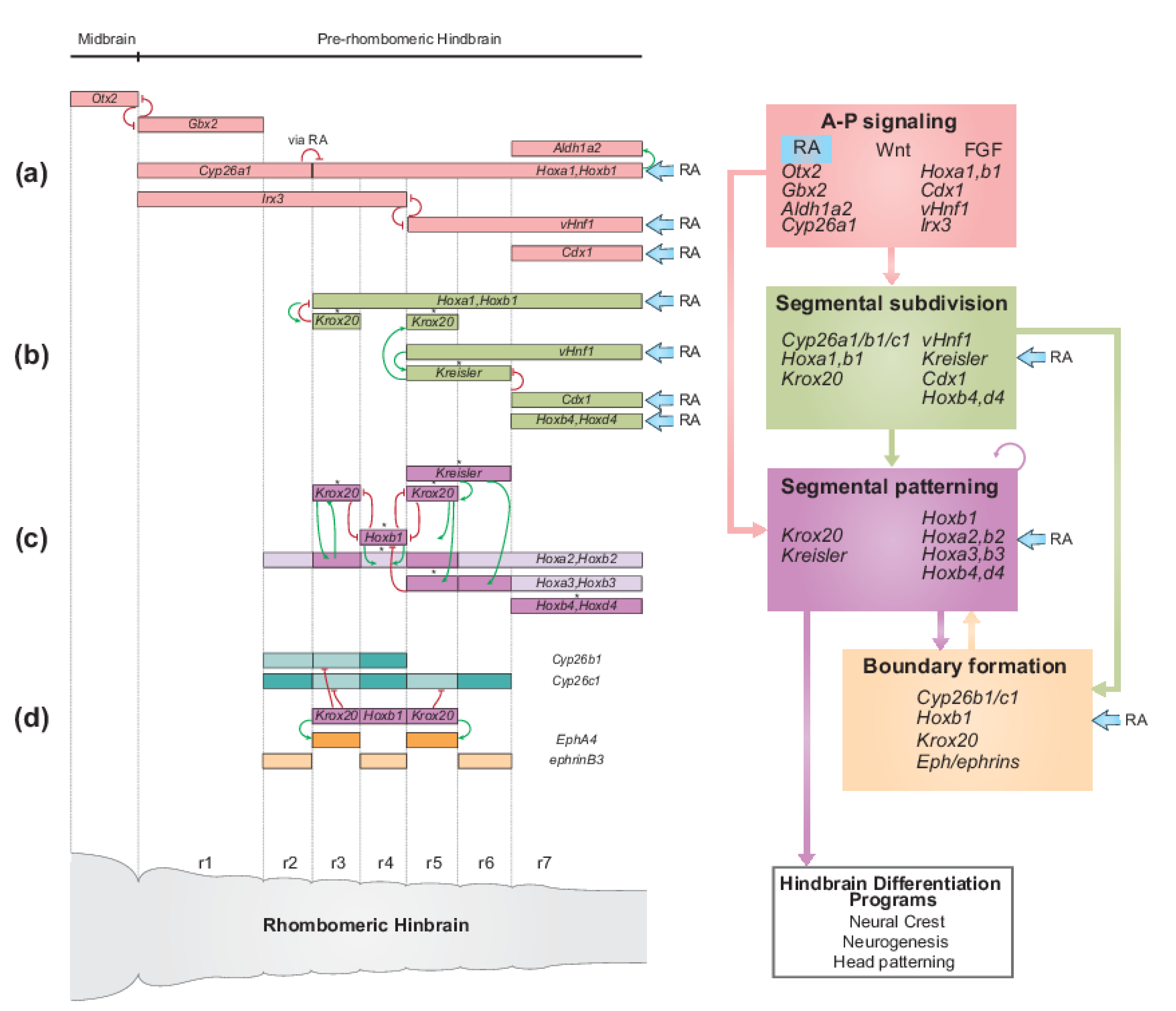
Analyses of patterns of gene expression, phenotypes arising from mutations and perturbation of expression and characterization of cis-regulatory elements in many different vertebrate species, particularly zebrafish, Xenopus, chicken, and mouse embryos, have revealed that the regulatory basis for establishing the Hox code is embedded in a conserved gene regulatory network (GRN) underlying hindbrain segmentation [34,38,42,43,100,101,103,143,144,177,178]. For example, a detailed list of cis-regulatory modules, activities, regulatory inputs, and species of origin used for constructing this GRN can be found in Table 1 of reference [34]. This GRN may be represented or visualized as a dynamic series of progressive steps or modules associated with the respective cell and developmental processes they regulate (Figure 1c) [43,100]. This GRN also provides a framework for understanding how signaling gradients and TFs, such as RA and Hox, are integrated into regulatory circuits that form and pattern hindbrain segments with distinct A-P identities. The FGF, Wnt, and RA signaling pathways govern initial steps of the GRN for hindbrain segmentation [93] and signaling cues from these pathways act as morphogens to dictate the molecular identity and organization of cells along the A-P axis. The RA morphogen is part of a well characterized signaling pathway which is of particular importance in precisely regulating the expression of key TFs, including Hox genes, in multiple modules in the GRN.
From an evolutionary perspective, hindbrain segmentation is a trait uniquely found in the vertebrate group and is remarkably conserved in all characterized vertebrate lineages [38,43]. Hindbrain segmentation appears to be an important vertebrate innovation that, in concert with the ability to form neural crest cells, is wired into conserved GRNs for developmental programs governing head development [34,100,164,179,180]. Recent studies in jawless vertebrates, such as lamprey, have revealed that many core components and TFs in the hindbrain GRN are deeply conserved to the base of the vertebrate tree [164,179,181], however, much less is known about the level of conservation of the roles of signaling pathways in the GRN of this vertebrate group. Because of their unique position as part of an early diverged vertebrate group, jawless vertebrate models provide an important opportunity to examine whether RA played an analogous role in different aspects of the hindbrain GRN early in vertebrate evolution and how this role might have evolved.
In this article, we give a brief overview of the current understanding of the GRN for vertebrate hindbrain segmentation, with a particular emphasis on the roles of RA signaling in regulating different aspects of segmentation via this GRN. In addition, we summarize how an RA morphogen gradient is set up in the developing hindbrain through a balance between synthesis and degradation and discuss the connection between the evolution of the RA signaling pathway and the hindbrain GRN in relation to diversity.

2. RA Signaling and Vertebrate Hindbrain Segmentation

2.1. RA Signaling and Its Roles in Development

RA signaling plays important roles in many fundamental biological processes. Cues from this pathway are involved in regulation of cell growth and proliferation, differentiation, and the control of homeostasis in multiple tissues. In development, RA can act as a morphogen, i.e., a signaling molecule that forms a concentration gradient over space and time which activates target genes in a concentration-dependent manner. In early vertebrate embryogenesis, RA is involved in the regulation of heart development [182,183], body axis formation [94,113,184] and the patterning and elongation of the A-P axis, through interactions with other signaling gradients (FGF and Wnt) [94,97,185]. In addition, RA signaling provides regulatory inputs into neural differentiation programs [113,186], pancreas specification and eye, kidney, and lung development [186,187,188]. Furthermore, RA signaling has a well-established role in A-P patterning of the hindbrain, where it contributes to the dynamic regulation of the process of segmentation, which lays down a basic ground plan for the elaboration of this key coordination center in the brain [43,100,103,144,178].
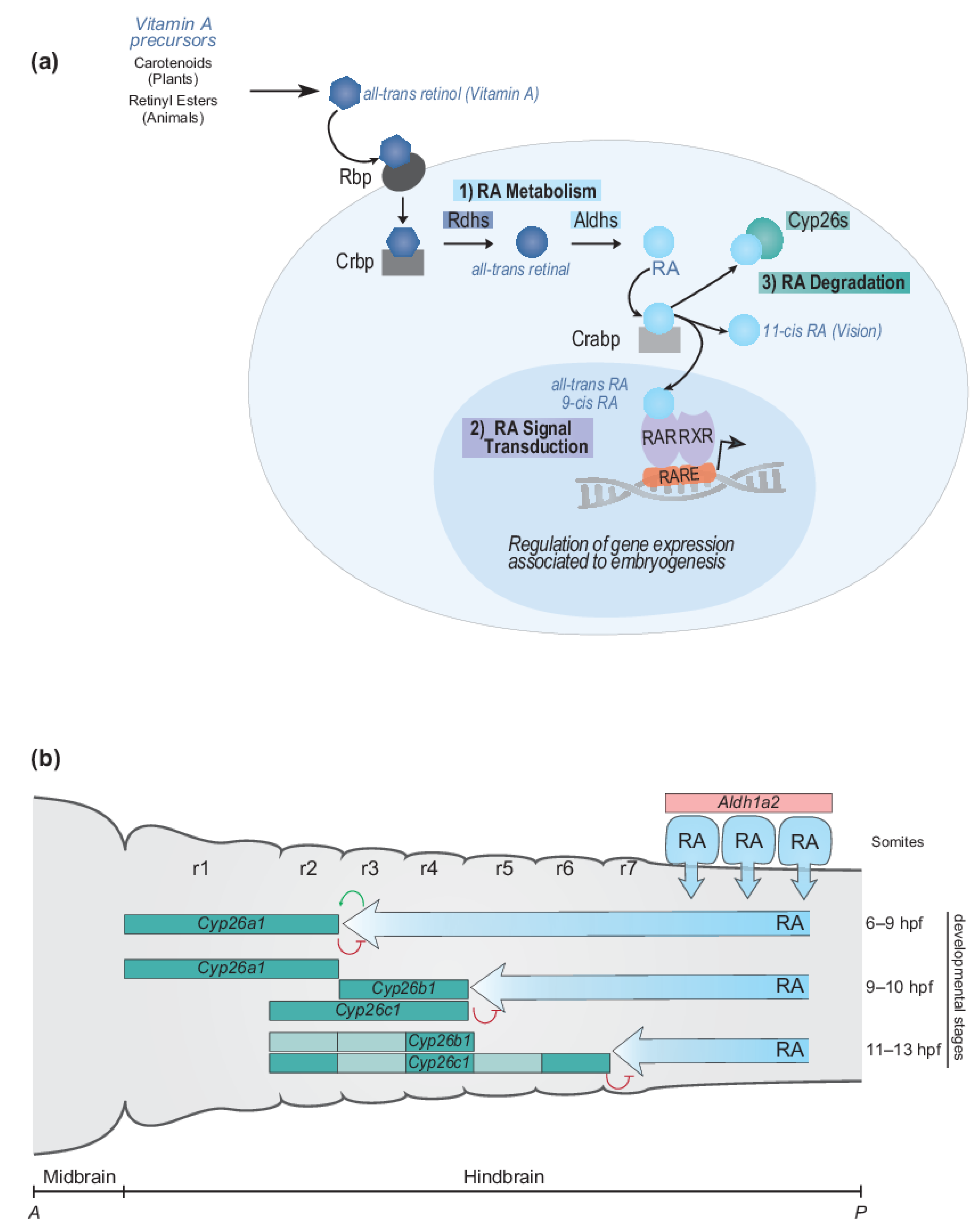
The RA signaling pathway can be broken down into three general steps: (1) RA metabolism to generate active ligands, (2) RA signal transduction to modulate gene expression, and (3) RA degradation to control levels of active ligand (Figure 2a). Components of the RA signaling pathway involve a Vitamin A precursor, binding proteins that mediate its extra- and intracellular transport, metabolic enzymes that convert it to an active ligand (e.g., all-trans RA and 9-cis RA) and enzymes that control the degradation of RA (Figure 2a). Signal transduction is mediated by the interaction of RA with members of the nuclear hormone receptor family of proteins (RAR and RXR) which bind directly to DNA regulatory elements in the genome and modulate patterns of gene expression [189,190]. The synthesizing and degrading enzymes, transport proteins, receptors and DNA regulatory elements together constitute the RA machinery and provide multiple opportunities for evolving and regulating the RA signaling pathway in various biological processes [187,188]. In this section, we summarize what is known about components of the RA signaling pathway and relate it to hindbrain segmentation, Hox genes, and the evolution of A-P patterning in vertebrates. The primary focus of this review is on the canonical pathway of RA signaling. However, there is emerging evidence concerning the non-canonical functions of certain components of the pathway, such as the cytoplasmic function of RARγ in cell death [191].

2.1.1. RA Metabolism, Signal Transduction and Degradation

RA is a fat-soluble, Vitamin A-derived signaling ligand that is coupled to the regulation of many processes in development, disease, and evolution. Vitamin A and its derivative retinoids are obtained from Vitamin A precursors found in the diet, either as carotenoids (mainly β-carotene) from plants, or retinyl esters from animals. In mammalian embryos, retinoids are provided by the mother via the placenta and yolk sac [192], while in other vertebrate embryos, it is mostly found in the egg yolk. In addition, the synthesis of retinal from carotenoids by bcox enzymes is a significant source of RA and is crucial for many biological processes, including hindbrain development [193]. With respect to the canonical RA signaling pathway, during the first step of the RA signaling pathway (RA metabolism in Figure 2a), RA is generated by a series of successive oxidative reactions from a Vitamin A precursor (Figure 2a). Briefly, all-trans retinol, an alcohol form of Vitamin A, is initially converted to all-trans retinal by retinol or alcohol dehydrogenases (Rdhs or Adhs). Retinal is in turn converted to RA by the retinal or aldehyde dehydrogenases (Raldhs or Aldhs). There are three main isomeric forms of RA: all-trans-RA and 9-cis-RA are associated with signal transduction in embryogenesis, cell growth and differentiation in chordates while 11-cis-RA plays a role in vision in most animals. While the different retinoids can play distinct roles with important physiological significance, this review will focus on roles of all-trans- and 9-cis- RA as they are able to bind and activate RARs in the tissues and processes most relevant to hindbrain segmentation and embryogenesis, as discussed below.
With respect to transport, retinol is delivered to cells by Retinol Binding Proteins (Rbp) in the bloodstream and within cells, it is bound by Cellular Retinol-Binding Proteins (Crbp1 and 2). Following synthesis of RA within a cell, it is bound by Cellular RA-Binding Proteins (Crabp1 and 2) and transported to the nucleus where it will play a key role in modulating the expression of downstream target genes (RA signal transduction in Figure 2a). This is achieved through the binding of RA to ligand-activated transcription factors which are part of the large family of nuclear hormone receptors [189,190,194]. There are two classes of receptors involved in this process, the retinoid X receptors (RXRs) and the retinoic acid receptors (RARs) (Figure 2a). The RAR and RXR proteins interact to form a dimer and they transduce the RA signal by binding directly to specific DNA motifs, called retinoic acid response elements (RAREs), found in the regulatory regions of target genes. RXRs are broadly expressed and serve as a common dimeric partner for a variety of other classes of nuclear hormone receptors, while the RARs are specific to transduction of retinoid signals. The RARE motifs have a consensus sequence that contains two short direct repeats (DR) generally separated by two or five nucleotides, e.g., a DR5 consists of two identical sequences separated by five nucleotides. However, in vivo, some RAREs display alternate spacing where the direct repeats are separated by anywhere from 1–5 nucleotides. In the absence of RA ligand, RARs-RXRs dimers can bind to RAREs of target genes and associate with transcriptional repressors (e.g., SMRT/NcoR) to inhibit gene expression [195,196]. Binding of RA ligands to RAR-RXR dimers induces a conformation change which results in the replacement of the associated repressor complexes with co-activators (NCOA) and the activation of target genes.
With respect to the turn-over of RA, members of the Cytochrome P450 family 26 (Cyp26) of enzymes play an important role in the modulating the levels of RA through degradation [101,103,197,198]. The dynamic patterns of expression of Cyp26 genes and the resulting activity of the Cyp26 enzymes, is involved in balancing the levels of RA generated by the synthesis pathway, shaping the range and levels of the RA morphogen gradient (Figure 2a RA degradation).

2.1.2. Dynamic Regulation of Endogenous RA Levels during Hindbrain Development

Aspects of processes underlying the development of the vertebrate hindbrain can illustrate how the dynamics of synthesis and degradation of RA play key roles in setting up an RA morphogen gradient involved in initiating the process of segmentation. Aldehyde Dehydrogenase 1a2 (Aldh1a2) (also known as Raldh2) is the major source of RA synthesis (Figure 2a) [199]. In mouse, chick, and zebrafish, at the onset of gastrulation Aldh1a2 is initially expressed in the presomitic mesoderm, positioned adjacent to the developing neural tube and Aldh1a2 remains active and synthesizes RA in newly formed somites, which will generate the axial skeleton (Figure 2b) [200,201,202]. RA then diffuses into the adjacent neural tube and spreads anteriorly. The anterior limit of its signaling activity in the neural tube is progressively restricted during development by the dynamic expression and activity of members of the Cyp26 family in the midbrain and rostral hindbrain (Figure 2b) [101,103,197]. Experimental studies have shown that the Cyp26 enzymes play a major role in facilitating where and when RA provides regulatory cues in the hindbrain GRN, by precisely regulating its endogenous levels. This fine-tuning of the concentration of RA is important as activation of many target genes, including Hox genes, display differential sensitivity to induction by RA [203,204]. Further support for the roles of synthesis and degradation of RA have been provided by genetic and pharmacological perturbations of endogenous Aldh1a2 and Cyp26 genes, which lead to defects in the hindbrain [103,187,197]. In multiple vertebrate models, phenotypes have shown a reduced forebrain/midbrain and expanded hindbrain region in embryos with elevated levels of RA, and a truncated hindbrain in embryos with low levels of RA. These phenotypes highlight the need to maintain adequete levels of RA for proper segmentation and organization of the hindbrain.
Retinoic acid is an important morphogen and the key role of its gradient in embryogenesis is widely recognized. Thus, many different approaches have been used to substantiate and quantify RA gradients in vertebrate embryos. Direct visualization of an endogenous RA gradient [205] and indirect evidence based on transgenic reporters for RA activity, pharmacological inhibitors of synthesizing and degrading enzymes, as well as genetic perturbations of key enzymes have led to the generation of different models to explain how an RA morphogen gradient is established in the hindbrain [101,206]. There are also examples of species-specific variations. However, consistent between models and species is the progressive integration of cues from opposing FGF and RA signaling pathways working in concert with the dynamic expression of Cyp26 enzymes to generate rapidly shifting boundaries of RA activity that regulate downstream events in the establishment of hindbrain segments [103,187,207].

2.1.3. Temporal Dynamics of Cyp26 Gene Expression in the Developing Hindbrain

There are three Cyp26 enzymes, Cyp26a1, b1, and c1, in all jawed vertebrates examined. The genes encoding these enzymes can display subtle differences in timing and domains of expression between species, but all three Cyp26 genes are dynamically expressed during hindbrain development (Figure 2b). Analyses of their patterns of expression and function in the hindbrain have been most extensively characterized in zebrafish and mouse models. In zebrafish, Cyp26a1 is first induced in the mesendoderm, adjacent to the future hindbrain, by low levels of RA [207]. At later stages of gastrulation (6–9 hpf), FGF signaling from the midbrain induces Cyp26a1 expression in the future r1–r2 domain of the hindbrain. This leads to degradation of RA in r1 and r2 and initially establishes a sharp anterior limit of RA activity at the r2/r3 boundary of the hindbrain (Figure 2b) [101,178,197]. Slightly later in development (9 to 10 hpf), Cyp26b1 is induced in r3 and r4 and Cyp26c1 in r2 to r4, which resets the anterior limit of RA activity to the r4/r5 boundary. By 11–13 hpf, the Cyp26b1 and Cyp26c1 expression domains expand and collectively cover the region from r2 to r6, resulting in a shift of the anterior limit of RA activity to the r6/r7 boundary [152,197]. Globally similar patterns of Cyp26 expression and regulation have been demonstrated in mouse, with the exception that RA has been shown to activate Cyp26c1 in r4 [103]. This illustrates how spatiotemporal changes in the expression of Cyp26s, with regulatory input from both FGF and RA signaling, can induce progressive changes in the pattern of degradation of RA, setting up shifting gradients and domains of RA which ultimately influence the formation of rhombomeric segments.

2.2. The Role of RA Signaling in the GRN for Hindbrain Segmentation

Studies have shown that the conserved functional properties of the vertebrate hindbrain are generated in part through a conserved program of segmentation, which organizes the ground plan for the development of this region. Comparative studies between jawed vertebrate species have aided our understanding of the genes, signals, and regulatory circuits that control the progressive steps of hindbrain segmentation [42,43,100]. These findings have been used as a basis to build a GRN that provides a core framework for understanding how the dynamic and progressive steps of hindbrain segmentation and A-P patterning are established and regulated (Figure 3) [34,93,100,179]. In addition, the GRN provides a foundation for exploring the evolution of hindbrain segmentation and A-P patterning systems in chordates. While multiple aspects of this GRN are highly conserved, there are some interesting differences between species, particularly with respect to the synthesis and degradation of RA, which will be discussed later in this review. This section focuses on the role that RA plays in different modules of the hindbrain GRN and explores the conservation of the relationship between RA signaling and the GRN.

2.2.1. RA Plays a Pivotal Role in Multiple Aspects of the Jawed Vertebrate Hindbrain GRN

RA initially impacts the GRN by providing input into the A-P signaling module, which along with Wnts and FGFs trigger the process of segmentation (Figure 3a). The A-P signaling module integrates signaling gradients and genes that respond to them, setting up spatially restricted domains of a set of TFs that begin to subdivide the hindbrain. For example, Gbx genes are upregulated caudally during gastrulation in response to posteriorizing Wnt signals, where they play a key role in positioning the midbrain–hindbrain boundary (MHB) by restricting Otx2 to the presumptive fore- and midbrain. The interface between Otx and Gbx expression domains is refined by mutual repression and sets the anterior limit of the hindbrain [208,209]. In addition, extensive analyses have revealed a downstream regulatory cascade involving members of the Pax2, En, FGF, and Wnt gene families that leads to lineage restriction between mid- and hindbrain territories and creates the MHB organizer, which acts as an influential signaling center for anterior hindbrain patterning [210,211,212,213,214,215].
The A-P signaling module also includes genes encoding key enzymes, such as Aldh1a2 and Cyp26a1, that shape the RA morphogen gradient and ultimately predefine future rhombomere territories. RA is then involved in the early spatial positioning of key posterior hindbrain patterning genes (vHnf1 and Cdx), as well as activating Hox genes of the first paralogous group (HoxPG1), Hoxa1 and Hoxb1 [163,168], whose expression domains are subsequently shaped by interactions with other patterning genes. For instance, Cyp26a1 is very important in setting up an anterior limit of RA activity at the future r2/r3 boundary, which in turn defines the expression domain of HoxPG1. Similarly, the mutual repressive relationship between Irx3/iro7 and vHnf1 is important for positioning the future r4/r5 boundary, which is later maintained by the expression of Cyp26b1 and Cyp26c1 in a domain abutting the anterior limit of vHnf1 expression [103,216].
In the next step of the process, the segmental subdivision module regulates the process of sub-dividing the hindbrain into segmental compartments which represent repeating metameric units. RA regulates genes encoding TFs implicated in the segmental subdivision of the presumptive hindbrain (Figure 3b) [34,43]. These segmentation genes (Krox20, Kreisler, vHnf1, HoxPG1, and HoxPG4 (Hoxb4 and Hoxd4)) are initially expressed in broad, partially overlapping domains that become progressively refined into distinct territories which define the future rhombomeric segments. In the next step, the segmental patterning module regulates the process which confers each segmental unit with a distinct molecular identity and set of characters (Figure 3c). In this module, many of these same TFs will contribute to the A-P patterning of individual segments in the hindbrain. In the segmental patterning module, a network of regulatory interactions sets up the hindbrain Hox code, where Hox genes are expressed and maintained in a rhombomeric-restricted fashion, providing regulatory information to direct differentiation programs in the hindbrain. RA further participates in the maintenance of some of these Hox expression domains.
In the boundary formation module, RA, via the activity of Cyp26b1 and Cyp26c1, plays a key role in mechanisms controlling cell segregation and boundary formation between segments, which involve inputs from Eph/Ephrin signaling, Krox20, and Hoxb1 (Figure 3d) [34,43,152,217,218]. Therefore, the GRN is defined by tightly coordinated interactions between Hox genes, their upstream segmental regulators, RA, and mechanisms controlling cell segregation and border formation. The coordination of these regulatory circuits generates precisely positioned domains of expression with sharp boundaries between adjacent rhombomeres, ensuring no overlap of cellular identity at the interface between cells in adjacent segments. RA plays a central and multifaceted role in this dynamic process.
In addition, feedback loops have an integral role in modulating RA signaling. For example, some targets of RA provide feedback to RA signaling itself, such as Hoxa1 and Hoxb1, which are transcriptionally activated by RA and contribute to the transcriptional control of Aldh1a2 expression, which is at the origin of RA synthesis [219]. In mouse, there is also evidence for a feedback circuit between HoxPG4 and rarβ, encoding for RARβ: rarβ is initially induced by RA and later upregulated by Hoxb4 and Hoxd4, which limits its expression posterior to the r6/r7 boundary. In addition, other RARs can indirectly maintain the expression of HoxPG4. These feedback loops between Hox and RARs are important for fine tuning expression domains and aligning the segmental expression borders of genes initially induced by RA [220]. Collectively, this work clearly illustrates that RA signaling plays diverse roles in different modules underlying the hindbrain GRN.

2.2.2. Retracing the Evolution of the Hindbrain GRN Using Jawless Vertebrates

Analyses of gene expression, functional perturbation experiments, and regulatory studies have revealed that many aspects of the GRN governing hindbrain segmentation are deeply conserved within the jawed vertebrate group [34]. For example, conserved cis-regulatory elements in segmental enhancers are similarly embedded in modules of the hindbrain GRN in different species, as illustrated by Krox20 element A [221] and the Hoxa2 r4 enhancer [222]. Moreover, several conserved RAREs have been identified in different modules of the GRN, such as in an enhancer of vHnf1 [223] as well as in many Hox loci; e.g., an RARE adjacent to Hoxb1 is required for restricting its expression to r4 [115,163]. This shows that many regulatory inputs to the GRN, including aspects of the machinery involved in their response to RA, i.e., RAREs, are deeply conserved in jawed vertebrates.
The conservation of the GRN across jawed vertebrates raises the question of whether these regulatory programs are also deployed earlier in the evolution of vertebrates. Recent studies have begun to investigate developmental GRNs in cyclostomes (lamprey and hagfish), which constitute the only extant group of jawless vertebrates, a sister group to jawed vertebrates [224]. Jawless vertebrate species have followed their own evolutionary paths since the split from the lineage that led to jawed vertebrates, and therefore represent a patchwork of ancestral and derived features [225]. Nevertheless, aspects of development that are shared across jawed and jawless vertebrates can point towards features of the vertebrate common ancestor. Thus, jawless vertebrates are valuable models to explore the origin and evolution of vertebrate-specific traits. Comparative studies with jawed vertebrates have shown that, despite their relatively simple and diverged morphology, e.g., lacking a jaw and paired limbs, they share many pan-vertebrate features with the jawed vertebrates. For example, lampreys possess a true neural crest and a core GRN governing neural crest development in jawed vertebrates is conserved in lamprey [180,226,227,228,229].
The characterization of the germline genome of the sea lamprey, Petromyzon marinus, has provided insights into major evolutionary events, such as pan-vertebrate or lineage-specific whole genome and chromosomal duplication events that occurred in vertebrates [71,72,230]. Of relevance to A-P patterning and the hindbrain GRN, comparative phylogenic studies have suggested that the six Hox clusters in the sea lamprey appear to be the result of at least one WGD event that occurred early in vertebrate evolution, before the divergence of the jawless vertebrate lineage. This was likely followed by chromosomal-scale and/or additional whole genome duplication event(s) and selective losses that occurred in the jawless vertebrate lineage (Figure 1a). Indeed, sea lamprey Hoxα and δ, and Hoxβ and ε are thought to be paralogous clusters derived from putative ancestral Hoxα/δ and Hoxβ/ε clusters respectively [71,72,230]. The sea lamprey Hox complement illustrates how major evolutionary events can influence the evolution of important gene families, providing various opportunities for molecular and morphological diversity.
Early studies in the Japanese lamprey, Lethenteron japonicum, revealed a transient period of hindbrain segmentation, with a segmental organization of reticulo-spinal neurons and cranial nerve roots positioned in similar arrangement to jawed vertebrates [231,232]. However, unlike in jawed vertebrates, these experiments demonstrated that some motor nuclei and Hox expression domains appeared not to be in register with rhombomeres, suggesting that Hox genes were not coupled to hindbrain segmentation. In addition, these early experiments did not reveal an apparent role of RA signaling in hindbrain segmentation in lamprey [231,233]. This led to the idea that lamprey might represent an intermediate organism between jawed vertebrates and non-vertebrate chordates, possessing an ‘early type’ of segmented hindbrain, independent of Hox genes and RA, which had not fully integrated aspects of the ancient A-P patterning system and coupled it to hindbrain segmentation [231].
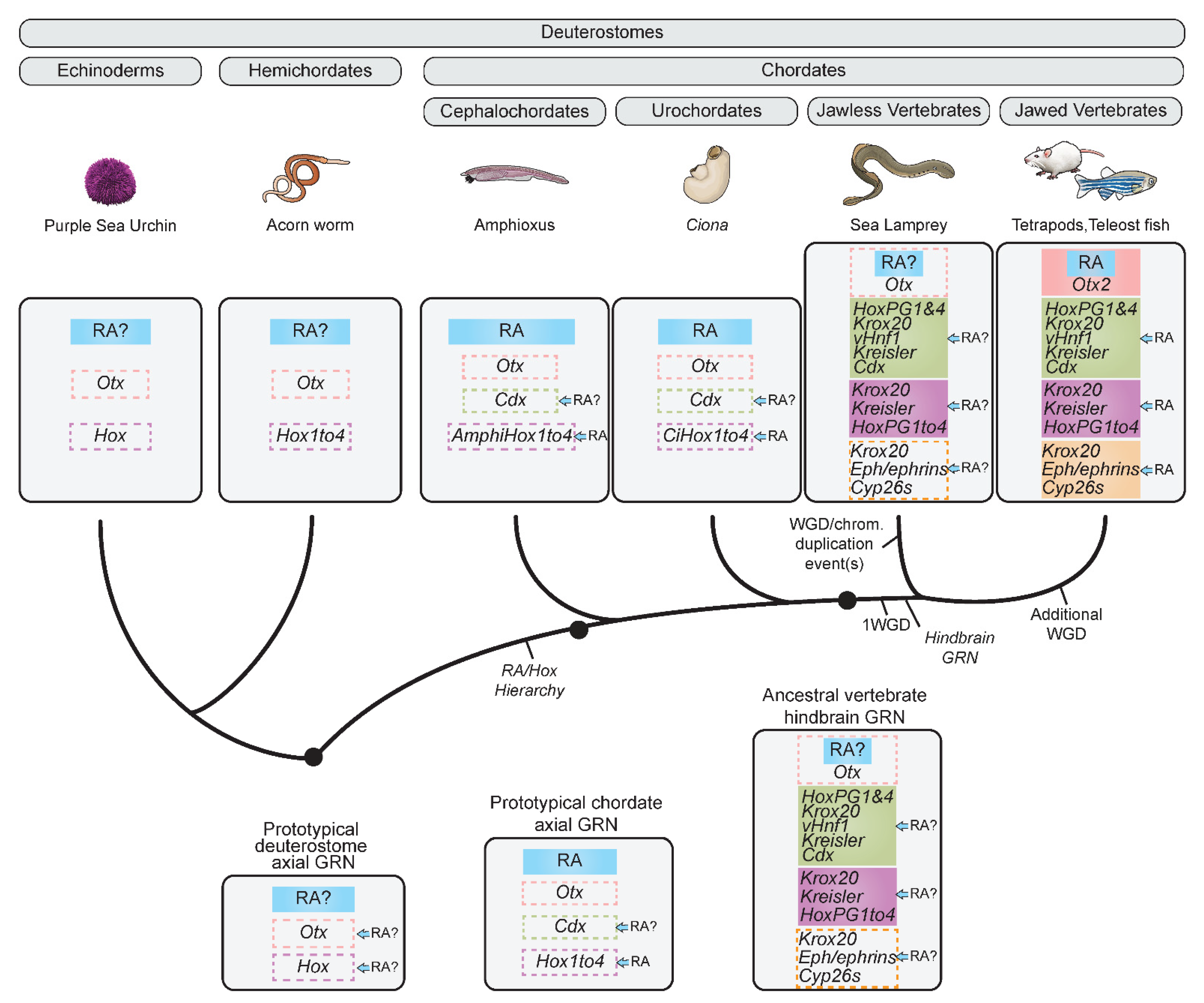
The early data on lamprey Hox expression focused on later stages with a few selected Hox genes. More recent studies, taking advantage of the accessible genome sequence to systematically evaluate expression of Hox genes over a broad range of early stages, have revealed that the expression of the HoxPG1–4 complement in the sea lamprey is tightly coupled to hindbrain segments. This showed that Hox genes are part of an ancestral core hindbrain GRN which arose in the evolution of vertebrates, prior to the divergence of extant jawed and jawless vertebrate lineages (Figure 4) [33,164,179,181]. For instance, many key TFs involved in different modules of the jawless vertebrate hindbrain GRN are expressed in similar restricted segmental domains in the lamprey hindbrain, such as Kreisler, Krox20, vHnf1, and HoxPG1 [33,231,234,235,236]. In addition, aspects of the boundary formation module appear to be conserved in jawless vertebrates, such as LjEphC, expressed in r3 and r5, reminiscent of the jawed vertebrate EphA4 [231]. Other examples of Eph/ephrin genes have been identified in the sea lamprey genome, but it is currently unknown whether they have rhombomeric expression [237]. Moreover, despite early similarities in rhombomeric gene expression between lamprey and jawed vertebrates, lamprey exhibits divergent Hox expression at later stages, with some Hox genes escaping rhombomeric restriction. This may reflect divergent regulatory inputs and patterning roles at these later stages [33,238].
In summary, many aspects of the GRN underlying hindbrain segmentation and segmental Hox expression can be traced to the base of vertebrates [33,164,180]. However, the precise roles of RA signaling in the lamprey hindbrain GRN are still unclear, and it is unknown whether it is integrated into the GRN in a manner analogous to jawed vertebrates.

2.3. Origins and Evolution of the Role of RA in the Hindbrain GRN

In light of the presence of Hox genes in most metazoans, evidence for an ancient A-P patterning system in chordates, and the high degree of conservation of aspects of the hindbrain GRN in vertebrates, it is interesting to question (1) when the RA/Hox regulatory hierarchy emerged during the course of evolution and (2) when this RA/Hox hierarchy become integrated into the GRN underlying hindbrain segmentation. This section describes efforts to understand its origin. Comparative analyses in various invertebrate models have sought to investigate the nature of the hindbrain GRN and how it relates to RA signaling in evolution. There is little clear evidence that RA is required for the development of protostomes, and in instances where developmental roles have been inferred, such as in neural differentiation in the marine annelid Platynereis dumerilii, these are not connected to Hox gene expression and A-P patterning [239]. Thus, in this review, we focus on chordates and non-chordate deuterostomes.

A Prototypical Axial GRN Integrating RA Can Be Rooted to the Base of Chordates

Despite having different anatomies and simpler brains, i.e., no segmented hindbrain, urochordates and cephalochordates both have a CNS that develops from a neural plate, as in vertebrates [240,241]. Analyses of embryonic gene expression domains in these models have provided evidence for some ancestral aspects of A-P brain regionalization at the molecular level, including equivalent forebrain, midbrain, hindbrain, and spinal territories [242,243]. These conserved A-P programs include aspects of the GRN involved in Hox-dependent patterning of the vertebrate hindbrain. For example, anterior neural expression of the forebrain–midbrain specifier Otx has been found in urochordates [242] and cephalochordates (Figure 4) [244], while both groups express Cdx in posterior tissues, including the neural tube [245,246]. Together, the A-P expression territories of these genes delimit a putative hindbrain-like region. Additionally, drug treatments in the cephalochordate amphioxus Branchiostoma floridae have revealed that perturbation of RA has a dramatic influence upon A-P patterning via these genes, although it is unclear whether they are direct targets of RA [246,247].
Studies in non-vertebrate chordate models support the idea that there is an ancestral role for RA signaling in patterning the nervous system via the Hox genes, which is conserved across many chordates. Urochordates, the closest phylogenetic group to vertebrates [248], possess a broken Hox cluster with only nine orthologs and partial spatial collinearity (Figure 4) [249]. Interestingly, Hox genes are up regulated in response to RA in Ciona intestinalis embryos, and CiHox1 was shown to respond to RA via upstream RAREs [250,251,252]. However, RA appears to have relatively minor influence on neural Hox expression in urochordates compared to vertebrates. Indeed, the larvacean Oikopleura dioica seems to have lost the ability to transduce RA signaling based on treatments with exogenous RA and the absence of multiple components of the RA machinery, such as Cyp26s and RARs. This suggests that some chordates can use mechanisms other than RA signaling to maintain their body plan, which may be linked to the altered organization of their Hox cluster [253].
Cephalochordates possess a single Hox cluster with 15 Hox genes (AmphiHox) (Figure 1a), that globally show colinear nested domains of expression along the A-P axis [254,255,256,257,258]. In amphioxus, RA also influences Hox expression along the A-P axis and in the nervous system (Figure 4). Ectopic RA treatment influences not only Hox expression, but motoneuron specification, reminiscent of what happens in vertebrates [247,258,259,260,261]. Cross-species regulatory analyses have uncovered the presence of two conserved DR5 RAREs located near AmphiHox1 and AmphiHox3 that are able to mediate reporter expression in the neural tube and neural crest of chicken and mouse embryos [262,263]. There are equivalent RAREs located in similar positions around the vertebrate Hox1 and Hox4 genes which are involved in neural and segmental expression [117,163,168,264]. This suggests that pre-existing RAREs in chordate Hox clusters may have been part of the ancient A-P patterning system and could have evolved new roles in Hox-dependent patterning of vertebrate innovations, such as the hindbrain. The apparent retention of RAREs in conserved locations within the Hox clusters may be important for both mediating conserved regulatory functions and establishing new roles [263].
In light of these similarities in the RA-regulated expression of Hox genes, amphioxus is generally considered to most closely represent the putative ancestral chordate state, which already integrated RA signaling as part of a mechanism for A-P patterning of the nervous system. Therefore, the evolution of the RA/Hox hierarchy is not specific to vertebrates but likely constitutes part of an ancient mechanism that shaped A-P patterning of the nervous system of the chordate ancestor. This roots Hox regulation by RA in the nervous system to the base of chordates.
In contrast to the conserved role of RA in regulating A-P Hox patterning, these invertebrate chordate models have revealed that certain aspects of the hindbrain GRN seem to have evolved later in the vertebrate group (Figure 4). Examples of such aspects include the cooption of key TFs such as Krox20 and Kreisler to the segmental subdivision module [265,266] and the cooption of the Eph/ephrin family in the evolution of the boundary formation module [237]. It seems likely that the role of these genes in the formation of rhombomeres and in the hindbrain GRN arose through new regulatory interactions and was an innovation of the vertebrate lineage [34,267,268].
In summary, a prototypical chordate axial GRN was likely composed of some early patterning genes, such as Otx and Cdx, that delimit a hindbrain-like territory by responding to graded A-P signaling inputs, including RA, combined with a nested deployment of Hox genes within this territory in response to RA signaling (Figure 4).

2.4. Evolution of the RA Signaling Pathway in Chordates

The ancestral role of RA in axial patterning in chordates raises the question of the degree to which the RA signaling pathway has been conserved or diversified in different chordate lineages in relation to A-P patterning. Assessing components of the RA machinery across chordates can help in exploring this issue. Comparing all components across numerous chordate models is beyond the scope of this review and others have already reviewed the status of this machinery extensively across bilaterians [269,270]. Here, we focus on the evolutionary status of a few critical, conserved, and well characterized components, as little is known about the evolution of several components, such as the Crbps and Crabps, in metazoans. In other instances, some components of the RA machinery are thought to be more ancient and deeply conserved in all metazoans, including cnidarians and bilaterians [269,271]. This is the case for the RXR nuclear receptors, which represent an essential component of the signal transduction pathway and are dimeric partners for many other classes of nuclear hormone receptors [189]. We do not ignore the possibility that such highly conserved components could have important evolving roles in the RA machinery but choose to focus on changes in aspects of the RA machinery related to the metabolic pathway (Rdh10 and Aldh1a2), the signal transduction pathway (RARs), and RA degradation (Cyp26s).

2.4.1. The Metabolic Pathway

The first step in metabolism involves the activity of enzymes in the Adh and Rdh families. Rdh10 is particularly important in vertebrates and is required for proper embryonic patterning and morphogenesis [272,273]. Rdh10 homologs have been detected in amphioxus, BfRdh10 [274], and Ciona, CiRdh10 [275], and shown to function as retinol dehydrogenases (Figure 5).
Aldh1a enzymes direct the next step of the pathway, converting retinal to different isomeric forms of RA. In vertebrates, tetrapods have 3–4 Aldh1a genes (Aldh1a1,2,3 and Aldh1a4/7), while teleosts appear to only have two (Aldh1a2 and Aldh1a3). Aldh1a2 is generally considered to be the primary RA generating enzyme in the developing trunk [199,200,201,276], while Aldh1a1 and Aldh1a3 play important roles in the developing optic vesicle and retina [277,278,279]. There are six putative Aldh1s in the cephalochordate amphioxus (BfAldh1a to f) and four in the urochordate Ciona (CiAldh1a to d) [280,281,282] (Figure 5). These duplicates are thought to have arisen from an ancestral Aldh1 gene via multiple duplication events that occurred independently in each of these chordate lineages. In both amphioxus and Ciona, only one Aldh1 (BfAldh1a and CiAldh1a) appears to be expressed in an equivalent spatiotemporal manner to vertebrate Aldh1a2, while the other paralogous Aldh1 genes display divergent expression patterns [280,281,282]. In addition to differences in expression between paralogs, it has been shown that some of the duplicates have evolved different protein structures, which appear to confer distinct functions. CiAldh1a, BfAldh1a, and vertebrate Aldh1a2 are involved in retinaldehyde processing and A-P patterning, while the other amphioxus and Ciona Aldh1s (BfAldh1b,c,e,f and CiAldh1b,c,d) have adopted a structure more similar to the vertebrate Aldh2s, which is important for detoxification of small aldehydes [280]. Thus, the duplication of the Aldh1 genes in chordate lineages appears to have been accompanied by divergence in their spatiotemporal expression as well as their biochemical functions, including roles beyond RA signaling.

2.4.2. RARs and the RA Signal Transduction Pathway

While vertebrates possess three RARs (RARα, RARβ and RARγ), each with various isoforms, only one RAR homolog has been identified in invertebrate chordate lineages, such as in Amphioxus [283] and in Ciona [284] (Figure 5). Both CiRAR and AmphiRAR have been shown to transduce RA signaling and activate transcription in the presence of RA in a broadly similar way to vertebrates [283,285]. Furthermore, AmphiRAR is expressed in the amphioxus nervous system and displays sensitivity to exogenous RA and RA antagonists, similar to the vertebrate RARs [283]. Interestingly, comparisons of ligand-binding specificities between vertebrate and invertebrate chordate RARs revealed that AmphiRAR has an RAR-α/β-like specificity while RAR–γ has a divergent ligand-binding capacity [286]. While the developmental significance of this divergence in vertebrates remains to be determined, this suggests that RARs may have acquired new functions that shaped their roles in RA signal transduction during vertebrate evolution.

2.4.3. RA Degradation by Cyp26s

The three vertebrate Cyp26 genes, Cyp26a1, Cyp26b1, and Cyp26c1 have a complex evolutionary history linked to whole-genome and tandem duplication events. Two Cyp26 genes have been identified in Ciona, CiCyp26a and CiCyp26b, including one that responds to exogenous levels of RA, but the evolutionary history of the Cyp26 family in this lineage remains unclear [284,287] (Figure 5). Three Cyp26 genes have been reported in two cephalochordate representatives, B. floridae [274] and B. lanceolatum. The Cyp26-1, Cyp26-2, and Cyp26-3 genes are clustered in the genome and are thought to have evolved via a cephalochordate-specific tandem duplication event from a single ancestral Cyp26 gene. The cephalochordate Cyp26 family has evolved two independent functions, the first being a role in initiating RA-dependent patterning of the A-P axis assumed by CYP26-2, reminiscent of the vertebrate Cyp26a1. A second role of the Cyp26 family is in homeostasis, where it maintains fluctuating levels of RA in the embryo, assumed by CYP26-1 and CYP26-3. These are more reactive to fluctuations in endogenous RA levels than CYP26-2 is, reminiscent of the vertebrate Cyp26a1 [101,197,207,287]. This suggests that an ancestral chordate Cyp26 may have been involved in both regulating RA-dependent nervous system patterning and maintaining RA homeostasis. It is possible that multiple lineage-specific duplication events of that gene, followed by sub-functionalization led to this dual activity being split between different members of the Cyp26 family in both cephalochordates and vertebrates. An alternative scenario is that an ancestral Cyp26 only possessed a single function and that the additional functions evolved independently in both lineages following the duplication of the ancestral gene. Investigating these two functions in non-chordate deuterostomes could help to clarify the origin of the different roles played by the Cyp26s in shaping the RA morphogen gradient in chordates.
In summary, a potential core ancestral RA machinery in the early evolution of chordates likely included at least one copy of each component—Rdh10, Aldh1a, RAR, and Cyp26. While fundamentals of the core RA signaling pathway are well conserved within chordates, it is clear that some aspects of the RA machinery have diversified independently in different chordate lineages, including in vertebrates. This is exemplified by the multiple lineage-specific duplications of the ancestral Aldh1 and Cyp26 genes, as well as the vertebrate-specific duplication and diversification of the RARs and Aldh1as. This may have provided many opportunities for altering the regulation of RA signaling and its incorporation into diverse biological processes during evolution.

2.5. Origins and Evolution of RA Signaling beyond Chordates—Insights from Non-Chordate Deuterostomes

Non-chordate deuterostomes have been investigated in an attempt to understand the evolution of chordate hindbrain patterning, the evolution of the RA signaling pathway, and when they might have become integrated.

2.5.1. Nervous System Patterning in Hemichordates and Echinoderms

Hemichordates and echinoderms, as sister groups to the chordates, are highly relevant for understanding the origins and coupling of A-P signaling gradients to nervous system development. A distinctive feature of hemichordates, including Saccoglossus kowalevskii (Sk), is the presence of a diffuse nerve net, as opposed to a more restricted dorsal CNS in chordates [288]. Multiple TFs and signaling components of the vertebrate A-P patterning GRN also play a role in the patterning of the hemichordate body axis (Figure 4). They also possess a single Hox cluster, with at least 11 Hox genes that display colinear expression, including in their nervous system (Figure 4) [131,289,290]. However, a role for RA as part of the hemichordate GRN for A-P patterning of the nerve-net has yet to be determined.
Echinoderms have many representatives, including sea urchins and sea stars, which usually display larval bilateral symmetry and adult radial symmetry. In these animals, the Hox complement can sometimes be disorganized, making these models difficult to relate to chordates with regard to axial patterning [290]. However, since recent research has revealed that a Hox-Gbx network controls radial segmentation of the larval endoderm in the sea anemone Nematostella vectensis, it seems that an axial Hox code may have controlled body patterning and segmentation before the evolution of the bilaterian A-P axis [291,292]. Additionally, there are examples of highly conserved A-P expression domains of patterning genes such as En or Otx, anterior to Hox domains in echinoderms (Figure 4) [293]. While RA seems to be involved in the metamorphosis of Sea Stars and Feather Stars [294,295], it has not been clearly implicated in the organization of the nervous system and/or in regulating Hox genes and is an area of interest for future research.

2.5.2. Hemichordates and Echinoderms Models and the Evolution of the RA Machinery

Despite the absence of a known role for RA in patterning the CNS of non-chordate deuterostomes and considering the conservation of a core RA machinery toolkit at the base of chordates, it is worth considering the origins and evolution of the RA machinery to explore whether some components are unique to chordates.
Searches for Rdh10 homologs in non-chordate deuterostomes have uncovered three isoforms, Rdh10A, Rdh10B, and Rdh10C, in the echinoderm Lumbriculus variegatus (Lv) [275] which have been shown to function as retinol dehydrogenases (Figure 5). These genes are expressed early in development, consistent with early roles in retinol metabolism. It is unclear if there is an Rdh10 homolog in hemichordates, but another enzyme belonging to a sister clade of the short chain dehydrogenases, DhrS3s, has been found in S. kowalevskii. Five predicted Aldh1s (SkAldh1a to e) have been found in S.kowalevskii while there is only evidence for a closely related Aldh2 in echinoderms [281]. Putative homologous RARs with a remarkably high degree of conservation in their amino acid sequence have been found in both S. kowalevskii and in the echinoderm Strongylocentrotus purpuratus (Sp) [270]. Two putative Cyp26 orthologs (SkCyp26a and SkCyp26b) and a single putative Cyp26 (SpCyp26) have been identified in S. kowalevskii and in S. purpuratus respectively [287].
These results indicate that multiple aspects of the RA machinery are more ancient than the chordate lineage and were likely already present at the origin of deuterostomes. The RA machinery of the deuterostome ancestor is for the most part presumptive, but potentially included at least one copy of the main components of the RA machinery, namely Rdh10, Aldh1, RAR, and Cyp26. Furthermore, these non-chordate deuterostome models provide further examples of lineage-specific duplication of these components, such as Aldhs and Cyp26s in S. kowalevskii, indicating that the duplication and diversification of RA machinery may be a widespread phenomenon across many taxa. Considering the putative presence of primary components of the RA machinery in non-chordate deuterostomes, it would be interesting to explore its role in the hemichordate nervous system, as this could provide insight into when the RA machinery became coupled to ancient Hox-dependent A-P patterning systems.

2.6. Regulatory Diversification of the RA Machinery and Evolution of the Vertebrate Hindbrain GRN

The deep conservation of the basic RA machinery toolkit (Rdh10, Aldh1, RAR, Cyp26), offers the opportunity to explore and speculate on the putative evolutionary history of its components. Despite the apparent deep conservation of many aspects of this machinery across chordates and even more ancient aspects present in all deuterostomes, some important features of how RA signaling components are regulated and deployed during hindbrain development appear to be unique to jawed vertebrates. This suggests that these may have evolved following the split between vertebrate and invertebrate chordates. These notably include novel expression domains of key components of the RA machinery with the potential to influence features of the RA gradient along the A-P axis, as well as evolving mechanisms of RA signal transduction, including the integration of new RAREs that could influence how downstream targets respond to RA.

2.6.1. Evolving Roles of RAREs

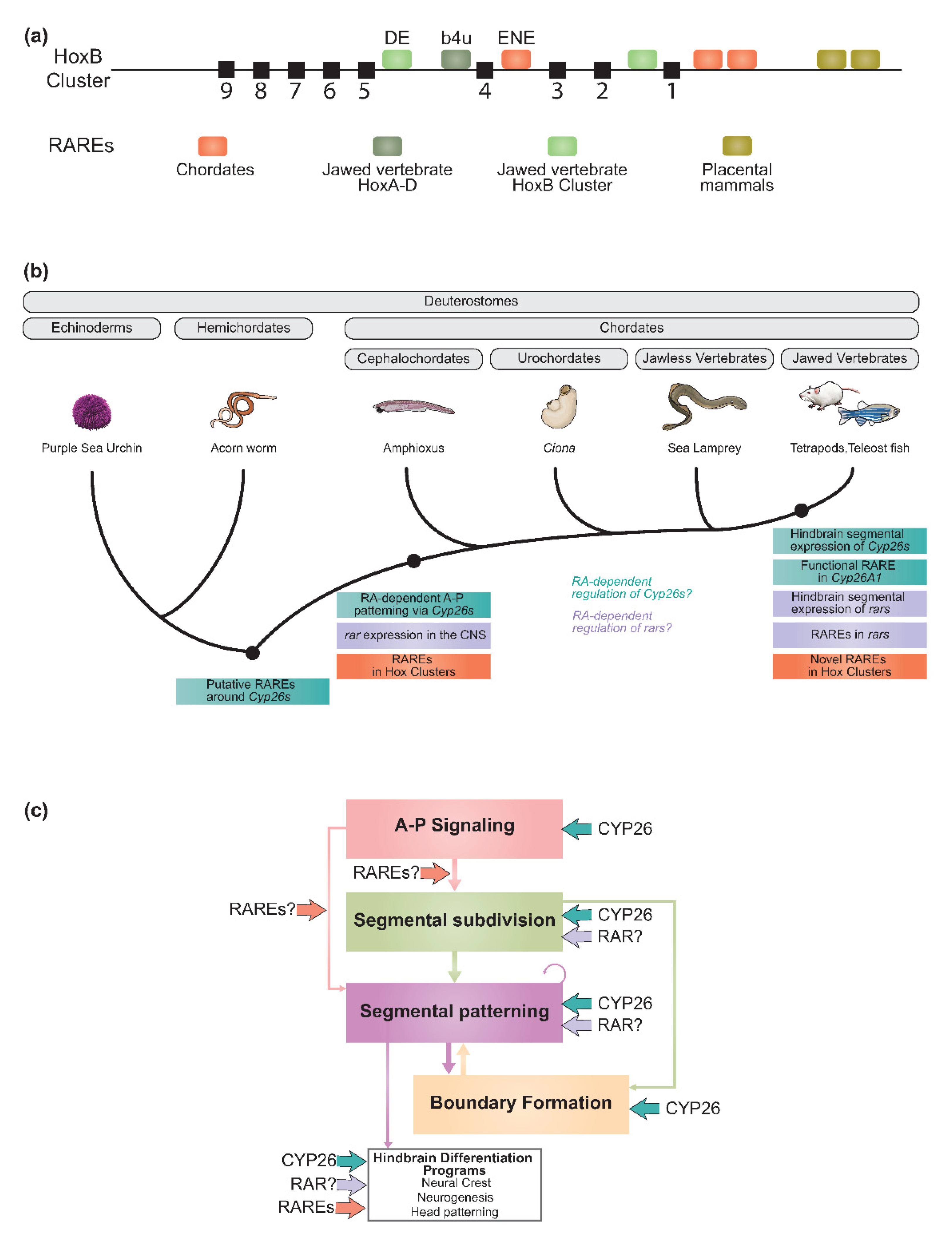
In the context of Hox genes, efforts have been made to characterize RAREs in multiple chordate lineages as these are important transducers of RA signaling in the regulation of gene expression. The data indicate an interesting degree of plasticity in their evolution, with instances where new RAREs appear in Hox clusters while ancestral ones may be retained [84,85,148,163,168,296,297,298,299,300]. Indeed, evidence indicates that many new RAREs have emerged in the vertebrate lineage, with some arising following WGD events. The jawed vertebrate Hox complement illustrates the diverse ways in which RAREs can evolve in a lineage (Figure 6a). The mouse HoxB cluster has retained ancestral RAREs, such as the one located in the early neural enhancer (ENE) region (ENE-RARE) of Hoxb4, that appears to be conserved in invertebrate chordates [117,262] (Figure 6a). In addition, 4U-RARE is present in the 5′ region of each Hox4 paralog of the mammalian Hox complement (a4u, b4u, c4u, d4u), suggesting that it is also present in the single Hox cluster of ancestral vertebrates and has been retained in each cluster through the multiple rounds of genome duplication [84]. In contrast, some RAREs appear to have evolved only in specific Hox clusters, such as the one present in the DE neural enhancer (DE-RARE), important for the regulation of Hoxb4 and Hoxb5, which is unique to the mammalian HoxB cluster [84,301,302]. Remarkably, some RAREs are unique to placental mammals [85,297]. This illustrates how the emergence and evolution of changes in cis-regulatory regions, such as RAREs, make it possible to integrate novel roles for RA in regulating Hox genes and other targets in the hindbrain GRN as well as other tissues. This could have emerged via the co-option of pre-existing RAREs or via the emergence of new RAREs during chordate evolution. Future efforts to identify the direct downstream targets of RA signaling during hindbrain development in vertebrates and invertebrate deuterostomes promise to elucidate how and when novel RA targets have been integrated into the hindbrain GRN during chordate evolution [303].

2.6.2. Regulatory Evolution of the Cyp26, Rar, and Crabp Gene Families

In addition to the evolution of new regulatory inputs of RA into gene expression, via novel or co-opted RAREs, the evolving roles of RA in the hindbrain GRN also seem to have emerged through the evolution of new expression domains of genes, such as Cyp26s, rars, and Crabps, that are important for shaping and interpreting the concentration of RA along the A-P axis and in many tissues.

Cyp26 Genes

Comparisons of Cyp26 gene expression patterns between vertebrates and amphioxus reveal both shared and derived features. These point toward an ancestral A-P patterning role for Cyp26s in chordates, as well as the elaboration of their inputs into hindbrain patterning in the vertebrate lineage.
The shared aspect of their expression is illustrated by an initial deployment at the anterior end of the presumptive hindbrain territory. In vertebrates, Cyp26a1 initially plays a fundamental role in delimiting the future anterior hindbrain, and Cyp26-2 is similarly expressed in an anterior domain of neural ectoderm during gastrulation in amphioxus. This suggests a conserved role for Cyp26s in setting up an anterior sink for lowering levels of RA that helps to position the future hindbrain-like territory. Consistent with this, the pharmacological manipulation of Cyp26 activity suggests roles for amphioxus Cyp26s in A-P patterning of the CNS [304]. In addition, it appears that the conserved aspect of Cyp26 expression is identifiable at the cis-regulatory level. In vertebrates, RA induces the embryonic expression of Cyp26a1 via a conserved upstream RARE [305,306] and in silico analyses have revealed that this RARE is conserved upstream of Cyp26-2 in amphioxus and binds RAR/RXR heterodimers in vitro [287]. Equivalent RARE sequences have been identified upstream of Cyp26s in a hemichordate (SkCyp26a) and in an echinoderm (SpCyp26) [287]. Thus, the RA-dependent regulation of Cyp26s via this element might be a conserved ancestral feature of deuterostomes. In vivo functional validation of this RARE in amphioxus and in hemichordates will help determine whether they have equivalent roles in driving early anterior Cyp26 expression during embryogenesis and could shed light on the evolutionary origin of such RA-dependent negative feedback mechanisms via Cyp26 RAREs (Figure 6b). Alternatively, they may have different roles in invertebrate deuterostomes and could have been co-opted to A-P patterning during chordate evolution. The evolution of such a robust feedback system for balancing endogenous fluctuations in levels of RA and protecting against toxic RA levels might have contributed to the establishment of a finely tuned system for precisely regulating cellular RA concentrations. Such a robust regulatory system may have facilitated its use in more complex patterning contexts in chordates.
Beyond an ancestral role for Cyp26s acting as anterior sinks for shaping the RA gradient in chordates, there are differences in their expression between vertebrates and amphioxus which suggest other roles. In vertebrates, the Cyp26s display dynamic segmental expression at multiple stages of hindbrain development, including during rhombomere formation (Figure 2b). This later expression is linked to their activity in generating shifting domains of RA degradation that set the anterior limit of expression of key genes that prefigure the future rhombomere boundaries. They are also involved in creating segmental variation in RA levels across odd and even rhombomeres, which influences signaling events important for the formation of sharp segmental boundaries (Figure 3d). In contrast, none of the amphioxus Cyp26s seem to be expressed in the developing hindbrain-like region itself [304]. This suggests that these later roles for Cyp26s which are coupled to hindbrain patterning might have evolved independently in the vertebrate lineage (Figure 6b). The dynamic, rhombomere-specific expression of vertebrate Cyp26 genes presumably evolved through the acquisition of new regulatory inputs from segmentally expressed genes, such as Krox20. However, relatively little is known about how Cyp26 genes are regulated in the hindbrain, which is an important topic for future research. Current evidence suggests that regulatory inputs into Cyp26 expression may vary between different vertebrate models. For example, in mouse, Cyp26c1 is induced by RA in r4 and plays an important role in maintaining adequate levels of RA, necessary for the induction of Hoxb1 [103], while zebrafish Cyp26c1 expression is not regulated by RA [197]. This suggests that the expression and regulation of Cyp26s during hindbrain development has continued to diversify in vertebrate evolution, potentially contributing to morphological diversity.

Rar Genes

In the vertebrate hindbrain, it has been suggested that the segmental expression of rars could be critical for the correct segmental expression of genes regulated by RA, such as the Hox genes [101,307]. In jawed vertebrates, the expression of multiple rars is regulated by RA (Figure 6b) and RAREs have been characterized in the regulatory region of these genes [308,309]. AmphiRAR is upregulated by RA [283], but no RARE has yet been found near this gene (Figure 6b). Similar to members of the Cyp26 family, some rars display a rhombomere-restricted expression pattern, e.g., murine rarα and rarβ (Figure 6b) [307]. These segmental expression domains arise in part through feedback circuits between RA target genes and rars, which serve to align the expression of multiple RA targets at segmental boundaries [220].

Crabp Genes

The evolution and diversification of the transport proteins, Crbps and Crabps, may have enabled further inputs into the diversification and fine-tuning of the RA signaling pathway in vertebrates. Crabp genes exhibit rhombomeric expression domains during development that are broadly conserved across vertebrate models [310,311]. They have been shown to be sensitive to RA and may be direct RA targets [311,312,313]. Crabp2a is essential for hindbrain patterning and segmental Hox expression in zebrafish, where it maintains the robustness of RA signaling by transporting RA to Cyp26 enzymes for degradation [311]. These features are analogous to the importance of the segmental expression of Cyp26 and rar genes in hindbrain segmentation. Thus, it is of value to understand when these roles for Crabp genes evolved. While a putative protostome Crabp-like receptor has previously been identified [314], this protein family has not been well-characterized beyond jawed vertebrates and therefore it is unclear whether the role of these transport proteins in the RA machinery is an innovation of vertebrates [269,315].
Thus, the co-option of the core vertebrate RA machinery to the process of hindbrain segmentation, together with the elaboration of robust feedback mechanisms from key components of the RA machinery to precisely maintain the levels of RA and shape the RA gradient, may have been a driving force in the evolution of the complex segmented structure of the vertebrate hindbrain. Furthermore, the acquisition of new downstream targets of RA signaling, via changes in the cohort of RAREs (Figure 6c), could have played an important role in setting up and maintaining the regulatory circuits in the different modules of the hindbrain GRN. Considering the segmental expression and the RA-dependent regulation of multiple core components of the RA machinery, it is interesting to consider the origin of mechanisms driving such regulatory feedbacks, as well as these segmental expression patterns. While very little is known about the evolution of these regulatory feedback loops, it is tempting to think that the segmental expression of components of the RA machinery could have been facilitated by their integration into the hindbrain GRN, e.g., via the influence of key members of the segmental patterning module, such as Krox20 and/or Hox proteins. This integration may have been important for building robustness and for the evolution of novel roles of RA in the hindbrain GRN.
In summary, the Cyp26s and RARs, with their complex evolutionary history, dynamic segmental expression patterns, and evolving roles, and the RAREs, with their evolutionary plasticity and potential for cooption into new functions, provide many opportunities for generating new interactions (Figure 6c). We favor the idea that the coupling of RA signaling to the vertebrate hindbrain GRN, together with the evolution of core aspects of the RA machinery, could have been major drivers of vertebrate hindbrain-related innovations by modulating the shape and timing of the RA morphogen gradient and modifying its regulatory output upon target genes (Figure 6c).

2.7. Lamprey as a Model for Understanding the Origin of the Hindbrain RA/Segmentation Hierarchy in Vertebrates

The important phylogenetic position of the sea lamprey as an extant jawless vertebrate, coupled to its relative tractability as a model organism, make it a prime model for investigating when and how the RA signaling pathway became integrated into the hindbrain GRN during vertebrate evolution. As described above, the precise roles for RA in hindbrain segmentation and Hox patterning in lamprey remain unclear, and it has previously been suggested that it may represent an intermediate state in which RA influences Hox expression but not hindbrain segmentation. Now that the Hox complement has been fully described, and the timing of hindbrain segmentation and rhombomeric gene expression are better understood, it is worth considering in more detail the role of RA in hindbrain patterning in lamprey [33]. The availability of a more complete lamprey germline genome assembly now enables a more thorough examination of the RA machinery, to further elucidate the evolution of RA signaling in relation to the vertebrate hindbrain. Duplication and reorganization of the genome, as exemplified by the six Hox clusters, has provided an opportunity for the amplification and divergence of the RA machinery in lamprey.
Previous studies in lamprey have identified components of the RA machinery. For example, in silico analyses predicted three Cyp26s, namely Cyp26a1, Cyp26b1/c1a, and Cyp26b1/c1b, but their evolutionary history remains unclear in different lamprey species, and their expression profiles have yet to be characterized (Figure 5) [287]. With respect to RA metabolism, while no Rdh10 has been reported in lamprey, an Aldh1a2 homolog has been identified and shown to be regulated in the spinal cord via the activity of a highly conserved intronic enhancer [316].
The rars have been relatively well studied in lamprey, with regards to their biochemical properties in RA signal transduction. Phylogenetic analyses and functional characterization of these receptors and their counterparts from jawed vertebrates and invertebrate chordates have suggested that duplication and divergence of the rar genes has led to functional changes in the RAR receptors during vertebrate evolution [286,317]. Four rars (rar1–4) have been characterized, but their relationships to the jawed vertebrate rars have not been unambiguously resolved (Figure 5) [317,318]. RAR3 is the putative ortholog of the jawed vertebrate RARα and amongst all vertebrate RARs these are thought to be the most similar to the ancestral vertebrate RAR in terms of sequence and function. Interestingly, these analyses indicate that none of the lamprey RARs show an RARγ-like ligand-binding specificity, suggesting RARγ has diverged in the gnathostome lineage. Collectively, these findings constitute examples of neofunctionalization and plasticity of the RA machinery in the vertebrate lineage and suggest an evolutionary scenario in which there were at least two rar paralogs in the vertebrate common ancestor, rarα/3 and rarβ/γ/1/2/4 (Figure 5). Subsequently, the RAR family diversified further in structure and ligand-binding specificity in the jawless and jawed vertebrate lineages. While the functional significance of this diversification remains unclear, such changes could be important for selectively transducing signals from different kinds of retinoids or different concentrations of RA. This would imply that aspects of RA/retinoid signaling may differ between jawed and jawless vertebrates and highlights the value of lamprey as a model for investigating steps in the evolution of RA signaling in vertebrates.
In summary, early vertebrates likely possessed two rars, at least two Cyp26s, and it is probable that the early vertebrate had at least one Aldh1a2 gene. However, the evolution of Rdh10 in vertebrates remains unclear (Figure 5). Considering the important roles of Cyp26b1 and c1 in hindbrain development in jawed vertebrates, characterizing the expression and function of Cyp26s in hindbrain development in the sea lamprey will be important to understand how and when these roles evolved. Examples of important remaining evolutionary questions that lamprey could help to answer include: (1) When did Cyp26b1/Cyp26c1 become involved in hindbrain development? (2) When were they coupled to hindbrain segmentation? It will be worth examining whether the RA-dependent feedback mechanisms characterized for some of the jawed vertebrate Cyp26 and rar genes are also active in lamprey.

3. Conclusions

The vertebrate hindbrain is a complex structure and represents an important example of how the integration of conserved signaling pathways and regulatory networks of axial patterning genes can influence the evolution of new traits. Hindbrain development in vertebrates is governed by a network of genetic interactions that orchestrates the formation and patterning of rhombomeres. The hindbrain GRN model provides a framework for interpreting data from multiple experimental sources and examining conservation of developmental mechanisms between species. RA signaling plays crucial roles at multiple stages of hindbrain development. An RA gradient initiates the early deployment of nested gene expression domains to subdivide the A-P axis. Subsequently, segment-specific variation in RA signaling influences the cross-regulation of these genes to pre-figure rhombomeres and mediates community signaling that refines rhombomere boundaries. RA signaling performs these roles by being wired into multiple modules of the GRN, either via RAREs in RA target genes, or through the segmental regulation of important components of the RA machinery, including Cyp26, rar, and Crabp genes.
From an evolutionary perspective, the GRN appears to be highly conserved across jawed vertebrates and studies on the sea lamprey have revealed that a segmented hindbrain with an underlying Hox code was an ancestral vertebrate feature. While multiple aspects of the hindbrain GRN appear to be conserved in lamprey, the roles for RA signaling in this process are currently poorly characterized and may have diverged between jawed and jawless vertebrates. Further studies of RA signaling in the context of hindbrain segmentation in lamprey promise to reveal how and when the roles for RA signaling in the hindbrain GRN evolved and diversified in the vertebrate lineage.
Looking deeper in evolution, studies in invertebrate models suggest that many components of the RA signaling machinery can be traced back to early metazoans, suggesting that RA signaling is an evolutionarily ancient mechanism. While a core RA signaling machinery appears to be present in deuterostomes, evidence suggests that the lineage-specific duplication of multiple RA signaling components may have enabled its functional diversification across deuterostome lineages. While the functional and evolutionary significance of such diversification is unclear, it is possible that major genome rearrangements, such as the whole genome duplication events that occurred in vertebrate evolution, may have provided important contexts for facilitating diversification in the role/s of RA signaling. Further functional characterization of the RA machinery in vertebrates and invertebrates, as well as the spatiotemporal detection of RA levels during hindbrain development, will reveal the contributions of these conserved and divergent aspects of RA signaling to the evolution of the hindbrain GRN.
Furthermore, the A-P deployment of cohorts of patterning genes, such as Hox genes, is highly conserved and probably dates to the bilaterian ancestor. However, the coupling of RA signaling to Hox genes and A-P patterning appears to have occurred in the deuterostome lineage. Indeed, comparisons with invertebrate chordates have revealed that the ancestral chordate employed an RA-Hox hierarchy to pattern the hindbrain territory, although the hindbrain GRN appears to have been modified/elaborated in early vertebrates by the evolution of novel regulatory interactions for segmental subdivision and rhombomeric segmentation. These could include the regulation of the RA machinery by segmentally restricted factors, as well as RA-dependent regulatory feedback mechanisms into the RA machinery. Comparisons of RA targets and the regulatory inputs into the RA machinery between vertebrate and invertebrate models promise to identify the nature of these novel regulatory interactions and how RA signaling became coupled to rhombomere formation and segment identity during vertebrate evolution.

Author Contributions

Conceptualization, A.M.H.B., H.J.P. and R.K.; manuscript writing, review, and editing, A.M.H.B., H.J.P. and R.K. All authors have read and agreed to the published version of the manuscript.

Funding

This work was supported by funds from the Stowers Institute for Medical Research (grant no: 1001) to R.K.

Institutional Review Board Statement

Not applicable.

Informed Consent Statement

Not applicable.

Data Availability Statement

All data and results referred to in this review have been previously published and cited with the appropriate reference.

Acknowledgments

The authors thank other members of the Krumlauf laboratory for helpful feedback and discussion on the manuscript and Mark Miller for providing illustrations. This work was done to fulfill, in part, requirements for A.M.H.B.’s PhD thesis research as a student registered with the Open University.

Conflicts of Interest

The authors declare no conflict of interest.

References

  1. Carroll, S.B. Homeotic genes and the evolution of arthropods and chordates. Nature 1995, 376, 479–485. [Google Scholar] [CrossRef] [PubMed]
  2. Martin, A.; Serano, J.M.; Jarvis, E.; Bruce, H.S.; Wang, J.; Ray, S.; Barker, C.A.; O’Connell, L.C.; Patel, N.H. CRISPR/Cas9 Mutagenesis Reveals Versatile Roles of Hox Genes in Crustacean Limb Specification and Evolution. Curr. Biol. 2016, 26, 14–26. [Google Scholar] [CrossRef] [Green Version]
  3. McGinnis, W.; Krumlauf, R. Homeobox genes and axial patterning. Cell 1992, 68, 283–302. [Google Scholar] [CrossRef]
  4. Mallo, M.; Wellik, D.M.; Deschamps, J. Hox genes and regional patterning of the vertebrate body plan. Dev. Biol. 2010, 344, 7–15. [Google Scholar] [CrossRef] [PubMed] [Green Version]
  5. Diederich, R.J.; Pattatucci, A.M.; Kaufman, T.C. Developmental and evolutionary implications of labial, Deformed and engrailed expression in the Drosophila head. Development 1991, 113, 273–281. [Google Scholar] [CrossRef] [PubMed]
  6. Lutz, B.; Lu, H.C.; Eichele, G.; Miller, D.; Kaufman, T.C. Rescue of Drosophila labial null mutant by the chicken ortholog Hoxb-1 demonstrates that the function of Hox genes is phylogenetically conserved. Genes Dev. 1996, 10, 176–184. [Google Scholar] [CrossRef] [PubMed] [Green Version]
  7. Powers, T.P.; Hogan, J.; Ke, Z.; Dymbrowski, K.; Wang, X.; Collins, F.H.; Kaufman, T.C. Characterization of the Hox cluster from the mosquito Anopheles gambiae (Diptera: Culicidae). Evol. Dev. 2000, 2, 311–325. [Google Scholar] [CrossRef]
  8. Hughes, C.L.; Kaufman, T.C. Exploring the myriapod body plan: Expression patterns of the ten Hox genes in a centipede. Development 2002, 129, 1225–1238. [Google Scholar] [CrossRef]
  9. Singh, N.P.; De Kumar, B.; Paulson, A.; Parrish, M.E.; Zhang, Y.; Florens, L.; Conaway, J.W.; Si, K.; Krumlauf, R. A six-amino-acid motif is a major determinant in functional evolution of HOX1 proteins. Genes Dev. 2020, 34, 1680–1696. [Google Scholar] [CrossRef]
  10. Ferrier, D.E.; Holland, P.W. Ancient origin of the Hox gene cluster. Nat. Rev. Genet. 2001, 2, 33–38. [Google Scholar] [CrossRef]
  11. Duboule, D.; Dolle, P. The structural and functional organization of the murine HOX gene family resembles that of Drosophila homeotic genes. EMBO J. 1989, 8, 1497–1505. [Google Scholar] [CrossRef]
  12. Graham, A.; Papalopulu, N.; Krumlauf, R. The murine and Drosophila homeobox gene complexes have common features of organization and expression. Cell 1989, 57, 367–378. [Google Scholar] [CrossRef]
  13. Lewis, E.B. A gene complex controlling segmentation in Drosophila. Nature 1978, 276, 565–570. [Google Scholar] [CrossRef] [PubMed]
  14. Scott, M.P.; Weiner, A.J. Structural relationships among genes that control development: Sequence homology between the Antennapedia, Ultrabithorax and fushi tarazu loci of Drosophila. Proc. Natl. Acad. Sci. USA 1984, 81, 4115–4119. [Google Scholar] [CrossRef] [Green Version]
  15. Scott, M.; Weiner, A.; Hazelrigg, T.; Polisky, B.; Pirotta, V.; Scalenghe, F.; Kaufman, M. The molecular organization of the Antennapedia locus of Drosophila. Cell 1983, 35, 763–776. [Google Scholar] [CrossRef]
  16. McGinnis, W.; Garber, R.L.; Wirz, J.; Kuroiwa, A.; Gehring, W. A homologous protein-coding sequence in Drosophila homeotic genes and its conservation in other metazoans. Cell 1984, 37, 403–408. [Google Scholar] [CrossRef]
  17. McGinnis, W.; Levine, M.S.; Hafen, E.; Kuroiwa, A.; Gehring, W. A conserved DNA sequence in homeotic genes of the Drosophila Antennapedia and bithorax complexes. Nature 1984, 308, 428–433. [Google Scholar] [CrossRef]
  18. Harding, K.; Wedeen, C.; McGinnis, W.; Levine, M. Spatially regulated expression of homeotic genes in Drosophila. Science 1985, 229, 1236–1242. [Google Scholar] [CrossRef] [PubMed]
  19. Dekker, E.J.; Pannese, M.; Houtzager, E.; Boncinelli, E.; Durston, A. Colinearity in the Xenopus laevis Hox-2 complex. Mech. Dev. 1992, 40, 3–12. [Google Scholar] [CrossRef]
  20. Kmita, M.; Duboule, D. Organizing axes in time and space; 25 years of colinear tinkering. Science 2003, 301, 331–333. [Google Scholar] [CrossRef] [Green Version]
  21. Deschamps, J.; Duboule, D. Embryonic timing, axial stem cells, chromatin dynamics, and the Hox clock. Genes Dev. 2017, 31, 1406–1416. [Google Scholar] [CrossRef] [Green Version]
  22. Duboule, D.; Morata, G. Colinearity and functional hierarchy among genes of the homeotic complexes. Trends Genet. 1994, 10, 358–364. [Google Scholar] [CrossRef]
  23. Lowe, C.J.; Wu, M.; Salic, A.; Evans, L.; Lander, E.; Stange-Thomann, N.; Gruber, C.E.; Gerhart, J.; Kirschner, M. Anteroposterior patterning in hemichordates and the origins of the chordate nervous system. Cell 2003, 113, 853–865. [Google Scholar] [CrossRef] [Green Version]
  24. Serano, J.M.; Martin, A.; Liubicich, D.M.; Jarvis, E.; Bruce, H.S.; La, K.; Browne, W.E.; Grimwood, J.; Patel, N.H. Comprehensive analysis of Hox gene expression in the amphipod crustacean Parhyale hawaiensis. Dev. Biol. 2016, 409, 297–309. [Google Scholar] [CrossRef] [PubMed]
  25. Bachiller, D.; Macias, A.; Duboule, D.; Morata, G. Conservation of a functional hierarchy between mammalian and insect Hox/HOM genes. EMBO J. 1994, 13, 1930–1941. [Google Scholar] [CrossRef] [PubMed]
  26. Gaunt, S.J.; Strachan, L. Temporal colinearity in expression of anterior Hox genes in developing chick embryos. Dev. Dyn. 1996, 207, 270–280. [Google Scholar] [CrossRef]
  27. Duboule, D. Temporal colinearity and phylotypic progression: A basis for the stability of a vertebrate Bauplan and the evolution of morphologies through heterochrony. Development 1994, 135–142. [Google Scholar] [CrossRef]
  28. Duboule, D. Vertebrate hox gene regulation: Clustering and/or colinearity? Curr. Opin. Genet. Dev. 1998, 8, 514–518. [Google Scholar] [CrossRef]
  29. Gaunt, S.J.; Sharpe, P.T.; Duboule, D. Spatially restricted domains of homeo-gene transcripts in mouse embryos: Relation to a segmented body plan. Development 1988, 104, 169–181. [Google Scholar] [CrossRef]
  30. Krumlauf, R. Hox genes in vertebrate development. Cell 1994, 78, 191–201. [Google Scholar] [CrossRef]
  31. Lowe, C.J.; Clarke, D.N.; Medeiros, D.M.; Rokhsar, D.S.; Gerhart, J. The deuterostome context of chordate origins. Nature 2015, 520, 456–465. [Google Scholar] [CrossRef]
  32. Kessel, M.; Gruss, P. Homeotic transformations of murine vertebrae and concomitant alteration of Hox codes induced by retinoic acid. Cell 1991, 67, 89–104. [Google Scholar] [CrossRef] [Green Version]
  33. Parker, H.J.; Bronner, M.E.; Krumlauf, R. An atlas of anterior hox gene expression in the embryonic sea lamprey head: Hox-code evolution in vertebrates. Dev. Biol. 2019, 453, 19–33. [Google Scholar] [CrossRef] [Green Version]
  34. Parker, H.J.; Krumlauf, R. Segmental arithmetic: Summing up the Hox gene regulatory network for hindbrain development in chordates. Wiley Interdiscip. Rev. Dev. Biol. 2017, 6, e286. [Google Scholar] [CrossRef]
  35. Wilkinson, D.G.; Bhatt, S.; Cook, M.; Boncinelli, E.; Krumlauf, R. Segmental expression of Hox-2 homoeobox-containing genes in the developing mouse hindbrain. Nature 1989, 341, 405–409. [Google Scholar] [CrossRef]
  36. Hunt, P.; Gulisano, M.; Cook, M.; Sham, M.H.; Faiella, A.; Wilkinson, D.; Boncinelli, E.; Krumlauf, R. A distinct Hox code for the branchial region of the vertebrate head. Nature 1991, 353, 861–864. [Google Scholar] [CrossRef]
  37. Prince, V.E.; Moens, C.B.; Kimmel, C.B.; Ho, R.K. Zebrafish hox genes: Expression in the hindbrain region of wild-type and mutants of the segmentation gene, valentino. Development 1998, 125, 393–406. [Google Scholar] [CrossRef] [PubMed]
  38. Lumsden, A. Segmentation and compartition in the early avian hindbrain. Mech. Dev. 2004, 121, 1081–1088. [Google Scholar] [CrossRef] [PubMed]
  39. Murphy, P.; Davidson, D.R.; Hill, R.E. Segment-specific expression of a homeobox-containing gene in the mouse hindbrain. Nature 1989, 341, 156–159. [Google Scholar] [CrossRef]
  40. Murphy, P.; Hill, R.E. Expression of the mouse labial-like homeobox-containing genes, Hox 2.9 and Hox 1.6, during segmentation of the hindbrain. Development 1991, 111, 61–74. [Google Scholar] [CrossRef] [PubMed]
  41. Kessel, M.; Balling, R.; Gruss, P. Variations of cervical vertebrae after expression of a Hox-1.1 transgene in mice. Cell 1990, 61, 301–308. [Google Scholar] [CrossRef]
  42. Alexander, T.; Nolte, C.; Krumlauf, R. Hox genes and segmentation of the hindbrain and axial skeleton. Annu. Rev. Cell Dev. Biol. 2009, 25, 431–456. [Google Scholar] [CrossRef] [PubMed] [Green Version]
  43. Krumlauf, R.; Wilkinson, D.G. Segmentation and patterning of the vertebrate hindbrain. Development 2021, 148, dev186460. [Google Scholar] [CrossRef]
  44. Balling, R.; Mutter, G.; Gruss, P.; Kessel, M. Craniofacial abnormalities induced by ectopic expression of the homeobox gene Hox-1.1 in transgenic mice. Cell 1989, 58, 337–347. [Google Scholar] [CrossRef] [Green Version]
  45. Zhang, M.; Kim, H.J.; Marshall, H.; Gendron-Maguire, M.; Lucas, D.A.; Baron, A.; Gudas, L.J.; Gridley, T.; Krumlauf, R.; Grippo, J.F. Ectopic Hoxa-1 induces rhombomere transformation in mouse hindbrain. Development 1994, 120, 2431–2442. [Google Scholar] [CrossRef]
  46. Alexandre, D.; Clarke, J.D.; Oxtoby, E.; Yan, Y.L.; Jowett, T.; Holder, N. Ectopic expression of Hoxa-1 in the zebrafish alters the fate of the mandibular arch neural crest and phenocopies a retinoic acid-induced phenotype. Development 1996, 122, 735–746. [Google Scholar] [CrossRef]
  47. Di Bonito, M.; Narita, Y.; Avallone, B.; Sequino, L.; Mancuso, M.; Andolfi, G.; Franze, A.M.; Puelles, L.; Rijli, F.M.; Studer, M. Assembly of the auditory circuitry by a Hox genetic network in the mouse brainstem. PLoS Genet. 2013, 9, e1003249. [Google Scholar] [CrossRef] [Green Version]
  48. Geisen, M.J.; Di Meglio, T.; Pasqualetti, M.; Ducret, S.; Brunet, J.F.; Chedotal, A.; Rijli, F.M. Hox paralog group 2 genes control the migration of mouse pontine neurons through slit-robo signaling. PLoS Biol. 2008, 6, e142. [Google Scholar] [CrossRef] [Green Version]
  49. Chatonnet, F.; Wrobel, L.J.; Mezieres, V.; Pasqualetti, M.; Ducret, S.; Taillebourg, E.; Charnay, P.; Rijli, F.M.; Champagnat, J. Distinct roles of Hoxa2 and Krox20 in the development of rhythmic neural networks controlling inspiratory depth, respiratory frequency, and jaw opening. Neural Dev. 2007, 2, 19. [Google Scholar] [CrossRef]
  50. Oury, F.; Murakami, Y.; Renaud, J.S.; Pasqualetti, M.; Charnay, P.; Ren, S.Y.; Rijli, F.M. Hoxa2- and rhombomere-dependent development of the mouse facial somatosensory map. Science 2006, 313, 1408–1413. [Google Scholar] [CrossRef] [Green Version]
  51. Studer, M.; Lumsden, A.; Ariza-McNaughton, L.; Bradley, A.; Krumlauf, R. Altered segmental identity and abnormal migration of motor neurons in mice lacking Hoxb-1. Nature 1996, 384, 630–634. [Google Scholar] [CrossRef] [PubMed]
  52. Gavalas, A.; Ruhrberg, C.; Livet, J.; Henderson, C.E.; Krumlauf, R. Neuronal defects in the hindbrain of Hoxa1, Hoxb1 and Hoxb2 mutants reflect regulatory interactions among these Hox genes. Development 2003, 130, 5663–5679. [Google Scholar] [CrossRef] [PubMed] [Green Version]
  53. Gavalas, A.; Studer, M.; Lumsden, A.; Rijli, F.M.; Krumlauf, R.; Chambon, P. Hoxa1 and Hoxb1 synergize in patterning the hindbrain, cranial nerves and second pharyngeal arch. Development 1998, 125, 1123–1136. [Google Scholar] [CrossRef]
  54. Davenne, M.; Maconochie, M.K.; Neun, R.; Pattyn, A.; Chambon, P.; Krumlauf, R.; Rijli, F.M. Hoxa2 and Hoxb2 control dorsoventral patterns of neuronal development in the rostral hindbrain. Neuron 1999, 22, 677–691. [Google Scholar] [CrossRef] [Green Version]
  55. Gaufo, G.O.; Wu, S.; Capecchi, M.R. Contribution of Hox genes to the diversity of the hindbrain sensory system. Development 2004, 131, 1259–1266. [Google Scholar] [CrossRef] [Green Version]
  56. Gaufo, G.O.; Thomas, K.R.; Capecchi, M.R. Hox3 genes coordinate mechanisms of genetic suppression and activation in the generation of branchial and somatic motoneurons. Development 2003, 130, 5191–5201. [Google Scholar] [CrossRef] [Green Version]
  57. Arenkiel, B.R.; Tvrdik, P.; Gaufo, G.O.; Capecchi, M.R. Hoxb1 functions in both motoneurons and in tissues of the periphery to establish and maintain the proper neuronal circuitry. Genes Dev. 2004, 18, 1539–1552. [Google Scholar] [CrossRef] [Green Version]
  58. Briscoe, J.; Wilkinson, D.G. Establishing neuronal circuitry: Hox genes make the connection. Genes Dev. 2004, 18, 1643–1648. [Google Scholar] [CrossRef] [Green Version]
  59. Ramirez-Solis, R.; Zheng, H.; Whiting, J.; Krumlauf, R.; Bradley, A. Hoxb-4 (Hox-2.6) mutant mice show homeotic transformation of a cervical vertebra and defects in the closure of the sternal rudiments. Cell 1993, 73, 279–294. [Google Scholar] [CrossRef]
  60. Condie, B.G.; Capecchi, M.R. Mice homozygous for a targeted disruption of Hoxd-3(Hox-4.1) exhibit anterior transformations of the first and second cervical vertebrae, the atlas and axis. Development 1993, 119, 579–595. [Google Scholar] [CrossRef]
  61. Kostic, D.; Capecchi, M.R. Targeted disruptions of the murine Hoxa-4 and Hoxa-6 genes result in homeotic transformations of components of the vertebral column. Mech. Dev. 1994, 46, 231–247. [Google Scholar] [CrossRef]
  62. Guerreiro, I.; Nunes, A.; Woltering, J.M.; Casaca, A.; Novoa, A.; Vinagre, T.; Hunter, M.E.; Duboule, D.; Mallo, M. Role of a polymorphism in a Hox/Pax-responsive enhancer in the evolution of the vertebrate spine. Proc. Natl. Acad. Sci. USA 2013, 110, 10682–10686. [Google Scholar] [CrossRef] [Green Version]
  63. Jungbluth, S.; Bell, E.; Lumsden, A. Specification of distinct motor neuron identities by the singular activities of individual Hox genes. Development 1999, 126, 2751–2758. [Google Scholar] [CrossRef]
  64. Meyer, A.; Schartl, M. Gene and genome duplications in vertebrates: The one-to-four (-to-eight in fish) rule and the evolution of novel gene functions. Curr. Opin. Cell Biol. 1999, 11, 699–704. [Google Scholar] [CrossRef] [Green Version]
  65. Taylor, J.S.; Braasch, I.; Frickey, T.; Meyer, A.; Van de Peer, Y. Genome duplication, a trait shared by 22,000 species of ray-finned fish. Genome Res. 2003, 13, 382–390. [Google Scholar] [CrossRef] [Green Version]
  66. Vandepoele, K.; De Vos, W.; Taylor, J.S.; Meyer, A.; Van de Peer, Y. Major events in the genome evolution of vertebrates: Paranome age and size differ considerably between ray-finned fishes and land vertebrates. Proc. Natl. Acad. Sci. USA 2004, 101, 1638–1643. [Google Scholar] [CrossRef] [PubMed] [Green Version]
  67. Siegel, N.; Hoegg, S.; Salzburger, W.; Braasch, I.; Meyer, A. Comparative genomics of ParaHox clusters of teleost fishes: Gene cluster breakup and the retention of gene sets following whole genome duplications. BMC Genom. 2007, 8, 312. [Google Scholar] [CrossRef] [Green Version]
  68. Kuraku, S.; Meyer, A.; Kuratani, S. Timing of genome duplications relative to the origin of the vertebrates: Did cyclostomes diverge before or after? Mol. Biol. Evol. 2009, 26, 47–59. [Google Scholar] [CrossRef] [Green Version]
  69. Holland, P.W.; Takahashi, T. The evolution of homeobox genes: Implications for the study of brain development. Brain Res. Bull. 2005, 66, 484–490. [Google Scholar] [CrossRef]
  70. Holland, P.W. Evolution of homeobox genes. Wiley Interdiscip. Rev. Dev. Biol. 2013, 2, 31–45. [Google Scholar] [CrossRef]
  71. Smith, J.J.; Keinath, M.C. The sea lamprey meiotic map improves resolution of ancient vertebrate genome duplications. Genome Res. 2015, 25, 1081–1090. [Google Scholar] [CrossRef] [Green Version]
  72. Smith, J.J.; Timoshevskaya, N.; Ye, C.; Holt, C.; Keinath, M.C.; Parker, H.J.; Cook, M.E.; Hess, J.E.; Narum, S.R.; Lamanna, F.; et al. The sea lamprey germline genome provides insights into programmed genome rearrangement and vertebrate evolution. Nat. Genet. 2018, 50, 270–277. [Google Scholar] [CrossRef]
  73. Duboule, D. The rise and fall of Hox gene clusters. Development 2007, 134, 2549–2560. [Google Scholar] [CrossRef] [PubMed] [Green Version]
  74. Kuraku, S.; Meyer, A. The evolution and maintenance of Hox gene clusters in vertebrates and the teleost-specific genome duplication. Int. J. Dev. Biol. 2009, 53, 765–773. [Google Scholar] [CrossRef] [Green Version]
  75. Pascual-Anaya, J.; Sato, I.; Sugahara, F.; Higuchi, S.; Paps, J.; Ren, Y.; Takagi, W.; Ruiz-Villalba, A.; Ota, K.G.; Wang, W.; et al. Hagfish and lamprey Hox genes reveal conservation of temporal colinearity in vertebrates. Nat. Ecol. Evol. 2018, 2, 859–866. [Google Scholar] [CrossRef] [PubMed]
  76. Mehta, T.K.; Ravi, V.; Yamasaki, S.; Lee, A.P.; Lian, M.M.; Tay, B.H.; Tohari, S.; Yanai, S.; Tay, A.; Brenner, S.; et al. Evidence for at least six Hox clusters in the Japanese lamprey (Lethenteron japonicum). Proc. Natl. Acad. Sci. USA 2013, 110, 16044–16049. [Google Scholar] [CrossRef] [PubMed] [Green Version]
  77. Holland, P.W.H.; Garcia-Fernandez, J. Hox genes and chordate evolution. Dev. Biol. 1996, 173, 382–395. [Google Scholar] [CrossRef] [Green Version]
  78. Shimeld, S.M.; Holland, P.W. Vertebrate innovations. Proc. Natl. Acad. Sci. USA 2000, 97, 4449–4452. [Google Scholar] [CrossRef] [Green Version]
  79. Duboule, D.; Deschamps, J. Colinearity loops out. Dev. Cell 2004, 6, 738–740. [Google Scholar] [CrossRef] [PubMed]
  80. Durston, A.J.; Jansen, H.J.; Wacker, S.A. Review: Time-space translation regulates trunk axial patterning in the early vertebrate embryo. Genomics 2010, 95, 250–255. [Google Scholar] [CrossRef] [PubMed] [Green Version]
  81. Narendra, V.; Rocha, P.P.; An, D.; Raviram, R.; Skok, J.A.; Mazzoni, E.O.; Reinberg, D. CTCF establishes discrete functional chromatin domains at the Hox clusters during differentiation. Science 2015, 347, 1017–1021. [Google Scholar] [CrossRef] [PubMed] [Green Version]
  82. Durston, A.J. Vertebrate hox temporal collinearity: Does it exist and what is it’s function? Cell Cycle 2019, 18, 523–530. [Google Scholar] [CrossRef] [Green Version]
  83. Kmita, M.; van Der Hoeven, F.; Zakany, J.; Krumlauf, R.; Duboule, D. Mechanisms of Hox gene colinearity: Transposition of the anterior Hoxb1 gene into the posterior HoxD complex. Genes Dev. 2000, 14, 198–211. [Google Scholar] [PubMed]
  84. Ahn, Y.; Mullan, H.E.; Krumlauf, R. Long-range regulation by shared retinoic acid response elements modulates dynamic expression of posterior Hoxb genes in CNS development. Dev. Biol. 2014, 388, 134–144. [Google Scholar] [CrossRef] [PubMed] [Green Version]
  85. Nolte, C.; Jinks, T.; Wang, X.; Martinez Pastor, M.T.; Krumlauf, R. Shadow enhancers flanking the HoxB cluster direct dynamic Hox expression in early heart and endoderm development. Dev. Biol. 2013, 383, 158–173. [Google Scholar] [CrossRef] [PubMed] [Green Version]
  86. Kondo, T.; Duboule, D. Breaking colinearity in the mouse HoxD complex. Cell 1999, 97, 407–417. [Google Scholar] [CrossRef]
  87. Darras, S.; Fritzenwanker, J.H.; Uhlinger, K.R.; Farrelly, E.; Pani, A.M.; Hurley, I.A.; Norris, R.P.; Osovitz, M.; Terasaki, M.; Wu, M.; et al. Anteroposterior axis patterning by early canonical Wnt signaling during hemichordate development. PLoS Biol. 2018, 16, e2003698. [Google Scholar] [CrossRef] [Green Version]
  88. Neijts, R.; Amin, S.; van Rooijen, C.; Tan, S.; Creyghton, M.P.; de Laat, W.; Deschamps, J. Polarized regulatory landscape and Wnt responsiveness underlie Hox activation in embryos. Genes Dev. 2016, 30, 1937–1942. [Google Scholar] [CrossRef] [PubMed] [Green Version]
  89. Pownall, M.; Tucker, A.; Slack, J.; Isaacs, H. eFGF, Xcad3 and Hox genes form a molecular pathway that establishes the anteroposterior axis in Xenopus. Development 1996, 122, 3881–3892. [Google Scholar] [CrossRef]
  90. Pownall, M.E.; Isaacs, H.V.; Slack, J.M. Two phases of Hox gene regulation during early Xenopus development. Curr. Biol. 1998, 8, 673–676. [Google Scholar] [CrossRef] [Green Version]
  91. Bel-Vialar, S.; Itasaki, N.; Krumlauf, R. Initiating Hox gene expression: In the early chick neural tube differential sensitivity to FGF and RA signaling subdivides the HoxB genes in two distinct groups. Development 2002, 129, 5103–5115. [Google Scholar] [CrossRef]
  92. In der Rieden, P.M.; Vilaspasa, F.L.; Durston, A.J. Xwnt8 directly initiates expression of labial Hox genes. Dev. Dyn. 2010, 239, 126–139. [Google Scholar] [CrossRef]
  93. Frank, D.; Sela-Donenfeld, D. Hindbrain induction and patterning during early vertebrate development. Cell Mol. Life Sci. 2019, 76, 941–960. [Google Scholar] [CrossRef]
  94. Deschamps, J.; van Nes, J. Developmental regulation of the Hox genes during axial morphogenesis in the mouse. Development 2005, 132, 2931–2942. [Google Scholar] [CrossRef] [PubMed] [Green Version]
  95. Young, T.; Rowland, J.E.; van de Ven, C.; Bialecka, M.; Novoa, A.; Carapuco, M.; van Nes, J.; de Graaff, W.; Duluc, I.; Freund, J.N.; et al. Cdx and Hox genes differentially regulate posterior axial growth in mammalian embryos. Dev. Cell 2009, 17, 516–526. [Google Scholar] [CrossRef] [Green Version]
  96. Diez del Corral, R.; Olivera-Martinez, I.; Goriely, A.; Gale, E.; Maden, M.; Storey, K. Opposing FGF and retinoid pathways control ventral neural pattern, neuronal differentiation, and segmentation during body axis extension. Neuron 2003, 40, 65–79. [Google Scholar] [CrossRef] [Green Version]
  97. Diez del Corral, R.; Storey, K.G. Opposing FGF and retinoid pathways: A signalling switch that controls differentiation and patterning onset in the extending vertebrate body axis. BioEssays 2004, 26, 857–869. [Google Scholar] [CrossRef] [PubMed]
  98. Ye, Z.; Kimelman, D. Hox13 genes are required for mesoderm formation and axis elongation during early zebrafish development. Development 2020, 147, dev185298. [Google Scholar] [CrossRef]
  99. Bel-Vialar, S.; Core, N.; Terranova, R.; Goudot, V.; Boned, A.; Djabali, M. Altered retinoic acid sensitivity and temporal expression of Hox genes in polycomb-M33-deficient mice. Dev. Biol. 2000, 224, 238–249. [Google Scholar] [CrossRef] [Green Version]
  100. Parker, H.J.; Krumlauf, R. A Hox gene regulatory network for hindbrain segmentation. Curr. Top. Dev. Biol. 2020, 139, 169–203. [Google Scholar] [CrossRef]
  101. White, R.J.; Schilling, T.F. How degrading: Cyp26s in hindbrain development. Dev. Dyn. 2008, 237, 2775–2790. [Google Scholar] [CrossRef] [Green Version]
  102. Sosnik, J.; Zheng, L.; Rackauckas, C.V.; Digman, M.; Gratton, E.; Nie, Q.; Schilling, T.F. Noise modulation in retinoic acid signaling sharpens segmental boundaries of gene expression in the embryonic zebrafish hindbrain. Elife 2016, 5, e14034. [Google Scholar] [CrossRef]
  103. Sirbu, I.O.; Gresh, L.; Barra, J.; Duester, G. Shifting boundaries of retinoic acid activity control hindbrain segmental gene expression. Development 2005, 132, 2611–2622. [Google Scholar] [CrossRef] [Green Version]
  104. Begemann, G.; Meyer, A. Hindbrain patterning revisited: Timing and effects of retinoic acid signalling. BioEssays 2001, 23, 981–986. [Google Scholar] [CrossRef]
  105. Durston, A.J.; Timmermans, J.P.; Hage, W.J.; Hendriks, H.F.; de Vries, N.J.; Heideveld, M.; Nieuwkoop, P.D. Retinoic acid causes an anteroposterior transformation in the developing central nervous system. Nature 1989, 340, 140–144. [Google Scholar] [CrossRef] [PubMed]
  106. Godsave, S.F.; Koster, C.H.; Getahun, A.; Mathu, M.; Hooiveld, M.; van der Wees, J.; Hendriks, J.; Durston, A.J. Graded retinoid responses inthe developing hindbrain. Dev. Dyn. 1998, 213, 39–49. [Google Scholar] [CrossRef]
  107. van der Wees, J.; Schilthuis, J.G.; Koster, C.H.; Diesveld-Schipper, H.; Folkers, G.E.; van der Saag, P.T.; Dawson, M.I.; Shudo, K.; van der Burg, B.; Durston, A.J. Inhibition of retinoic acid receptor-mediated signalling alters positional identity in the developing hindbrain. Development 1998, 125, 545–556. [Google Scholar] [CrossRef] [PubMed]
  108. Gale, E.; Prince, V.; Lumsden, A.; Clarke, J.; Holder, N.; Maden, M. Late effects of retinoic acid on neural crest and aspects of rhombomere. Development 1996, 122, 783–793. [Google Scholar] [CrossRef] [PubMed]
  109. Maden, M.; Hunt, P.; Eriksson, U.; Kuroiwa, A.; Krumlauf, R.; Summerbell, D. Retinoic acid-binding protein, rhombomeres and the neural crest. Development 1991, 111, 35–43. [Google Scholar] [CrossRef]
  110. Maden, M.; Holder, N. Retinoic acid and development of the central nervous system. Bioessays 1992, 14, 431–438. [Google Scholar] [CrossRef]
  111. Maden, M.; Gale, E.; Kostetskii, I.; Zile, M. Vitamin A deficient quail embryos have half a hindbrain and other neural defects. Curr. Biol. 1996, 6, 417–426. [Google Scholar] [CrossRef] [Green Version]
  112. Gale, E.; Zile, M.; Maden, M. Hindbrain respecification in the retinoid-deficient quail. Mech. Dev. 1999, 89, 43–54. [Google Scholar] [CrossRef]
  113. Maden, M. Retinoid signalling in the development of the central nervous system. Nat. Rev. Neurosci. 2002, 3, 843–853. [Google Scholar] [CrossRef] [PubMed]
  114. Marshall, H.; Nonchev, S.; Sham, M.H.; Muchamore, I.; Lumsden, A.; Krumlauf, R. Retinoic acid alters hindbrain Hox code and induces transformation of rhombomeres 2/3 into a 4/5 identity. Nature 1992, 360, 737–741. [Google Scholar] [CrossRef] [PubMed]
  115. Studer, M.; Popperl, H.; Marshall, H.; Kuroiwa, A.; Krumlauf, R. Role of a conserved retinoic acid response element in rhombomere restriction of Hoxb-1. Science 1994, 265, 1728–1732. [Google Scholar] [CrossRef] [PubMed] [Green Version]
  116. Marshall, H.; Morrison, A.; Studer, M.; Popperl, H.; Krumlauf, R. Retinoids and Hox genes. FASEB J. 1996, 10, 969–978. [Google Scholar] [CrossRef] [Green Version]
  117. Gould, A.; Itasaki, N.; Krumlauf, R. Initiation of rhombomeric Hoxb4 expression requires induction by somites and a retinoid pathway. Neuron 1998, 21, 39–51. [Google Scholar] [CrossRef] [Green Version]
  118. Sundin, O.; Eichele, G. An early marker of axial pattern in the chick embryo and its respecification by retinoic acid. Development 1992, 114, 841–852. [Google Scholar] [CrossRef]
  119. Dupe, V.; Lumsden, A. Hindbrain patterning involves graded responses to retinoic acid signalling. Development 2001, 128, 2199–2208. [Google Scholar] [CrossRef]
  120. Lloret-Vilaspasa, F.; Jansen, H.J.; de Roos, K.; Chandraratna, R.A.; Zile, M.H.; Stern, C.D.; Durston, A.J. Retinoid signalling is required for information transfer from mesoderm to neuroectoderm during gastrulation. Int. J. Dev. Biol. 2010, 54, 599–608. [Google Scholar] [CrossRef] [Green Version]
  121. Dekker, E.J.; Pannese, M.; Houtzager, E.; Timmermans, A.; Boncinelli, E.; Durston, A. Xenopus Hox-2 genes are expressed sequentially after the onset of gastrulation and are differentially inducible by retinoic acid. Dev. Suppl. 1992, 195–202. [Google Scholar] [CrossRef]
  122. Durston, A.J. What are the roles of retinoids, other morphogens, and Hox genes in setting up the vertebrate body axis? Genesis 2019, 57, e23296. [Google Scholar] [CrossRef] [PubMed]
  123. Papalopulu, N.; Clarke, J.D.; Bradley, L.; Wilkinson, D.; Krumlauf, R.; Holder, N. Retinoic acid causes abnormal development and segmental patterning of the anterior hindbrain in Xenopus embryos. Development 1991, 113, 1145–1158. [Google Scholar] [CrossRef] [PubMed]
  124. Skromne, I.; Thorsen, D.; Hale, M.; Prince, V.E.; Ho, R.K. Repression of the hindbrain developmental program by Cdx factors is required for the specification of the vertebrate spinal cord. Development 2007, 134, 2147–2158. [Google Scholar] [CrossRef] [Green Version]
  125. Simeone, A.; Acampora, D.; Arcioni, L.; Andrews, P.W.; Boncinelli, E.; Mavilio, F. Sequential activation of HOX2 homeobox genes by retinoic acid in human embryonal carcinoma cells. Nature 1990, 346, 763–766. [Google Scholar] [CrossRef] [PubMed]
  126. Simeone, A.; Acampora, D.; Nigro, V.; Faiella, A.; D’Esposito, M.; Stornaiuolo, A.; Mavilio, F.; Boncinelli, E. Differential regulation by retinoic acid of the homeobox genes of the four HOX loci in human embryonal carcinoma cells. Mech. Dev. 1991, 33, 215–227. [Google Scholar] [CrossRef]
  127. Papalopulu, N.; Lovell-Badge, R.; Krumlauf, R. The expression of murine Hox-2 genes is dependent on the differentiation pathway and displays a collinear sensitivity to retinoic acid in F9 cells and Xenopus embryos. Nucleic Acids Res. 1991, 19, 5497–5506. [Google Scholar] [CrossRef] [PubMed]
  128. De Kumar, B.; Parrish, M.E.; Slaughter, B.D.; Unruh, J.R.; Gogol, M.; Seidel, C.; Paulson, A.; Li, H.; Gaudenz, K.; Peak, A.; et al. Analysis of dynamic changes in retinoid-induced transcription and epigenetic profiles of murine Hox clusters in ES cells. Genome Res. 2015, 25, 1229–1243. [Google Scholar] [CrossRef] [Green Version]
  129. Mazzoni, E.O.; Mahony, S.; Peljto, M.; Patel, T.; Thornton, S.R.; McCuine, S.; Reeder, C.; Boyer, L.A.; Young, R.A.; Gifford, D.K.; et al. Saltatory remodeling of Hox chromatin in response to rostrocaudal patterning signals. Nat. Neurosci. 2013, 16, 1191–1198. [Google Scholar] [CrossRef] [Green Version]
  130. Pani, A.M.; Mullarkey, E.E.; Aronowicz, J.; Assimacopoulos, S.; Grove, E.A.; Lowe, C.J. Ancient deuterostome origins of vertebrate brain signalling centres. Nature 2012, 483, 289–294. [Google Scholar] [CrossRef] [Green Version]
  131. Gerhart, J.; Lowe, C.; Kirschner, M. Hemichordates and the origin of chordates. Curr. Opin. Genet. Dev. 2005, 15, 461–467. [Google Scholar] [CrossRef] [Green Version]
  132. Kiecker, C.; Lumsden, A. Compartments and their boundaries in vertebrate brain development. Nat. Rev. Neurosci. 2005, 6, 553–564. [Google Scholar] [CrossRef]
  133. Gilland, E.; Baker, R. Conservation of neuroepithelial and mesodermal segments in the embryonic vertebrate head. Acta Anat. 1993, 148, 110–123. [Google Scholar] [PubMed]
  134. Gilland, E.; Baker, R. Evolutionary patterns of cranial nerve efferent nuclei in vertebrates. Brain Behav. Evol. 2005, 66, 234–254. [Google Scholar] [CrossRef]
  135. Pasqualetti, M.; Diaz, C.; Renaud, J.S.; Rijli, F.M.; Glover, J.C. Fate-mapping the mammalian hindbrain: Segmental origins of vestibular projection neurons assessed using rhombomere-specific Hoxa2 enhancer elements in the mouse embryo. J. Neurosci. 2007, 27, 9670–9681. [Google Scholar] [CrossRef] [PubMed]
  136. Samad, O.A.; Geisen, M.J.; Caronia, G.; Varlet, I.; Zappavigna, V.; Ericson, J.; Goridis, C.; Rijli, F.M. Integration of anteroposterior and dorsoventral regulation of Phox2b transcription in cranial motoneuron progenitors by homeodomain proteins. Development 2004, 131, 4071–4083. [Google Scholar] [CrossRef] [PubMed] [Green Version]
  137. Lumsden, A.; Keynes, R. Segmental patterns of neuronal development in the chick hindbrain. Nature 1989, 337, 424–428. [Google Scholar] [CrossRef]
  138. Minoux, M.; Rijli, F.M. Molecular mechanisms of cranial neural crest cell migration and patterning in craniofacial development. Development 2010, 137, 2605–2621. [Google Scholar] [CrossRef] [Green Version]
  139. Le Douarin, N. The Neural Crest; Cambridge University Press: Cambridge, UK, 1983. [Google Scholar]
  140. Le Douarin, N.; Kalcheim, C. The Neural Crest, 2nd ed.; Cambridge University Press: Cambridge, UK; New York, NY, USA, 1999; 445p. [Google Scholar]
  141. Keynes, R.; Lumsden, A. Segmentation and the origins of regional diversity in the vertebrate central nervous system. Neuron 1990, 4, 1–9. [Google Scholar] [CrossRef]
  142. Guthrie, S.; Lumsden, A. Formation and regeneration of rhombomere boundaries in the developing chick hindbrain. Development 1991, 112, 221–229. [Google Scholar] [CrossRef]
  143. Lumsden, A.; Krumlauf, R. Patterning the vertebrate neuraxis. Science 1996, 274, 1109–1115. [Google Scholar] [CrossRef]
  144. Moens, C.B.; Prince, V.E. Constructing the hindbrain: Insights from the zebrafish. Dev. Dyn. 2002, 224. [Google Scholar] [CrossRef] [PubMed]
  145. Moens, C.B.; Kimmel, C.B. Hindbrain patterning in the zebrafish embryo. Soc. Neurosci. Abstr. 1995, 21, 118.118. [Google Scholar]
  146. Moens, C.B.; Cordes, S.P.; Giorgianni, M.W.; Barsh, G.S.; Kimmel, C.B. Equivalence in the genetic control of hindbrain segmentation in fish and mouse. Development 1998, 125, 381–391. [Google Scholar] [CrossRef]
  147. Waskiewicz, A.J.; Rikhof, H.A.; Moens, C.B. Eliminating zebrafish pbx proteins reveals a hindbrain ground state. Dev. Cell 2002, 3, 723–733. [Google Scholar] [CrossRef] [Green Version]
  148. Kolm, P.; Apekin, V.; Sive, H. Xenopus hindbrain patterning requires retinoid signaling. Dev. Biol. 1997, 192, 1–16. [Google Scholar] [CrossRef]
  149. Godsave, S.; Dekker, E.J.; Holling, T.; Pannese, M.; Boncinelli, E.; Durston, A. Expression patterns of Hoxb genes in the Xenopus embryo suggest roles in anteroposterior specification of the hindbrain and in dorsoventral patterning of the mesoderm. Dev. Biol. 1994, 166, 465–476. [Google Scholar] [CrossRef]
  150. Wilkinson, D.G.; Bhatt, S.; Chavrier, P.; Bravo, R.; Charnay, P. Segment-specific expression of a zinc-finger gene in the developing nervous system of the mouse. Nature 1989, 337, 461–464. [Google Scholar] [CrossRef] [PubMed]
  151. Xu, Q.; Mellitzer, G.; Robinson, V.; Wilkinson, D.G. In vivo cell sorting in complementary segmental domains mediated by Eph receptors and ephrins. Nature 1999, 399, 267–271. [Google Scholar] [CrossRef]
  152. Addison, M.; Xu, Q.; Cayuso, J.; Wilkinson, D.G. Cell Identity Switching Regulated by Retinoic Acid Signaling Maintains Homogeneous Segments in the Hindbrain. Dev. Cell 2018, 45, 606–620.e3. [Google Scholar] [CrossRef] [Green Version]
  153. Morrison, A.; Chaudhuri, C.; Ariza-McNaughton, L.; Muchamore, I.; Kuroiwa, A.; Krumlauf, R. Comparative analysis of chicken Hoxb-4 regulation in transgenic mice. Mech. Dev. 1995, 53, 47–59. [Google Scholar] [CrossRef]
  154. Gould, A.; Morrison, A.; Sproat, G.; White, R.A.; Krumlauf, R. Positive cross-regulation and enhancer sharing: Two mechanisms for specifying overlapping Hox expression patterns. Genes Dev. 1997, 11, 900–913. [Google Scholar] [CrossRef] [Green Version]
  155. Manzanares, M.; Nardelli, J.; Gilardi-Hebenstreit, P.; Marshall, H.; Giudicelli, F.; Martinez-Pastor, M.T.; Krumlauf, R.; Charnay, P. Krox20 and kreisler co-operate in the transcriptional control of segmental expression of Hoxb3 in the developing hindbrain. EMBO J. 2002, 21, 365–376. [Google Scholar] [CrossRef] [PubMed] [Green Version]
  156. Manzanares, M.; Bel-Vialer, S.; Ariza-McNaughton, L.; Ferretti, E.; Marshall, H.; Maconochie, M.K.; Blasi, F.; Krumlauf, R. Independent regulation of initiation and maintenance phases of Hoxa3 expression in the vertebrate hindbrain involves auto and cross-regulatory mechanisms. Development 2001, 128, 3595–3607. [Google Scholar] [CrossRef] [PubMed]
  157. Nonchev, S.; Maconochie, M.; Vesque, C.; Aparicio, S.; Ariza-McNaughton, L.; Manzanares, M.; Maruthainar, K.; Kuroiwa, A.; Brenner, S.; Charnay, P.; et al. The conserved role of Krox-20 in directing Hox gene expression during vertebrate hindbrain segmentation. Proc. Natl. Acad. Sci. USA 1996, 93, 9339–9345. [Google Scholar] [CrossRef] [Green Version]
  158. Nonchev, S.; Vesque, C.; Maconochie, M.; Seitanidou, T.; Ariza-McNaughton, L.; Frain, M.; Marshall, H.; Sham, M.H.; Krumlauf, R.; Charnay, P. Segmental expression of Hoxa-2 in the hindbrain is directly regulated by Krox-20. Development 1996, 122, 543–554. [Google Scholar] [CrossRef]
  159. Vesque, C.; Maconochie, M.; Nonchev, S.; Ariza-McNaughton, L.; Kuroiwa, A.; Charnay, P.; Krumlauf, R. Hoxb-2 transcriptional activation in rhombomeres 3 and 5 requires an evolutionarily conserved cis-acting element in addition to the Krox-20 binding site. EMBO J. 1996, 15, 5383–5396. [Google Scholar] [CrossRef]
  160. Sham, M.-H.; Hunt, P.; Nonchev, S.; Papalopulu, N.; Graham, A.; Boncinelli, E.; Krumlauf, R. Analysis of the murine Hox-2.7 gene: Conserved alternative transcripts with differential distributions in the nervous system and the potential for shared regulatory regions. EMBO J. 1992, 11, 1825–1836. [Google Scholar] [CrossRef]
  161. Morrison, A.; Moroni, M.C.; Ariza-McNaughton, L.; Krumlauf, R.; Mavilio, F. In vitro and transgenic analysis of a human HOXD4 retinoid-responsive enhancer. Development 1996, 122, 1895–1907. [Google Scholar] [CrossRef]
  162. Popperl, H.; Bienz, M.; Studer, M.; Chan, S.K.; Aparicio, S.; Brenner, S.; Mann, R.S.; Krumlauf, R. Segmental expression of Hoxb-1 is controlled by a highly conserved autoregulatory loop dependent upon exd/pbx. Cell 1995, 81, 1031–1042. [Google Scholar] [CrossRef] [Green Version]
  163. Marshall, H.; Studer, M.; Popperl, H.; Aparicio, S.; Kuroiwa, A.; Brenner, S.; Krumlauf, R. A conserved retinoic acid response element required for early expression of the homeobox gene Hoxb-1. Nature 1994, 370, 567–571. [Google Scholar] [CrossRef]
  164. Parker, H.J.; Bronner, M.E.; Krumlauf, R. A Hox regulatory network of hindbrain segmentation is conserved to the base of vertebrates. Nature 2014, 514, 490–493. [Google Scholar] [CrossRef] [Green Version]
  165. McNulty, C.L.; Peres, J.N.; Bardine, N.; van den Akker, W.M.; Durston, A.J. Knockdown of the complete Hox paralogous group 1 leads to dramatic hindbrain and neural crest defects. Development 2005, 132, 2861–2871. [Google Scholar] [CrossRef] [Green Version]
  166. Tvrdik, P.; Capecchi, M.R. Reversal of hox1 gene subfunctionalization in the mouse. Dev. Cell 2006, 11, 239–250. [Google Scholar] [CrossRef] [PubMed] [Green Version]
  167. Rijli, F.; Gavalas, A.; Chambon, P. Segmentation and specification in the branchial region of the head: The role of Hox selector genes. Int. J. Dev. Biol. 1998, 42, 393–401. [Google Scholar] [PubMed]
  168. Dupe, V.; Davenne, M.; Brocard, J.; Dolle, P.; Mark, M.; Dierich, A.; Chambon, P.; Rijli, F.M. In vivo functional analysis of the Hoxa-1 3’ retinoic acid response element (3′RARE). Development 1997, 124, 399–410. [Google Scholar] [CrossRef] [PubMed]
  169. Rijli, F.M.; Mark, M.; Lakkaraju, S.; Dierich, A.; Dollé, P.; Chambon, P. A homeotic transformation is generated in the rostral branchial region of the head by disruption of Hoxa-2, which acts as a selector gene. Cell 1993, 75, 1333–1349. [Google Scholar] [CrossRef]
  170. Parker, H.J.; Pushel, I.; Krumlauf, R. Coupling the roles of Hox genes to regulatory networks patterning cranial neural crest. Dev. Biol. 2018, 444 (Suppl. 1), S67–S78. [Google Scholar] [CrossRef] [PubMed]
  171. Baltzinger, M.; Ori, M.; Pasqualetti, M.; Nardi, I.; Rijli, F.M. Hoxa2 knockdown in Xenopus results in hyoid to mandibular homeosis. Dev. Dyn. 2005, 234, 858–867. [Google Scholar] [CrossRef] [PubMed]
  172. Santagati, F.; Minoux, M.; Ren, S.Y.; Rijli, F.M. Temporal requirement of Hoxa2 in cranial neural crest skeletal morphogenesis. Development 2005, 132, 4927–4936. [Google Scholar] [CrossRef] [Green Version]
  173. Kulesa, P.M.; Fraser, S.E. In ovo time-lapse analysis of chick hindbrain neural crest cell migration shows cell interactions during migration to the branchial arches. Development 2000, 127, 1161–1172. [Google Scholar] [CrossRef] [PubMed]
  174. Trainor, P.A.; Sobieszczuk, D.; Wilkinson, D.; Krumlauf, R. Signalling between the hindbrain and paraxial tissues dictates neural crest migration pathways. Development 2002, 129, 433–442. [Google Scholar] [CrossRef] [PubMed]
  175. Graham, A.; Heyman, I.; Lumsden, A. Even-numbered rhombomeres control the apoptotic elimination of neural crest cells from odd-numbered rhombomeres in the chick hindbrain. Development 1993, 119, 233–245. [Google Scholar] [CrossRef]
  176. Sechrist, J.; Serbedzija, G.N.; Scherson, T.; Fraser, S.E.; Bronner-Fraser, M. Segmental migration of the hindbrain neural crest does not arise from its segmental generation. Development 1993, 118, 691–703. [Google Scholar] [CrossRef] [PubMed]
  177. Maconochie, M.; Nonchev, S.; Morrison, A.; Krumlauf, R. Paralogous Hox genes: Function and regulation. Annu. Rev. Genet. 1996, 30, 529–556. [Google Scholar] [CrossRef]
  178. Schilling, T.F.; Nie, Q.; Lander, A.D. Dynamics and precision in retinoic acid morphogen gradients. Curr. Opin. Genet. Dev. 2012, 22, 562–569. [Google Scholar] [CrossRef] [Green Version]
  179. Parker, H.J.; Bronner, M.E.; Krumlauf, R. The vertebrate Hox gene regulatory network for hindbrain segmentation: Evolution and diversification: Coupling of a Hox gene regulatory network to hindbrain segmentation is an ancient trait originating at the base of vertebrates. BioEssays 2016, 38, 526–538. [Google Scholar] [CrossRef]
  180. Sauka-Spengler, T.; Meulemans, D.; Jones, M.; Bronner-Fraser, M. Ancient evolutionary origin of the neural crest gene regulatory network. Dev. Cell 2007, 13, 405–420. [Google Scholar] [CrossRef] [Green Version]
  181. Parker, H.J.; De Kumar, B.; Green, S.A.; Prummel, K.D.; Hess, C.; Kaufman, C.K.; Mosimann, C.; Wiedemann, L.M.; Bronner, M.E.; Krumlauf, R. A Hox-TALE regulatory circuit for neural crest patterning is conserved across vertebrates. Nat. Commun. 2019, 10, 1189. [Google Scholar] [CrossRef] [Green Version]
  182. Buckingham, M.; Meilhac, S.; Zaffran, S. Building the mammalian heart from two sources of myocardial cells. Nat. Rev. Genet. 2005, 6, 826–835. [Google Scholar] [CrossRef]
  183. Vincent, S.D.; Buckingham, M.E. How to make a heart: The origin and regulation of cardiac progenitor cells. Curr. Top. Dev. Biol. 2010, 90, 1–41. [Google Scholar] [CrossRef]
  184. Uehara, M.; Yashiro, K.; Takaoka, K.; Yamamoto, M.; Hamada, H. Removal of maternal retinoic acid by embryonic CYP26 is required for correct Nodal expression during early embryonic patterning. Genes Dev. 2009, 23, 1689–1698. [Google Scholar] [CrossRef] [Green Version]
  185. Kudoh, T.; Wilson, S.W.; Dawid, I.B. Distinct roles for Fgf, Wnt and retinoic acid in posteriorizing the neural ectoderm. Development 2002, 129, 4335–4346. [Google Scholar] [CrossRef] [PubMed]
  186. Kam, R.K.; Deng, Y.; Chen, Y.; Zhao, H. Retinoic acid synthesis and functions in early embryonic development. Cell Biosci. 2012, 2, 11. [Google Scholar] [CrossRef] [Green Version]
  187. Rhinn, M.; Dolle, P. Retinoic acid signalling during development. Development 2012, 139, 843–858. [Google Scholar] [CrossRef] [Green Version]
  188. Niederreither, K.; Dolle, P. Retinoic acid in development: Towards an integrated view. Nat. Rev. Genet. 2008, 9, 541–553. [Google Scholar] [CrossRef] [PubMed]
  189. Evans, R.M.; Mangelsdorf, D.J. Nuclear Receptors, RXR, and the Big Bang. Cell 2014, 157, 255–266. [Google Scholar] [CrossRef] [Green Version]
  190. Mangelsdorf, D.J.; Thummel, C.; Beato, M.; Herrlich, P.; Schutz, G.; Umesono, K.; Blumberg, B.; Kastner, P.; Mark, M.; Chambon, P.; et al. The nuclear receptor superfamily: The second decade. Cell 1995, 83, 835–839. [Google Scholar] [CrossRef] [Green Version]
  191. Xu, Q.; Jitkaew, S.; Choksi, S.; Kadigamuwa, C.; Qu, J.; Choe, M.; Jang, J.; Liu, C.; Liu, Z.G. The cytoplasmic nuclear receptor RARgamma controls RIP1 initiated cell death when cIAP activity is inhibited. Nat. Commun. 2017, 8, 425. [Google Scholar] [CrossRef] [Green Version]
  192. Spiegler, E.; Kim, Y.K.; Wassef, L.; Shete, V.; Quadro, L. Maternal-fetal transfer and metabolism of vitamin A and its precursor beta-carotene in the developing tissues. Biochim. Biophys. Acta 2012, 1821, 88–98. [Google Scholar] [CrossRef] [Green Version]
  193. Lampert, J.M.; Holzschuh, J.; Hessel, S.; Driever, W.; Vogt, K.; von Lintig, J. Provitamin A conversion to retinal via the beta,beta-carotene-15,15′-oxygenase (bcox) is essential for pattern formation and differentiation during zebrafish embryogenesis. Development 2003, 130, 2173–2186. [Google Scholar] [CrossRef] [Green Version]
  194. Mahony, S.; Mazzoni, E.O.; McCuine, S.; Young, R.A.; Wichterle, H.; Gifford, D.K. Ligand-dependent dynamics of retinoic acid receptor binding during early neurogenesis. Genome Biol. 2011, 12, R2. [Google Scholar] [CrossRef] [Green Version]
  195. Zechel, C. Synthetic retinoids dissociate coactivator binding from corepressor release. J. Recept. Signal. Transduct. Res. 2002, 22, 31–61. [Google Scholar] [CrossRef]
  196. Chen, J.D.; Evans, R.M. A transcriptional co-repressor that interacts with nuclear hormone receptors. Nature 1995, 377, 454–457. [Google Scholar] [CrossRef]
  197. Hernandez, R.E.; Putzke, A.P.; Myers, J.P.; Margaretha, L.; Moens, C.B. Cyp26 enzymes generate the retinoic acid response pattern necessary for hindbrain development. Development 2007, 134, 177–187. [Google Scholar] [CrossRef] [Green Version]
  198. Sakai, Y.; Meno, C.; Fujii, H.; Nishino, J.; Shiratori, H.; Saijoh, Y.; Rossant, J.; Hamada, H. The retinoic acid-inactivating enzyme CYP26 is essential for establishing an uneven distribution of retinoic acid along the anterio-posterior axis within the mouse embryo. Genes Dev. 2001, 15, 213–225. [Google Scholar] [CrossRef] [Green Version]
  199. Zhao, D.; McCaffery, P.; Ivins, K.J.; Neve, R.L.; Hogan, P.; Chin, W.W.; Drager, U.C. Molecular identification of a major retinoic-acid-synthesizing enzyme, a retinaldehyde-specific dehydrogenase. Eur. J. Biochem. 1996, 240, 15–22. [Google Scholar] [CrossRef]
  200. Niederreither, K.; McCaffery, P.; Drager, U.C.; Chambon, P.; Dolle, P. Restricted expression and retinoic acid-induced downregulation of the retinaldehyde dehydrogenase type 2 (RALDH-2) gene during mouse development. Mech. Dev. 1997, 62, 67–78. [Google Scholar] [CrossRef]
  201. Begemann, G.; Schilling, T.F.; Rauch, G.J.; Geisler, R.; Ingham, P.W. The zebrafish neckless mutation reveals a requirement for raldh2 in mesodermal signals that pattern the hindbrain. Development 2001, 128, 3081–3094. [Google Scholar] [CrossRef]
  202. Berggren, K.; McCaffery, P.; Drager, U.; Forehand, C.J. Differential distribution of retinoic acid synthesis in the chicken embryo as determined by immunolocalization of the retinoic acid synthetic enzyme, RALDH-2. Dev. Biol. 1999, 210, 288–304. [Google Scholar] [CrossRef] [Green Version]
  203. Lin, C.; Garrett, A.S.; De Kumar, B.; Smith, E.R.; Gogol, M.; Seidel, C.; Krumlauf, R.; Shilatifard, A. Dynamic transcriptional events in embryonic stem cells mediated by the super elongation complex (SEC). Genes Dev. 2011, 25, 1486–1498. [Google Scholar] [CrossRef] [PubMed] [Green Version]
  204. Chan, S.K.; Ryoo, H.D.; Gould, A.; Krumlauf, R.; Mann, R.S. Switching the in vivo specificity of a minimal HOX-responsive element. Development 1997, 124, 2007–2014. [Google Scholar] [CrossRef]
  205. Shimozono, S.; Iimura, T.; Kitaguchi, T.; Higashijima, S.; Miyawaki, A. Visualization of an endogenous retinoic acid gradient across embryonic development. Nature 2013, 496, 363–366. [Google Scholar] [CrossRef]
  206. Schilling, T.F.; Sosnik, J.; Nie, Q. Visualizing retinoic acid morphogen gradients. Methods Cell Biol. 2016, 133, 139–163. [Google Scholar] [CrossRef] [PubMed] [Green Version]
  207. White, R.J.; Nie, Q.; Lander, A.D.; Schilling, T.F. Complex regulation of cyp26a1 creates a robust retinoic acid gradient in the zebrafish embryo. PLoS Biol. 2007, 5, e304. [Google Scholar] [CrossRef]
  208. Rhinn, M.; Lun, K.; Luz, M.; Werner, M.; Brand, M. Positioning of the midbrain-hindbrain boundary organizer through global posteriorization of the neuroectoderm mediated by Wnt8 signaling. Development 2005, 132, 1261–1272. [Google Scholar] [CrossRef] [Green Version]
  209. Kiecker, C.; Lumsden, A. The role of organizers in patterning the nervous system. Annu. Rev. Neurosci. 2012, 35, 347–367. [Google Scholar] [CrossRef] [Green Version]
  210. Joyner, A.; Herrup, K.; Auerbach, A.; Davis, C.; Rossant, J. Subtle cerebellar phenotype in mice homozygous for a targeted deletion of the En-2 homeobox. Science 1991, 251, 1239–1243. [Google Scholar] [CrossRef]
  211. McMahon, A.P.; Joyner, A.L.; Bradley, A.; McMahon, J.A. The midbrain-hindbrain phenotype of Wnt-1-/Wnt-1- mice results from stepwise deletion of engrailed-expressing cells by 9.5 days postcoitum. Cell 1992, 69, 581–595. [Google Scholar] [CrossRef]
  212. Millen, K.J.; Wurst, W.; Herrup, K.; Joyner, A.L. Abnormal embryonic cerebellar development and patterning of postnatal foliation in two mouse Engrailed-2 mutants. Development 1993, 120, 695–706. [Google Scholar] [CrossRef]
  213. Joyner, A.L. Engrailed, Wnt and Pax genes regulate midbrain--hindbrain development. Trends Genet. 1996, 12, 15–20. [Google Scholar] [CrossRef]
  214. Wassarman, K.M.; Lewandoski, M.; Campbell, K.; Joyner, A.L.; Rubenstein, J.L.; Martinez, S.; Martin, G.R. Specification of the anterior hindbrain and establishment of a normal mid/hindbrain organizer is dependent on Gbx2 gene function. Development 1997, 124, 2923–2934. [Google Scholar] [CrossRef]
  215. Millet, S.; Campbell, K.; Epstein, D.J.; Losos, K.; Harris, E.; Joyner, A.L. A role for Gbx2 in repression of Otx2 and positioning the mid/hindbrain organizer. Nature 1999, 401, 161–164. [Google Scholar] [CrossRef]
  216. Lecaudey, V.; Anselme, I.; Rosa, F.; Schneider-Maunoury, S. The zebrafish Iroquois gene iro7 positions the r4/r5 boundary and controls neurogenesis in the rostral hindbrain. Development 2004, 131, 3121–3131. [Google Scholar] [CrossRef] [PubMed] [Green Version]
  217. Giudicelli, F.; Taillebourg, E.; Charnay, P.; Gilardi-Hebenstreit, P. Krox-20 patterns the hindbrain through both cell-autonomous and non cell-autonomous mechanisms. Genes Dev. 2001, 15, 567–580. [Google Scholar] [CrossRef] [Green Version]
  218. Labalette, C.; Wassef, M.A.; Desmarquet-Trin Dinh, C.; Bouchoucha, Y.X.; Le Men, J.; Charnay, P.; Gilardi-Hebenstreit, P. Molecular dissection of segment formation in the developing hindbrain. Development 2015, 142, 185–195. [Google Scholar] [CrossRef] [Green Version]
  219. Vitobello, A.; Ferretti, E.; Lampe, X.; Vilain, N.; Ducret, S.; Ori, M.; Spetz, J.F.; Selleri, L.; Rijli, F.M. Hox and Pbx factors control retinoic acid synthesis during hindbrain segmentation. Dev. Cell 2011, 20, 469–482. [Google Scholar] [CrossRef] [Green Version]
  220. Serpente, P.; Tumpel, S.; Ghyselinck, N.B.; Niederreither, K.; Wiedemann, L.M.; Dolle, P.; Chambon, P.; Krumlauf, R.; Gould, A.P. Direct crossregulation between retinoic acid receptor {beta} and Hox genes during hindbrain segmentation. Development 2005, 132, 503–513. [Google Scholar] [CrossRef] [PubMed] [Green Version]
  221. Torbey, P.; Thierion, E.; Collombet, S.; de Cian, A.; Desmarquet-Trin-Dinh, C.; Dura, M.; Concordet, J.P.; Charnay, P.; Gilardi-Hebenstreit, P. Cooperation, cis-interactions, versatility and evolutionary plasticity of multiple cis-acting elements underlie krox20 hindbrain regulation. PLoS Genet. 2018, 14, e1007581. [Google Scholar] [CrossRef] [PubMed]
  222. Tümpel, S.; Cambronero, F.; Ferretti, E.; Blasi, F.; Wiedemann, L.M.; Krumlauf, R. Expression of Hoxa2 in rhombomere 4 is regulated by a conserved cross-regulatory mechanism dependent upon Hoxb1. Dev. Biol. 2007, 302, 646–660. [Google Scholar] [CrossRef] [Green Version]
  223. Pouilhe, M.; Gilardi-Hebenstreit, P.; Desmarquet-Trin Dinh, C.; Charnay, P. Direct regulation of vHnf1 by retinoic acid signaling and MAF-related factors in the neural tube. Dev. Biol. 2007, 309, 344–357. [Google Scholar] [CrossRef] [PubMed] [Green Version]
  224. Shimeld, S.M.; Donoghue, P.C. Evolutionary crossroads in developmental biology: Cyclostomes (lamprey and hagfish). Development 2012, 139, 2091–2099. [Google Scholar] [CrossRef] [PubMed] [Green Version]
  225. Miyashita, T.; Gess, R.W.; Tietjen, K.; Coates, M.I. Non-ammocoete larvae of Palaeozoic stem lampreys. Nature 2021, 591, 408–412. [Google Scholar] [CrossRef]
  226. McCauley, D.W.; Bronner-Fraser, M. Neural crest contributions to the lamprey head. Development 2003, 130, 2317–2327. [Google Scholar] [CrossRef] [Green Version]
  227. Green, S.A.; Simoes-Costa, M.; Bronner, M.E. Evolution of vertebrates as viewed from the crest. Nature 2015, 520, 474–482. [Google Scholar] [CrossRef] [Green Version]
  228. Square, T.; Jandzik, D.; Romasek, M.; Cerny, R.; Medeiros, D.M. The origin and diversification of the developmental mechanisms that pattern the vertebrate head skeleton. Dev. Biol. 2017, 427, 219–229. [Google Scholar] [CrossRef]
  229. Martik, M.L.; Gandhi, S.; Uy, B.R.; Gillis, J.A.; Green, S.A.; Simoes-Costa, M.; Bronner, M.E. Evolution of the new head by gradual acquisition of neural crest regulatory circuits. Nature 2019, 574, 675–678. [Google Scholar] [CrossRef] [Green Version]
  230. Smith, J.J.; Kuraku, S.; Holt, C.; Sauka-Spengler, T.; Jiang, N.; Campbell, M.S.; Yandell, M.D.; Manousaki, T.; Meyer, A.; Bloom, O.E.; et al. Sequencing of the sea lamprey (Petromyzon marinus) genome provides insights into vertebrate evolution. Nat. Genet. 2013, 45, 415–421. [Google Scholar] [CrossRef]
  231. Murakami, Y.; Pasqualetti, M.; Takio, Y.; Hirano, S.; Rijli, F.M.; Kuratani, S. Segmental development of reticulospinal and branchiomotor neurons in lamprey: Insights into the evolution of the vertebrate hindbrain. Development 2004, 131, 983–995. [Google Scholar] [CrossRef] [Green Version]
  232. Kuratani, S.; Horigome, N.; Ueki, T.; Aizawa, S.; Hirano, S. Stereotyped axonal bundle formation and neuromeric patterns in embryos of a cyclostome, Lampetra japonica. J. Comp. Neurol. 1998, 391, 99–114. [Google Scholar] [CrossRef]
  233. Murakami, Y.; Uchida, K.; Rijli, F.M.; Kuratani, S. Evolution of the brain developmental plan: Insights from agnathans. Dev. Biol. 2005, 280, 249–259. [Google Scholar] [CrossRef]
  234. Kuratani, S.; Horigome, N.; Hirano, S. Developmental morphology of the head mesoderm and reevaluation of segmental theories of the vertebrate head: Evidence from embryos of an agnathan vertebrate, Lampetra japonica. Dev. Biol. 1999, 210, 381–400. [Google Scholar] [CrossRef] [PubMed] [Green Version]
  235. Kuratani, S.; Ueki, T.; Hirano, S.; Aizawa, S. Rostral truncation of a cyclostome, Lampetra japonica, induced by all-trans retinoic acid defines the head/trunk interface of the vertebrate body. Dev. Dyn. 1998, 211, 35–51. [Google Scholar] [CrossRef]
  236. Jimenez-Guri, E.; Pujades, C. An ancient mechanism of hindbrain patterning has been conserved in vertebrate evolution. Evol. Dev. 2011, 13, 38–46. [Google Scholar] [CrossRef]
  237. Arcas, A.; Wilkinson, D.G.; Nieto, M.A. The Evolutionary History of Ephs and Ephrins: Toward Multicellular Organisms. Mol. Biol. Evol. 2020, 37, 379–394. [Google Scholar] [CrossRef]
  238. Takio, Y.; Kuraku, S.; Murakami, Y.; Pasqualetti, M.; Rijli, F.M.; Narita, Y.; Kuratani, S.; Kusakabe, R. Hox gene expression patterns in Lethenteron japonicum embryos--insights into the evolution of the vertebrate Hox code. Dev. Biol. 2007, 308, 606–620. [Google Scholar] [CrossRef]
  239. Handberg-Thorsager, M.; Gutierrez-Mazariegos, J.; Arold, S.T.; Kumar Nadendla, E.; Bertucci, P.Y.; Germain, P.; Tomancak, P.; Pierzchalski, K.; Jones, J.W.; Albalat, R.; et al. The ancestral retinoic acid receptor was a low-affinity sensor triggering neuronal differentiation. Sci. Adv. 2018, 4, eaao1261. [Google Scholar] [CrossRef] [Green Version]
  240. Bertrand, S.; Escriva, H. Evolutionary crossroads in developmental biology: Amphioxus. Development 2011, 138, 4819–4830. [Google Scholar] [CrossRef] [Green Version]
  241. Lemaire, P. Evolutionary crossroads in developmental biology: The tunicates. Development 2011, 138, 2143–2152. [Google Scholar] [CrossRef] [Green Version]
  242. Schilling, T.F.; Knight, R.D. Origins of anteroposterior patterning and Hox gene regulation during chordate evolution. Philos. Trans. R. Soc. Lond. B Biol. Sci. 2001, 356, 1599–1613. [Google Scholar] [CrossRef] [Green Version]
  243. Albuixech-Crespo, B.; Lopez-Blanch, L.; Burguera, D.; Maeso, I.; Sanchez-Arrones, L.; Moreno-Bravo, J.A.; Somorjai, I.; Pascual-Anaya, J.; Puelles, E.; Bovolenta, P.; et al. Molecular regionalization of the developing amphioxus neural tube challenges major partitions of the vertebrate brain. PLoS Biol. 2017, 15, e2001573. [Google Scholar] [CrossRef] [Green Version]
  244. Williams, N.A.; Holland, P.W. Gene and domain duplication in the chordate Otx gene family: Insights from amphioxus Otx. Mol. Biol. Evol. 1998, 15, 600–607. [Google Scholar] [CrossRef] [PubMed] [Green Version]
  245. Imai, K.S.; Stolfi, A.; Levine, M.; Satou, Y. Gene regulatory networks underlying the compartmentalization of the Ciona central nervous system. Development 2009, 136, 285–293. [Google Scholar] [CrossRef] [Green Version]
  246. Onai, T.; Lin, H.C.; Schubert, M.; Koop, D.; Osborne, P.W.; Alvarez, S.; Alvarez, R.; Holland, N.D.; Holland, L.Z. Retinoic acid and Wnt/beta-catenin have complementary roles in anterior/posterior patterning embryos of the basal chordate amphioxus. Dev. Biol. 2009, 332, 223–233. [Google Scholar] [CrossRef] [PubMed] [Green Version]
  247. Koop, D.; Holland, N.D.; Semon, M.; Alvarez, S.; de Lera, A.R.; Laudet, V.; Holland, L.Z.; Schubert, M. Retinoic acid signaling targets Hox genes during the amphioxus gastrula stage: Insights into early anterior-posterior patterning of the chordate body plan. Dev. Biol. 2010, 338, 98–106. [Google Scholar] [CrossRef] [Green Version]
  248. Delsuc, F.; Brinkmann, H.; Chourrout, D.; Philippe, H. Tunicates and not cephalochordates are the closest living relatives of vertebrates. Nature 2006, 439, 965–968. [Google Scholar] [CrossRef] [PubMed]
  249. Spagnuolo, A.; Ristoratore, F.; Di Gregorio, A.; Aniello, F.; Branno, M.; Di Lauro, R. Unusual number and genomic organization of Hox genes in the tunicate Ciona intestinalis. Gene 2003, 309, 71–79. [Google Scholar] [CrossRef]
  250. Ishibashi, T.; Usami, T.; Fujie, M.; Azumi, K.; Satoh, N.; Fujiwara, S. Oligonucleotide-based microarray analysis of retinoic acid target genes in the protochordate, Ciona intestinalis. Dev. Dyn. 2005, 233, 1571–1578. [Google Scholar] [CrossRef]
  251. Kanda, M.; Ikeda, T.; Fujiwara, S. Identification of a retinoic acid-responsive neural enhancer in the Ciona intestinalis Hox1 gene. Dev. Growth Differ. 2013, 55, 260–269. [Google Scholar] [CrossRef]
  252. Kanda, M.; Wada, H.; Fujiwara, S. Epidermal expression of Hox1 is directly activated by retinoic acid in the Ciona intestinalis embryo. Dev. Biol. 2009, 335, 454–463. [Google Scholar] [CrossRef] [Green Version]
  253. Canestro, C.; Postlethwait, J.H. Development of a chordate anterior-posterior axis without classical retinoic acid signaling. Dev. Biol. 2007, 305, 522–538. [Google Scholar] [CrossRef] [Green Version]
  254. Garcia-Fernandez, J.; Holland, P.W. Archetypal organization of the amphioxus Hox gene cluster. Nature 1994, 370, 563–566. [Google Scholar] [CrossRef] [PubMed]
  255. Ferrier, D.E.; Minguillon, C.; Holland, P.W.; Garcia-Fernandez, J. The amphioxus Hox cluster: Deuterostome posterior flexibility and Hox14. Evol. Dev. 2000, 2, 284–293. [Google Scholar] [CrossRef] [Green Version]
  256. Wada, H.; Garcia-Fernandez, J.; Holland, P.W. Colinear and segmental expression of amphioxus Hox genes. Dev. Biol. 1999, 213, 131–141. [Google Scholar] [CrossRef] [PubMed] [Green Version]
  257. Pascual-Anaya, J.; Adachi, N.; Alvarez, S.; Kuratani, S.; D’Aniello, S.; Garcia-Fernandez, J. Broken colinearity of the amphioxus Hox cluster. EvoDevo 2012, 3, 28. [Google Scholar] [CrossRef] [PubMed] [Green Version]
  258. Schubert, M.; Holland, N.D.; Laudet, V.; Holland, L.Z. A retinoic acid-Hox hierarchy controls both anterior/posterior patterning and neuronal specification in the developing central nervous system of the cephalochordate amphioxus. Dev. Biol. 2006, 296, 190–202. [Google Scholar] [CrossRef] [PubMed]
  259. Schubert, M.; Yu, J.K.; Holland, N.D.; Escriva, H.; Laudet, V.; Holland, L.Z. Retinoic acid signaling acts via Hox1 to establish the posterior limit of the pharynx in the chordate amphioxus. Development 2005, 132, 61–73. [Google Scholar] [CrossRef] [PubMed] [Green Version]
  260. Holland, L.Z.; Holland, N.D. Expression of AmphiHox-1 and AmphiPax-1 in amphioxus embryos treated with retinoic acid: Insights into evolution and patterning of the chordate nerve cord and pharynx. Development 1996, 122, 1829–1838. [Google Scholar] [CrossRef]
  261. Zieger, E.; Candiani, S.; Garbarino, G.; Croce, J.C.; Schubert, M. Roles of Retinoic Acid Signaling in Shaping the Neuronal Architecture of the Developing Amphioxus Nervous System. Mol. Neurobiol. 2018, 55, 5210–5229. [Google Scholar] [CrossRef]
  262. Manzanares, M.; Wada, H.; Itasaki, N.; Trainor, P.A.; Krumlauf, R.; Holland, P.W. Conservation and elaboration of Hox gene regulation during evolution of the vertebrate head. Nature 2000, 408, 854–857. [Google Scholar] [CrossRef]
  263. Wada, H.; Escriva, H.; Zhang, S.; Laudet, V. Conserved RARE localization in amphioxus Hox clusters and implications for Hox code evolution in the vertebrate neural crest. Dev. Dyn. 2006, 235, 1522–1531. [Google Scholar] [CrossRef]
  264. Studer, M.; Gavalas, A.; Marshall, H.; Ariza-McNaughton, L.; Rijli, F.M.; Chambon, P.; Krumlauf, R. Genetic interactions between Hoxa1 and Hoxb1 reveal new roles in regulation of early hindbrain patterning. Development 1998, 125, 1025–1036. [Google Scholar] [CrossRef]
  265. Knight, R.D.; Panopoulou, G.D.; Holland, P.W.; Shimeld, S.M. An amphioxus Krox gene: Insights into vertebrate hindbrain evolution. Dev. Genes Evol. 2000, 210, 518–521. [Google Scholar] [CrossRef]
  266. Jackman, W.R.; Kimmel, C.B. Coincident iterated gene expression in the amphioxus neural tube. Evol. Dev. 2002, 4, 366–374. [Google Scholar] [CrossRef] [PubMed]
  267. Natale, A.; Sims, C.; Chiusano, M.L.; Amoroso, A.; D’Aniello, E.; Fucci, L.; Krumlauf, R.; Branno, M.; Locascio, A. Evolution of anterior Hox regulatory elements among chordates. BMC Evol. Biol. 2011, 11, 330. [Google Scholar] [CrossRef] [PubMed] [Green Version]
  268. Locascio, A.; Aniello, F.; Amoroso, A.; Manzanares, M.; Krumlauf, R.; Branno, M. Patterning the ascidian nervous system: Structure, expression and transgenic analysis of the CiHox3 gene. Development 1999, 126, 4737–4748. [Google Scholar] [CrossRef] [PubMed]
  269. Albalat, R. The retinoic acid machinery in invertebrates: Ancestral elements and vertebrate innovations. Mol. Cell Endocrinol. 2009, 313, 23–35. [Google Scholar] [CrossRef] [PubMed]
  270. Gutierrez-Mazariegos, J.; Schubert, M.; Laudet, V. Evolution of Retinoic Acid Receptors and Retinoic Acid Signaling. In The Biochemistry of Retinoic Acid Receptors I: Structure, Activation, and Function at the Molecular Level; Asson-Batres, M.A., Rochette-Egly, C., Eds.; Springer: Dordrecht, The Netherlands, 2014; pp. 55–73. [Google Scholar]
  271. Escriva, H.; Delaunay, F.; Laudet, V. Ligand binding and nuclear receptor evolution. BioEssays 2000, 22, 717–727. [Google Scholar] [CrossRef]
  272. Sandell, L.L.; Lynn, M.L.; Inman, K.E.; McDowell, W.; Trainor, P.A. RDH10 oxidation of Vitamin A is a critical control step in synthesis of retinoic acid during mouse embryogenesis. PLoS ONE 2012, 7, e30698. [Google Scholar] [CrossRef]
  273. Sandell, L.L.; Sanderson, B.W.; Moiseyev, G.; Johnson, T.; Mushegian, A.; Young, K.; Rey, J.P.; Ma, J.X.; Staehling-Hampton, K.; Trainor, P.A. RDH10 is essential for synthesis of embryonic retinoic acid and is required for limb, craniofacial, and organ development. Genes Dev. 2007, 21, 1113–1124. [Google Scholar] [CrossRef] [Green Version]
  274. Albalat, R.; Brunet, F.; Laudet, V.; Schubert, M. Evolution of retinoid and steroid signaling: Vertebrate diversification from an amphioxus perspective. Genome Biol. Evol. 2011, 3, 985–1005. [Google Scholar] [CrossRef] [PubMed] [Green Version]
  275. Belyaeva, O.V.; Chang, C.; Berlett, M.C.; Kedishvili, N.Y. Evolutionary origins of retinoid active short-chain dehydrogenases/reductases of SDR16C family. Chem. Biol. Interact. 2015, 234, 135–143. [Google Scholar] [CrossRef] [Green Version]
  276. Niederreither, K.; Vermot, J.; Le Roux, I.; Schuhbaur, B.; Chambon, P.; Dolle, P. The regional pattern of retinoic acid synthesis by RALDH2 is essential for the development of posterior pharyngeal arches and the enteric nervous system. Development 2003, 130, 2525–2534. [Google Scholar] [CrossRef] [Green Version]
  277. Li, H.; Wagner, E.; McCaffery, P.; Smith, D.; Andreadis, A.; Dräger, U.C. A retinoic acid synthesizing enzyme in ventral retina and telencephalon of the embryonic mouse. Mech. Dev. 2000, 95, 283–289. [Google Scholar] [CrossRef]
  278. Mic, F.A.; Molotkov, A.; Fan, X.; Cuenca, A.E.; Duester, G. RALDH3, a retinaldehyde dehydrogenase that generates retinoic acid, is expressed in the ventral retina, otic vesicle and olfactory pit during mouse development. Mech. Dev. 2000, 97, 227–230. [Google Scholar] [CrossRef]
  279. Fan, X.; Molotkov, A.; Manabe, S.; Donmoyer, C.M.; Deltour, L.; Foglio, M.H.; Cuenca, A.E.; Blaner, W.S.; Lipton, S.A.; Duester, G. Targeted disruption of Aldh1a1 (Raldh1) provides evidence for a complex mechanism of retinoic acid synthesis in the developing retina. Mol. Cell Biol. 2003, 23, 4637–4648. [Google Scholar] [CrossRef] [Green Version]
  280. Sobreira, T.J.; Marletaz, F.; Simoes-Costa, M.; Schechtman, D.; Pereira, A.C.; Brunet, F.; Sweeney, S.; Pani, A.; Aronowicz, J.; Lowe, C.J.; et al. Structural shifts of aldehyde dehydrogenase enzymes were instrumental for the early evolution of retinoid-dependent axial patterning in metazoans. Proc. Natl. Acad. Sci. USA 2011, 108, 226–231. [Google Scholar] [CrossRef] [PubMed] [Green Version]
  281. Canestro, C.; Postlethwait, J.H.; Gonzalez-Duarte, R.; Albalat, R. Is retinoic acid genetic machinery a chordate innovation? Evol. Dev. 2006, 8, 394–406. [Google Scholar] [CrossRef] [PubMed]
  282. Nagatomo, K.; Fujiwara, S. Expression of Raldh2, Cyp26 and Hox-1 in normal and retinoic acid-treated Ciona intestinalis embryos. Gene Expr. Patterns 2003, 3, 273–277. [Google Scholar] [CrossRef]
  283. Escriva, H.; Holland, N.D.; Gronemeyer, H.; Laudet, V.; Holland, L.Z. The retinoic acid signaling pathway regulates anterior/posterior patterning in the nerve cord and pharynx of amphioxus, a chordate lacking neural crest. Development 2002, 129, 2905–2916. [Google Scholar] [CrossRef] [PubMed]
  284. Fujiwara, S.; Kawamura, K. Acquisition of retinoic acid signaling pathway and innovation of the chordate body plan. Zool. Sci. 2003, 20, 809–818. [Google Scholar] [CrossRef]
  285. Kamimura, M.; Fujiwara, S.; Kawamura, K.; Yubisui, T. Functional retinoid receptors in budding ascidians. Dev. Growth Differ. 2000, 42, 1–8. [Google Scholar] [CrossRef] [PubMed]
  286. Escriva, H.; Bertrand, S.; Germain, P.; Robinson-Rechavi, M.; Umbhauer, M.; Cartry, J.; Duffraisse, M.; Holland, L.; Gronemeyer, H.; Laudet, V. Neofunctionalization in vertebrates: The example of retinoic acid receptors. PLoS Genet. 2006, 2, e102. [Google Scholar] [CrossRef] [Green Version]
  287. Carvalho, J.E.; Theodosiou, M.; Chen, J.; Chevret, P.; Alvarez, S.; De Lera, A.R.; Laudet, V.; Croce, J.C.; Schubert, M. Lineage-specific duplication of amphioxus retinoic acid degrading enzymes (CYP26) resulted in sub-functionalization of patterning and homeostatic roles. BMC Evol. Biol. 2017, 17, 24. [Google Scholar] [CrossRef] [Green Version]
  288. Nomaksteinsky, M.; Rottinger, E.; Dufour, H.D.; Chettouh, Z.; Lowe, C.J.; Martindale, M.Q.; Brunet, J.F. Centralization of the deuterostome nervous system predates chordates. Curr. Biol. 2009, 19, 1264–1269. [Google Scholar] [CrossRef] [PubMed]
  289. Aronowicz, J.; Lowe, C.J. Hox gene expression in the hemichordate Saccoglossus kowalevskii and the evolution of deuterostome nervous systems. Integr. Comp. Biol. 2006, 46, 890–901. [Google Scholar] [CrossRef] [PubMed]
  290. Mooi, R.; David, B. Radial Symmetry, the Anterior/Posterior Axis, and Echinoderm Hox Genes. Annu. Rev. Ecol. Evol. Syst. 2008, 39, 43–62. [Google Scholar] [CrossRef]
  291. He, S.; Del Viso, F.; Chen, C.Y.; Ikmi, A.; Kroesen, A.E.; Gibson, M.C. An axial Hox code controls tissue segmentation and body patterning in Nematostella vectensis. Science 2018, 361, 1377–1380. [Google Scholar] [CrossRef] [Green Version]
  292. Arendt, D. Hox genes and body segmentation. Science 2018, 361, 1310–1311. [Google Scholar] [CrossRef]
  293. David, B.; Mooi, R. How Hox genes can shed light on the place of echinoderms among the deuterostomes. EvoDevo 2014, 5, 22. [Google Scholar] [CrossRef] [Green Version]
  294. Yamakawa, S.; Morino, Y.; Honda, M.; Wada, H. The role of retinoic acid signaling in starfish metamorphosis. EvoDevo 2018, 9, 10. [Google Scholar] [CrossRef] [Green Version]
  295. Yamakawa, S.; Morino, Y.; Kohtsuka, H.; Wada, H. Retinoic Acid Signaling Regulates the Metamorphosis of Feather Stars (Crinoidea, Echinodermata): Insight into the Evolution of the Animal Life Cycle. Biomolecules 2019, 10, 37. [Google Scholar] [CrossRef] [Green Version]
  296. Mainguy, G.; In der Rieden, P.M.; Berezikov, E.; Woltering, J.M.; Plasterk, R.H.; Durston, A.J. A position-dependent organisation of retinoid response elements is conserved in the vertebrate Hox clusters. Trends Genet. 2003, 19, 476–479. [Google Scholar] [CrossRef]
  297. Nolte, C.; De Kumar, B.; Krumlauf, R. Hox genes: Downstream "effectors" of retinoic acid signaling in vertebrate embryogenesis. Genesis 2019, 57, e23306. [Google Scholar] [CrossRef]
  298. Nolte, C.; Amores, A.; Nagy Kovacs, E.; Postlethwait, J.; Featherstone, M. The role of a retinoic acid response element in establishing the anterior neural expression border of Hoxd4 transgenes. Mech. Dev. 2003, 120, 325–335. [Google Scholar] [CrossRef]
  299. Nolte, C.; Rastegar, M.; Amores, A.; Bouchard, M.; Grote, D.; Maas, R.; Kovacs, E.N.; Postlethwait, J.; Rambaldi, I.; Rowan, S.; et al. Stereospecificity and PAX6 function direct Hoxd4 neural enhancer activity along the antero-posterior axis. Dev. Biol. 2006, 299, 582–593. [Google Scholar] [CrossRef] [Green Version]
  300. Morrison, A.; Ariza-McNaughton, L.; Gould, A.; Featherstone, M.; Krumlauf, R. HOXD4 and regulation of the group 4 paralog genes. Development 1997, 124, 3135–3146. [Google Scholar] [CrossRef] [PubMed]
  301. Oosterveen, T.; Niederreither, K.; Dolle, P.; Chambon, P.; Meijlink, F.; Deschamps, J. Retinoids regulate the anterior expression boundaries of 5’ Hoxb genes in posterior hindbrain. EMBO J. 2003, 22, 262–269. [Google Scholar] [CrossRef] [Green Version]
  302. Qian, P.; De Kumar, B.; He, X.C.; Nolte, C.; Gogol, M.; Ahn, Y.; Chen, S.; Li, Z.; Xu, H.; Perry, J.M.; et al. Retinoid-Sensitive Epigenetic Regulation of the Hoxb Cluster Maintains Normal Hematopoiesis and Inhibits Leukemogenesis. Cell Stem Cell 2018, 22, 740–754.e7. [Google Scholar] [CrossRef] [PubMed] [Green Version]
  303. Gil-Gálvez, A.; Jiménez-Gancedo, S.; Acemel, R.D.; Bertrand, S.; Schubert, M.; Escrivá, H.; Tena, J.J.; Gómez-Skarmeta, J.L. Gain of gene regulatory network interconnectivity at the origin of vertebrates. bioRxiv 2020. [Google Scholar] [CrossRef]
  304. Carvalho, J.E.; Lahaye, F.; Croce, J.C.; Schubert, M. CYP26 function is required for the tissue-specific modulation of retinoic acid signaling during amphioxus development. Int. J. Dev. Biol. 2017, 61, 733–747. [Google Scholar] [CrossRef]
  305. Hu, P.; Tian, M.; Bao, J.; Xing, G.; Gu, X.; Gao, X.; Linney, E.; Zhao, Q. Retinoid regulation of the zebrafish cyp26a1 promoter. Dev. Dyn. 2008, 237, 3798–3808. [Google Scholar] [CrossRef] [PubMed] [Green Version]
  306. Loudig, O.; Babichuk, C.; White, J.; Abu-Abed, S.; Mueller, C.; Petkovich, M. Cytochrome P450RAI(CYP26) promoter: A distinct composite retinoic acid response element underlies the complex regulation of retinoic acid metabolism. Mol. Endocrinol. 2000, 14, 1483–1497. [Google Scholar] [CrossRef]
  307. Dolle, P. Developmental expression of retinoic acid receptors (RARs). Nucl. Recept. Signal. 2009, 7, e006. [Google Scholar] [CrossRef] [PubMed] [Green Version]
  308. Hoffmann, B.; Lehmann, J.M.; Zhang, X.K.; Hermann, T.; Husmann, M.; Graupner, G.; Pfahl, M. A retinoic acid receptor-specific element controls the retinoic acid receptor-beta promoter. Mol. Endocrinol. 1990, 4, 1727–1736. [Google Scholar] [CrossRef] [PubMed] [Green Version]
  309. Lehmann, J.M.; Zhang, X.K.; Pfahl, M. RAR gamma 2 expression is regulated through a retinoic acid response element embedded in Sp1 sites. Mol. Cell Biol. 1992, 12, 2976–2985. [Google Scholar] [CrossRef] [PubMed] [Green Version]
  310. Lyn, S.; Giguere, V. Localisation of CRABP-I and CRABP-II mRNA in the early mouse embryo by whole-mount in situ hybridisation: Implications for teratogenesis and neural development. Dev. Dyn. 1994, 199, 280–291. [Google Scholar] [CrossRef] [PubMed]
  311. Cai, A.Q.; Radtke, K.; Linville, A.; Lander, A.D.; Nie, Q.; Schilling, T.F. Cellular retinoic acid-binding proteins are essential for hindbrain patterning and signal robustness in zebrafish. Development 2012, 139, 2150–2155. [Google Scholar] [CrossRef] [PubMed] [Green Version]
  312. Aström, A.; Pettersson, U.; Chambon, P.; Voorhees, J.J. Retinoic acid induction of human cellular retinoic acid-binding protein-II gene transcription is mediated by retinoic acid receptor-retinoid X receptor heterodimers bound to one far upstream retinoic acid-responsive element with 5-base pair spacing. J. Biol. Chem. 1994, 269, 22334–22339. [Google Scholar] [CrossRef]
  313. Durand, B.; Saunders, M.; Leroy, P.; Leid, M.; Chambon, P. All-trans and 9-cis retinoic acid induction of CRABPII transcription is mediated by RAR-RXR heterodimers bound to DR1 and DR2 repeated motifs. Cell 1992, 71, 73–85. [Google Scholar] [CrossRef]
  314. Mansfield, S.G.; Cammer, S.; Alexander, S.C.; Muehleisen, D.P.; Gray, R.S.; Tropsha, A.; Bollenbacher, W.E. Molecular cloning and characterization of an invertebrate cellular retinoic acid binding protein. Proc. Natl. Acad. Sci. USA 1998, 95, 6825–6830. [Google Scholar] [CrossRef] [PubMed] [Green Version]
  315. Zieger, E.; Schubert, M. New Insights Into the Roles of Retinoic Acid Signaling in Nervous System Development and the Establishment of Neurotransmitter Systems. Int. Rev. Cell Mol. Biol. 2017, 330, 1–84. [Google Scholar] [CrossRef] [PubMed]
  316. Castillo, H.A.; Cravo, R.M.; Azambuja, A.P.; Simoes-Costa, M.S.; Sura-Trueba, S.; Gonzalez, J.; Slonimsky, E.; Almeida, K.; Abreu, J.G.; de Almeida, M.A.; et al. Insights into the organization of dorsal spinal cord pathways from an evolutionarily conserved raldh2 intronic enhancer. Development 2010, 137, 507–518. [Google Scholar] [CrossRef] [Green Version]
  317. Gutierrez-Mazariegos, J.; Nadendla, E.K.; Studer, R.A.; Alvarez, S.; de Lera, A.R.; Kuraku, S.; Bourguet, W.; Schubert, M.; Laudet, V. Evolutionary diversification of retinoic acid receptor ligand-binding pocket structure by molecular tinkering. R. Soc. Open Sci. 2016, 3, 150484. [Google Scholar] [CrossRef] [PubMed]
  318. Campo-Paysaa, F.; Jandzik, D.; Takio-Ogawa, Y.; Cattell, M.V.; Neef, H.C.; Langeland, J.A.; Kuratani, S.; Medeiros, D.M.; Mazan, S.; Kuraku, S.; et al. Evolution of retinoic acid receptors in chordates: Insights from three lamprey species, Lampetra fluviatilis, Petromyzon marinus, and Lethenteron japonicum. EvoDevo 2015, 6, 18. [Google Scholar] [CrossRef] [Green Version]
Figure 1. Hox genes and vertebrate hindbrain segmentation. (a) Schematic representation of the evolution of Hox clusters in different chordate models-amphioxus, sea lamprey and mouse, illustrating the different ways gene families can evolve following major genome rearrangements. Amphioxus displays one Hox cluster, suggesting that the chordate ancestor likely possessed one Hox cluster. In vertebrates, multiple whole genome duplication (WGD) and/or chromosomal scale duplication events, led to the amplification of the ancestral Hox cluster which is thought to have been important for generating morphological diversity. Resulting Hox complements are depicted, and paralog groups (PG) are numbered 1 to 15. HoxPG1–PG4 are color-coded to reflect their role in axial patterning of the developing brain region, including in the vertebrate hindbrain. In the early diverged jawless vertebrate group (cyclostomes), the sea lamprey Hox complement is composed of 6 clusters denoted Hoxα to ξ and Hox clusters are displayed to reflect their putative evolutionary history [33]. In contrast, in jawed vertebrates, the mammalian Hox complement is composed of 4 Hox clusters and denoted HoxA to D. The colinear property of Hox gene expression is thought to be directly linked to their response to major signaling gradients acting as morphogens such as Retinoic Acid (RA) and Fibroblast Growth Factor (FGF). In vertebrates, the colinear expression of HoxPG1-PG4 is important for the segmentation of the hindbrain. (b) Schematic representation of the segmented vertebrate hindbrain and important structures emanating from individual segments (rhombomeres (r)). In the hindbrain, Hox PG1–PG4 establish a ‘Hindbrain Hox code’ in response to RA and FGF, which strongly influences hindbrain segmentation. The Hindbrain Hox code is represented following the HoxPG1–PG4 color code, with darker shades representing higher levels of expression in specific rhombomeres. The influence of the Hox code in segmentally derived motor neurons and neural crest cells (NCCs) migrating into the pharyngeal arches (PA) is represented by colored arrows. Streams of NCCs migrating into PA1 are not influenced by the Hox code (black arrows). Panel modified from [34]. (c) Diagram representing the vertebrate hindbrain Gene Regulatory Network (GRN). The Hox code and signaling gradients constitute important aspects of a broader GRN underlying hindbrain segmentation, composed of different color-coded modules. Colored arrows indicate the regulatory relationships between different modules.
Diversity 13 00398 g001
Figure 2. RA signaling pathway and RA morphogen gradient during hindbrain development. (a) Simplified schematic representation of major steps of the RA signaling pathway and main components of the RA machinery. The RA signaling pathway converts Vitamin A to RA following three important steps: (1) RA metabolism, (2) RA signal transduction and (3) RA degradation. The RA machinery includes main actors of the signaling pathway: RA synthesizing and degrading enzymes (Rdhs; Aldhs; Cyp26s), transport proteins (Rbp; Crbp; Crabp), nuclear hormone receptors (RAR-RXRs), and Retinoic Acid Response Elements (RAREs). (b) Schematic depiction of the dynamic regulation of endogenous levels of RA during hindbrain development. The RA morphogen gradient is set up by synthesizing and degrading enzymes of the RA machinery in the context of the vertebrate hindbrain. Aldh1a2 is expressed in the somites where Aldh1a2 localizes and synthesizes RA. The spatially and temporally dynamic segmental expression of Cyp26a1, Cyp26b1, and Cyp26c1 induces shifting domains of Cyp26 degrading activity, which act as anterior sinks of RA activity, therefore generating a precise and gradual set up of the RA morphogen gradient. The developmental time points correspond to zebrafish development. A, anterior; P, posterior; r, rhombomere.
Diversity 13 00398 g002
Figure 3. Regulatory interactions underlying the GRN for hindbrain segmentation. Diagram depicting regulatory interactions between major components of the GRN for hindbrain segmentation. (a) The A-P signaling module (pink box), defined by major signaling pathways (FGF, RA, Wnt) and their initial A-P patterning target genes, initiate the process of segmentation. The spatial organization of these transcription factor (TFs)-encoding genes (e.g., Otx2, Gbx2, Irx3) and their regulatory interactions (red and green arrows signify negative and positive regulatory interaction respectively) define the future hindbrain territory. RA, in association with key genes of the RA machinery (Aldh1a2, Cyp26a1), is also part of this module and plays a particularly important initial role in the hindbrain GRN by triggering the expression of many of the A-P genes (e.g., HoxPG1, vHnf1, Cdx1) (blue arrows). (b) In the segmental subdivision module (lime green box), RA will maintain the expression of many of these same genes, that together with additional important TF-encoding patterning genes (Krox20, Kreisler) will subdivide the hindbrain into nested territories prefiguring future rhombomeric expression domains. This process is tightly controlled by additional mechanisms of cross-repression, activation, and autoregulation (*). (c) In the segmental patterning module (purple box), further regulatory interactions between different patterning genes will set up and maintain the segmental Hox code, important for dictating hindbrain differentiation programs. RA maintains an important role in this module. (d) In the boundary formation module (orange box), members of different modules (Krox20, Hoxb1) together with Eph/ephrin, coordinate the establishment and maintenance of sharp boundaries between adjacent rhombomeres. Importantly, the segmental expression of Cyp26b1 and Cyp26c1 allows RA to modulate this module. Colored arrows indicate the regulatory interactions between different modules. Gene names are based on mouse and zebrafish models. Adapted from [43].
Diversity 13 00398 g003
Figure 4. Origins and evolution of the hindbrain GRN. Schematic phylogenetic tree representing a model for the evolution of the vertebrate hindbrain GRN based on comparative evolutionary analyses of key components of the axial patterning GRN in deuterostomes, in relation to major events of genome rearrangement. Examples of patterning genes that display homologous expression domains with respect to vertebrates are depicted and considered to be part of a putative ancestral axial GRN (e.g., Cdx, Otx). Color-coded dotted box are used to illustrate the putative evolutionary history of these conserved genes with respect to the module they belong to in the vertebrate hindbrain GRN (in accordance with Figure 3). Ancestral roles of RA in regulating aspects of the axial GRN, including Hox genes, (blue arrow) and presumptive roles of RA (blue arrow-question mark) are depicted. Important evolutionary events with respects to this GRN are indicated, such as the emergence of the Hox/RA hierarchy. Prototypical axial GRN models with respect to different evolutionary time points are summarized at the bottom. Jawed vertebrate gene names are based on mouse and zebrafish models. Ci, Ciona; Amphi, Amphioxus. Cartoons of different model organisms are adapted from [34].
Diversity 13 00398 g004
Figure 5. Origins and evolution of the RA machinery in deuterostomes. Schematic phylogenetic tree of the evolution of the RA machinery in deuterostomes in relation to major events of genome rearrangement. This diagram combines components of the RA machinery that have been functionally characterized with some previously published in silico work. Gene families of the major components of the RA machinery that this review focuses on—the Rdh10, Aldh1, RAR and Cyp26 families—are color-coded in accordance with Figure 2. Genes found in silico that have not been functionally validated are indicated with an asterisk (*). Question marks in colored boxes indicate that no gene has been described to this date. The RA-dependent regulation (blue arrow), e.g., Cyp26a1, and presumptive RA-dependent regulation (blue arrow-question mark) of certain components of the RA machinery are indicated. Important events in the evolution and elaboration of the RA machinery, i.e., independent lineage-specific duplications of certain components, are indicated in the tree. Inferred models for the RA machinery with respect to different evolutionary time points are summarized at the bottom. Jawed vertebrate gene names are based on mouse and zebrafish models. Lv, L. variegatus; Sp, S. purpuratus; Sk, S. kowalevskii; Ci, C. intestinalis; Bf, B. floridae; Amphi, Amphioxus. Cartoons of different model organisms are adapted from [34].
Diversity 13 00398 g005
Figure 6. Diversification of the RA machinery and evolution of the vertebrate hindbrain GRN. (a) Evolving roles of RAREs in Hox clusters. Schematic illustrating different ways RAREs can evolve using the context of the mammalian HoxB cluster. Black boxes represent HoxB genes (HoxB1B9). RAREs are represented on the cluster and color coded in accordance with their known evolutionary history. (b) Schematic phylogenetic tree summarizing important evolutionary changes in the regulation of major components of the RA machinery, represented in colored boxes, in accordance with the color code used in Figure 2 and Figure 5. Important remaining evolutionary questions are indicated. Cartoons of different model organisms are adapted from [34]. (c) Diagram summarizing known inputs from different components of the vertebrate RA machinery on different modules of vertebrate hindbrain GRN (colored arrows) in addition to further possible ways other components of the RA machinery could have influenced and contributed to the elaboration of this GRN (colored arrows-question mark).
Diversity 13 00398 g006
Publisher’s Note: MDPI stays neutral with regard to jurisdictional claims in published maps and institutional affiliations.