Article

Retrospective Long-Term Evaluation of Conventional Transarterial Chemoembolization for Hepatocellular Carcinoma over 20 Years

by
Thomas J. Vogl
1,*,†,
Hamzah Adwan
1,†,
Leonard Wolff
1,
Maximilian Lahrsow
1,
Tatjana Gruber-Rouh
1,
Nour-Eldin Abdelrehim Nour-Eldin
1,2,
Jörg Trojan
3,
Wolf-Otto Bechstein
4 and
Nagy N. N. Naguib
5,6
1
Clinic for Radiology and Nuclear Medicine, University Hospital Frankfurt, Goethe University, 60590 Frankfurt, Germany
2
Department of Diagnostic and Interventional Radiology, Faculty of Medicine, Cairo University, Cairo 12613, Egypt
3
Department of Gastroenterology, University Hospital Frankfurt, Goethe University, 60590 Frankfurt, Germany
4
Department of General and Visceral Surgery, University Hospital Frankfurt, Goethe University, 60590 Frankfurt, Germany
5
Radiology Department, AMEOS Hospital Halberstadt, 38820 Halberstadt, Germany
6
Department of Diagnostic and Interventional Radiology, Faculty of Medicine, Alexandria University, Alexandria 21526, Egypt
*
Author to whom correspondence should be addressed.
These authors contributed equally to this work and share first authorship.
Cancers 2024, 16(8), 1498; https://doi.org/10.3390/cancers16081498
Submission received: 12 March 2024 / Revised: 10 April 2024 / Accepted: 11 April 2024 / Published: 14 April 2024
(This article belongs to the Collection Treatment of Hepatocellular Carcinoma and Cholangiocarcinoma)

Abstract

:

Simple Summary

HCC is the most common malignant primary hepatic tumor, which can be treated by various therapies including surgery, ablation, or transarterial methods such as cTACE. Each of these treatments has its own indications depending on the stage of the disease. cTACE is mostly performed in patients with intermediate stage HCC. This large retrospective single-center study shows the long-term results including survival and prognostic factors of patients with HCC treated by TACE in different treatment settings over 20 years.

Abstract

The aim of this study was to retrospectively evaluate the effects of conventional transarterial chemoembolization (cTACE) for the treatment of hepatocellular carcinoma over 20 years regarding overall survival (OS) and prognostic factors for OS. During the period from 1996 to 2016, 836 patients with HCC were treated with cTACE. Data evaluation was performed on the basis of pre- and postinterventional MRI and CT scans. Survival analysis was performed by Kaplan–Meier estimator; prognostic factors were determined by the use of Cox regression analysis. Overall, 4084 (mean 4.89 TACE sessions/patient) procedures were assessed. Median OS was 700 days (99% CI, 632.8–767.2). Depending on the indication, patients treated with a neoadjuvant intention showed the best OS (1229 days, 99% CI 983.8–1474.2) followed by curative intention (787 days, 99% CI 696.3–877.7), and then palliative intention (360 days, 99% CI 328.4–391.6). Portal vein thrombosis (HR 2.19, CI 1.63–2.96, and p < 0.01) and Child–Pugh class B or worse (HR 1.44, CI 1.11–1.86, and p < 0.001) were significantly associated with shorter OS. Patients with HCC benefit from TACE after careful patient selection. Portal vein thrombosis and Child–Pugh class B or worse are significantly unfavorable prognostic factors for patients’ survival.

1. Introduction

Hepatocellular carcinoma (HCC) remains the most common primary tumor of the liver [1], making up about 75–85% of primary liver cancers [2]. The five-year survival rate of patients with HCC is more than fifty percent in curative situations [3]. However, the incidences are increasing in the European countries, as well as Oceania, and the Americas [4]. HCC is the fifth most common cancer in men worldwide [5].
HCC has many risk factors, with the most frequent ones being an infection with a hepatotropic virus, mainly Hepatitis B and C Virus, chronic alcohol consumption, exposure to aflatoxin, or smoking [6,7].
The treatment of HCC mostly depends on the disease situation of the patient. Liver transplantation (LT) represents an optimal treatment for HCC [8]. Beneficial hereby is the fact that not only the HCC is treated, but also the underlying liver disease [8]. Other treatment methods include surgical resection, interventional treatments, for instance percutaneous ethanol injection (PEI), radiofrequeny ablation (RFA), microwave ablation (MWA), cryoablation, or transarterial chemoembolization (TACE) [9].
TACE is a first-line treatment for HCC patients with an intermediate stage of disease with well-defined tumors according to the latest version of the Barcelona Clinic Liver Cancer (BCLC) guideline [10]. TACE also several other indications and can be performed symptomatically or palliatively, [11] as well as in a combination treatment with thermal ablation [11,12] as shown by several studies [13,14,15,16,17,18,19,20] with very promising results. Patients with a BCLC stages of 0 or A can be treated in a curative intention by thermal ablation, resection, or LT [10].
TACE is based on the injection of chemotherapeutic and embolic agents in the tumor-supplying arteries [21]. Conventional TACE (cTACE) and drug-eluting bead (DEB)-TACE are the two main sorts of TACE [22,23]. cTACE is carried out by applying chemotherapeutical drugs which are emulsified in lipiodol, which is then followed by the application of embolizing material to halt the blood flow [24]. On the other hand, DEB-TACE is performed by the injection of DEBs in which the chemotherapeutic agent is fixated in the tumor arteries [12]. cTACE and DEB-TACE provide comparable outcomes [25].
While the subject of TACE for the management of HCC has been sufficiently addressed in the literature, to our knowledge, none of the available studies examined the very long-term outcome of TACE in a large patient cohort.
The purpose of this study is to retrospectively evaluate the outcomes of cTACE for the treatment of HCC over a period of twenty years mainly regarding overall survival (OS) and prognostic factors for OS.

2. Materials and Methods

2.1. Patients

This retrospective study was performed following the approval of the local ethics committee for all patients with HCC who underwent TACE between 1996 and 2016. Patients’ clinical information was extracted from the institute’s database, which contains all patients who received interventional procedures of the liver. All patients who underwent TACE due to HCC were included in this study, hence a total of 836 patients (653 men and 183 women; mean age: 74.18 years) were acquired. Hereby, exclusion criteria were age < 18 years or insufficient data. As radiological images were available going onwards from 2006, patients treated between 2006 and 2016 were additionally evaluated by size measurement of the index lesion in cm3. The included patients underwent multiple cycles each consisting of several separate sessions of TACE. We mainly focused on the OS as well as prognostic factors for OS. The evaluated parameters were patients’ Child–Pugh class, indication for TACE, size of spleen, presence of a portal vein thrombosis (PVT), and the applied chemotherapeutics.
Most of the included patients had Child–Pugh class A followed by Child–Pugh class B at rates of 59.21% and 22.49%, respectively. Eighty-six patients had complete PVT and fifty-seven had partial PVT. Otherwise, most of the included patients did not have PVT. The number of patients with splenomegaly was 365 at a rate of 43.66%.
A total of 4084 sessions of TACE were performed with a mean number of 4.89 sessions per patient. Four hundred sixty patients underwent ≤3 TACE sessions at a rate of 55% and two hundred and forty-three patients (29.1%) were treated by 4–6 sessions of TACE. The rest 133 patients (15.9%) underwent >6 TACE sessions.
Three hundred ninety-four patients were treated in a neoadjuvant setting and four hundred and thirty-one patients in either curative or palliative intentions. In 11 patients, the indication could not be elicited retrospectively. Of the patients with a neoadjuvant TACE, 112 patients received MWA after TACE, 78 patients were treated with laser induced thermotherapy (LITT), 49 patients underwent LT, 39 patients underwent surgical resection, and 34 were treated with RFA. Two patients underwent selective internal radiation therapy (SIRT). In a total of 80 cases, the subsequent therapy after TACE was unknown. Details of patients and treatments are summarized in Table 1.

2.2. Imaging Protocol

Cross-sectional imaging techniques including magnetic resonance imaging (MRI) and computed tomography (CT) examinations were used to perform the imaging studies. Non-enhanced and contrast-enhanced MR imaging was performed in all patients prior to the first interventional procedure. The pre-interventional MRI protocol included T1-weighted gradient-echo sequences (FLASH-2D) with transverse and sagittal slice orientation (TR/TE: 135/6 ms; FA 80°; FOV 350 mm; matrix 134 × 256; slice thickness 8 mm; interslice gap 0.8 mm). In addition, non-enhanced T2-weighted breath-hold turbo-spin-echo sequences (TR/TE: 3800/92 ms; FA 150°; FOV 350 mm; matrix 115 × 256; slice thickness 8 mm; interslice gap 0.8 mm) and contrast-enhanced dynamic VIBE sequences (TR/TE: 4.5/1.8 ms; FA 15°; FOV 350 mm; matrix 128 × 256; slice thickness 8 mm; interslice gap 0.8 mm) were performed. The patients underwent CT scans mainly after the TACE sessions to evaluate the distribution of lipiodol within the liver and detect possible spreading into other organs. Further follow-up examinations were conducted using MRI directly before the next TACE session.

2.3. TACE Technique

All patients were informed of the risks, side effects, and alternative therapeutic options at least 24 h before therapy and informed consent was obtained. After sterile covering of the inguinal region and the injection of a local anesthetic, a catheter was introduced through the femoral artery using the Seldinger technique [26]. A 5F-pigtail catheter was first used to perform angiography to visualize the vessels supplying the tumor, and then a switch was made to a 5F sidewinder catheter via the guide wire. This catheter was then used to selectively catheterize and visualize the superior mesenteric artery and the coeliac trunk. Following that, a catheter was proceeded into the supplying arteries of the tumor. After reaching the target position, the application of the chemotherapeutic agent was initiated, as well as the subsequent application of the embolic materials. Once embolization was complete, sufficient occlusion of the vessels was checked by means of another angiography.
We used various chemotherapeutic drugs while performing TACE. However, mitomycin C, gemcitabine, and cisplatin were mainly used. These anticancer drugs were applied alone or in combination with each other. The occlusion of the targeted vessels was accomplished with a maximum of 10 mL of lipiodol, followed by an injection of 60–180 mg of microspheres. Application of a compression bandage or a percutaneous closure device on the site of the puncture concluded the procedure.
The decision to perform further TACE sessions was made depending on the tumor response. Usually, the number of required procedures was a minimum of three interventions. In case of a tumor response or stable disease situation, further TACE sessions were performed. However, if a disease progression occurred after two successive TACE sessions, no further sessions were usually performed.

2.4. Statistical Analysis

Statistical analysis was performed using “BiAS.” version 11.08. p-values < 0.05 were considered statistically significant. OS was determined by the Kaplan–Meier estimator. Prognostic factors concerning the risk of death were determined by the Cox regression analysis. All patients were divided into four groups, each covering an interval of five years.

3. Results

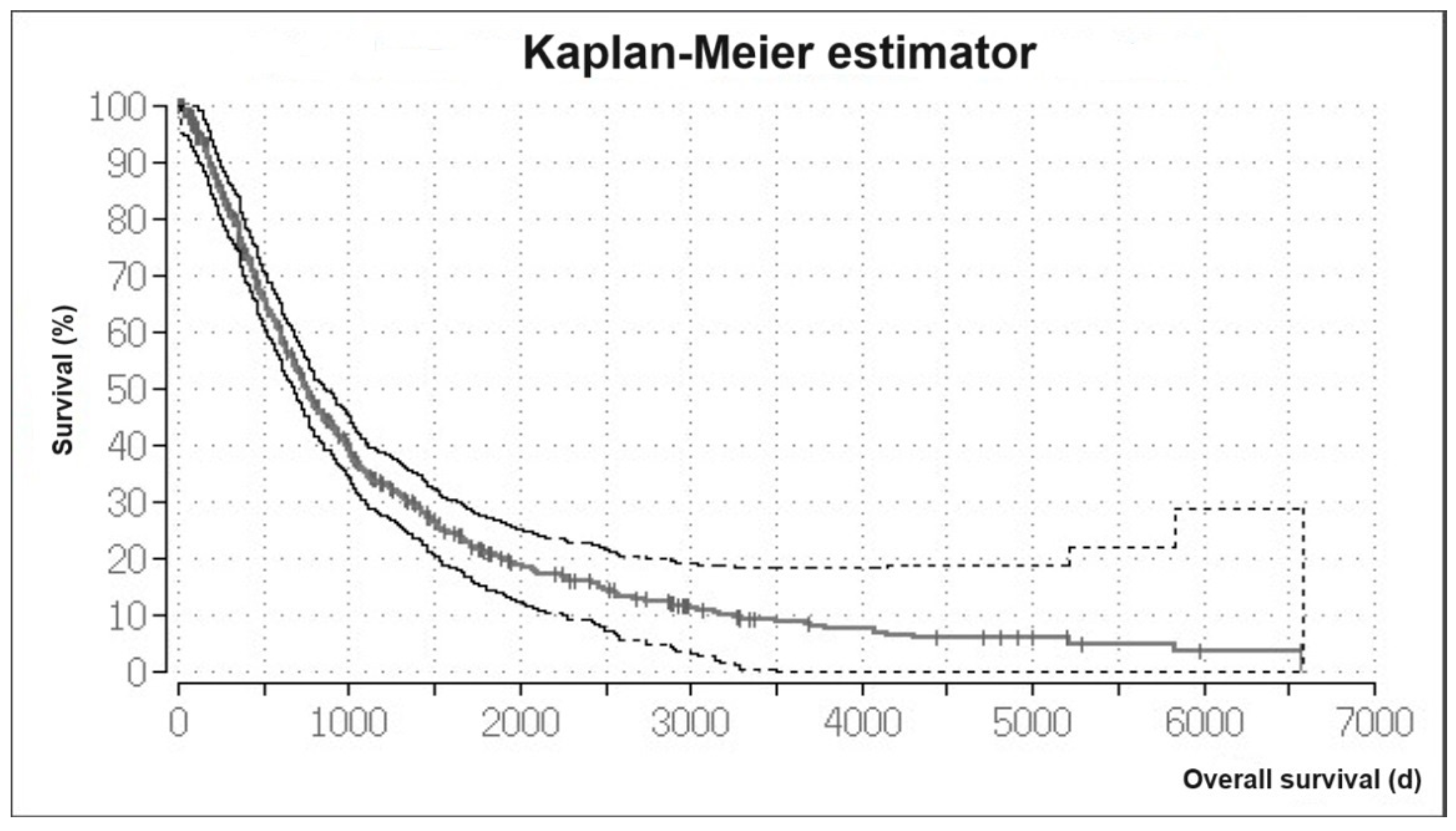
3.1. Overall Survival

The mean OS time was 1223.76 days and the median OS time was 700 days (99% CI, 632.8–767.2) for all treated patients. The Kaplan–Meier analysis of the OS showed a 1-year survival rate of 80%. The 5-year survival rate was 21%. The Kaplan–Meier curve for all patients is shown in Figure 1.
Survival analysis regarding different indications showed that the median OS time in patients with a neoadjuvant indication was 1229 days (99% CI, 983.8–1474.2), and the mean OS time was 1700 days. Patients with a curative indication had a median OS time of 787 days (99% CI, 696.3–877.7) and a mean OS time of 1048 days. Patients in a palliative setting had a median OS time of 360 days (99% CI, 328.4–391.6) and a mean OS time of 429.9 days.
Survival analysis for neoadjuvant treated patients sorted by their definitive treatment showed that patients who received LT had a median OS time of 3175 days (95% CI 1682.5–4667.5). Those treated with MWA, RFA, LITT, and surgery had similar OS times (MWA: 1308 days, 95% CI 1082.1–1533.9; RFA: 1545 days, 95% CI 416.7–2673.3; LITT: 1385 days, 95% CI 1174.3–1595.7; surgery: 1437 days, 95% CI 89.7–2784.3). Patients with unknown treatment post-TACE had a median OS time of 693 days (95% CI 540.1–845.9) (Figure 2).

3.2. Determination of Predictors for Survival

Cox regression analysis was performed to investigate whether there are predictors for death due to HCC. Examined parameters were sex, Child–Pugh class, splenomegaly, presence of PVT, and the number of treatment sessions. Those parameters were dichotomized as follows: men and women, average sized (<12 cm) versus enlarged spleen (≥12 cm), no PVT versus partial or complete PVT, Child–Pugh class A versus Child–Pugh classes B and C, and the number of treatment sessions in patients with 1–6 sessions versus ≥7 sessions. The relative hazard ratios (HR) regarding the parameters sex, splenomegaly, and number of treatment sessions were not statistically significant. The relative HR of existing PVT was 2.19 (CI 1.63–2.96 and p < 0.01), and the relative HR of Child–Pugh class B or worse was 1.44 (CI 1.11–1.86 and p < 0.001).

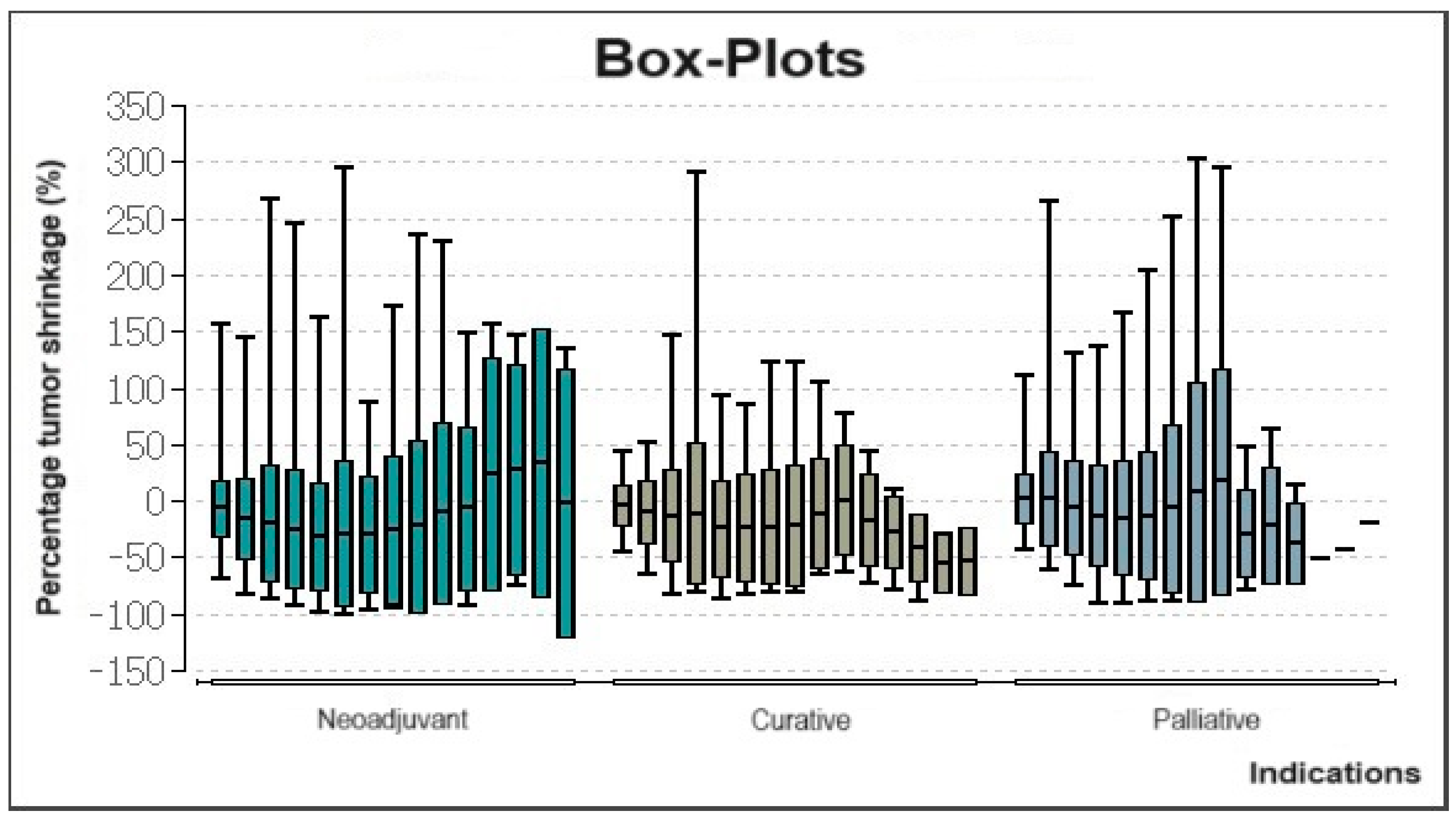
3.3. Evaluation of Tumor Volume Reduction

The percentage change in tumor volume was evaluated in a total of 358 patients with HCC treated between 2006 and 2016. Box-plots of this data show that treatment in neoadjuvant scenarios led to tumor shrinkage especially up until the 6th or 7th treatment session (Figure 3). Patients treated in a curative intention showed a similar course. All patients, regardless of indication, experienced an increase in tumor volume after at least the 9th consecutive interventional treatment.

3.4. Chemotherapeutics

The data regarding the used chemotherapeutics was available for 96% (3921/4084) of the performed TACE sessions. In the period between 1996 and 2001, the most used chemotherapeutic agent was mitomycin C alone at a rate of 81.6% followed by the combination of mitomycin C and gemcitabine at a rate of 10.8%.
In the period between 2002 and 2006, the combination of mitomycin C and gemcitabine was most frequently applied at a rate of 45.1% followed by mitomycin C alone at a rate of 39.8%.
In the period between 2007 and 2011, the most used chemotherapeutic agent was mitomycin C alone at a rate of 31.5% followed by the combination of mitomycin C, gemcitabine, and cisplatin at a rate of 27.3%.
Lastly, in the period between 2012 and 2016, the most commonly used chemotherapeutic agent was the combination of mitomycin C, gemcitabine, and cisplatin at a rate of 41.8% followed by mitomycin C alone at a rate of 27.6%. It can be concluded that over time, the application of mitomycin C alone was regressive, whereas the use of mitomycin C combined with cisplatin and gemcitabine increased. The comparison of the usage of the chemotherapeutic agents within the aforementioned periods is shown in Table 2.

4. Discussion

TACE is the most common treatment for HCC among all stages, especially in Europe and North America [27]. The most common second treatment for HCC after surgical resection in Europe is ablation using RFA/PEI followed by TACE [27]. TACE is the treatment of choice for patients with intermediate disease stage (BCLC B). These patients have a survival time of 16 months without treatment and 20 months when treated by TACE [28].
In this study, we investigated the long-term outcome of cTACE for HCC over 20 years in a large cohort and evaluated the prognostic factors for survival time. We could report a mean OS time of 1223.76 days (40 months) and a median OS time of 700 days (23 months) for all 836 patients. The associated 1- and 5-year survival rates in this study were 80% and 21%, respectively.
There are many studies that investigated TACE as a treatment for HCC in different settings. For example, Kong et al. [29] and Akarapatima et al. [30] found that TACE significantly improves the survival time of HCC patients compared to the best supportive care. In our study the best survival times were reported in patients treated neoadjuvantly with TACE, followed by other treatments including ablation and surgery. A systemic review and meta-analysis by Huo et al. compared the combination of radiotherapy and TACE with TACE alone for HCC [31]. They found that the combination therapy of both modalities was superior to TACE alone. Su et al. investigated in their retrospective study the long-term survival of HCC patients with macroscopic vascular invasion, who were treated by the combination therapy of TACE and radiotherapy compared to radiotherapy alone [32]. They found that the combination therapy was significantly superior regarding OS and progression-free survival in comparison to radiotherapy alone. Yang et al. showed in their systematic review and meta-analysis that OS and PFS are significantly improved when TACE and ablation are combined [33], supporting our findings that the best OS is accomplished when TACE is combined with adjuvant ablation or surgery. Parikh et al. [34] showed that almost half of all included patients outside the Milan criteria could be successfully downstaged. Further, they could show that multimodal locoregional therapy concept had significantly higher downstaging success rates compared to transarterial radioembolization/TACE. These studies provide further evidence of the benefits of multimodal therapy concepts. On the other side, patients undergoing interventional treatment with a palliative indication had the worst results as shown in this study. The studies by Mao et al. [35] and Wu et al. [36] who could both show that high tumor burden and poor function of liver have an effect on OS and prognosis of patients with HCC. Nevertheless, TACE should not be withheld from other indications, as the multimodal use of TACE, for instance in a palliative setting, showed promising results as well.
The conducted Cox regression analyses underlined that the stated pre-interventional patient selection could be of great value as we could prove that patients with HCC and PVT or a Child–Pugh class B or worse had a significantly higher risk of death, leading to the conclusion that pre-interventional patient selection should especially be focused on the patient’s Child–Pugh class and presence of PVT to improve therapy results. Similarly, Chang et al. [37] included in their study 108 patients with small HCCs who were treated by interventional treatments. They showed that the survival rates in patients with Child–Pugh class B were significantly worse compared to patients with Child–Pugh class A (HR = 2.68 and 95% CI = 1.52–4.73). Furthermore, Liu et al. investigated TACE for advanced HCC patients with PVT [38]. They included a total of 188 patients and found that amongst others, Child–Pugh class (HR = 2.981, 95% CI: 1.919–4.631, and p < 0.001) and PVT (HR = 2.806, 95% CI: 2.024–3.890, and p < 0.001) were independent predictors of survival.
This study has several limitations that should be considered. Firstly, the retrospective nature of the study. Secondly, the heterogeneity of the patients included in the current study and the non-standardization of the type and regime of the used chemotherapeutic agents. Thirdly, the study did not evaluate occurring complications as well as progression-free survival. Lastly, imaging studies of patients treated between 1996 and 2005 were not available and therefore, the evaluation was based on the written radiological reports.

5. Conclusions

In conclusion, this study demonstrates the possibilities and limitations of TACE as patients profit from TACE especially in the medium term, for instance, as part of neoadjuvant multimodal strategies. Though, the effectiveness is reduced in the long term. Additionally, our results underline the importance of pre-interventional patient selection as patients with PVT or Child–Pugh class B or worse tend to have poorer outcomes and survival.

Author Contributions

Conceptualization, T.J.V., J.T. and W.-O.B.; methodology, T.J.V. and H.A.; validation, T.J.V., H.A. and N.N.N.N.; formal analysis, T.J.V., H.A., M.L., T.G.-R., N.-E.A.N.-E. and N.N.N.N.; investigation, T.J.V., H.A., L.W., M.L., T.G.-R., N.-E.A.N.-E., J.T., W.-O.B. and N.N.N.N.; data curation, L.W.; writing—original draft, H.A., L.W. and N.N.N.N.; writing—review and editing, T.J.V., H.A., L.W. and N.N.N.N.; visualization, T.J.V., H.A., M.L., T.G.-R. and N.-E.A.N.-E.; supervision, T.J.V. and H.A.; project administration, T.J.V. and H.A. All authors have read and agreed to the published version of the manuscript.

Funding

This research received no external funding.

Institutional Review Board Statement

The study was conducted according to the guidelines of the Declaration of Helsinki and approved by the Institutional Review Board of University Hospital Frankfurt (approval code: 373/16 and date of approval: 10 February 2017).

Informed Consent Statement

Informed consent was obtained from all subjects involved in the study.

Data Availability Statement

The data may be requested from the corresponding author. All requests must be reasonable.

Conflicts of Interest

The authors declare no conflicts of interest.

References

  1. Balogh, J.; Victor, D., 3rd; Asham, E.H.; Burroughs, S.G.; Boktour, M.; Saharia, A.; Li, X.; Ghobrial, R.M.; Monsour, H.P., Jr. Hepatocellular carcinoma: A review. J. Hepatocell. Carcinoma 2016, 3, 41–53. [Google Scholar] [CrossRef]
  2. Sung, H.; Ferlay, J.; Siegel, R.L.; Laversanne, M.; Soerjomataram, I.; Jemal, A.; Bray, F. Global Cancer Statistics 2020: GLOBOCAN Estimates of Incidence and Mortality Worldwide for 36 Cancers in 185 Countries. CA A Cancer J. Clin. 2021, 71, 209–249. [Google Scholar] [CrossRef]
  3. Forner, A.; Llovet, J.M.; Bruix, J. Hepatocellular carcinoma. Lancet 2012, 379, 1245–1255. [Google Scholar] [CrossRef]
  4. Valery, P.C.; Laversanne, M.; Clark, P.J.; Petrick, J.L.; McGlynn, K.A.; Bray, F. Projections of primary liver cancer to 2030 in 30 countries worldwide. Hepatology 2018, 67, 600–611. [Google Scholar] [CrossRef]
  5. Philips, C.A.; Rajesh, S.; Nair, D.C.; Ahamed, R.; Abduljaleel, J.K.; Augustine, P. Hepatocellular Carcinoma in 2021: An Exhaustive Update. Cureus 2021, 13, e19274. [Google Scholar] [CrossRef]
  6. Herbst, D.A.; Reddy, K.R. Risk factors for hepatocellular carcinoma. Clin. Liver Dis. 2013, 1, 180–182. [Google Scholar] [CrossRef]
  7. Ghouri, Y.A.; Mian, I.; Rowe, J.H. Review of hepatocellular carcinoma: Epidemiology, etiology, and carcinogenesis. J. Carcinog. 2017, 16, 1. [Google Scholar] [CrossRef]
  8. Shimamura, T.; Goto, R.; Watanabe, M.; Kawamura, N.; Takada, Y. Liver Transplantation for Hepatocellular Carcinoma: How Should We Improve the Thresholds? Cancers 2022, 14, 419. [Google Scholar] [CrossRef]
  9. Lurje, I.; Czigany, Z.; Bednarsch, J.; Roderburg, C.; Isfort, P.; Neumann, U.P.; Lurje, G. Treatment Strategies for Hepatocellular Carcinoma—A Multidisciplinary Approach. Int. J. Mol. Sci. 2019, 20, 1465. [Google Scholar] [CrossRef]
  10. Reig, M.; Forner, A.; Rimola, J.; Ferrer-Fàbrega, J.; Burrel, M.; Garcia-Criado, Á.; Kelley, R.K.; Galle, P.R.; Mazzaferro, V.; Salem, R.; et al. BCLC strategy for prognosis prediction and treatment recommendation: The 2022 update. J. Hepatol. 2022, 76, 681–693. [Google Scholar] [CrossRef]
  11. Vogl, T.J.; Zangos, S.; Balzer, J.O.; Nabil, M.; Rao, P.; Eichler, K.; Bechstein, W.O.; Zeuzem, S.; Abdelkader, A. Transarterial chemoembolization (TACE) in hepatocellular carcinoma: Technique, indication and results]. RoFo Fortschritte Geb. Rontgenstrahlen Nukl. 2007, 179, 1113–1126. [Google Scholar] [CrossRef]
  12. Vogl, T.J.; Gruber-Rouh, T. HCC: Transarterial Therapies-What the Interventional Radiologist Can Offer. Dig. Dis. Sci. 2019, 64, 959–967. [Google Scholar] [CrossRef]
  13. Zhao, M.; Wang, J.P.; Li, W.; Huang, Z.L.; Zhang, F.J.; Fan, W.J.; Zhang, L.; Li, X.S.; Pan, C.C.; Wu, P.H. Comparison of safety and efficacy for transcatheter arterial chemoembolization alone and plus radiofrequency ablation in the treatment of single branch portal vein tumor thrombus of hepatocellular carcinoma and their prognosis factors. Zhonghua Yi Xue Za Zhi 2011, 91, 1167–1172. [Google Scholar]
  14. Kim, J.W.; Shin, S.S.; Kim, J.K.; Choi, S.K.; Heo, S.H.; Lim, H.S.; Hur, Y.H.; Cho, C.K.; Jeong, Y.Y.; Kang, H.K. Radiofrequency ablation combined with transcatheter arterial chemoembolization for the treatment of single hepatocellular carcinoma of 2 to 5 cm in diameter: Comparison with surgical resection. Korean J. Radiol. 2013, 14, 626–635. [Google Scholar] [CrossRef]
  15. Peng, Z.W.; Zhang, Y.J.; Chen, M.S.; Xu, L.; Liang, H.H.; Lin, X.J.; Guo, R.P.; Zhang, Y.Q.; Lau, W.Y. Radiofrequency ablation with or without transcatheter arterial chemoembolization in the treatment of hepatocellular carcinoma: A prospective randomized trial. J. Clin. Oncol. Off. J. Am. Soc. Clin. Oncol. 2013, 31, 426–432. [Google Scholar] [CrossRef]
  16. Li, S.; Zhang, L.; Huang, Z.M.; Wu, P.H. Transcatheter arterial chemoembolization combined with CT-guided percutaneous thermal ablation versus hepatectomy in the treatment of hepatocellular carcinoma. Chin. J. Cancer 2015, 34, 254–263. [Google Scholar] [CrossRef]
  17. Zhang, R.; Shen, L.; Zhao, L.; Guan, Z.; Chen, Q.; Li, W. Combined transarterial chemoembolization and microwave ablation versus transarterial chemoembolization in BCLC stage B hepatocellular carcinoma. Diagn. Interv. Radiol. 2018, 24, 219–224. [Google Scholar] [CrossRef]
  18. Zhang, Y.J.; Chen, M.S.; Chen, Y.; Lau, W.Y.; Peng, Z. Long-term Outcomes of Transcatheter Arterial Chemoembolization Combined with Radiofrequency Ablation as an Initial Treatment for Early-Stage Hepatocellular Carcinoma. JAMA Netw. Open 2021, 4, e2126992. [Google Scholar] [CrossRef]
  19. Hendriks, P.; Sudiono, D.R.; Schaapman, J.J.; Coenraad, M.J.; Tushuizen, M.E.; Takkenberg, R.B.; Oosterveer TT, M.; de Geus-Oei, L.F.; van Delden, O.M.; Burgmans, M.C. Thermal ablation combined with transarterial chemoembolization for hepatocellular carcinoma: What is the right treatment sequence? Eur. J. Radiol. 2021, 144, 110006. [Google Scholar] [CrossRef]
  20. Sun, Y.; Zhang, H.; Long, J.; Zhang, Y.; Zheng, J.; Yuan, C. Percutaneous thermal ablation combined with transcatheter arterial chemoembolization for hepatitis C virus-related hepatocellular carcinoma: Efficacy and survival. Front. Oncol. 2022, 12, 978614. [Google Scholar] [CrossRef]
  21. Bryce, K.; Tsochatzis, E.A. Downstaging for hepatocellular cancer: Harm or benefit? Transl. Gastroenterol. Hepatol. 2017, 2, 106. [Google Scholar] [CrossRef]
  22. Renzulli, M.; Peta, G.; Vasuri, F.; Marasco, G.; Caretti, D.; Bartalena, L.; Spinelli, D.; Giampalma, E.; D’Errico, A.; Golfieri, R. Standardization of conventional chemoembolization for hepatocellular carcinoma. Ann. Hepatol. 2021, 22, 100278. [Google Scholar] [CrossRef]
  23. Ikeda, M.; Arai, Y.; Inaba, Y.; Tanaka, T.; Sugawara, S.; Kodama, Y.; Aramaki, T.; Anai, H.; Morita, S.; Tsukahara, Y.; et al. Conventional or Drug-Eluting Beads? Randomized Controlled Study of Chemoembolization for Hepatocellular Carcinoma: JIVROSG-1302. Liver Cancer 2022, 11, 440–450. [Google Scholar] [CrossRef]
  24. Lencioni, R. Loco-regional treatment of hepatocellular carcinoma. Hepatology 2010, 52, 762–773. [Google Scholar] [CrossRef]
  25. Song, J.E.; Kim, D.Y. Conventional vs drug-eluting beads transarterial chemoembolization for hepatocellular carcinoma. World J. Hepatol. 2017, 9, 808–814. [Google Scholar] [CrossRef]
  26. Seldinger, S.I. Catheter replacement of the needle in percutaneous arteriography; a new technique. Acta Radiol. 1953, 39, 368–376. [Google Scholar] [CrossRef]
  27. Park, J.W.; Chen, M.; Colombo, M.; Roberts, L.R.; Schwartz, M.; Chen, P.J.; Kudo, M.; Johnson, P.; Wagner, S.; Orsini, L.S.; et al. Global patterns of hepatocellular carcinoma management from diagnosis to death: The BRIDGE Study. Liver Int. Off. J. Int. Assoc. Study Liver 2015, 35, 2155–2166. [Google Scholar] [CrossRef]
  28. Verslype, C.; Rosmorduc, O.; Rougier, P.; ESMO Guidelines Working Group. Hepatocellular carcinoma: ESMO-ESDO Clinical Practice Guidelines for diagnosis, treatment and follow-up. Ann. Oncol. Off. J. Eur. Soc. Med. Oncol. 2012, 23 (Suppl. 7), vii41–vii48. [Google Scholar] [CrossRef]
  29. Kong, J.Y.; Li, S.M.; Fan, H.Y.; Zhang, L.; Zhao, H.J.; Li, S.M. Transarterial chemoembolization extends long-term survival in patients with unresectable hepatocellular carcinoma. Medicine 2018, 97, e11872. [Google Scholar] [CrossRef]
  30. Akarapatima, K.; Chang, A.; Prateepchaiboon, T.; Pungpipattrakul, N.; Songjamrat, A.; Pakdeejit, S.; Rattanasupar, A. Comparison of Overall Survival between Transarterial Chemoembolization and Best Supportive Care in Intermediate- Stage Hepatocellular Carcinoma. Asian Pac. J. Cancer Prev. 2022, 23, 3173–3178. [Google Scholar] [CrossRef]
  31. Huo, Y.R.; Eslick, G.D. Transcatheter Arterial Chemoembolization Plus Radiotherapy Compared with Chemoembolization Alone for Hepatocellular Carcinoma: A Systematic Review and Meta-analysis. JAMA Oncol. 2015, 1, 756–765. [Google Scholar] [CrossRef]
  32. Su, T.S.; Li, L.Q.; Meng, W.W.; Wang, Y.D.; Chen, Y.T.; Li, J.X.; Du, Y.Q.; Qu, S.; Zhao, C.; Huang, D.J.; et al. Long-Term Survival Analysis of Transarterial Chemoembolization Plus Radiotherapy vs. Radiotherapy for Hepatocellular Carcinoma with Macroscopic Vascular Invasion. Front Oncol. 2020, 10, 1205. [Google Scholar] [CrossRef]
  33. Yang, Y.; Yu, H.; Qi, L.; Liu, C.; Feng, Y.; Qi, J.; Li, J.; Zhu, Q. Combined radiofrequency ablation or microwave ablation with transarterial chemoembolization can increase efficiency in intermediate-stage hepatocellular carcinoma without more complication: A systematic review and meta-analysis. Int. J. Hyperth. 2022, 39, 455–465. [Google Scholar] [CrossRef]
  34. Parikh, N.D.; Waljee, A.K.; Singal, A.G. Downstaging hepatocellular carcinoma: A systematic review and pooled analysis. Liver Transpl. 2015, 21, 1142–1152. [Google Scholar] [CrossRef]
  35. Mao, Y.M.; Luo, Z.Y.; Li, B.; Hu, T.Y. Prospective study on the survival of HCC patients treated with transcatheter arterial lipiodol chemoembolization. Asian Pac. J. Cancer Prev. 2012, 13, 1039–1042. [Google Scholar] [CrossRef]
  36. Wu, K.T.; Wang, C.C.; Lu, L.G.; Zhang, W.D.; Zhang, F.J.; Shi, F.; Li, C.X. Hepatocellular carcinoma: Clinical study of long-term survival and choice of treatment modalities. World J. Gastroenterol. 2013, 19, 3649–3657. [Google Scholar] [CrossRef]
  37. Chang, H.C.; Lin, Y.M.; Yen, A.M.; Chen, S.L.; Wu, W.Y.; Chiu, S.Y.; Fann, J.C.; Lin, Y.S.; Chen, H.H.; Liao, C.S. Predictors of long-term survival in hepatocellular carcinomas: A longitudinal follow-up of 108 patients with small tumors. Anticancer Res. 2013, 33, 5171–5178. [Google Scholar]
  38. Liu, L.; Zhang, C.; Zhao, Y.; Qi, X.; Chen, H.; Bai, W.; He, C.; Guo, W.; Yin, Z.; Fan, D.; et al. Transarterial chemoembolization for the treatment of advanced hepatocellular carcinoma with portal vein tumor thrombosis: Prognostic factors in a single-center study of 188 patients. BioMed Res. Int. 2014, 2014, 194278. [Google Scholar] [CrossRef]
Figure 1. Kaplan–Meier estimator. Overall survival of HCC patients with interventional treatment.
Cancers 16 01498 g001
Figure 2. Survival in patients with neoadjuvant indication sorted by their subsequent therapy after TACE.
Cancers 16 01498 g002
Figure 3. Box-plots showing percentage changes in tumor volume for all patients with HCC treated during the period of 2006 until 2016.
Cancers 16 01498 g003
Table 1. Summarization of relevant characteristics of patients and treatments.
Characteristic
Total number of patients836
Mean age in years 74.18
Sex
Male (%)653 (78.1%)
Female (%)183 (21.9%)
Child–Pugh class (%)
A495 (59.21%)
B188 (22.49%)
C122 (14.6%)
Unknown31 (3.7%)
Portal vein thrombosis
Complete86 (10.3%)
Partial57 (6.8%)
None629 (75.2%)
Unknown64 (7.7%)
Mean spleen size12.8 cm
Splenomegaly (size ≥ 12.0 cm)365 (43.66%)
Total number of cTACE4084
Mean number of TACE sessions/ patient4.89
Patients with ≤3 TACE performed460 (55%)
Patients with 4–6 TACE performed243 (29.1%)
Patients with >6 TACE performed133 (15.9%)
Indications
Neoadjuvant394 (47.1%)
Curative or Palliative431 (51.6%)
Unknown11 (1.3%)
Interventions/treatments after neoadjuvant TACE
Microwave ablation 112 (28.43%)
Radiofrequency ablation 34 (8.63%)
Laser-induced thermotherapy 78 (19.8%)
Selective internal radiation therapy (SIRT)2 (0.5%)
Surgical resection39 (9.9%)
Liver transplantation 49 (12.44%)
Unknown80 (20.3%)
Table 2. Listing of the most frequently used chemotherapeutic agents in chronological order.
Chemotherapeutic
Regime
1996–20012002–20062007–20112012–2016
Mitomycin C
Gemcitabine
Cisplatin
6 (1%)44 (3.4%)342 (27.3%)308 (41.8%)
Mitomycin C
Cisplatin
3 (0.5%)19 (1.4%)245 (19.6%)183 (24.9%)
Mitomycin C506 (81.6%)522 (39.8%)395 (31.5%)203 (27.6%)
Mitomycin C
Gemcitabine
67 (10.8%)592 (45.1%)156 (12.4%)7 (1%)
Other combinations/ chemotherapeutics38 (6.1%)135 (10.3%)115 (9.2)35 (4.7%)
∑ (%)620 (100%)1312 (100%)1253 (100%)736 (100%)
Disclaimer/Publisher’s Note: The statements, opinions and data contained in all publications are solely those of the individual author(s) and contributor(s) and not of MDPI and/or the editor(s). MDPI and/or the editor(s) disclaim responsibility for any injury to people or property resulting from any ideas, methods, instructions or products referred to in the content.

