Error in Figure
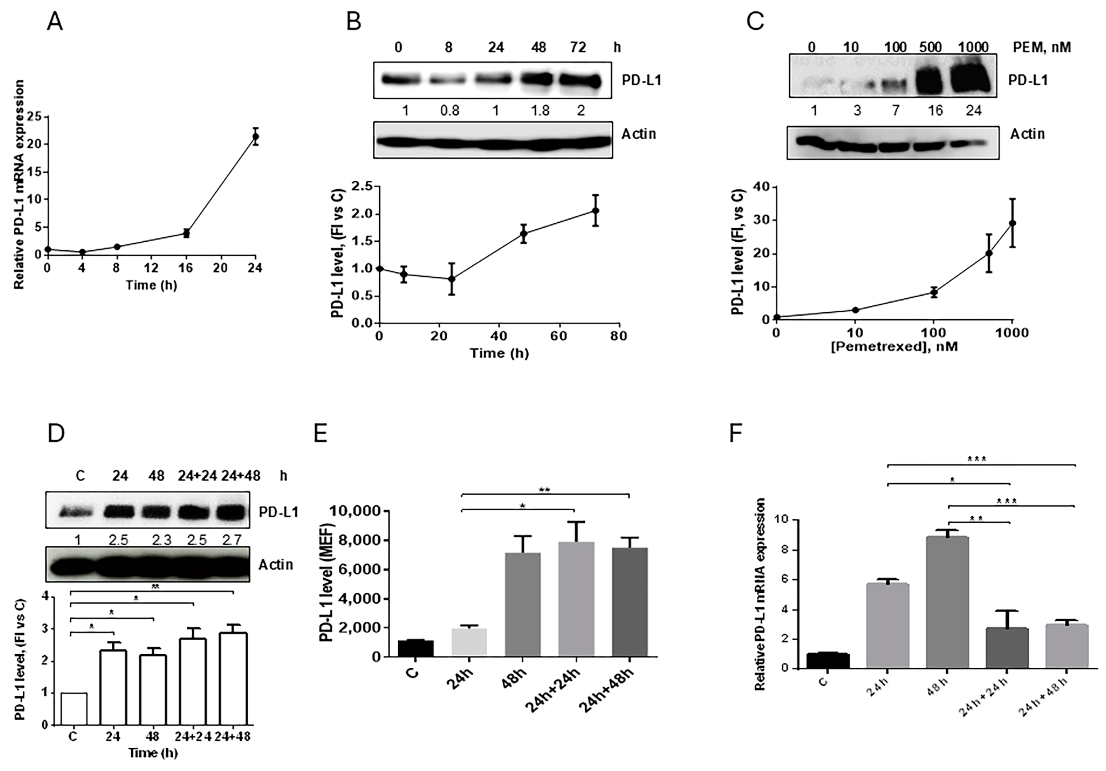
In the original publication [1], there was a mistake in Figure 2C,D as published. An incorrect image of Actin was inserted as a loading control. The corrected Figure 2 appears below. Additionally, the associated Supplementary Figure S7 has been updated. The authors apologize for any inconvenience caused and state that the scientific conclusions are unaffected. This correction was approved by the Academic Editor. The original publication has also been updated.
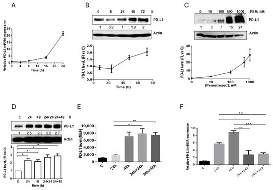
Figure 2.
Effect of pemetrexed on PD-L1 expression in A549 cell line. (A) A549 cells were treated with 100 nM pemetrexed for the indicated period of time and PD-L1 mRNA level, evaluated by RT-PCR, was reported. (B) Time-dependent modulation (100 nM pemetrexed) and (C) dose-dependent modulation (72 h) of PD-L1 protein expression in A549 cells were evaluated by western blotting. A549 cells were continuously exposed to 500 nM pemetrexed for the indicated period of time or treated for 24 h and, after drug removal, the cells were incubated with fresh medium for 24 h or 48 h. At the indicated times, total PD-L1 protein, membrane PD-L1 protein, and PD-L1 mRNA were quantified by western blotting (D), flow cytometry (E), and RT-PCR (F), respectively. * p < 0.05; ** p < 0.01; *** p < 0.001. Data in (A), (E), and (F) are mean values ± SD of three independent experiments. Results in (B–D) are representative of three independent experiments.
Reference
- Cavazzoni, A.; Digiacomo, G.; Alfieri, R.; La Monica, S.; Fumarola, C.; Galetti, M.; Bonelli, M.; Cretella, D.; Barili, V.; Zecca, A.; et al. Pemetrexed Enhances Membrane PD-L1 Expression and Potentiates T Cell-Mediated Cytotoxicity by Anti-PD-L1 Antibody Therapy in Non-Small-Cell Lung Cancer. Cancers 2020, 12, 666. [Google Scholar] [CrossRef] [PubMed]
Disclaimer/Publisher’s Note: The statements, opinions and data contained in all publications are solely those of the individual author(s) and contributor(s) and not of MDPI and/or the editor(s). MDPI and/or the editor(s) disclaim responsibility for any injury to people or property resulting from any ideas, methods, instructions or products referred to in the content. |
© 2025 by the authors. Licensee MDPI, Basel, Switzerland. This article is an open access article distributed under the terms and conditions of the Creative Commons Attribution (CC BY) license (https://creativecommons.org/licenses/by/4.0/).