Article

Effect of Calcination Temperature of SiO2/TiO2 Photocatalysts on UV-VIS and VIS Removal Efficiency of Color Contaminants

by
Aleksandra Babyszko
,
Agnieszka Wanag
*,
Ewelina Kusiak-Nejman
and
Antoni Waldemar Morawski
*
Faculty of Chemical Technology and Engineering, Department of Inorganic Chemical Technology and Environment Engineering, West Pomeranian University of Technology in Szczecin, Pułaskiego 10, 70-322 Szczecin, Poland
*
Authors to whom correspondence should be addressed.
Catalysts 2023, 13(1), 186; https://doi.org/10.3390/catal13010186
Submission received: 28 November 2022 / Revised: 5 January 2023 / Accepted: 10 January 2023 / Published: 13 January 2023

Abstract

:
This paper presents the effect of fumed silica modification and calcination temperature on the physicochemical properties of photocatalysts and their activity under the UV-VIS and VIS light range. The materials were obtained by hydrolysis of titanium tetraisopropoxide (TTIP) combined with a calcination step. The obtained nanomaterials were characterized using analytical methods such as X-ray diffraction XRD, FT-IR/DRS infrared spectroscopy, UV-Vis/DRS spectroscopy and SEM scanning electron microscopy. BET specific surface area and zeta potential were also measured. It was observed that SiO2 modification inhibited the transformation phase of anatase to rutile and the increase in crystallite size during calcination. The calcination process contributed to a change in the surface character of photocatalysts under study from positively to negatively charged. The photocatalytic activity of samples was identified by determining the methylene blue decomposition under UV-VIS and VIS light. Experimental results showed that the addition of SiO2 and the calcination process increased the photoactivity. The obtained materials showed higher activity compared to the reference samples. It was found that the degree of dye removal increased along with increased calcination temperature. The highest activity was observed for photocatalyst SiO2(11.1%)/TiO2_600.

1. Introduction

Colored sewage belongs to a group of industrial effluents, which are classified as very troublesome substances from the treatment process’s point of view. Due to rapid changes in production technology and the variety of dyes used in various industries, it is challenging to develop a simple, universal, effective and economical method of removing colored substances from wastewater. Colored contaminants contained in the effluent are a major environmental problem since they cause a reduction in the permeability of sunlight, which interferes with the proper functioning of aquatic ecosystems. In the treatment processes of colored sewage, the photocatalytic oxidation process using titanium dioxide (TiO2) is increasingly applied to improve the decomposition process of contaminants. Sultana et al. [1] prepared TiO2/chitosan nanohybrids to remove azo dye Remazol Orange 3R (RO) by a precipitation technique. They concluded that the high photocatalytic activity was due to the improved adsorption capacity of materials obtained. They also suggested the influence of electrostatic interactions on RO adsorption. Shathy et al. [2] investigated the effect of several parameters, such as initial dye concentration, amount of photocatalyst, pH and exposure time on the activity of B-ZnO/TiO2 nanocomposites. The activity was determined based on the photooxidation of methylene blue. The increase in activity was attributed to a slowdown in electron-hole pair recombination and a larger specific surface area. In their study, Mondol et al. [3] decomposed Reactive Red-35 using AC/TiO2 nanohybrids obtained by a hydrothermal method combined with a calcination step under sunlight. They found that the improvement in photocatalytic capacity was related to the adsorption properties of the materials tested. In addition, they showed that the photodegradation of a dye was mainly controlled by OH and O2 radicals.
The photocatalytic properties of TiO2 with the possibility of use in the removal of organic compounds from both water and air have been studied for years [4,5,6,7,8]. Unfortunately, titanium dioxide absorbs almost exclusively ultraviolet radiation, representing ca. 3–5% of the sunlight. Therefore, intensive research is being carried out worldwide to improve and increase the efficiency of photocatalytic processes. New requirements placed on titanium photocatalysts are mainly the ability to absorb light in a wider range of radiation (especially VIS), reducing the recombination level of charge carriers by controlling the size and shape of particles, as well as the development of the specific surface of photocatalyst. In order to increase the efficiency and effectiveness of the photocatalytic process, various modifications of the photocatalyst structure or surface are carried out [9,10,11,12,13,14].
The modification of TiO2 with SiO2 is one of the greatest interests. It is proved that silica doping enhances the photocatalytic activity, thermal stability, and mechanical strength of TiO2 photocatalyst [15,16]. Furthemore, among non-metallic dopants, silica is one of the compounds that enhance the photocatalytic activity of TiO2 under both visible and ultraviolet light. It has been demonstrated that the addition of SiO2 increases the surface area of the composite, increasing the adsorption rate of contaminants [17]. In turn, the increased adsorption of contaminants on the silica surface improves the photocatalytic activity of mixed SiO2-TiO2 oxides compared to unmodified TiO2 [18,19]. In addition, silica has hydrophilic properties so that its surface strongly interacts with water molecules, leading to the strong adsorption of water on its surface [20,21,22]. This phenomenon is beneficial for the photocatalytic properties of TiO2.
Iwamoto et al. [23] found that silica-modified anatase TiO2 showed excellent thermal stability. Moreover, they demonstrated that SiO2 atoms incorporate into vacancy sites. The silica-doped TiO2 prepared by Cheng et al. [24] exhibited oxygen vacancy formation and high photocatalytic activity due to the inhibition of phase transition from anatase to rutile form. Nilchi et al. [25] wrote about the effect of SiO2 doping on the photocatalytic properties of TiO2 and suggested that photoactivity depends on the size of BET specific surface area, crystallinity, and crystallite size. Zhang et al. [16], based on the decomposition of phenol in the presence of SiO2-TiO2 composites, found that SiO2-mediated modification delayed the anatase-to-rutile phase transformation and fixed the ordered mesoporous structure of TiO2 at high calcination temperatures. In contrast, Wang et al. [26] showed that the presence of SiO2 inhibited the growth of anatase TiO2. In addition, they found that the higher the SiO2 content in the sample, the higher the degree of adsorption and degradation of color impurities.
The novelty of the presented research was the proposal and development of a new method to obtain TiO2 photocatalysts modified with fumed silica active not only in the UV-VIS range but also in visible light. Our previous studies have shown that, from an economic point of view, the best photocatalytic properties are shown by a sample with 11.1 wt% SiO2 [27]. Considering the above, a photocatalyst weighting of 11.1% by weight was selected for further studies to determine the effect of annealing temperature on the photocatalyst’s photoactivity. To our knowledge, this is the first paper to show that fumed silica modification positively affects visible light activity. From the information available on the manufacturer’s website [28], it appears that the silica used has a well-developed specific surface area (>200 m2/g). The nanomaterials were prepared by the sol-gel method and then calcined at different temperature ranges in an inert gas atmosphere. The the heating phase was intended to increase the crystallization degree of titanium dioxide. The photocatalytic activity was determined based on the distribution of methylene blue under ultraviolet and visible light irradiation. An element of novelty was also the determination of the effect of calcination temperature on the enhancement of the activity of the obtained photocatalysts in the visible range.

2. Results and Discussion

2.1. Characterization of the Photocatalysts

X-ray diffractograms of the investigated photocatalysts are presented in Figure 1A,B. The phase compositions and crystallite sizes for these samples are summarized in Table 1. However, it should be mentioned that the presented results consider only the crystalline phase. From the obtained results, it was found that almost all samples showed reflections characteristic of the anatase phase: (101), (004), (200), (105), (211), (204), (116) and (215) located at 25.3, 38.2, 48.4, 54.6, 55.4, 63.5, 69.5 and 75.9°, respectively (JCPDS 04-002-8296 PDF4+ card). Reflections (111) and (121) corresponding to the brookite phase were recorded at 25.3 and 30.3°, respectively (JCPDS 04-007-0758 PDF4+ card). The reflection (111) characteristic of the brookite phase recorded at 2θ = 25.6° coincided with that of anatase. For this reason, the average size of brookite crystallites was calculated based on the reflection located at 2θ = 30.3°. The presence of brookite in the materials was related to the use of hydrochloric acid as a catalyst for the hydrolysis reaction of titanium(IV) isopropoxide. Reflections characteristic of the brookite phase were recorded for starting TiO2, starting TiO2_400, SiO2(11.1%)/TiO2, SiO2(11.1%)/TiO2_400 and SiO2(11.1%)/TiO2_600 photocatalysts. No reflections from brookite were recorded for starting TiO2_600 and for samples annealed at 800 °C. For the reference photocatalysts, calcination at 600 °C resulted in the transformation of brookite into rutile and a sharp increase in the size of anatase crystallites (up to 57 nm). Hu et al. [29] proved that the phase transition to rutile is faster for materials containing, in addition to anatase, some amounts of brookite. This is because the brookite-to-rutile phase transition occurs more easily than anatase-rutile. For the reference samples calcined above 600 °C, reflections characteristic of the rutile phase (110), (101), (200), (111), (210), (211), (220), (002), (310), (301) (112) were observed, which are located at 27.4, 35.9, 39.5, 41.2, 44.2, 54.4, 56.6, 62.6, 64.5, 68.9 and 69.9° (JCPDS 01-076-0318 PDF4+ card). It is worth mentioning that the phase transition of anatase to rutile is irreversible and occurs in the temperature range of 600−700 °C [30]. In our case, starting TiO2 was completely converted to rutile at 800 °C. While after calcination at 800 °C, the SiO2(11.1%)/TiO2_800 sample contained 100% anatase. Modification with silica as a silicon source contributed to the effective inhibition of the phase transformation of anatase to rutile [31,32]. As can be seen from the data presented in Table 1, the temperature of the photocatalysts’ calcination process also had a significant effect on the growth of the average crystallite size. Anatase crystallites increased from 5 nm to 57 nm for heat-treated starting TiO2 and silica-modified TiO2 from 5 nm to 29 nm. The size of brookite crystallites ranged from 2 to 9 for heat-treated starting TiO2 and 5–21 nm for silica-modified TiO2. The size of rutile crystallites was in the range of 132−228 nm for heat-treated starting TiO2. This indicated that the crystallinity of studied samples increased after calcination. It was also observed that the crystallite size of all polymorphic varieties of TiO2 was smaller for silica-modified photocatalysts than for materials without SiO2 addition. For example, the size of anatase crystallites for the starting TiO2_600 sample was 57 nm, while for SiO2(11.1%)/TiO2_600, it was only 21 nm. This is since the silica effectively prevents the growth of TiO2 crystallites during the calcination process [32,33].
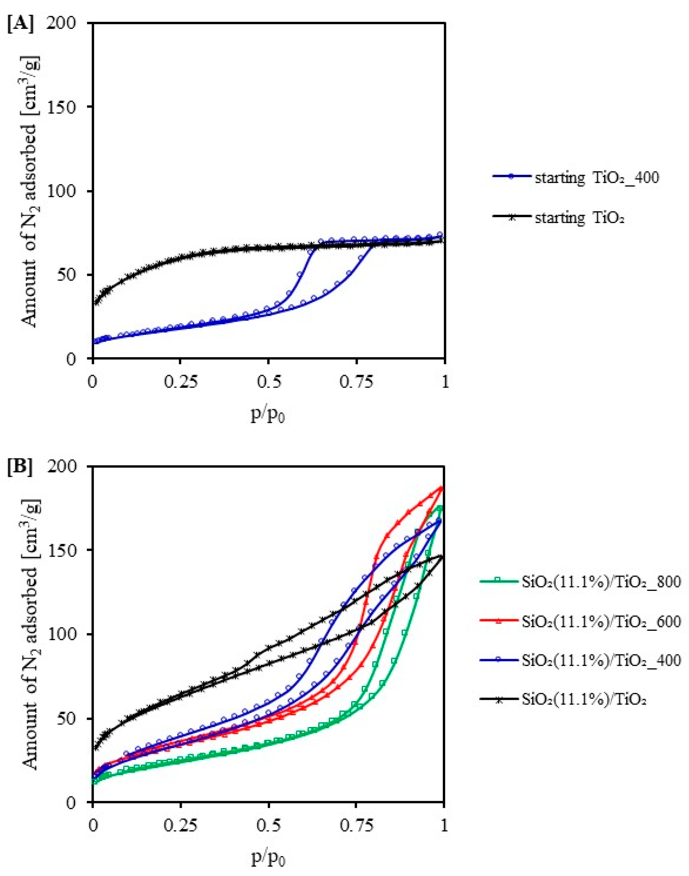
Figure 2 shows the nitrogen adsorption-desorption isotherms of the studied photocatalysts. According to the IUPAC classification, most of the presented isotherms (SiO2(11.1%)/TiO2, SiO2(11.1%)/TiO2_400–800) could be classified as type IV, characteristic for mesoporous materials [34]. Only the nitrogen adsorption-desorption isotherm for the control material (starting TiO2) could be described as a type I (Langmuir) isotherm. The nanomaterials modified with fumed silica presented a type H3 hysteresis loop. The isotherm of the starting TiO2_400 sample showed an asymmetric and triangular hysteresis loop of type H2. This loop is characteristic of materials having bottle pores or networked pore systems [35]. In the case of the starting TiO2, the hysteresis loop did not occur. It is worth noting that for photocatalysts modified with fumed silica (see Figure 2B), the size of the hysteresis loop decreased along with the increase in calcination temperature.
The values of determined specific surface and pore size distribution for the obtained photocatalysts are presented in Table 2. According to the data presented in Table 2, the control sample (starting TiO2) was a microporous material. Except for this sample, all the tested photocatalysts were mesoporous materials with a small percentage of micropores. Modification with fumed silica increased the specific surface area of BET and total pore volume from 193 m2/g, 0.109 cm3/g for starting TiO2 to 208 m2/g, 0.265 cm3/g for SiO2(11.1%)/TiO2 (SBET of applied fumed silica > 200 m2/g). A literature review shows that the use of silica to modify TiO2 contributes to an increase in the specific surface area [36]. After heat treatment, the specific surface of photocatalysts decreased from 208 m2/g for SiO2(11.1%)/TiO2 to 81 m2/g for the same sample calcined at 800 °C. The increase in calcination temperature caused the sintering and agglomeration of TiO2 particles, resulting in a decrease in the specific surface of materials [37]. It is also worth noting that the modification with SiO2 caused the inhibition of the decrease in the specific surface after calcination. The size of specific surface for starting TiO2_400 sample was 54 m2/g and for SiO2(11.1%)/TiO2_400 sample was 121 m2/g [36].
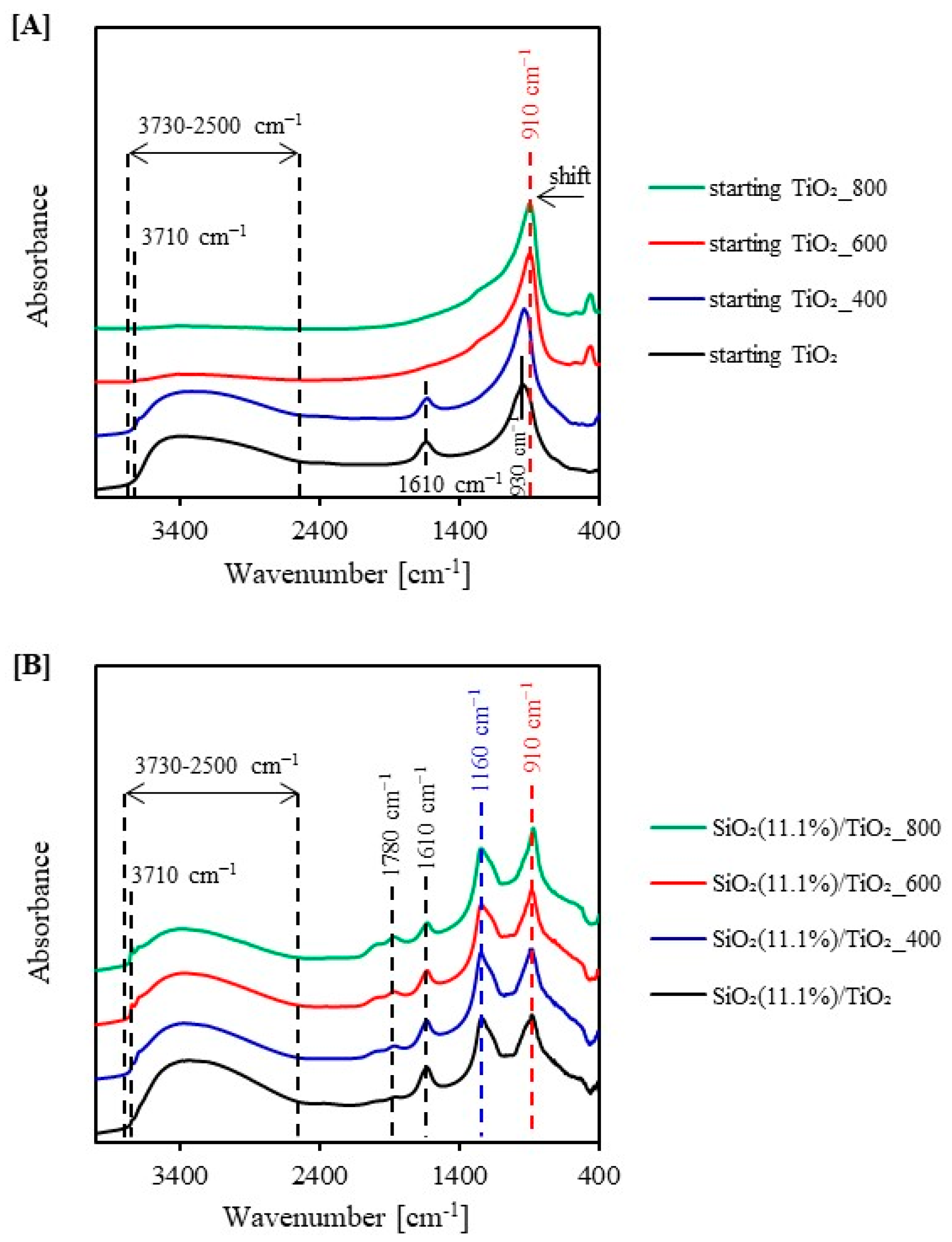
To investigate the surface character of obtained photocatalysts, FT-IR/DRS analysis was conducted. Figure 3A,B shows the FT-IR/DR spectra determined for starting TiO2, reference samples and SiO2/TiO2 photocatalysts. All the spectra presented showed an extensive band in the wavenumber range 3730–2500 cm−1, a narrow band at 3710 cm−1 and a peak at 930 cm−1. The broad band at 3730–2500 cm−1 is from adsorbed water and hydroxyl groups [38]. It was observed that increasing calcination temperature causes a decrease in the intensity of this band, especially in the case of reference samples. This was attributed to the removal of weakly adsorbed water and the disappearance of –OH groups. However, for the samples after silica modification and annealing at 600 and 800 °C, the mentioned peaks are visible due to the presence of Si–OH groups and adsorbed water [39]. The band at 3710 cm−1 corresponds to the OH groups surface bound to titanium [40]. The narrow band at a wave number value of 1610 cm−1, originates from the bending vibrations of molecular water [41]. An intense band at about 930 cm−1 is attributed to O–Ti–O stretching modes. After the modification process, this band is slightly shifted, indicating the interaction between titanium and silicon [42]. As a result of the modification, the appearance of a new band at 1160 cm−1 derived from silicon and corresponding to asymmetric stretching vibrations of the Si–O–Si bond was observed [43]. The band at 1780 cm−1 is characteristic of C=O stretching vibrations.
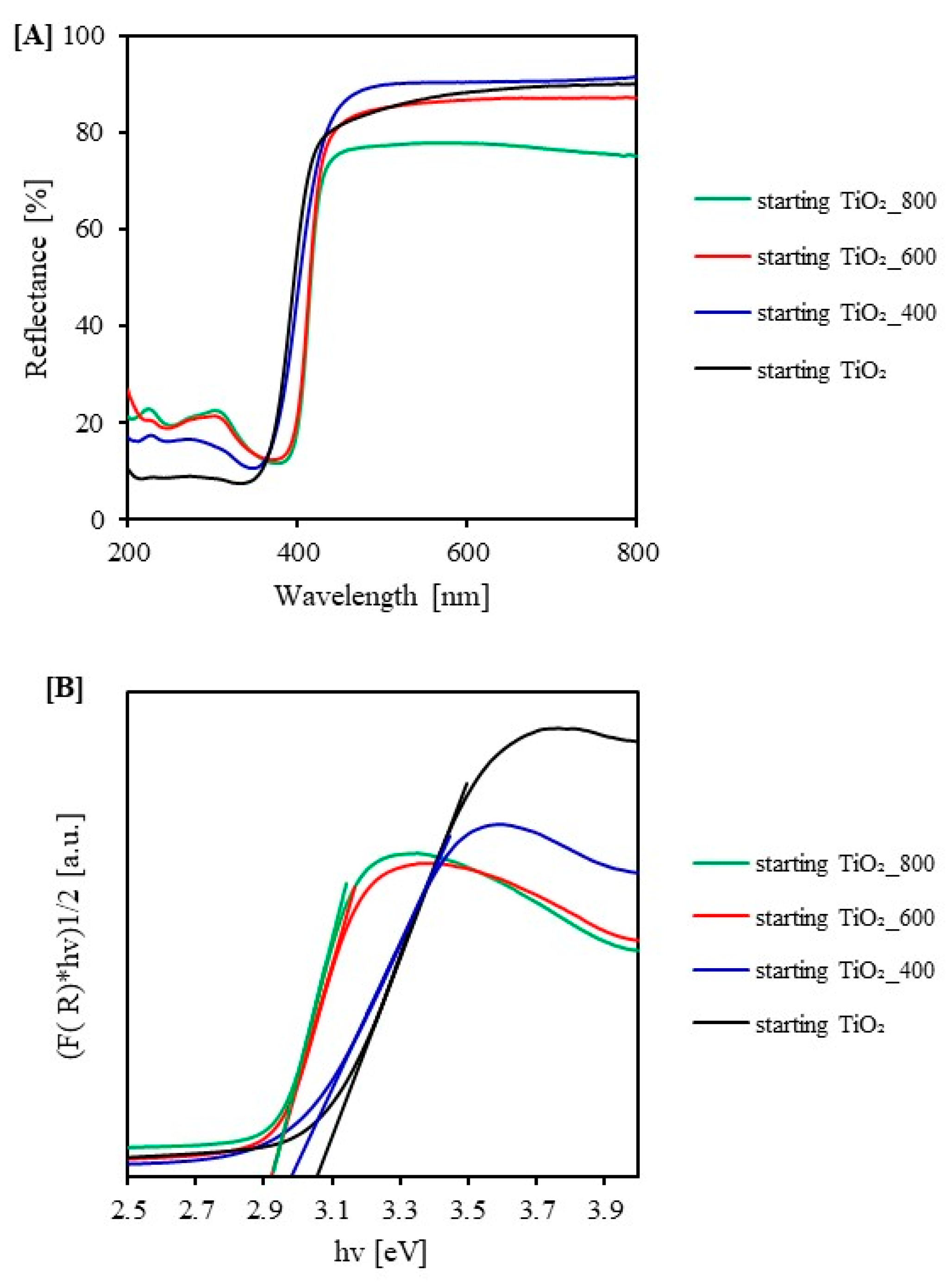
Figure 4 and Figure 5 show UV-Vis/DR absorption spectra determined for starting TiO2, reference samples calcined under argon atmosphere and SiO2/TiO2 photocatalysts. All the materials obtained showed absorption of radiation in the UV region. Only the starting TiO2_800 sample showed absorption in the visible light region due to the photocatalyst’s color change from white to dark grey. For the reference samples, calcination in an argon atmosphere leads to a slight shift of the absorption edge towards the visible radiation. Small changes in the band gap energy were observed for samples calcined at 600 and 800 °C. The band gap energy for these photocatalysts was 2.92 eV. The decrease in the band gap energy was also attributed to the presence of rutile in the TiO2 phase composition. It is well known that rutile has a lower band gap energy value than anatase. The value of band gap energy for rutile is 3.02 eV [44]. For the reference samples heated in an argon atmosphere, an absorption peak was observed at 318 nm, which is related to the transition of an electron from the valence band of O 2p to the conduction band of Ti 3d [25]. In the case of silica-modified photocatalysts, no absorption peak was observed at 318 nm. This is due to the the presence of silica effectively inhibits the phase transformation of anatase to rutile [43]. After the modification process with fumed silica, all the tested photocatalysts showed a shift of the absorption edge towards lower wavelengths (blue shift). An increase in the value of band gap energy was noted for these samples. The increase in band gap energy was attributed to the quantum effect [36]. An increase in the band gap energy value contributes to a decrease in the valence band and an increase in the edge of the conduction band, thus slowing down the recombination process. In turn, slowing down the recombination time of electron-hole pairs translates into higher activity.
The zeta potential values for the modified photocatalysts and the reference samples, are summarized in Table 3. Based on the results of zeta potential measurements, it was found that the application of the calcination process changed the nature of the surface of the studied nanomaterials from positively to negatively charged. As the calcination temperature increased (above 400 °C), the zeta potential value decreased. The surface of photocatalysts calcined at 600 and 800 °C was negatively charged. The starting TiO2_800 (−25.8 mV) and SiO2(11.1%)/TiO2_800 (−29.5 mV) samples showed the lowest zeta potential. Suttiponparnit et al. [45] found that the larger the crystallite size, the lower the zeta potential. Moreover, they showed that the surface of TiO2 changes to the negative character when the size of crystallites is larger than 20 nm. In our case, the size of crystallites also greatly influenced the change of surface character. As the calcination temperature increased, the size of crystallites increased (see Table 1), which caused the electrokinetic potential to decrease. The zeta potential of the SiO2/TiO2 samples changed from positive to negative when the crystallite size exceeded 20 nm.
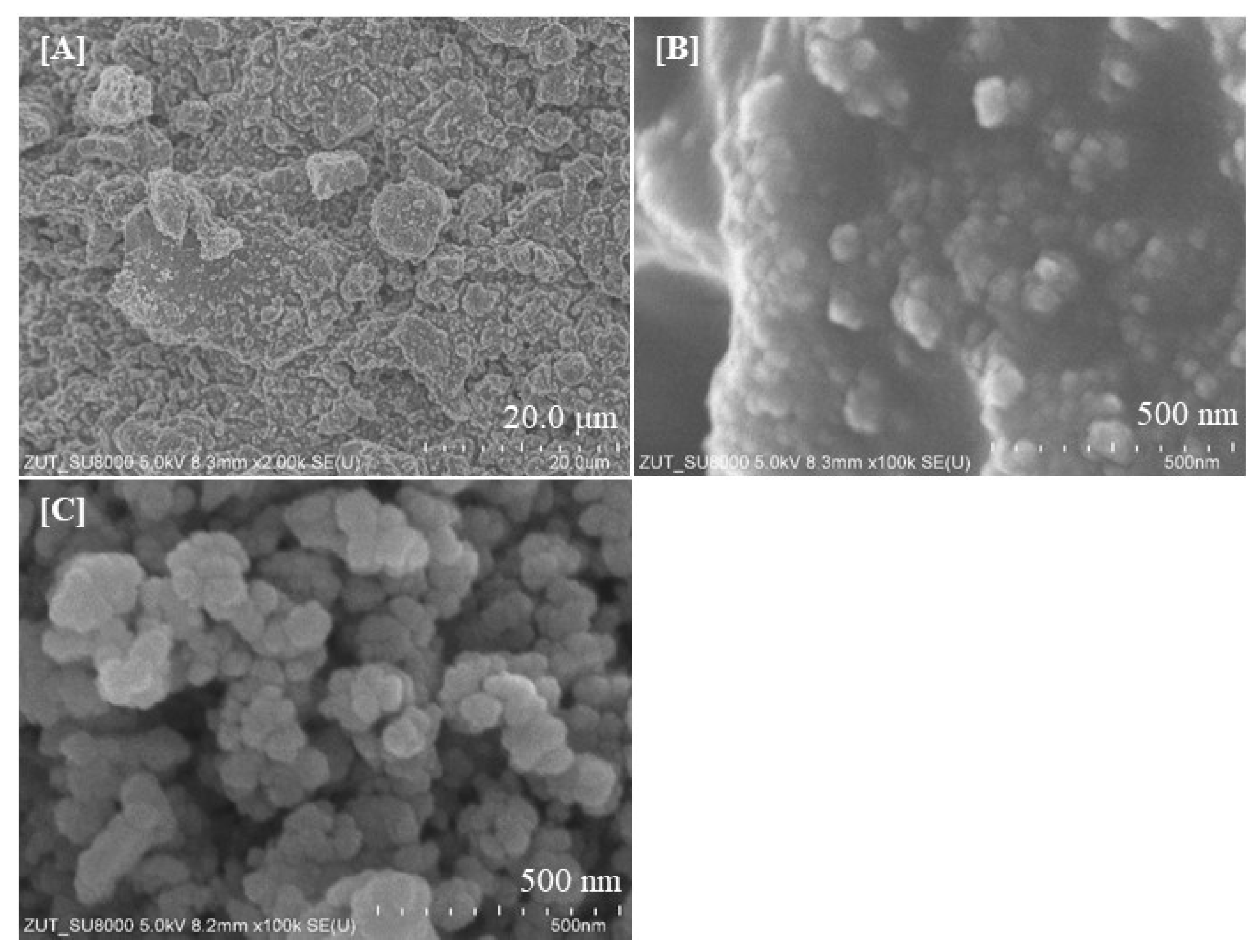
Scanning electron microscopy studies were conducted to provide information on the morphology of materials obtained. Figure 6A–C present selected SEM images for starting the TiO2 photocatalyst and sample SiO2(11.1%)/TiO2_800. As can be seen from the above images (Figure 5A,B) showing the surface structure, the particles of starting TiO2 were characterized by an irregular and undefined shape. Moreover, the particles form large aggregates (150–350 nm) consisting of agglomerated particles. It is worth noting that the particles of starting TiO2 are embedded in the unreacted TTIP matrix. The particles of SiO2-modified photocatalyst were characterized by a more pronounced circularity and regularity of shape compared to starting TiO2. Moreover, due to the modification and calcination at 800 °C, the aggregates were larger in size (400–550 nm). This is since annealing of titanium dioxide leads to the sintering of photocatalyst grains, which promotes the agglomeration of particles, causing an increase in their size [37]. In addition, silica particles tend to form larger structures [46]. In both cases, the particles tended to aggregate into larger particle clusters. However, it was noted that the tendency of particles to form aggregates increased due to SiO2 modification.

2.2. Adsorption Test

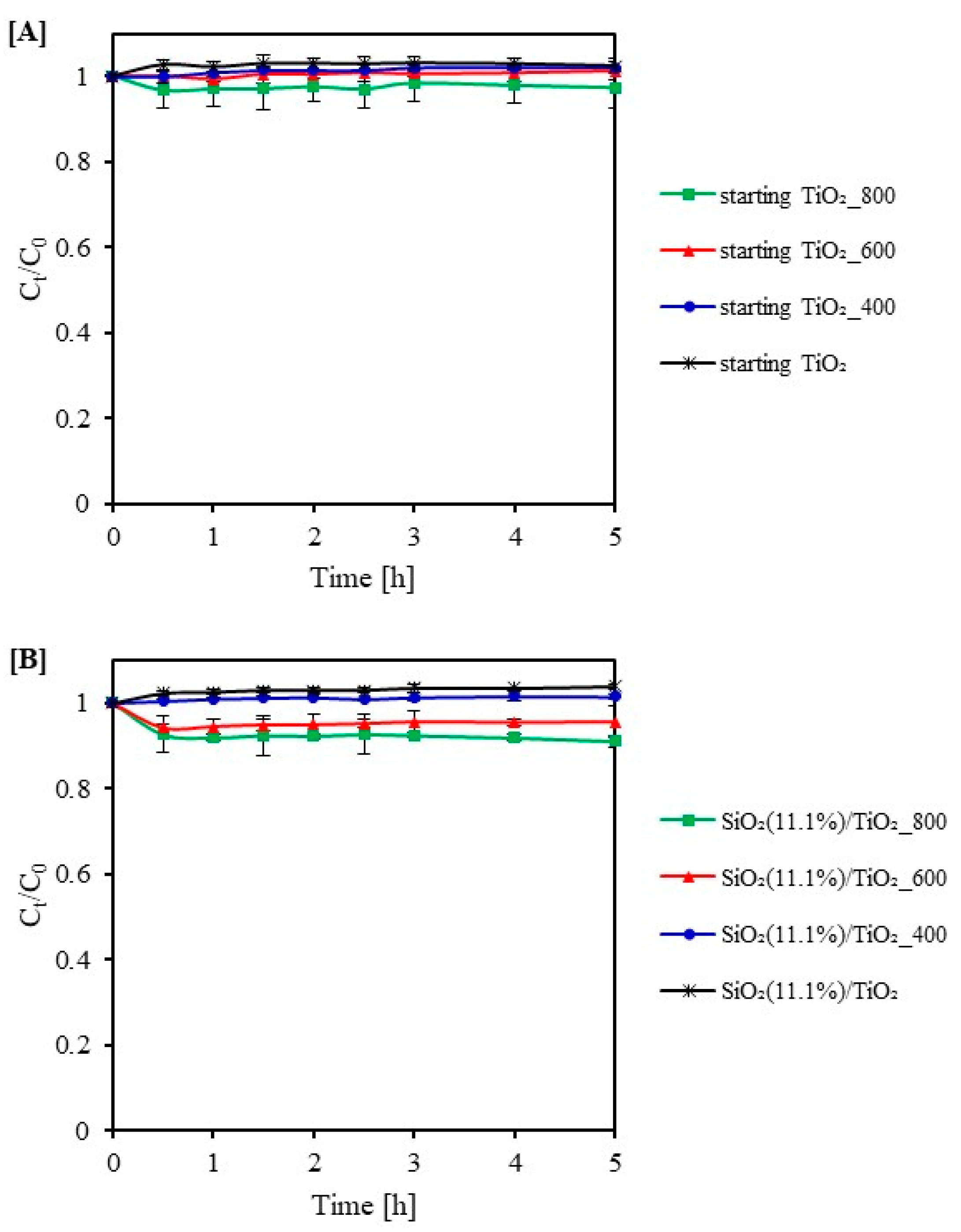
The adsorption properties of photocatalysts were investigated before the irradiation process. The adsorption process of methylene blue on the surface of reference materials and SiO2/TiO2 photocatalysts is shown in Figure 7A,B. The obtained results found that the adsorption equilibrium was established after 1 h for all samples. For the reference materials, SiO2(11.1%)/TiO2 and SiO2(11.1%)/TiO2_400, a slight desorption of MB (about 2–4%) was observed. The MB adsorption for SiO2(11.1%)/TiO2_600 and SiO2(11.1%)/TiO2_800 was negligible and was 2 and 8% after 5 h, respectively. The SiO2/TiO2_600 °C photocatalyst adsorption was higher than for the SiO2/TiO2_400 °C sample. This is due to a change in the surface nature. The surface for SiO2(11.1%)/TiO2_600 has changed from positively charged to negatively charged, showing better interaction with the cationic dye.

2.3. Photocatalytic Activity Test

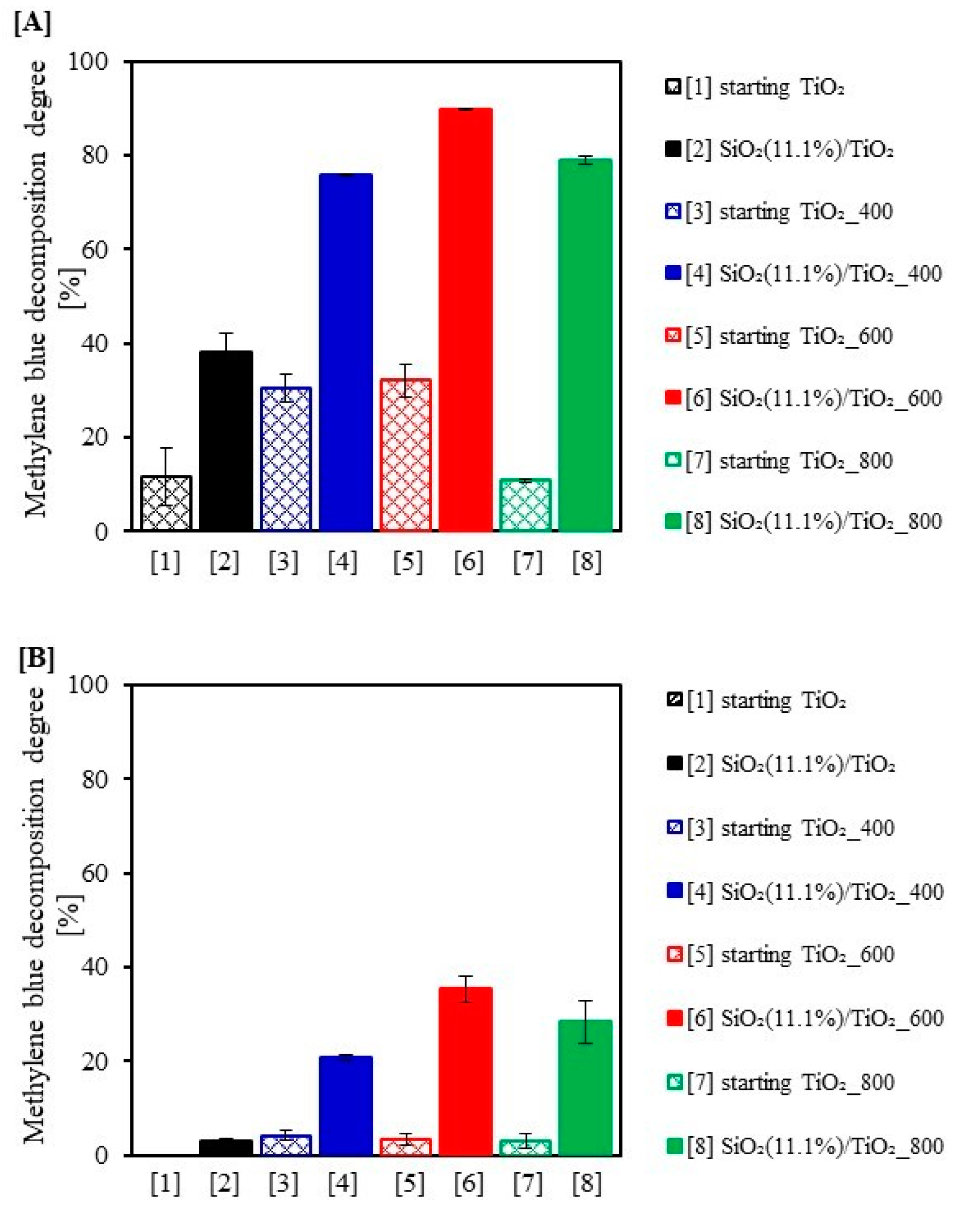
The photocatalytic activity of SiO2/TiO2 photocatalysts was determined by the degradation of methylene blue under UV-VIS and VIS irradiation. The results of dye degradation degree after 5 h of irradiation are shown in Figure 8A,B. All the obtained nanomaterials showed higher activity than the reference samples. It can be observed that the methylene blue decomposition degree increased as the calcination temperature increased to 600 °C. The SiO2(11.1%)/TiO2_600 sample showed the highest activity under UV-VIS and VIS irradiation. The MB decomposition degree was 89.60 and 35.27%, respectively. In comparison, the dye decomposition degree for starting TiO2_600 sample, obtained by the same method but without SiO2 addition, was 32.04 and 3.41% under UV-VIS and VIS light, respectively. However, it should be noted that although the activity of SiO2-modified materials was higher compared to the starting TiO2, the performance of dye removal process under visible light wasgenerally low. This confirms the fact that these materials are mainly activated by ultraviolet radiation. Therefore, the limitation of VIS availability significantly limits the use of tested photocatalysts. It is important to note that the SiO2(11.1%)/TiO2_600 photocatalyst, which showed the highest activity in both cases, consists of 83% anatase and 17% brookite. Our observations were consistent with the results of Mutuma et al. [47] who found that the presence of a mixed phase (anatase and brookite) improved photocatalytic properties. They recorded the highest activity for a sample of 80% anatase and 20% brookite. In addition, the SiO2(11.1%)/TiO2_600 sample had a negative zeta potential, and as is known, methylene blue is a cationic dye and shows better interaction with negatively charged surfaces. Improvement in photocatalytic activity resulting from a change in the surface charge of the photocatalyst was also noted by Sienkiewicz et al. [43]. It was found that both the addition of SiO2 and the calcination process increased the photoactivity. An important parameter affecting the activity was the composition phase of photocatalysts. As can be seen, materials containing more anatase phase showed higher activity compared to reference samples, which contained more brookite and rutile. It is well known that rutile is a less active form of titanium dioxide than anatase, as it has fewer active sites and hydroxyl groups on the surface [48]. It is worth noting the high activity obtained for the photocatalyst SiO2(11.1%)/TiO2_800. The high activity of this sample was due to the 100% content of the anatase phase. In comparison, the starting TiO2_800 photocatalyst without adding SiO2 showed significantly weaker activity. The lower activity of this material was due to its 100% rutile phase content. This shows that the modification of SiO2 effectively inhibited the transformation phase of anatase to rutile and the growth of TiO2 crystallites after calcination. Increasing the crystallinity of anatase after calcination resulted in a high rate of electron diffusion by slowing the recombination of electron-hole pairs [49]. The crystallization level was also an important parameter determining the activity. The better the crystallized material, the higher the activity. It is generally accepted that the optimal size of anatase crystallites for high photocatalytic activity is around 10 nm. However, Almquist and Biswas [50] found that the activity is highest when the crystallite size is in the range of 25–40 nm. In our case, the highest photoactivity was shown by SiO2(11.1%)/TiO2_600 and SiO2(11.1%)/TiO2_800 samples with anatase crystallite sizes of 25 and 29 nm, respectively. Under visible radiation, the activity of tested photocatalysts was much lower than in the case of UV-VIS radiation. This was explained by the fact, the materials obtained are mainly activated by ultraviolet radiation. After the modification process with fumed silica, all the tested photocatalysts showed a shift of the absorption edge towards lower wavelengths (blue shift). An increase in the value of band gap energy was noted for these samples.
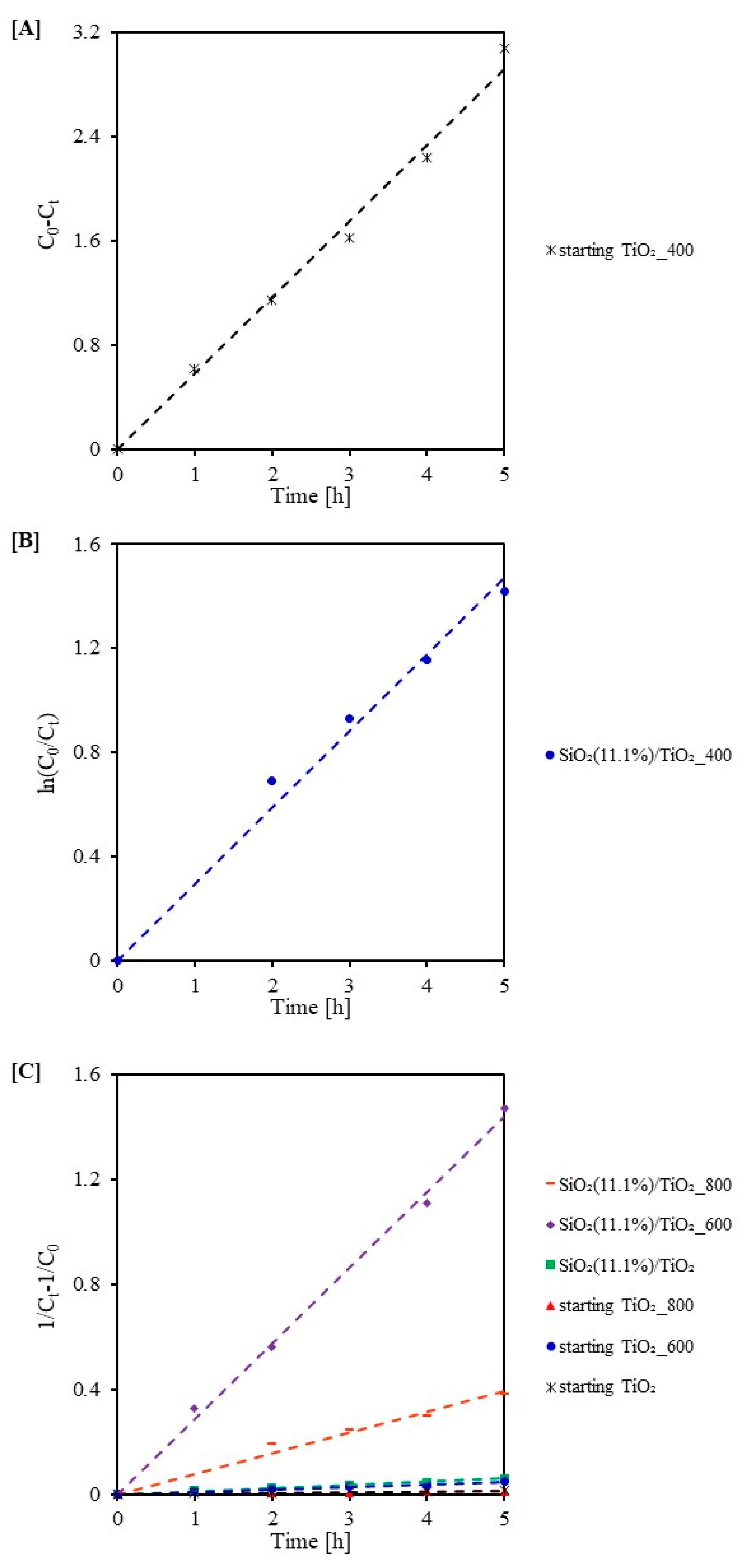
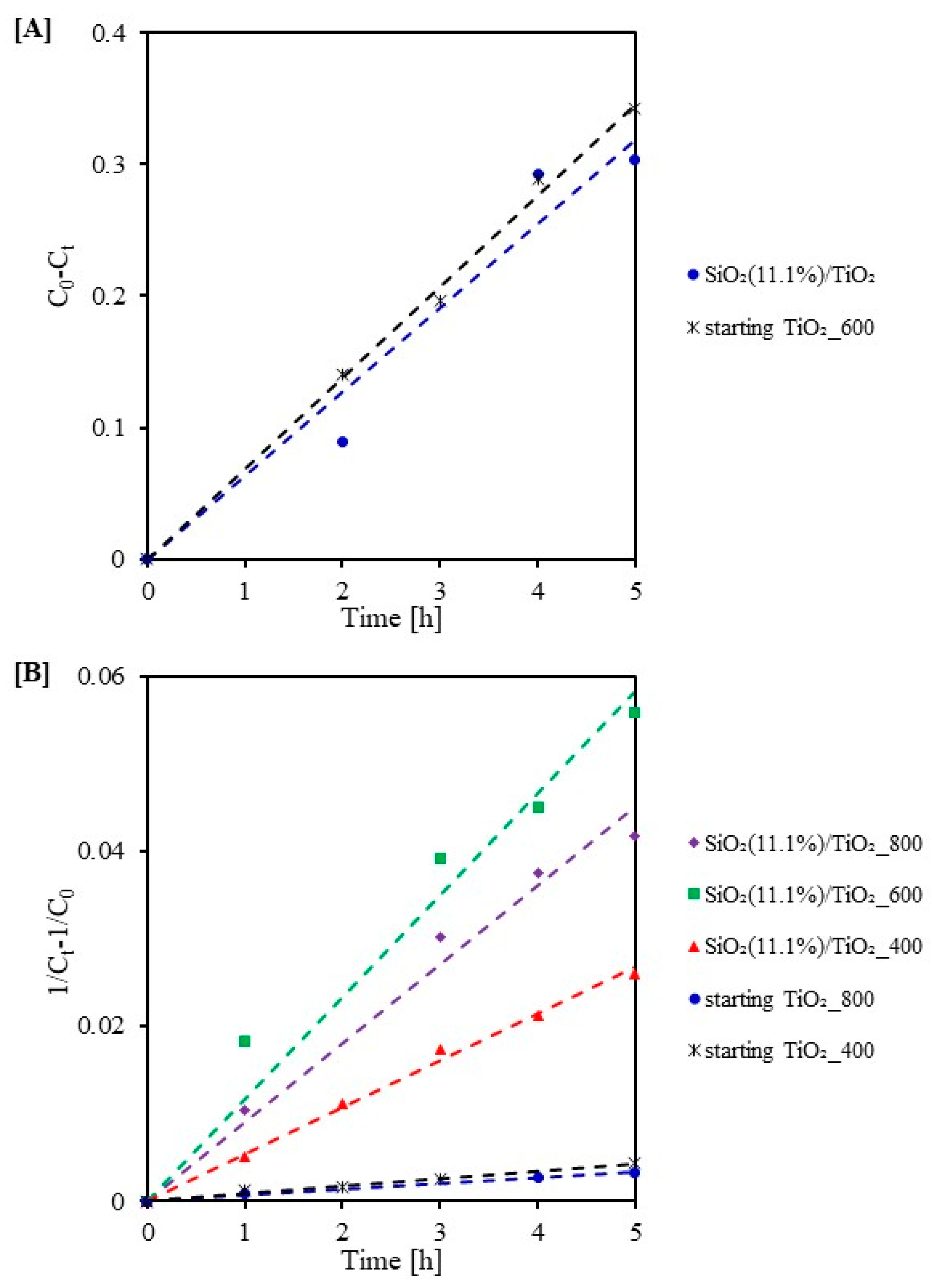
In order to further describe the degradation of methylene blue under both UV-VIS and VIS irradiation, apparent reaction rate constants were determined. The zero-order, pseudo-first-order, and pseudo-second-order linear transformations are shown for UV-VIS in Figure 9A–C and for VIS in Figure 10A,B, respectively. The obtained values of reaction rate constants and fit coefficients are summarized in Table 4 and Table 5. The dye degradation under UV-VIS irradiation for starting TiO2_400 followed a zero-order model. According to the pseudo-first-order Langmuir-Hinshelwood model for SiO2(11.1%)/TiO2_400. For all other samples, the degradation followed the pseudo-second-order model. The highest k2 values of methylene blue degradation were recorded for photocatalysts modified with SiO2 and then calcined at higher temperatures. It is worth noting that the highest reaction rate constant (0.288 L/(min-mg) was obtained for SiO2(11.1%)/TiO2_600, and this value is 96 and 24 times higher than that for starting TiO2 and SiO2(11.1%)/TiO2, respectively. The decomposition of methylene blue under visible radiation for SiO2(11.1%)/TiO2 and starting TiO2_600 followed the zero-order model (see Figure 10A). However, for the other nanomaterials, the decomposition followed a pseudo-second-order model (see Figure 10B). Similar to UV-VIS, higher k2 values were recorded for SiO2-modified samples. It was also observed that after 3 h of exposure to VIS light, the points on the graphs started to deviate from the typical linear curve. The formation of intermediates caused the decrease in reaction rate during dye degradation.

2.4. Photocatalytic Mechanism

The mechanism of photocatalytic decomposition of methylene blue in the presence of SiO2/TiO2 under UV-VIS and VIS irradiation is presented in Figure 11. The first step in the photocatalysis process is the activation of the photocatalyst by absorption of a photon with energy equal to or greater than the band gap energy. This is followed by electron transfer from the valence band to the conduction band, which leads to the formation of electron-hole charge carrier pairs (e–h+). The hydroxyl radicals produced are strong oxidants, thus able to oxidize methylene blue to simple inorganic compounds. As is commonly known, too rapid charge recombination adversely affects the efficiency of photooxidation process. According to the literature [51,52], modification of TiO2 with SiO2 as a result of the separation of photo-induced charges contributes to slowing down electron-hole recombination, thus improving photocatalytic performance. In the case of VIS radiation, the effect of the photosensitization process on the increase in activity of tested photocatalysts can also not be excluded.

3. Materials and Methods

3.1. Materials

Titanium(IV) isopropoxide (TTIP) (≥97%, Sigma-Aldrich Co., Saint Louis, MO, USA) was used as a TiO2 precursor in the preparation of SiO2/TiO2 photocatalysts by the sol-gel method. Fumed silica (purity ≥ 99.8%, specific surface area > 200 m2/g, average primary particle size 7–14 nm) was used as the silicon source from the German company PlasmaChem GmbH.. Isopropyl alcohol (pure p.a) produced by Firma Chempur® Company (Piekary Śląskie, Poland) was used as a solvent. Hydrochloric acid (35–38% pure, Firma Chempur®, Piekary Śląskie, Poland) was used as a catalyst for the hydrolysis reaction. Methylene blue (purity ≥ 82%), used as a model organic pollutant, was produced by Firma Chempur® Company (Piekary Śląskie, Poland).

3.2. Preparation of Photocatalysts

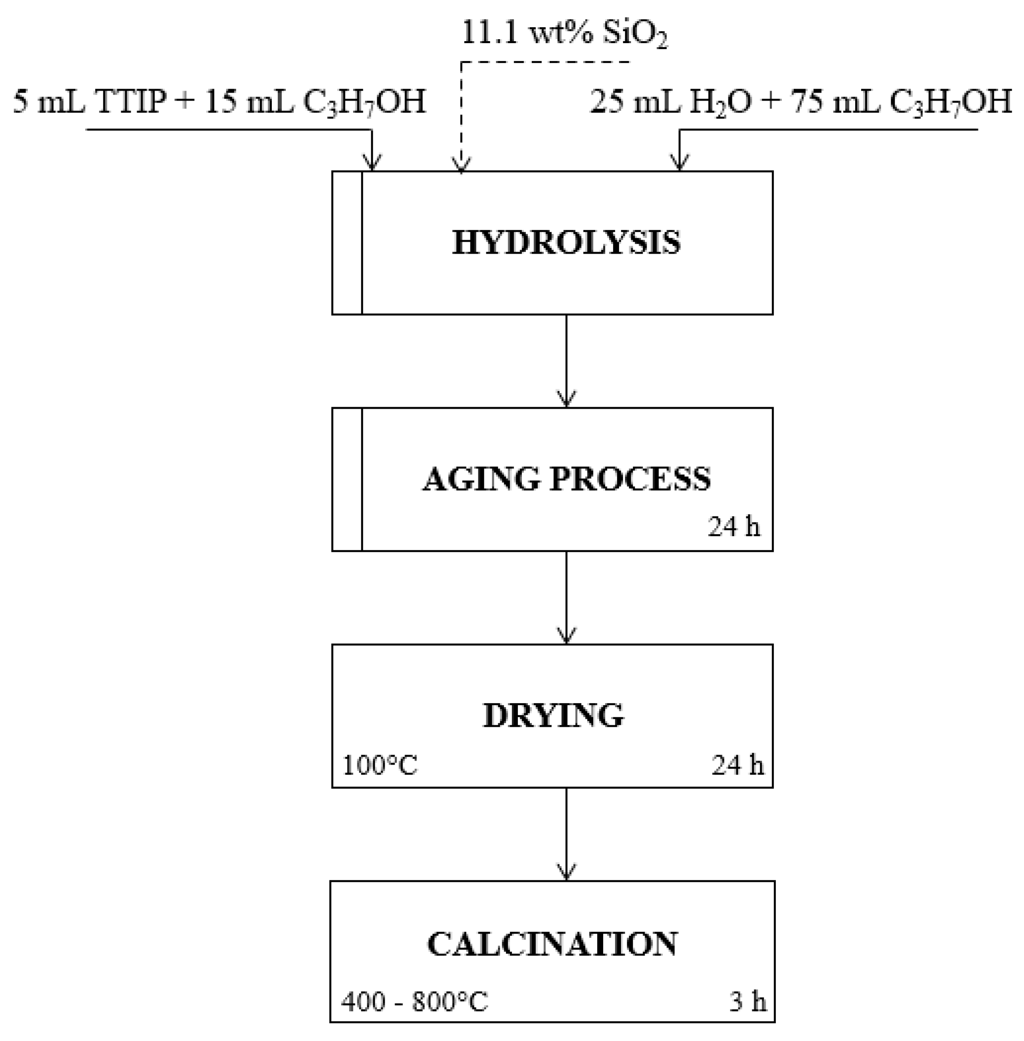
The SiO2/TiO2 photocatalysts were prepared by a sol-gel method in which TTIP and fumed silica were the precursors. First, 5 mL of TTIP was dropped into 15 mL of isopropyl alcohol. Then an appropriate amount of silica precursor (11.1 wt%) was added. Next, the pH of the resulting solution was brought to 2 using HCl. Then 100 mL of water: isopropyl alcohol mixture (25:75 v/v) was added dropwise with intense stirring to initiate hydrolysis. Stirring was continued for 1 h. The next step was a 24-h ageing process, after which the resulting gel was dried at 100 °C for another 24 h. Finally, the material was subjected to heat treatment in an argon atmosphere (purity 5.0, Messer Polska Sp. z o.o., Chorzów, Poland). For this purpose, 2.5 g of the photocatalyst was placed in a quartz crucible and introduced into a quartz tube in a tube furnace (Program Controller R 40/250/12-C40, Nabertherm®, Lilienthal, Germany). The system was then flushed with argon over 30 min to remove air. The heating process was carried out over a temperature range of 400 °C to 800 °C (Δt = 200 °C) for 3 h after the set temperature, with a constant flow of argon (60 mL/min). The obtained samples were named SiO2(11.1%)/TiO2_t, where (11.1%) indicates the weight percentage of SiO2 and t indicates the calcination temperature. The control material, designated as (starting TiO2), was obtained by the same method but without adding fumed silica. The schematic diagram of the preparation of SiO2/TiO2 photocatalysts is shown in Figure 12. Table 6 summarizes the methods for the preparation of SiO2-modified TiO2 nanomaterials.

3.3. Characterization Methods

The crystal structure of investigated samples was characterized by XRD analysis (Empyrean X-ray diffractometer, PANalytical, Almelo, The Netherlands) using Cu Kα radiation with a wavelength of 1.54056 Å. Diffractograms were conducted in the 2θ angular range from 20 to 80°. The International Diffraction Data Center PDF-4+ 2014 database (04-002-8296 PDF4+ card for anatase, 04-007-0758 PDF4+ card for brookite and 04-005-5923 PDF4+ card for rutile) was used to interpret the obtained diffractograms. The average crystallite size was calculated based on the Rietveld method. The specific surface area and pore size distribution of the obtained materials were determined by analysis of N2 adsorption-desorption isotherms at 77 K. The study was carried out using a QUADRASORB evoTM apparatus (Anton Paar GmbH, Graz, Austria). Before the measurement, the samples were degassed under vacuum at 100 °C for 16 h to pre-clean the surfaces of the photocatalysts tested. The total pore volume (Vtotal) was calculated from the adsorbed nitrogen after pore condensation at a relative pressure p/p0 = 0.99. The volume of micropores (Vmicro) was estimated by the Dubinin-Radushkevich method. The volume of mesopores (Vmeso) was determined as the difference between Vtotal and Vmicro. Surface composition analysis of the photocatalysts was conducted using an FT-IR-4200 spectrometer (JASCO International Co. Ltd., Tokyo, Japan) equipped with a DiffuseIR accessory (PIKE Technologies, Cottonwood Dr, Fitchburg, WI, USA. The FT-IR spectra were recorded in the wavenumber range of 4000–400 cm−1. The optical properties of the samples were investigated using a UV-Vis/DR V-650 spectrophotometer (JASCO International Co., Tokyo, Japan) equipped with a diffuse-reflection accessory. The diffuse reflectance spectra were recorded in the 200–800 nm wavelength range. Spectralon (Spectralon® Diffuse Reflectance Material, Labsphere, North Sutton, NH, USA) was used as the standard sample. The energy gap was calculated by plotting the function [F(R)hυ]1/2 as a function of photon energy (hυ) and extrapolating the linear part to [F(R)hυ]1/2 = 0 [53]. The zeta potential was measured using a ZetaSizer NanoSeries ZS instrument (Malvern Panalytical Ltd., Malvern, UK). The SU8020 Ultra-High Resolution Field Emission Scanning Electron Microscope (Hitachi Ltd., Tokyo, Japan) was used to determine the surface character of the materials obtained.

3.4. Photocatalytic Activity Measurements

The photocatalytic activity of fumed silica-modified materials was determined by the degradation of methylene blue (MB) under UV-VIS and VIS irradiation. A lamp (Philips), consisting of six bulbs of 20 W each, was used as the source of UV-VIS radiation. The measured intensity of UV radiation was 138 W/m2 (for the 280–400 nm range), and that of VIS radiation was 167 W/m2 (for the 300–2800 nm range). However, a lamp with an intensity of 1.3 W/m2 for UV (for 280–400 nm range) and 54 W/m2 for VIS (for 300–2800 nm range) was used as a visible radiation source. The experiment was conducted in a 0.6 L glass beaker containing 0.2 g/L photocatalyst and 0.5 L methylene blue solution. The initial dye concentration was 10 mg/L. Before irradiation, the suspension was stirred on a magnetic stirrer without light exposure for 1 h to ensure the adsorption-desorption equilibrium. After this time, the suspension was irradiated using a suitable radiation source. The irradiation was carried out for 5 h. At equal intervals, 10 mL of the suspension was collected, and this volume was centrifuged to remove the TiO2 nanoparticles. The concentration of methylene blue in the solution was measured using a UV-VIS V-630 spectrophotometer (Jasco International Co., Tokyo, Japan). The reaction rate constants for the decomposition of methylene blue were determined from the obtained results. The zero reaction rate constant was determined from the zero-order model (Equation (1)) [42]:
C0 − Ct = k0t
The pseudo-first reaction rate constant was calculated using the Langmuir-Hinshelwood kinetics model (Equation (2)) [54]:
ln(C0/Ct) = kKt = k1t
While the pseudo-second reaction rate constant was determined according to the pseudo-second-order model (Equation (3)) [55]:
1/Ct − 1/C0 = k2t
where: C0—initial concentration of the methylene blue (mg/L), Ct—concentration of the methylene blue at time t (mg/L), k0, k1 and k2—zero-order, pseudo-first and pseudo-second reaction rate constants [mg/(L·min), 1/min, L/(min·mg)], K—adsorption coefficient of the reactant (L/mg) and t—time of illumination (min).

4. Conclusions

The SiO2/TiO2 nanomaterials were prepared by the TTIP hydrolysis method and then subjected to calcination in the temperature range of 400–800 °C under an argon atmosphere. The effect of modification with fumed silica and the effect of calcination temperature on the activity of TiO2 photocatalysts under visible light were studied for the first time in this paper. It was found that modification of TiO2 with silica inhibited anatase transition to rutile and crystallite size increase during calcination. In addition, the use of silica to modify TiO2 contributed to an increase in specific surface area and total pore volume. The calcination process changed the surface character of studied photocatalysts. The photoactivity of obtained materials was tested in the degradation reaction of methylene blue in an aqueous solution in the presence of light from the UV-VIS and visible range. In general, it was found that the modification carried out resulted in improved photocatalytic performance. The photocatalytic activity increased as the calcination temperature increased to 600 °C. The best result under UV-VIS and VIS irradiation was observed for the SiO2(11.1%)/TiO2_600 sample. Under UV-VIS irradiation, it removed 89.60%, and under VIS irradiation, the degree of methylene blue removal was 35.27%.

Author Contributions

Conceptualization: A.B. and A.W.; investigation: A.B., A.W.; data curation: A.W., E.K.-N., A.W.M.; writing—original draft preparation: A.B.; writing—review and editing: A.W., E.K.-N., A.W.M.; project administration: A.W.M.; funding acquisition: A.W.M. All authors have read and agreed to the published version of the manuscript.

Funding

This work was supported by grant 2017/27/B/ST8/02007 from the National Science Centre, Poland.

Data Availability Statement

The data presented in this study are available on request from the corresponding author.

Conflicts of Interest

The authors declare no conflict of interest.

References

  1. Sultana, T.; Dey, S.C.; Molla, M.; Islam, A.; Hossain, M.R.; Rahman, M.M.; Quddus, M.S.; Moniruzzaman, M.; Shamsuddin, S.M.; Sarker, M. Facile synthesis of TiO2/Chitosan nanohybrid for adsorption-assisted rapid photodegradation of an azo dye in water. React. Kinet. Mech. Catal. 2021, 133, 1121–1139. [Google Scholar] [CrossRef]
  2. Shathy, R.A.; Fahim, S.A.; Sarker, M.; Quddus, M.S.; Moniruzzaman, M.; Masum, S.M.; Molla, M.A.I. Natural Sunlight Driven Photocatalytic Removal of Toxic Textile Dyes in Water Using B-Doped ZnO/TiO2 Nanocomposites. Catalysts 2022, 12, 308. [Google Scholar] [CrossRef]
  3. Mondol, B.; Sarker, A.; Shareque, A.M.; Dey, S.C.; Islam, M.T.; Das, A.K.; Shamsuddin, S.M.; Molla, M.A.I.; Sarker, M. Preparation of activated carbon/TiO2 nanohybrids for photodegradation of reactive red-35 dye using sunlight. Photochem 2021, 1, 54–66. [Google Scholar] [CrossRef]
  4. Guo, Z.; Ma, R.; Li, G. Degradation of phenol by nanomaterial TiO2 in wastewater. Chem. Eng. J. 2006, 119, 55–59. [Google Scholar] [CrossRef]
  5. Grabowska, E.; Reszczyńska, J.; Zaleska, A. RETRACTED: Mechanism of phenol photodegradation in the presence of pure and modified-TiO2: A review. Water Res. 2012, 46, 5453–5471. [Google Scholar] [CrossRef] [PubMed]
  6. Borges, M.E.; Sierra, M.; Cuevas, E.; García, R.D.; Esparza, P. Photocatalysis with solar energy: Sunlight-responsive photocatalyst based on TiO2 loaded on a natural material for wastewater treatment. Sol. Energy 2016, 135, 527–535. [Google Scholar] [CrossRef]
  7. Boyjoo, Y.; Sun, H.; Liu, J.; Pareek, V.K.; Wang, S. A review on photocatalysis for air treatment: From catalyst development to reactor design. Chem. Eng. J. 2017, 310, 537–559. [Google Scholar] [CrossRef]
  8. Shayegan, Z.; Lee, C.S.; Haghighat, F. TiO2 photocatalyst for removal of volatile organic compounds in gas phase—A review. Chem. Eng. J. 2018, 334, 2408–2439. [Google Scholar] [CrossRef] [Green Version]
  9. Adnan, R.; Shah, I. A mini-review on rare earth metal-doped TiO2 for photocatalytic remediation of wastewater. Environ. Sci. Pollut. Res. 2016, 23, 15941–15951. [Google Scholar]
  10. Khalid, N.R.; Majid, A.; Tahir, M.B.; Niaz, N.A.; Khalid, S. Carbonaceous-TiO2 nanomaterials for photocatalytic degradation of pollutants: A review. Ceram. Int. 2017, 43, 14552–14571. [Google Scholar] [CrossRef]
  11. Mazierski, P.; Mikolajczyk, A.; Bajorowicz, B.; Malankowska, A.; Zaleska-Medynska, A.; Nadolna, J. The role of lanthanides in TiO2-based photocatalysis: A review. Appl. Catal. B Environ. 2018, 233, 301–317. [Google Scholar] [CrossRef]
  12. Yoo, D.K.; Ahmed, I.; Sarker, M.; Lee, H.J.; Vinu, A.; Jhung, S.H. Metal–organic frameworks containing uncoordinated nitrogen: Preparation, modification, and application in adsorption. Mater. Today 2021, 51, 566–585. [Google Scholar] [CrossRef]
  13. Sarker, M.; Yoo, D.K.; Lee, S.; Kim, T.W.; Kim, C.U.; Jhung, S.H. Conversion of Y into SSZ-13 zeolite, in the absence of extra silica, alumina and seed crystals, with N, N, N-dimethylethylcyclohexylammonium bromide, and application of the SSZ-13 zeolite in the propylene production from ethylene. Catal. Today 2021, 375, 94–100. [Google Scholar] [CrossRef]
  14. Khanna, S.; Chaliyawala, H.; Paneliya, S.; Roy, D.; Mukhopadhyay, K.; Banerjee, R.; Mukhopadhyay, I. Systematic investigation of close-packed silica nanospheres monolayer under sintering conditions. J. Eur. Ceram. Soc. 2019, 39, 1411–1419. [Google Scholar] [CrossRef]
  15. Zhang, Y.P.; Lee, S.H.; Reddy, K.R.; Gopalan, A.I.; Lee, K.P. Synthesis and characterization of core-shell SiO2 nanoparticles/poly (3-aminophenylboronic acid) composites. J. Appl. Polym. Sci. 2007, 104, 2743–2750. [Google Scholar] [CrossRef]
  16. Zhang, L.; Xing, Z.; Zhang, H.; Li, Z.; Wu, X.; Zhang, X.; Zhang, Y.; Zhou, W. High thermostable ordered mesoporous SiO2–TiO2 coated circulating-bed biofilm reactor for unpredictable photocatalytic and biocatalytic performance. Appl. Catal. B Environ. 2016, 180, 521–529. [Google Scholar] [CrossRef]
  17. Pal, A.; Jana, T.K.; Chatterjee, K. Silica supported TiO2 nanostructures for highly efficient photocatalytic application under visible light irradiation. Mater. Res. Bull. 2016, 76, 353–357. [Google Scholar] [CrossRef]
  18. Raj, K.; Smith, Y.R.; Subramanian, V.R.; Viswanathan, B. Structural studies of silica modified titania and its photocatalytic activity of 4-chlorophenol oxidation in aqueous medium. Indian J. Chem. 2010, 49A, 867–875. [Google Scholar]
  19. Fu, X.; Clark, L.A.; Yang, Q.; Anderson, M.A. Enhanced photocatalytic performance of titania-based binary metal oxides: TiO2/SiO2 and TiO2/ZrO2. Environ. Sci. Technol. 1996, 30, 647–653. [Google Scholar] [CrossRef]
  20. Chun, H.; Yizhong, W.; Hongxiao, T. Influence of adsorption on the photodegradation of various dyes using surface bond-conjugated TiO2/SiO2 photocatalyst. Appl. Catal. B Environ. 2001, 35, 95–105. [Google Scholar] [CrossRef]
  21. Xie, C.; Xu, Z.; Yang, Q.; Xue, B.; Du, Y.; Zhang, J. Enhanced photocatalytic activity of titania–silica mixed oxide prepared via basic hydrolyzation. Mater. Sci. Eng. B 2004, 112, 34–41. [Google Scholar] [CrossRef]
  22. Ding, Z.; Lu, G.Q.; Greenfield, P.F. Role of the crystallite phase of TiO2 in heterogeneous photocatalysis for phenol oxidation in water. J. Phys. Chem. B 2000, 104, 4815–4820. [Google Scholar] [CrossRef]
  23. Iwamoto, S.; Tanakulrungsank, W.; Inoue, M.; Kagawa, K.; Praserthdam, P. Synthesis of large-surface area silica-modified titania ultrafine particles by the glycothermal method. J. Mater. Sci. Lett. 2000, 19, 1439–1443. [Google Scholar] [CrossRef]
  24. Cheng, P.; Zheng, M.; Jin, Y.; Huang, Q.; Gu, M. Preparation and characterization of silica–doped titania photocatalyst through sol–gel method. Mater. Lett. 2003, 57, 2989–2994. [Google Scholar]
  25. Nilchi, A.; Janitabar-Darzi, S.; Mahjoub, A.R.; Rasouli-Garmarodi, S. New TiO2/SiO2 nanocomposites—Phase transformations and photocatalytic studies. Colloids Surf. A Physicochem. Eng. Asp. 2010, 361, 25–30. [Google Scholar] [CrossRef]
  26. Wang, J.; Zhang, W.; Zhu, B.; Xiao, Y. Microstructure and photocatalytic activity of TiO2-SiO2 composite materials. In Proceedings of the 2020 6th International Conference on Materials, Mechanical Engineering and Automation Technology, Zhuhai, China, 1–3 May 2020. [Google Scholar] [CrossRef]
  27. Babyszko, A.; Wanag, A.; Sadłowski, M.; Kusiak-Nejman, E.; Morawski, A.W. Synthesis and Characterization of SiO2/TiO2 as Photocatalyst on Methylene Blue Degradation. Catalysts 2022, 12, 1372. [Google Scholar] [CrossRef]
  28. SiO2—Krzemionka Koloidalna, Nanoproszek, Hydrofilowy. Available online: https://3d-nano.com/pl/catalogue/sio2-krzemionka-koloidalna-nanoproszek-hydrofilowy/ (accessed on 18 November 2022).
  29. Hu, Y.; Tsai, H.L.; Huang, C.L. Effect of brookite phase on the anatase–rutile transition in titania nanoparticles. J. Eur. Ceram. Soc. 2003, 23, 691–696. [Google Scholar] [CrossRef]
  30. Pelaez, M.; Nolan, N.T.; Pillai, S.C.; Seery, M.K.; Falaras, P.; Kontos, A.G.; Dunlop, P.S.M.; Hamilton, J.W.J.; Byrne, J.A.; O’Shea, K.; et al. A review on the visible light active titanium dioxide photocatalysts for environmental applications. Appl. Catal. B Environ. 2012, 125, 331–349. [Google Scholar] [CrossRef] [Green Version]
  31. Tobaldi, D.M.; Tucci, A.; Škapin, A.S.; Esposito, L. Effects of SiO2 addition on TiO2 crystal structure and photocatalytic activity. J. Eur. Ceram. Soc 2010, 30, 2481–2490. [Google Scholar] [CrossRef]
  32. Lu, Z.; Jiang, X.; Zhou, B.; Wu, X.; Lu, L. Study of effect annealing temperature on the structure, morphology and photocatalytic activity of Si doped TiO2 thin films deposited by electron beam evaporation. Appl. Surf. Sci. 2011, 257, 10715–10720. [Google Scholar] [CrossRef]
  33. Xu, G.; Zheng, Z.; Wu, Y.; Feng, N. Effect of silica on the microstructure and photocatalytic properties of titania. Ceram. Int. 2009, 35, 1–5. [Google Scholar] [CrossRef]
  34. Sing, K.S.W. Reporting physisorption data for gas/solid systems with special reference to the determination of surface area and porosity (Provisional). Pure Appl. Chem. 1982, 54, 2201–2218. [Google Scholar] [CrossRef]
  35. Leofanti, G.; Padovan, M.; Tozzola, G.; Venturelli, B.J.C.T. Surface area and pore texture of catalysts. Catal. Today 1998, 41, 207–219. [Google Scholar] [CrossRef]
  36. Bao, N.; Wei, Z.; Ma, Z.; Liu, F.; Yin, G. Si-doped mesoporous TiO2 continuous fibers: Preparation by centrifugal spinning and photocatalytic properties. J. Hazard. Mater. 2010, 174, 129–136. [Google Scholar] [CrossRef]
  37. Nyamukamba, P.; Tichagwa, L.; Greyling, C. The influence of carbon doping on TiO2 nanoparticle size, surface area, anatase to rutile phase transformation and photocatalytic activity. Mater. Sci. Forum 2012, 712, 49–63. [Google Scholar] [CrossRef]
  38. Winter, M.; Hamal, D.; Yang, X.; Kwen, H.; Jones, D.; Rajagopalan, S.; Klabunde, K.J. Defining reactivity of solid sorbents: What is the most appropriate metric? Chem. Mater. 2009, 21, 2367–2374. [Google Scholar] [CrossRef]
  39. Resende, S.F.; Nunes, E.H.M.; Houmard, M.; Vasconcelos, W.L. Simple sol–gel process to obtain silica-coated anatase particles with enhanced TiO2-SiO2 interfacial area. J. Colloid Interface Sci. 2014, 433, 211–217. [Google Scholar] [CrossRef] [Green Version]
  40. Yang, G.; Jiang, Z.; Shi, H.; Xiao, T.; Yan, Z. Preparation of highly visible-light active N-doped TiO2 photocatalyst. J. Mater. Chem. 2010, 20, 5301–5309. [Google Scholar] [CrossRef]
  41. Rokicka-Konieczna, P.; Markowska-Szczupak, A.; Kusiak-Nejman, E.; Morawski, A.W. Photocatalytic water disinfection under the artificial solar light by fructose-modified TiO2. Chem. Eng. J. 2019, 372, 203–215. [Google Scholar] [CrossRef]
  42. Abdullah, M.A.; Chong, F.K. Dual-effects of adsorption and photodegradation of methylene blue by tungsten-loaded titanium dioxide. Chem. Eng. J. 2010, 158, 418–425. [Google Scholar]
  43. Sienkiewicz, A.; Rokicka-Konieczna, P.; Wanag, A.; Kusiak-Nejman, E.; Morawski, A.W. Artificial Solar Light-Driven APTES/TiO2 Photocatalysts for Methylene Blue Removal from Water. Molecules 2022, 27, 947. [Google Scholar] [CrossRef] [PubMed]
  44. Banerjee, S.; Gopal, J.; Muraleedharan, P.; Tyagi, A.K.; Raj, B. Physics and chemistry of photocatalytic titanium dioxide: Visualization of bactericidal activity using atomic force microscopy. Curr. Sci. 2006, 90, 1378–1383. [Google Scholar]
  45. Suttiponparnit, K.; Jiang, J.; Sahu, M.; Suvachittanont, S.; Charinpanitkul, T.; Biswas, P. Role of surface area, primary particle size, and crystal phase on titanium dioxide nanoparticle dispersion properties. Nanoscale Res. Lett. 2011, 6, 27. [Google Scholar] [CrossRef] [PubMed] [Green Version]
  46. Jesionowski, T.; Żurawska, J.; Krysztafkiewicz, A.; Pokora, M.; Waszak, D.; Tylus, W. Physicochemical and morphological properties of hydrated silicas precipitated following alkoxysilane surface modification. Appl. Surf. Sci. 2003, 205, 212–224. [Google Scholar] [CrossRef]
  47. Mutuma, B.K.; Shao, G.N.; Kim, W.D.; Kim, H.T. Sol–gel synthesis of mesoporous anatase–brookite and anatase–brookite–rutile TiO2 nanoparticles and their photocatalytic properties. J. Colloid Interface Sci. 2015, 442, 1–7. [Google Scholar] [CrossRef] [PubMed]
  48. Herrmann, J.M. Heterogeneous photocatalysis: Fundamentals and application to the removal of various types of aqueous pollutants. Catal. Today 1999, 53, 115–129. [Google Scholar] [CrossRef]
  49. Sienkiewicz, A.; Wanag, A.; Kusiak-Nejman, E.; Ekiert, E.; Rokicka-Konieczna, P.; Morawski, A.W. Effect of calcination on the photocatalytic activity and stability of TiO2 photocatalysts modified with APTES. J. Environ. Chem. Eng. 2021, 9, 104794. [Google Scholar] [CrossRef]
  50. Almquist, C.B.; Biswas, P. Role of synthesis method and particle size of nanostructured TiO2 on its photoactivity. J. Catal. 2002, 212, 145–156. [Google Scholar] [CrossRef]
  51. Yang, Z.Y.; Shen, G.Y.; He, Y.P.; Liu, X.X.; Yang, S.J. Preparation of TiO2/SiO2 composite oxide and its photocatalytic degradation of rhodamine B. J. Porous Mater. 2016, 23, 589–599. [Google Scholar] [CrossRef]
  52. Zhang, W.; Zhang, Y.; Yang, K.; Yang, Y.; Jia, J.; Guo, L. Photocatalytic performance of SiO2/CNOs/TiO2 to accelerate the degradation of Rhodamine B under visible light. Nanomaterials 2019, 9, 1671. [Google Scholar] [CrossRef] [Green Version]
  53. Chen, D.; Zou, L.; Li, S.; Zheng, F. Nanospherical like reduced graphene oxide decorated TiO2 nanoparticles: An advanced catalyst for the hydrogen evolution reaction. Sci. Rep. 2016, 6, 1–8. [Google Scholar] [CrossRef] [Green Version]
  54. Sahoo, C.; Gupta, A.K.; Sasidharan Pillai, I.M. Photocatalytic degradation of methylene blue dye from aqueous solution using silver ion-doped TiO2 and its application to the degradation of real textile wastewater. J. Environ. Sci. Health 2012, 47, 1428–1438. [Google Scholar] [CrossRef]
  55. Khan, F.; Wahab, R.; Hagar, M.; Alnoman, R.; Rashid, M. Nanotransition materials (NTMs): Photocatalysis, validated high effective sorbent models study for organic dye degradation and precise mathematical data’s at standardized level. Nanomaterials 2018, 8, 134. [Google Scholar] [CrossRef] [PubMed]
Figure 1. XRD patterns of starting TiO2 and reference materials (A) and fumed silica-modified TiO2 prior and after heat treatment (B).
Catalysts 13 00186 g001
Figure 2. N2 adsorption-desorption isotherms for starting TiO2 and starting TiO2_400 (A) and fumed silica-modified TiO2 prior and after heat treatment (B).
Catalysts 13 00186 g002
Figure 3. FT-IR/DR spectra of starting TiO2 and reference materials (A) and fumed silica-modified TiO2 prior and after heat treatment (B).
Catalysts 13 00186 g003
Figure 4. UV-Vis/DR spectra (A) and Tauc plot (B) of starting TiO2 and reference materials.
Catalysts 13 00186 g004
Figure 5. UV-Vis/DR spectra (A) and Tauc plot (B) for fumed silica-modified TiO2 before and after heat treatment.
Catalysts 13 00186 g005
Figure 6. SEM microscopic images of (A,B) starting TiO2 taken at different magnifications, (C) SiO2(11.1%)/TiO2_800.
Catalysts 13 00186 g006
Figure 7. Adsorption of methylene blue on the surface of starting TiO2 and reference materials (A) and fumed silica-modified TiO2 prior and after heat treatment (B).
Catalysts 13 00186 g007
Figure 8. Degree of methylene blue decomposition after 5 h of irradiation under UV-VIS (A) and VIS (B). Process conditions: CMB = 10 mg/L, Vs = 0.5 L, Cphotocat. = 0.2 g/L, adsorption time—1 h.
Catalysts 13 00186 g008
Figure 9. The zero-order plot (A), the pseudo-first-order plot (B), and the pseudo-second-order plot (C) of methylene blue decomposition during 5 h of irradiation under UV-VIS. Process conditions: CMB = 10 mg/L, Vs = 0.5 L, Cphotocat. = 0.2 g/L, adsorption time—1 h.
Catalysts 13 00186 g009
Figure 10. The zero-order plot (A) and the pseudo-second-order plot (B) of methylene blue decomposition during 5 h of irradiation under VIS. Process conditions: CMB = 10 mg/L, Vs = 0.5 L, Cphotocat. = 0.2 g/L, adsorption time—1 h.
Catalysts 13 00186 g010
Figure 11. Degradation mechanism of methylene blue under UV-VIS and VIS (based on [52]).
Catalysts 13 00186 g011
Figure 12. Schematic diagram of the preparation of SiO2/TiO2 photocatalysts by sol-gel method.
Catalysts 13 00186 g012
Table 1. XRD phase composition and crystallite size.
Sample CodePhase Composition [%]Mean Crystallite Size [nm]
AnataseBrookiteRutileAnataseBrookiteRutile
starting TiO25743-52-
starting TiO2_4006337-149-
starting TiO2_6002-9857-132
starting TiO2_800--100--228
SiO2(11.1%)/TiO28119-55-
SiO2(11.1%)/TiO2_4006733-107-
SiO2(11.1%)/TiO2_6008317-2521-
SiO2(11.1%)/TiO2_800100--29--
Table 2. Specific surface area and band gap energy.
Sample CodeSBET [m2/g]Vtotal [cm3/g]Vmicro [cm3/g]Vmeso [cm3/g]Eg [eV]
starting TiO21930.1090.0790.0303.05
starting TiO2_400540.1010.0200.0812.98
starting TiO2_600----2.92
starting TiO2_800----2.92
SiO2(11.1%)/TiO22080.2650.0800.1853.22
SiO2(11.1%)/TiO2_4001210.2590.0430.2163.23
SiO2(11.1%)/TiO2_6001150.2900.0440.2463.19
SiO2(11.1%)/TiO2_800810.2700.0310.2393.08
Table 3. The zeta potential values of starting TiO2, reference samples and fumed silica-modified photocatalysts prior to and after heat treatment.
Sample CodepHδ
[mV]
starting TiO23.1+38.9
starting TiO2_4004.1+39.0
starting TiO2_6005.4−13.2
starting TiO2_8005.6−25.8
SiO2(11.1%)/TiO23.3+33.6
SiO2(11.1%)/TiO2_4004.4+19.7
SiO2(11.1%)/TiO2_6005.0−8.7
SiO2(11.1%)/TiO2_8005.4−29.5
Table 4. The fitting parameters, zero, pseudo-first, and pseudo-second reaction rate constants for methylene blue decomposition (after 5 h of UV-VIS radiation).
Sample Codek0
(mg/(L·min))
R2Sample Codek1
(1/min)
R2Sample Codek2
(L/(min·mg))
R2
starting TiO2_4000.5830.99SiO2(11.1%)/TiO2_4000.2940.98starting TiO2_8000.0020.95
starting TiO20.0030.99
starting TiO2_6000.0090.99
SiO2(11.1%)/TiO20.0120.99
SiO2(11.1%)/TiO2_8000.0780.98
SiO2(11.1%)/TiO2_6000.2880.99
Table 5. The fitting parameters, zero and pseudo-second reaction rate constants for methylene blue decomposition (after 5 h of VIS radiation).
Sample Codek0
(mg/(L·min))
R2Sample Codek2
(L/(min·mg))
R2
SiO2(11.1%)/TiO20.0640.95starting TiO2_8000.0010.99
starting TiO2_6000.0690.99starting TiO2_4000.0010.98
SiO2(11.1%)/TiO2_4000.0050.99
SiO2(11.1%)/TiO2_8000.0090.98
SiO2(11.1%)/TiO2_6000.0120.96
Table 6. Comparison of preparation methods for silica-modified TiO2 photocatalysts.
Photocatalysts Preparation MethodPreparation ConditionsPrecursor of TiO2 and SilicaDetermination of PhotoactivityRadiation SourceLiterature
sol-gel methodhydrolysis polycondensation of titanium n-butoxide and tetraethyl orthosilicate (TEOS), calcination at 400–950 °C/2 htitanium n-butoxide (Ti(OBun)4), tetraethyl orthosilicate (TEOS)decomposition of methyl orangeUV and solar irradiation[24]
sol-gel methodhydrolysis, calcination at 400, 600, 800, 950 and 1000 °C/1 htitanium tetrachloride (TiCl4), tetraethyl orthosilicate (TEOS)photocatalytic degradation of Congo RedUV lamp (254 nm, 18 W)[25]
glycothermal methodsilica-modified titanias were directly synthesized by the reaction of TIP and TEOS in 1.4-butanediol, calcination at 800, 1000, and 1200 °C/30 mintitanium tetraisopropoxide (TIP), tetraethyl orthosilicate (TEOS)--[23]
sol-gel methodhydrolysis, calcination at 400 °C/2 h and 500 °C/1 htetrabutyl titanate, tetraethyl orthosilicate (TEOS)decomposition of phenol and 2,4,5-trichlorophenolUV lamp (365 nm, 8 W)[16]
sol-gel methodhydrolysis, calcination at 500 °C/1 htetrabutyl titanate, ethyl orthosilicate decomposition of methylene bluehigh-voltage halogen tungsten lamp[26]
sol-gel methodhydrolysis TTIP, sol drying at 100 °C/24 h, calcination at 400–800 °C/3 htitanium(IV) isopropoxide (TTIP), fumed silicadecomposition of methylene bluethe intensity of UV radiation was 138 W/m2 (for 280–400 nm range) and that of VIS radiation was 167 W/m2 (for 300–2800 nm range)our research
Disclaimer/Publisher’s Note: The statements, opinions and data contained in all publications are solely those of the individual author(s) and contributor(s) and not of MDPI and/or the editor(s). MDPI and/or the editor(s) disclaim responsibility for any injury to people or property resulting from any ideas, methods, instructions or products referred to in the content.