Article

Endophytic Fungi Inoculation Reduces Ramulosis Severity in Gossypium hirsutum Plants

by
Isabella de Oliveira Silva
1,2,
Layara Alexandre Bessa
2,3,
Mateus Neri Oliveira Reis
3,
Damiana Souza Santos Augusto
2,3,
Charlys Roweder
4,
Edson Luiz Souchie
1 and
Luciana Cristina Vitorino
1,2,*
1
Laboratory of Agricultural Microbiology, Federal Institute Goiano, Rio Verde Campus, Rio Verde 75901-970, Brazil
2
Simple Agro Corporation, 400 Parque General Borges Forte St., Jardim Goiás, Rio Verde 75903-421, Brazil
3
Laboratory of Metabolism and Genetics of Biodiversity, Federal Institute Goiano, Rio Verde Campus, Rio Verde 75901-970, Brazil
4
Laboratory of Silviculture and Forestry Production, Federal Institute Goiano, Rio Verde Campus, Rio Verde 75901-970, Brazil
*
Author to whom correspondence should be addressed.
Microorganisms 2024, 12(6), 1124; https://doi.org/10.3390/microorganisms12061124
Submission received: 14 May 2024 / Revised: 28 May 2024 / Accepted: 29 May 2024 / Published: 31 May 2024

Abstract

:
Biotic stress in cotton plants caused by the phytopathogenic fungus Colletotrichum gossypii var. cephalosporioides triggers symptoms of ramulosis, a disease characterized by necrotic spots on young leaves, followed by death of the affected branch’s apical meristem, plant growth paralysis, and stimulation of lateral bud production. Severe cases of ramulosis can cause up to 85% yield losses in cotton plantations. Currently, this disease is controlled exclusively by using fungicides. However, few studies have focused on biological alternatives for mitigating the effects of contamination by C. gossypii var. cephalosporioides on cotton plants. Thus, the hypothesis raised is that endophytic fungi isolated from an Arecaceae species (Butia purpurascens), endemic to the Cerrado biome, have the potential to reduce physiological damage caused by ramulosis, decreasing its severity in these plants. This hypothesis was tested using plants grown from seeds contaminated with the pathogen and inoculated with strains of Gibberella moniliformis (BP10EF), Hamigera insecticola (BP33EF), Codinaeopsis sp. (BP328EF), G. moniliformis (BP335EF), and Aspergillus sp. (BP340EF). C. gossypii var. cephalosporioides is a leaf pathogen; thus, the evaluations were focused on leaf parameters: gas exchange, chlorophyll a fluorescence, and oxidative metabolism. The hypothesis that inoculation with endophytic strains can mitigate physiological and photochemical damage caused by ramulosis in cotton was confirmed, as the fungi improved plant growth and stomatal index and density, increased net photosynthetic rate (A) and carboxylation efficiency (A/Ci), and decreased photochemical stress (ABS/RC and DI0/RC) and oxidative stress by reducing enzyme activity (CAT, SOD, and APX) and the synthesis of malondialdehyde (MDA). Control plants developed leaves with a low adaxial stomatal index and density to reduce colonization of leaf tissues by C. gossypii var. cephalosporioides due to the absence of fungal antagonism. The Codinaeopsis sp. strain BP328EF can efficiently inhibit C. gossypii var. cephalosporioides in vitro (81.11% relative inhibition), improve gas exchange parameters, reduce photochemical stress of chlorophyll-a, and decrease lipid peroxidation in attacked leaves. Thus, BP328EF should be further evaluated for its potential effect as a biological alternative for enhancing the resistance of G. hirsutum plants and minimizing yield losses caused by C. gossypii var. cephalosporioides.

1. Introduction

Plants under biotic stress experience metabolic disturbances induced by pathogenic microorganisms [1]. These disturbances can lead to diseases with a variety of symptoms, including chlorosis, wilting, localized lesions, and large necroses. Currently, cotton (Gossypium hirsutum L.) is one of the most affected crops by biotic stress, as many pests, such as insects and phytopathogenic microorganisms, attack plants throughout their cycle, causing damage to different plant parts and resulting in significant economic losses [2]. Cotton is the world’s leading source of natural fibers, with an estimated planted area of more than 32 million hectares in the 2023–2024 crop season [3], although the negative impacts of pathogen attacks gradually increase global production costs [4]. Bacteria and fungi that attack leaves, stems, roots, and fruits are among the most important causes of cotton diseases.
The pathogenic fungi include Fusarium oxysporum f. sp. vasinfectum, which causes Fusarium wilt [5]; Verticillium dahliae, which causes Verticillium wilt [6]; Mycosphaerella areola, which causes Ramularia leaf spot [7]; Sclerotium rolfsii and Rhizoctonia solani, which cause root rot [8]; and Colletotrichum gossypii and Colletotrichum gossypii var. cephalosporioides, which cause anthracnose and ramulosis, respectively [9,10].
The main cotton-producing regions in South America are affected by C. gossypii var. cephalosporioides. Severe outbreaks cause significant reductions in production, often associated with meristem necrosis, excessive sprouting, branching, and stunting [11]. This fungus infects leaves, petioles, and stems, hindering bract formation and, consequently, cotton yield [12]. Damage caused by ramulosis to cotton crops varies from 20% to 30%, reaching 85% in severe cases [13]. This disease has been exclusively controlled using chemical products, which leads to disease outbreaks by promoting pathogen resistance. Persistent challenges associated with the use of traditional fungicides also include toxicity to humans and non-target organisms, as well as environmental pollution [14]. Thus, the development of biological alternatives for ramulosis management in cotton plantations has been encouraged. In this context, the use of endophytic fungi is a promising option, as several species act as biocontrol agents against pests and diseases, enabling a sustainable suppression of phytopathogens [15,16,17]. Many endophytic fungi are potential resources for biocontrol, as they reduce the effects or prevent diseases in plants not only through direct or indirect antibiosis but also by promoting growth and improving resistance in host plants [18]. On the other hand, tests need to be seriously conducted, as endophytic isolates can, under some conditions, exert negative effects on the host plant [19].
Endophytic fungi have been tested for biocontrol of important cotton diseases such as Verticillium wilt e.g., [20,21,22] and root rot e.g., [23]. However, few studies have been conducted under in vitro conditions, focusing on the biological control of C. gossypii [24,25], and even fewer under greenhouse or field conditions. Investigations into alternative biological perspectives for controlling C. gossypii var. cephalosporioides are also scarce e.g., [26].
Studies have shown that C. gossypii var. cephalosporioides is transmitted externally and internally by seeds, which are the most efficient dissemination agents. Seeds carry this pathogen over short and long distances, leading to the introduction of ramulosis in new areas [27]. Therefore, cotton seeds were infected by C. gossypii var. cephalosporioides to test the hypothesis that endophytic fungi isolated from an Arecaceae species (Butia purpurascens), which is endemic to the Cerrado biome [28], have the potential to mitigate physiological damage caused by ramulosis, decreasing its severity in G. hirsutum plants. The present study contributes to the search for conservation strategies based on the use of available biodiversity resources. In this context, the Cerrado biome has been an important source of microorganisms of biotechnological interest that can establish symbiotic relationships with agricultural species [29,30]. The use of strains from the Cerrado microbiota for producing biocontrol agents promotes the valorization of the biome’s biodiversity and awareness for its conservation.
C. gossypii var. cephalosporioides is a hemibiotrophic pathogen that initially infects through a biotrophic stage, associated with large primary intracellular hyphae, and subsequently in the necrotrophic stage, when the fungus causes significant changes in the cotton physiology due to secretion of lytic enzymes and nonspecific toxins [12], as narrower secondary hyphae spread throughout the host’s tissues [31]. From a physiological perspective, photosynthetic processes such as gas exchange and chlorophyll fluorescence are among the most damaged by pathogens that infect leaves, such as C. gossypii var. cephalosporioides [32]. Several studies have shown that pathogen infections lead to reduced photosynthesis [33,34,35] and changes in photosystems [36]. These plants are affected by mesophyll cell damage, colonization of intra- and intercellular spaces, and stomatal closure, affecting transpiration, CO2 influx, and photosynthetic rate [37,38]. Thus, cotton plants biotically stressed by C. gossypii var. cephalosporioides and, consequently, exhibiting ramulosis symptoms were chosen for evaluation to better understand the effects of inoculation with endophytic fungi on the physiology of these plants.
Besides the biocontrol of cotton diseases, endophytic fungi can increase the availability of organic cotton in the market, whose production is encouraged by consumer interests and industry certification standards [39]. Organic fibers are used in several products, and organic cottonseeds are utilized in animal feed and organic oil manufacturing. Therefore, induced resistance and biological control resulting from inoculation are expected to minimize impacts caused by biotic stress in cotton plants, as it is a key practice in sustainable agriculture, not only to control diseases caused by phytopathogens but also to reduce production costs [40]. Thus, the objective of this study was to assess the effect of endophytic fungi inoculation on plant growth, gas exchange, photochemistry, and oxidative stress of G. hirsutum plants infected by C. gossypii var. cephalosporioides and, therefore, ramulosis exhibiting symptoms. This study focused on developing an alternative for minimizing losses caused by C. gossypii in cotton yield by improving plant performance under biotic stress.

2. Materials and Methods

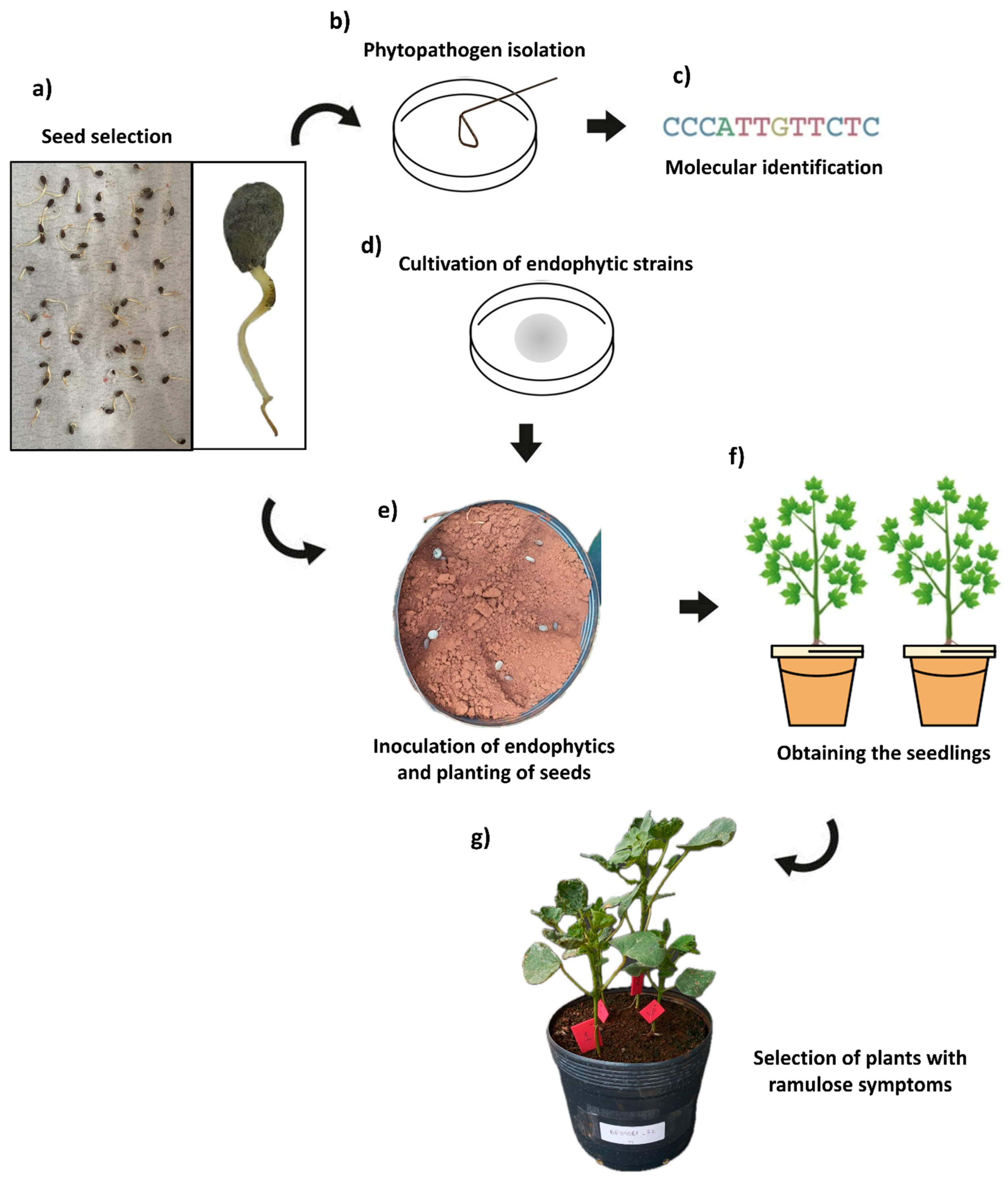
2.1. Isolated Fungi and Seeds Contaminated with C. gossypii var. cephalosporioides

Tests were conducted using root-endophytic strains isolated from Butia purpurascens (Arecaceae). These strains are currently part of the culture collection at the Agricultural Microbiology Laboratory of the Federal Institute Goiano, Rio Verde, GO, Brazil. The strains were cultured on Potato Dextrose Agar medium (infusion of 200 g of potato, 20 g of dextrose, and 15 g of agar) for 7 days at 30 °C to obtain replicates of each culture. The evaluated strains were: BP10EF (Gibberella moniliformis), BP33EF (Hamigera insecticola), BP328EF (Codinaeopsis sp.), BP335EF (Gibberella moniliformis), and BP340EF (Aspergillus sp.). These strains were chosen because they exhibited antibiosis to C. gossypii var. cephalosporioides in previously conducted tests (see Section 2.2).
Cotton seeds contaminated with C. gossypii var. cephalosporioides were obtained through phytosanitary quality tests. Seeds of the variety TMG47-B2RF/2021 from 4 seed lots, free from treatment with fungicide or insecticide, were evaluated on germination paper. They were arranged on paper sheets moistened with distilled water, covered with plastic film, and placed in a BOD chamber, where they remained for 4 days at 35 °C (Figure 1a). Infestation with C. gossypii var. cephalosporioides was confirmed in 63% of the seeds, and one of the seed lots was used for the in vivo experiment.
Three seeds contaminated with C. gossypii var. cephalosporioides were aseptically placed on a plate containing PDA medium. These seeds were used to establish fungal cultures, which were purified and taken to the Biological Institute of São Paulo for molecular identification (Figure 1b,c). All cultures were molecularly identified as belonging to the species C. gossypii var. cephalosporioides. The identification was performed by partial sequencing of the internal transcribed spacer and the calmodulin and β-tubulin genes. For this, amplicons of 575, 532, and 478 nucleotides were obtained for these respective genes. Sequencing was performed using the Sanger method; for phylogenetic inference, sequences were paired by similarity to sequences in GenBank using BLASTn while considering homology greater than 99%.

2.2. In Vitro Antibiosis Tests

In vitro antibiosis tests using the paired culture technique were performed to better understand the interaction between C. gossypii var. cephalosporioides and the endophytic strains tested. Thus, 5-cm-diameter mycelial discs of the phytopathogen and endophytic fungi were placed equidistantly on plates containing BDA medium. These plates were then incubated at 30 °C and left to rest for 72 h, when the diameters of colony halos were measured. Plates with the phytopathogen, without inoculation with the endophytic strains, were used as controls.
The test was conducted in triplicate for each endophytic strain tested, and the data were used to estimate the percentage of inhibition of phytopathogenic fungus growth induced by the endophytic strains. This was calculated through the relative inhibition index (RII):
RII   ( 100 % ) = RC RX RC × 100
where
RC = radius of the phytopathogen colony in the control treatment; and
RX = radius of the phytopathogen colony paired with the endophytic strain.

2.3. Preparation of the Plant Substrate and Seed Planting

The experiment was conducted in a greenhouse at the Tissue Culture Laboratory of the Federal Institute Goiano, in Rio Verde, GO, Brazil, from June to August 2022, at mean air temperature of 30.26 °C and relative air humidity of 29.43%.
The seeds were sanitized before planting through the asepsis process described by Reis et al. [41] to remove epiphytic microorganisms and ensure that only the endogenous contaminant C. gossypii var. cephalosporioides remained. The seeds were left to rest for 30 min and then planted in 3-kg pots containing a mixture of soil and nutritional substrate (Bioplant Garden®, Bio Plant Life, Santa Ana, CA, USA) at concentrations of 70% and 30%, respectively. This mixture was previously sterilized at 121 °C for 30 min to avoid interaction of seeds with microorganisms and subsequently kept in impermeable bags; it was placed in the pots only at the time of planting.
Five seeds were sown per pot, arranged in 3-cm-deep furrows; 5-mm-diameter mycelial discs of the tested endophytic fungi were inoculated directly onto the seeds to provide a simultaneous development of hyphae and radicles (Figure 1d,e).
The plants were evaluated daily for visual symptoms of ramulosis up to the V2 developmental stage. Some plants presented disease symptoms at the V0 stage (cotyledon) 14 days after planting, presenting small, dark, circular necrotic lesions, like those described by Talhinhas and Baroncelli [31] for lesions caused by C. gossypii var. cephalosporioides in the early stages of cotton development. Most plants presented differentiated leaf development and wrinkling of leaf blades 21 days after planting. Thinning was carried out at this time, maintaining only seedlings with ramulosis symptoms in the pots (two plants per pot) (Figure 1f,g). Emergence of leaf lesions, followed by a halt in branch growth, as well as emergence of new lateral buds, were found 30 days after planting. These new branches tended to form clusters characterized by an excess of nodes and internodes, resulting in plants with a bushy appearance, consistent with those described by Araújo [27] for infections by C. gossypii var. cephalosporioides in cotton plants. Thus, the occurrence of ramulosis was confirmed in all evaluated plants. The plants were irrigated daily according to their needs over the experimental period.

2.4. Biometric, Gas Exchange, and Chlorophyll-a Fluorescence Evaluations

Biometric and physiological evaluations were performed 30 days after planting, when ramulosis symptoms were confirmed in all cotton plants, at the vegetative phenological stage. Data on plant height (cm), stem diameter (cm), number of leaves, and shoot fresh and dry weights (g) were obtained. Dry weight was obtained after drying the plants in a forced air circulation oven at 65 °C until constant weight.
Physiology and antioxidant metabolism analyses were performed when the plants reached the V5 stage. Gas exchanges were evaluated using an infrared gas analyzer equipped with a fluorometer (LI-6400xt; LI-COR, Lincoln, NE, USA) to determine net photosynthetic rate (A; μmol m−2 s−1); intercellular CO2 concentration (Ci); stomatal conductance of water vapor (gs); transpiration rate (E mmol m−2 s−1); and the ratio of intercellular to ambient CO2 concentration (Ci/Ca). Measurements were always made on the youngest fully expanded leaf facing the sun, between 08:00 h and 11:00 h, using constant photosynthetically active radiation (1000 μmol photons m−2 s−1), with records of atmospheric CO2 concentration, relative air humidity, air temperature, and radiation. The carboxylation efficiency of plants was calculated using A/Ci.
The OJIP chlorophyll-a fluorescence was estimated using a portable fluorometer (FluorPen FP 100; Photon Systems Instruments, Drasov, Czech Republic). The fourth leaf of all sampled units was previously dark-adapted for 30 min for complete oxidation of the photosynthetic electron transport system and then subjected to a 3000 µmol m−2 s−1 pulse of blue light to measure the minimum fluorescence (F0) at 50 μs, when all photosystem II (PSII) reaction centers are open and defined as step O, followed by step J (2 ms), step I (30 ms), and maximum fluorescence (FM), when all PSII reaction centers are closed, known as step P. These values were used to estimate several bioenergetic indices of PSII, according to Strasser et al. [42].
The parameters estimated were: relatively low values of specific light absorption flux per active reaction center (ABS/RC); trapped per reaction center (TR0/RC); electron transport flux per reaction center (ET0/RC); specific energy dissipation flux at the antenna chlorophyll level (DI0/RC); photosynthetic performance index (PIABS), which incorporates the cascade of energy events from initial absorption to PQ reduction; maximum quantum yield of primary photochemistry (PHIP0); quantum yield of energy dissipation (PHID0); and quantum yield of electron transport (PHIE0).

2.5. Extraction and Activity of Antioxidant Metabolism Enzymes and Malondialdehyde (MDA)

The activity of enzymes from the antioxidant and lipid peroxidation systems was quantified. Samples were collected, placed in liquid nitrogen, and stored in an ultrafreezer at −80 °C.
Enzyme extraction was carried out as follows: 200 mg of leaf tissues were macerated in liquid nitrogen with 50% PVPP and following the extraction protocol proposed by Biemelt et al. [43], with an extraction buffer composed of 100 mM potassium phosphate (pH 7.8), 0.1 mM EDTA, and 10 mM ascorbic acid. The extract was centrifuged at 13,000× g for 10 min at 4 °C, and the supernatant was used to evaluate the activity of catalase (CAT), ascorbate peroxidase (APX), guaiacol peroxidase (POD), and superoxide dismutase (SOD).
CAT activity was evaluated according to the methodology proposed by Havir and McHale [44]: An aliquot of the enzyme extract was added to an incubation medium containing 100 mM potassium phosphate (pH 7.0) and 12.5 mM hydrogen peroxide. Enzyme activity was determined based on the consumption of H2O2 every 15 s for 3 min at 240 nm in a spectrophotometer. The molar extinction coefficient used was 36 mM−1 cm−1. CAT activity was expressed as µmol H2O2 min−1 mg−1 protein.
APX activity was evaluated using the methodology of Nakano and Asada [45], considering an ascorbate oxidation rate of 290 nm every 15 s for 3 min. An aliquot of the enzyme extract was added to a medium containing 100 mM potassium phosphate buffer (pH 7.0), 0.5 mM ascorbic acid, and 0.1 mM peroxide of hydrogen. The molar extinction coefficient used was 2.8 mM−1 cm−1. APX activity was expressed as µmol AsA min−1 mg−1 protein.
POD activity was evaluated using the methodology of Fang and Kao [46], considering the formation of tetraguaiacol by the increase in absorbance. An aliquot of the enzyme extract was added to a medium containing 50 mM sodium phosphate buffer (pH 6.0) and 0.13% guaiacol; 0.15% H2O2 was added before the spectrophotometric readings at 470 nm for 3 min. The molar extinction coefficient used was 26.6 mM−1 cm−1. POD activity was expressed as µmol H2O2 min−1 mg−1 protein. Proteins from leaf samples were quantified according to the Bradford method [47]. The absorbances were read at 595 nm, and the final data were used to express the enzyme activities.
SOD activity was determined based on the methodology of Giannopolitis and Ries [48], considering the enzyme’s ability to inhibit photoreduction of nitro-blue tetrazolium (NBT). An aliquot of the extract was incubated in a medium containing 50 mM potassium phosphate (pH 7.8), 14 mM methionine, 0.1 µM EDTA, 75 µM NBT, and 2 µM riboflavin. The samples, along with the incubation medium, were illuminated with a 20-W fluorescent lamp for 7 min. Readings were taken with a spectrophotometer at 560 nm. SOD activity was expressed as U mg−1 protein (1U = quantity of enzymes needed to inhibit NBT photoreduction by 50%).
Malondialdehyde (MDA) was quantified by macerating 200 mg of leaf tissue in liquid nitrogen and PVPP, followed by homogenization in 0.1% (m v−1) trichloroacetic acid (TCA) and centrifugation at 10,000× g for 15 min at 4 °C. The amount of MDA was determined using the methodology proposed by Buege and Aust [49].

2.6. Experimental Design and Statistical Analyses

The in vitro antibiosis experiment was conducted in a completely randomized experimental design with 5 treatments (exposure of the phytopathogen to five endophytic fungal strains), and the greenhouse experiment was conducted in a randomized block design with 6 treatments (5 endophytic fungal strains and a control). Plants inoculated with culture medium discs without mycelium were used as controls. The experiments were conducted with five replications per treatment, considering two plants per pot as one replication, totaling 60 units. The data obtained for the treatments were subjected to a one-way ANOVA to evaluate the effect of inoculation with endophytic strains. When the effects were significant, the means were evaluated by Tukey’s test at a 5% significance level. Subsequently, all variables that showed significant differences were jointly evaluated in a correlation matrix and connected by means of principal component analysis (PCA). Considering that these variables had different units of measurement, PCA was recovered using standardized data to obtain a mean of 0 and a standard deviation of 1. The number of principal components was defined according to eigenvalues (>1.0) and variance explained (>70%). The statistical tests were conducted using the R 4.3.2 program [50].
The similarity matrix was developed to estimate similarities or differences among plants from different treatments. The similarity index was obtained using the Pearson correlation coefficient, with values of r transformed into d = (1 − r) × 100 to estimate the distance (d). A dendrogram was then recovered using the unweighted pair group method with arithmetic mean (UPGMA), with an adjustment between the distance matrix and the dendrogram estimated by the cophenetic correlation coefficient [51]. This analysis was conducted using the DendroUPGMA program [52].

3. Results

3.1. Antibiosis of Endophytic Strains of Colletotrichum gossypii var. cephalosporioides

The antibiosis test showed expressive mycelial development of all endophytic fungal strains in relation to the phytopathogen, which resulted in the inhibition of growth of C. gossypii var. cephalosporioides (Figure 2a–e). The estimated relative inhibition index (RII) confirmed the potential of all endophytic strains to inhibit the in vitro growth of C. gossypii var. cephalosporioides, as the mean RIIs found were, in general, higher than 72% (Figure 2b). However, the comparison of strains showed that BP328EF (Codinaeopsis sp.) and BP335 (Gibberella moniliformis) were more effective in inhibiting the phytopathogen, showing the highest RII means (81.11% and 79.81%, respectively).

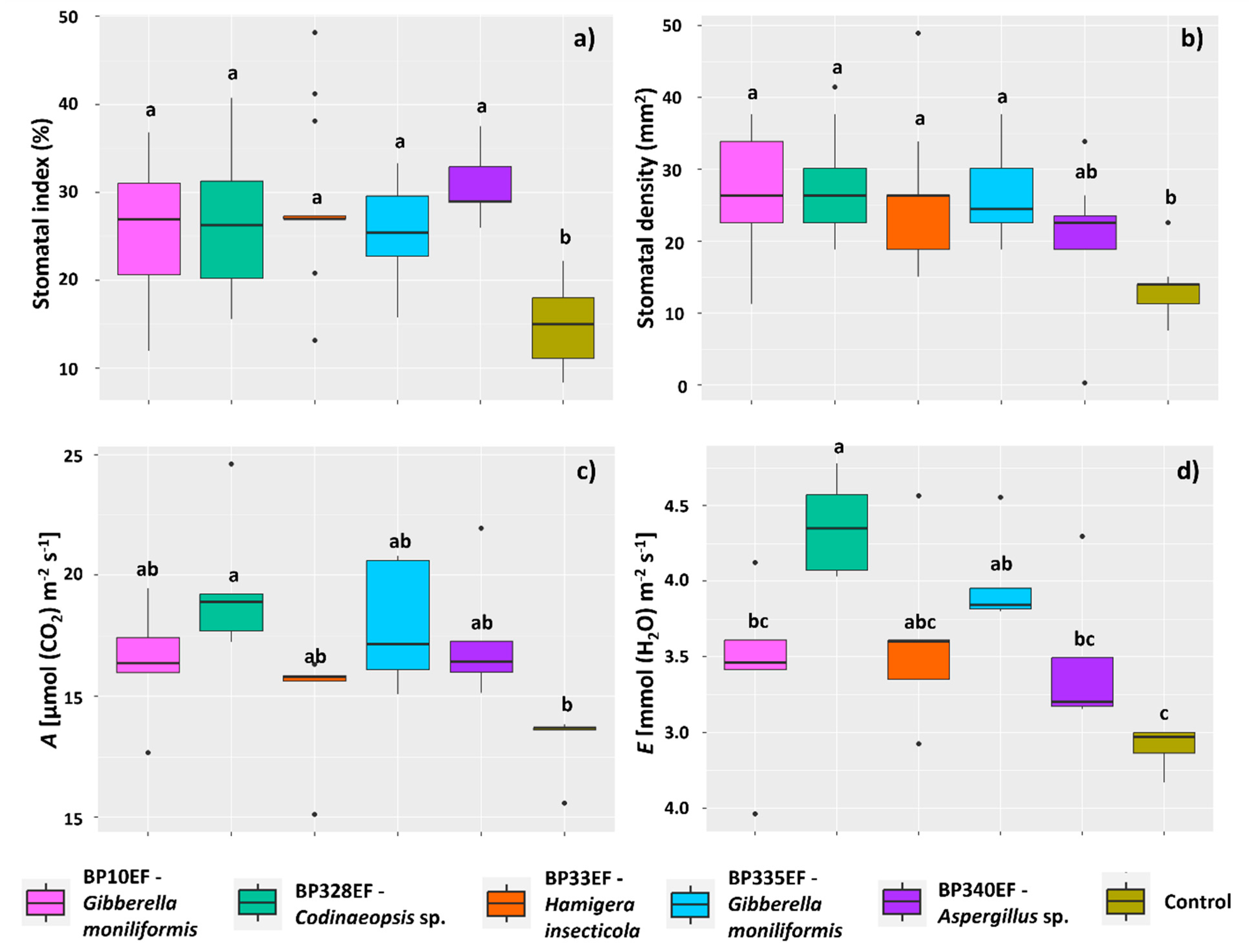
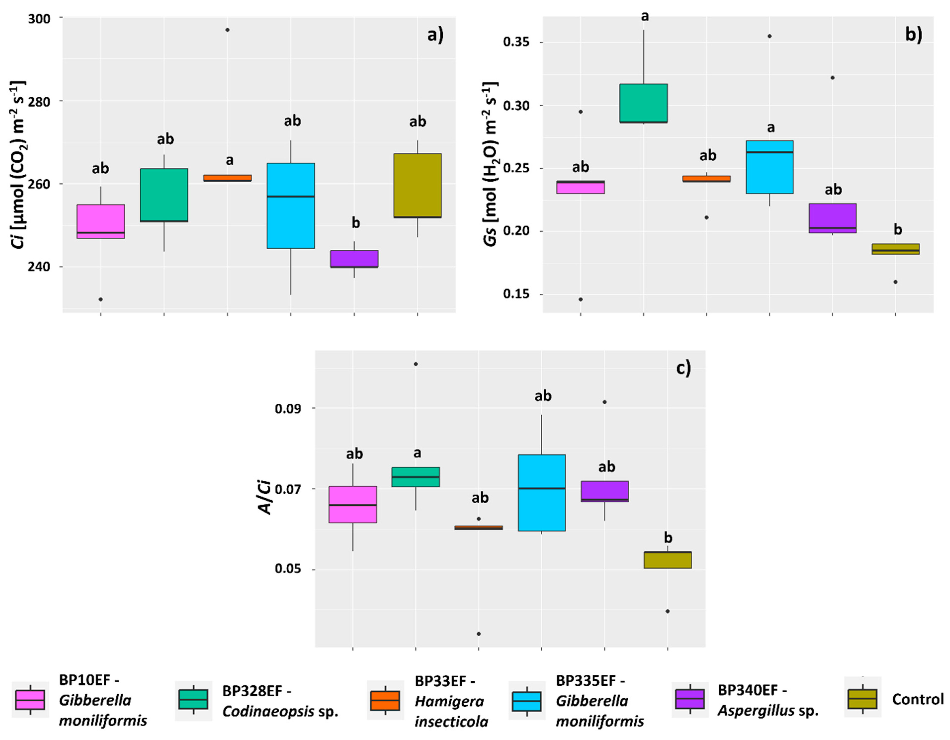
3.2. Growth and Physiology of Cotton Plants Infected by Colletotrichum gossypii var. cephalosporioides and Subjected to Inoculation with Endophytic Strains

In general, the inoculation with endophytic fungi positively affected the development of cotton plants infected with C. gossypii var. cephalosporioides. Control plants presented a lower mean height (17.31 cm) than plants inoculated with BP10EF (24.17 cm) (Figure 3a). Stem diameter was also affected by fungal treatments; the highest means were found for plants inoculated with BP33EF (0.20 cm) (Figure 3b). Shoot fresh weight (33.50 g) and shoot dry weight (6.42 g) of control plants were also lower than those of inoculated plants; the highest means were found for plants inoculated with BP33EF (51.50 and 12.82 g, respectively) (Figure 3c,d).
Inoculation with endophytic fungi had no effect on the stomatal index or stomatal density on the adaxial surface of cotton leaves affected by ramulosis; however, on the adaxial surface, control plants had the lowest percentage of stomata (14.96%). However, plants subjected to the different endophytic strains did not show any difference in adaxial stomatal index (Figure 4a). Stomatal density showed similar results, with lower means in control plants (12.87) and in plants inoculated with BP340EF (20.72) (Figure 4b).
Regarding gas exchanges, fungal inoculation, in general, tended to improve the net photosynthetic rate (A) in plants affected by ramulosis; however, higher A means were found for plants inoculated with BP328EF (19.53 µmol (CO2) m−2 s−1) (Figure 4c). Inoculation with BP328EF, however, significantly increased the transpiration rate (4.36 mmol (H2O) m−2 s−1); control plants had the lowest water loss (2.90 mmol (H2O) m−2 s−1) (Figure 4d).
The inoculation treatments had no effect on intercellular CO2 concentration (Ci), which showed similar means to the control; however, Ci tended to increase in plants inoculated with BP33EF (268.24 µmol (CO2) m−2 s−1) and decrease in plants inoculated with BP340EF (241.49 µmol (CO2) m−2 s−1) (Figure 5a). Stomatal conductance (Gs) was higher in plants inoculated with BP328EF and BP335EF (respectively 0.30 and 0.26 mol (H2O) m−2 s−1) compared with control plants (0.23 mol (H2O) m−2 s−1) (Figure 5b). Carboxylation efficiency (A/Ci) was also affected by inoculation with endophytic strains, tending to be higher in plants inoculated with BP328EF (0.07) and lower in control plants (0.05).
The inoculation treatments tended to reduce the light absorption flux per active reaction center (ABS/RC) of chlorophyll-a, as this index was high only in control plants (2.53) and in plants inoculated with BP33EF (2.58) (Figure 6a). The electron transport flux per reaction center (ET0/RC) was also higher in control plants (1.22), but was significantly lower in plants inoculated with BP328EF (1.07) (Figure 6b). The results found for the trapped energy flux per reaction center (TR0/RC) were similar to those of ET0/RC, with higher means in control plants (1.88) and lower in plants inoculated with BP328EF (1.78) (Figure 6c). The results found for the specific energy dissipation flux at the antenna chlorophyll level (DI0/RC) were identical to those found for ABS/RC, denoting higher energy dissipation as heat in control plants (0.55) and plants inoculated with BP33EF (0.54) (Figure 6d).
Inoculation with endophytic fungal strains maintained higher levels of maximum quantum yield of primary photochemistry (PHIP0) in cotton plants compared with control plants (0.78), except for plants inoculated with BP33EF (0.78) (Figure 7a). The quantum yield of energy dissipation (PHID0), however, showed opposite results, with the highest means in these same treatments, i.e., plants inoculated with BP33EF and control plants (0.21 and 0.20, respectively) (Figure 7b). However, the highest quantum yield of electron transport (PHIE0) was found in plants inoculated with BP340EF (0.49) (Figure 7c). The photosynthetic performance index (PIABS) was lower for chlorophylls of plants inoculated with BP33EF (2.07) and control plants (2.08) (Figure 7d).
Endophytic fungi inoculation in cotton plants exhibiting ramulosis symptoms considerably decreased CAT synthesis in leaf tissues; the highest mean CAT activity was found in control plants (260.27 µmol (H2O) min−1 mg−1 protein) (Figure 8a). The enzyme POD showed opposite results, with lower mean activity in control plants (26,795.18 µmol (H2O) min−1 mg−1 protein) and in plants inoculated with BP33EF (26,212.17 µmol (H2O) min−1 mg−1 protein); this enzyme showed the highest mean activity in leaves of plants inoculated with BP335EF (85,554.79 µmol (H2O) min−1 mg−1 protein) (Figure 8b).
The activity of the enzymes SOD and APX was similar to that found for CAT; it was stimulated mainly in control plants (0.010 U mg−1 protein for SOD and 3309.91 µmol AsA min−1 mg−1 protein for APX). However, plants in the different inoculation treatments showed no significant differences in the activity of these enzymes (Figure 9a,b). Lipid peroxidation (given by the amount of MDA) in control plants and plants inoculated with BP340EF tended to be higher (179.28 and 165.60 ηmol g−1, respectively) (Figure 9c).
Principal Components 1 and 2 together explained 99.0% of the data variance. This analysis confirmed the trend that oxidative metabolism (activity of enzymes CAT, APX, and SOD) and the cell damage caused by it, given the MDA production, were more active in control plants (Figure 10a). Similarly, control plants and plants inoculated with the endophytic strain BP33EF showed higher results for chlorophyll-a fluorescence parameters (ABS/RC, ET0/RC, TR0/RC, and DI0/RC), which are indicators of photochemical stress. Plants inoculated with BP33EF showed the best plant growth performance (stem diameter and shoot fresh and dry weights). Plants inoculated with BP10EF, BP328EF, BP335EF, and BP340EF showed the best photosynthetic indices and photochemical yields. The treatment with BP328EF explained the largest variations in the means of A, E, Gs, and A/Ci. The cluster analysis showed two stable clusters, confirming the efficiency of fungal inoculation and isolated control plants in the individual cluster (Figure 10b). The similarity between BP328EF and BP10EF grouped these plants in the same cluster, connected to BP33EF. However, divergent means of plant growth and photochemical variables found for BP33EF resulted in a slight separation. Another grouping was established by the similarity between the means found for BP335EF and BP340EF.

4. Discussion

4.1. Inoculation with Endophytic Fungi Mitigates Physiological and Photochemical Damage by Ramulosis in Cotton Plants

The results corroborate those presented by several studies in the literature, confirming the ability of some endophytic fungi to reduce lesions caused by pathogens through direct antibiosis, production of lytic enzymes, or activation of hormones [1]. However, beneficial microorganisms compete strongly with pathogens for niche colonization and nutrient acquisition [53]. Therefore, endophytic fungal strains can assist in the development of resistance to pests and diseases by affecting the pathogen’s development or reproduction. Studies have confirmed that these fungi can activate ISR (induced systemic resistance) and ASR (acquired systemic resistance) by activating microbial-associated molecular patterns (MAMPs) [54,55,56]. These patterns result in the production of signaling molecules, such as salicylic acid and ethylene. Thus, the colonization of endophytic fungi causes a first activation, making plants more capable of responding to phytopathogenic microorganisms and nematodes [57]. However, the endophytic relationship possibly confers additional defense mechanisms to modulate the plant immune system because of the manipulation of antimicrobial metabolites, either directly, such as alkaloids, or indirectly, such as phytohormones, jasmonic acid, or salicylic acid [55].
The present study confirms the potential of endophytic fungi, biotrophic fungi, and/or necrotrophic fungi to mitigate the biotic stress caused by ramulosis in cotton. The tested strains BP10EF and BP335EF of G. moniliformis (Fusarium verticillioides anamorph) can switch from biotrophic to necrotrophic states, as the biotrophic state can encompass an endophytic condition. Many studies have reported the occurrence of G. moniliformis with an endophytic habit [58,59] and confirmed the biotechnological potential of endophytic strains of this species, including for biocontrol [60,61]. The efficacy of BP10EF and BP335EF in controlling ramulosis can be explained by the synthesis of metabolites such as trioleoylglycerol (triolein), naphthoquinone (lawsone), and tricarballylic acid (Fumonisin A–C and P) [60,62].
The strains BP340EF (Aspergillus sp.) and BP335EF (G. moniliformis) showed similar behavior, forming one cluster. Endophytic Aspergillus species also produce active molecules associated with the biocontrol functional trait, such as butyrolactones, stigmasterol derivatives, and meroterpenoids e.g., [63,64]. El-hawary et al. [65] reported that different Aspergillus species can produce secondary metabolites, including butenolides, alkaloids, terpenoids, cytochalasins, phenalenones, ρ-terphenyls, xanthones, steroids, diphenyl ether, and anthraquinone derivatives, with diverse biological activities, including antifungal and antibacterial effects. Verma et al. [66] indicated that the biosynthesis of silver nanoparticles using an endophytic strain of Aspergillus clavatus produces an efficient fungicidal compound for the control of Candida albicans, thus reaffirming the importance of species of this genus in biocontrol processes.
The potential of lesser-known fungi was also evaluated. The endophytic strain BP328EF (Codinaeopsis sp.) showed a relative inhibition of in vitro growth of C. gossypii var. cephalosporioides by 81.11%. The genus Codinaeopsis encompasses soil fungi capable of synthesizing polyketide codinaeopsin. This metabolite contains an unusual heterocyclic unit that binds indole and decalin fragments and exhibits antimalaria activity [67,68]. Little is known about the effects of inoculating agronomically important plants with fungi of this genus. However, the Hamigera insecticola strain (BP33EF) showed potential as a growth promoter in cotton plants, considering its effect on the evaluated biometric characteristics. Studies have confirmed the antifungal potential of species in this genus due to the synthesis of hamigerone and dihydrohamigerone metabolites [69] and silver nanoparticles produced from Hamigera terricola, which showed antifungal potential against phytopathogenic species [70].
Besides biocontrol processes and the induction of resistance, studies have confirmed that endophytic fungi can improve plant growth and development [71,72]. Russo et al. [73] showed that species of endophytic entomopathogenic fungi can exhibit traits resulting in the promotion of soybean (Glycine max) growth, improving plant biometric development, and increasing grain yield under field conditions. Galeano et al. [74] indicated that the potential of Aspergillus species to promote plant growth should be considered. Hamayum et al. [75] showed that Aspergillus flavus can mitigate the effects of biotic stress from high temperatures on soybean and sunflower plants; they found significant quantities of indoleacetic acid (IAA), salicylic acid (SA), flavonoids, and phenolic compounds in cultures of this fungus; inoculated plants showed higher dry weight accumulation and chlorophyll contents and lower quantities of abscisic acid (ABA) and proline. This species can mitigate the effects of stress from high salt concentrations and high temperatures on soybean and sunflower plants by regulating endogenous hormones and the antioxidant system [76,77]. Gibberellins produced by Aspergillus fumigatus significantly increased shoot length, fresh and dry weights, leaf area, chlorophyll contents, and photosynthetic rate of soybean plants under salt stress [78]. Additionally, Saxena et al. [79] showed that Aspergillus niger can promote the growth of G. max through phosphate solubilization. In contrast to its saprophytic and pathogenic identity, the ability of filamentous Aspergillus fungi to solubilize insoluble phosphates, such as Ca, Fe, and Al phosphates, has stood out [80]. Similarly, Radhakrishnan et al. [81] highlighted the potential of G. moniliformis to promote plant growth through phosphate solubilization. They showed that soybean plants inoculated with G. moniliformis and subjected to salt stress became more resistant, as this fungus solubilized large quantities of phosphates, reducing oxidative damage and ABA concentrations in leaves, and increasing salicylic acid contents. This explains the growth promotion effects found using the strain BP340EF, mainly in terms of shoot dry weight, stomatal index, carboxylation efficiency, and photochemical yield, as well as the strains BP10EF and BP335EF, mainly in terms of the activation of oxidative stress enzymes.
Fungal inoculation increased stomatal index and density on the adaxial surface of cotton leaves affected by ramulosis, resulting in increased net photosynthetic rate, transpiration, and stomatal conductance with greater carboxylation efficiency. This occurs because stomata serve as an innate immune barrier against infections [82]. Thus, stomatal closure and decreased stomatal index and density are plant strategies to minimize pathogen infection e.g., [83]. Decreases in stomatal density and/or size can significantly affect photosynthesis; thus, plants seem to have a compensation system involving the advantages of reducing gas exchange to prevent the penetration of pathogens [84]. Therefore, as the seeds used in the present study were infected with C. gossypii var. cephalosporioides, control plants developed leaves with a low stomatal index and density to decrease the possibility of colonization of leaf tissues by C. gossypii or other pathogens, which could make the situation of a plant affected by ramulosis even more critical.

4.2. Endophytic Fungi Differentially Affect Growth, Gas Exchange, and Primary Photochemistry of Cotton Plants Affected by Ramulosis

These results enable discussions about specific functional traits expressed by different lines. Considering the widespread use of biological products to improve disease control and yield in agriculture, species of interest still need to be evaluated and utilized, considering their specific biological functionality, for a better understanding of the effect of mechanisms underlying microbial activity on the plant-microorganism interaction. For instance, the potential of the H. insecticola strain BP33EF to promote plant growth was evaluated in the present study. This strain, however, did not alleviate the primary photochemical stress induced by ramulosis in cotton leaves, showing similar ABS/RC and DI0/RC to those found in control plants. Consequently, photosynthetic performance (PIABS) was low in these plants. In such cases, compensation studies should be conducted to assess the actual yield gain when using growth-promoting strains that either trigger or do not mitigate metabolic stress processes.

4.3. The Codinaeopsis sp. Strain BP328EF Inhibits the In Vitro Growth of Colletotrichum gossypii var. cephalosporioides, Positively Affects Gas Exchange, and Reduces Chlorophyll-a Photochemical Stress and Lipid Peroxidation

Promising results were found for the use of the Codinaeopsis sp. strain BP328EF to mitigate the effects of ramulosis in cotton plants. This genus is not reported in the literature as associated with phytopathogenic characteristics or pathogenicity in animals. Codinaeopsis (=Codinea) is a polyphyletic genus encompassing phialidic and dematiaceous hyphomycetes [85], known for their intriguing morphology and turbulent taxonomic history. Codinaea and its segregates thrive on decomposing plants, rarely occurring as endophytes or plant pathogens. Environmental DNA and ITS sequences indicate their common occurrence in bulk soils. These fungi evolved mainly in Eurasia and the Americas, with subsequent transitions to Africa and Australasia [86]. Little is known about the effects of fungi in this genus on plant growth promotion and stress mitigation. However, the results found in the present study are confirmed by those found by Reis et al. [41], who not only evaluated the potential of Codinaeopsis sp. to promote plant growth but also its ability to improve nutrient absorption by G. max plants; plant responses regarding chlorophyll index, shoot dry weight, and nutrient concentration (N, P, and Mg) were similar to those of plants treated with a commercial product (Biomaphos®, Bioma SA, Quartino, Switzerland) composed of phosphate-solubilizing bacteria. Similarly, an endophytic strain of Codinaea sp. significantly affected the elongation of rooted cuttings from different cranberry cultivars [87].
Therefore, field tests should be conducted to assess the effects of applying Codinaeopsis sp. to inhibit ramulosis occurrence in cotton plantations. This study contributes new approaches, proposing a biological alternative to improve G. hirsutum plants and minimize yield losses due to colonization by C. gossypii var. cephalosporioides. Thus, mitigating damage caused by endophytic fungi in ramulosis-affected plants encourages the use of new sustainable management practices regarding phytopathogen control in cotton fields. However, the results presented in this study are expected to stimulate prospective studies of endophytic microorganisms in endemic plants of the Cerrado biome, opening prospects for new applications for biological control of pests and diseases in cotton and other important agricultural crops.

5. Conclusions

The hypothesis that inoculation of cotton plants with endophytic fungi can attenuate the physiological and photochemical damage caused by ramulosis was confirmed. Overall, endophytic fungi improved plant growth, stomatal index and density, net photosynthetic rate, and carboxylation efficiency while decreasing photochemical and oxidative stresses. Control plants developed leaves with a low adaxial stomatal index and density as a strategy to reduce the likelihood of colonization of leaf tissues by Colletotrichum gossypii var. cephalosporioides due to the absence of fungal antagonism. The effects of relative inhibition of in vitro growth of C. gossypii var. cephalosporioides by the activity of the Codinaeopsis sp. strain BP328EF were explained as improvements in gas exchange parameters and reductions in chlorophyll-a photochemical stress and lipid peroxidation in cotton plants. This study aims to contribute to the development of biological alternatives for improving resistance in cotton (G. hirsutum) plants and minimizing yield losses caused by colonization by C. gossypii.

Author Contributions

Conceptualization, L.C.V. and L.A.B.; methodology, E.L.S. and C.R.; software, M.N.O.R.; formal analysis, I.d.O.S. and D.S.S.A.; investigation, I.d.O.S. and D.S.S.A.; resources, L.C.V. and L.A.B.; writing—original draft preparation, I.d.O.S.; writing—review and editing, L.C.V.; visualization, M.N.O.R. and L.C.V.; supervision, L.A.B.; project administration, L.A.B.; funding acquisition, L.C.V. All authors have read and agreed to the published version of the manuscript.

Funding

This research received no external funding.

Data Availability Statement

All the data relevant to this manuscript are available on request from the corresponding author.

Acknowledgments

The authors thank the Federal Institute Goiano (IFGoiano, Rio Verde campus) for allowing the use of their Laboratories and supplies, and the students involved in this study. They also thank the Foundation for Research Support of the State of Goiás, the Brazilian Coordination for the Improvement of Higher Education Personnel (CAPES), and the Brazilian National Council for Scientific and Technological Development (CNPq) for supporting many research projects of this study group through the granting of scientific initiation scholarships to Isabella de Oliveira Silva and productivity scholarships to Luciana Cristina Vitorino and Layara Alexandre Bessa.

Conflicts of Interest

Authors Isabella de Oliveira Silva, Layara Alexandre Bessa, Damiana Souza Santos Augusto and Luciana Cristina Vitorino are employed by the company Simple Agro Corporation. The remaining authors declare that the research was conducted in the absence of any commercial or financial relationships that could be construed as a potential conflict of interest.

References

  1. Tyagi, J.; Chaudhary, P.; Jyotsana, U.B.; Bhandari, G.; Chaudhary, A. Impact of endophytic fungi in biotic stress management. In Plant Protection: From Chemicals to Biologicals; Co, K.G., Ed.; Walter de Gruyter GmbH: Berlin, Germany, 2022; pp. 447–462. [Google Scholar] [CrossRef]
  2. Kamburova, V.; Salakhutdinov, I.; Abdurakhmonov, I.Y. Cotton breeding in the view of abiotic and biotic stresses: Challenges and perspectives. In Cotton; Abdurakhmonov, I.Y., Ed.; IntechOpen: London, UK, 2022. [Google Scholar] [CrossRef]
  3. Meyer, L.A.; Dew, T. Cotton and Wool Outlook: December 2021; US Department of Agriculture, Economic Research Service: Washington, DC, USA, 2021; p. 5542. Available online: https://www.ers.usda.gov/webdocs/outlooks/102828/cws-21k.pdf (accessed on 27 March 2023).
  4. Tarazi, R.; Jimenez, J.L.S.; Vaslin, M.F. Biotechnological solutions for major cotton (Gossypium hirsutum) pathogens and pests. Biotechnol. Res. Innov. 2019, 3, 19–26. [Google Scholar] [CrossRef]
  5. Cox, K.L.; Babilonia, T.W.; He, P.; Shan, L. Return of old foes—Recurrence of bacterial blight and Fusarium wilt of cotton. Curr. Opin. Plant Biol. 2019, 50, 95–103. [Google Scholar] [CrossRef] [PubMed]
  6. Zhu, Y.; Zhao, M.; Li, T.; Wang, L.; Liao, C.; Liu, D.; Li, B. Interactions between Verticillium dahliae and cotton: Pathogenic mechanism and cotton resistance mechanism to Verticillium wilt. Front. Plant Sci. 2023, 14, 1174281. [Google Scholar] [CrossRef] [PubMed]
  7. Shete, P.P.; Kasal, Y.G.; Perane, R.R. Screening of the cotton genotypes against Ramularia areola atk. under field condition. Plant Arch. 2018, 18, 734–736. [Google Scholar]
  8. Ghaffar, A. Biological control of sclerotial diseases. In Biocontrol of Plant Diseases; Mukerji, K.G., Garg, K.L., Eds.; CRC Press: Boca Raton, FL, USA, 2023; pp. 153–176. [Google Scholar]
  9. Makwana, N.; Rawal, P. Cultural, morphological and pathogenic variability of Colletotrichum gossypii causing anthracnose of cotton. J. Mycol. Plant Pathol. 2022, 52, 395–404. [Google Scholar]
  10. Salustiano, M.E.; Rondon, M.N.; Abreu, L.M.; Costa, S.S.; Costa, J.C.; Machado, L.H. The etiological agent of cotton ramulosis represents a single phylogenetic lineage within the Colletotrichum gloeosporioides species complex. Trop. Plant Pathol. 2014, 39, 357–367. [Google Scholar] [CrossRef]
  11. Moreno-Moran, M.; Burbano-Figueroa, O. Field resistance of advanced breeding lines of upland cotton to ramulosis caused by Colletotrichum gossypii var. cephalosporioides. Crop Prot. 2019, 122, 49–56. [Google Scholar] [CrossRef]
  12. Guerra, A.M.N.D.M.; Rodrigues, F.Á.; Lima, T.C.; Berger, P.G.; Barros, A.F.; Silva, Y.C.R.D. Photosynthetic capacity of cotton boll rot infected plants and supplied with silicon. Bragantia 2014, 73, 50–64. [Google Scholar] [CrossRef]
  13. De Araújo, A.E.; Ferreira, A.D.B.; Morello, C.D.L. Damage caused in cotton by different levels of ramulosis in Brazil. In Proceedings of the World Cotton Research Conference-5, Mumbai, India, 7–11 November 2011; Volume 7, pp. 290–292. [Google Scholar]
  14. Joshua, J.; Mmbaga, M.T. Potential biological control agents for soilborne fungal pathogens in Tennessee snap bean farms. HortScience 2020, 755, 988–994. [Google Scholar] [CrossRef]
  15. Adeleke, B.S.; Ayilara, M.S.; Akinola, S.A.; Babalola, O.O. Biocontrol mechanisms of endophytic fungi. Egypt. J. Biol. Pest Control 2022, 32, 46. [Google Scholar] [CrossRef]
  16. Fontana, D.C.; De Paula, S.; Torres, A.G.; De Souza, V.H.M.; Pascholati, S.F.; Schmidt, D.; Dourado Neto, D. Endophytic fungi: Biological control and induced resistance to phytopathogens and abiotic stresses. Pathogens 2021, 10, 570. [Google Scholar] [CrossRef]
  17. Santra, H.K.; Banerjee, D. Fungal endophytes: A source for biological control agents. In Agriculturally Important Fungi for Sustainable Agriculture: Functional Annotation for Crop Protection; Yadav, A., Mishra, S., Kour, D., Yadav, N., Kumar, A., Eds.; Springer: Cham, Switzerland, 2020; Volume 2, pp. 181–216. [Google Scholar] [CrossRef]
  18. Hassani, M.A.; Duran, P.; Hacquard, S. Microbial interactions within the plant holobiont. Microbiome 2018, 6, 58. [Google Scholar] [CrossRef] [PubMed]
  19. Bard, N.W.; Cronk, Q.C.; Davies, T.J. Fungal endophytes can modulate plant invasion. Biol. Rev. 2024. [Google Scholar] [CrossRef] [PubMed]
  20. Jin, L.; Yang, L.; Li, W.; Xu, D.; Yang, N.; Li, G.; Wan, P. Diversity and biocontrol potential of culturable endophytic fungi in cotton. Front. Microbiol. 2021, 12, 698930. [Google Scholar] [CrossRef] [PubMed]
  21. Wei, F.; Zhang, Y.; Shi, Y.; Feng, H.; Zhao, L.; Feng, Z.; Zhu, H. Evaluation of the biocontrol potential of endophytic fungus Fusarium solani CEF559 against Verticillium dahliae in cotton plant. Biomed. Res. Int. 2019, 2019, 3187943. [Google Scholar] [CrossRef] [PubMed]
  22. Yuan, Y.; Feng, H.; Wang, L.; Li, Z.; Shi, Y.; Zhao, L.; Zhu, H. Potential of endophytic fungi isolated from cotton roots for biological control against verticillium wilt disease. PLoS ONE 2017, 12, e0170557. [Google Scholar] [CrossRef]
  23. Gasoni, L.; de Gurfinkel, S. Biocontrol of Rhizoctonia solani by the endophytic fungus Cladorrhinum foecundissimum in cotton plants. Australas. Plant Pathol. 2009, 38, 389–391. [Google Scholar] [CrossRef]
  24. Nawaz, H.H.; Rajaofera, M.N.; He, Q.; Anam, U.; Lin, C.; Miao, W. Evaluation of antifungal metabolites activity from Bacillus licheniformis OE-04 against Colletotrichum gossypii. Pestic. Biochem. Physiol. 2018, 146, 33–42. [Google Scholar] [CrossRef]
  25. Yadav, L.; Yadav, N.K.; Malik, V.K.; Yadav, P.; Yadav, N.; Vashisth, P.; Dhariwal, S. Evaluation of biological control agents against Colletotrichum gossypii under in vitro condition. Pharma Innov. J. 2012, 11, 3830–3832. [Google Scholar]
  26. Ferro, H.M.; Souza, R.; Lelis, F.; Vieira, M.; Silva, J.C.P.D.; Medeiros, F.H.V.D. Bacteria for cotton plant protection: Disease control, crop yield, and fiber quality. Rev. Caatinga 2020, 33, 43–53. [Google Scholar] [CrossRef]
  27. Araújo, D.V.; Zambenedetti, G.B.; Dallacort, R.; Azevedo, V.H.; Mainardi, J.T. Progresso da ramulose em campo a partir de sementes de algodoeiro inoculadas com Colletotrichum gossypii var. cephalosporioides. Trop. Plant Pathol. 2012, 37, 204–209. [Google Scholar] [CrossRef]
  28. Da Silva, C.F.; Vitorino, L.C.; Soares, M.A.; Souchie, E.L. Multifunctional potential of endophytic and rhizospheric microbial isolates associated with Butia purpurascens roots. Antonie Van Leeuwenhoek 2018, 111, 2157–2174. [Google Scholar] [CrossRef] [PubMed]
  29. Dos Reis, J.B.A.; do Vale, H.M.M.; Lorenzi, A.S. Insights into taxonomic diversity and bioprospecting potential of Cerrado endophytic fungi: A review exploring a unique Brazilian biome and methodological limitations. World J. Microbiol. Biotechnol. 2022, 38, 202. [Google Scholar] [CrossRef] [PubMed]
  30. Noriler, S.A.; Savi, D.C.; Aluizio, R.; Palacio-Cortes, A.M.; Possiede, Y.M.; Glienke, C. Bioprospecting and structure of fungal endophyte communities found in the Brazilian biomes, Pantanal, and Cerrado. Front. Microbiol. 2018, 9, 1526. [Google Scholar] [CrossRef] [PubMed]
  31. Talhinhas, P.; Baroncelli, R. Colletotrichum species and complexes: Geographic distribution, host range and conservation status. Fungal Divers. 2021, 110, 109–198. [Google Scholar] [CrossRef]
  32. Chhabra, R.; Kaur, S.; Vij, L.; Gaur, K. Exploring physiological and biochemical factors governing plant pathogen interaction: A review. Int. J. Curr. Microbiol. App. Sci. 2020, 9, 1650–1666. [Google Scholar] [CrossRef]
  33. Xing, J.; Li, M.; Li, J.; Shen, W.; Li, P.; Zhao, J.; Zhang, Y. Stem canker pathogen Botryosphaeria dothidea inhibits poplar leaf photosynthesis in the early stage of inoculation. Front. Plant Sci. 2022, 13, 1008834. [Google Scholar] [CrossRef] [PubMed]
  34. Yang, H.; Luo, P. Changes in photosynthesis could provide important insight into the interaction between wheat and fungal pathogens. Int. J. Mol. Sci. 2021, 22, 8865. [Google Scholar] [CrossRef] [PubMed]
  35. Yahya, M.; Saeed, N.A.; Nadeem, S.; Hamed, M.; Saleem, K. Effect of leaf rust disease on photosynthetic rate, chlorophyll contents, and grain yield of wheat. Arch. Phytopathol. Plant Prot. 2020, 53, 425–439. [Google Scholar] [CrossRef]
  36. Lu, Y.; Yao, J. Chloroplasts at the crossroad of photosynthesis, pathogen infection and plant defense. Int. J. Mol. Sci. 2018, 19, 3900. [Google Scholar] [CrossRef] [PubMed]
  37. Gahir, S.; Bharath, P.; Raghavendra, A.S. Stomatal closure sets in motion long-term strategies of plant defense against microbial pathogens. Front. Plant Sci. 2021, 12, 761952. [Google Scholar] [CrossRef] [PubMed]
  38. Grimmer, M.K.; John Foulkes, M.; Paveley, N.D. Foliar pathogenesis and plant water relations: A review. J. Exp. Bot. 2012, 63, 4321–4331. [Google Scholar] [CrossRef] [PubMed]
  39. Goyal, A.; Parashar, M. Organic cotton and BCI-certified cotton fibres. In Sustainable Fibres for Fashion and Textile Manufacturing; Nayak, R., Ed.; Woodhead Publishing: Sawston, UK, 2023; pp. 51–74. [Google Scholar]
  40. Cabanillas, C.; Tablada, M.; Ferreyra, L.; Pérez, A.; Sucani, G. Sustainable management strategies focused on native bio-inputs in Amaranthus cruentus L. in agroecological farms in transition. J. Clean. Prod. 2017, 142, 343–350. [Google Scholar] [CrossRef]
  41. Reis, M.N.O.; Vitorino, L.C.; Lourenço, L.L.; Bessa, L.A. Microbial inoculation improves growth, nutritional and physiological aspects of Glycine max (L.) Merr. Microorganisms 2022, 10, 1386. [Google Scholar] [CrossRef] [PubMed]
  42. Strasser, R.J.; Srivastava, A.; Tsimilli-Michael, M. The fluorescence transient as a tool to characterize and screen photosynthetic samples. In Probing Photosynthesis: Mechanisms, Regulation and Adaptation; CRC Press: Boca Raton, FL, USA, 2000; pp. 445–483. [Google Scholar]
  43. Biemelt, S.; Keetman, U.; Albrecht, G. Re-aeration following hypoxia or anoxia leads to activation of the antioxidative defense system in roots of wheat seedlings. Plant Physiol. 1998, 116, 651–658. [Google Scholar] [CrossRef] [PubMed]
  44. Havir, E.A.; McHale, N.A. Biochemical and developmental characterization of multiple forms of catalase in tobacco leaves. Plant Physiol. 1987, 84, 450–455. [Google Scholar] [CrossRef] [PubMed]
  45. Nakano, Y.; Asada, K. Hydrogen peroxide is scavenged by ascorbate-specific peroxidase in spinach chloroplasts. Plant Cell Physiol. 1981, 22, 867–880. [Google Scholar] [CrossRef]
  46. Fang, W.-C.; Kao, C.H. Enhanced peroxidase activity in rice leaves in response to excess iron, copper and zinc. Plant Sci. 2000, 158, 71–76. [Google Scholar] [CrossRef] [PubMed]
  47. Bradford, M.M. A rapid and sensitive method for the quantitation of microgram quantities of protein utilizing the principle of protein-dye binding. Anal. Biochem. 1976, 72, 248–254. [Google Scholar] [CrossRef] [PubMed]
  48. Giannopolitis, C.N.; Ries, S.K. Superoxide dismutases: I. Occurrence in higher plants. Plant Physiol. 1977, 59, 309–314. [Google Scholar] [CrossRef] [PubMed]
  49. Buege, J.A.; Aust, S.D. Microsomal lipid peroxidation. In Methods in Enzymology; Qin, P.Z., Ed.; Academic Press: New York, NY, USA, 1978; pp. 302–310. [Google Scholar] [CrossRef]
  50. R Core Team. R: A Language and Environment for Statistical Computing [Software]; R Foundation for Statistical Computing: Vienna, Austria, 2024; Available online: https://www.R-project.org/ (accessed on 27 March 2023).
  51. Sokal, R.R.; Rohlf, F.J. The comparison of dendrograms by objective methods. Taxon 1962, 11, 33–40. [Google Scholar] [CrossRef]
  52. Garcia-Vallve, S.; Palau, J.; Romeu, A. Horizontal gene transfer in glycosyl hydrolases inferred from codon usage in Escherichia coli and Bacillus subtilis. Mol. Biol. Evol. 1999, 16, 1125–1134. [Google Scholar] [CrossRef] [PubMed]
  53. Haas, D.; Keel, C. Regulation of antibiotic production in root-colonizing Pseudomonas spp. and relevance for biological control of plant disease. Annu. Rev. Phytopathol. 2003, 41, 117–153. [Google Scholar] [CrossRef] [PubMed]
  54. Lu, H.; Wei, T.; Lou, H.; Shu, X.; Chen, Q. A critical review on communication mechanism within plant-endophytic fungi interactions to cope with biotic and abiotic stresses. J. Fungi 2021, 7, 719. [Google Scholar] [CrossRef] [PubMed]
  55. Yan, L.; Zhu, J.; Zhao, X.; Shi, J.; Jiang, C.; Shao, D. Beneficial effects of endophytic fungi colonization on plants. Appl. Microbiol. Biotechnol. 2019, 103, 3327–3340. [Google Scholar] [CrossRef] [PubMed]
  56. Latz, M.A.; Jensen, B.; Collinge, D.B.; Jørgensen, H.J. Endophytic fungi as biocontrol agents: Elucidating mechanisms in disease suppression. Plant Ecol. Divers. 2018, 11, 555–567. [Google Scholar] [CrossRef]
  57. Poveda, J. Trichoderma as biocontrol agent against pests: New uses for a mycoparasite. Biol. Control 2021, 159, 104634. [Google Scholar] [CrossRef]
  58. Eo, J.K.; Choi, M.S.; Eom, A.H. Diversity of endophytic fungi isolated from Korean ginseng leaves. Mycobiology 2014, 42, 147–151. [Google Scholar] [CrossRef] [PubMed]
  59. Desjardins, A.E.; Busman, M.; Muhitch, M.; Proctor, R.H. Complementary host–pathogen genetic analyses of the role of fumonisins in the Zea mays–Gibberella moniliformis interaction. Physiol. Mol. Plant Pathol. 2007, 70, 149–160. [Google Scholar] [CrossRef] [PubMed]
  60. Kim, J.W.; Ryu, J.; Shim, S.H. Chemical investigation on an endophytic fungus Gibberella moniliformis JS1055 derived from a halophyte Vitex rotundifolia. Nat. Prod. Sci. 2018, 24, 189–193. [Google Scholar] [CrossRef]
  61. Sarang, H.; Rajani, P.; Vasanthakumari, M.M.; Kumara, P.M.; Siva, R.; Ravikanth, G.; Uma Shaanker, R. An endophytic fungus, Gibberella moniliformis from Lawsonia inermis L. produces lawsone, an orange-red pigment. Antonie Van Leeuwenhoek 2017, 110, 853–862. [Google Scholar] [CrossRef] [PubMed]
  62. Proctor, R.H.; Brown, D.W.; Plattner, R.D.; Desjardins, A.E. Co-expression of 15 contiguous genes delineates a fumonisin biosynthetic gene cluster in Gibberella moniliformis. Fungal Genet. Biol. 2003, 38, 237–249. [Google Scholar] [CrossRef] [PubMed]
  63. Ibrahim, S.R.; Elkhayat, E.S.; Mohamed, G.A.; Khedr, A.I.; Fouad, M.A.; Kotb, M.H.; Ross, S.A. Aspernolides F and G, new butyrolactones from the endophytic fungus Aspergillus terreus. Phytochem. Lett. 2015, 14, 84–90. [Google Scholar] [CrossRef] [PubMed]
  64. Bai, Z.-Q.; Lin, X.; Wang, J.; Wang, Y.; Zhou, X.; Yang, B.; Liu, J.; Wang, Y.; Liu, Y. New phenyl derivatives from endophytic fungus Aspergillus flavipes AIL8 derived of mangrove plant Acanthus ilicifolius. Fitoterapia 2014, 95, 194–202. [Google Scholar] [CrossRef] [PubMed]
  65. El-Hawary, S.S.; Moawad, A.S.; Bahr, H.S.; Abdelmohsen, U.R.; Mohammed, R. Natural product diversity from the endophytic fungi of the genus Aspergillus. RSC Adv. 2020, 10, 22058–22079. [Google Scholar] [CrossRef] [PubMed]
  66. Verma, V.C.; Kharwar, R.N.; Gange, A.C. Biosynthesis of antimicrobial silver nanoparticles by the endophytic fungus Aspergillus clavatus. Nanomedicine 2010, 5, 33–40. [Google Scholar] [CrossRef] [PubMed]
  67. Ramanathan, M.; Tan, C.J.; Chang, W.J.; Tsai, H.H.G.; Hou, D.R. Synthesis of the decalin core of codinaeopsin via an intramolecular Diels–Alder reaction. Org. Biomol. Chem. 2013, 11, 3846–3854. [Google Scholar] [CrossRef] [PubMed]
  68. Kontinik, R.; Clardy, J. Codinaeopsin, an antimalarial fungal polyketide. Org. Lett. 2008, 10, 4149–4151. [Google Scholar] [CrossRef] [PubMed]
  69. Breinholt, J.; Kjoer, A.; Olsen, C.E.; Rassing, B.R. Hamigerone and dihydrohamigerone: Tvvo acetate-derived, antifungal metabolites from Hamigera. Acta Chem. Scand. 1997, 51, 1241–1244. [Google Scholar] [CrossRef]
  70. Mistry, H.; Thakor, R.; Polara, H.; Shah, T.; Bariya, H. Biogenically efficient production and characterization of silver nanoparticles using the marine fungus Hamigera terricola along with their antimicrobial and antioxidative efficacy. In Nanotechnology and In Silico Tools; Kaneria, M., Rakholiya, K., Egbuna, C., Eds.; Elsevier: Amsterdam, The Netherlands, 2024; pp. 89–96. [Google Scholar] [CrossRef]
  71. Baron, N.C.; Rigobelo, E.C. Endophytic fungi: A tool for plant growth promotion and sustainable agriculture. Mycology 2022, 13, 39–55. [Google Scholar] [CrossRef] [PubMed]
  72. Poveda, J.; Eugui, D.; Abril-Urías, P.; Velasco, P. Endophytic fungi as direct plant growth promoters for sustainable agricultural production. Symbiosis 2021, 85, 1–19. [Google Scholar] [CrossRef]
  73. Russo, M.L.; Pelizza, S.A.; Vianna, M.F.; Allegrucci, N.; Cabello, M.N.; Toledo, A.V.; Scorsetti, A.C. Effect of endophytic entomopathogenic fungi on soybean Glycine max (L.) Merr. growth and yield. J. King Saud Univ.-Sci. 2019, 31, 728–736. [Google Scholar] [CrossRef]
  74. Galeano, R.M.S.; Franco, D.G.; Chaves, P.O.; Giannesi, G.C.; Masui, D.C.; Ruller, R.; Zanoelo, F.F. Plant growth-promoting potential of endophytic Aspergillus niger 9-p isolated from native forage grass in Pantanal of Nhecolândia region, Brazil. Rhizosphere 2021, 18, 100332. [Google Scholar] [CrossRef]
  75. Hamayun, M.; Hussain, A.; Khan, S.A.; Iqbal, A.; Lee, I.J. Aspergillus flavus promoted the growth of soybean and sunflower seedlings at elevated temperature. BioMed Res. Int. 2019, 2019, 1295457. [Google Scholar] [CrossRef] [PubMed]
  76. Ismail; Hamayun, M.; Hussain, A.; Iqbal, A.; Khan, S.A.; Lee, I.J. Aspergillus niger boosted heat stress tolerance in sunflower and soybean via regulating their metabolic and antioxidant system. J. Plant Interact. 2020, 15, 223–232. [Google Scholar] [CrossRef]
  77. Asaf, S.; Hamayun, M.; Khan, A.L.; Waqas, M.; Khan, M.A.; Jan, R.; Hussain, A. Salt tolerance of Glycine max L. induced by endophytic fungus Aspergillus flavus CSH1, via regulating its endogenous hormones and antioxidative system. Plant Physiol. Biochem. 2018, 128, 13–23. [Google Scholar] [CrossRef] [PubMed]
  78. Khan, A.L.; Hamayun, M.; Kim, Y.H.; Kang, S.M.; Lee, J.H.; Lee, I.J. Gibberellins producing endophytic Aspergillus fumigatus sp. LH02 influenced endogenous phytohormonal levels, isoflavonoids production and plant growth in salinity stress. Process Biochem. 2011, 46, 440–447. [Google Scholar] [CrossRef]
  79. Saxena, J.; Rawat, J.; Sanwal, P. Enhancement of growth and yield of Glycine max plants with inoculation of phosphate solubilizing fungus Aspergillus niger K7 and biochar amendment in soil. Commun. Soil Sci. Plant Anal. 2016, 47, 2334–2347. [Google Scholar] [CrossRef]
  80. Nayak, S.; Samanta, S.; Mukherjee, A.K. Beneficial role of Aspergillus sp. in agricultural soil and environment. In Frontiers in Soil and Environmental Microbiology; CRC Press: Boca Raton, FL, USA, 2020; Volume 1, pp. 17–36. ISBN 9780429485794. [Google Scholar]
  81. Radhakrishnan, R.; Khan, A.L.; Kang, S.M.; Lee, I.J. A comparative study of phosphate solubilization and the host plant growth promotion ability of Fusarium verticillioides RK01 and Humicola sp. KNU01 under salt stress. Ann. Microbiol. 2015, 65, 585–593. [Google Scholar] [CrossRef]
  82. Melotto, M.; Zhang, L.; Oblessuc, P.R.; He, S.Y. Stomatal defense a decade later. Plant Physiol. 2017, 174, 561–571. [Google Scholar] [CrossRef] [PubMed]
  83. Murray, R.R.; Emblow, M.S.; Hetherington, A.M.; Foster, G.D. Plant virus infections control stomatal development. Sci. Rep. 2016, 6, 34507. [Google Scholar] [CrossRef] [PubMed]
  84. Muir, C.D. A stomatal model of anatomical tradeoffs between gas exchange and pathogen colonization. Front. Plant Sci. 2020, 11, 518991. [Google Scholar] [CrossRef] [PubMed]
  85. Maire, R. Fungi Catalaunici: Series Altera. Contributions à L’étude de la Flore Mycologique de la Catalogne; Institut Botànic de Barcelona: Barcelona, Spain, 1937; Volume 3, pp. 1–128. [Google Scholar]
  86. Réblová, M.; Kolařík, M.; Nekvindová, J.; Réblová, K.; Sklenář, F.; Miller, A.N.; Hernández-Restrepo, M. Phylogenetic reassessment, taxonomy, and biogeography of Codinaea and similar fungi. J. Fungi 2021, 7, 1097. [Google Scholar] [CrossRef] [PubMed]
  87. Salhi, L.N.; Bustamante Villalobos, P.; Forget, L.; Burger, G.; Lang, B.F. Endosymbionts in cranberry: Diversity, effect on plant growth, and pathogen biocontrol. Plants People Planet 2022, 4, 511–522. [Google Scholar] [CrossRef]
Figure 1. Procedures used to obtain Gossypium hirsutum plants with ramulosis symptoms and inoculated with endophytic fungal strains. Obtaining seeds colonized by Colletotrichum gossypii var. cephalosporioides (a); isolation and identification of the phytopathogen (b,c); cultivation of endophytic fungal strains (d); exposure of Gossypium hirsutum seeds to endophytic fungi (e); obtaining seedlings (f); and selection of symptomatic seedlings for ramulosis (g).
Microorganisms 12 01124 g001
Figure 2. Compatibility between Colletotrichum gossypii var. cephalosporioides and the endophytic fungi: BP10EF: Gibberella moniliformis (a), BP33EF: Hamigera insecticola (b), BP328EF: Codinaeopsis sp. (c), BP335EF: Gibberella moniliformis (d), and BP340EF: Aspergillus sp. (e). Antibiosis of endophytic fungi to C. gossypii var. cephalosporioides in the paired colony test (a) and relative inhibition index (%) (f). In (ae), the colonies on the left are the endophytic fungi, and those on the right are the phytopathogens. In (f), black horizontal bars within the boxplots represent the median. Vertical bars show the maximum and minimum values, and the points outside the box are outlier values. Equal letters above the boxes represent statistically equal means (Tukey’s test, p < 0.05).
Microorganisms 12 01124 g002
Figure 3. Growth of Gossypium hirsutum plants under attack of Colletotrichum gossypii var. cephalosporioides and inoculated with endophytic fungi. Plant height (cm) (a); stem diameter (cm) (b); shoot fresh weight (g) (c); and shoot dry weight (g) (d). Black horizontal bars within the boxplots represent the median. Vertical bars show the maximum and minimum values, and the points outside the box are outlier values. Equal letters above the boxes represent statistically equal means (Tukey’s test, p < 0.05).
Microorganisms 12 01124 g003
Figure 4. Stomatal parameters on the adaxial leaf surface and gas exchanges in Gossypium hirsutum plants under attack of Colletotrichum gossypii var. cephalosporioides and inoculated with endophytic fungi. Stomatal index (%) (a); stomatal density (mm2) (b); net photosynthetic rate: A (c); and transpiration rate: E (d). Black horizontal bars within the boxplots represent the median. Vertical bars show the maximum and minimum values, and the points outside the box are outlier values. Equal letters above the boxes represent statistically equal means (Tukey’s test, p < 0.05).
Microorganisms 12 01124 g004
Figure 5. Gas exchange parameters in Gossypium hirsutum plants under attack of Colletotrichum gossypii var. cephalosporioides and inoculated with endophytic fungi. Intercellular CO2 concentration: Ci (a); stomatal conductance: Gs (b); and carboxylation efficiency: A/Ci (c). Black horizontal bars within the boxplots represent the median. Vertical bars show the maximum and minimum values, and the points outside the box are outlier values. Equal letters above the boxes represent statistically equal means (Tukey’s test, p < 0.05).
Microorganisms 12 01124 g005
Figure 6. Primary photochemistry by chlorophyll-a fluorescence in Gossypium hirsutum plants under attack of Colletotrichum gossypii var. cephalosporioides and inoculated with endophytic fungi. Light absorption flux per active reaction center (ABS/RC) (a); electron transport flux per reaction center (ET0/RC) at t = 0 (b); trapped energy flux per reaction center (TR0/RC) at t = 0 (c); specific energy dissipation flux (DI0/RC) (d). Black horizontal bars within the boxplots represent the median. Vertical bars show the maximum and minimum values, and the points outside the box are outlier values. Equal letters above the boxes represent statistically equal means (Tukey’s test, p < 0.05).
Microorganisms 12 01124 g006
Figure 7. Primary photochemistry by chlorophyll-a fluorescence in Gossypium hirsutum plants under attack of Colletotrichum gossypii var. cephalosporioides and inoculated with endophytic fungi. Maximum quantum yield of primary photochemistry: PHIP0 (a); quantum yield of energy dissipation: PHID0 (b); quantum yield of electron transport: PHIE0 (c); and photosynthetic performance index: PHIABS (d). Black horizontal bars within the boxplots represent the median. Vertical bars show the maximum and minimum values, and the points outside the box are outlier values. Equal letters above the boxes represent statistically equal means (Tukey’s test, p < 0.05).
Microorganisms 12 01124 g007
Figure 8. Activity of enzymes of oxidative metabolism in leaves of Gossypium hirsutum plants under attack of Colletotrichum gossypii var. cephalosporioides and inoculated with endophytic fungi. Catalase: CAT (a); and peroxidase: POD (b). Black horizontal bars within the boxplots represent the median. Vertical bars show the maximum and minimum values, and the points outside the box are outlier values. Equal letters above the boxes represent statistically equal means (Tukey’s test, p < 0.05).
Microorganisms 12 01124 g008
Figure 9. Activity of enzymes of oxidative metabolism and lipid peroxidation in leaves of Gossypium hirsutum plants under attack of Colletotrichum gossypii var. cephalosporioides and inoculated with endophytic fungi. Superoxide dismutase: SOD (a); ascorbate peroxidase: APX (b); and malondialdehyde: MDA (c). Black horizontal bars within the boxplots represent the median. Vertical bars show the maximum and minimum values, and the points outside the box are outlier values. Equal letters above the boxes represent statistically equal means (Tukey’s test, p < 0.05).
Microorganisms 12 01124 g009
Figure 10. Principal Component Analysis (a) and cluster analysis (b) for data of plant growth, gas exchanges, fluorescence of chlorophyll-a, and oxidative metabolism of Gossypium hirsutum plants under attack of Colletotrichum gossypii var. cephalosporioides and inoculated with endophytic fungal strains (BP10EF: Gibberella moniliformis; BP33EF: Hamigera insecticola; BP328EF: Codinaeopsis sp.; BP335EF: Gibberella moniliformis; and BP340EF: Aspergillus sp.). In (a): relative inhibition index: RII, net photosynthetic rate: A; transpiration rate: E, intercellular CO2 concentration: Ci, stomatal conductance: Gs, carboxylation efficiency: A/Ci, light absorption flux per active reaction center: ABS/RC, electron transport flux per reaction center (ET0/RC) at t = 0; trapped energy flux per reaction center (TR0/RC) at t = 0; specific energy dissipation flux (DI0/RC), maximum quantum yield of primary photochemistry: PHIP0, quantum yield of energy dissipation: PHID0, quantum yield of electron transport: PHIE0, photosynthetic performance index: PHIABS, catalase: CAT, peroxidase: POD, superoxide dismutase: SOD, ascorbate peroxidase: APX, and malondialdehyde: MDA.
Microorganisms 12 01124 g010
Disclaimer/Publisher’s Note: The statements, opinions and data contained in all publications are solely those of the individual author(s) and contributor(s) and not of MDPI and/or the editor(s). MDPI and/or the editor(s) disclaim responsibility for any injury to people or property resulting from any ideas, methods, instructions or products referred to in the content.

