Article

AoRan1 Is Involved in Regulating Conidiation, Stress Resistance, Secondary Metabolism, and Pathogenicity in Arthrobotrys oligospora

State Key Laboratory for Conservation and Utilization of Bio-Resources in Yunnan, Key Laboratory for Southwest Microbial Diversity of the Ministry of Education, and School of Life Science, Yunnan University, Kunming 650032, China
*
Author to whom correspondence should be addressed.
Microorganisms 2024, 12(9), 1853; https://doi.org/10.3390/microorganisms12091853
Submission received: 10 August 2024 / Revised: 27 August 2024 / Accepted: 5 September 2024 / Published: 6 September 2024
(This article belongs to the Section Environmental Microbiology)

Abstract

:
Arthrobotrys oligospora is a representative nematode-trapping (NT) fungus that is able to capture, kill, and digest nematodes by producing specialized three-dimensional networks (traps) under nutrient-deprived conditions. Ran1 is a serine/threonine protein kinase that can act as a negative regulator of sexual conjugation and meiosis. However, the specific role of Ran1 remains largely unknown in NT fungi. Here, we identified AoRan1 (AOL_s00004g277) via gene disruption, phenotypic analysis, and metabolomic analysis. Our findings reveal that Aoran1 knockout caused a remarkable increase in conidial production, traps, and nematode feeding efficiency. In addition, the absence of Aoran1 resulted in the accumulation of lipid droplets and increased autophagic levels as well as increased tolerance to cell wall synthesis-disturbing reagents and oxidants. Metabolomic analyses also suggested that AoRan1 is involved in multiple metabolic processes, such as fatty acid biosynthesis. In summary, our results suggest that AoRan1 is crucial in conidiation, pathogenicity, and secondary metabolism. This study’s results further our understanding of the molecular mechanisms by which AoRan1 regulates conidiation and trap formation in A. oligospora.

1. Introduction

Plant parasitic nematodes (PPNs) are plant pathogens that cause a significant impact on annual global crop productivity [1,2]. Current control measures primarily rely on chemical nematicides, but there is growing concern about their environmental impact [3]. Therefore, there is an urgent need to find an efficient and non-polluting nematode control measure [4]. Nematode-trapping (NT) fungi are promising potential biological control agents against PPNs due to their ability to trap, kill, and consume nematodes by producing diverse trapping devices (traps), including constricting rings and adhesive traps (adhesive knobs, adhesive nets, adhesive columns, and non-constricting rings) [5,6,7,8,9]. Most NT fungi live saprophytically, but once nematodes are present, their vegetative hyphae differentiate into traps to capture the nematodes as a supplement [10]; this also represents a shift in NT fungi towards a predatory lifestyle [11,12]. As a representative NT fungus, A. oligospora is capable of producing three-dimensional networks to consume nematodes; because of this, it is widely used as a model strain to explore fungal interactions with nematodes [11,13]. The number and morphology of traps are critical for the nematode predation efficiency of NT fungi; thus, the molecular mechanisms of the signaling pathways associated with trap formation are becoming a focus of research.
Studies have shown that protein kinases are the largest family of signal transduction proteins in eukaryotic cells, and the majority of them are serine/threonine protein kinases [14,15,16]. The phosphorylation of serine/threonine protein kinases has been proven to be involved in regulating various biological pathways, such as metabolism, cytoskeletal reassembly, mitotic cell division, and the regulation of membrane receptors [14,17,18,19]. Ran1 is a conserved serine/threonine protein kinase and a negative regulator of sex conjugation and meiosis, and its function is well understood in the fission yeast Schizosaccharomyces pombe [20,21,22]. For instance, when nutritional deficiencies occur, cells end the mitotic process and instead undergo conjugation and meiosis, and Ran1 plays an important role in this process. Ran1 kinase functions as a negative regulator of Mei2, an RNA-binding protein that is essential for meiotic initiation. The inactivation of Ran1 is followed by the activation of Mei2, which initiates meiosis under nutrient-deficient conditions via a signaling cascade involving Ste11 [22,23,24,25]. The Ste11 protein is also a component of the pheromone-responsive mitogen-activated protein kinase (MAPK) cascade containing the MAPK Fus3 protein [26]. Previous studies have also shown that the MAPK pathway is phosphorylation-dependent and activated by different protein kinases in which the serine/threonine protein kinases play an important role [27,28]. A study demonstrated that the deletion of Ran1 significantly affects the transcription levels of genes regulating the MAPK signaling pathway [29]. The MAPK signaling pathway has been found and proven to regulate multiple biological processes in filamentous fungi, including NT fungi [30,31]. These results suggest that Ran1 may be associated with the MAPK signaling pathway. However, the specific role of Ran1 remains largely unknown in NT fungi. Considering the above results, we speculate that Ran1 plays a key regulatory role in multiple biological processes in A. oligospora, such as conidiation and nematode-trapping ability.
Here, we investigated the roles of the Aoran1 gene via multiple phenotypic and metabolomic analyses. Our results reveal that AoRan1 plays a crucial role in conidiation, trap formation, lipid droplet (LD) accumulation, the autophagic level, pathogenicity, and secondary metabolism.

2. Materials and Methods

2.1. Strains and Culture Conditions

The wild-type (WT) A. oligospora (ATCC24927), obtained from the Microbial Library of the Germplasm Bank of Wild Species from Southwest China, and the WT and deletion strains used in this study were incubated routinely in potato dextrose agar (PDA; 200 g potato, 20 g dextrose, and 20 g agar per 1 L) medium under dark conditions. The pCSN44 plasmid used to amplify the hph cassette and pRS426 plasmid for the construction of knockout vectors are conserved in DH5α (TaKaRa, Dalian, China). Saccharomyces cerevisiae FY834 was cultured on yeast extract peptone dextrose (YPD; 10 g yeast extract, 20 g peptone, 20 g dextrose, and 20 g agar per 1 L) medium. The Caenorhabditis elegans N2 strain was cultured in oatmeal medium and used to induce trap formation.

2.2. Phylogenetic Analysis of AoRan1

On the basis of the amino acid sequence of homologous Ran1 encoded by the model fungi Aspergillus fumigatus and Neurospora crassa, the AoRan1 protein was identified in A. oligospora through comparative analysis using the NCBI database. The isoelectric point (PI) and molecular weight (MW) of AoRan1 were calculated using ExPASy-ProtParam (http://web.expasy.org/compute_pi/) (accessed on 5 July 2024). The conserved functional domains were predicted using InterProScan (http://www.ebi.ac.uk/Tools/pfa/iprscan/) (accessed on 5 July 2024). On the basis of the amino acid sequences of Ran1 homologs in diverse fungi, a phylogenetic tree was built using MEGA 7.0 [32].

2.3. Deletion of Aoran1

The target gene Aoran1 (AOL_s00004g277) was deleted by means of homologous recombination [33]. In brief, the 5′ and 3′ flanking regions of the Aoran1 open reading frame were amplified from A. oligospora genomic DNA using the designed primers; the hph fragment with hygromycin resistance was amplified from the pCSN44 plasmid and used as a screening marker. The linearized plasmid pRS426 (EcoRI and XhoI) was co-transduced into S. cerevisiae FY834 with the amplified fragments to construct a knockout vector for the target gene. The plasmids we used in this study are shown in Figure S1. Eventually, the correctly constructed knockout vector was inserted into the protoplasts via PEG-mediated transformation [34]. Then, transformants were screened on a PDAS (PDA supplemented with 10 g/L molasses and 0.4 M saccharose) medium with 200 µg/mL of hygromycin B (Amresco, Solon, OH, USA) [35] and validated using the PCR and quantitative real-time PCR (RT-qPCR) methods. RT-qPCR was applied to detect the transcription level of the target gene in positive transformants. The primers used for the knockout of the target gene and validation of the transformants are shown in Table S1.

2.4. Comparison of Hyphal Growth and Sporulation

The growth rates of the fungal strains under diverse nutritional circumstances were compared by removing discs of the same size from the colony edge after 5 days of activation on PDA and inoculating them into three different media, PDA, TG (10 g tryptone, 10 g glucose, and 20 g agar per 1 L), and TYGA (10 g tryptone, 5 g yeast extract, 10 g dextrose, 5 g molasses, and 20 g agar per 1 L), respectively. After incubation for 5 days at 28 °C under dark conditions, the colony diameter was measured and recorded every 24 h [30]. To calculate the production of conidia, the mycelia were eluted and filtered with 10 mL ddH2O from the fungal strains incubated in corn meal yeast extract (CMY, 20 g corn starch, 5 g yeast extract, and 20 g agar per 1 L) medium. Then, the spore suspension in 1 μL was examined using a microscope as previously described [36,37]. The number of conidia in each sample was counted three times.

2.5. Comparison of Mycelial Growth under Different External Pressures

To explore the ability of AoRan1 to respond to external stressors, the fungal strains were incubated in TG solid medium with different concentration gradients of chemical stress reagents, including oxidants (H2O2 and menadione), osmotic stressors (NaCl and sorbitol), and cell wall synthesis-disturbing reagents (SDS and Congo red) for 5 days under dark conditions. TG medium without any added reagents was used as a control. The relative growth inhibition (RGI) values were calculated as previously described [34,37]. Three biological replications were performed for each sample.

2.6. Comparison of Pathogenicity and Observation of Trap Morphology

To compare the capacity to produce traps between the WT and ∆Aoran1 mutant strains, approximately 20,000 spores eluted from the CMY medium were spread evenly in water agar (WA, 20 g agar per 1 L deionized water) medium. Approximately 400 nematodes (C. elegans N2) were transferred into the medium to induce trap formation [38]. The nematode capture rate was observed, and the number of traps was counted every 12 h up to 48 h. The morphology of mature traps across the fungal strains was observed by means of calcofluor white (CFW) staining. In addition, discs of the same size were removed from the colony edge of the fungal strains incubated in PDA medium and then incubated in potato dextrose broth for two days in a shaker flask at 180 rpm. To compare the size of the protease hydrolysis circles, the same amounts of fermentation broth from the fungal strains were dropped into wells punched out of a WA plate containing skim milk [39].

2.7. Staining Analysis

The fungal strains were cultured in PDA medium with coverslips inserted into the medium to allow mycelia to attach to them. For the staining of septa and nuclei, the mycelia were stained with 20 μg/mL CFW (Sigma-Aldrich, St. Louis, MO, USA) and 20 μg/mL 4′, 6-diamidino-2-phenylindole (DAPI) (Sigma-Aldrich). In order to observe differences in fatty acid metabolism and autophagic levels, the mycelia of the fungal strains were stained with 10 µg/mL of boron dipyrromethene (BODIPY) dye (Sigma-Aldrich) and 10 µg/mL of monodansylcadaverine (MDC, Sigma-Aldrich), respectively. The staining time was approximately 5–10 min. All of the above staining results were visualized using an inverted fluorescence microscope (ECLIPSE Ni-E, Nikon, Tokyo, Japan) [36,40]. In addition, transmission electron microscopy (TEM) (JEM-1400Plus, Hitachi, Tokyo, Japan) was applied to observe the ultrastructure of hyphal cells.

2.8. RT-qPCR Analysis

The fungal strains were inoculated in PDA medium covered with a cellophane membrane, and mycelia were collected on day 5. The RNA of the mycelial samples was extracted with an RNA extraction kit (Axygen Scientific, Union City, CA, USA). Then, the extracted RNA was reversely transcribed into stable cDNA using a PrimeScript RT Reagent Kit with a gDNA Eraser (TaKaRa, Dalian, China). The β-tubulin gene (AOL_s00076g640) was used as the internal standard. The primers used to detect the transcription levels of genes related to sporulation, serine protease, fatty acid metabolism, and the autophagic process are shown in Table S2. The transcription levels of the genes examined in this study were obtained using the 2−ΔΔCT method [41].

2.9. Metabolomics Analysis

The fungal strains were incubated in PDA medium for 5 days and then inoculated into 250 mL of PD broth (200 g potato, and 20 g dextrose per 1 L) and cultured in shake flasks for 7 days. Then, 250 mL of ethyl acetate was mixed with the fermentation broth and ultrasonically shaken for 20 min; shaking was repeated three times. The mixed solution was then left for approximately 12 h. The supernatant solution was extracted using a vacuum rotary evaporator, and then the extracted crude extract was dissolved in 1.5 mL of methanol (chromatographic grade ≥99%). The solution was filtered into a light-protected bottle. Meanwhile, the fermentation broth was filtered through a vacuum pump to collect the mycelia, and the collected mycelia were dried and weighed. Finally, the extracted individual samples were quantified based on their dry weights and then subjected to liquid chromatography–mass spectrometry (LC-MS) analysis. Thermo Xcalibur software (Version 3.0, Thermo Fisher Scientific, Waltham, MA, USA) and Discoverer software 3.0 were applied to analyze the metabolic profiles and perform non-targeted metabolomics, respectively [42].

2.10. Statistical Analysis

To make the experimental results more precise, three replications of each experiment were performed. The experimental data are expressed as the mean ± standard deviation (SD), and significance was defined at p < 0.05. GraphPad Prism Software (version 9) was applied to perform analyses on the experimental data.

3. Results

3.1. Sequence Analysis of AoRan1

AoRan1 contains 399 amino acid residues with an MW of 44.02 kDa and a PI of 6.41. The phylogenetic tree shows that AoRan1 shares very high sequence similarity (89.47–97.99%) with the homologous proteins of the other two NT fungi (Arthrobotrys flagrans and Dactylellina haptotyla), and they also belong to the same branch in the phylogenetic tree. In addition, AoRan1 shares a moderate degree of similarity (62.68–66.67%) with the homologs of certain filamentous fungi, including Aspergillus nidulans, Beauveria bassiana, Metarhizium robertsii, Neurospora crassa, and Magnaporthe oryzae. The major functional domain of AoRan1 and its homologous proteins is the STKc_Pat1_like domain, which catalyzes the transfer of γ-phosphoryl groups from ATP to serine/threonine residues on the protein substrate and is highly conserved across fungi (Figure 1A).

3.2. Verification of Positive Transformants

To further investigate the function of Aoran1, homologous recombination was used to disrupt the target gene by replacing it with an hph cassette (Figure 1B). The transformants were verified by means of PCR using primers YZ-AoRan1-F and YZ-AoRan1-R (Figure 1C). The positive transformants were further verified via RT-qPCR to check the transcription level of AoRan1 (Figure 1D). Then, three positive transformants (ΔAoran1–3, ΔAoran1–27, and ΔAoran1–46) were randomly selected for a series of subsequent experiments.

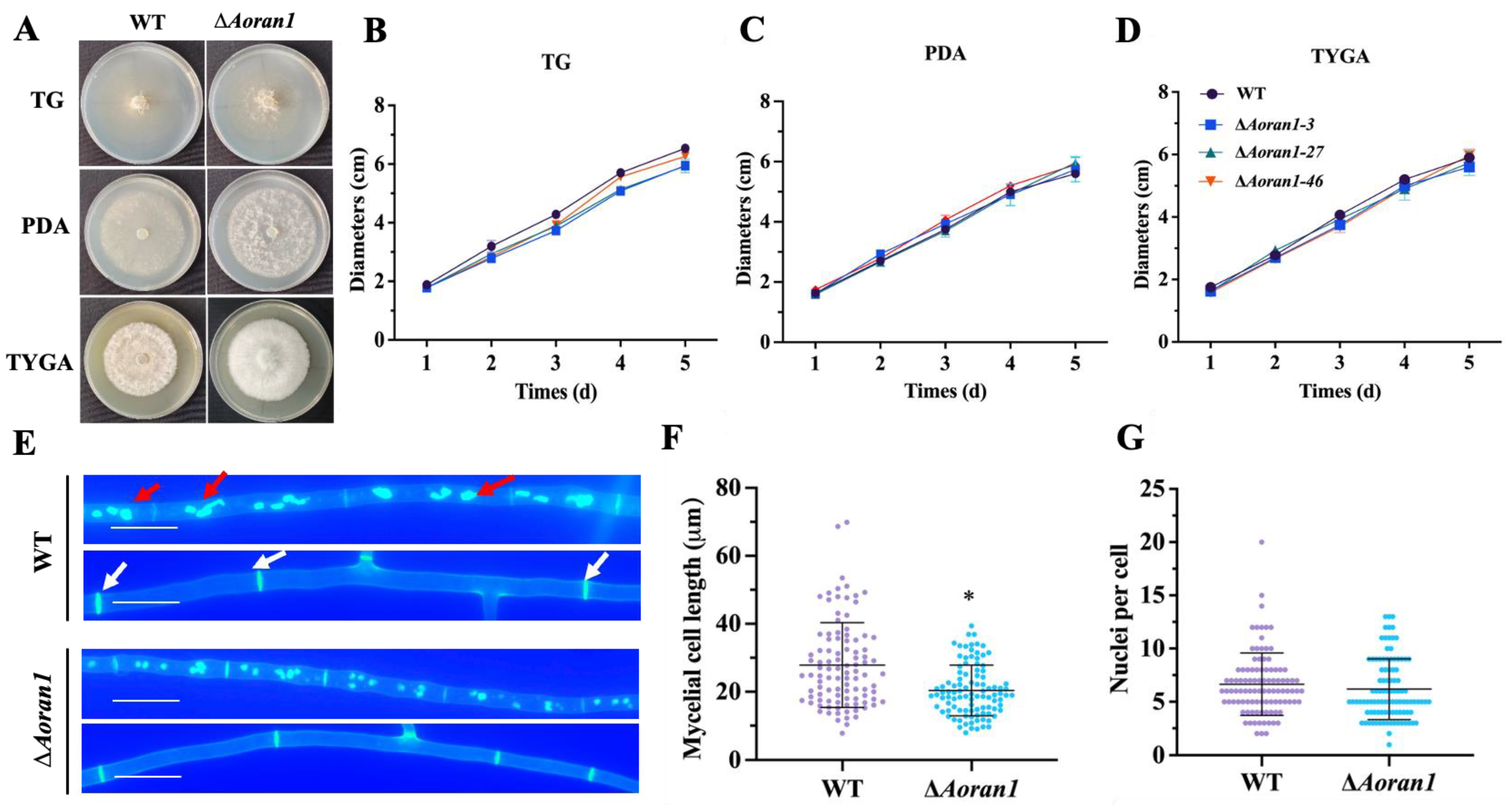
3.3. Comparison of Growth Rates between WT and ∆Aoran1 Mutant Strains

To explore the impact of ∆Aoran1 on the growth rate of A. oligospora, the fungal strains were incubated in PDA, TG, and TYGA media for 5 days. Compared to the WT strain, the growth rates of the ΔAoran1 mutant strains were not obviously different in any of the three media. However, the aerial mycelia of the ΔAoran1 mutant strains were denser than those of the WT strain (Figure 2A–D). Meanwhile, CFW staining suggested that the septum number in the ΔAoran1 mutant strains was remarkably increased, which caused a remarkable decrease in the mycelial cell length (Figure 2E,F); however, DAPI staining showed no remarkable change in the average number of nuclei (Figure 2E,G).

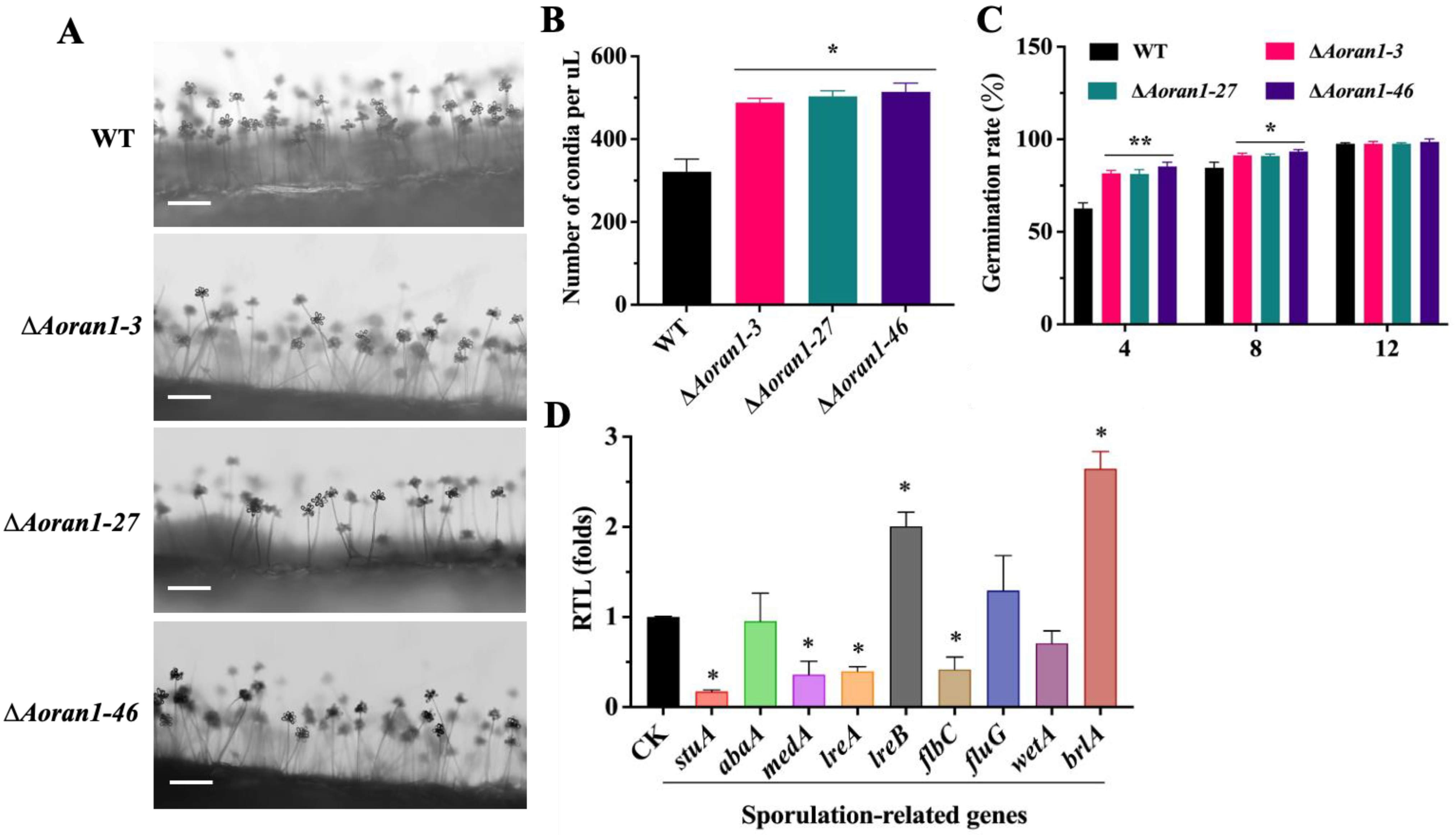
3.4. AoRan1 Plays a Vital Role in Conidiation

In the absence of Aoran1, there were no significant differences in the number of conidiophores or the morphology of conidia attached to conidiophores (Figure 3A). However, the absence of ΔAoran1 caused a statistically significant increase in the conidial number of A. oligospora (Figure 3B). Additionally, the conidial germination rate was significantly increased (Figure 3C). We examined the transcription levels of nine sporulation-related genes by means of RT-qPCR technology; among these genes, the expressions of lreB and brlA (one of the core regulatory genes of conidiation) were significantly upregulated, whereas the expressions of stuA, medA, lreA, and flbC were remarkably downregulated (Figure 3D).

3.5. AoRan1 Is Critical to Trap Formation and Pathogenicity

To further investigate whether AoRan1 affects trap formation, fresh spores from fungal strains grown on CMY medium were collected; then, approximately 20,000 spores were evenly spread in WA medium and incubated for 4 days. After the addition of approximately 400 nematodes to the medium, trap formation was observed every 12 h (Figure 4A). The morphology of the traps was observed using CFW staining. The mature traps of the WT strain consisted of 5–8 mycelial rings; in contrast, traps in the ΔAoran1 mutant strains contained 7–10 mycelial rings, and the conjunction between mycelial rings in the traps was denser, which may indicate a significant increase in hyphal fusion in the traps (Figure 4B). The results show that the deletion of Aoran1 caused a remarkable increase in the number of traps at all time points (Figure 4C). The nematode-trapping ability was also significantly increased at 12–36 h and then converged to that of the WT strain at 48 h (Figure 4D). Moreover, we compared the proteolytic activity of the ΔAoran1 mutant strain with that of the WT strain, and the size of the protease hydrolysis circle did not differ significantly between them (Figure 4E). The transcription levels of nine genes encoding serine proteases were examined via the RT-qPCR method, and the transcription levels of 43g49, 75g8, 176g95, 188g273, and 215g702 were remarkably upregulated, while those of the remaining genes were remarkably downregulated. In particular, the transcription level of 54g992 was close to zero (Figure 4F). The above results reveal that AoRan1 is crucial in regulating trap formation and virulence.

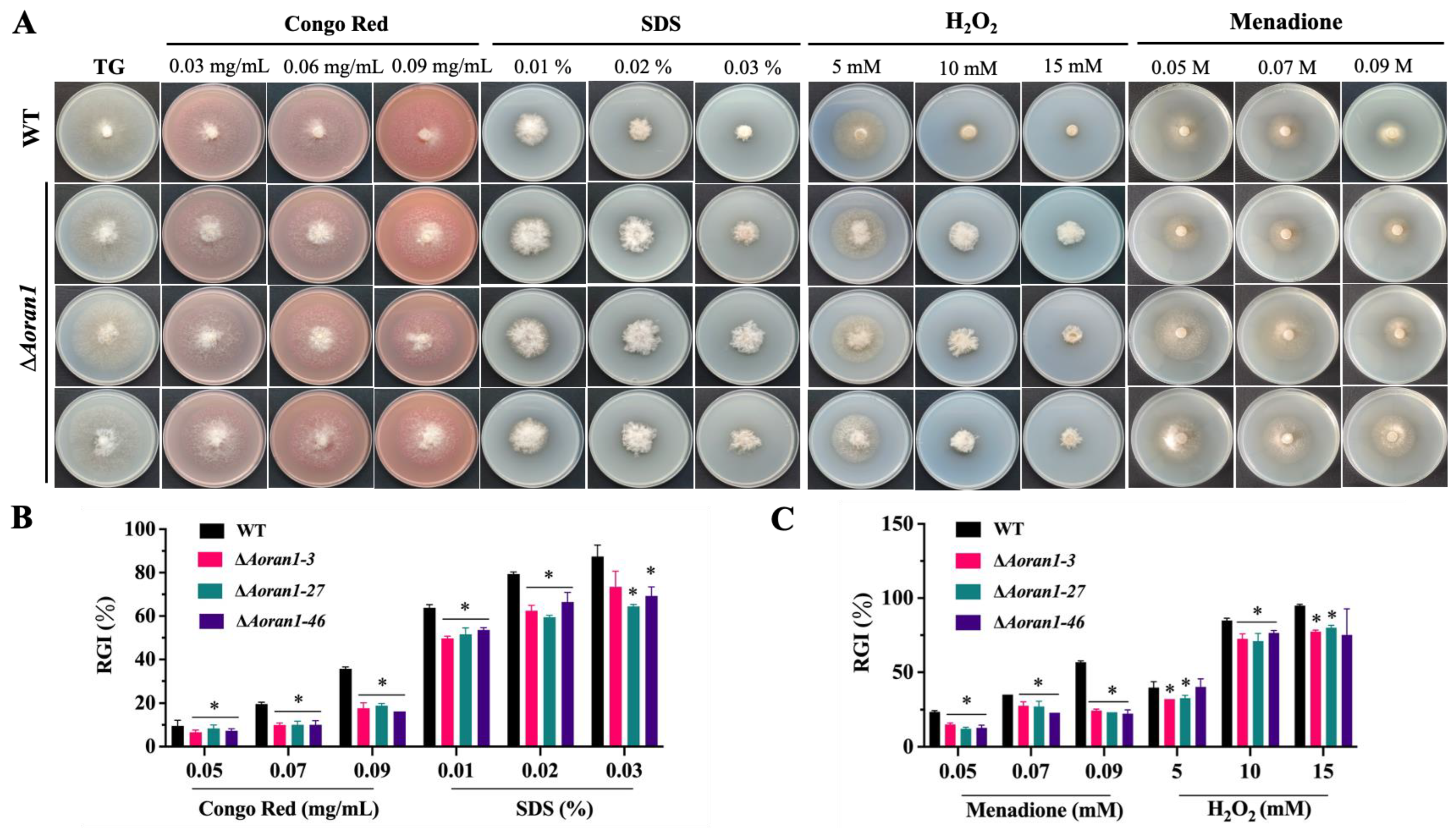
3.6. AoRan1 Is Involved in Stress Response

The ΔAoran1 mutant strains showed a significant increase in tolerance to different concentration gradients of cell wall synthesis disruptors (Congo red and SDS) and oxidants (H2O2 and menadione) (Figure 5A), and the deletion of Aoran1 caused a faster growth rate compared to the WT strain, with a corresponding significant decrease in RGI values (Figure 5B,C). Compared with the WT, the strains with Aoran1 deletion showed a remarkable increase in tolerance to low concentrations of NaCl (0.1 M); however, there was no significant difference in tolerance to higher concentrations of NaCl (0.2–0.3 M) or sorbitol (0.25–0.75 M) (Figure S2A,B).

3.7. AoRan1 Regulates LD Accumulation and Autophagic Level

BODIPY staining showed a significant increase in LD accumulation in the ΔAoran1 mutant strains compared with the WT strain (Figure 6A). The transcription levels of eight genes related to LD metabolism were examined via RT-qPCR, among which the transcription levels of genes encoding short-chain dehydrogenase reductase (AOL_s00004g288), peroxisomal ABC transporter (AOL_s00004g606), peroxisomal hydratase-dehydrogenase-epimerase (AOL_s00054g29), and 3-ketoacyl-CoA ketothiolase (AOL_s00210g122) were significantly upregulated, and the transcription levels of acyl-CoA dehydrogenase (AOL_s00079g276), phosphatidylinositol transfer protein (AOL_s00081g51), and 3-hydroxybutyryl-CoA dehydrogenase (AOL_s00110g113) were considerably downregulated (Figure 6B). MDC staining showed a remarkable decrease in the number but an increase in the volume of autophagosomes (Figure 6C). Among the six autophagy-related genes examined, the transcription levels of atg1 and atg8 were significantly upregulated, and the transcription levels of atg9, atg13, and atg17 were significantly downregulated (Figure 6D). In addition, the TEM images show more significant LD accumulation and autophagic levels in the ΔAoran1 mutant strains than in the WT strain (Figure 6E).

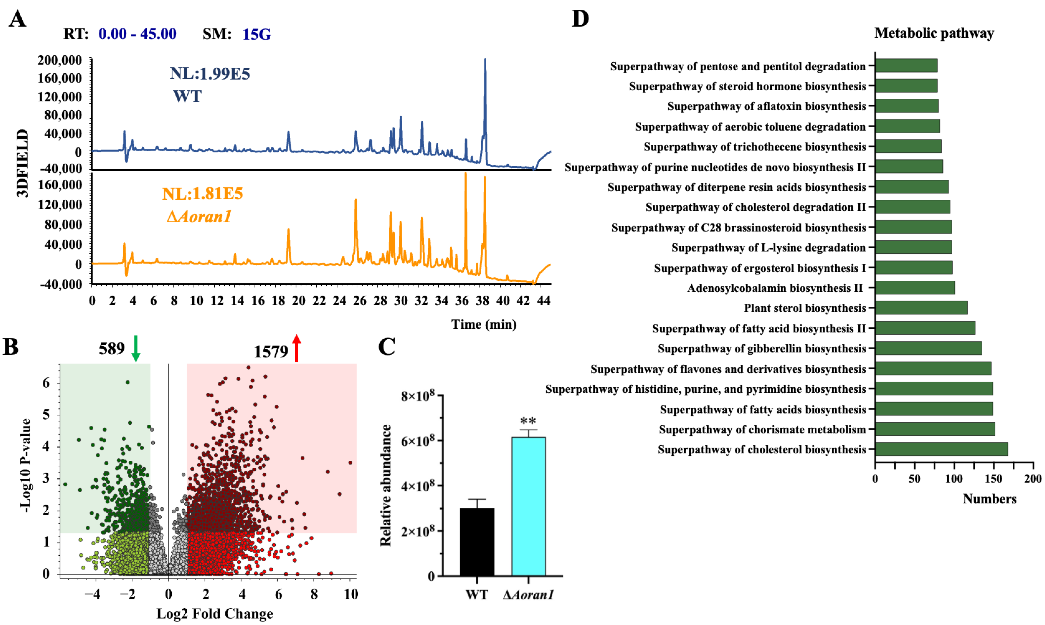
3.8. AoRan1 Contributes to Secondary Metabolism

The 7-day fermentation broth of the WT and ΔAoran1 mutant strains was processed using LC-MS to analyze the differential compounds between them. The chromatograms showed a remarkable increase in the abundance of metabolic compounds in the ΔAoran1 mutant strain (Figure 7A). The clustered volcano plot revealed that 1579 compounds were upregulated and 589 compounds were considerably downregulated upon the deletion of Aoran1 (Figure 7B). Arthrobotrisins, specialized metabolic compounds that can be produced by several NT fungi [43,44], were found in both the WT and ΔAoran1 mutant strains (Figure S3A,B), and the relative abundance of arthrobotrisins was significantly increased in the ΔAoran1 mutant strain (Figure 7C). The compounds with the most significant differences (top twenty) in the ΔAoran1 mutant strain compared to the WT strain are shown in Table S3; among them, the main compounds are tridecyl benzenesulfonate, 16-(3,4-dimethoxybenzylidene), rost-4-ene-3,17-dione, and S_S-dimethyl-beta-propiothetin. The main metabolic pathways associated with these differentially expressed compounds consist of the superpathways of cholesterol biosynthesis, chorismite biosynthesis, fatty acid biosynthesis, and the biosynthesis of histidine, purine, and pyrimidine (Figure 7D).

4. Discussion

In recent years, NT fungi have been recognized as efficient and promising nematode control agents [9,45]. The traps produced by NT fungi are crucial tools that enable them to obtain nutrition and also give them a competitive advantage over other non-predatory fungi [5,6], but the regulatory mechanisms associated with trap formation are still not well understood. Therefore, it is important to gain a deeper understanding of how these traps are differentiated, how NT fungi respond to the external environment, and what the relationship is between NT fungi and other organisms [6,46].
The serine/threonine protein kinase Ran1 is a conserved protein from yeast to humans [47]. In this study, A. oligospora was used as a model fungus, and the Aoran1 gene was knocked out using homologous recombination to further characterize its function. After obtaining the mutant strains, we performed a series of phenotypic analyses on them. The results show that the deletion of Aoran1 did not cause a significant change in the growth rate. However, our results indicate that the resistance of the ΔAoran1 mutant strain to cell wall synthesis-disrupting reagents and oxidants was significantly increased compared to that of the WT strain. A previous study demonstrated that in S. pombe, the deletion of Ran1 leads to a remarkable impact on the transcriptional induction of cell wall integrity (CWI) MAPK pathway-regulated genes, suggesting that Ran1 may be a regulator of CWI-responsive genes [15], which correlates with our findings. The above results suggest that the role of Ran1 in response to external stress is conserved across fungi.
Studies have shown that for filamentous fungi, conidiogenesis is the most common reproductive strategy for dispersal, invasion, and proliferation in the environment [48]. Previous studies showed that in S. pombe, in the presence of mating pheromones, cells will conjugate with the opposite mating type and undergo meiosis and sporulation regardless of nutrient starvation, whereas the role of Ran1 is to prevent the onset of meiosis until conjugation occurs. Therefore, we speculate that Ran1 plays a role in sporulation [49,50]. Here, we found that the knockout of the Aoran1 gene led to a remarkable increase in conidial production in the mutant strains; they also had significantly higher rates of conidial germination. We also found that brlA, one of the key genes controlling the central regulatory pathway of sporulation [51], had a significantly increased transcription level. A previous study showed that the absence of brlA leads to a complete loss of the ability to form conidiophores and produce conidia in A. oligospora [52]. In A. fumigatus, the deletion of brlA also caused a complete loss of the ability to produce conidia [53]. These results correlate with our findings, all of which indicate that brlA has a crucial role in regulating conidiation. Therefore, AoRan1 is a crucial regulator of conidiation in A. oligospora.
As a representative NT fungus, it is interesting to note that A. oligospora can form a three-dimensional adhesion network to prey on nematodes [11,13]. The process of trap formation is very complicated and has been proven to be the result of multiple cells or hyphae undergoing fusion [36,54]. Recently, it was proven that the highly conserved Fus3 MAPK of the pheromone response pathway plays a key role in signaling transduction during trap formation [55]. For instance, in A. oligospora, the deletion of Aoham5 and Aoso results in the mycelial ring in the trap being incompletely closed, showing a spiral shape. As a result, nematodes still have a chance of escaping after being captured, resulting in a significant reduction in the nematode predation efficiency [36]. Additionally, in S. pombe, it was demonstrated that the activation of pheromone-responsive MAPK plays a vital role in inducing cells to undergo phenotypically normal meiosis [56], and it is interesting to note that Ran1 performs a similar function [47]. Therefore, we wondered whether AoRan1 has a regulatory role in trap formation. In this study, we found that the deletion of Aoran1 caused a remarkable increase in the number of traps at all time points. In addition, the transcription levels of genes encoding serine proteases were remarkably increased in the mutant strains, and all of the above changes may contribute to the remarkable increase in nematode predation ability in the mutant strains. Our results indicate that AoRan1 is crucial for trap formation and proper trap morphogenesis in A. oligospora.
A noteworthy point is that autophagy-related pathways participate in regulating various biological processes, such as conidiation, conidial germination, and development, in filamentous fungi [57,58,59]. In this study, we found that the absence of Aoran1 caused a reduction in the number but a significant increase in the volume of autophagosomes in A. oligospora. Interestingly, there were also significant changes in the number of conidia and the conidium germination rate in the mutant strain. A previous study suggested that autophagy is crucial in trap formation in A. oligospora; it demonstrated that the absence of Aoatg8 remarkably reduced the autophagic level, disrupted trap formation, and ultimately severely affected the ability of A. oligospora to prey on nematodes [38]. In the present study, we observed a remarkably enhanced autophagic level in the mutant strains, and at the same time, the number of traps and the nematode-trapping ability were significantly increased. These results may help to elucidate the relationship between the autophagy process and other biological processes, such as conidiation and trap formation.
Currently, metabolomics has been widely used to illustrate the mechanisms underlying fungal growth and virulence [40,60]. Metabolomics analyses revealed multiple up- and downregulations of secondary metabolic compounds in the mutant strains. In particular, the superpathway of fatty acid biosynthesis was significantly enriched, which is consistent with the results we obtained in relation to fatty acid metabolism. It has been shown that arthrobotrisins are able to inhibit trap formation [44,61,62]. In this study, we found that the relative abundance of arthrobotrisins was significantly increased, which is in contrast to the increased number of traps we found. The most likely explanation is that trap formation is jointly regulated by various biological processes, and the effect of arthrobotrisins on trap formation may be compensated by other biological processes. Therefore, mechanisms related to trap formation in mutant strains still need to be further investigated. In conclusion, AoRan1 is crucial in regulating secondary metabolism in A. oligospora.

5. Conclusions

This study is the first to characterize the functions of serine/threonine protein kinase Ran1 in NF fungi. Our results reveal that AoRan1 plays crucial roles in various important biological processes, including sporulation, stress response, pathogenicity, and secondary metabolism. In addition, AoRan1 regulates LD accumulation and autophagic levels. These results may expand our understanding of the roles that serine/threonine protein kinase play in A. oligospora and lay the foundation for further research on the mechanisms by which Ran1 regulates conidiation, pathogenicity, and other biological processes in NT fungi.

Supplementary Materials

The following supporting information can be downloaded at: https://www.mdpi.com/article/10.3390/microorganisms12091853/s1, Figure S1: Mapping of pRS426 and pCSN44 plasmids used to construct knockout vector for target gene; Figure S2: Comparison of fungal strains in response to osmotic stress reagents; Figure S3: Mass spectra of arthrobotrisins in fungal strains; Table S1: Primers used to knock out the target gene; Table S2: Primers for RT-qPCR detection of genes related to sporulation, fatty acid metabolism, and autophagy in A. oligospora; Table S3: The compounds with the most significant differences (top twenty) in ΔAoran1 mutant strain.

Author Contributions

Methodology, investigation, and writing—original draft preparation, S.D.; software, data curation, and resources, Q.L. and Y.S.; resources and data curation, L.Z. and H.Y.; conceptualization, funding acquisition, and writing—review and editing, J.Y. All authors have read and agreed to the published version of the manuscript.

Funding

This study was financially supported by the National Natural Science Foundation of China (32370043), the Applied Basic Research Foundation of Yunnan Province (202201BC070004), the Yunling Scholar of Yunnan Province (to Jinkui Yang), and the Scientific Research Fund Project of the Yunnan Provincial Department of Education (2024Y018).

Data Availability Statement

All data generated or analyzed during this study are included in the published paper and associated Supplementary Files.

Acknowledgments

We are grateful to Ying-qi Guo and Xing-cai Wu (Kunming Institute of Zoology, Chinese Academy of Sciences) for their help with taking and analyzing the TEM images.

Conflicts of Interest

The authors declare no conflict of interest.

References

  1. Abad, P.; Gouzy, J.; Aury, J.-M.; Castagnone-Sereno, P.; Danchin, E.G.J.; Deleury, E.; Perfus-Barbeoch, L.; Anthouard, V.; Artiguenave, F.; Blok, V.C.; et al. Genome sequence of the metazoan plant-parasitic nematode Meloidogyne incognita. Nat. Biotechnol. 2008, 26, 909–915. [Google Scholar] [CrossRef] [PubMed]
  2. Coyne, D.L.; Cortada, L.; Dalzell, J.J.; Claudius-Cole, A.O.; Haukeland, S.; Luambano, N.; Talwana, H. Plant-parasitic nematodes and food security in sub-Saharan Africa. Annu. Rev. Phytopathol. 2018, 25, 381–403. [Google Scholar] [CrossRef]
  3. Tzean, Y.; Chou, T.-H.; Hsiao, C.-C.; Shu, P.-Y.; Walton, J.D.; Tzean, S.-S. Cloning and characterization of cuticle-degrading serine protease from nematode-trapping fungus Arthrobotrys musiformis. Mycoscience 2016, 57, 136–143. [Google Scholar] [CrossRef]
  4. Ahmad, G.; Khan, A.; Khan, A.A.; Ali, A.; Mohhamad, H.I. Biological control: A novel strategy for the control of the plant parasitic nematodes. Antonie Van Leeuwenhoek 2021, 114, 885–912. [Google Scholar] [CrossRef] [PubMed]
  5. Liu, K.; Tian, J.; Xiang, M.; Liu, X. How carnivorous fungi use three-celled constricting rings to trap nematodes. Protein Cell 2012, 3, 325–328. [Google Scholar] [CrossRef]
  6. Yang, Y.; Yang, E.; An, Z.; Liu, X. Evolution of nematode-trapping cells of predatory fungi of the orbiliaceae based on evidence from rRNA-Encoding DNA and multiprotein sequences. Proc. Natl. Acad. Sci. USA 2007, 104, 8379–8384. [Google Scholar] [CrossRef]
  7. Larsen, M. Prospects for controlling animal parasitic nematodes by predacious micro fungi. Parasitology 2000, 120, 121–131. [Google Scholar] [CrossRef] [PubMed]
  8. Kerry, B.R. Rhizosphere interactions and the exploitation of microbial agents for the biological control of plant-parasitic nematodes. Annu. Rev. Phytopathol. 2000, 38, 423–441. [Google Scholar] [CrossRef]
  9. Jiang, X.; Xiang, M.; Liu, X. Nematode-trapping fungi. Microbiol. Spectr. 2017, 5, FUNK-0022-2016. [Google Scholar] [CrossRef]
  10. Wang, S.; Liu, X. Tools and basic procedures of gene manipulation in nematode-trapping fungi. Mycology 2023, 10, 75–90. [Google Scholar] [CrossRef]
  11. Yang, J.; Wang, L.; Ji, X.; Feng, Y.; Li, X.; Zou, C.; Xu, J.; Ren, Y.; Mi, Q.; Wu, J.; et al. Genomic and proteomic analyses of the fungus Arthrobotrys oligospora provide insights into nematode-trap formation. PLoS Pathog. 2011, 7, e1002179. [Google Scholar] [CrossRef] [PubMed]
  12. Hu, X.; Hoffmann, D.S.; Wang, M.; Schuhmacher, L.; Stroe, M.C.; Schreckenberger, B.; Elstner, M.; Fischer, R. GprC of the nematode-trapping fungus Arthrobotrys flagrans activates mitochondria and reprograms fungal cells for nematode hunting. Nat. Microbiol. 2024, 9, 1752–1763. [Google Scholar] [CrossRef]
  13. Lin, H.C.; de Ulzurrun, G.V.; Chen, S.A.; Yang, C.T.; Tay, R.J.; Iizuka, T.; Huang, T.Y.; Kuo, C.Y.; Gonçalves, A.P.; Lin, S.Y.; et al. Key processes required for the different stages of fungal carnivory by a nematode-trapping fungus. PLoS Biol. 2023, 21, e3002400. [Google Scholar] [CrossRef]
  14. Park, G.; Servin, J.A.; Turner, G.E.; Altamirano, L.; Colot, H.V.; Collopy, P.; Litvinkova, L.; Li, L.; Jones, C.A.; Diala, F.-G.; et al. Global analysis of serine-threonine protein kinase genes in Neurospora crassa. Eukaryot. Cell 2011, 10, 1553–1564. [Google Scholar] [CrossRef] [PubMed]
  15. Li, P.; McLeod, M. Molecular mimicry in development: Identification of Ste11+ as a substrate and Mei3+ as a pseudosubstrate inhibitor of Ran1+ kinase. Cell 1996, 87, 869–880. [Google Scholar] [CrossRef]
  16. Goldsmith, E.J.; Akella, R.; Min, X.; Zhou, T.; Humphreys, J.M. Substrate and docking interactions in serine/threonine protein kinases. Chem. Rev. 2007, 107, 5065–5081. [Google Scholar] [CrossRef]
  17. Burgess, A.; Vigneron, S.; Brioudes, E.; Labbé, J.-C.; Lorca, T.; Castro, A. Loss of human greatwall results in G2 arrest and multiple mitotic defects due to deregulation of the cyclin B-Cdc2/PP2A balance. Proc. Natl. Acad. Sci. USA 2010, 107, 12564–12569. [Google Scholar] [CrossRef] [PubMed]
  18. Herzig, S.; Neumann, J. Effects of serine/threonine protein phosphatases on ion channels in excitable membranes. Physiol. Rev. 2000, 80, 173–210. [Google Scholar] [CrossRef]
  19. Lorca, T.; Bernis, C.; Vigneron, S.; Burgess, A.; Brioudes, E.; Labbé, J.-C.; Castro, A. Constant regulation of both the MPF amplification loop and the greatwall-PP2A pathway is required for metaphase II arrest and correct entry into the first embryonic cell cycle. J. Cell Sci. 2010, 123, 2281–2291. [Google Scholar] [CrossRef]
  20. Cipak, L.; Hyppa, R.W.; Smith, G.R.; Gregan, J. ATP analog-sensitive Pat1 protein kinase for synchronous fission yeast meiosis at physiological temperature. Cell Cycle 2012, 11, 1626–1633. [Google Scholar] [CrossRef]
  21. Bhola, T.; Kapuy, O.; Vinod, P.K. Computational modelling of meiotic entry and commitment. Sci. Rep. 2018, 8, 180. [Google Scholar] [CrossRef]
  22. Kitamura, K.; Katayama, S.; Dhut, S.; Sato, M.; Watanabe, Y.; Yamamoto, M.; Toda, T. Phosphorylation of Mei2 and Ste11 by Pat1 kinase inhibits sexual differentiation via ubiquitin proteolysis and 14-3-3 protein in fission yeast. Dev. Cell 2001, 1, 389–399. [Google Scholar] [CrossRef] [PubMed]
  23. Nurse, P. Mutants of the fission yeast Schizosaccharomyces pombe which alter the shift between cell proliferation and sporulation. Mol. Gen. Genet. 1985, 198, 497–502. [Google Scholar] [CrossRef]
  24. Watanabe, Y.; Yamamoto, M.S. Pombe Mei2+ encodes an RNA-binding protein essential for premeiotic DNA synthesis and meiosis I, which cooperates with a novel RNA species meiRNA. Cell 1994, 78, 487–498. [Google Scholar] [CrossRef]
  25. Beach, D.; Rodgers, L.; Gould, J. RAN1 + controls the transition from mitotic division to meiosis in fission yeast. Curr. Genet. 1985, 10, 297–311. [Google Scholar] [CrossRef]
  26. Madhani, H.D.; Fink, G.R. Combinatorial control required for the specificity of yeast MAPK signaling. Science 1997, 275, 1314–1317. [Google Scholar] [CrossRef]
  27. Gustin, M.C.; Albertyn, J.; Alexander, M.; Davenport, K. MAP kinase pathways in the yeast Saccharomyces cerevisiae. Microbiol. Mol. Biol. Rev. 1998, 62, 1264–1300. [Google Scholar] [CrossRef]
  28. Li, D.; Qin, L.; Wang, Y.; Xie, Q.; Li, N.; Wang, S.; Yuan, J. AflSte20 regulates morphogenesis, stress response, and aflatoxin biosynthesis of Aspergillus flavus. Toxins 2019, 11, 73. [Google Scholar] [CrossRef] [PubMed]
  29. Pulido, V.; Rodríguez-Peña, J.M.; Alonso, G.; Sanz, A.B.; Arroyo, J.; García, R. mRNA decapping activator Pat1 is required for efficient yeast adaptive transcriptional responses via the cell wall integrity MAPK pathway. J. Mol. Biol. 2024, 436, 1685700. [Google Scholar] [CrossRef]
  30. Xie, M.; Bai, N.; Yang, X.; Liu, Y.; Zhang, K.-Q.; Yang, J. Fus3 regulates asexual development and trap morphogenesis in the nematode-trapping fungus Arthrobotrys oligospora. iScience 2023, 26, 107404. [Google Scholar] [CrossRef]
  31. Xie, M.; Ma, N.; Bai, N.; Yang, L.; Yang, X.; Zhang, K.-Q.; Yang, J. PKC-SWI6 signaling regulates asexual development, cell wall integrity, stress response, and lifestyle transition in the nematode-trapping fungus Arthrobotrys oligospora. Sci. China Life Sci. 2022, 65, 2455–2471. [Google Scholar] [CrossRef]
  32. Kumar, S.; Stecher, G.; Tamura, K. MEGA7: Molecular evolutionary genetics analysis version 7.0 for bigger datasets. Mol. Biol. Evol. 2016, 33, 1870–1874. [Google Scholar] [CrossRef] [PubMed]
  33. Colot, H.V.; Park, G.; Turner, G.E.; Ringelberg, C.; Crew, C.M.; Litvinkova, L.; Weiss, R.L.; Borkovich, K.A.; Dunlap, J.C. A high-throughput gene knockout procedure for Neurospora reveals functions for multiple transcription factors. Proc. Natl. Acad. Sci. USA 2006, 103, 10352–10357. [Google Scholar] [CrossRef]
  34. Zhu, Y.; Yang, X.; Bai, N.; Liu, Q.; Yang, J. AoRab7A interacts with AoVps35 and AoVps41 to regulate vacuole assembly, trap formation, conidiation, and functions of proteasomes and ribosomes in Arthrobotrys oligospora. Microbiol. Res. 2024, 280, 127573. [Google Scholar] [CrossRef]
  35. Tunlid, A.; Ahman, J.; Oliver, R.P. Transformation of the nematode-trapping fungus Arthrobotrys oligospora. FEMS Microbiol. Lett. 1999, 173, 111–116. [Google Scholar] [CrossRef] [PubMed]
  36. Wang, W.; Liu, Y.; Duan, S.; Bai, N.; Zhu, M.; Yang, J. Cellular communication and fusion regulate cell fusion, trap morphogenesis, conidiation, and secondary metabolism in Arthrobotrys oligospora. Microbiol. Res. 2024, 278, 127516. [Google Scholar] [CrossRef]
  37. Zhao, X.; Fan, Y.; Xiang, M.; Kang, S.; Wang, S.; Liu, X. DdaCrz1, a C2H2-Type transcription factor, regulates growth, conidiation, and stress resistance in the nematode-trapping fungus Drechslerella dactyloides. J. Fungi 2022, 8, 750. [Google Scholar] [CrossRef]
  38. Chen, Y.; Gao, Y.; Zhang, K.; Zou, C. Autophagy is required for trap formation in the nematode-trapping fungus Arthrobotrys oligospora. Environ. Microbiol. Rep. 2013, 5, 511–517. [Google Scholar] [CrossRef]
  39. Yang, L.; Li, X.; Ma, Y.; Zhu, M.; Zhang, K.-Q.; Yang, J. The Arf-GAPs, AoAge1 and AoAge2, regulate diverse cellular processes, conidiation, trap formation, and secondary metabolism in Arthrobotrys oligospora. Microbiol. Res. 2024, 285, 127779. [Google Scholar] [CrossRef] [PubMed]
  40. Liu, Q.; Bai, N.; Duan, S.; Shen, Y.; Zhu, L.; Yang, J. Characterizing the role of AosfgA and AofluG in mycelial and conidial development in Arthrobotrys oligospora and their role in secondary metabolism. Microorganisms 2024, 12, 615. [Google Scholar] [CrossRef]
  41. Livak, K.J.; Schmittgen, T.D. Analysis of relative gene expression data using real-time quantitative PCR and the 2−ΔΔCT method. Methods 2001, 25, 402–408. [Google Scholar] [CrossRef] [PubMed]
  42. Teng, L.L.; Song, T.Y.; Xu, Z.F.; Liu, X.; Dai, R.; Chen, Y.H.; Li, S.H.; Zhang, K.Q.; Niu, X.M. Selected mutations revealed intermediates and key precursors in the biosynthesis of polyketide-terpenoid hybrid sesquiterpenyl epoxy-cyclohexenoids. Org. Lett. 2017, 19, 3923–3926. [Google Scholar] [CrossRef]
  43. Zhou, J.; Wu, Q.-F.; Li, S.-H.; Yan, J.-X.; Wu, L.; Cheng, Q.-Y.; He, Z.-Q.; Yue, X.-T.; Zhang, K.-Q.; Zhang, L.-L.; et al. The multifaceted gene 275 embedded in the PKS-PTS gene cluster was involved in the regulation of arthrobotrisin biosynthesis, TCA Cycle, and septa formation in nematode-trapping fungus Arthrobotrys oligospora. J. Fungi 2022, 8, 1261. [Google Scholar] [CrossRef] [PubMed]
  44. He, Z.-Q.; Tan, J.-L.; Li, N.; Zhang, H.-X.; Chen, Y.-H.; Wang, L.-J.; Zhang, K.-Q.; Niu, X.-M. Sesquiterpenyl epoxy-cyclohexenoids and their signaling functions in nematode-trapping fungus Arthrobotrys oligospora. J. Agric. Food Chem. 2019, 67, 13061–13072. [Google Scholar] [CrossRef]
  45. Andersson, K.-M.; Kumar, D.; Bentzer, J.; Friman, E.; Ahrén, D.; Tunlid, A. Interspecific and host-related gene expression patterns in nematode-trapping fungi. BMC Genom. 2014, 15, 968. [Google Scholar] [CrossRef]
  46. Zhang, D.; Zhu, X.; Sun, F.; Zhang, K.; Niu, S.; Huang, X. The roles of actin cytoskeleton and actin-associated protein Crn1p in trap formation of Arthrobotrys oligospora. Res. Microbiol. 2017, 168, 655–663. [Google Scholar] [CrossRef]
  47. McLeod, M.; Shor, B.; Caporaso, A.; Wang, W.; Chen, H.; Hu, L. Cpc2, a fission yeast homologue of mammalian RACK1 protein, interacts with Ran1 (Pat1) kinase to regulate cell cycle progression and meiotic development. Mol. Cell. Biol. 2000, 20, 4016–4027. [Google Scholar] [CrossRef]
  48. Liu, X.; Miao, Q.; Zhou, Z.; Lu, S.; Li, J. Identification of three novel conidiogenesis-related genes in the nematode-trapping fungus Arthrobotrys oligospora. Pathogens 2022, 11, 717. [Google Scholar] [CrossRef] [PubMed]
  49. Yamamoto, H.; Tsukahara, K.; Kanaoka, Y.; Jinno, S.; Okayama, H. Isolation of a mammalian homologue of a fission yeast differentiation regulator. Mol. Cell. Biol. 1999, 19, 3829–3841. [Google Scholar] [CrossRef]
  50. Egel, R.; Egel-Mitani, M. Premeiotic DNA synthesis in fission yeast. Exp. Cell Res. 1974, 88, 127–134. [Google Scholar] [CrossRef]
  51. Chen, M.; Yang, H.-Y.; Cao, Y.-R.; Hui, Q.-Q.; Fan, H.-F.; Zhang, C.-C.; Han, J.-J.; Guo, Z.-Y.; Xu, J.; Zhang, K.-Q.; et al. Functional characterization of core regulatory genes involved in sporulation of the nematophagous fungus Purpureocillium lavendulum. mSphere 2020, 5, e00932-20. [Google Scholar] [CrossRef] [PubMed]
  52. Bai, N.; Xie, M.; Liu, Q.; Zhu, Y.; Yang, X.; Zhang, K.-Q.; Yang, J. AoMedA has a complex regulatory relationship with AoBrlA, AoAbaA, and AoWetA in conidiation, trap formation, and secondary metabolism in the nematode-trapping fungus Arthrobotrys oligospora. Appl. Environ. Microbiol. 2023, 89, e00983-23. [Google Scholar] [CrossRef]
  53. Twumasi-Boateng, K.; Yu, Y.; Chen, D.; Gravelat, F.N.; Nierman, W.C.; Sheppard, D.C. Transcriptional profiling identifies a role for BrlA in the response to nitrogen fepletion and for StuA in the regulation of secondary metabolite clusters in Aspergillus fumigatus. Eukaryot. Cell 2009, 8, 104–115. [Google Scholar] [CrossRef] [PubMed]
  54. Fischer, M.S.; Glass, N.L. Communicate and fuse: How filamentous fungi establish and maintain an interconnected mycelial network. Front. Microbiol. 2019, 10, 619. [Google Scholar] [CrossRef] [PubMed]
  55. Chen, S.-A.; Lin, H.-C.; Schroeder, F.C.; Hsueh, Y.-P. Prey sensing and response in a nematode-trapping fungus is governed by the MAPK pheromone response pathway. Genetics 2021, 217, iyaa008. [Google Scholar] [CrossRef]
  56. Yamamoto, T.G.; Chikashige, Y.; Ozoe, F.; Kawamukai, M.; Hiraoka, Y. Activation of the pheromone-responsive MAP kinase drives haploid cells to undergo ectopic meiosis with normal telomere clustering and sister chromatid segregation in fission yeast. J. Cell Sci. 2004, 117, 3875–3886. [Google Scholar] [CrossRef]
  57. Pinan-Lucarré, B.; Balguerie, A.; Clavé, C. Accelerated cell death in Podospora autophagy mutants. Eukaryot. Cell 2005, 4, 1765–1774. [Google Scholar] [CrossRef]
  58. Kikuma, T.; Ohneda, M.; Arioka, M.; Kitamoto, K. Functional analysis of the ATG8 homologue AoAtg8 and role of autophagy in differentiation and germination in Aspergillus oryzae. Eukaryot. Cell 2006, 5, 1328–1336. [Google Scholar] [CrossRef]
  59. Deng, Y.Z.; Ramos-Pamplona, M.; Naqvi, N.I. Autophagy-assisted glycogen catabolism regulates asexual differentiation in Magnaporthe oryzae. Autophagy 2009, 5, 33–43. [Google Scholar] [CrossRef]
  60. Li, X.; Wu, Y.; Liu, Z.; Zhang, C. The function and transcriptome analysis of a bZIP transcription factor CgAP1 in Colletotrichum gloeosporioides. Microbiol. Res. 2017, 197, 39–48. [Google Scholar] [CrossRef]
  61. Wei, L.-X.; Zhang, H.-X.; Tan, J.-L.; Chu, Y.-S.; Li, N.; Xue, H.-X.; Wang, Y.-L.; Niu, X.-M.; Zhang, Y.; Zhang, K.-Q. Arthrobotrisins A–C, oligosporons from the nematode-trapping fungus Arthrobotrys oligospora. J. Nat. Prod. 2011, 74, 1526–1530. [Google Scholar] [CrossRef] [PubMed]
  62. Yu, X.; Hu, X.; Pop, M.; Wernet, N.; Kirschhöfer, F.; Brenner-Weiß, G.; Keller, J.; Bunzel, M.; Fischer, R. Fatal attraction of Caenorhabditis elegans to predatory fungi through 6-methyl-salicylic acid. Nat. Commun. 2021, 12, 5462. [Google Scholar] [CrossRef] [PubMed]
Figure 1. A phylogenetic analysis of AoRan1 and the deletion and verification of the Aoran1 gene in A. oligospora. (A) A neighbor-joining tree of orthologous Ran1 in different fungi, which was built using MEGA7.0 (Left), and a prediction of the functional domains of AoRan1 homologs in diverse fungi (Right). (B) A schematic diagram of the homologous recombination strategy used to knock out Aoran1. (C) The validation of ΔAoran1 transformants using PCR. (D) The validation of ΔAoran1 transformants using RT-qPCR. RTL: the relative transcription level of gene Aoran1. Asterisk: Significantly different compared to the WT strain (*** p < 0.001).
Microorganisms 12 01853 g001
Figure 2. Role of Aoran1 in vegetative growth and mycelial growth. (A) Colony morphology after 5 days of incubation in three basal media. (BD) Colony diameters in PDA (A), TG (B), and TYGA (C) media. (E) Mycelia stained with calcofluor white and 4′, 6-diamidino-2-phenylindole to observe septa and nuclei. Bar: 10 μm. White arrow, hyphal septa; red arrow, nuclei. (F,G) Statistical analysis of mycelial cell length (F) and nuclear number (G) in fungal strains. WT, wild-type strain. Asterisk: Significantly different compared to WT strain (* p < 0.05).
Microorganisms 12 01853 g002
Figure 3. Role of AoRan1 in conidiation. (A) Observations of conidiophores in fungal strains. Bar = 100 μm. (B) Conidial production in 1 μL conidial suspension, comparing WT and mutant strains. (C) Conidial germination rates in fungal strains. (D) Relative transcription level (RTL) of several sporulation-related genes in mutant strain versus WT strain, detected using RT-qPCR. CK was denoted as standard for statistical analysis of relative transcription level of gene in mutant versus WT strain. Asterisk: Significantly different compared to WT strain (* p < 0.05; ** p < 0.01).
Microorganisms 12 01853 g003
Figure 4. Role of AoRan1 in trap formation, nematode-trapping ability, and proteolytic activity. (A) Microscopic images of trap formation and predation on nematodes at 12–48 h. Bar = 100 μm. (B) Traps were stained with CFW to observe their morphology. Red arrows: traps. Bar = 20 μm. (C) Trap yields of fungal strains at 12–48 h. (D) Percentages of nematodes captured by fungal strains at 12–48 h. (E) Comparison of hydrolysis circle of protease activity between WT and mutant strains. (F) Relative transcription level (RTL) of several genes encoding serine proteases in mutant strains, detected using RT-qPCR. CK was denoted as standard for statistical analysis of relative transcription level of gene in mutant versus WT strain. Asterisk: Significantly different compared to WT strain (* p < 0.05).
Microorganisms 12 01853 g004
Figure 5. Role of AoRan1 in response to external stress. (A) Colonial morphology supplemented with different concentration gradients of cell wall synthesis-disturbing (CWSD) reagents and oxidants. (B,C) Relative growth inhibition (RGI) rate of fungal strains after 5 days of culture in TG media treated with different concentrations of CWSD reagents (Congo red and SDS) (B) and oxidants (H2O2 and menadione) (C). Asterisk: Significantly different compared to WT strain (* p < 0.05).
Microorganisms 12 01853 g005
Figure 6. Observation of LD accumulation and autophagy of fungal strains. (A,C) Microscopic images of LDs and autophagosomes in mycelial cells. LDs in (A) and autophagosomes in (C) were stained by means of BODIPY dye and MDC staining, respectively. White arrows: LDs. Red arrows: autophagosomes. Bar = 5 μm. (B,D) Relative transcription level (RTL) of genes related to fatty acid metabolism (B) and autophagy (D) examined in mutant strain versus WT strain at day 5. CK in (B,D) was denoted as standard for statistical analysis of relative transcription level of gene in mutant versus WT strain. (E) TEM showed internal ultrastructure of hyphae. Red arrow: autophagosomes. White arrows: LDs. Asterisk: Significantly different compared to WT strain (* p < 0.05).
Microorganisms 12 01853 g006
Figure 7. Role of AoRan1 in secondary metabolism. (A) Comparison of metabolic profiling between WT and ΔAoran1 mutant strains. (B) Volcano plot of differential metabolites between WT and ΔAoran1 mutant strains. Green arrow represents the number of downregulated compounds, and red arrow represents the number of upregulated compounds. (C) Relative abundance of arthrobotrisins between WT and ΔAoran1 mutant strains. (D) Top 20 metabolic pathways enriched in ΔAoran1 mutant strain. Asterisk: Significantly different compared to WT strain (** p < 0.01).
Microorganisms 12 01853 g007
Disclaimer/Publisher’s Note: The statements, opinions and data contained in all publications are solely those of the individual author(s) and contributor(s) and not of MDPI and/or the editor(s). MDPI and/or the editor(s) disclaim responsibility for any injury to people or property resulting from any ideas, methods, instructions or products referred to in the content.

