Article

SARS-CoV-2 Genotyping Highlights the Challenges in Spike Protein Drift Independent of Other Essential Proteins

by
Jeremy W. Prokop
1,2,*,
Sheryl Alberta
3,
Martin Witteveen-Lane
1,
Samantha Pell
3,
Hosam A. Farag
1,
Disha Bhargava
2,
Robert M. Vaughan
2,
Austin Frisch
2,
Jacob Bauss
2,
Humza Bhatti
2,
Sanjana Arora
1,
Charitha Subrahmanya
1,
David Pearson
1,
Austin Goodyke
1,
Mason Westgate
1,
Taylor W. Cook
2,
Jackson T. Mitchell
2,
Jacob Zieba
4,
Matthew D. Sims
5,6,
Adam Underwood
7,
Habiba Hassouna
8,
Surender Rajasekaran
1,2,
Maximiliano A. Tamae Kakazu
2,9,
Dave Chesla
1,2,
Rosemary Olivero
2,10 and
Adam J. Caulfield
11,*
add Show full author list remove Hide full author list
1
Office of Research, Corewell Health, Grand Rapids, MI 49503, USA
2
College of Human Medicine, Michigan State University, Grand Rapids, MI 49503, USA
3
Advanced Technology Lab, Corewell Health, Grand Rapids, MI 49503, USA
4
Genetics and Genome Sciences Program, BioMolecular Science, Michigan State University, East Lansing, MI 48824, USA
5
Section of Infectious Diseases, Corewell Health, Royal Oak, MI 48073, USA
6
Department of Internal Medicine, Oakland University William Beaumont School of Medicine, Auburn Hills, MI 48309, USA
7
Division of Mathematics and Science, Walsh University, North Canton, OH 44720, USA
8
Adult Infectious Disease, Corewell Health, Grand Rapids, MI 49503, USA
9
Division of Pulmonary and Critical Care Medicine, Corewell Health, Grand Rapids, MI 49503, USA
10
Pediatric Infectious Disease, Helen DeVos Children’s Hospital, Corewell Health, Grand Rapids, MI 49503, USA
11
Microbiology Lab, Corewell Health, Grand Rapids, MI 49503, USA
*
Authors to whom correspondence should be addressed.
Microorganisms 2024, 12(9), 1863; https://doi.org/10.3390/microorganisms12091863
Submission received: 1 August 2024 / Revised: 22 August 2024 / Accepted: 27 August 2024 / Published: 9 September 2024

Abstract

:
As of 2024, SARS-CoV-2 continues to propagate and drift as an endemic virus, impacting healthcare for years. The largest sequencing initiative for any species was initiated to combat the virus, tracking changes over time at a full virus base-pair resolution. The SARS-CoV-2 sequencing represents a unique opportunity to understand selective pressures and viral evolution but requires cross-disciplinary approaches from epidemiology to functional protein biology. Within this work, we integrate a two-year genotyping window with structural biology to explore the selective pressures of SARS-CoV-2 on protein insights. Although genotype and the Spike (Surface Glycoprotein) protein continue to drift, most SARS-CoV-2 proteins have had few amino acid alterations. Within Spike, the high drift rate of amino acids involved in antibody evasion also corresponds to changes within the ACE2 binding pocket that have undergone multiple changes that maintain functional binding. The genotyping suggests selective pressure for receptor specificity that could also confer changes in viral risk. Mapping of amino acid changes to the structures of the SARS-CoV-2 co-transcriptional complex (nsp7-nsp14), nsp3 (papain-like protease), and nsp5 (cysteine protease) proteins suggest they remain critical factors for drug development that will be sustainable, unlike those strategies targeting Spike.

1. Introduction

SARS-CoV-2 currently ranks as the most sequenced virus in human history, with nearly 9 million genomes sequenced as of early 2024 within NCBI [1] and 16.8 million in GISAID [2], the two largest repositories of viral genomics. Within NCBI Virus, most (~97%) of the sequenced SARS-CoV-2 genomes were isolated from oronasopharynx swabs, with less than one percent from feces and blood (Figure 1A).
The early genomic datasets suggested that the virus could accumulate one substitution every 11 days, with extensive focus on how changes in the genome could alter the viral spread to inform the pandemic response [3]. Frequent reports of genomic changes have contributed to the public perception that the virus is rapidly changing, a perception that has influenced scientists to focus on genomic change rather than conservation. Although extensive work addressed the changes to the Spike (S) protein and how they result in immune escape [4], the virus contains additional proteins coded by the genes ORF1ab (ORF1a and ORF1b), ORF3a, E, M, ORF6, ORF7a, ORF8, N, and ORF10 [5] that have remained poorly characterized for variant impacts. SAR-CoV-2 genomes contain each of these proteins at similar detection as Spike (Figure 1B), but assessments of protein-coding changes within them have been less of a focus throughout the literature [6].Based on the NCBI, the majority of our genomes for SARS-CoV-2 were generated in Europe and North America (Figure 1C), with the USA, United Kingdom, Germany, Denmark, and Switzerland depositing the most genomes as countries (Figure 1D). Within the USA, many of the states with the largest populations generated the most genomes (Figure 1E), including California (CA), Florida (FL), and Texas (TX). Several states sequenced a higher number of genomes relative to their population (Figure 1F), including Vermont (VT), Massachusetts (MA), and Minnesota (MN). As there is a clear disbalance in genotyping of SARS-CoV-2 relative to geography, there has also been a lag in the publications of data analysis for these genotypes from diverse geographies, emphasizing individual groups to define dynamics of the virus in unique locations [7,8,9,10]. Yet, there has been a lack of strategy for exploring the full viral genome drift to proteins outside Spike.
The SARS-CoV-2 genomics field has been centered around phylogenetics and “high-impact” variants [11], which was beneficial for predicting new risk strains in the pandemic response. As data accumulated over the years, the extensive volumes of genomic data, the complexities of sequencing bias, the challenge of rare variants to “potential” risks, and the geographical complexities [12] have challenged the genomic utility of the massive sequencing investments during the pandemic. As the field transitions into the endemic phase of SARS-CoV-2, we must strategize how our investments in genomics can highlight aspects of targeted treatment dynamics and understand selective pressures between viral survival and virulence within each geographical region.
This paper combines our deep evolutionary and structural dynamics of SARS-CoV-2 [13] with two years of genotyping within a community hospital system. The analysis integrates the current state of SARS-CoV-2 genomic and structural knowledge, elucidating opportunities to move forward in the COVID-19 endemic era that other teams can utilize to transition from phylogenetics into protein impacts and drug targeting.

2. Materials and Methods
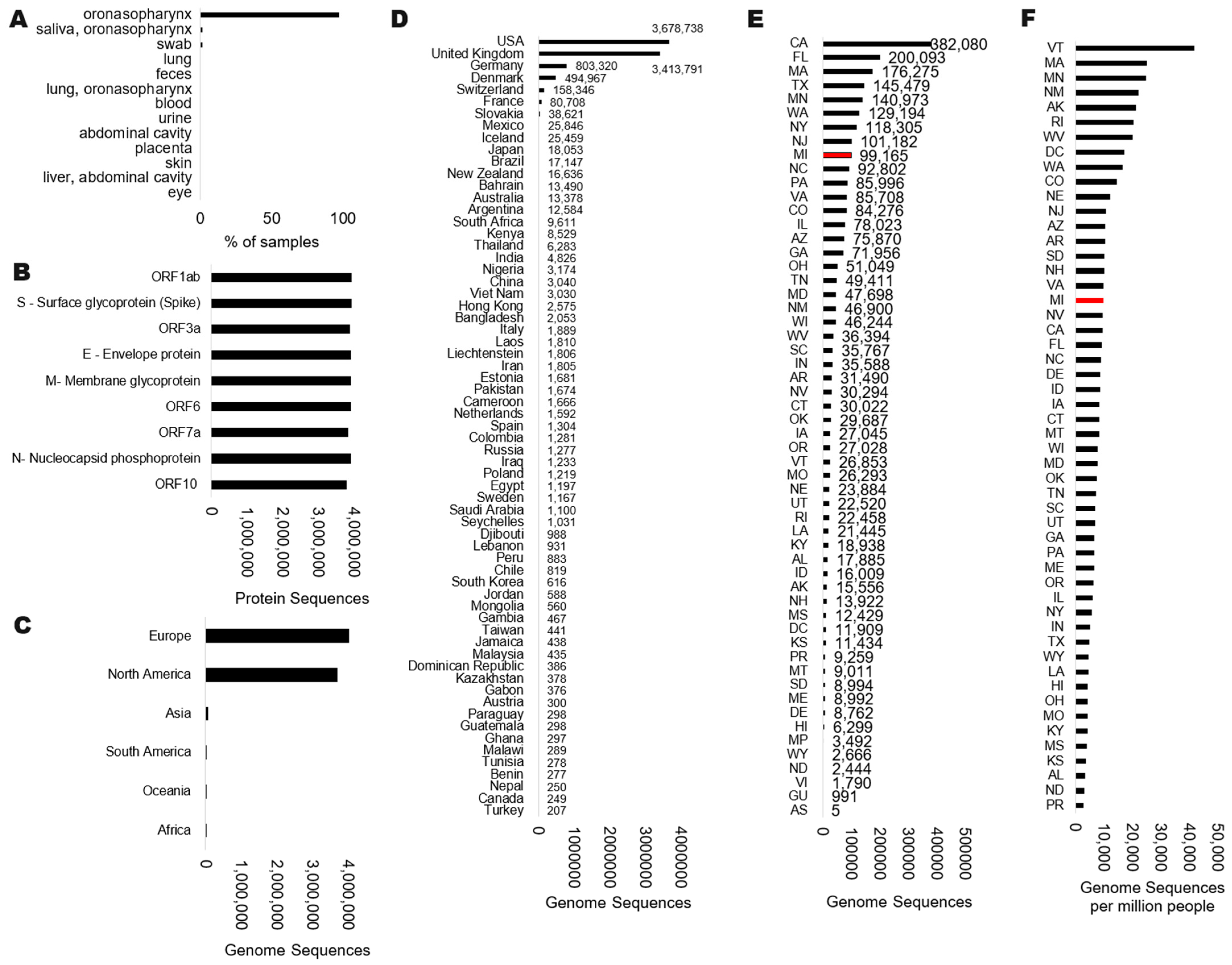
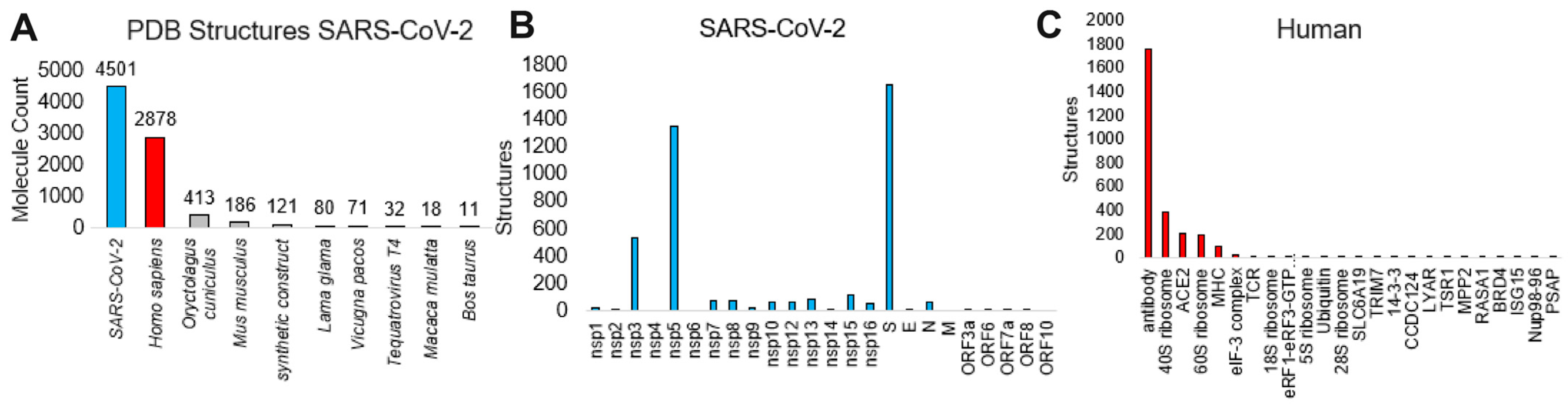
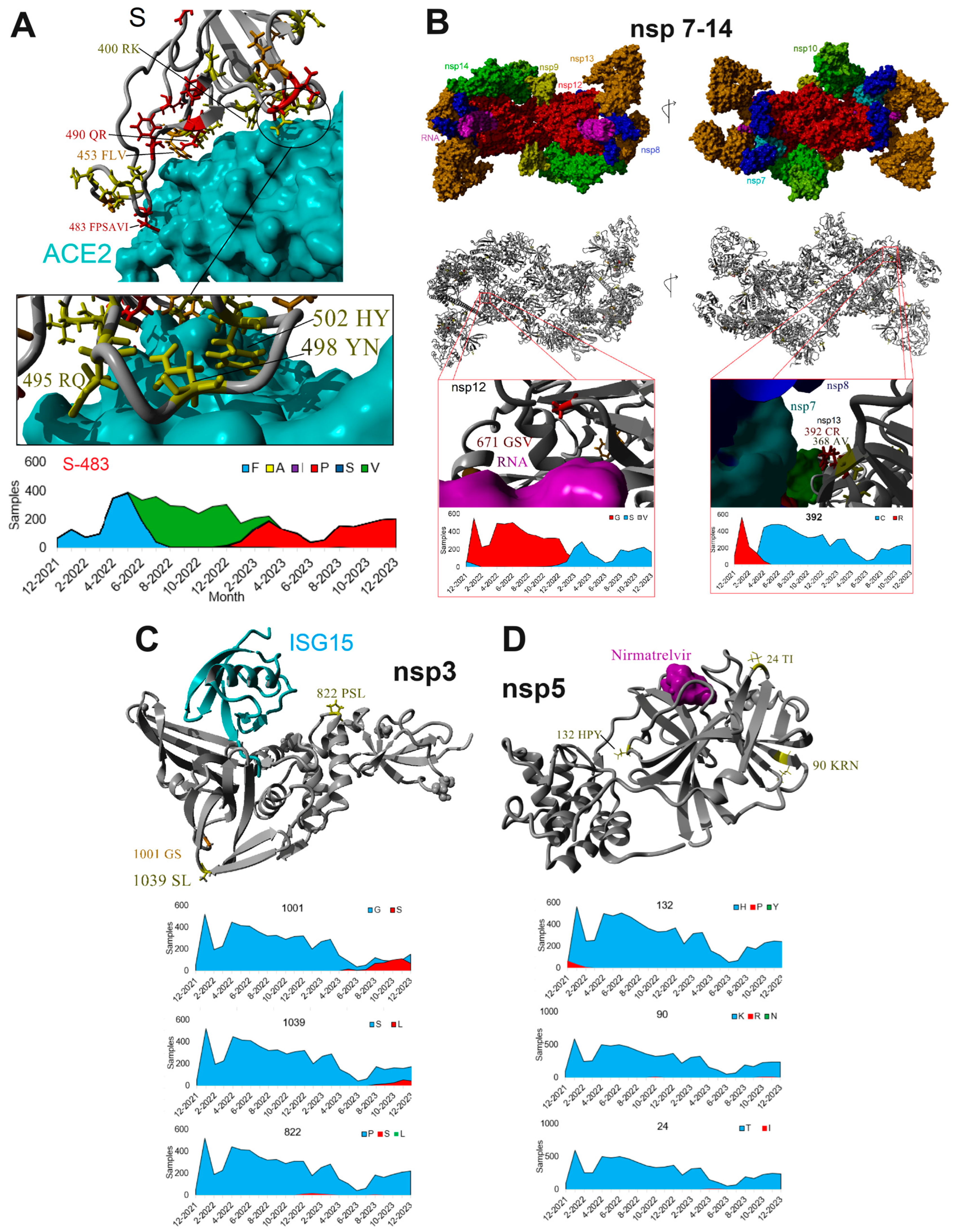
This work was approved by the Corewell Health West Institutional Review Board (IRB #CHW 2020-470). Nasal swabs were obtained from routine clinical testing at Corewell Health regional hospitals and collection sites throughout Michigan (MI), United States. Samples positive for SARS-CoV-2 (cycle threshold Ct value ≤ 32) were extracted using a QIAcube HT (Qiagen, Hilden, Germany) with a QIAamp 96 Viral RNA kit with Offboard Lysis 1NS-2 protocol. Illumina’s COVIDSeq Test kit was used for sequencing in rounds of 96-well plates consisting of 94 samples, a positive control, and a no template control for each run. IDT for Illumina DNA/RNA UD Index sets A through D (96 indexes per set) were used to identify each unique sample. Individual libraries were pooled per plate, quantified, and then normalized to 4 nm. Four normalized libraries were pooled together and sequenced on the NextSeq 500 using a mid-output sequencing cartridge. FASTQ analysis was performed using Illumina’s DRAGEN COVID Lineage application, and the flattened FASTA file for each sample was exported to NCBI and GISAID with collection dates for further protein annotations. NCBI processing of sample uploads as the routine part of NCBI virus annotations yielding updated Pangolin and protein annotations [14].
FASTA files for each SARS-CoV-2 protein of all sequenced samples were exported from NCBI Virus tool and aligned with the Kalign algorithm [15] using UGENE tools [16] with the gap open penalty set to 54.95, gap extension penalty at 8.52, terminal gap penalty at 4.42, a bonus score of 0.02, and the standard genetic code option. Only missense variants were assessed for the sequenced samples, with all synonymous changes of the viral genomes not analyzed in the current study. Following alignments within the UGENE tool, the consensus of all aligned sequences for each protein was calculated using the default consensus mode set to a threshold of 10%. The consensus sequence was added to each alignment as the top listed sequence, and alignments were transformed into matrix positioning using Mega software (version 5) [17]. Within the matrix, any sequences with uncertain base calls (N to ? annotation in FASTA files) over the protein resulted in that entire protein sequence being removed, yielding a highly curated alignment with uncertain error removed. After sequence removal, a unique list of amino acids at each position was calculated from the matrix, and the percent of sequences with the same amino acid as the consensus was calculated, with one minus that number serving as the drift rate for each amino acid.
The Protein Data Bank (PDB) [18] was quired on 3/14/2024 for all structures deposited containing the species “SARS-CoV-2”. Results were extracted for the per-molecule species annotations and manually curated for the protein annotations of human and SARS-CoV-2 molecules to fix uncommon annotation use. The Spike-ACE2 integrated structure [13], nsp7-14 complex (PDB file 7egq) [19], nsp3-ISG15 (6xa9) [20], and nsp5-Nirmatrelvir (8b2t) [21] files were used to map variants using YASARA software (version 24) [22]. In short, each molecule of SARS-CoV-2 was homology modeled from the source PDB file using the protein consensus sequence above. Coloring of amino acid drift was done using YASARA python coding. Data for all USA and international genotypes were extracted for SARS-CoV-2 (taxon ID 2697049) using NCBI Virus on 24 July 2024. Each variant identified in structures was annotated to lineages using the NCBI Virus mutation tool.

3. Results

Corewell Health clinical sites are in a mixed community of a city (Grand Rapids) with a regional triage center and additional hospitals that service areas of rural Michigan, USA. The state of Michigan has ranked as one of the top geographical areas for SARS-CoV-2 genomes (Figure 1E, red), with a median density of genomes relative to the population size (Figure 1F, red). Corewell Health is an extensive hospital system that serves over 1.3 million health plan members. Corewell Health sequenced 7518 SARS-CoV-2 genomes from December 2021 through December 2023 that passed quality control with >90% coverage and a median of 500 reads (Figure 2A). These samples were all used to process genome annotations with the NCBI standard SARS-CoV-2 workflow. There were a few delta strains throughout the two years of sample/data collection and sequencing, but most of the sequencing was of various omicron annotations (Figure 2B). The genotyping data were 82% concordant with national data (USA) over the study period based on a per genotype per month annotation of NCBI SRA data relative to Corewell Health samples (Figure 2C). This suggests that our genotyping data represent similar trends to national data for month-over-month sampling and genotype calls, opening the door for variant and protein analyses using our subset of data.
As large volumes of samples (in the millions) are computationally limiting due to the volume, random sampling is often used to assess viral drift that is not centered on ultra-rare variant detection. Utilizing four months throughout the sampling, we further compared the breakdown of Pangolin annotations at Corewell Health relative to all samples in the state of Michigan, the USA, and any deposited sample (internationally) to determine the utility of a single hospital system analysis. The absence of alpha and low levels of delta observed early in the study are in concordance with the level of USA cases (Figure 2D). Corewell Health accurately reflects the genotyping in the state of Michigan and USA, while the international data do have some difference in timing, likely the result of migration of genotypes being different over geographical regions month-to-month. In the case of this work, the high correlation of the Corewell Health month-to-month genotype levels is in high agreement with NCBI USA data and thus represents a sampling strategy for variants arising over lineages and not focusing on the ultra-rare changes that occurred throughout the pandemic.
Each protein of SARS-CoV-2 was aligned at an amino acid level and assessed for coverage and drift relative to the consensus sequence (Figure 3). Extensive parsing of ambiguous protein sequences was used to remove uncertain sequences, resulting in variability for each protein coverage. The ORF1ab protein was broken into the various protein cleavage products nsp1-nsp16. A total of 166,741 protein sequences for 9634 amino acids were assessed, yielding 64,979,205 positions assessed in our genotyping. The envelope protein (E) has a drift rate of 1.21% per amino acid, followed by ORF6a (0.90%) and Spike (S, 0.76%), representing the proteins with the most changes relative to the consensus sequence of each protein. Many of the nonstructural proteins have a low drift rate, including nsp7 (0.01%), nsp10 (0.02%), and nsp5 (0.02%). Spike (S) has 20 different amino acids with >10% drift from the consensus, which, when normalized for protein size, is the highest along with ORF6a (Figure 3A). Proteins such as nsp2, nsp5, nsp7, nsp8, nsp9, nsp10, nsp14, nsp16, ORF7a, ORF8, and ORF10 had no amino acid variants with >10% frequency (Figure 3B). A total of 43 amino acids has a drift rate of >10% relative to the consensus (Table 1). Spike, ORF6a, E, nsp1, and nsp6 had the highest frequency of variants over 10% of samples.
To identify variant outcomes relative to known protein structures, we curated the known structural knowledge of SARS-CoV-2 within the RCSB PDB as of 14 March 2024 (Figure 4). The RCSB PDB is the main repository for all solved protein structures [23]. A total of 4501 SARS-CoV-2 unique molecules have had a structure determined, including 2878 human proteins bound to them (Figure 4A). Other species bound are primarily based on antibody molecules produced in non-human organisms. Of the SARS-CoV-2 molecules with structures, Spike is the most prevalent, followed by the two proteases nsp3 and nsp5 (Figure 4B). Of the human-bound proteins, the majority were antibodies or MHC complexes (Figure 4C), with a few Spike-ACE2 structures and several large ribosome complexes.
Structure files were used to align the genomic variants of SARS-CoV-2 for Spike-ACE2 (Figure 5A), the multi-protein nsp7-nsp14 SARS-CoV-2 co-transcriptional complex (Figure 5B), nsp3-ISG15 (Figure 5C), and nsp5- nirmatrelvir (PAXLOVID, Figure 5D) structure files. Spike has multiple variants within the ACE2 interaction region (Figure 5A). A cluster of variants at amino acids 495, 498, and 502 contact ACE2 but maintain functional conservation. Amino acid 495 uses arginine (R) or glutamine (Q), while 498 uses tyrosine (Y) or asparagine (N), all of which maintain hydrogen bonding to ACE2 sites. Amino acid 502 uses histidine and tyrosine, maintaining aromatic ACE2 pi orbital interactions. Additional functional conservation can be found for ACE2 interactions at amino acids 400 (R or K), 490 (Q or R), 453 (F, L, or V), and 483. Amino acid 483 transitions throughout the two years of sequencing from phenylalanine (F) to valine (V) to proline (P), which all maintain the hydrophobic ACE2 interactions. This variant also correlates to changing lineage annotations with this as position 486 of BA.5.1 with a valine and XBB.1.5 with a proline. The proline has previously been suggested as a variant of concern due to risks in increasing binding affinity to ACE2 [24,25].
In contrast to the high drift of Spike, the entire multi-protein SARS-CoV-2 co-transcriptional complex (Figure 5B) only contains two variants of potential alteration impact. The nsp12 amino acid 671 transitions from a glycine (G) to a serine (S) in early 2023, allowing for the flexible bend angle near the RNA contact points. This variant (RDRP: G671S) represents 2% of the GenBank deposited samples from 2/2024 to 7/2024 with the highest frequency in un-assignable and GE.1.2.1 lineages according to the NCBI Virus mutation tool. The most impactful change within the complex occurs at nsp13 amino acid 392, where in early 2022, it changed from an arginine (R) to cysteine (C). This location falls near the nsp7 and nsp8 interaction sites and thus could potentially alter interactions. Within GenBank, 99% of deposited samples from 2/2024 to 7/2024 contain this change over all major annotated lineages.
The top studied drug target sites are the proteases nsp3 (Figure 5C) and nsp5 (Figure 5D) of ORF1ab, where no variants above 1% of our sequenced samples are found at the targetable active sites. Those variants that are above 1% all fall on nonfunctional sites such as 822, 1001, and 1039 of nsp3 or 24, 90, or 132 of nsp5.

4. Discussion

As any virus continues to drift and change over time, there is a critical need to focus on the biological outcomes of the changes rather than over-focusing on genotypes. Although genotypes can inform the kinetics of viral propagation, yielding insights into how a virus’s geographical and temporal movements happen [26], they can also result in confusion and fear when at the forefront of the conversation about a pandemic or endemic. As the COVID-19 pandemic became so focused on genotypes and the rapid changes of SARS-CoV-2, the general population and policymakers often over-reacted [27,28]. As the current work shows, many SARS-CoV-2 proteins have had few changes outside Spike, with even fewer that are functional relative to our known structural knowledge and dynamics [13] of the virus. At the onset of the pandemic, groups such as ours called for drug development based on shared function with other Coronaviruses [13,29,30]. However, international investments in Spike-based therapies far outpaced that of other protein complexes. Although viruses such as HIV have high levels of protein changes throughout based on replication dynamics that make drug development complex [29], more focus should be placed on genotypes’ structural impacts, molecular outcomes, and drug-targeting dynamics for each virus.
A simple word search of relevant terms to this paper shows that surface glycoprotein and Spike terms far outnumber any of the other proteins of SARS-CoV-2 (Table 2). The general terms for the virus, such as COVID-19 and SARS-CoV-2, are higher in Google relative to both PubMed and Google Scholar. Surface glycoprotein is the highest returned term within PubMed and far outpaces other proteins in both PubMed and Google Scholar, suggesting that, as noted above, there is an overfocus on surface glycoprotein (Spike, S) that does not match the genotyping conservation discussed within the current work. A search of “SARS-CoV-2 mutations” and “Spike” returns 4920 returns on Google Scholar, while “SARS-CoV-2 mutations” and “ORF” or “nsp” returns only 929 and 598 hits. Both ORF and nsp represent multiple proteins, further highlighting the contrast relative to Spike. This highlights a fundamental issue with our current genotyping interpretation and overfocus on genomic changes relative to conservation analysis.
It is well known that Spike continues to drift, which has led to the iterative development of changes to COVID-19 vaccines, a likely never-ending process based on the current genotyping insights. The changes in Spike were a major factor for the elevation of pathogenicity for SARS-CoV-2 over other coronaviruses [31], and several of the amino acid changes observed in Omicron Spike also modulate ACE2 and immune evasion [32,33]. As our data show, what is less appreciated is the functionally conserved changes happening at the ACE2 binding site, while no other protein shows such levels of functional drift. The data suggest that pressure for Spike changes is not found in other SARS-CoV-2 coded proteins. These insights are highly relevant to moving forward in the virus’s treatment and public health dynamics in the endemic phase, suggesting a significant need to transition from Spike-based therapies to other proteins. Others have noted that this drift makes it very challenging also to target the Spike-ACE2 interaction with drugs [34]. As we continue to engage the arms race of Spike antibody targeting with Spike drift for ACE2 interactions, it is feasible to drive selective pressure to elevate Spike dynamics that could enhance virulence, viral dynamics, and increase binding affinity [35]. This suggests pivoting to non-Spike therapies and strategies [36].
So why does Spike have amino acid drift at a rate so different from many other viral proteins? The answer is likely that the surface proteins need not be maintained for a virus to replicate; they are only required for spreading [37,38,39]. All a virus needs the surface proteins to do is evade immune detection and get inside a cell, and there are many proteins they can find to get inside a cell with many ways to evade immune detection. Viruses are always finding new cell epitopes with which to integrate. As the protein sequences change on surface proteins like Spike, there may be some selection of binding surfaces, as we have shown for ACE2-Spike interactions in SARS-CoV-2, while changes also enhance immune evasion and confirm selective benefit [40]. It is uncertain and unlikely vaccinations impact drift of the surface proteins as this drift is already naturally occurring. Even without vaccinations, the freedom of the surface protein to change due to its limited role in viral replication, the proteins behave much as any protein with reduced selective pressure [41]. If the protein changes enough, selection finds another protein partner in the host to enter a cell, as evolution has shown often in large evolutionary changes that give rise to new viral species. This is a similar concept throughout evolution when large genomic segments duplicate and reduce the selective pressure of one of the two copies of paralogs to give radiant evolution potential [42]. However, when the sequence changes for the inner machinery of proteins in a virus, like the proteases and polymerases, the virus cannot survive and the change is quickly lost to subsequent viral replications due to selective pressures [43]. In the process of the constant viral turnover, these pressures on protein drift can be seen and are likely the result of protein-centric genotyping insights from our work.
A genome is only a coding material for the biological functions manifested from protein structures. In virology, the focus has often been the DNA/RNA sequence, as the material is more straightforward to observe and make sense of. That gives rise to assessments of biology using phylogenetics and explaining viral drift at DNA/RNA level where the genotypes make it appear a virus rapidly changes. As the work here shows, the drift of DNA often looks chaotic, while the proteins look stable. Thus, the use of genomes in drug design has a view of chaos and constant change. As most of our drugs target proteins, the assessment of viral drift requires a protein-centric assessment. The research field did this well throughout the pandemic for Spike, but the rest of the SARS-CoV-2 proteome was left overlooked.
One of the most prolific examples of protein structural drug design is that of HIV, where the use of structures and the rapid drift have opened up a toolbox of drugs [44]. As viruses such as HIV rapidly change due to the reverse transcriptase process, these viruses highlight how challenging it is to treat, where focus on the conserved mechanisms that, when drifted, impact the viral function are strategic targets of our emerging drugs [45]. As viruses often share these critical protein mechanisms, the drugs that target conserved protein pockets can often open rapid treatment approaches for emerging viruses, as was so well done in targeting the proteases of SARS-CoV-2 that originated from the structure-based design within SARS-CoV [46]. The structure-based drug design can be seen emerging in dengue [47], Zika [48], Hepatitis [49], and influenzas [50], to name a few.
Our data are supported by the real-world continued and growing success of the antiviral medication nirmatrelvir-ritonavir (brand name Paxlovid) and nsp5-targeting, whose efficacy has not been impacted by the two years of viral drift [51,52]. Our early work in viral evolution for SARS-CoV-2 relative to other coronaviruses suggested the same outcomes [13], and the current genotyping data also highly support non-Spike therapies. It also suggests that some T-cell receptor and antibody-active proteins, such as the nucleocapsid, may represent more sustainable models for public health vaccinations.
As epidemiology begins to decipher the long-term risks of SARS-CoV-2, including in non-severe COVID-19 [53], and as the virus becomes endemic, similar to Epstein Barr virus with rare risks to populations [54], we need to consider the role of viral drift heavily in our future therapy strategies against SARS-CoV-2. We must also enhance our knowledge of proteins that are not drifting and conserved across multiple Coronaviruses. For example, the role of NSP3 and other proteins in immune suppression [29], which remain unchanged throughout the SARS-CoV-2 drift and are present in many patients’ blood responses [55], yet have largely been ignored and underfunded. Suppose we were to invest as heavily into these unknown mechanisms that remain highly conserved within the SARS-CoV-2 proteins as we did for Spike and vaccine strategies. In that case, it is likely that we would transform not only the knowledge and treatments for COVID-19 but also make major strides for numerous other viruses that share mechanisms. Continuous integrated surveillance systems and research are necessary to monitor the virus’s evolution, evaluate the effectiveness of vaccines and treatments, and rapidly respond to emerging variants to ensure public health safety.

Author Contributions

Conceptualization, J.W.P., S.A. (Sheryl Alberta), D.C., R.O. and A.J.C.; methodology, J.W.P., S.A. (Sheryl Alberta), M.W.-L. and S.P.; validation, J.W.P., M.W.-L. and S.P.; formal analysis, J.W.P., M.W.-L., S.P., H.A.F., D.B., R.M.V., A.F., J.B., H.B., S.A. (Sanjana Arora), C.S., D.P., A.G., M.W., T.W.C., J.T.M., J.Z., M.D.S., A.U., H.H., S.R. and M.A.T.K.; investigation, J.W.P., S.A. (Sheryl Alberta), D.C., R.O. and A.J.C.; resources, D.C.; data curation, J.W.P., M.W.-L. and S.P.; writing—original draft preparation, J.W.P. and A.J.C.; writing—review and editing, J.W.P., S.A. (Sheryl Alberta), M.W.-L., S.P., H.A.F., D.B., R.M.V., A.F., J.B., H.B., S.A. (Sanjana Arora), C.S., D.P., A.G., M.W., T.W.C., J.T.M., J.Z., M.D.S., A.U., H.H., S.R., M.A.T.K., D.C., R.O. and A.J.C.; visualization, J.W.P.; supervision, J.W.P., S.A. (Sheryl Alberta), D.C., R.O. and A.J.C.; project administration, D.C.; funding acquisition, J.W.P., S.A. (Sheryl Alberta), D.C., R.O. and A.J.C. All authors have read and agreed to the published version of the manuscript.

Funding

This research was funded by MI-SAPPHIRE, a grant from the Michigan Department of Health and Human Services through the Centers for Disease Control.

Data Availability Statement

All sequencing data annotations are available at https://doi.org/10.6084/m9.figshare.25988050.v1. The drift calculation for each amino acid is located at https://doi.org/10.6084/m9.figshare.26364514.v1.

Conflicts of Interest

None of the authors have declared any significant conflicts of interest relative to this work. Corewell Health is a not for profit hospital system in the USA.

References

  1. Hatcher, E.L.; Zhdanov, S.A.; Bao, Y.; Blinkova, O.; Nawrocki, E.P.; Ostapchuck, Y.; Schäffer, A.A.; Brister, J.R. Virus Variation Resource—Improved Response to Emergent Viral Outbreaks. Nucleic Acids Res. 2017, 45, D482–D490. [Google Scholar] [CrossRef] [PubMed]
  2. Shu, Y.; McCauley, J. GISAID: Global Initiative on Sharing All Influenza Data—From Vision to Reality. Euro Surveill. Bull. Eur. Sur Mal. Transm. Eur. Commun. Dis. Bull. 2017, 22, 30494. [Google Scholar] [CrossRef] [PubMed]
  3. Martin, M.A.; VanInsberghe, D.; Koelle, K. Insights from SARS-CoV-2 Sequences. Science 2021, 371, 466–467. [Google Scholar] [CrossRef]
  4. Harvey, W.T.; Carabelli, A.M.; Jackson, B.; Gupta, R.K.; Thomson, E.C.; Harrison, E.M.; Ludden, C.; Reeve, R.; Rambaut, A.; COVID-19 Genomics UK (COG-UK) Consortium; et al. SARS-CoV-2 Variants, Spike Mutations and Immune Escape. Nat. Rev. Microbiol. 2021, 19, 409–424. [Google Scholar] [CrossRef]
  5. Kim, D.; Lee, J.-Y.; Yang, J.-S.; Kim, J.W.; Kim, V.N.; Chang, H. The Architecture of SARS-CoV-2 Transcriptome. Cell 2020, 181, 914–921.e10. [Google Scholar] [CrossRef] [PubMed]
  6. Tao, K.; Tzou, P.L.; Nouhin, J.; Gupta, R.K.; de Oliveira, T.; Kosakovsky Pond, S.L.; Fera, D.; Shafer, R.W. The Biological and Clinical Significance of Emerging SARS-CoV-2 Variants. Nat. Rev. Genet. 2021, 22, 757–773. [Google Scholar] [CrossRef] [PubMed]
  7. Ba, A.A.; Coppée, R.; Dieng, A.; Manneh, J.; Fall, M.; Gueye, K.; Sene, P.Y.; Ndiour, S.; Samaté, D.; Manga, P.; et al. Genomic Epidemiology of SARS-CoV-2 in Senegal in 2020–2021. J. Infect. Dev. Ctries. 2024, 18, 851–861. [Google Scholar] [CrossRef]
  8. Machado, L.C.; Dezordi, F.Z.; de Lima, G.B.; de Lima, R.E.; Silva, L.C.A.; Pereira, L.d.M.; da Silva, A.F.; Silva Neto, A.M.d.; Oliveira, A.L.S.d.; Armstrong, A.d.C.; et al. Spatiotemporal Transmission of SARS-CoV-2 Lineages during 2020-2021 in Pernambuco-Brazil. Microbiol. Spectr. 2024, 12, e0421823. [Google Scholar] [CrossRef]
  9. Khairnar, K.; Tomar, S.S. COVID-19 Genome Surveillance: A Geographical Landscape and Mutational Mapping of SARS-CoV-2 Variants in Central India over Two Years. Virus Res. 2024, 344, 199365. [Google Scholar] [CrossRef]
  10. Lagare, A.; Faye, M.; Issa, M.; Hamidou, O.; Bienvenu, B.; Mohamed, A.; Aoula, B.; Moumouni, K.; Hassane, F.; Otto, Y.A.; et al. First Identification of the SARS-COV-2/XBB.1.5 Sublineage among Indigenous COVID-19 Cases through the Influenza Sentinel Surveillance System in Niger. Heliyon 2023, 9, e20916. [Google Scholar] [CrossRef]
  11. Walensky, R.P.; Walke, H.T.; Fauci, A.S. SARS-CoV-2 Variants of Concern in the United States-Challenges and Opportunities. JAMA 2021, 325, 1037–1038. [Google Scholar] [CrossRef]
  12. Chiara, M.; D’Erchia, A.M.; Gissi, C.; Manzari, C.; Parisi, A.; Resta, N.; Zambelli, F.; Picardi, E.; Pavesi, G.; Horner, D.S.; et al. Next Generation Sequencing of SARS-CoV-2 Genomes: Challenges, Applications and Opportunities. Brief. Bioinform. 2021, 22, 616–630. [Google Scholar] [CrossRef] [PubMed]
  13. Gupta, R.; Charron, J.; Stenger, C.L.; Painter, J.; Steward, H.; Cook, T.W.; Faber, W.; Frisch, A.; Lind, E.; Bauss, J.; et al. SARS-CoV-2 (COVID-19) Structural and Evolutionary Dynamicome: Insights into Functional Evolution and Human Genomics. J. Biol. Chem. 2020, 295, 11742–11753. [Google Scholar] [CrossRef]
  14. Brister, J.R.; Ako-Adjei, D.; Bao, Y.; Blinkova, O. NCBI Viral Genomes Resource. Nucleic Acids Res. 2015, 43, D571–D577. [Google Scholar] [CrossRef] [PubMed]
  15. Lassmann, T.; Sonnhammer, E.L.L. Kalign—An Accurate and Fast Multiple Sequence Alignment Algorithm. BMC Bioinform. 2005, 6, 298. [Google Scholar] [CrossRef]
  16. Okonechnikov, K.; Golosova, O.; Fursov, M. UGENE team Unipro UGENE: A Unified Bioinformatics Toolkit. Bioinformatics 2012, 28, 1166–1167. [Google Scholar] [CrossRef] [PubMed]
  17. Kumar, S.; Tamura, K.; Nei, M. MEGA: Molecular Evolutionary Genetics Analysis Software for Microcomputers. Comput. Appl. Biosci. CABIOS 1994, 10, 189–191. [Google Scholar] [CrossRef] [PubMed]
  18. Berman, H.; Henrick, K.; Nakamura, H.; Markley, J.L. The Worldwide Protein Data Bank (wwPDB): Ensuring a Single, Uniform Archive of PDB Data. Nucleic Acids Res. 2007, 35, D301–D303. [Google Scholar] [CrossRef]
  19. Yan, L.; Yang, Y.; Li, M.; Zhang, Y.; Zheng, L.; Ge, J.; Huang, Y.C.; Liu, Z.; Wang, T.; Gao, S.; et al. Coupling of N7-Methyltransferase and 3′-5′ Exoribonuclease with SARS-CoV-2 Polymerase Reveals Mechanisms for Capping and Proofreading. Cell 2021, 184, 3474–3485.e11. [Google Scholar] [CrossRef]
  20. Klemm, T.; Ebert, G.; Calleja, D.J.; Allison, C.C.; Richardson, L.W.; Bernardini, J.P.; Lu, B.G.; Kuchel, N.W.; Grohmann, C.; Shibata, Y.; et al. Mechanism and Inhibition of the Papain-like Protease, PLpro, of SARS-CoV-2. EMBO J. 2020, 39, e106275. [Google Scholar] [CrossRef]
  21. Brewitz, L.; Dumjahn, L.; Zhao, Y.; Owen, C.D.; Laidlaw, S.M.; Malla, T.R.; Nguyen, D.; Lukacik, P.; Salah, E.; Crawshaw, A.D.; et al. Alkyne Derivatives of SARS-CoV-2 Main Protease Inhibitors Including Nirmatrelvir Inhibit by Reacting Covalently with the Nucleophilic Cysteine. J. Med. Chem. 2023, 66, 2663–2680. [Google Scholar] [CrossRef] [PubMed]
  22. Krieger, E.; Koraimann, G.; Vriend, G. Increasing the Precision of Comparative Models with YASARA NOVA--a Self-Parameterizing Force Field. Proteins 2002, 47, 393–402. [Google Scholar] [CrossRef]
  23. Kouranov, A.; Xie, L.; de la Cruz, J.; Chen, L.; Westbrook, J.; Bourne, P.E.; Berman, H.M. The RCSB PDB Information Portal for Structural Genomics. Nucleic Acids Res. 2006, 34, D302–D305. [Google Scholar] [CrossRef] [PubMed]
  24. Parums, D.V. Editorial: The XBB.1.5 (‘Kraken’) Subvariant of Omicron SARS-CoV-2 and Its Rapid Global Spread. Med. Sci. Monit. Int. Med. J. Exp. Clin. Res. 2023, 29, e939580. [Google Scholar] [CrossRef] [PubMed]
  25. Callaway, E. Coronavirus Variant XBB.1.5 Rises in the United States—Is It a Global Threat? Nature 2023, 613, 222–223. [Google Scholar] [CrossRef]
  26. Mercatelli, D.; Giorgi, F.M. Geographic and Genomic Distribution of SARS-CoV-2 Mutations. Front. Microbiol. 2020, 11, 1800. [Google Scholar] [CrossRef] [PubMed]
  27. Mirza, N.; Rahat, B.; Naqvi, B.; Rizvi, S.K.A. Impact of Covid-19 on Corporate Solvency and Possible Policy Responses in the EU. Q. Rev. Econ. Finance J. Midwest Econ. Assoc. 2023, 87, 181–190. [Google Scholar] [CrossRef]
  28. Alvarez, E.; Bielska, I.A.; Hopkins, S.; Belal, A.A.; Goldstein, D.M.; Slick, J.; Pavalagantharajah, S.; Wynfield, A.; Dakey, S.; Gedeon, M.-C.; et al. Limitations of COVID-19 Testing and Case Data for Evidence-Informed Health Policy and Practice. Health Res. Policy Syst. 2023, 21, 11. [Google Scholar] [CrossRef]
  29. Hartog, N.; Faber, W.; Frisch, A.; Bauss, J.; Bupp, C.P.; Rajasekaran, S.; Prokop, J.W. SARS-CoV-2 Infection: Molecular Mechanisms of Severe Outcomes to Suggest Therapeutics. Expert Rev. Proteomics 2021, 18, 105–118. [Google Scholar] [CrossRef]
  30. Sirpilla, O.; Bauss, J.; Gupta, R.; Underwood, A.; Qutob, D.; Freeland, T.; Bupp, C.; Carcillo, J.; Hartog, N.; Rajasekaran, S.; et al. SARS-CoV-2-Encoded Proteome and Human Genetics: From Interaction-Based to Ribosomal Biology Impact on Disease and Risk Processes. J. Proteome Res. 2020, 19, 4275–4290. [Google Scholar] [CrossRef]
  31. Shang, J.; Ye, G.; Shi, K.; Wan, Y.; Luo, C.; Aihara, H.; Geng, Q.; Auerbach, A.; Li, F. Structural Basis of Receptor Recognition by SARS-CoV-2. Nature 2020, 581, 221–224. [Google Scholar] [CrossRef] [PubMed]
  32. Yin, W.; Xu, Y.; Xu, P.; Cao, X.; Wu, C.; Gu, C.; He, X.; Wang, X.; Huang, S.; Yuan, Q.; et al. Structures of the Omicron Spike Trimer with ACE2 and an Anti-Omicron Antibody. Science 2022, 375, 1048–1053. [Google Scholar] [CrossRef] [PubMed]
  33. Pastorio, C.; Zech, F.; Noettger, S.; Jung, C.; Jacob, T.; Sanderson, T.; Sparrer, K.M.J.; Kirchhoff, F. Determinants of Spike Infectivity, Processing, and Neutralization in SARS-CoV-2 Omicron Subvariants BA.1 and BA.2. Cell Host Microbe 2022, 30, 1255–1268.e5. [Google Scholar] [CrossRef]
  34. Verkhivker, G. Coevolution, Dynamics and Allostery Conspire in Shaping Cooperative Binding and Signal Transmission of the SARS-CoV-2 Spike Protein with Human Angiotensin-Converting Enzyme 2. Int. J. Mol. Sci. 2020, 21, 8268. [Google Scholar] [CrossRef] [PubMed]
  35. Zhu, R.; Canena, D.; Sikora, M.; Klausberger, M.; Seferovic, H.; Mehdipour, A.R.; Hain, L.; Laurent, E.; Monteil, V.; Wirnsberger, G.; et al. Force-Tuned Avidity of Spike Variant-ACE2 Interactions Viewed on the Single-Molecule Level. Nat. Commun. 2022, 13, 7926. [Google Scholar] [CrossRef]
  36. Kakavandi, S.; Zare, I.; VaezJalali, M.; Dadashi, M.; Azarian, M.; Akbari, A.; Ramezani Farani, M.; Zalpoor, H.; Hajikhani, B. Structural and Non-Structural Proteins in SARS-CoV-2: Potential Aspects to COVID-19 Treatment or Prevention of Progression of Related Diseases. Cell Commun. Signal. CCS 2023, 21, 110. [Google Scholar] [CrossRef]
  37. Hynes, R.O. Cell Surface Proteins and Malignant Transformation. Biochim. Biophys. Acta 1976, 458, 73–107. [Google Scholar] [CrossRef]
  38. Choppin, P.W.; Scheid, A. The Role of Viral Glycoproteins in Adsorption, Penetration, and Pathogenicity of Viruses. Rev. Infect. Dis. 1980, 2, 40–61. [Google Scholar] [CrossRef]
  39. Hynes, R.O. Alteration of Cell-Surface Proteins by Viral Transformation and by Proteolysis. Proc. Natl. Acad. Sci. USA 1973, 70, 3170–3174. [Google Scholar] [CrossRef]
  40. Vossen, M.T.M.; Westerhout, E.M.; Söderberg-Nauclér, C.; Wiertz, E.J.H.J. Viral Immune Evasion: A Masterpiece of Evolution. Immunogenetics 2002, 54, 527–542. [Google Scholar] [CrossRef]
  41. Lynch, M. Mutation Pressure, Drift, and the Pace of Molecular Coevolution. Proc. Natl. Acad. Sci. USA 2023, 120, e2306741120. [Google Scholar] [CrossRef]
  42. Magadum, S.; Banerjee, U.; Murugan, P.; Gangapur, D.; Ravikesavan, R. Gene Duplication as a Major Force in Evolution. J. Genet. 2013, 92, 155–161. [Google Scholar] [CrossRef] [PubMed]
  43. Koonin, E.V.; Dolja, V.V.; Krupovic, M. Origins and Evolution of Viruses of Eukaryotes: The Ultimate Modularity. Virology 2015, 479–480, 2–25. [Google Scholar] [CrossRef]
  44. Turner, B.G.; Summers, M.F. Structural Biology of HIV. J. Mol. Biol. 1999, 285, 1–32. [Google Scholar] [CrossRef] [PubMed]
  45. Gaikwad, S.Y.; Phatak, P.; Mukherjee, A. Cutting Edge Strategies for Screening of Novel Anti-HIV Drug Candidates against HIV Infection: A Concise Overview of Cell Based Assays. Heliyon 2023, 9, e16027. [Google Scholar] [CrossRef]
  46. Lu, I.-L.; Mahindroo, N.; Liang, P.-H.; Peng, Y.-H.; Kuo, C.-J.; Tsai, K.-C.; Hsieh, H.-P.; Chao, Y.-S.; Wu, S.-Y. Structure-Based Drug Design and Structural Biology Study of Novel Nonpeptide Inhibitors of Severe Acute Respiratory Syndrome Coronavirus Main Protease. J. Med. Chem. 2006, 49, 5154–5161. [Google Scholar] [CrossRef]
  47. Noble, C.G.; Shi, P.-Y. Structural Biology of Dengue Virus Enzymes: Towards Rational Design of Therapeutics. Antiviral Res. 2012, 96, 115–126. [Google Scholar] [CrossRef]
  48. Cox, B.D.; Stanton, R.A.; Schinazi, R.F. Predicting Zika Virus Structural Biology: Challenges and Opportunities for Intervention. Antivir. Chem. Chemother. 2015, 24, 118–126. [Google Scholar] [CrossRef] [PubMed]
  49. Penin, F.; Dubuisson, J.; Rey, F.A.; Moradpour, D.; Pawlotsky, J.-M. Structural Biology of Hepatitis C Virus. Hepatol. Baltim. Md 2004, 39, 5–19. [Google Scholar] [CrossRef] [PubMed]
  50. Liu, C.; Hu, L.; Dong, G.; Zhang, Y.; Ferreira da Silva-Júnior, E.; Liu, X.; Menéndez-Arias, L.; Zhan, P. Emerging Drug Design Strategies in Anti-Influenza Drug Discovery. Acta Pharm. Sin. B 2023, 13, 4715–4732. [Google Scholar] [CrossRef]
  51. Hashemian, S.M.R.; Sheida, A.; Taghizadieh, M.; Memar, M.Y.; Hamblin, M.R.; Bannazadeh Baghi, H.; Sadri Nahand, J.; Asemi, Z.; Mirzaei, H. Paxlovid (Nirmatrelvir/Ritonavir): A New Approach to Covid-19 Therapy? Biomed. Pharmacother. Biomed. Pharmacother. 2023, 162, 114367. [Google Scholar] [CrossRef] [PubMed]
  52. Focosi, D.; Maggi, F.; McConnell, S.; Casadevall, A. Very Low Levels of Remdesivir Resistance in SARS-COV-2 Genomes after 18 Months of Massive Usage during the COVID19 Pandemic: A GISAID Exploratory Analysis. Antiviral Res. 2022, 198, 105247. [Google Scholar] [CrossRef]
  53. Jahankhani, K.; Ahangari, F.; Adcock, I.M.; Mortaz, E. Possible Cancer-Causing Capacity of COVID-19: Is SARS-CoV-2 an Oncogenic Agent? Biochimie 2023, 213, 130–138. [Google Scholar] [CrossRef]
  54. Fugl, A.; Andersen, C.L. Epstein-Barr Virus and Its Association with Disease—A Review of Relevance to General Practice. BMC Fam. Pract. 2019, 20, 62. [Google Scholar] [CrossRef] [PubMed]
  55. Prokop, J.W.; Hartog, N.L.; Chesla, D.; Faber, W.; Love, C.P.; Karam, R.; Abualkheir, N.; Feldmann, B.; Teng, L.; McBride, T.; et al. High-Density Blood Transcriptomics Reveals Precision Immune Signatures of SARS-CoV-2 Infection in Hospitalized Individuals. Front. Immunol. 2021, 12, 694243. [Google Scholar] [CrossRef] [PubMed]
Figure 1. Status of SARS-CoV-2 genomes (taxid:2697049) within NCBI Virus. Data extractions were performed on 24 July 2024. Data are shown for the isolate (A), protein annotations (B), or geographical region based on continent (C), country (D), or state within the USA (E). Panel (F) shows the normalized genome sequences in each state of the USA relative to the million individuals within the state based on the 2020 census levels. Red boxes indicate the location of current genotyping data.
Microorganisms 12 01863 g001
Figure 2. Corewell Health COVID-19 sequencing data. (A) Sequencing statistics for samples analyzed at Corewell Health using Illumina chemistry. (B) SARS-CoV-2 genotypes per month from December 2021 through December 2023. The “*” indicates months with further processing in panel D. (C) Correlation analysis of the Pangolin annotation each month from the USA (with <100 ambiguous N’s per sequence) relative to Corewell Health. (D) Bar plots for the percent of samples each of four months (starred in panel (D)) for data from Corewell Health, the state of Michigan, the USA, and any NCBI deposited sample (Internationally). Data are shown with the percent of the top 20 Pangolin annotations (top 5 labeled), with the % listed next to the stacked plot representing how many of the total samples are covered by the top 20. Colors correspond to panel (B) annotations for higher-level genotypes.
Microorganisms 12 01863 g002
Figure 3. SARS-CoV-2 protein drift. (A) Per protein amino acid drift relative to the consensus sequence. Shown next to each protein name is the number of sequences without ambiguity. The x-axis shows the amino acid number of each protein, and the y-axis shows the % of samples with drift relative to the consensus amino acid. (B) The per protein sequence coverage relative to the number of amino acids with >10% variation.
Microorganisms 12 01863 g003
Figure 4. Protein structure analysis of SARS-CoV-2. (A) The number of SARS-CoV-2 protein structure molecules for various species in the Protein Databank (PDB) as of 14 March 2024. (B) The number of unique structures for each of the SARS-CoV-2 proteins (cyan). (C) The number of human structures (red) interacting with SARS-CoV-2 proteins.
Microorganisms 12 01863 g004
Figure 5. Genomic drift mapped to SARS-CoV-2 protein structures. (A) Analysis of variants in Spike (S) at the interface of ACE2 interaction. (B) Analysis of variants for the SARS-CoV-2 co-transcriptional complex. (C) Analysis of variants for the nsp3 papain-like protease with the human ISG15 interaction site. (D) Analysis of variants for the nsp5 SARS-CoV-2 protein with nirmatrelvir (PAXLOVID) bound.
Microorganisms 12 01863 g005
Table 1. Amino acids that have the highest drift rate from each protein’s consensus sequence.
ProteinComplete Sequences for ProteinAmino Acid NumberConsensusAmino Acids UsedAmino Acids Used% Drift
surface glycoprotein5102483F6FPSAVI73.11
membrane glycoprotein36173D4DHNG62.37
surface glycoprotein5102449R5RLMWQ53.21
ORF6740561L5LIDFP49.99
surface glycoprotein510266H5H-YFS47.53
surface glycoprotein510267V3V-I47.43
surface glycoprotein5102343R5RTISK46.37
NSP47365438L3LFY46.06
surface glycoprotein5102457N5NKISY41.40
surface glycoprotein5102141Y2Y-36.50
surface glycoprotein5102443G5GSTDV35.59
surface glycoprotein5102210G4GERV34.77
surface glycoprotein5102336D5DHVYG33.83
surface glycoprotein5102442V8VPSHALIF31.145
surface glycoprotein5102143H7HQK-YPL29.60
surface glycoprotein5102487F3FSP29.38
surface glycoprotein510280V2VA29.09
surface glycoprotein5102365L2LI29.09
surface glycoprotein5102180Q6QEVHKL28.68
NSP127233671G3GSV28.61
surface glycoprotein5102249G3GVD26.81
envelope protein743711T3TAM26.45
NSP1646847K2KR25.57
surface glycoprotein5102490Q2QR24.52
NSP13723136S2SP24.31
NSP16468135R4RSKN18.69
NSP364341892A3ATG17.28
NSP47365264F2FL17.05
NSP47365327I4ITVF17.03
ORF3a7465223I2IT17.03
nucleocapsid phosphoprotein7016410R5RLSHC17.00
NSP157426112I3ITN16.89
NSP137231392C2CR16.60
NSP3643424I2IT16.52
NSP36434489S2SG16.47
NSP3643438K2KR15.99
NSP364341265S2S-15.96
NSP364341266L3LIV15.96
NSP67472105L2LF15.91
NSP67472186I2IV15.79
NSP67472257L3LFH12.17
surface glycoprotein5102441K5KTNRM11.00
surface glycoprotein5102453F3FLV10.80
Table 2. A search for relevant words from SARS-CoV-2 in various databases. Each database was searched on 24 July 2024.
PubMed (All)PubMed (Since 2020)Google Scholar (All)Google Scholar (Since 2020)Google (All)Ratio Google to PubMed (Since 2020)
surface glycoprotein788,63692,4841,840,00026,70045,000,000487
Membrane glycoprotein786,28991,8631,920,00024,20038,400,000418
COVID-19438,172292,4935,040,0001,020,0006,400,000,00021,881
pandemic274,892171,3204,220,000683,0001,510,000,0008814
SARS-CoV-2231,386186,7172,500,000781,000459,000,0002458
viral genome117,24319,2414,220,00030,400120,000,0006237
viral genotype64,22410,6742,130,00016,70023,500,0002202
Envelope protein52,39159392,110,00023,90046,300,0007796
RNA-directed RNA polymerase38,009414235,100719040,200,0009705
Helicase36,6227834332,00025,20019,500,0002489
Nucleocapsid28,3115146208,00027,5007,970,0001549
surface glycoprotein spike9146226991,90018,2001,210,000533
papain like protease203157495,10017,000776,0001352
3C like protease1580986850,00016,3008,810,0008935
Nucleocapsid phosphoprotein145269419,7006350131,000189
nsp2118030220,8008450459,0001520
nsp1112432426,60012,400428,0001321
nsp3104937720,50011,700269,000714
nsp494920317,0006880208,0001025
ORF1ab67644817,90017,400307,000685
ORF654213116,7007070192,0001466
nsp1248332910,6008830470,0001429
nsp547817411,6006780212,0001218
ORF3a38024088408040145,000604
nsp1430116979406430235,0001391
nsp1325615472806080540,0003506
nsp1025413666404640176,0001294
2′-O-methyltransferase23765130,00018,50016,800,000258,462
nsp1623012368805690169,0001374
nsp1521511860304910224,0001898
nsp720511469205260189,0001658
nsp92009653503500189,0001969
nsp819111668505170136,0001172
ORF10 190678350445073,9001103
ORF7a1891145350483096,300845
Guanine-N7 methyltransferase175342230107053,7001579
nsp61708976604880152,0001708
Uridylate-specific endoribonuclease865874548017,200297
Disclaimer/Publisher’s Note: The statements, opinions and data contained in all publications are solely those of the individual author(s) and contributor(s) and not of MDPI and/or the editor(s). MDPI and/or the editor(s) disclaim responsibility for any injury to people or property resulting from any ideas, methods, instructions or products referred to in the content.

