Article

The Combination of Diode Laser and Ozonated Water in the Treatment of Complicated Pulp Gangrene

1
Faculty of Dental Medicine, Grigore T. Popa University of Medicine and Pharmacy, 16 University Str., 700115 Iasi, Romania
2
Faculty of Medicine, Grigore T. Popa University of Medicine and Pharmacy, 16 University Str., 700115 Iasi, Romania
*
Authors to whom correspondence should be addressed.
Appl. Sci. 2020, 10(12), 4203; https://doi.org/10.3390/app10124203
Submission received: 12 May 2020 / Revised: 15 June 2020 / Accepted: 15 June 2020 / Published: 19 June 2020
(This article belongs to the Special Issue Applied Biomaterials in Oral Surgery and Personalized Dentistry)

Abstract

:
The study aimed to investigate the effects of endodontic space decontamination using a laser combined with ozonated water in the therapy of complicated pulp gangrene. The subject of this in vivo study was a 12-year-old patient diagnosed with extensive periapical periodontitis on the mandibular right first molar. Biological samples were initially collected to identify the active pathogen—Enterococcus faecalis, then the nonsurgical endodontic treatment was performed in a single visit, according to the active disinfection guidelines of the identified pathogen. Two-month postoperative, clinical and radiological examination revealed a complete healing of the periapical lesion. The correct diagnosis of this endodontic–periodontal pathology, and the unconventional treatment of the complex system of infected root canals, allowed a favorable treatment result without any surgical intervention. This unconventional approach, which combines a laser technique with ozonated water, allows for predictable results in periapical lesion treatment.

1. Introduction

Over the years, considerable effort has been made to find new prognostic and predictive biomarkers for oral pathologies, to develop new technologies for identifying and controlling etiological factors and to use new methods in the treatment and follow-up of oral diseases [1,2,3,4].
Most prevalent oral conditions, such as dental caries and periodontal and peri-implant disease, are plaque-related. Pulpal and periapical pathology are the consequence of intracanal bacteria and their products. Mechanical preparation of root canals targets the removal of infected dentin and the enlargement of canals to favor irrigant penetration during the disinfecting process. However, literature data reveal that complete bacterial removal is not constantly achieved with current conventional chemical disinfectants [1,2,3,4,5,6,7,8].
The antiseptic properties of dental lasers and ozone have been analyzed and extensively discussed in the literature in recent years [1,2,3,4,5,6,7,8]. Their bactericidal, fungicidal and virucidal effects have been highlighted since the early twentieth century [9,10]. Since then, lasers and ozone have had a wide applicability in several medical fields, including dentistry. Chemomechanical debridement of the infected root canals, especially on teeth with persistent apical periodontitis, is a modern minimally invasive treatment. It is extremely attractive for both dental practitioner and patient because it is painless and does not require any anesthesia. A correct decontamination of the endodontic space is the key to success in the treatment of chronic apical periodontitis. The intervention procedures include the reduction of bacterial microflora by mechanical removal of pulp tissue and the infected residual smear layer [11].
It is unanimously accepted that anaerobic gram-negative bacteria are involved in infections of root canals and the periapical area. Enterococcus faecalis (E. faecalis) plays an important role in endodontic and periapical infections, especially due to its highly pathogenic characteristics [12]. While some studies have considered E. faecalis to be one of the most common pathogens involved in persistent root infections, other studies have shown the contrary. Antunes et al. identified in 2015 the presence of E. faecalis in only 14% of the evaluated cases of persistent apical periodontitis. The samples were obtained by cryopulverization and tested by polymerase chain reaction (PCR) [13]. The low frequency of E. faecalis in the root apex makes its involvement in endodontic treatment failures questionable. Nevertheless, E. faecalis proved to be one of the most resilient contaminants, showing resistance to antimicrobial and disinfecting agents, such as chlorhexidine, NaOCl, ethylenediamine tetraacetic acid (EDTA), citric acid, halogens and hydrogen peroxide [11,12,13,14]. Additionally, for most of these agents toxicity in high concentrations is a significant factor.
The main goal of combining endodontic ozone therapy with diode laser therapy is to significantly reduce or eradicate the microbial load on the complex root canal system, facilitating the completion of the mechanical debridement. The three-dimensional obturation is meant to prevent bacterial recolonization [15,16].
Compared to laser therapy, which is a radiative technique, ozone therapy uses a highly instable chemical compound (O3) that is much more oxidizing than oxygen. Ozone is naturally produced in the upper layer of the atmosphere, as a result of ultraviolet energy or lightning. Clinically, an ozone generator produces an electrical discharge field that increases the energy of oxygen molecules (O2), causing atom separation and temporary recombination of oxygen atoms in triatomic molecules.
Ozone has a remarkable antimicrobial capacity, and bacteria do not become resistant to its action, which is an enormous advantage when compared to antibiotic therapy [17]. As reported in the in vitro studies, the ozonated water (O3 aq) applied for one minute with a concentration up to 20 μg × mL−1 has no toxic effects on oral tissue [18,19]. Moreover, it has been shown that ozone has good biocompatibility with oral epithelial cells (BHY), gingival fibroblasts (HGF-1) and periodontal cells [20,21]. Currently, ozone in endodontic therapy is often discussed as an alternative antiseptic agent due to its antimicrobial capacity. Ozone is a strong oxidizing agent, reported to have similar bactericidal properties to sodium hypochlorite but with lower toxicity [22].
In endodontics, antimicrobial photodynamic therapy (aPDT) involves the application of a photosensitizer in an infected root canal that sensitizes the microorganism, followed by exposure to a 360° uniformly distributed light. The light source, usually with red/near-infrared wavelengths, generates the excitation of the photosensitizer, leading to reactive oxygen production that induces a biocidal reaction in the target cells, in this case the microorganisms, causing their death [23,24]. Currently, dental lasers are considered an additional therapy to conventional protocols used to disinfect the complex root canal system [25,26].
Due to the lack of in vivo studies that evaluate the cumulative effect of aPDT associated with ozonated water (-) in the treatment of extensive chronic apical periodontitis, this research aimed to investigate the efficiency of the controlled application of these two different techniques in the context of completely replacing conventional chemical endodontic disinfectants.

2. Materials and Methods

2.1. Study Participant

The subject enrolled in this in vivo study was a 12-year-old patient (PI) complaining of a moderate pain on the mandibular right first molar (MRFM), especially during mastication. The patient presented with diabetes type I with a blood glucose level of 248 mg/dL and a glycated hemoglobin level of 7.6%. He also mentioned that 1 year and 7 months ago his MRFM was restored with a direct composite restoration due to a caries lesion (Figure 1).
No significant signs or symptoms were identified on extraoral examination. During the intraoral inspection, a coronal composite resin restoration was noticed on the MRFM. A microleakage space was identified by probing when evaluating the restoration. A fistula in the right mandibular vestibule adjacent to the root apex of the MRFM was identified. The palpations of the mucosa and the percussion in the axis of the investigated tooth were painful. The periodontal evaluation of the mandibular right first and second molars did not detect any periodontal pockets. Thermal sensitivity tests using cold (Pharmaethyl Tetrafluoroethane Spray, Septodont®, St. Maur-des-Fossés, France) and hot (heated gutta-percha bars—H01061, Coltene Whaledent GmBH + Co KG, Langenau, Switzerland) stimuli were negative when applied on the MRFM. The results for the pulp sensitivity test were normal when used for the two consecutive molars. The radiographic evaluation revealed for the MRFM a considerable widening of the periapical space of the mesial roots—periapical index (PAI index) score 4—and a slight widening of the periapical space of the distal roots—PAI index score 3 (Figure 2). The pulp gangrene was complicated with chronic apical periodontitis extended on both roots, but it was more extended on the mesial root of the tooth.
Figure 2 shows a periapical osteolysis with a well-defined contour. According to anamnesis, and clinical and radiographic findings, the diagnosis was of chronic apical periodontitis on the MRFM.

2.2. Materials, Devices, and Techiques

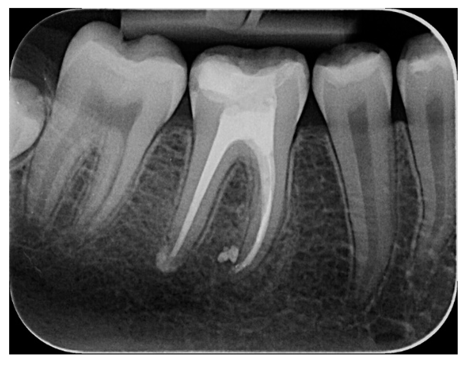
2.2.1. Chemomechanical Debridement of Endodontic Space and Root Canal Filling

The endodontic treatment for the MRFM was performed in a single visit.
The crown-down technique was used alternately with Gates–Glidden drills #3, #4 and #5 and ProTaper Gold rotary endodontic files (Dentsply Maillefer®, Ballaigues, Switzerland) to shape the canals. An apex-locator device was used to measure the working length (Root ZX II, J. Morita Corp., Kyoto, Japan), and an aqueous solution of 17% EDTA (Cerkamed Medical Company, Stalowa, Poland) was used to remove the smear layer.
Antimicrobial photodynamic therapy was performed using the debridement function of SOL device (DenMat, Lompoc, California, United States)—2 Watts output continuous wave (CW) and 808 ± 5 nm wavelength. An optical fiber with a diameter of 400 μm was inserted into the root canal, which was 3 mm shorter than the working length. The irradiation of the endodontic space was repeated four times with 15 s intervals. CW-2-W aPDT was applied alternately in continuous mode and in pulse mode (PM)—2 W, in direction from the crown to the apical area. The laser pulse mode application was performed after the root canals were dry using paper points.
The O3 aq was obtained by bubbling ozone in the gaseous state (75 μg × mL−1) in distilled water for 15 min using an ozone generator (Ozonytron, Biozonix GmbH, Munich, Germany). The measurement of the O3 aq concentration was made by oxidizing a colorless indicator (diethyl-p-phenylene diamine), which turns pink in the presence of ozone. As a reference, a sample with a saturation point of 20 μg × mL−1, produced by the Palintest Ozone Meter (Palintest® Ltd., Gateshead, UK) was used. The resulting product was introduced into the intracanal space after each size change of the endodontic files.
The final root canal filling was performed using a vertical condensation technique of warm gutta-percha (Fast-Fill system, Eighteeth, Changzhou, China), with a bioceramic-based sealant Bio-C Sealer (Angelus, Londrina, Paraná, Brazil). A temporary coronal filling with Dent-a-Cav (WP Dental GmbH, Barmstedt, Germany) was placed for 2 weeks, and periapical radiography was indicated for the MRFM to check the root canal treatment (Figure 3). The final coronal restoration was performed using a composite resin (Competence Special Set, WP Dental GmbH, Barmstedt, Germany), 2 weeks after the root canal treatment. The patient recall for clinical and radiographic examination was established in two months. The periapical radiography after two months is presented in Figure 4.

2.2.2. Identification and Quantification of E. faecalis

Endodontic Sample Collection

Biological samples were collected from the root canals using sterile endodontic absorbent paper points (ISO 20, Dentsply DeTrey®, Konstanz, Germany) by leaving each paper point in the root canal for 20 s. The points were then placed in a 1.5-mL cryotube containing 1 mL of RNAlater solution. DNA extraction was performed with the commercial MagaZorb DNA Mini-Prep Kit (Promega, Madison, WI, USA) following the manufacturer’s instructions. The quantity and the purity of the isolated DNA were determined by spectrophotometry (NanoPhotometer®, Implen GmbH, Munich, Germany). The quantification was performed by measuring the optical density (OD) at 260 nm. The ratio of the absorbance at 260 and 280 nm indicated the DNA purity.
The biological samples were collected in four distinct protocol stages:
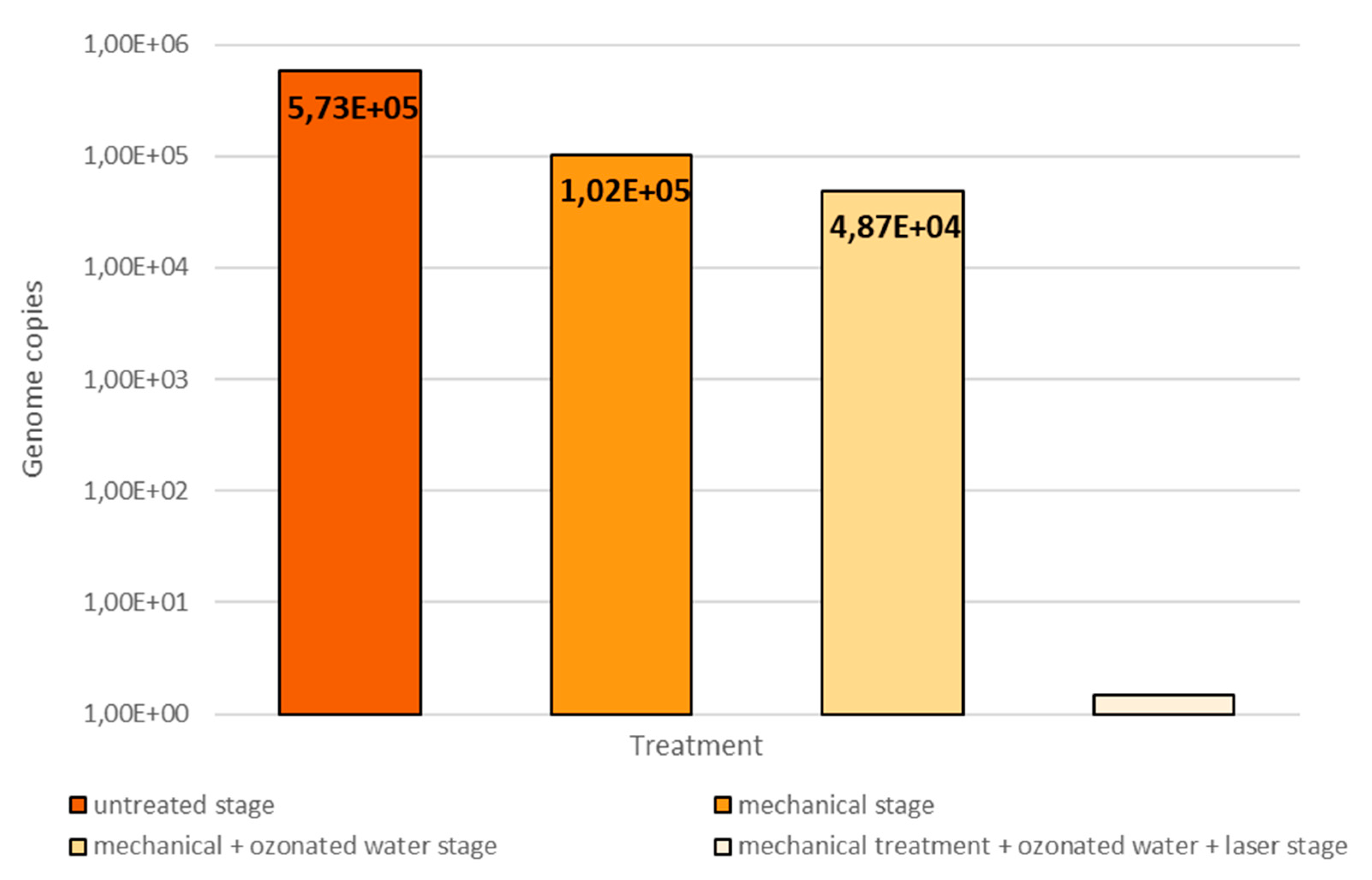
  • after opening the pulp chamber, before starting the chemomechanical decontamination of the endodontic space (untreated stage)
  • after mechanical debridement with physiological serum (mechanical stage)
  • after mechanical debridement and ozone therapy, where ozone was the substitute for chemical decontaminants (mechanical + ozonated water stage)
  • after mechanical debridement, ozone therapy and diode laser therapy, the equivalent of a complete chemomechanical preparation (mechanical + ozonated water + laser stage)

Quantification of Enterococcus faecalis

The determination of E. faecalis bacterial load was performed according to the protocol established by Bourgeois et al. in 2017 [27]. The quantification of the pathogenic bacteria was performed by quantitative real-time PCR; the method with intercalated fluorochromes, using the Mx3005P qPCR platform (Stratagene, La Jolla, CA, USA) [28]. In the qPCR reactions, a total volume of 25 µL solution was used, which contained 2 µL DNA solution isolated from the sample to be analyzed, 12.5 GoTaq® GREEN qPCR Master Mix solution, 0.6 R 10 µM primer solution, 0.6 µL F 10µM primer solution, 0.4 µL ROX solution and 8.9 µL biologically pure water. These reactions were used to determine the effective concentration of the microorganism. For the construction of the standard curve, a recombinant plasmid was used in which the DNA sequence of interest was incorporated and flanked by sequences complementary to the primers to allow their attachment.
The study was approved by the Ethical Committee of the “Grigore T. Popa” University of Medicine and Pharmacy of Iasi. A written informed consent form for the harvest and use of biological materials during endodontic treatment was voluntarily obtained from the patient (protocol number 10353). The research was conducted in full accordance with the World Medical Association Declaration of Helsinki.

3. Results and Discussion

The biocidal effect on E. faecalis of three different protocols was evaluated by qPCR reactions.
In the qPCR tests, an E. faecalis concentration value of 5.7 × 10⁵ was identified before applying any type of treatment. The mechanical treatment reduced the E. faecalis concentration to 1.02 × 10⁵. The mechanical and O3 aq treatment induced an additional reduction to 4.87 × 10⁴. The complete treatment protocol (mechanical + ozonated water + laser) led to no E. faecalis being identified in the endodontic space (Figure 5).
The diagnosis for the MRFM was established by considering the patient symptoms and clinical signs and by using additional X-ray investigations that showed the well-defined area of radiolucency, with the disappearance of the lamina dura and periodontal ligaments, associated with the negative response to pulpal vitality tests. Clinical evaluation of the patient two weeks after the endodontic treatment showed no clinical symptomatology, with negative percussion and palpation, and the visual intraoral evaluation showed healing of the fistula tract. The radiographic evaluation after two months indicated that the pathological apical radiolucency was reduced to a minimum.
The number of therapy sessions needed for complete decontamination of the root canals is still among controversial debate topics in the endodontic literature. In this study we chose to make the treatment and microbiological analysis in a single visit. This decision relies on the results of Cochrane systemic analysis conducted in 2008, which showed that there is no significant difference in treatment efficiency according to the number of dental visits [29].
Supplementarily, in our in vivo study the conventional chemical disinfectants were completely replaced by ozone and antimicrobial photodynamic therapy. Our data obtained by associating these two methods in root canal decontamination (laser radiation and ozone water) represent a novelty in the research field.
Cardoso et al. in 2008 showed that O3 aq used as an irrigation agent significantly reduces the number of Candida albicans [30] and E. faecalis in human root canals [31,32]. Huth et al. in 2007 scientifically proved that ozone has anti-inflammatory and immunomodulatory properties, exerting inhibitory effects on the NF-kB system, which is crucially implicated in the inflammatory, immune and apoptosis processes [33]. Due to these results, the use of ozone in dentistry has become more frequent. In endodontics the use of ozone relies on its antimicrobial efficacy, as well as on its oxidation capacity to reduce the number of microorganisms in the root canal [34,35,36]. Nevertheless, the conclusion of a recent systematic review was that ozone therapy should not replace or complement the use of NaOCl, revealing that the performance in microbial load reduction of ozone is inferior to that of NaOCl [37].
Instead, the laser interacts with water molecules (by irradiation) and produces a cavitation phenomenon that allows better intracanalar penetration of the irrigation solution [38,39]. One of the most important actions performed by the dental laser is the heating action, but this can also be considered as a disadvantage. A 10 °C rise in temperature for 1 min can irreversibly damage the periodontal tissues. The heating effect can be reduced by optimizing the application process (time and intensity).
Special attention was paid to all operating components and stages of the proposed experimental therapeutic protocol, which already were optimized in previous studies [1,35]. Thus, in our study diode laser irradiation was performed at C/P-Mode-2 W (four times) for 15 s, and each irradiation was followed by 20 s of O3 aq irrigation.
According to the literature data and preliminary results obtained in our study, it appears that the involvement of the three techniques—mechanical debridement, ozone therapy and the diode laser technique—brings a synergistic effect in achieving E. faecalis negative samples.
Within the limitations of the study (only one patient was taken into account), the findings sustain our previous clinical observations that the combination of a dental laser and ozonated water can replace the traditional chemical agents in complete decontamination of the endodontic space.

4. Conclusions

In this particular case, excellent results were obtained by a new procedure for periapical lesion treatment using a dental laser and ozonated water. The beneficial effect of this nonconventional therapy was highlighted by radiographic examination two months after the root canal filling. This treatment is based on the interaction of two components: a photosensitizer dye and ozone. The healing of the periapical area by restitutio ad integrum in conjunction with the absence of clinical signs and symptoms confirms the effectiveness of this alternative treatment.
Further studies are required to confirm the biological safety of diode laser application in this procedure and the efficiency of the two therapies in combination.

Author Contributions

All authors have equally contributed to the paper: conceptualization, C.L.G. and S.A.; methodology, S.A.; validation, O.Ț., S.A. and S.S.; formal analysis, V.P. and M.S. (Mihaela Sălceanu); investigation, C.L.G.; resources, C.L.G.; data curation, V.P. and M.S. (Mihaela Scurtu); writing—original draft preparation, C.L.G.; writing—review and editing, C.L.G. and O.Ț.; visualization, V.P. and S.S.; supervision, S.A.; project administration, M.S. (Mihaela Sălceanu); funding acquisition, M.S. (Mihaela Scurtu) and O.Ț. All authors have read and agreed to the published version of the manuscript.

Funding

This research received no external funding.

Conflicts of Interest

The authors declare no conflicts of interest.

References

  1. Shah, D.; Lynd, T.; Ho, D.; Chen, J.; Vines, J.; Jung, H.D.; Kim, J.H.; Zhang, P.; Wu, H.; Jun, H.W.; et al. Pulp-Dentin Tissue Healing Response: A Discussion of Current Biomedical Approaches. J. Clin. Med. 2020, 9, 434. [Google Scholar] [CrossRef] [PubMed] [Green Version]
  2. Yoshizawa, J.M.; Schafer, C.A.; Schafer, J.J.; Farrell, J.J.; Paster, B.J.; Wong, D.T.W. Salivary Biomarkers: Toward Future Clinical and Diagnostic Utilities. Clin. Microbiol. Rev. 2013, 26, 781–791. [Google Scholar] [CrossRef] [PubMed] [Green Version]
  3. Isola, G.; Alibrandi, A.; Rapisarda, E.; Matarese, G.; Williams, R.C.; Leonardi, R. Association of vitamin D in patients with periodontitis: A cross-sectional study. J Periodontal Res. 2020. [Google Scholar] [CrossRef] [PubMed]
  4. Isola, G.; Alibrandi, A.; Currò, M.; Matarese, M.; Ricca, S.; Matarese, G.; Ientile, R.; Kocher, T. Evaluation of salivary and serum ADMA levels in patients with periodontal and cardiovascular disease as subclinical marker of cardiovascular risk. J Periodontol. 2020. [Google Scholar] [CrossRef] [PubMed]
  5. Bordea, I.R.; Hanna, R.; Chinifoursh, N.; Gradinaru, E.; Campean, R.S.; Sîrbu, A.; Amaroli, A.; Benedicenti, S. Evaluation the Outcome of Various Laser Therapy Applications in Root Canal Disinfection: A Systematic Review. Photodiagnosis Photodyn. Ther. 2020, 29, 101611. [Google Scholar] [CrossRef] [PubMed]
  6. Pourhajibagher, M.; Chinifoursh, N.; Bahador, A. Antimicrobial action photoactivated C-Phycocyanin against Enterococcus faecalis biofilms: Attenuation of quorum-sensing system. Photodiagnosis Photodyn. Ther. 2019, 28, 286–291. [Google Scholar] [CrossRef]
  7. Zan, R.; Kutlu, G.; Hubbezoglu, I.; Sumer, Z.; Tunc, T.; Mutlu, Z. Bactericidal effects of various irrigation solutions against staphylococcus aureus in human root canal. J. Istanb. Univ. Fac. Dent. 2015, 49, 19–26. [Google Scholar] [CrossRef]
  8. Mohammadi, Z.; Shalavi, S.; Soltani, M.K.; Asgary, S. A review of the properties and applications of ozone in endodontics: An update. Iran Endod. J. 2013, 8, 40–43. [Google Scholar]
  9. Burke, F.J. Ozone and caries: A review of the literature. Dent. Update 2012, 39, 271–278. [Google Scholar] [CrossRef]
  10. Azarpazhooh, A.; Limeback, H. The application of ozone in dentistry: A systematic review of literature. J. Dent. 2008, 36, 104–116. [Google Scholar] [CrossRef]
  11. Ricucci, D.; Siqueira, J.F., Jr. Biofilms and apical periodontitis: Study of prevalence and association with clinical and histopathologic findings. J. Endod. 2010, 36, 1277–1288. [Google Scholar] [CrossRef] [PubMed]
  12. Love, R.M. Enterococcus faecalis—A mechanism for its role in endodontic failure. Int. Endod. J. 2001, 34, 399–405. [Google Scholar] [CrossRef] [PubMed]
  13. Antunes, H.S.; Rocas, I.N.; Alves, F.R.; Siqueira, J.F., Jr. Total and Specific Bacterial Levels in the Apical Root Canal System of Teeth with Post-treatment Apical Periodontitis. J. Endod. 2015, 41, 1037–1042. [Google Scholar] [CrossRef]
  14. Nair, P.N. On the causes of persistent apical periodontitis: A review. Int. Endod. J. 2006, 39, 249–281. [Google Scholar] [CrossRef]
  15. Cobzeanu, B.M.; Costan, V.D.; Danciu, M.; Pasca, S.A.; Sulea, D.; Ungureanu, L.B.; Moscalu, M.; Cobzeanu, M.D.; Popescu, E. Environmental factors involved in genesis of retromolar oropharynx junction cancer. Environ. Eng. Manag. J. 2017, 16, 1101–1106. [Google Scholar] [CrossRef]
  16. Prada, I.; Mico-Munoz, P.; Giner-Lluesma, T.; Mico-Martinez, P.; Muwaquet-Rodiquez, S.; Albero-Monteagudo, A. Update of therapeutic planning of irrigation and intracanal medication in root canal treatment. A literature review. J. Clin. Exp. Dent. 2019, 11, 185–193. [Google Scholar] [CrossRef]
  17. Restaino, L.; Frampton, E.W.; Hemphill, J.B.; Palnikar, P. Efficacy of ozonated water against various food-related microorganisms. Appl. Environ. Microbiol. 1995, 61, 3471–3475. [Google Scholar] [CrossRef] [Green Version]
  18. Estrela, C.; Estrela, C.R.; Decurcio, D.A.; Hollanda, A.C.; Silva, J.A. Antimicrobial efficacy of ozonated water, gaseous ozone, sodium hypochlorite and chlorhexidine in infected human root canals. Int. Endod. J. 2007, 40, 85–93. [Google Scholar] [CrossRef] [PubMed]
  19. Noguchi, F.; Kitamura, C.; Nagayoshi, M.; Chen, K.K.; Terashita, M.; Nishihara, T. Ozonated water improves lipopolysaccharide-induced responses of an odontoblast-like cell line. J. Endod. 2009, 35, 668–672. [Google Scholar] [CrossRef] [PubMed]
  20. Huth, K.C.; Jakob, F.M.; Saugel, B.; Cappello, C.; Paschos, E.; Hollweck, R.; Hickel, R.; Brand, K. Effect of ozone on oral cells compared with established antimicrobials. Eur. J. Oral. Sci. 2006, 114, 435–440. [Google Scholar] [CrossRef] [PubMed]
  21. Filippi, A. The effects of ozonized water on epithelial wound healing. Dtsch. Zahnarztl. Z. 2001, 56, 104–108. [Google Scholar]
  22. Nagayoshi, M.; Kitamura, C.; Fukuizumi, T.; Nishihara, T.; Terashita, M. Antimicrobial effect of ozonated water on bacteria invading dentinal tubules. J. Endod. 2004, 30, 778–781. [Google Scholar] [CrossRef]
  23. Dai, T.; Huang, Y.Y.; Hamblin, M.R. Photodynamic therapy for localized infections—State of the art. Photodiagnosis Photodyn. Ther. 2009, 6, 170–188. [Google Scholar] [CrossRef] [PubMed] [Green Version]
  24. Oliveira, B.P.; Aguiar, C.M.; Camara, A.C. Photodynamic therapy in combating the causative microorganisms from endodontic infections. Eur. J. Dent. 2014, 8, 424–430. [Google Scholar] [CrossRef] [PubMed] [Green Version]
  25. Ricardo, A.L.; Britto, M.L.; Genovese, W.J. In vivo study of the Nd:YAP laser in persistent periapical lesion. Photomed. Laser Surg. 2005, 23, 582–585. [Google Scholar] [CrossRef] [PubMed]
  26. Ozel, E.; Yucekal-Tuncer, B.; Firatli, E. Short-term treatment of periapical lesion of anterior tooth affected by microleakage using Nd:YAG laser. Case report. N. Y. State Dent. J. 2010, 76, 48–51. [Google Scholar] [PubMed]
  27. Bourgeois, D.; David, A.; Inquimbert, C.; Tramini, P.; Molinari, N.; Carrouel, F. Quantification of carious pathogens in the interdental microbiota of young caries-free adults. PLoS ONE 2017, 12, e0185804. [Google Scholar] [CrossRef] [PubMed] [Green Version]
  28. Jelihovschi, I.; Drochioi, C.; Badescu, A.C.; Lupusoru, R.V.; Munteanu, A.E.; Baranov, N.; Manuc, D.; Serban, R.I.; Cobzaru, R.G.; Ripa, C.V.; et al. Comparison of Sampling Techniques for qPCR Quantification of Periodontal Pathogens. Rev. Chim. 2017, 68, 2853–2856. [Google Scholar] [CrossRef]
  29. Figini, L.; Lodi, G.; Gorni, F.; Gagliani, M. Single versus multiple visits for endodontic treatment of permanent teeth: A Cochrane systematic review. J. Endod. 2008, 34, 1041–1047. [Google Scholar] [CrossRef]
  30. Cardoso, M.G.; Oliveira, L.D.; Koga-Ito, C.Y.; Jorge, A.O. Effectiveness of ozonated water on Candida albicans, Enterococcus faecalis, and endotoxins in root canals. Oral Surg. Oral Med. Oral Pathol. Oral Radiol. Endod. 2008, 105, 85–91. [Google Scholar] [CrossRef]
  31. Tuncay, O.; Dincer, A.N.; Kustarci, A.; Er, O.; Dinc, G.; Demirbuga, S. Effects of ozone and photo-activated disinfection against E. Faecalis biofilms In Vitro. Niger J. Clin. Pract. 2015, 18, 814–818. [Google Scholar] [CrossRef] [PubMed] [Green Version]
  32. Bocci, V. Biological and clinical effects of ozone. Has ozone therapy a future in medicine? Br. J. Biomed. Sci. 1999, 56, 270–279. [Google Scholar] [PubMed]
  33. Huth, K.C.; Saugel, B.; Jakob, F.M.; Cappelo, C.; Quirling, M.; Paschos, E.; Ern, K.; Hickel, R.; Brand, K. Effect of aqueous ozone on the NF-kappa B system. J. Dent. Res. 2007, 86, 451–456. [Google Scholar] [CrossRef] [PubMed] [Green Version]
  34. Baysan, A.; Lynch, E. Effect of ozone on the oral microbiota and clinical severity of primary root caries. Am. J. Dent. 2004, 17, 56–60. [Google Scholar]
  35. Lezcano, I.; Rey, R.P.; Gutiérrez, M.S.; Baluja, C.; Sánchez, E. Ozone inactivation of microorganisms in water. Gram-positive bacteria and yeast. Ozone Sci. Eng. 2001, 23, 183–187. [Google Scholar] [CrossRef]
  36. Giuroiu, C.; Dragomir, R.; Hamburda, T.; Salceanu, M.; Aminov, L.; Melian, A.; Andrian, S.; Vataman, M. The role of ozonated water in the success of therapy of chronic apical periodontitis. Case report. Rom. J. Oral Reabil. 2014, 6, 71–75. [Google Scholar]
  37. Silva, E.J.N.L.; Prado, M.C.; Soares, D.N.; Hecksher, F.; Martins, J.N.R.; Fidalgo, T.K.S. The effect of ozone therapy in root canal disinfection: A systematic review. Int. Endod. J. 2020, 53, 317–332. [Google Scholar] [CrossRef] [Green Version]
  38. De Groot, S.D.; Verhaagen, B.; Versluis, M.; Wu, M.K.; Wesselink, P.R.; van der Sluis, L.W. Laser-activated irrigation within root canals: Cleaning efficacy and flow visualization. Int. Endod. J. 2009, 42, 1077–1083. [Google Scholar] [CrossRef]
  39. Garcez, A.S.; Nuñez, S.C.; Hamblin, M.R.; Ribeiro, M.S. Antimicrobial effects of photodynamic therapy on patients with necrotic pulps and periapical lesion. J. Endod. 2008, 34, 138–142. [Google Scholar] [CrossRef] [Green Version]
Figure 1. Orthopantomography (OPG) taken immediately after carious lesion treatment in the mandibular right first molar (MRFM).
Applsci 10 04203 g001
Figure 2. Initial radiographic investigation.
Applsci 10 04203 g002
Figure 3. Postoperative periapical radiographic aspect.
Applsci 10 04203 g003
Figure 4. Two-month postoperative periapical radiographic aspect reveals the healing of the periapical area.
Applsci 10 04203 g004
Figure 5. qPCR test values of E. faecalis for the four protocol stages.
Applsci 10 04203 g005

