Article

A Potential “Vitaminic Strategy” against Caries and Halitosis

1
Department of Medicine and Health Sciences “V. Tiberio”, Università degli Studi del Molise, 86100 Campobasso, Italy
2
Department of Drug and Health Sciences, Section of Microbiology, University of Catania, 95125 Catania, Italy
*
Author to whom correspondence should be addressed.
Appl. Sci. 2022, 12(5), 2457; https://doi.org/10.3390/app12052457
Submission received: 29 December 2021 / Revised: 17 February 2022 / Accepted: 23 February 2022 / Published: 26 February 2022
(This article belongs to the Special Issue Advances in Natural Bioactive Compounds and Biological Effects)

Abstract

:

Featured Application

Here, we propose an alternative use of Vea® Oris, a commercial product recommended at present for the maintenance of general oral health, specifically against caries and halitosis. Made by only vitamin E and capric/caprylic acid, it could represent a “more natural strategy” than the chemical-derived products normally proposed for this aim.

Abstract

Streptococcus mutans and Fusobacterium nucleatum are two key bacteria of the oral microbiota. Due to their ability to form biofilms on oral tissues, they are both involved in the onset of the most common oral diseases. F. nucleatum is also the principal producer of hydrogen sulfide (H2S), causative of the awkward bad breath of halitosis. In this study, the oral product Vea® Oris, made by vitamin E and capric/caprylic acid only, was evaluated as a potential treatment for the most common oral diseases. Different concentrations of the product were tested against both S. mutans and F. nucleatum. The effect on planktonic and biofilm growth was investigated for both strains, and for F. nucleatum, the influence on H2S production was evaluated. From our data, the product did not relevantly reduce the planktonic growth of both strains, whereas it validly counteracted biofilm assemblage. Moreover, an interesting trend of H2S reduction was highlighted. Overall, these results suggested, on the one hand, a synergistic antimicrobial–antibiofilm action of two Vea® Oris components and, together, potential modulation activity on H2S production. However, the study should be implemented to confirm these only preliminary findings, certainly extending the panel of tested bacteria and using alternative methods of detection.

1. Introduction

The oral cavity is one of the most complex environments in the human body due to the continuous variation of homeostasis conditions. Over 700 diverse bacterial species have been identified as potential colonizers of the oral niches [1,2,3]. The bacterial composition of the oral microbiota is responsive to different intrinsic and extrinsic factors. These intrinsic factors are not changeable by the host, related to genetic factors (e.g., ethnicity, gender, immune response, medical conditions, etc.), characteristics of the oral cavity such as temperature and saliva composition (pH, buffer capacity, hormones, and secreted substances), and characteristics of adhesion surfaces (e.g., roughness of tooth surface). Extrinsic factors are modifiable by the host, related to the environment, habits and lifestyle, and regard hygiene, diet, the use of drugs, smoking, medications, sex, access to dental care, etc. [4,5,6,7]. Intrinsic and extrinsic factors contribute, together with host aging, to colonization by different microbial pools of the oral cavity. As a result of this complex interaction, the balance between beneficial and pathogen species is continuously altered, passing from eubiosis to a dysbiosis state and, as a consequence, to the onset of oral pathologies such as caries, gingivitis, and periodontitis [2,4].
Innovative metagenomic approaches have greatly benefited researchers and clinicians to correlate the modulation of the diverse bacterial species in the oral cavity with all possible stimuli acting in this body environment [3,8]. Through sequencing methods, the relationships between beneficial and pathogenic species have been extensively studied, and the composition of the polymicrobial biofilms on teeth, gingiva, tongue surface, and other soft tissues has been deeply characterized [7]. As is now well known, the biofilm structure constitutes a protected sub-environment in which various bacterial species interact to protect themselves from possible surrounding perturbations, as a kind of “mutualist survival strategy” of the microbial community. This microbial assembly is quite difficult to inhibit and remove from the adhesion surface, and this is also verified for the biofilms formed in the oral cavity [9,10,11]. Indeed, the most common oral pathologies, such as plaque and dental caries, gingivitis, and periodontitis, are due to biofilm deposition [7,12]. The process of biofilm formation in the oral cavity occurs in three subsequent steps, i.e., first, adhesion to the surface; then, the colonization step; and finally, biofilm assemblage and maturation [13]. Diverse bacterial species participate in biofilm development as “early” or “late” colonizers [14]. The “early” colonizers are involved in the first two steps of biofilm formation. Because of their ability to bind salivary proteins, they adhere to hard or soft tissues, colonize the oral niches, and provide an adhesive substratum for the “late” colonizers [6,13,14,15].
The early colonizers belong mainly to the Streptococcus genus. S. mutans is the most represented species in supragingival plaque, childhood caries, and dentinal and root lesions. Due to its rapid metabolism and strong acid tolerance, it is recognized as the most cariogenic bacteria in the oral cavity [6,12,13].
On the other hand, the Gram-negative anaerobe F. nucleatum, acting halfway between the early and late colonizers, can be considered a key player of the oral biofilm maturation stage. Due to its coating adhesion molecules and polysaccharide receptors, it co-aggregates both with the early and late streptococcal colonizers; these latter, mainly Gram-negative, anaerobes belong to the genera of Bacteroidetes and Spirochaetes [13,14,15]. In addition to this bridging role in the biofilm network, the enhanced prevalence of F. nucleatum within the deep periodontal pockets suggests that this germ also plays an active role as a periodontal pathogen [16,17]. In this niche, F. nucleatum stimulates the production of host matrix metalloproteinases, the increased levels of which concur to the initial periodontal inflammation in periodontal diseases [14]. This pathogen is also provided by significant hemolytic activity and, more interestingly, is the main producer of hydrogen sulfide (H2S) in the oral cavity. H2S, together with methyl mercaptan (CH3SH) and dimethyl sulfide (CH3SCH3)24, is one of the volatile sulfur compounds (VSCs) produced by periodontopathic anaerobic bacteria and principally responsible for typical malodor in halitosis [18,19,20,21,22]. Consistently, the uncomfortable phenomenon of halitosis is associated with an increase in VSCs producer species, principally F. nucleatum but also Porphyromonas gingivalis, Treponema denticola, Prevotella intermedia, and Eubacterium [18,19,21,22]. On these assumptions, inhibiting F. nucleatum in the oral cavity could represent an excellent double-sided strategy to reduce both the formation of the oral biofilm causative of oral diseases and the uneasiness of halitosis. An even more efficient approach to massively prevent/reduce oral pathologies could also be represented by the simultaneous inhibition of the main early colonizer, S. mutans. At present, rather than specific action against these key oral germs, lifestyle modulation and the use of innovative cosmetics are recognized as the only strategies to maintain a generally balanced microbiome in the oral cavity [2,4].
Indeed, mouthwashes and many dentistry products made by antimicrobial compounds, such as chlorhexidine, triclosan, cetylpyridinium chloride, and chlorine dioxide, are often used against the oral biofilm to treat bad breath [19,20]. Nevertheless, the use of such products is more often associated with an increase in bacterial resistance, as well as with the undesirable killing of nonpathogenic commensal species and a dysbiosis state [23,24]. Surely, the use of natural products is, at least conceptually, a more appreciable strategy to prevent and contrast oral pathologies, even more if these products can selectively impact the pathogenic species of the buccal microbiota. A potential “natural therapy” could also modify the applicability of the treatment, not only as curative but also as a preventive therapy, both in pediatric and old-aged patients. Natural derivatives such as propolis, cranberry, tea, Galla chinensis, grapes, coffee, and cacao-containing polyphenols have already demonstrated their activity against oral biofilms [20,25,26,27,28]. Similarly, hinokitiol, green tea powder, and eucalyptus extract have been beneficially used against oral malodor [19,29,30,31].
Further, oxidative stress was found as an important causative factor of dysbiosis and oral disorders, and the action of diverse antioxidant substances against oral bacteria has also been studied. Fat-soluble vitamins (vitamin A, vitamin E-tocopherol and b-carotene), water-soluble vitamins (vitamin C and vitamin B complex), trace elements (zinc, magnesium), and bioflavonoids (plant derived) neutralize the reactive oxygen species (ROS) causative of many oral pathologies [32,33,34,35,36,37,38,39]. These antioxidant compounds are generally consumed through fruits and vegetables, and they are maintained at an optimal level in individuals by a well-balanced diet [40,41]. Conversely, the low quotes of these antioxidants found in individuals affected by oral diseases clearly suggest their potential role against oral pathogens [4,32,42].
Among all the known compounds, vitamin E is the major fat-soluble antioxidant in cell membranes [43,44]. It exerts a considerable anti-inflammatory action and enhances the humoral immune response [43,45,46,47,48]. Its beneficial effect on oral health has already been documented through improvements in all periodontal parameters in patients treated with vitamin E (decrease in plaque and biofilm formation indexes, probing depth, clinical attachment level, and bleeding on probing) [32,39,42,49]. Literature findings regarding the action of naturally derived compounds and those considered above are summarized in Table 1.
Based on these assumptions, here, we tested in vitro the two key oral bacteria S. mutans and F. nucleatum using the innovative product Vea® Oris made by only two components, vitamin E (alpha-tocopherol) and caprylic/capric triglyceride obtained from coconut oil and glycerin. It is already available on the market as a safe product recommended for oral mucosa protection and maintenance. Although no specific germ action has been previously demonstrated, the interesting “vitaminic” formulation encouraged us to consider it a “natural alternative strategy” against S. mutans and F. nucleatum and, consequently, against caries and halitosis.
We therefore tested the effect of different concentrations of Vea® Oris on the planktonic and biofilm growth of S. mutans and F. nucleatum. For F. nucleatum, the influence of Vea® Oris on H2S production was also evaluated.

2. Materials and Methods

The commercial product Vea® Oris (by Hulka s.r.l.) was tested as inhibition treatment against S. mutans DSM 20523 and F. nucleatum subsp. vincentii DSM 19507.

2.1. Effect of Vea® Oris Treatments on Planktonic and Biofilm Growth

Strains were cultured in Difco™ Columbia Broth (BD, Biosciences) at 37 °C for 48 h under static conditions. The optical density at 600 nm (600 nm O.D.) was measured after incubation using the Lambda 25 spectrophotometer (PerkinElmer). Dilutions were prepared to obtain inocula with a starting O.D. of 0.1. For each strain, aliquots of 150 µL were distributed into the wells of a 96-well plate. Vea® Oris was added at concentrations of 5, 10, and 20% (% v/v), i.e., volumes of 50, 100, and 187 µL/mL, respectively (Figure 1, yellow and green wells with the drop symbol). Aliquots of strains not added with Vea® Oris preparation were used as controls for bacterial growth (Figure 1, yellow and green wells without the drop symbol). All treatments and controls were produced in triplicates, and triplicates of 96-well plates were assessed. Liquid sterile paraffin was added to cover the surface of each inoculated well, providing suitable anaerobiosis conditions. Blanks for the spectrophotometric measurement were also produced as described. Triplicates of the same volume (150 µL) were prepared with Columbia Broth not added (Figure 1, gray wells without the drop symbol) or added with 5, 10, and 20% Vea® Oris (Figure 1, gray wells with the drop symbol).
All 96-well plates were sealed, and the O.D. at 600 nm was measured at the starting point using the VICTOR X5 multilabel plate reader (PerkinElmer). The plates were then incubated at 37 °C for 48 h under static conditions. After incubation, the O.D. was measured again to evaluate the variation of growth [53,54]. The O.D. measurement was performed in triplicates, and the O.D. value was calculated as the mean value of replicates. The values obtained for all inocula strains, without and with Vea® Oris, were normalized for the respective experimental blanks, i.e., the broth without and with Vea® Oris. The growth with Vea® Oris was then compared to that without, and the concentration-effect of the product was finally evaluated (ANOVA, Tukey’s HSD with p ≤ 0.05).
After the final O.D. measurement, biofilm produced under Vea® Oris treatments with respect to the control was estimated. All inoculated wells were subjected to the staining procedure implemented by Stepanovic et al. (2000), with some experimental modifications [55,56,57]. The experimental procedure up to this point described is also schematically represented in Figure 2.
Then, the wells were emptied and washed three times using 250 µL of 0.9% NaCl. A 200 µL volume of methanol solution was added to each well and maintained in static incubation for 15 min to fix the adherent cells to the plastic surface. The methanol solution was discarded, and the plates were dried under biological laminar flow in an upside-down position. Subsequently, 200 µL of 1% crystal violet solution (Gram staining kit, Biolife Italiana srl) was added and maintained in static incubation for 5 min. The staining solution was removed, and the plates were washed under moderate tap water flow. The dye trapped in adherent cells was resolubilized, and 160 µL of 33% glacial acetic acid was added. Finally, the O.D. at 570 nm of each well was measured in triplicates using the VICTOR X5 multilabel plate reader (PerkinElmer) [55,56]. The O.D. value for each treatment and the control was calculated as the average of measures.
The O.D. values are proportional to the amount of the resuspended staining and the number of cells fixed at the well surface as a biofilm.
Therefore, the biofilm formed by strains with 5, 10, and 20% (% v/v) Vea® Oris was compared to the biofilm formed by controls without the product. The effect of increasing concentrations of Vea® Oris on biofilm production was then evaluated by comparing the values obtained for different treatments between each other. More precisely, the biofilm formed by the strains in control conditions (without Vea® Oris) was considered 100% biofilm formation. The reduction of biofilm due to Vea® Oris was estimated as the percentage difference between the biofilm without (100% biofilm) and with 5, 10, and 20% Vea® Oris (ANOVA, Tukey’s HSD with p ≤ 0.05).

2.2. Potential Ability of Vea® Oris Treatments to Modulate H2S Production

Since Fusobacterium has been largely demonstrated as the most relevant genus associated with halitosis [19,20,21], F. nucleatum subsp. vincentii DSM 19507 was also investigated for its ability to form a malodorous H2S product under Vea® Oris treatments. Our experimental setup is described in detail and also schematically shown in Figure 3.
F. nucleatum was cultivated in Columbia Broth at 37 °C for 48 h. The culture was centrifuged at 5000 rcf for 3 min, and the cells were collected at the bottom of the culturing tube. The bacterial pellet was resuspended in 40 mL of sulfide indole motility (SIM) medium (Thermo Scientific™). SIM medium allows for the detection of H2S produced due to its specific formulation. It contains ferrous ammonium sulfate that, by reacting with the volatile compound, forms ferrous sulfide, a black precipitate that ultimately acts as a detector of H2S produced [58,59].
Three-milliliter aliquots of SIM cultures were distributed in 15 mL falcon tubes containing 150 (5% v/v), 300 (10% v/v), and 600 (20% v/v) µL of Vea® Oris, respectively. Each Vea® Oris treatment was assessed in triplicates. Three-milliliter triplicates of controls and spectrophotometric blanks were also produced with F. nucleatum cultures without Vea® Oris and with SIM broth alone, respectively. One milliliter of liquid sterile paraffin was added to the top of all tubes to provide anaerobiosis conditions. The tubes were incubated at 37 °C for 48 h. After incubation, the formation of the black compound was spectrophotometrically estimated. The absorbance of all inocula was measured first within the wide range of 400–700 nm at 1 nm steps with the Lambda 25 spectrophotometer (PerkinElmer). The final values of absorbance in this range for different treatments were calculated as the average of values measured for each set of triplicates. All values were previously normalized for blanks. The resulting absorbance spectra of Vea® Oris treatments were compared to the control (no Vea® Oris addition). The normality of data was checked using the Shapiro–Wilk test (α = 0.05), and the significance of comparisons was assessed using the Student’s t-test with p ≤ 0.05 applied to triplicates of the normalized absorbance values from all treatments.
The wavelength at which the darkened control of F. nucleatum produced the maximal absorbance was considered the detection wavelength of the black compound and consequently of H2S. This control absorbance was considered to be 100% of H2S production. The variation of H2S induced by Vea® Oris treatments was calculated as a percentage variation with respect to the control value.

3. Results

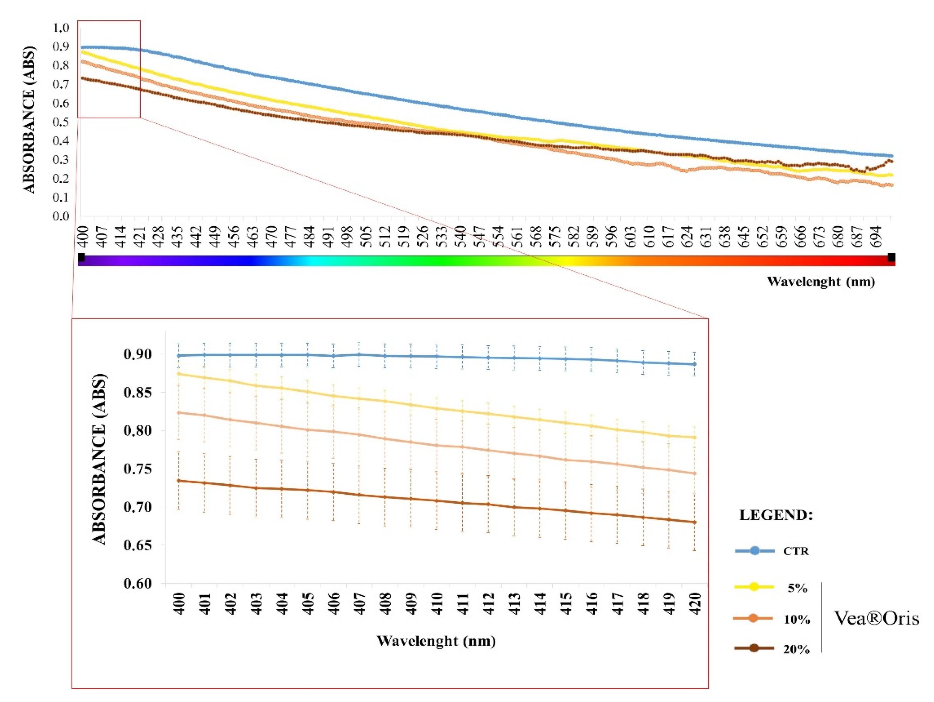
3.1. Variation of Planktonic and Biofilm Growth

All the notable variations induced by Vea® Oris treatments on the planktonic and biofilm growth of S. mutans and F. nucleatum are described below.
As shown in Figure 4, compared with the control, the planktonic growth of S. mutans was significantly but only slightly increased by Vea® Oris at 5 or 10% (% v/v), with increments of 17 and 14% (p < 0.01), respectively. Conversely, the 20% concentration did not exert a significant modulation of the planktonic bacterial growth (p = 0.90).
Interestingly, all the Vea® Oris concentrations significantly reduced the biofilm growth of the cariogenic species S. mutans. A biofilm reduction of 26% was obtained with Vea® Oris at 5% (% v/v) and a decrement of 32% with Vea® Oris at 10 and 20% (% v/v) (p < 0.01).
Similarly, for F. nucleatum, as highlighted in Figure 5, Vea® Oris did not influence planktonic growth (p > 0.05), whereas it significantly reduced the ability of the bacterium to form a biofilm, regardless of the applied concentration (p < 0.01). Even more interesting is that all the tested concentrations of 5, 10, and 20% induced similar biofilm decrements, of 50, 52, and 40%, respectively.

3.2. Variation of H2S Production

Regarding the effect of Vea® Oris on the H2S production of F. nucleatum, after the incubation time (see Section 2), the darkening of the SIM medium was observed for all inocula, both without and with Vea® Oris (Figure 6). However, already with the naked eye, slight differences in medium darkening were appreciable between the treated and untreated cultures (Figure 6). Moreover, compared with the control, a gradual reduction in dark color resulted in incremental concentrations of Vea® Oris.
These by-eye outcomes were confirmed through the spectrophotometric analysis. The absorbance spectra of the treated samples clearly differed from the untreated samples (Figure 7A). The range of maximal absorbance for all samples was between the wavelengths of 400 and 420 nm. Within this range, the curves for Vea® Oris treatments showed a gradual reduction of absorbance values, proportional to the increments of concentrations (Figure 7B).
These results suggest that the production of H2S was inversely influenced by the addition of Vea® Oris in a concentration-dependent manner.
To get inside the quantitative differences of H2S production in the absence/presence of Vea® Oris, the wavelength of maximum absorbance for the control was first identified. The absorbance value measured for the control at that wavelength was compared with those of samples treated with Vea® Oris (see Section 2).
In detail, the maximum absorbance value was measured for the control at the wavelength of 407 nm. Effectively, at this wavelength, the treated samples registered a gradual reduction in the absorbance value, and then H2S produced, along with the increments of Vea® Oris (Figure 8). The measured O.D. values were normally distributed (Shapiro–Wilk test, p = 0.07157), and then a parametric Student’s t-test was performed to evaluate the significance of comparisons between treatments. Nonsignificant differences were found between treatments (Student’s t-test, with p ≤ 0.05), and only negligible percentage decrements of absorbance of 6, 9, and 13% were calculated with respect to the control. Despite the low rates of decrement obtained, the graduality of the H2S reduction trend along with the increments of Vea® Oris is worthy of note (Figure 8).

4. Discussion

In this study, the action of the oral preparation Vea® Oris was investigated against S. mutans and F. nucleatum, two oral bacteria that can be considered key players of the most common oral diseases.
S. mutans is one of the most cariogenic species of the mouth environment [6,20,60]. Due to its ability to colonize oral surfaces, rapid metabolism, and strong acid tolerance, it manages the first stage of biofilm formation and is recognized as the principal early colonizer [12,14]. On the other hand, F. nucleatum acts in the later stage of oral biofilm maturation as a bridging species between the early and late colonizers. The latter are needed to assemble the complex structure of a mature and resistant oral biofilm [6,13,14]. Moreover, F. nucleatum is involved in the onset of gingivitis and periodontal diseases [17,61]. Due to its great ability to produce the malodorous sulfur compound H2S, its enrichment in the oral cavity is also associated with the onset of halitosis [18,19,22,62].
The product Vea® Oris tested in this study is made by only two components from natural sources, vitamin E (α-tocopherol acetate) and caprylic/capric triglyceride. This formulation was tested at concentrations of 5, 10, and 20% (% v/v) on growing cultures of S. mutans and F. nucleatum strains. The effect of the Vea® Oris treatments on both planktonic and biofilm growth was evaluated. In addition to F. nucleatum, the influence on H2S production was verified.
From our results, the planktonic growth of F. nucleatum was not modulated by the product at any tested concentration. For S. mutans, no significant variation was similarly registered with the higher 20% (% v/v) concentration, and only a weak stimulation was obtained with the 5 and 10% (% v/v) concentrations.
On the contrary, regarding biofilm formation, the results suggested that the Vea® Oris mixture of vitamin E and capric/caprylic acid clearly decreased the ability of both S. mutans and F. nucleatum to assemble the biofilm structure. Depending on the Vea® Oris concentration, a biofilm reduction of 26–32% for S. mutans and 40–52% for F. nucleatum was estimated.
Although no studies have so far been conducted with a similar mixture of vitamin E (alpha-tocopherol) and capric/caprylic acid, these results lead us to estimate a possible synergistic antimicrobial–antibiofilm action of the two components. Undoubtedly, vitamin E as alpha-tocopherol acetate has already confirmed its antibiofilm action against bacteria associated with the urinary tract, mainly belonging to the genera Staphylococcus and Proteus [63,64]. Regardless of the methods used for biofilm estimation and the strain tested, the application of alpha-tocopherol acetate induced a reduction in biofilm of at least 50% [63,64]. However, although the specific action of tocopherols on the planktonic and biofilm growth of the buccal bacteria has been little investigated, Smolarek et al. showed that toothpaste added with tocopherols produced in vitro antimicrobial activity against S. mutans and E. faecalis [65]. At the same time, the bactericidal effects of medium-chain fatty acids (MCFAs), to which the capric/caprylic acid of Vea® Oris belongs, was also highlighted. Huang et al. demonstrated that short-, medium-, and long-chain fatty acids exhibit patterns of inhibition against a wide panel of oral bacteria, comprising S. mutans, Streptococcus gordonii, Streptococcus sengis, Candida albicans, Aggregatibacter actinomycetemcomitans, Fusobacterium nucleatum, and Porphyromonas gingivalis [66]. This inhibitory action of fatty acids is clearly species specific, since it is related to the profile of fatty acids produced by each species itself. It could be thought of as a “system of cross-balancing” of the oral microbiota ecology based on a pool of molecules active against competitors of the same environment. For instance, it was demonstrated that caprylic acid at a dose of 25 μg/mL completely inhibited the growth of F. nucleatum and reduced the growth of S. mutans by 50% [66]. Indeed, F. nucleatum produces butyric, isovaleric, and propionic acids but does not produce capric/caprylic acid to which, therefore, it is sensitive. On the contrary, since S. mutans is the major acidogenic and aciduric microorganism in the oral cavity, it showed higher resistance to the effect of many fatty acids comprising capric/caprylic acid [66].
However, no potential antibiofilm action of capric/caprylic acid has been highlighted. Rather, 1-monoglyceride of capric acid, monocaprin, tested against S. aureus, C. albicans, and S. mutans, showed inhibitory action against strains in the planktonic phase but no effect on the same strains in the biofilm growing state [67].
Then, considering our data in the light of these previous findings, we can infer that the effect of Vea® Oris against S. mutans and F. nucleatum is effectively the result of the two combined actions of the two components. The planktonic growth of the two bacteria may be held off by the more prevalent antimicrobial effect of caprylic/capric acid, whereas the antibiofilm effect of Vea® Oris may be principally ascribed to alpha-tocopherol acetate. The possibility of a potential synergistic interaction seems to also be supported by the following discussed studies. Medium-chain fatty acids such as caprylic and lauric acids have been demonstrated to enhance their bactericidal effect when used in association with essential oils (i.e., carvacrol, eugenol, b-resorcylic acid, trans-cinnamaldehyde, thymol, and vanillin), which, similarly to alpha-tocopherol, are extracted from plants and seeds [68,69]. Consistently, we are encouraged to speculate a similar relationship of synergism between caprylic/capric acid and alpha-tocopherol acetate in the Vea® Oris formulation. The enhanced activity of the mixture may be due to the same mechanism of synergism identified by Chapple et al. (2013) for ascorbate and α-tocopherol in association. These two compounds, recycling each other, produce an increased antioxidant effect with respect to the ascorbate alone [70].
Our results also suggested the potential of using Vea® Oris to modulate the H2S production of F. mutans in halitosis. Although the tested concentrations of Vea® Oris in our experiments induced only weak and nonsignificant decreases in H2S of 5–12%, an interesting concentration-dependent trend of reduction was obtained. Although further investigations are needed to investigate this H2S-inhibitory putative action, we can confidently speculate that the observed effect is principally attributable to vitamin E. Indeed, Lagha et al. (2017) demonstrated that green tea extract and the principal tea catechin, epigallocatechin-3-gallate (EGCG), at a high concentration of 2000 ug/mL markedly reduced the H2S production of F. nucleatum (reduction of about 80%) [51]. A weaker but significant reduction was also verified at lower concentrations (≤500 ug/mL) for both substances and theaflavins [51]. A similar inhibitory action was verified for thymoquinone, the prevalent aromatic compound of the essential oil obtained from N. sativa seeds [50]. Used at the concentration of 100 ug/mL, it reduced 40% of the H2S level in F. nucleatum cultures and, at the even lower concentration of 6.25 µg/mL, 45% of H2S production in Porphyromonas gingivalis cultures. These data lead us to suppose, also for alpha-tocopherol, a possible involvement of its similar aromatic structure in inhibitory action. Compounds with this aromatic architecture could interfere with competitors involved at currently unknown steps of the H2S pathway. Going deeper inside the possible molecular mechanism of inhibition, we could speculate action at the genetic level, as is for cetylpyridinium chloride (CPC), a heteroaromatic ammonium salt often used as a component of oral hygiene products [71].
Daily use of mouth rinses containing CPC was found to reduce VSCs and oral malodor through both direct and indirect activities of the aromatic compound. It exerts inhibitory activity on the growth of the oral bacteria F. nucleatum and P. gingivalis and acts at the genetic level, suppressing the expression of their mgl and cdl genes involved in VSC production [71]. Nevertheless, due to the uncertainty of data obtained in this preliminary study, we undoubtedly recognize that deeper investigations are needed to confirm the same or at least a similar genetic mechanism of inhibition for Vea® Oris. Overall, such encouraging results certainly set the stage for further in vitro, and possibly in vivo, studies to confirm the beneficial use of Vea® Oris seen herein against caries and halitosis.

5. Conclusions

The present study focused on two key bacteria mainly involved in the onset of oral pathologies, i.e., S. mutans and F. nucleatum. The principal aim was to hopefully find a “more natural” strategy that could simultaneously act on the pathogenic processes that these germs put in place in the oral cavity. Overall, the results we obtained were quite encouraging, although they need to be confirmed and implemented. Indeed, our data suggested that the Vea® Oris preparation, made by vitamin E and caprylic/capric acid only, has good potential as a nonchemical strategy to counteract biofilm formation in the oral cavity, rather than the planktonic growth of bacteria. Conversely, its action on H2S production, causative of halitosis, was not clearly shown. However, a weak trend of H2S decrease dependent on Vea® Oris concentrations was found at least. Even if the applicability of the product as a treatment of the main oral pathologies is still to be validated, in our opinion, these results certainly lay the foundations for implementing and deepening the study.

Limitations of the Study

This study was conceived as a pilot study and focused on only two tested bacterial strains, i.e., S mutans and F. nucleatum. Although these bacteria play a key role in the onset of the most common oral diseases, there is certainly a need for the study to be extended to other oral germs and bacterial strains to confirm the results and to eventually evaluate the species-specific effects of the tested product. At the same time, other detection methods should be implemented and applied to compare the results obtained with different methodologies.

Author Contributions

Conceptualization, L.P. and R.D.M.; methodology, M.A.C., N.V. and A.P.; validation, D.N. and R.D.M.; formal analysis, L.P., M.A.C., N.V. and A.P; investigation, L.P., G.P.P. and I.M.; resources, R.D.M.; data curation, M.A.C., N.V. and A.P.; writing—original draft preparation, L.P., G.P.P. and I.M.; writing—review and editing, G.P.P. and I.M.; supervision, D.N., G.G. and R.D.M.; funding acquisition, G.G. and R.D.M. All authors have read and agreed to the published version of the manuscript.

Funding

This research was funded by a donation assigned from “Hulka s.r.l.” to Roberto Di Marco and Germano Guerra at the University of Molise. The funding covered the costs of the materials, analysis and APC. The sponsor had no role in any other step of the editing of the manuscript.

Data Availability Statement

All the data generated and analyzed during this study are included in this article.

Acknowledgments

The authors thank everyone who technically supported our experiments and in particular Francesca Brazzo of Hulka srl, who provided us with adequate stocks of the tested product.

Conflicts of Interest

The authors declare no conflict of interest.

References

  1. Lu, M.; Xuan, S.; Wang, Z. Oral microbiota: A new view of body health. Food Sci. Hum. Wellness 2019, 8, 8–15. [Google Scholar] [CrossRef]
  2. Kilian, M.; Chapple, I.L.C.; Hannig, M.; Marsh, P.D.; Meuric, V.; Pedersen, A.M.L.; Tonetti, M.S.; Wade, W.G.; Zaura, E. The oral microbiome-An update for oral healthcare professionals. Br. Dent. J. 2016, 221, 657–666. [Google Scholar] [CrossRef] [PubMed]
  3. Palmer, R.J., Jr. Composition and development of oral bacterial communities. Periodontology 2000 2014, 64, 20–39. [Google Scholar] [CrossRef] [PubMed] [Green Version]
  4. Cornejo Ulloa, P.; van der Veen, M.H.; Krom, B.P. Review: Modulation of the oral microbiome by the host to promote ecological balance. Odontology 2019, 107, 437–448. [Google Scholar] [CrossRef] [Green Version]
  5. Willis, J.R.; Gabaldón, T. The human oral microbiome in health and disease: From sequences to ecosystems. Microorganisms 2020, 8, 308. [Google Scholar] [CrossRef] [Green Version]
  6. Guo, L.; Shokeen, B.; He, X.; Shi, W.; Lux, R. Streptococcus mutans SpaP binds to RadD of Fusobacterium nucleatum ssp. polymorphum. Mol. Oral Microbiol. 2017, 32, 355–364. [Google Scholar] [CrossRef] [PubMed]
  7. Xu, P.; Gunsolley, J. Application of metagenomics in understanding oral health and disease. Virulence 2014, 5, 424–432. [Google Scholar] [CrossRef] [Green Version]
  8. Belda-Ferre, P.; Alcaraz, L.D.; Cabrera-Rubio, R.; Romero, H.; Simón-Soro, A.; Pignatelli, M.; Mira, A. The oral metagenome in health and disease. ISME J. 2012, 6, 46–56. [Google Scholar] [CrossRef] [Green Version]
  9. Singh, S.; Singh, S.K.; Chowdhury, I.; Singh, R. Understanding the Mechanism of Bacterial Biofilms Resistance to Antimicrobial Agents. Open Microbiol. J. 2017, 11, 53–62. [Google Scholar] [CrossRef] [Green Version]
  10. Yin, W.; Wang, Y.; Liu, L.; He, J. Biofilms: The microbial “protective clothing” in extreme environments. Int. J. Mol. Sci. 2019, 20, 3423. [Google Scholar] [CrossRef] [Green Version]
  11. Harper, R.A.; Carpenter, G.H.; Proctor, G.B.; Harvey, R.D.; Gambogi, R.J.; Geonnotti, A.R.; Hider, R.; Jones, S.A. Diminishing biofilm resistance to antimicrobial nanomaterials through electrolyte screening of electrostatic interactions. Colloids Surf. B Biointerfaces 2019, 173, 392–399. [Google Scholar] [CrossRef] [PubMed]
  12. Jwa, S.K. Efficacy of moringa oleifera leaf extracts against cariogenic biofilm. Prev. Nutr. Food Sci. 2019, 24, 308–312. [Google Scholar] [CrossRef]
  13. Benítez-Páez, A.; Belda-Ferre, P.; Simón-Soro, A.; Mira, A. Microbiota diversity and gene expression dynamics in human oral biofilms. BMC Genom. 2014, 15, 1–13. [Google Scholar] [CrossRef] [PubMed]
  14. Thurnheer, T.; Karygianni, L.; Flury, M.; Belibasakis, G.N. Fusobacterium Species and Subspecies Differentially Affect the Composition and Architecture of Supra- and Subgingival Biofilms Models. Front. Microbiol. 2019, 10, 1716. [Google Scholar] [CrossRef] [PubMed] [Green Version]
  15. Karched, M.; Bhardwaj, R.G.; Asikainen, S.E. Coaggregation and biofilm growth of Granulicatella spp. with Fusobacterium nucleatum and Aggregatibacter actinomycetemcomitans. BMC Microbiol. 2015, 15, 1–10. [Google Scholar] [CrossRef] [PubMed] [Green Version]
  16. Lenartova, M.; Tesinska, B.; Janatova, T.; Hrebicek, O.; Mysak, J.; Janata, J.; Najmanova, L. The Oral Microbiome in Periodontal Health. Front. Cell. Infect. Microbiol. 2021, 11, 629723. [Google Scholar] [CrossRef]
  17. Saputri, D.; Abrar, M.; Mubarak, Z.; Mudatsir. The Role of Fusobacterium Nucleatum on Chronic Periodontitis (Literature Review). Adv. Health Sci. Res. 2021, 32, 17–21. [Google Scholar] [CrossRef]
  18. Washio, J.; Sato, T.; Koseki, T.; Takahashi, N. Hydrogen sulfide-producing bacteria in tongue biofilm and their relationship with oral malodour. J. Med. Microbiol. 2005, 54, 889–895. [Google Scholar] [CrossRef] [Green Version]
  19. Suzuki, N.; Nakano, Y.; Watanabe, T.; Yoneda, M.; Hirofuji, T.; Hanioka, T. Two mechanisms of oral malodor inhibition by zinc ions. J. Appl. Oral Sci. 2018, 26, 1–7. [Google Scholar] [CrossRef] [Green Version]
  20. Kuang, X.; Chen, V.; Xu, X. Novel Approaches to the Control of Oral Microbial Biofilms. Biomed. Res. Int. 2018, 2018, 6498932. [Google Scholar] [CrossRef] [Green Version]
  21. Kato, H.; Yoshida, A.; Awano, S.; Ansai, T.; Takehara, T. Quantitative detection of volatile sulfur compound-producing microorganisms in oral specimens using real-time PCR. Oral Dis. 2005, 11, 67–71. [Google Scholar] [CrossRef]
  22. Yoshida, A.; Yoshimura, M.; Ohara, N.; Yoshimura, S.; Nagashima, S.; Takehara, T.; Nakayama, K. Hydrogen Sulfide Production From Cysteine and Homocysteine by Periodontal and Oral Bacteria. J. Periodontol. 2009, 80, 1845–1851. [Google Scholar] [CrossRef] [PubMed]
  23. Verspecht, T.; Rodriguez Herrero, E.; Khodaparast, L.; Khodaparast, L.; Boon, N.; Bernaerts, K.; Quirynen, M.; Teughels, W. Development of antiseptic adaptation and cross-adapatation in selected oral pathogens in vitro. Sci. Rep. 2019, 9, 8326. [Google Scholar] [CrossRef] [PubMed]
  24. Bescos, R.; Ashworth, A.; Cutler, C.; Brookes, Z.L.; Belfield, L.; Rodiles, A.; Casas-Agustench, P.; Farnham, G.; Liddle, L.; Burleigh, M.; et al. Effects of Chlorhexidine mouthwash on the oral microbiome. Sci. Rep. 2020, 10, 5254. [Google Scholar] [CrossRef] [Green Version]
  25. Jeon, J.G.; Rosalen, P.L.; Falsetta, M.L.; Koo, H. Natural products in caries research: Current (limited) knowledge, challenges and future perspective. Caries Res. 2011, 45, 243–263. [Google Scholar] [CrossRef] [PubMed]
  26. Yoo, S.; Murata, R.M.; Duarte, S. Antimicrobial traits of tea- and cranberry-derived polyphenols against streptococcus mutans. Caries Res. 2011, 45, 327–335. [Google Scholar] [CrossRef]
  27. Cheng, L.; Li, J.; He, L.; Zhou, X. Natural products and caries prevention. Caries Res. 2015, 49, 38–45. [Google Scholar] [CrossRef] [PubMed]
  28. Gazzani, G.; Daglia, M.; Papetti, A. Food components with anticaries activity. Curr. Opin. Biotechnol. 2012, 23, 153–159. [Google Scholar] [CrossRef] [PubMed]
  29. Iha, K.; Suzuki, N.; Yoneda, M.; Takeshita, T.; Hirofuji, T. Effect of mouth cleaning with hinokitiol-containing gel on oral malodor: A randomized, open-label pilot study. Oral Surg. Oral Med. Oral Pathol. Oral Radiol. 2013, 116, 433–439. [Google Scholar] [CrossRef]
  30. Lodhia, P.; Yaegaki, K.; Khakbaznejad, A.; Imai, T.; Sato, T.; Tanaka, T.; Murata, T.; Kamoda, T. Effect of green tea on volatile sulfur compounds in mouth air. J. Nutr. Sci. Vitaminol. 2008, 54, 89–94. [Google Scholar] [CrossRef] [Green Version]
  31. Tanaka, M.; Toe, M.; Nagata, H.; Ojima, M.; Kuboniwa, M.; Shimizu, K.; Osawa, K.; Shizukuishi, S. Effect of Eucalyptus-Extract Chewing Gum on Oral Malodor: A Double-Masked, Randomized Trial. J. Periodontol. 2010, 81, 1564–1571. [Google Scholar] [CrossRef] [PubMed]
  32. Gomes Muniz, F.W.M.; Nogueira, S.B.; Vasconcelos Mendes, F.L.; Rösing, C.K.; Mendes Moreira, M.M.S.; De Andrade, G.M.; De Sousa Carvalho, R. The impact of antioxidant agents complimentary to periodontal therapy on oxidative stress and periodontal outcomes: A systematic review. Arch. Oral Biol. 2015, 60, 1203–1214. [Google Scholar] [CrossRef] [PubMed]
  33. Genovese, C.; Davinelli, S.; Mangano, K.; Tempera, G.; Nicolosi, D.; Corsello, S.; Vergalito, F.; Tartaglia, E.; Scapagnini, G.; Di Marco, R. Effects of a new combination of plant extracts plus d-mannose for the management of uncomplicated recurrent urinary tract infections. J. Chemother. 2018, 30, 107–114. [Google Scholar] [CrossRef]
  34. Platel, K.; Srinivasan, K. Bioavailability of micronutrients from plant foods: An update. Crit. Rev. Food Sci. Nutr. 2016, 56, 1608–1619. [Google Scholar] [CrossRef] [PubMed]
  35. Zhao, M.; Liu, X.; Luo, Y.; Guo, H.; Hu, X.; Chen, F. Evaluation of Protective Effect of Freeze-Dried Strawberry, Grape, and Blueberry Powder on Acrylamide Toxicity in Mice. J. Food Sci. 2015, 80, H869–H874. [Google Scholar] [CrossRef] [PubMed]
  36. Kopec, R.E.; Cooperstone, J.L.; Schweiggert, R.M.; Young, G.S.; Harrison, E.H.; Francis, D.M.; Clinton, S.K.; Schwartz, S.J. Avocado consumption enhances human postprandial provitamin a absorption and conversion from a novel high-β-carotene tomato sauce and from carrots. J. Nutr. 2014, 144, 1158–1166. [Google Scholar] [CrossRef] [PubMed] [Green Version]
  37. Chandra, H.M.; Shanmugaraj, B.M.; Srinivasan, B.; Ramalingam, S. Influence of Genotypic Variations on Antioxidant Properties in Different Fractions of Tomato. J. Food Sci. 2012, 77, C1174–C1178. [Google Scholar] [CrossRef] [PubMed]
  38. Bohlooli, S.; Barmaki, S.; Khoshkhahesh, F.; Nakhostin-Roohi, B. The effect of spinach supplementation on exercise-induced oxidative stress. J. Sports Med. Phys. Fit. 2015, 55, 609–614. [Google Scholar]
  39. Van Der Velden, U.; Kuzmanova, D.; Chapple, I.L.C. Micronutritional approaches to periodontal therapy. J. Clin. Periodontol. 2011, 38, 142–158. [Google Scholar] [CrossRef]
  40. Thompson, C.A.; Habermann, T.M.; Wang, A.H.; Vierkant, R.A.; Folsom, R.; Ross, J.A.; Cerhan, J.R. Antioxidant intake from fruits, vegetables and other sources and risk of non-Hodgkin’s lymphoma: The Iowa Women’s Health Study. Int. J. Cancer. 2010, 126, 992–1003. [Google Scholar] [CrossRef]
  41. Sdona, E.; Ekström, S.; Andersson, N.; Hallberg, J.; Rautiainen, S.; Håkansson, N.; Wolk, A.; Kull, I.; Melén, E.; Bergström, A. Fruit, vegetable and dietary antioxidant intake in school age, respiratory health up to young adulthood. Clin. Exp. Allergy 2021, 52, 104–114. [Google Scholar] [CrossRef] [PubMed]
  42. Hernández-Ríos, P.; Pussinen, P.J.; Vernal, R.; Hernández, M. Oxidative stress in the local and systemic events of apical periodontitis. Front. Physiol. 2017, 8, 1–8. [Google Scholar] [CrossRef] [PubMed] [Green Version]
  43. Wang, X.; Quinn, P.J. The location and function of vitamin E in membranes (Review). Mol. Membr. Biol. 2000, 17, 143–156. [Google Scholar] [CrossRef] [PubMed]
  44. Rizvi, S.; Raza, S.T.; Ahmed, F.; Ahmad, A.; Abbas, S.; Mahdi, F. The role of Vitamin E in human health and some diseases. Sultan Qaboos Univ. Med. J. 2014, 14, 157–165. [Google Scholar]
  45. Lee, G.Y.; Han, S.N. The role of vitamin E in immunity. Nutrients 2018, 10, 1614. [Google Scholar] [CrossRef] [PubMed] [Green Version]
  46. Singh, U.; Devaraj, S.; Jialal, I. Vitamin E, oxidative stress, and inflammation. Annu. Rev. Nutr. 2005, 25, 151–174. [Google Scholar] [CrossRef]
  47. Ohta, Y.; Kobayashi, T.; Imai, Y.; Inui, K.; Yoshino, J.; Nakazawa, S. Effect of oral vitamin E administration on acute gastric mucosal lesion progression in rats treated with compound 48/80, a mast cell degranulator. Biol. Pharm. Bull. 2006, 29, 675–683. [Google Scholar] [CrossRef] [Green Version]
  48. Kamsiah, J.; Muhaizan, W.; Gapor, M.; Roslin, O. Mucosal Protective Effects of Vitamin E on Aspirin-Induced Gastric Lesions in Rats. Int. J. Pharmacol. 2005, 1, 93–97. [Google Scholar]
  49. Kensche, A.; Reich, M.; Kümmerer, K.; Hannig, M.; Hannig, C. Lipids in preventive dentistry. Clin. Oral Investig. 2013, 17, 669–685. [Google Scholar] [CrossRef]
  50. Khader, M.; Eckl, P.M. Thymoquinone: An emerging natural drug with a wide range of medical applications. Iran. J. Basic Med. Sci. 2014, 17, 950–957. [Google Scholar] [CrossRef]
  51. Ben Lagha, A.; Haas, B.; Grenier, D. Tea polyphenols inhibit the growth and virulence properties of Fusobacterium nucleatum. Sci. Rep. 2017, 7, 44815. [Google Scholar] [CrossRef] [PubMed] [Green Version]
  52. Gossweiler, A.G.; Martinez-Mier, E.A. Chapter 6: Vitamins and oral health. Monogr. Oral Sci. 2020, 28, 59–67. [Google Scholar] [CrossRef] [PubMed]
  53. Beal, J.; Farny, N.G.; Haddock-Angelli, T.; Selvarajah, V.; Baldwin, G.S.; Buckley-Taylor, R.; Gershater, M.; Kiga, D.; Marken, J.; Sanchania, V.; et al. Robust estimation of bacterial cell count from optical density. Commun. Biol. 2020, 3, 512. [Google Scholar] [CrossRef] [PubMed]
  54. McBirney, S.E.; Trinh, K.; Wong-Beringer, A.; Armani, A.M. Wavelength-normalized spectroscopic analysis of Staphylococcus aureus and Pseudomonas aeruginosa growth rates. Biomed. Opt. Express 2016, 7, 4034. [Google Scholar] [CrossRef] [PubMed] [Green Version]
  55. Stepanović, S.; Vuković, D.; Dakić, I.; Savić, B.; Švabić-Vlahović, M. A modified microtiter-plate test for quantification of staphylococcal biofilm formation. J. Microbiol. Methods 2000, 40, 175–179. [Google Scholar] [CrossRef]
  56. Stepanović, S.; Ćirković, I.; Ranin, L.; Švabić-Vlahović, M. Biofilm formation by Salmonella spp. and Listeria monocytogenes on plastic surface. Lett. Appl. Microbiol. 2004, 38, 428–432. [Google Scholar] [CrossRef]
  57. Wilson, C.; Rutering, J.; Ilmer, M.; Recio, A.; Coleman, M.; Vykoukal, J.; Alt, E.; Orleans, N. Quantitative and Qualitative Assessment Methods for Biofilm Growth: A Mini-review Christina. Nat. Rev. Drug Discov. 2016, 5, 1–8. [Google Scholar]
  58. Mahon, C.R.; Lehman, D.C.; Manuselis, G. Textbook of Diagnostic Microbiology-E-Book; Elsevier Health Sciences: Amsterdam, The Netherlands, 2014. [Google Scholar]
  59. Pommerville, J.C. Alcamo’s Fundamentals of Microbiology; Jones and Bartlett Publishers: Burlington, MA, USA, 2004. [Google Scholar]
  60. Anderson, A.C.; Rothballer, M.; Altenburger, M.J.; Woelber, J.P.; Karygianni, L.; Lagkouvardos, I.; Hellwig, E.; Al-Ahmad, A. In-vivo shift of the microbiota in oral biofilm in response to frequent sucrose consumption. Sci. Rep. 2018, 8, 1–13. [Google Scholar] [CrossRef]
  61. Han, Y.W. Fusobacterium nucleatum: A commensal-turned pathogen NIH Public Access. Curr. Opin. Microbiol. 2016, 23, 141–147. [Google Scholar] [CrossRef] [PubMed] [Green Version]
  62. Basic, A.; Blomqvist, M.; Dahlén, G.; Svensäter, G. The proteins of Fusobacterium spp. involved in hydrogen sulfide production from L-cysteine. BMC Microbiol. 2017, 17, 1–10. [Google Scholar] [CrossRef] [Green Version]
  63. Bidossi, A.; Bortolin, M.; Toscano, M.; De Vecchi, E.; Romanò, C.L.; Mattina, R.; Drago, L. In vitro comparison between α-tocopheryl acetate and α-tocopheryl phosphate against bacteria responsible of prosthetic and joint infections. PLoS ONE 2017, 12, e1–e12. [Google Scholar] [CrossRef] [Green Version]
  64. Vergalito, F.; Pietrangelo, L.; Petronio Petronio, G.; Colitto, F.; Alfio Cutuli, M.; Magnifico, I.; Venditti, N.; Guerra, G.; Di Marco, R. Vitamin E for prevention of biofilm-caused Healthcare-associated infections. Open Med. 2020, 15, 14–21. [Google Scholar] [CrossRef] [PubMed] [Green Version]
  65. de Camargo Smolarek, P.; Esmerino, L.A.; Chibinski, A.C.; Bortoluzzi, M.C.; dos Santos, E.B.; Kozlowski, V.A. In vitro antimicrobial evaluation of toothpastes with natural compounds. Eur. J. Dent. 2015, 9, 580–586. [Google Scholar] [CrossRef] [Green Version]
  66. Huang, C.B.; Alimova, Y.; Myers, T.M.; Ebersole, J.L. Short- and medium-chain fatty acids exhibit antimicrobial activity for oral microorganisms. Arch. Oral Biol. 2011, 56, 650–654. [Google Scholar] [CrossRef] [Green Version]
  67. Thorgeirsdóttir, T.Ó.; Kristmundsdóttir, T.; Thormar, H.; Axelsdóttir, Í.; Holbrook, W.P. Antimicrobial activity of monocaprin: A monoglyceride with potential use as a denture disinfectant. Acta Odontol. Scand. 2006, 64, 21–26. [Google Scholar] [CrossRef]
  68. Kim, S.A.; Rhee, M.S. Highly enhanced bactericidal effects of medium chain fatty acids (caprylic, capric, and lauric acid) combined with edible plant essential oils (carvacrol, eugenol, β-resorcylic acid, trans-cinnamaldehyde, thymol, and vanillin) against Escherichia coli O15. Food Control 2016, 60, 447–454. [Google Scholar] [CrossRef]
  69. Cho, T.J.; Park, S.M.; Yu, H.; Seo, G.H.; Kim, H.W.; Kim, S.A.; Rhee, M.S. Recent advances in the application of antibacterial complexes using essential oils. Molecules 2020, 25, 1752. [Google Scholar] [CrossRef] [PubMed]
  70. Chapple, I.L.; Matthews, J.B.; Wright, H.J.; Scott, A.E.; Griffiths, H.R.; Grant, M.M. Ascorbate and α-tocopherol differentially modulate reactive oxygen species generation by neutrophils in response to FcγR and TLR agonists. Innate Immun. 2013, 19, 152–159. [Google Scholar] [CrossRef]
  71. Liu, J.; Ling, J.; Wu, C.D. ScienceDirect Cetylpyridinium chloride suppresses gene expression associated with halitosis. Arch. Oral Biol. 2013, 8, 2–7. [Google Scholar] [CrossRef]
Figure 1. Experimental setup. S. mutans without Vea® Oris (yellow wells) and with 5, 10, and 20% Vea® Oris (yellow wells with the drop symbol); F. nucleatum without Vea® Oris (green wells) and with 5, 10, and 20 (green wells with the drop symbol). Blanks were made by Columbia Broth not added with Vea® Oris (gray wells) and added with the tested Vea® Oris concentrations (gray wells with the drop symbol).
Applsci 12 02457 g001
Figure 2. Experimental procedure to evaluate the planktonic and biofilm growth of S. mutans and F. nucleatum strains.
Applsci 12 02457 g002
Figure 3. Experimental procedure to detect H2S produced by F. nucleatum in SIM broth under the shown treatments.
Applsci 12 02457 g003
Figure 4. Vea® Oris activity on S. mutans growth. (a) The planktonic (gray triangles) and biofilm (blue circles) growth of S. mutans untreated (0%, CTR) and treated with the indicated concentrations of Vea® Oris. The error bars indicate the standard deviation (SD) of the O.D. values. (b) Mean values and standard deviation (SD) of planktonic and biofilm growth; (c) statistical significance of all comparisons through ANOVA analysis with Tukey’s HSD test, p ≤ 0.05.
Applsci 12 02457 g004
Figure 5. Vea® Oris activity on F. nucleatum growth. (a) The planktonic (gray triangles) and biofilm (blue circles) growth of F. nucleatum untreated (0%, CTR) and treated with the indicated concentrations of Vea® Oris. The error bars indicate the standard deviation (SD) of the O.D. values; (b) mean values and standard deviation (SD) of planktonic and biofilm growth; (c) statistical significance of all comparisons through the ANOVA analysis with Tukey’s HS test, p ≤ 0.05.
Applsci 12 02457 g005
Figure 6. Effect of the Vea® Oris addition on H2S production. On the right: darkened F. nucleatum culture with no addition (0% v/v) and with the addition of 5, 10, and 20% (% v/v) Vea® Oris. On the left: blanks used for the spectrophotometric normalization made by SIM medium with no addition (0% v/v) and with the addition of 5, 10, and 20% (% v/v) Vea® Oris.
Applsci 12 02457 g006
Figure 7. Variation of H2S production. Absorbance spectra of F. nucleatum cultures untreated (gray curve, CTR) and treated with 5, 10, and 20% (% v/v) Vea® Oris (yellow, orange, and brown curves, respectively). (a) Absorbance spectra along the wide wavelength range of 400–700 nm; (b) absorbance spectra within the range of 400–420 nm, where the maximal absorbance was registered for all treatments.
Applsci 12 02457 g007
Figure 8. Modulation of H2S production. Colored bars indicate the absorbance values registered at the wavelength of H2S detection (407 nm) for the untreated culture (gray bar, CTR) and cultures treated with the indicated percentage (% v/v) of Vea® Oris (yellow, orange, and brown bars respectively). The error bars indicate the standard deviation (SD) of measures, and the dashed lines show the decreasing trend.
Applsci 12 02457 g008
Table 1. Action of naturally derived compounds in the oral cavity. Summary of literature findings.
SourceActive CompoundsEffect on Oral Microbiota/TissuesReferences
Tea, propolis, cranberry, Galla chinensis, grapes, coffee, cacaoPolyphenolsReduction of oral biofilm[20,25,26,27,28]
Hinokitiol (from Cupressaceae trees), green tea powder, eucalyptus extract, oil from N. sativa seedsTerpenoids, terpenes, theaflavins, catechinsReduction of oral malodor[19,29,30,31,50,51]
Blueberries, strawberries, grapes, avocado, tomatoes, spinach, and carrots Fat-soluble vitamins (vitamin A, vitamin E-tocopherol and b-carotene); water-soluble vitamins (vitamin C and vitamin B complex); trace elements (zinc, magnesium); and bioflavonoids (plant derived)Oxidative stress was demonstrated as being causative of oral dysbiosis and oral pathologies. Antioxidant compounds counteract the oxidative stress factors restoring oral eubiosis[4,32,33,34,35,36,37,38,39,42]
Oils from seeds (almonds, sunflowers, pine nuts, olives, peanuts, coconut, corn, hemp, wheat, etc.)Vitamin E, alpha-tocopherolTreatment with vitamin E induces in the oral cavity a sensitive decrease in plaque and biofilm formation, probing depth, clinical attachment level, and bleeding on probing[39,42,49,52]
Publisher’s Note: MDPI stays neutral with regard to jurisdictional claims in published maps and institutional affiliations.

