Article

The Effect of Customized Insole Pads on Plantar Pressure Distribution in a Diabetic Foot with Neuropathy: Material and Design Study Using Finite Element Analysis Approach

by
Muhammad Nouman
1,
Desmond Y. R. Chong
2,
Satta Srewaradachpisal
3 and
Surapong Chatpun
4,5,*
1
Sirindhorn School of Prosthetics and Orthotics, Faculty of Medicine, Siriraj Hospital, Mahidol University, Bangkok 10700, Thailand
2
Engineering Cluster, Singapore Institute of Technology, Singapore 138683, Singapore
3
Department of Mechanical and Mechatronics Engineering, Faculty of Engineering, Prince of Songkla University, Hat Yai, Songkhla 90110, Thailand
4
Department of Biomedical Sciences and Biomedical Engineering, Faculty of Medicine, Prince of Songkla University, Hat Yai, Songkhla 90110, Thailand
5
Institute of Biomedical Engineering, Faculty of Medicine, Prince of Songkla University, Hat Yai, Songkhla 90110, Thailand
*
Author to whom correspondence should be addressed.
Appl. Sci. 2023, 13(1), 399; https://doi.org/10.3390/app13010399
Submission received: 19 November 2022 / Revised: 21 December 2022 / Accepted: 26 December 2022 / Published: 28 December 2022
(This article belongs to the Section Applied Biosciences and Bioengineering)

Abstract

:
To reduce the trial and error in a real clinical scenario, the finite element analysis (FEA) can be effectively used to simulate various effective pad designs and a material selection to reduce and redistribute peak plantar pressure in a diabetic foot with neuropathy. The aim of this study was to investigate the effect of pad design and material stiffness on the reduction in plantar pressure in a diabetic foot with neuropathy using FEA. Three-dimensional foot models with a customized insole (CMI) were created to study the peak contact pressure. Ethylene vinyl acetate, Nora® Lunalastike, and thermoplastic polyurethane were assigned to the top, middle, and base layers of the CMI, respectively. Two types of pads were proposed: a heel pad and a heel–forefoot pad. Four different materials with different stiffnesses were assigned as pad materials including a void pad. The FEA revealed that pads with soft materials reduced peak plantar pressure more effectively than stiffer pads. The use of a softer heel–forefoot pad reduced the peak plantar pressure at the midfoot and forefoot compared with other pads. The findings suggest that the material and design selection for the fabrication of CMIs with pads are important factors in reducing plantar pressure and may be useful in the management of a neuropathic diabetic foot.

1. Introduction

Approximately 20 million people with diabetes have diabetic foot ulceration and another 130 million are at risk to develop diabetic foot ulcers [1,2]. Diabetic foot ulceration and re-ulceration are leading causes of prolonged hospitalization and are associated with a socio-economic burden and a high rate of mortality [3]. Foot complications are commonly seen with increased plantar pressure, especially in a diabetic foot with neuropathy causing non-traumatic lower extremity amputation [4,5]. Foot complications resulting in amputation begin with elevated plantar pressure and shear stresses that are directly associated with ulceration [6]. The occurrence of diabetic foot ulcers resulting from abnormal peak plantar pressure are found under the metatarsal heads and hindfoot, with a recurrence rate of more than 80% within three years after recovery [7]. More than 80% of all lower limb amputations in diabetes are led by long-term ulcerations and re-ulcerations [8].
In-shoe plantar pressure is usually measured to evaluate the plantar pressure distribution and sites of high plantar pressure. However, the plantar pressure measurement is time-consuming with prolonged trial and error procedures [9]. Furthermore, the cost of the measuring instruments and evaluation procedure is high. The finite element analysis (FEA) provides reliable results to evaluate various protocols without a patient’s direct involvement. It also helps to understand the foot biomechanics and the effects of various interventions [10]. Researchers so far have focused on the effect of insole stiffness and thickness on plantar pressure distribution [11,12]. Therapeutic footwear with insoles play an important role in the prevention of ulcers [13]. There are several design parameters that can be used to reduce peak plantar pressure in the feet of people with diabetic neuropathy. Shoe design can help to distribute weight more evenly across a foot and reduce the peak plantar pressure [14]. An insole that is designed to provide cushioning and support can help to reduce the forefoot and hindfoot peak plantar pressure [15]. In the case of material selection, different materials can also reduce peak plantar pressure. For example, foam materials may be more effective at absorbing shock than hard materials [16]. It is important to consider all these design parameters in combination when designing footwear or insoles for people with diabetic neuropathy. Insole shapes and material selections are strongly influenced by the experience of a pedorthist. Therefore, insole designs and materials are effective factors in reducing plantar pressure and in the alleviation of heel pain when compared with simulated flat insoles [17,18].
A reduction in peak plantar pressure, foot stability, arch support, foot comfort, and shock absorption are some of the key criteria to design and prescribe customized insoles (CMIs) for people with different needs [19,20,21,22]. Painful heels are addressed by providing a soft heel cup orthosis that provides comfort and a better reduction in the peak contact pressure under painful heels [23]. However, these soft heel cups are unable to provide stability for the foot. Additionally, adding dome-shaped metatarsal pads to an insole could reduce the forefoot peak contact pressure [24]. However, an addition of such a structure causes discomfort, and an inappropriate placement might result in adverse consequences with increased plantar contact pressure, especially in a diabetic foot with neuropathy [25]. The addition of a heel cup and metatarsal pads to a CMI to appropriately capture the heel morphology and the addition of a void in the base layer at the hindfoot and foot that can be filled with a softer material might be better to reduce and redistribute the pressure from the hindfoot and forefoot.
To estimate plantar pressure distribution, the FEA provides a better understanding of the interaction of a foot with a CMI. Furthermore, the FEA can be effectively used to simulate various effective designs and material combinations to study peak plantar pressure reduction and redistribution. However, limited studies have been focused on design parameters that provide a better reduction in peak plantar pressure at the forefoot and hindfoot in combination with the materials that are best suited to substantially lower peak plantar pressure in diabetic neuropathic feet [26,27]. Metatarsal pads are mostly applied to an insole to reduce and redistribute peak plantar pressure under the metatarsal heads. Moreover, these pads are in a dome-shape that might cause irritation or flatten out due to a poor selection of material, and prolonged use by the subject might be a secondary cause. The approach to reduce and redistribute the peak contact pressure by varying materials and designs in a diabetic foot is by placing padding at the base layer. The inclusion of padding at the base layer eliminates the difficulty of the proper placement of padding, and it strengthens the soft material surrounded and sandwiched by the stiffer material to prolong its durability. Therefore, the aim of this study was to computationally evaluate the effect of pad shape and material stiffness on plantar pressure distribution and the reduction in diabetic neuropathic foot using the FEA. Selecting an appropriate design and material for an insole pad will help to reduce and redistribute peak plantar pressure under pressure-sensitive foot regions.

2. Materials and Methods

2.1. Foot Model Reconstruction

The current study employed a validated FEA methodology developed by Nouman et al. [28,29]. A model of a foot with a CMI was developed to investigate various parameters affecting the plantar pressure distribution in a diabetic neuropathic foot. The subject’s foot geometry was acquired after approval from the institutional human research ethics committees (EC-63-219-25-2). The foot model is based on the unloaded left foot Digital Imaging and Communications in Medicine (DICOM) data of a 57-year-old male subject with an 84 kg weight. The foot bones and soft tissue geometries were segmented manually from computed tomography scan images of the left foot to acquire a three-dimensional (3D) foot model using Mimics software version 20 (Materialise, Leuven, Belgium). To reduce the computational modeling cost, the bones were merged to form a single body surrounded by soft tissues [28,29].

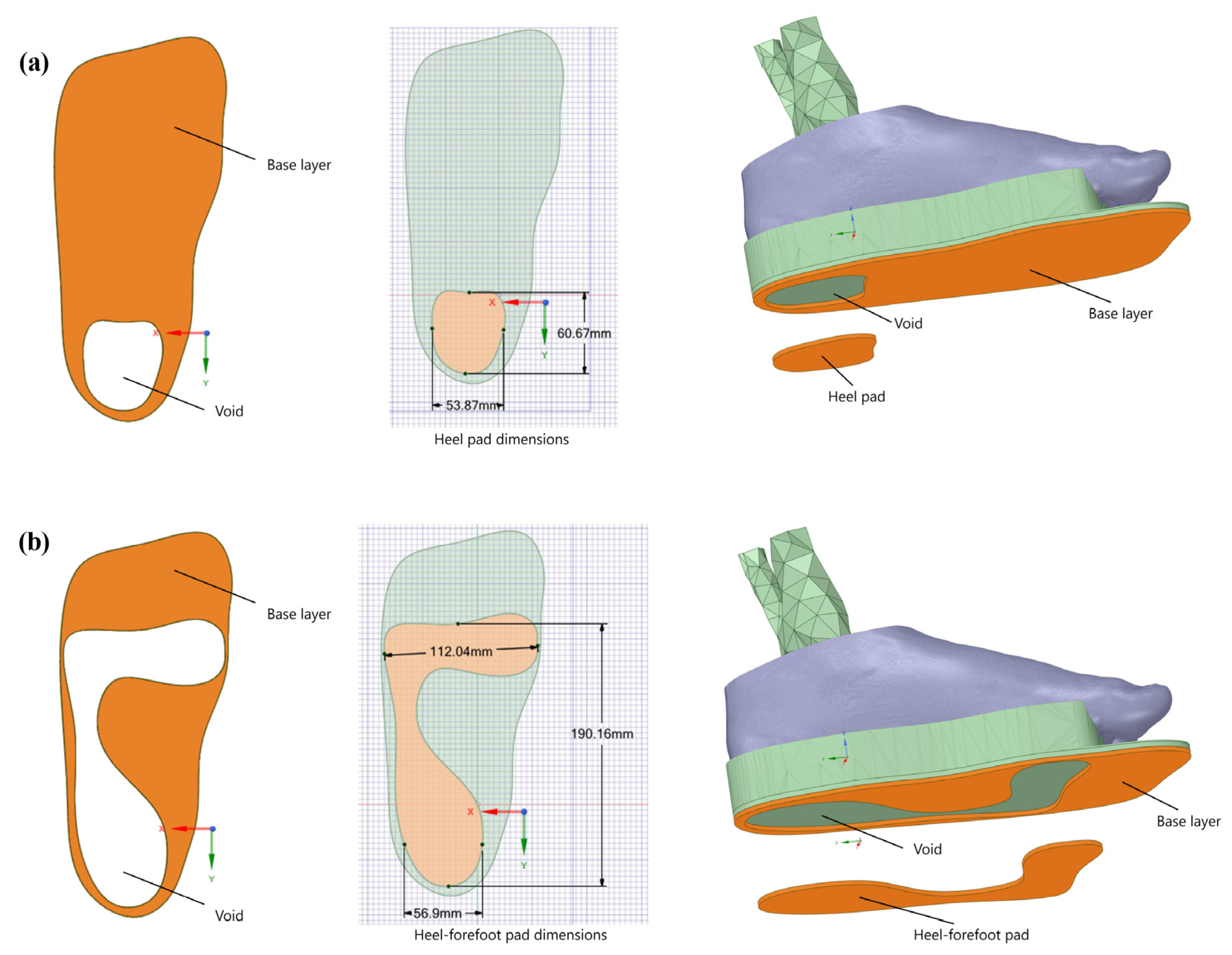
2.2. Pad Designs and Materials

A three-layer CMI as a control CMI with the top layer as ethylene vinyl acetate (EVA), the middle layer as Nora® Lunalastike, and the base layer as thermoplastic polyurethane (TPU) was designed. A void was designed under the anatomical regions of high peak plantar pressure to reduce the peak plantar pressure under the heel and forefoot (Figure 1). Two types of insole pads, a heel pad and heel–forefoot pad with an overall 3 mm thickness, were embedded in the base layer. The length and width of the heel pad were 60.67 mm and 53.87 mm, respectively (Figure 1a). Moreover, the heel–forefoot pad was designed from heel to forefoot with a heel width of 56.90 mm and a forefoot width of 112.04 mm, with a total length of 190.16 mm (Figure 1b).
The pad was embedded in the base layer of the CMI with varying material properties (Table 1). Ansys SpaceClaim (ANSYS Inc., Canonsburg, PA, USA) was used to create a 3D foot model with the CMI and with various pad designs for a diabetic foot with neuropathy.

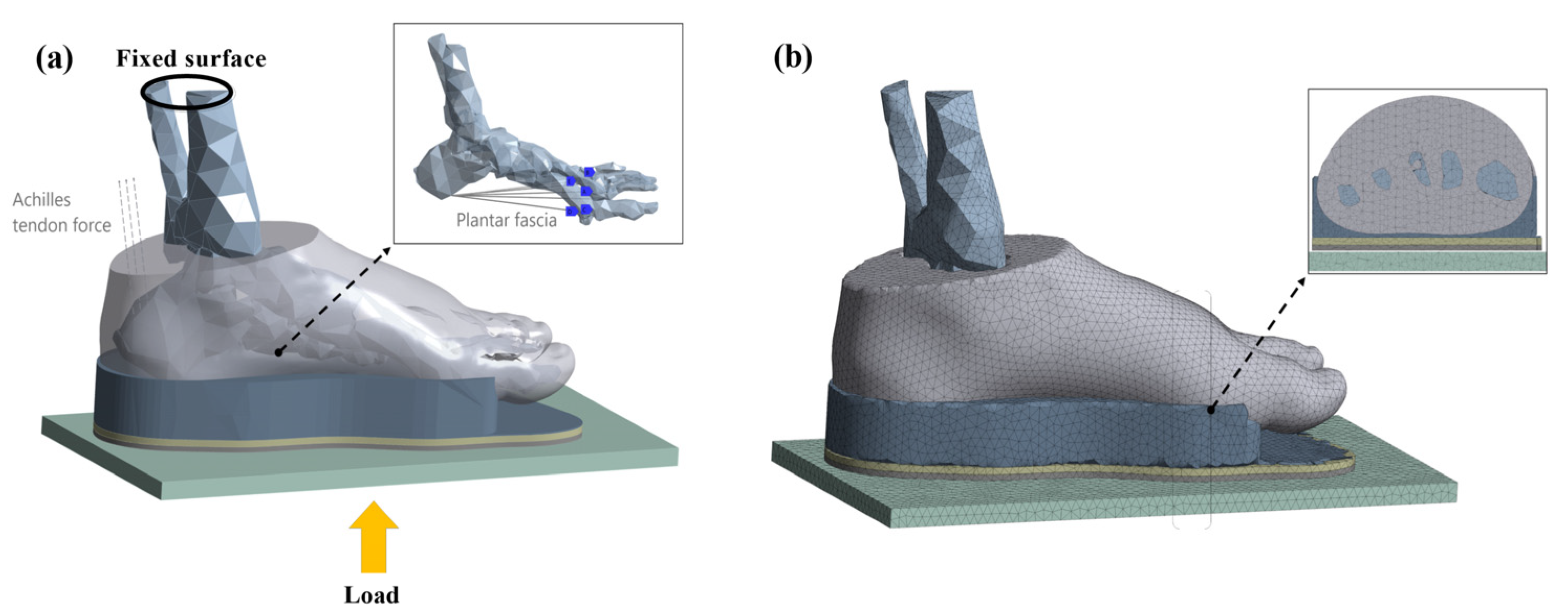
2.3. Boundary and Loading Conditions
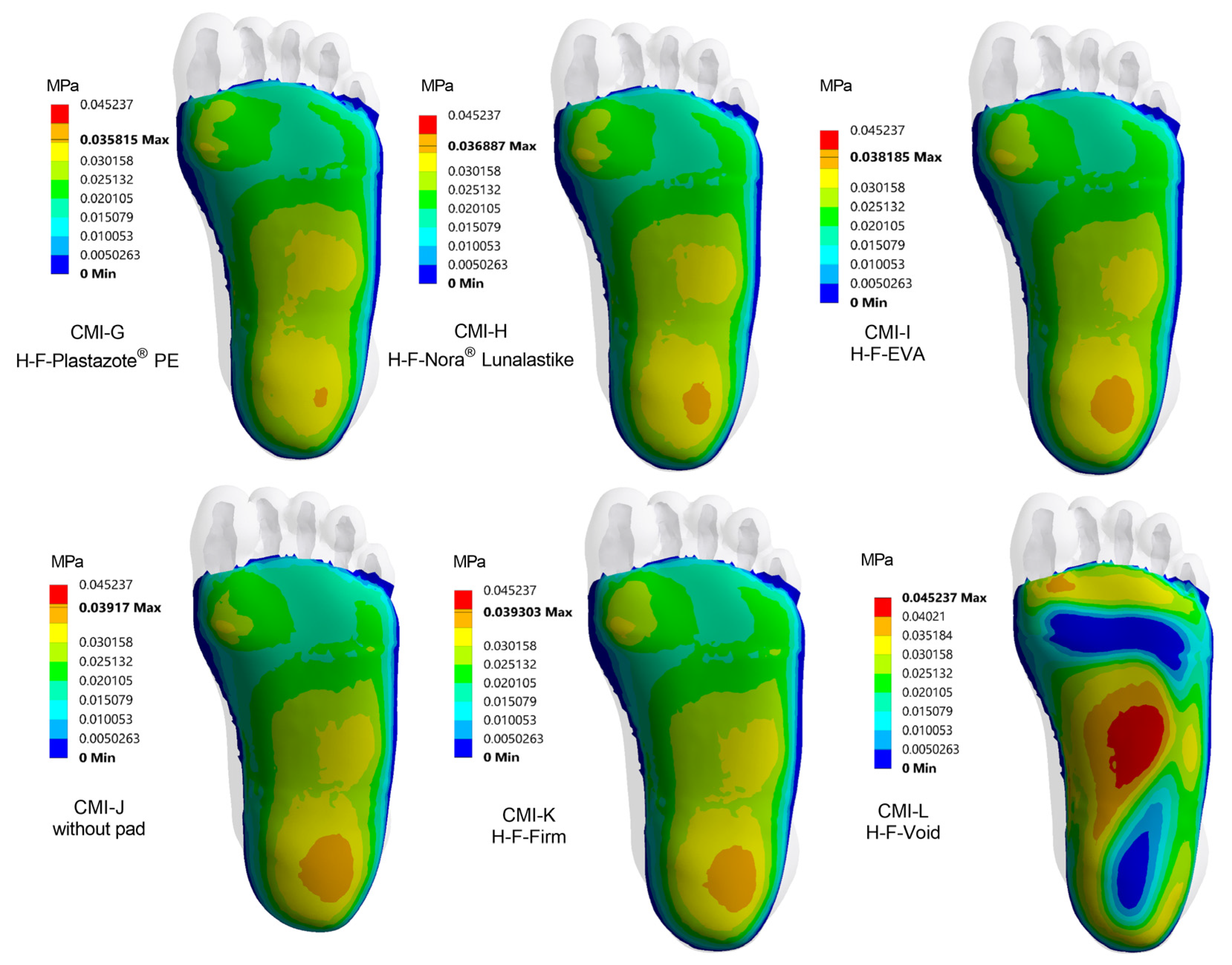
Ansys SpaceClaim was used to build the finite element (FE) foot model with the CMI along with a ground (Figure 2). The fused foot bones as one lump encapsulated by the soft tissues were assigned to be bonded. The foot–insole interaction and the insole–ground interface had a frictional coefficient of 0.3 [24] and 0.6 [25], respectively. The ends of the tibia and fibula were assigned to be fixed. A load of 420 N was regarded as a ground reaction force representing half of the body weight during balanced standing. In addition, based on earlier research, an upward-directed force of about 50% of Achilles tendon force during balanced standing was prescribed in the FE model [28]. To mimic the plantar fascia, 5 tension-only link segments were employed, and origin and insertion locations were determined by their anatomical location in accordance with an anatomy atlas [29]. A mesh convergence study was conducted for five different element sizes, and a tetrahedral 5 mm element size was used as a computationally optimal mesh for further analysis [28,29].

3. Results

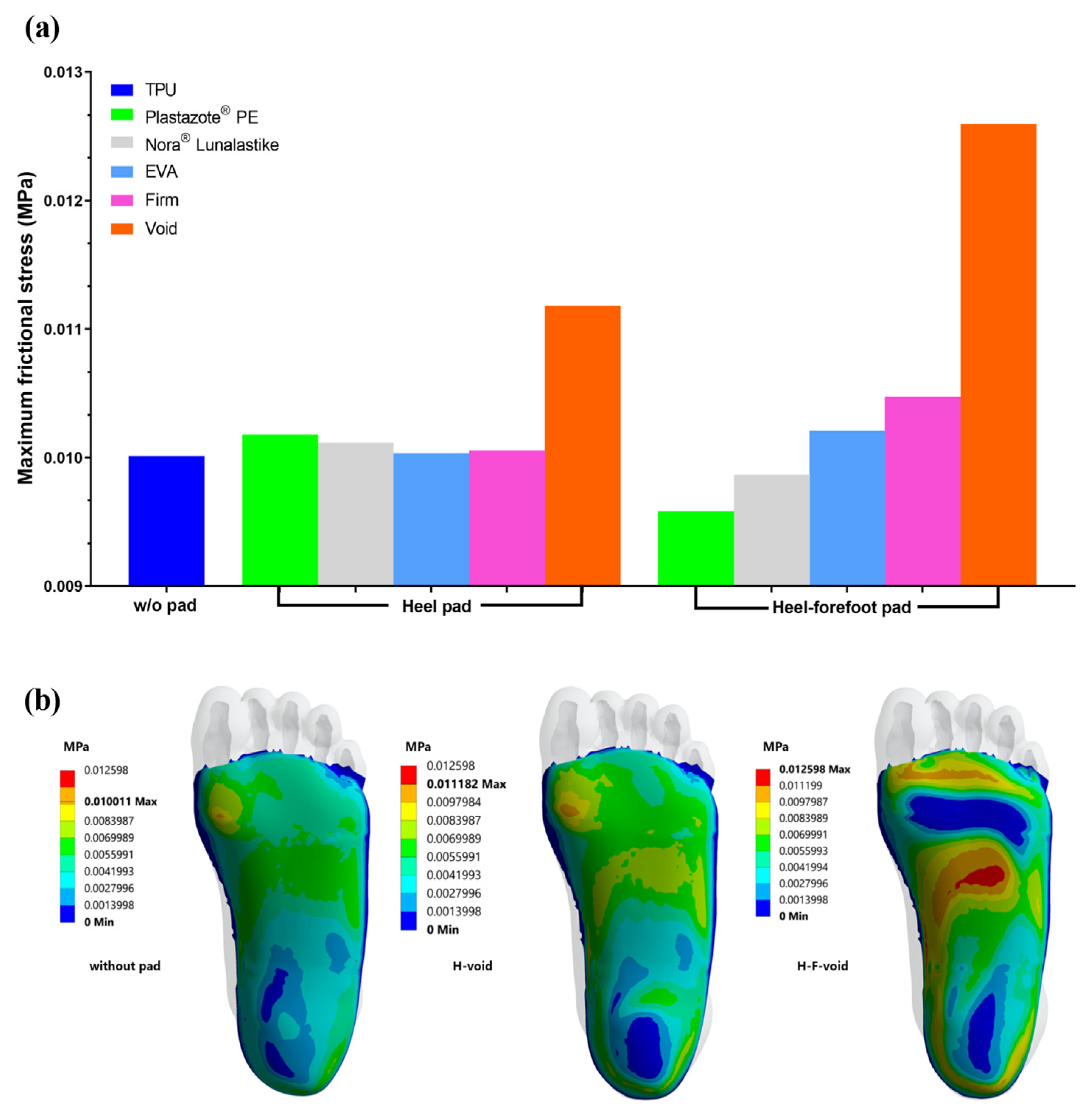
3.1. Frictional Stress

The peak frictional stress occurred under the first metatarsal head with the CMI without a heel pad and with a heel-to-forefoot pad and heel pad fabricated from various materials (Figure 3). The frictional stress with the heel–forefoot pad was reduced by 6.01% as compared with the heel pad only fabricated from Plastazote® PE. However, the frictional stress with the heel–forefoot pad with a firm material was increased by 4.08% as compared with the heel pad with a similar material. The maximum frictional stress increased by 4.52% with the heel–forefoot pad fabricated from a firm material compared with the CMI without a pad (Figure 2a). Conversely, with the heel–forefoot void, the maximum frictional stress shifted from the medial forefoot to the medial midfoot (Figure 3b). The maximum frictional stress increased when the stiffness of the pad was higher for the heel–forefoot pad. However, this observation could not be found for the heel pad only. The heel–forefoot pad made with Plastazote® PE reduced the maximum frictional stress by 27.18% compared with the heel–forefoot void. A similar effect was found with the heel void, where the frictional stress at the medial forefoot was increased by 9.40% compared with the heel pad fabricated from Plastazote® PE.

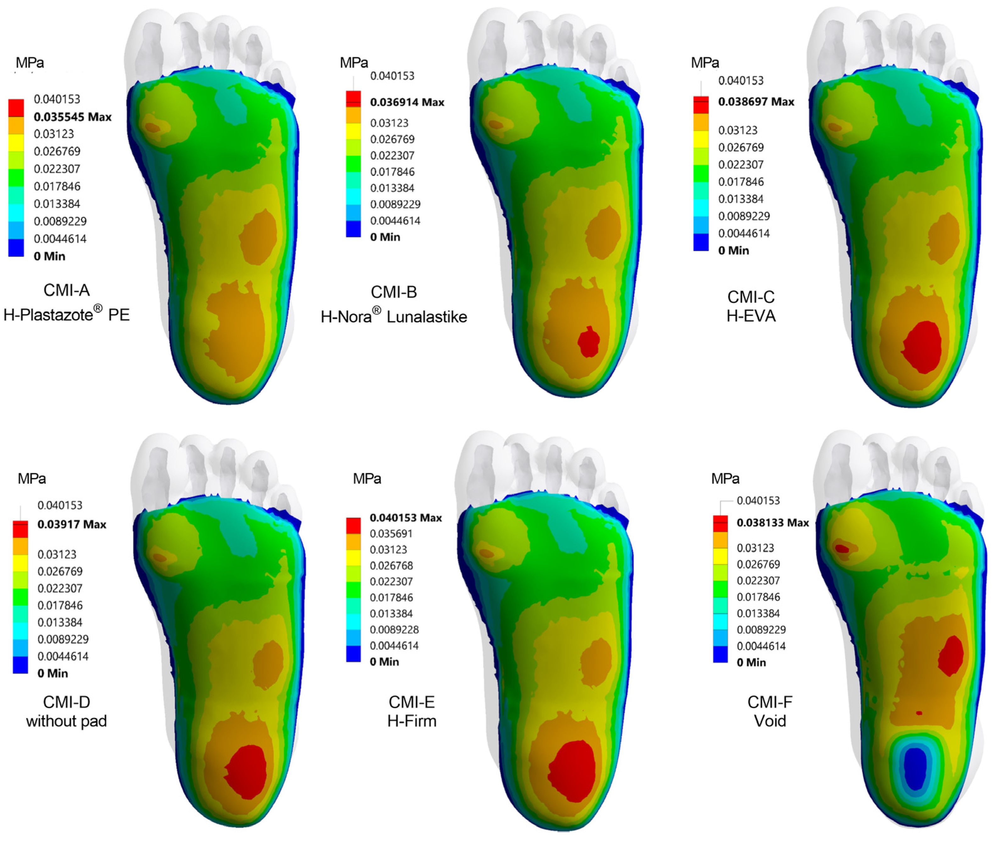
3.2. Plantar Pressure Distribution with Heel Pad

The softest heel pad (CMI-A) provided better planar pressure distribution, especially at the medial forefoot and hindfoot compared with the CMI with a stiffer heel pad and the CMI without pad (Figure 4). The results showed that the heel had the highest plantar contact pressure compared with the other foot regions, except for the model of the insole with a void heel (CMI-F). Furthermore, the area of high plantar contact pressure became larger when using a high-stiffness material for the heel pad. CMI-A had the softest heel pad, and the peak contact pressure was 9.02% lowered with CMI-A compared with the CMI without a heel pad (CMI-D). Moreover, there was a maximum reduction in the peak contact pressure by 12.41% with CMI-A compared with the firm material pad (CMI-E). On the contrary, the peak contact pressure increased by 7.06% with the heel void (CMI-F) and shifted from the hindfoot to the forefoot, followed by the midfoot, compared with CMI-A. In addition, the plantar pressure distribution at the midfoot was highest with CMI-F, followed by the first metatarsal head, compared with CMI-E, CMI-D, and CMI-A.

3.3. Plantar Pressure Distribution with Heel–Forefoot Pad

The heel–forefoot pad fabricated from a softer material provided a better distribution of plantar pressure at the medial forefoot and hindfoot compared with stiffer materials and the insole without a pad (Figure 5). In contrast with stiffer materials, the soft material heel–forefoot pad reduced the peak contact pressure at the hindfoot. The maximum peak contact pressure was reduced by 20.84% with the softest heel pad (CMI-G) compared with the insole without a heel pad (CMI-J). Moreover, the reduction in the peak contact pressure was 23.24% with CMI-G compared with the firm heel–forefoot pad (CMI-K). On the other hand, the peak contact pressure using the heel–forefoot void (CMI-L) was increased by 34.80% and shifted from the hindfoot to the medial midfoot compared with CMI-G. The regions of high contact pressure under the 1st to 5th metatarsal heads were shifted to a distal part of the metatarsal heads with CMI-L.

3.4. The Influence of Pad Design on Each Region of the Foot

The peak contact pressure was reduced using heel–forefoot and heel pads with softer materials at the hindfoot area (Table 2). The heel–forefoot pad made from Plastazote® PE (CMI-G) reduced the peak contact pressure by 6.98% compared with the heel pad only fabricated from the same material (CMI-A). However, the heel–forefoot void (CMI-L) had a negative effect with an increased medial forefoot peak contact pressure of 16.18% and 11.00% compared with the softer heel–forefoot pad (CMI-G) and the CMI without a pad, respectively. Moreover, the central forefoot peak contact pressure was reduced with the Plastazote® PE heel pad (CMI-A) compared with the heel–forefoot pad (CMI-G) and that with voids.
The medial and lateral peak contact pressures at the midfoot increased with the voids as compared with the heel and heel–forefoot pads and the insole without a pad. Moreover, the peak contact pressure was increased by 34.42% at the medial midfoot with the heel–forefoot void compared with the insole without a pad. A similar increased trend was found with the heel pad compared with the insole without a pad.

4. Discussion

This study proposed to computationally evaluate the effect of pad shape and material stiffness on plantar pressure distribution and the reduction in diabetic foot with neuropathy using the FEA. The current study found a softer material used in the heel–forefoot pad tends to reduce the frictional stresses at the forefoot and hindfoot compared with the heel pad and the insole without a pad. The heel pad designed from a softer material distributed the plantar pressure from the hindfoot compared with stiffer materials and the heel–forefoot pad fabricated from soft to stiff materials. Likewise, the peak contact pressure at the medial and central forefoot was reduced by the soft heel–forefoot pad compared with the soft heel pad only and the insole without a pad. The principles of the reduction in peak contact pressure in a diabetic foot with neuropathy require the consideration of various designs and material selections with the inclusion of frictional stress.
A CMI is beneficial for a diabetic neuropathic foot for the reduction in peak plantar pressure, especially at the forefoot [37,38,39,40]. However, some patients require a further reduction in peak plantar pressure either at the forefoot or hindfoot to prevent ulceration and to reduce the occurrence of pain. Experimental and FEA studies have highlighted the heel as one of the most frequently encountered regions with pain in all populations, including athletes and diabetic patients with a high risk of foot ulceration [41,42]. Metatarsal pads and small plugs are used to reduce the peak plantar pressure under metatarsal heads [37,43]. Using a CMI causes comfort in the foot due to the reduction and uniform distribution of the plantar pressure [44]. A previous experiment indicated that the CMI material and its modifications significantly lowered peak contact pressure at the forefoot and hindfoot, including shifting contact pressure to the midfoot [45]. A similar trend was found in this current study with a reduced peak planar pressure with the use of softer pads compared with the insole without a pad, firm pads, and voids.
Appropriate CMI material selection and pad design play a major impact on contact pressure reduction and redistribution under the hindfoot, and forefoot compared with a flat insole and no insole. The thickness and stiffness of the CMI material reduced the plantar pressure distribution in [46]. In our work, a softer material sandwiched between stiffer materials in addition to varying pad designs fabricated from a low Young’s modulus material could further reduce the peak contact pressure, especially at the heel. A similar trend of reduction was found with an appropriate selection of CMI material and thickness for people with prolonged standing at work [15,17,42]. By changing the designs of CMIs including pads, we can further reduce and redistribute the peak contact pressure in a diabetic neuropathic foot. Evidence suggests using a CMI with a metatarsal pad and a metatarsal bar can redistribute the plantar pressure. However, whether the metatarsal pad is positioned proximal or distal to the metatarsal heads influences the plantar pressure distribution at the forefoot [25,47]. Our study also demonstrated that the heel–forefoot pad made from a softer material reduced the medial forefoot peak contact pressure compared with using a heel pad only. However, a heel pad made from softer material reduced the central forefoot peak contact pressure as compared with the heel–forefoot pad and one with voids. A similar plantar pressure reduction was found with low- and medium-density CMIs fabricated from polyurethane and EVA [48].
The FEA technique allows an efficient analysis of the peak contact pressure that is frequently shown in cases of foot complications. The FEA approach highlights some of the important parameters that play an important role, especially in subjects prone to frequent ulceration and re-ulceration. However, frictional stress measurement, which is useful to be evaluated in clinical practice using in-shoe pressure sensors, is rarely reported and remains difficult in actual clinical settings in diabetic foot with neuropathy. Researchers attached sensors to the callus site to determine the forces that trigger callus formation, and it was found that in-shoe shear stresses are a causative factor for foot disorders [49,50]. The unique aspect of our study was using the FEA to evaluate the frictional stress that occurred with pads fabricated from various materials and those with voids in comparison with insoles without any pads. In our current study, the maximum frictional stress was much higher at the forefoot than at the hindfoot. This higher frictional stress at the forefoot might help in the development of ulceration and re-ulceration [51,52]. The results of this current study showed, similarly, a trend of maximum frictional stress under the first metatarsal head with pads and without pads compared with at the hindfoot. However, the maximum frictional stress shifted to the midfoot while using a heel–forefoot void. The secondary component causing ulceration can be detected as differences in the peak contact pressure and maximum frictional stress [53]. Recently, many researchers applied artificial intelligence for the purpose of diabetic foot screening and a future advancement [54,55]. Further improvement can be achieved by combining the FEA approach with artificial intelligence.
There were several limitations in this FEA study while investigating the CMI with two types of pads in comparison with that without a pad. Firstly, the soft tissues and insole material were simplified as linear elastic material properties to reduce the computational load acquired by the FEA. Therefore, the material properties in the current models were assigned as linear elastic, as our focus was to forecast the pattern of plantar pressure distribution while changing the pad designs and materials for a diabetic foot with neuropathy. Moreover, to identify the exact values it would be recommended to investigate more CMI designs wherein hyperelastic material properties could be assigned to the soft tissues and insoles during a gait cycle. Secondly, the current FEA study assumed a balanced standing with a ground reaction force, representing half of the body weight. The application of force following the center of pressure might represent a more realistic condition during gait. Thirdly, the bones were fused together as one entity to represent the bones to reduce the complexity of the foot model, as the region of interest was the plantar surface of the foot, rather than the bones in the foot. Lastly, the results are based on a single model of foot. Therefore, future studies are necessary to focus on types of foot and the effectiveness of CMIs. For further FE foot model development, the load bearing condition during different phases of the gait cycle requires muscle loading input. In addition, the motion within foot joints during walking might give more insight into the diabetic foot with the use of CMIs with different interventions.

5. Conclusions

It was demonstrated that the pad design and material selection had an impact on the plantar pressure reduction and distribution, especially in the forefoot and hindfoot regions. It was observed that the use of a heel–forefoot void further reduced the hindfoot and forefoot maximum frictional stresses compared with a heel–forefoot pad with softer materials. Using a softer material as a heel–forefoot pad and heel pad reduced the peak plantar pressure compared with the stiffer material and the insole without a pad. Our results suggest that the employment of the FEA while designing and selecting materials to fabricate a CMI is able to minimize prolonged clinical trial and error to reduce abnormal peak contact pressure in a diabetic foot with neuropathy.

Author Contributions

Conceptualization, M.N. and S.C.; methodology, M.N., D.Y.R.C. and S.C.; validation, M.N., D.Y.R.C., S.S. and S.C.; formal analysis, M.N., D.Y.R.C., S.S. and S.C.; investigation, M.N., D.Y.R.C., S.S. and S.C.; data curation, M.N., D.Y.R.C., S.S. and S.C.; writing—original draft preparation, M.N. and S.C.; writing—review and editing, M.N., D.Y.R.C., S.S. and S.C.; visualization, M.N., D.Y.R.C. and S.C.; supervision, D.Y.R.C. and S.C.; project administration, M.N. and S.C.; funding acquisition, M.N. and S.C. All authors have read and agreed to the published version of the manuscript.

Funding

We would like to thank the Graduate School and Faculty of Medicine, Prince of Songkla University for their financial support.

Institutional Review Board Statement

The study was conducted in accordance with the Declaration of Helsinki and approved by the Ethics Committee of Faculty of Medicine, Prince of Songkla University (REC.63-219-25-2).

Informed Consent Statement

Informed consent was obtained from a subject involved in the study.

Data Availability Statement

Not applicable.

Acknowledgments

We also would like to thank the participant for their active participation.

Conflicts of Interest

The authors declare no conflict of interest.

References

  1. Armstrong, D.G.; Boulton, A.J.M.; Bus, S.A. Diabetic Foot Ulcers and Their Recurrence. N. Engl. J. Med. 2017, 376, 2367–2375. [Google Scholar] [CrossRef] [PubMed]
  2. Lazzarini, P.A.; Pacella, R.E.; Armstrong, D.G.; van Netten, J.J. Diabetes-related lower-extremity complications are a leading cause of the global burden of disability. Diabet. Med. 2018, 35, 1297–1299. [Google Scholar] [CrossRef] [PubMed]
  3. Lazzarini, P.A.; Hurn, S.E.; Kuys, S.S.; Kamp, M.C.; Ng, V.; Thomas, C.; Jen, S.; Wills, J.; Kinnear, E.M.; d’Emden, M.C.; et al. The silent overall burden of foot disease in a representative hospitalised population. Int. Wound J. 2017, 14, 716–728. [Google Scholar] [CrossRef] [Green Version]
  4. Armstrong, D.G.; Lavery, L.A. Diabetic foot ulcers: Prevention, diagnosis and classification. Am. Fam. Physician 1998, 57, 1325–1332. [Google Scholar] [PubMed]
  5. Reiber, G.E.; Pecoraro, R.E.; Koepsell, T.D. Risk factors for amputation in patients with diabetes mellitus. A case-control study. Ann. Intern. Med. 1992, 117, 97–105. [Google Scholar] [CrossRef] [PubMed]
  6. van Netten, J.J.; Lazzarini, P.A.; Armstrong, D.G.; Bus, S.A.; Fitridge, R.; Harding, K.; Kinnear, E.; Malone, M.; Menz, H.B.; Perrin, B.M.; et al. Diabetic Foot Australia guideline on footwear for people with diabetes. J. Foot Ankle Res. 2018, 11, 2. [Google Scholar] [CrossRef] [Green Version]
  7. Shaw, J.E.; Sicree, R.A.; Zimmet, P.Z. Global estimates of the prevalence of diabetes for 2010 and 2030. Diabetes Res. Clin. Pract. 2010, 87, 4–14. [Google Scholar] [CrossRef]
  8. Pecoraro, R.E.; Reiber, G.E.; Burgess, E.M. Pathways to diabetic limb amputation. Basis for prevention. Diabetes Care 1990, 13, 513–521. [Google Scholar] [CrossRef]
  9. Woolard, R.H.; Carty, K.; Wirtz, P.; Longabaugh, R.; Nirenberg, T.D.; Minugh, P.A.; Becker, B.; Clifford, P.R. Research fundamentals: Follow-up of subjects in clinical trials: Addressing subject attrition. Acad. Emerg. Med. 2004, 11, 859–866. [Google Scholar] [CrossRef]
  10. Wang, D.; Cai, P. Finite Element Analysis of the Expression of Plantar Pressure Distribution in the Injury of the Lateral Ligament of the Ankle. Nano Biomed. Eng. 2019, 11, 290–296. [Google Scholar] [CrossRef]
  11. Ghassemi, A.; Mossayebi, A.R.; Jamshidi, N.; Naemi, R.; Karimi, M.T. Manufacturing and finite element assessment of a novel pressure reducing insole for Diabetic Neuropathic patients. Australas. Phys. Eng. Sci. Med. 2015, 38, 63–70. [Google Scholar] [CrossRef] [PubMed]
  12. Cheung, J.T.; Zhang, M. A 3-dimensional finite element model of the human foot and ankle for insole design. Arch. Phys. Med. Rehabil. 2005, 86, 353–358. [Google Scholar] [CrossRef] [PubMed]
  13. Cavanagh, P.R. Therapeutic footwear for people with diabetes. Diabetes Metab. Res. Rev. 2004, 20 (Suppl. S1), S51–S55. [Google Scholar] [CrossRef] [PubMed]
  14. Bus, S.A.; Maas, J.C.; Otterman, N.M. Lower-extremity dynamics of walking in neuropathic diabetic patients who wear a forefoot-offloading shoe. Clin. Biomech. 2017, 50, 21–26. [Google Scholar] [CrossRef]
  15. Anderson, J.; Williams, A.E.; Nester, C. Development and evaluation of a dual density insole for people standing for long periods of time at work. J. Foot Ankle Res. 2020, 13, 42. [Google Scholar] [CrossRef]
  16. Ahmed, S.; Barwick, A.; Butterworth, P.; Nancarrow, S. Footwear and insole design features that reduce neuropathic plantar forefoot ulcer risk in people with diabetes: A systematic literature review. J. Foot Ankle Res. 2020, 13, 30. [Google Scholar] [CrossRef]
  17. Goske, S.; Erdemir, A.; Petre, M.; Budhabhatti, S.; Cavanagh, P.R. Reduction of plantar heel pressures: Insole design using finite element analysis. J. Biomech. 2006, 39, 2363–2370. [Google Scholar] [CrossRef]
  18. Wibowo, D.B.; Widodo, A.; Haryadi, G.D.; Caesarendra, W.; Harahap, R. Effect of in-shoe foot orthosis contours on heel pain due to calcaneal spurs. Appl. Sci. 2019, 9, 495. [Google Scholar] [CrossRef] [Green Version]
  19. Collings, R.; Freeman, J.; Latour, J.M.; Paton, J. Footwear and insole design features for offloading the diabetic at risk foot-A systematic review and meta-analyses. Endocrinol. Diabetes Metab. 2021, 4, e00132. [Google Scholar] [CrossRef] [Green Version]
  20. Raspovic, A.; Landorf, K.B.; Gazarek, J.; Stark, M. Reduction of peak plantar pressure in people with diabetes-related peripheral neuropathy: An evaluation of the DH Pressure Relief Shoe™. J. Foot Ankle Res. 2012, 5, 25. [Google Scholar] [CrossRef]
  21. Shi, Q.Q.; Li, P.L.; Yick, K.-L.; Li, N.-W.; Jiao, J. Effects of contoured insoles with different materials on plantar pressure offloading in diabetic elderly during gait. Sci. Rep. 2022, 12, 15395. [Google Scholar] [CrossRef] [PubMed]
  22. Chatzistergos, P.E.; Naemi, R.; Healy, A.; Gerth, P.; Chockalingam, N. Subject specific optimisation of the stiffness of footwear material for maximum plantar pressure reduction. Ann. Biomed. Eng. 2017, 45, 1929–1940. [Google Scholar] [CrossRef] [PubMed] [Green Version]
  23. Ohuchi, H.; Chavez, J.S.; Alvarez, C.A.D. Changes in calcaneal pitch and heel fat pad thickness in static weight bearing radiographs while wearing shoes with arch support and heel cup orthotics. Asia-Pac. J. Sports Med. Arthrosc. Rehabil. Technol. 2019, 17, 21–24. [Google Scholar] [CrossRef] [PubMed]
  24. Seligman, D.A.; Dawson, D.R. Customized heel pads and soft orthotics to treat heel pain and plantar fasciitis. Arch. Phy. Med. Rehabil. 2003, 84, 1564–1567. [Google Scholar] [CrossRef] [PubMed]
  25. Hastings, M.K.; Mueller, M.J.; Pilgram, T.K.; Lott, D.J.; Commean, P.K.; Johnson, J.E. Effect of metatarsal pad placement on plantar pressure in people with diabetes mellitus and peripheral neuropathy. Foot Ankle Int. 2007, 28, 84–88. [Google Scholar] [CrossRef] [PubMed]
  26. Chatzistergos, P.E.; Naemi, R.; Chockalingam, N. A method for subject-specific modelling and optimisation of the cushioning properties of insole materials used in diabetic footwear. Med. Eng. Phys. 2015, 37, 531–538. [Google Scholar] [CrossRef] [PubMed]
  27. Chatzistergos, P.E.; Gatt, A.; Formosa, C.; Farrugia, K.; Chockalingam, N. Optimised cushioning in diabetic footwear can significantly enhance their capacity to reduce plantar pressure. Gait Posture 2020, 79, 244–250. [Google Scholar] [CrossRef]
  28. Nouman, M.; Dissaneewate, T.; Chong, D.Y.R.; Chatpun, S. Effects of custom-made insole materials on frictional stress and contact pressure in diabetic foot with neuropathy: Results from a finite element analysis. Appl. Sci. 2021, 11, 3412. [Google Scholar] [CrossRef]
  29. Nouman, M.; Chong, D.Y.R.; Chatpun, S. Finite Element Approach Towards Selection of Appropriate Materials to Redistribute Peak Plantar Pressure in Diabetic Foot with Neuropathy: Selecting Appropriate Materials for Diabetic Foot. PSU Med. J. 2021, 1, 43–54. [Google Scholar] [CrossRef]
  30. Brilakis, E.; Kaselouris, E.; Xypnitos, F.; Provatidis, C.G.; Efstathopoulos, N. Effects of foot posture on fifth metatarsal fracture healing: A finite element study. J. Foot Ankle Surg. 2012, 51, 720–728. [Google Scholar] [CrossRef]
  31. Chen, W.P.; Ju, C.W.; Tang, F.T. Effects of total contact insoles on the plantar stress redistribution: A finite element analysis. Clin. Biomech. 2003, 18, S17–S24. [Google Scholar] [CrossRef] [PubMed]
  32. Wright, D.G.; Rennels, D.C. A study of the elastic properties of plantar fascia. J. Bone Joint Surg. Am. 1964, 46, 482–492. [Google Scholar] [CrossRef] [PubMed]
  33. Lo, W.T.; Yick, K.; Ng, Z.; Yip, J. Numerical simulation of orthotic insole deformation for diabetic foot. J. Fiber Bioeng. Inform. 2015, 8, 401–411. [Google Scholar] [CrossRef] [Green Version]
  34. Lewis, G. Finite element analysis of a model of a therapeutic shoe: Effect of material selection for the outsole. Biomed. Mater. Eng. 2003, 13, 75–81. [Google Scholar] [PubMed]
  35. Frick, A.; Rochman, A. Characterization of TPU-elastomers by thermal analysis (DSC). Polym. Test. 2004, 23, 413–417. [Google Scholar] [CrossRef]
  36. Su, S.; Mo, Z.; Guo, J.; Fan, Y. The effect of arch height and material hardness of personalized insole on correction and tissues of flatfoot. J. Healthc. Eng. 2017, 8614341. [Google Scholar] [CrossRef] [Green Version]
  37. Mueller, M.J.; Lott, D.J.; Hastings, M.K.; Commean, P.K.; Smith, K.E.; Pilgram, T.K. Efficacy and mechanism of orthotic devices to unload metatarsal heads in people with diabetes and a history of plantar ulcers. Phys. Ther. 2006, 86, 833–842. [Google Scholar] [CrossRef] [PubMed] [Green Version]
  38. Bus, S.A.; Ulbrecht, J.S.; Cavanagh, P.R. Pressure relief and load redistribution by custom-made insoles in diabetic patients with neuropathy and foot deformity. Clin. Biomech. 2004, 19, 629–638. [Google Scholar] [CrossRef]
  39. Ashry, H.R.; Lavery, L.A.; Murdoch, D.P.; Frolich, M.; Lavery, D.C. Effectiveness of diabetic insoles to reduce foot pressures. J. Foot Ankle Surg. 1997, 36, 268–271. [Google Scholar] [CrossRef]
  40. Tsung, B.Y.; Zhang, M.; Mak, A.F.; Wong, M.W. Effectiveness of insoles on plantar pressure redistribution. J. Rehabil. Res. Dev. 2004, 41, 767–774. [Google Scholar] [CrossRef]
  41. Hellstrand Tang, U.; Zügner, R.; Lisovskaja, V.; Karlsson, J.; Hagberg, K.; Tranberg, R. Comparison of plantar pressure in three types of insole given to patients with diabetes at risk of developing foot ulcers—A two-year, randomized trial. J. Clin. Transl. Endocrinol. 2014, 1, 121–132. [Google Scholar] [CrossRef] [PubMed] [Green Version]
  42. Luo, G.; Houston, V.L.; Garbarini, M.A.; Beattie, A.C.; Thongpop, C. Finite element analysis of heel pad with insoles. J. Biomech. 2011, 44, 1559–1565. [Google Scholar] [CrossRef] [PubMed]
  43. Actis, R.L.; Ventura, L.B.; Lott, D.J.; Smith, K.E.; Commean, P.K.; Hastings, M.K.; Mueller, M.J. Multi-plug insole design to reduce peak plantar pressure on the diabetic foot during walking. Med. Biol. Eng. Comput. 2008, 46, 363–371. [Google Scholar] [CrossRef] [PubMed]
  44. Mündermann, A.; Nigg, B.M.; Humble, R.N.; Stefanyshyn, D.J. Orthotic comfort is related to kinematics, kinetics, and EMG in recreational runners. Med. Sci. Sports Exerc. 2003, 35, 1710–1719. [Google Scholar] [CrossRef]
  45. Nouman, M.; Dissaneewate, T.; Leelasamran, W.; Chatpun, S. The insole materials influence the plantar pressure distributions in diabetic foot with neuropathy during different walking activities. Gait Posture 2019, 74, 154–161. [Google Scholar] [CrossRef]
  46. Cheung, J.T.; Zhang, M. Parametric design of pressure-relieving foot orthosis using statistics-based finite element method. Med. Eng. Phys. 2008, 30, 269–277. [Google Scholar] [CrossRef]
  47. Martinez-Santos, A.; Preece, S.; Nester, C.J. Evaluation of orthotic insoles for people with diabetes who are at-risk of first ulceration. J Foot Ankle Res 2019, 12, 35. [Google Scholar] [CrossRef] [Green Version]
  48. Healy, A.; Dunning, D.N.; Chockalingam, N. Effect of insole material on lower limb kinematics and plantar pressures during treadmill walking. Prosthetics Orthotics Int. 2012, 36, 53–62. [Google Scholar] [CrossRef]
  49. Amemiya, A.; Noguchi, H.; Oe, M.; Takehara, K.; Ohashi, Y.; Suzuki, R.; Yamauchi, T.; Kadowaki, T.; Sanada, H.; Mori, T. Shear Stress-Normal Stress (Pressure) Ratio Decides Forming Callus in Patients with Diabetic Neuropathy. J. Diabetes Res. 2016, 2016, 3157123. [Google Scholar] [CrossRef] [Green Version]
  50. Cong, Y.; Cheung, J.T.; Leung, A.K.; Zhang, M. Effect of heel height on in-shoe localized triaxial stresses. J. Biomech. 2011, 44, 2267–2272. [Google Scholar] [CrossRef]
  51. Caselli, A.; Pham, H.; Giurini, J.M.; Armstrong, D.G.; Veves, A. The forefoot-to-rearfoot plantar pressure ratio is increased in severe diabetic neuropathy and can predict foot ulceration. Diabetes Care 2002, 25, 1066–1071. [Google Scholar] [CrossRef] [Green Version]
  52. Zou, D.; Mueller, M.J.; Lott, D.J. Effect of peak pressure and pressure gradient on subsurface shear stresses in the neuropathic foot. J. Biomech. 2007, 40, 883–890. [Google Scholar] [CrossRef] [PubMed]
  53. Amin, N.; Doupis, J. Diabetic foot disease: From the evaluation of the "foot at risk" to the novel diabetic ulcer treatment modalities. World J. Diabetes 2016, 7, 153–164. [Google Scholar] [CrossRef] [PubMed]
  54. Chemello, G.; Salvatori, B.; Morettini, M.; Tura, A. Artificial Intelligence Methodologies Applied to Technologies for Screening, Diagnosis and Care of the Diabetic Foot: A Narrative Review. Biosensors 2022, 12, 985. [Google Scholar] [CrossRef] [PubMed]
  55. Song, Y.; Cen, X.; Zhang, Y.; Bíró, I.; Ji, Y.; Gu, Y. Development and validation of a subject-specific coupled model for foot and sports shoe complex: A pilot computational study. Bioengineering 2022, 9, 553. [Google Scholar] [CrossRef] [PubMed]
Figure 1. The pad designs. (a) Heel pad and (b) heel–forefoot pad added to the base layer.
Applsci 13 00399 g001
Figure 2. (a) The 3D foot model is reconstructed with fixed points and applied load, and (b) the 3D foot model is meshed with tetrahedral elements.
Applsci 13 00399 g002
Figure 3. (a) The maximum frictional stress obtained for CMI with pad materials ranging from soft to hard compared with CMI without pad; (b) frictional stress distribution with the highest maximum frictional stress, especially with a heel void (H-void) and a heel–forefoot void (H-F-void) compared with CMI without pad.
Applsci 13 00399 g003
Figure 4. The plantar pressure obtained from FEA models using heel (H) pad made from various materials from soft to hard (CMI-A to CMI-E), without heel pad (CMI-D), and with void (CMI-F).
Applsci 13 00399 g004
Figure 5. The plantar pressure obtained from FEA models using the heel–forefoot pad fabricated from soft to hard material properties (CMI-G to CMI-K), without heel–forefoot pad (CMI-J), and with a void (CMI-L).
Applsci 13 00399 g005
Table 1. Material properties of components of the foot model with customized insole without heel pad and with heel pad and heel–forefoot pad used in the finite element analysis.
Components Young’s Modulus (MPa)Poisson’s Ratio vReferences
Bone73000.3[30]
Soft tissue0.190.49[31]
Plantar fascia3500.35[32]
Plastazote® PE 0.450.38[33]
Nora® Lunalastike 1.040.25[33]
EVA 50.40[34]
TPU 110.45[35]
Firm insole 10000.40[12]
Ground 21,000,0000.3[36]
Note: PE—polyethylene, EVA—ethylene vinyl acetate, TPU—thermoplastic polyurethane.
Table 2. Regional peak contact pressure of the foot with heel pad and heel–forefoot pad with varying materials.
Foot Region Peak Contact Pressure (kPa)
Plastazote® PENora® LunalastikeEVATPUFirmVoid
HH-FHH-FHH-Fw/o PadHH-FHH-F
Whole foot35.535.836.936.938.738.239.240.239.338.145.2
Forefoot34.131.833.932.933.634.033.533.734.938.138.6
Medial forefoot34.131.833.932.933.634.033.533.734.938.137.4
Central forefoot24.626.424.525.924.525.724.024.425.928.037.9
Lateral forefoot26.126.826.026.225.826.325.726.226.628.238.6
Midfoot34.032.233.832.633.633.233.533.633.737.745.2
Medial midfoot32.231.832.030.731.030.431.531.830.634.744.6
Lateral midfoot34.032.233.832.633.633.233.533.633.737.745.2
Hindfoot35.535.836.936.938.738.239.240.239.336.137.8
Medial hindfoot34.235.136.434.738.337.038.038.838.536.138.2
Lateral hindfoot35.535.836.936.938.738.239.240.239.335.933.8
Note: H—heel pad, H-F—heel–forefoot pad, and w/o—without.
Disclaimer/Publisher’s Note: The statements, opinions and data contained in all publications are solely those of the individual author(s) and contributor(s) and not of MDPI and/or the editor(s). MDPI and/or the editor(s) disclaim responsibility for any injury to people or property resulting from any ideas, methods, instructions or products referred to in the content.

