Communication

Novel Instrument for Clinical Evaluations of Active Extraocular Muscle Tension

1
Department of Ophthalmology, Research Institute of Medical Science, Konkuk University Medical Center, Konkuk University School of Medicine, Seoul 05030, Republic of Korea
2
Department of Mechatronics Engineering, Glocal Campus, Konkuk University, 268 Chungwondaero, Chungju-si 27478, Republic of Korea
3
Department of Ophthalmology, Blanton Eye Institute, Houston Methodist Hospital, Houston, TX 77030, USA
4
Department of Ophthalmology, Neurology, Neurosurgery, Weill Cornell Medicine, New York, NY 10065, USA
5
Department of Ophthalmology, University of Texas Medical Branch, Galveston, TX 77555, USA
6
Department of Ophthalmology, UT MD Anderson Cancer Center, Houston, TX 77030, USA
7
Department of Ophthalmology, Texas A and M College of Medicine, College Station, TX 77807, USA
8
Department of Ophthalmology, University of Iowa Hospitals and Clinics, Iowa City, IA 52242, USA
9
Department of Ophthalmology, Baylor College of Medicine and the Center for Space Medicine, Houston, TX 77030, USA
10
Department of Ophthalmology, University of Buffalo, Buffalo, NY 14214, USA
*
Author to whom correspondence should be addressed.
Appl. Sci. 2023, 13(20), 11431; https://doi.org/10.3390/app132011431
Submission received: 15 July 2023 / Revised: 15 October 2023 / Accepted: 16 October 2023 / Published: 18 October 2023

Abstract

:
Strabismus can be caused by abnormal tension of the extraocular muscles (EOMs) attached to the eyeball in superior, inferior, lateral, medial, superior oblique, and inferior oblique positions. Evaluating the tension in each EOM is crucial for surgical planning in strabismus, which is conducted by adjusting the tension on the EOM. The purpose of this study was to develop a compact measuring device to non-invasively evaluate the active EOM tension. The proposed device employed a cotton-tipped medical swab to transfer the EOM tension connected to the force sensor as a non-invasive medium. The tilting angle of the swab and the force of active EOM tension were wirelessly transferred to a laptop computer for recording and real-time displaying of the measured values. The active EOM tensions for the four recti muscles were 101.7 ± 15.0 g (mean ± SD) for the lateral rectus; 88.0 ± 15.4 g for the medial rectus; 61.3 ± 6.8 g for the inferior rectus; and 121.3 ± 38.5 g for the superior rectus. These values were higher than the reported values of 45–60 g measured in previous studies. In the previous studies, however, the EOM was detached from the globe and attached to a strain gauge, and, thus, there were no passive elastic forces from ocular connective tissue, resulting in lower values compared with the current study. The previous methods were also complex and not suitable for clinical measurement. Thus, the proposed method, which is non-invasive and mimics the conventional force generation test with a cotton-tipped swab, could facilitate the evaluation of active EOM tension, both clinically in strabismus management and in research into understanding its pathophysiology.

1. Introduction

Strabismus is a condition in which the visual axes of the eyes are misaligned, resulting in ocular misalignment and symptomatic diplopia. The alignment of the visual axis is controlled by the six extraocular muscles (EOMs) attached to the eyeball in the superior, inferior, lateral, medial, superior oblique, and inferior oblique positions. Contemporary hypotheses regarding the etiology of strabismus have given rise to the “sensory versus motor” theory. In this context, an abnormal binocular sensory system is characterized by the absence of binocular fixation with normal visual acuity in each eye, the presence of strabismus, diplopia, abnormal retinal correspondence, and impaired stereopsis. An abnormal motor system is characterized by the abnormal full duction and version as well as abnormal speed saccades [1,2,3].
In this regard, abnormal EOM tension leads to an imbalance in the eyeball that results in a disconjugate gaze. Strabismus is not merely a cosmetic problem because, if untreated in childhood, the turned eye could lead to amblyopia. In addition, ocular misalignment can cause double vision, visual confusion, and abnormal head posture in adults [4,5]. Typically, strabismus surgeries are planned based on measuring the degree of squint in prism diopters, rather than assessing muscle tension. Nevertheless, postoperative challenges, like overcorrection and undercorrection, are common occurrences [6,7,8]. Strabismus is often treated by surgically adjusting the tension on the EOM [9]. Knowledge of EOM tension is, therefore, important for the diagnosis of and surgical planning for strabismus [10,11].
The forced duction test (FDT) or force generation test (FGT) have traditionally been carried out in typical clinical situations to evaluate EOM tension. The passive EOM tension is evaluated using the FDT, in which the clinician grips and moves the eyeball using forceps to perceive the mechanical restriction of the EOM with the eyeball facing away from the direction of the applied force [12,13]. On the other hand, active EOM tension can be evaluated using the FGT, which checks for EOM weakness. The clinician fastens a cotton-tipped swab above the limbus of the cornea in order to prevent the eyeball from moving. The patient then tries to move the eyeball toward the direction of the cotton-tipped swab [12]. For example, in a patient with lateral rectus (LR) paresis, the eye is held at the temporal limbus with a cotton swap and is adducted. The patient is then asked to abduct the eye, and the force generated (by the LR) is estimated.
The conventional FDT and FGT methods are simple and easy for clinical evaluations of EOM tension. However, these clinical tests are often performed based on the subjective and qualitative perceived assessments of tension by the hand of the clinician. The results of the FDT and FGT are, therefore, subjective and highly dependent on the experience and skill of the clinician [14]. To overcome these limitations, several trials have been conducted to measure EOM tension by using force transducers or strain gauges [15,16]. The authors previously designed a simple and compact device that integrated forceps, load cells, and tilting sensors [11]. It provided quantitative and continuous measurements of passive EOM tension and could be applied to the treatment of patients with intermittent exotropia [17,18,19].
Collins et al. measured active EOM tension generated by saccadic eyeball movement by implanting a force transducer in the EOMs [15]. Lennerstrand et al. measured active isometric EOM tension by connecting strain gauge probes to detached EOMs [16]. Although there are several currently available quantitative instruments and recording systems for measuring active EOM tension, most are restricted to use in the research laboratory setting because they are expensive, complicated to operate, or too complex for use in the clinic or operating room.
In the current study, we developed a compact measuring device to non-invasively evaluate active EOM tension, which would facilitate objective and quantitative estimations of the EOMs. The proposed device employed a medical swab to transfer the EOM tension connected to the force sensor as a non-invasive medium. The tilting angle of the swab and EOM tension force were wirelessly transferred to a laptop for recording and real-time displaying of the measured values. We believe that this novel device is an easy-to-use tool for making quantitative measurements that could provide valuable information about the active tension in EOMs, making it suitable for use both clinically in strabismus management and in research into understanding its pathophysiology.

2. Materials and Methods

The FGT was conducted in this trial to measure the active EOM tension in three subjects using the proposed ATMD device. This clinical study was approved by the Institutional Review Board (IRB) and Ethics Committee at Konkuk University Medical Center (registration number: 2023-03-050-001). The study was conducted according to the principles expressed in the Declaration of Helsinki, including obtaining written informed consent from each participant included in the study.

2.1. Design of Active EOM Tension Measuring Device

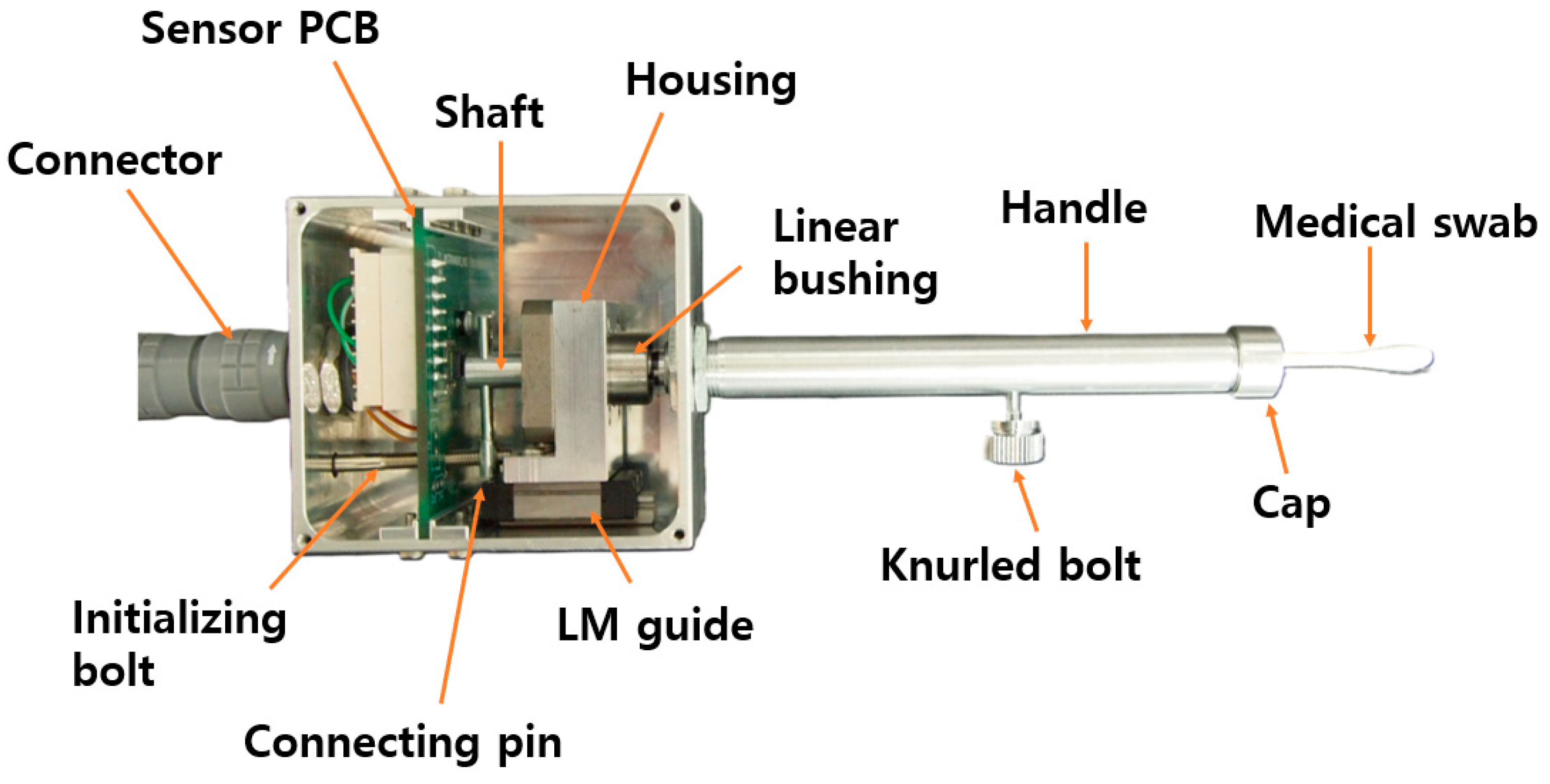
An active tension measuring device (ATMD) for EOM tension was designed in this study to allow for quantitative and objective measurements using the clinical FGT. The ATMD comprised sensing, control, and input–output (I/O) units. During the FGT, the ophthalmic surgeon grips and places a cotton-tipped swab on the eyeball; then, the eye is restricted from horizontal movement. The subject then tries to move their eyeball toward the swab. Consequentially, the active force pushing the cotton-tipped swab is recognized when the eye attempts to move against the placed cotton-tipped swab, as shown in Figure 1a. A sensing unit was first designed for the ATMD to measure the pushing force applied to the cotton swab tip. The ATMD device is comprised of a medical swab, a shaft, a knurled bolt, a linear bushing, a handle, and an integrated printed circuit board (PCB) with force and tilting sensors, as shown in Figure 2. The shaft was constructed as a single-side long hole for medical swab placement. The swab was interposed inside the shaft and then fixed by the knurled bolt. The other side of the shaft was connected to the piezoresistive-based force sensor (FMAMSDXX025WCSC3, Honeywell, NC, USA) mounted on the PCB [20]. The sensor was 5 mm × 5 mm in size and had a full-scale accuracy of within 2%. The linear bushing (LMK6, THK, Tokyo, Japan) could support the shaft by compensating for the mechanical friction in the axial plane. Forces on the top of the swab could be transferred to the force sensor through a single-side hole shaft with minimized mechanical losses. A tilting sensor (EBIMU-9DOFV5, E2BOX, Gyeonggi-do, Korea) was also mounted on the PCB to measure the tilting angle during the FGT [21]. The ophthalmic surgeon can refer to the tilting angle to uniformly perform the FGT in real time.
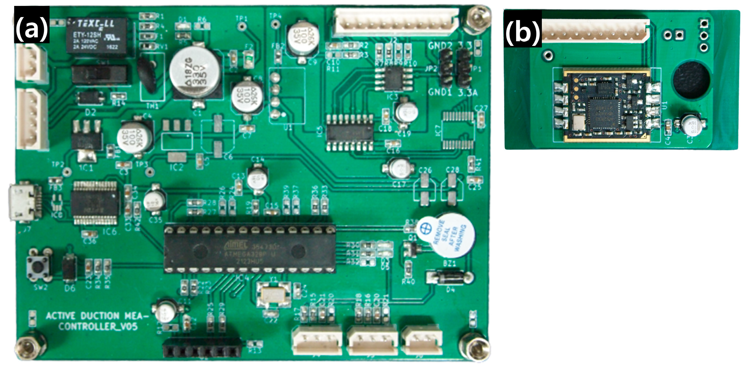
The control unit comprised a microcontroller and Bluetooth and power modules. The microcontroller (ATMEGA328P-PU, Atmel, CA, USA) collected data on the tilting angle and force by using an inter-integrated circuit and serial communication in a sampling period of 90 ms [22]. The collected data were wirelessly transferred to a laptop via a Bluetooth module (HC-05, HC Information Technology, Guangzhou, China) at a baud rate of 9600 bps [23]. In the power module, a 12 V input was transformed to 5 V and 3.3 V using a linear regulator (LM1117MPX-5.0, Texas Instruments, Dallas, TX, USA) and a switching converter (MUS-0503.3, Danube, Kaohsiung, Taiwan) to operate the control and sensing units, respectively. Figure 3a,b present the PCBs for the sensing and control units and their sensory and electrical components.
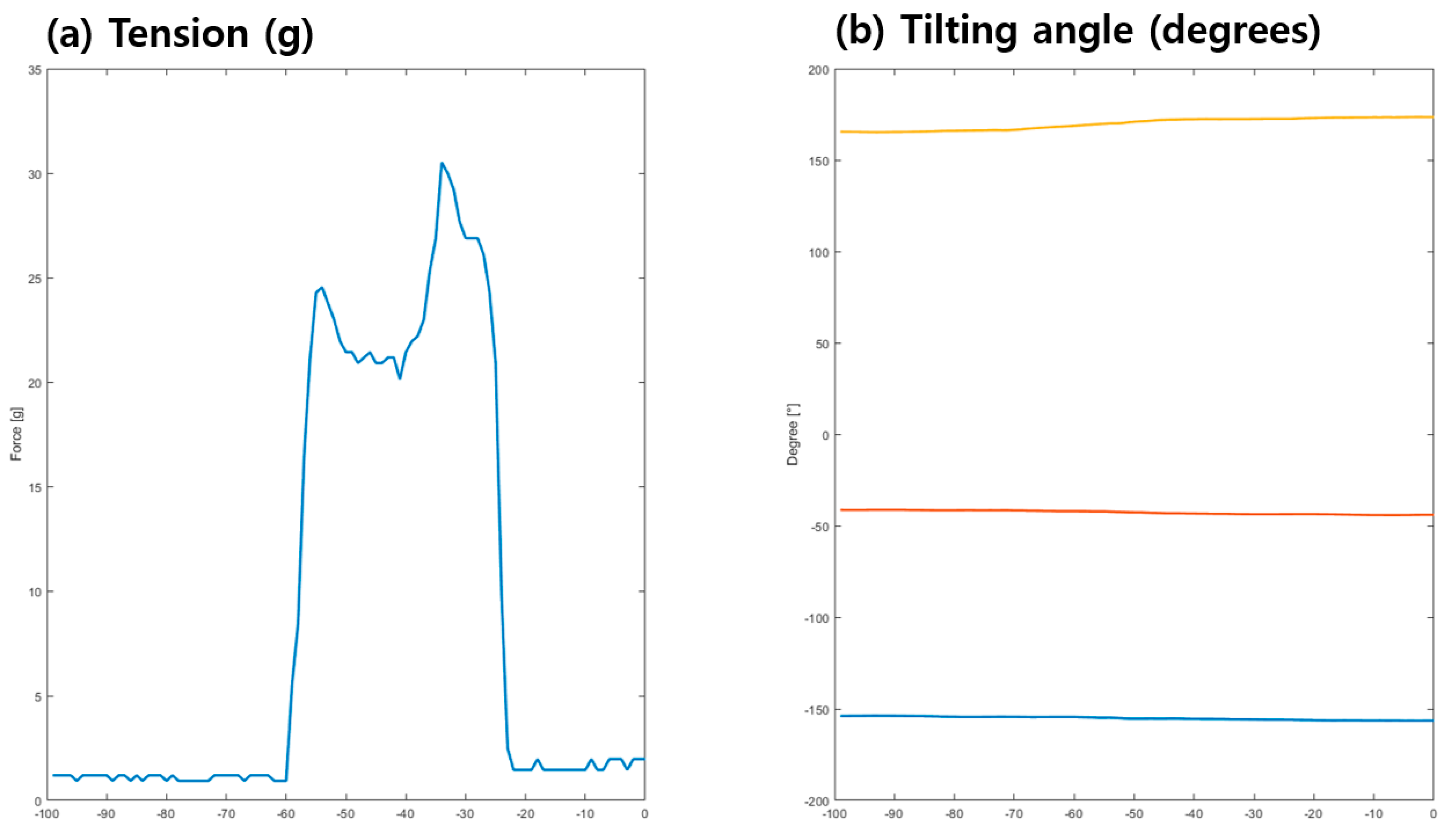
The I/O unit comprised foot and emergency switches for input and a laptop for real-time displaying and data storing of measured signals. Start and termination commands were provided by the foot switch. The entire power of the ATMD could immediately be cut off by pressing the emergency switch in order to avoid electrical injuries. The graphs could display force and three-axes tilting angles as real-time waveforms on the laptop display, as shown in Figure 4. All of the data could also be stored using the commercially available computing software MATLAB R2019b (MathWorks, Natick, MA, USA). A video detailing the device’s clinical application is available as Supplementary Material.

2.2. Structural Analysis and Calibration

Structural analysis was performed on the ATMD to investigate the sensitivity and mechanical stability of the composed components, as shown in Figure 5. The sensing unit was modelled in three dimensions using CAD software Solidworks 2019 SP3.0 (Dassault Systemes, Vélizy-Villacoublay, France) and assembled, as shown in Figure 5a. Model simplification was carried out for efficient analysis and rapid computation: (1) screw threads were replaced as bonded contacts, (2) linear bushing and housing were integrated as single parts, and (3) additional components were excluded, including the cap, connector, bolt, and swab.
Boundary conditions were determined and are displayed in Figure 5b [24,25]. The enclosure parts (e.g., handle and case) were fixed, while the internal parts (e.g., linear motion (LM) guide, housing, shaft, and sensor PCB) were bonded. The shaft and housing were subjected to friction, where the pressure applied to the swab might be transferred to the force sensor. Pressure on the top of the swab and axial force by initializing the bolt were included as external loads, as shown in Figure 5c. Preloading using the initializing bolt could influence the normal friction force, and, thus, the resistance against the force transferred to the sensor could be varied. These were crucial factors for precise measurements using FGT as an ATMD [26,27]. Finally, structural analysis was conducted to evaluate the stress distribution of each component, as shown in Figure 5e.
The stress distribution of the sensing unit was calculated, as shown in Figure 6. Most of the stress was generated in the connecting pin that vertically supported the loading of the shaft. The maximum von Mises stresses of the pin were 2.896 × 10⁶ N/m2, 5.793 × 10⁶ N/m2, 1.448 × 10⁶ N/m2, and 2.897 × 10⁷ N/m2 for axial forces of the initializing bolt of 0.98 N, 1.96 N, 4.9 N, and 9.8 N, respectively, as shown in Figure 6. Aluminum (Al) 6061, which is one of the most common Al alloys in general-purpose use, was employed as the raw material. The yield stress of Al 6061 was 2.757 × 10⁷ N/m2. We assumed that the maximum stress should be at least two-times lower than the yield stress for structural safety, and so an axial force of 4.9 N was determined for the initializing bolt.
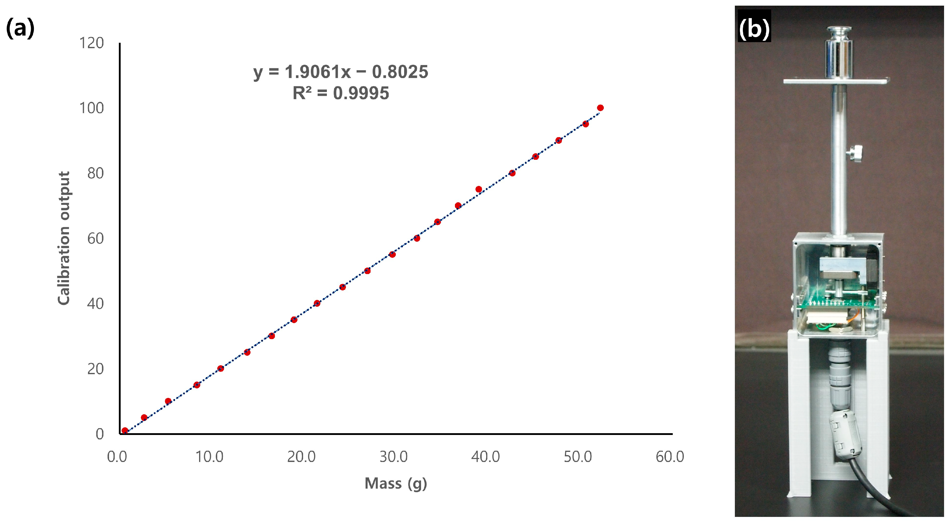
The calibration process was performed for the assembled sensing unit of ATMD, as shown in Figure 7a. A tailored housing was three-dimensionally printed to stand upright beside the sensing unit to precisely measure the counterweight, as shown in Figure 7b. The correlation between the force sensor output and counterweight was determined as
y = 1.9061x − 0.8025
where y is the mass in grams and x is the force sensor output.
The R2 value for Equation (1) was 99.95%, and the Pearson correlation coefficient, calculated using statistical software SigmaXL version 9.1, was 0.9985. These results indicate that the calibrated sensor unit is highly precise for FGT measurements [28,29,30].

3. Results

Three male subjects aged 26.3 ± 2.3 years (mean ± SD; range: 25–29 years) had heights of 169.3 ± 2.1 cm (range: 167–171 cm) and body masses of 72 ± 6.6 kg (range: 65–78 kg). None of the subjects had any limitations in eye movement, and all were confirmed as physical status classification I on the American Society of Anesthesiologists score. The exclusion criteria were the presence of other ocular diseases (e.g., strabismus, nystagmus, ptosis, blepharospasm, or glaucoma), thyroid disorder, muscular or neurological diseases (e.g., Parkinson’s disease or myasthenia gravis), history of ocular trauma, previous strabismus or ocular surgery, or a history of receiving medications known to affect muscle tension (e.g., muscle relaxants).
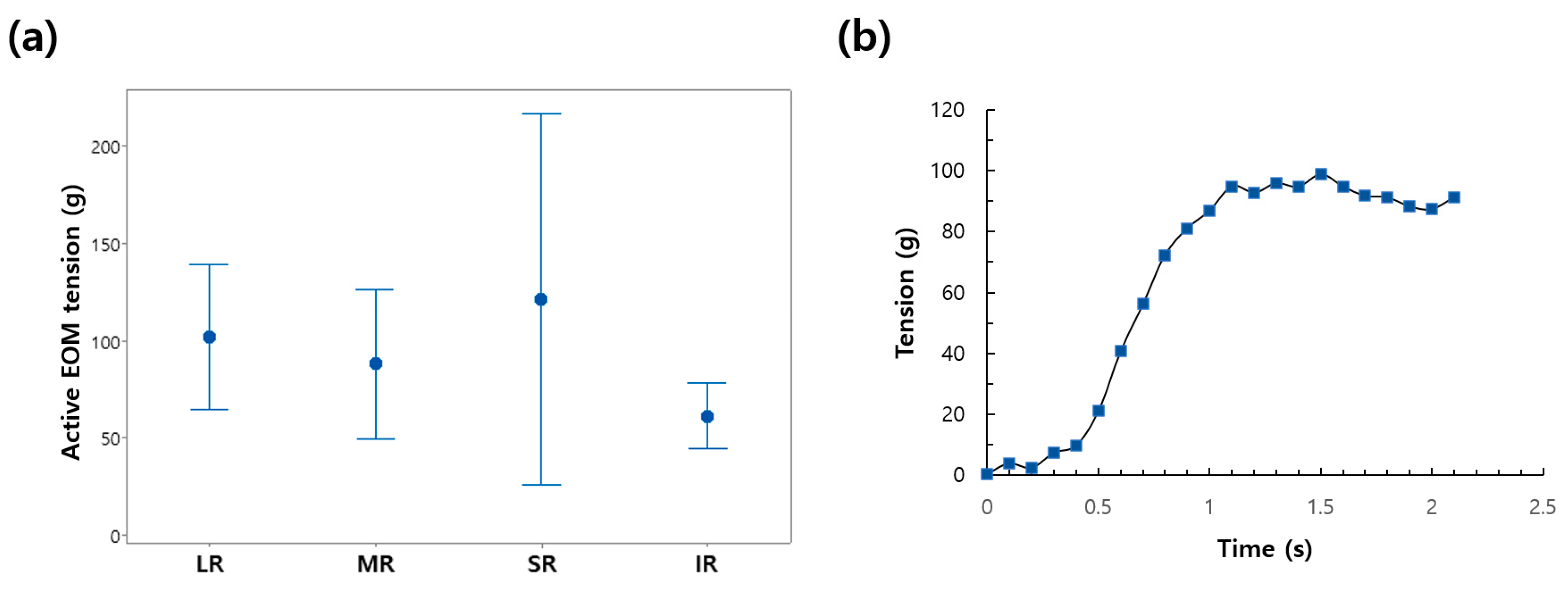
After topical anesthetic instillation, the cotton-tipped swab of the ATMD was placed on the limbus of the eyeball that was looking in the direction of the muscle that would be measured. The subject then tried to move the eyeball toward the direction of the cotton swab. The ATMD could measure active EOM tension while restricting eyeball movement, as shown in Figure 8b. The maximum tension in each rectus muscle was collected.
The active EOM tensions for the four rectus muscles were 101.7 ± 15.0 g, 88.0 ± 15.4 g, 61.3 ± 6.8 g, and 121.3 ± 38.5 g for the LR, medial rectus, inferior rectus, and superior rectus, respectively, as shown in Figure 8a. The substantial variation observed in SR values may be attributed to a unique anatomical feature distinguishing it from other muscles. Unlike other muscles, the SR forms a complex with the levator muscle and shares neural innervation. It is impractical to exclude the possibility that the force of the levator muscle has a concurrent influence when measuring the active force of the SR. This could potentially be a significant contributing factor to the observed variation. In future research endeavors, we plan to conduct prospective analyses using large datasets to address this issue. In addition, EOM tensions in Figure 8a were higher than those measured in previous studies of 45 g to 60 g [16,31]. Those previous studies only measured the EOM tension directly in detached EOMs during saccadic eye movements. The EOM was detached from the globe and attached to a strain gauge system using a silk suture. However, in the current study, we did not detach the EOM and measured the active EOM force under a typical clinical condition. Our measurements, therefore, contained passive elastic tensions that arose from ocular connective tissue, which represents the counteracting elasticity of the orbit that attempts to move the eye back to its central position. Our method is non-invasive and similar to the traditional method of conventional FGT using a cotton-tipped swab, making it easy to perform, thus lowering the barrier of application for clinicians.

4. Conclusions

We developed a compact measuring instrument to evaluate the active EOM tension utilizing the FDT. This instrument expedites objective and quantitative estimations of active EOM tension. The proposed device employs a medical swab, frictionless bushing, and an LM guide to reduce mechanical losses during the force transfer to the sensor, which can enhance the measurement accuracy, as shown in Table 1.
The cotton-tipped medical swab transfers the EOM tension connected to the force sensor as a non-invasive medium. The tilting angle of the swab and force of EOM tension were wirelessly transferred to the laptop for recording and real-time displaying of the measured values. The performance of the proposed instrument was investigated in one eye in each of the three test subjects. The measured values of the tension in all rectus muscles were higher than those of previous studies due to the different measurement methodologies. We believe that the proposed ATMD can be applied to obtain normative measurements of active EOM tension. Future work is necessary to confirm the validity, reliability, reproducibility, and generalizability of the ATMD for possible future use in clinical practice.

Supplementary Materials

The following supporting information can be downloaded at: https://www.mdpi.com/article/10.3390/app132011431/s1.

Author Contributions

Conceptualization, H.J.S.; methodology, H.K.; software, H.K.; validation, H.J.S.; formal analysis, H.K. and A.G.L.; investigation, S.K.; data curation, H.J.S.; writing—original draft preparation, H.K.; writing—review and editing, A.G.L. and H.J.S.; visualization, S.K.; supervision, A.G.L.; project administration, H.J.S.; funding acquisition, H.J.S. All authors have read and agreed to the published version of the manuscript.

Funding

This work was supported by Konkuk University Medical Center Research Grant 2023 (K230102). This sponsor had no role in the design or conduct of this research.

Institutional Review Board Statement

The study was conducted in accordance with the Declaration of Helsinki and approved by the Institutional Review Board of Konkuk University Medical Center (registration number: 2023-03-050-001).

Informed Consent Statement

Informed consent was obtained from all subjects involved in the study.

Data Availability Statement

All relevant data are within the manuscript.

Conflicts of Interest

The authors declare no conflict of interest.

References

  1. Lennerstrand, G. Strabismus and sensory-motor function of eye muscles. J. Integr. Neurosci. 2005, 48, 437–444. [Google Scholar] [CrossRef] [PubMed]
  2. Lennerstrand, G. Strabismus and eye muscle function. Acta Ophthalmol. Scand. 2007, 85, 711–723. [Google Scholar] [CrossRef] [PubMed]
  3. Gauthier, G.M.; Nommay, D.; Vercher, J.-L. The role of ocular muscle proprioception in visual localization of targets. Science 1990, 249, 58–61. [Google Scholar] [CrossRef]
  4. Franzblau, S.C. The diagnosis of amblyopia and strabismus: A programmed approach. J. Am. Optom. Assoc. 1977, 48, 1248–1257. [Google Scholar] [PubMed]
  5. Carlton, J.; Kaltenthaler, E. Health-related quality of life measures (HRQoL) in patients with amblyopia and strabismus: A systematic review. Br. J. Ophthalmol. 2011, 95, 325–330. [Google Scholar] [CrossRef] [PubMed]
  6. Jung, S.H.; Rah, S.H. The Clinical Course of Consecutive Esotropia after Surgical Correction. Korean J. Ophthalmol. 2007, 21, 228–231. [Google Scholar] [CrossRef]
  7. Baik, D.J.; Ha, S.G.; Kim, S.H. Clinical Manifestations of Delayed-onset Consecutive Esotropia after Surgical Correction of Intermittent Exotropia. Korean J. Ophthalmol. 2020, 34, 121–125. [Google Scholar] [CrossRef]
  8. Lekskul, A.; Supakitvilekarn, T.; Padungkiatsagul, T. Outcomes of undercorrection in surgical management and binocular vision gained of adult intermittent exotropia. Clin. Ophthalmol. 2018, 12, 1763–1767. [Google Scholar] [CrossRef]
  9. Kushner, B.J.; Vrabec, M. Theoretical effects of surgery on length tension relationships in extraocular muscles. J. Pediatr. Ophthalmol. Strabismus 1987, 24, 126–131. [Google Scholar] [CrossRef]
  10. Jung, J.H.; Holmes, J.M. Quantitative Intraoperative Torsional Forced Duction Test. Ophthalmology 2015, 122, 1932–1938. [Google Scholar] [CrossRef]
  11. Shin, H.J.; Lee, S.J.; Oh, C.S.; Kang, H. Novel compact device for clinically measuring extraocular muscle (EOM) tension. J. Biomech. 2020, 109, 109955. [Google Scholar] [CrossRef] [PubMed]
  12. Metz, H.S. Forced duction, active force generation, and saccadic velocity tests. Int. Ophthalmol. Clin. 1976, 16, 47–73. [Google Scholar] [CrossRef] [PubMed]
  13. Scott, A.B.; Collins, C.C.; O’Meara, D.M. A Forceps to Measure Strabismus Forces. Arch. Ophthalmol. 1972, 88, 330–333. [Google Scholar] [CrossRef] [PubMed]
  14. Kim, S.; Yang, H.K.; Hwang, J.M. Surgical outcomes of unilateral recession and resection in intermittent exotropia according to forced duction test results. PLoS ONE 2018, 13, e0200741. [Google Scholar] [CrossRef]
  15. Collins, C.C.; Carlson, M.R.; Scott, A.B.; Jampolsky, A. Extraocular muscle forces in normal human subjects. Investig. Ophthalmol. Vis. Sci. 1981, 20, 652–664. [Google Scholar]
  16. Lennerstrand, G.; Schiavi, C.; Tian, S.; Benassi, M.; Campos, E.C. Isometric force measured in human horizontal eye muscles attached to or detached from the globe. Graefes Arch. Clin. Exp. Ophthalmol. 2006, 244, 539–544. [Google Scholar] [CrossRef]
  17. Kang, H.; Lee, S.H.; Shin, H.J.; Lee, A.G. New instrument for quantitative measurements of passive duction forces and its clinical implications. Graefes Arch. Clin. Exp. Ophthalmol. 2020, 258, 2841–2848. [Google Scholar] [CrossRef]
  18. Kang, H.; Lee, S.H.; Oh, C.S.; Shin, H.J.; Lee, A.G. Quantitative measurement of passive duction force tension in intermittent exotropia and its clinical implications. Graefes Arch. Clin. Exp. Ophthalmol. 2021, 259, 1617–1623. [Google Scholar] [CrossRef]
  19. Kang, H.; Shin, H.J.; Lee, A.G. Risk of consecutive esotropia after surgery for intermittent exotropia according to passive duction force. PLoS ONE 2023, 18, e0281392. [Google Scholar] [CrossRef]
  20. Amann, P.; Nicolay, P.; Chatziioannou, V.; Willmann, R. A “digital mouthpiece” for the autonomous analysis and improvement of brass musician performances. Proc. Mtgs. Acoust. 2022, 49, 035022. [Google Scholar] [CrossRef]
  21. Park, J.; Kim, J.; Kim, D.H.; Hwang, J.; Kim, Y.G.; Hyung, S.Y.; Ko, S.-H.; Lee, M. Design of a Soft Wearable Passive Fitness Device for Upper Limb Resistance Exercise. In Proceedings of the 2022 IEEE/RSJ International Conference on Intelligent Robots and Systems (IROS), Kyoto, Japan, 23–27 October 2022; pp. 8488–8493. [Google Scholar] [CrossRef]
  22. Caruso, M.; Miceli, R.; Romano, P.; Schettino, G.; Spataro, C.; Viola, F. A low-cost, real-time monitoring system for PV plants based on ATmega 328P-PU microcontroller. In Proceedings of the 2015 IEEE International Telecommunications Energy Conference (INTELEC), Osaka, Japan, 18–22 October 2015; pp. 1–5. [Google Scholar] [CrossRef]
  23. Mori, G.N.; Swaminarayan, P.R. Measuring IoT Security Issues and Control Home Lighting System by Android Application Using Arduino Uno and HC-05 Bluetooth Module. In Data Science and Intelligent Applications; Kotecha, K., Piuri, V., Shah, H., Patel, R., Eds.; Lecture Notes on Data Engineering and Communications Technologies; Springer: Singapore, 2021; Volume 52, pp. 375–382. [Google Scholar] [CrossRef]
  24. Xia, Z.; Wang, Q.; Liu, Q.; Wang, Y.; Liu, J.; Chen, G. A novel approach for automatic reconstruction of boundary condition in structure analysis. Adv. Eng. Softw. 2016, 96, 38–57. [Google Scholar] [CrossRef]
  25. Beer, G.; Duenser, C. Advanced Boundary Element analysis of geotechnical problems with geological inclusions. Comput. Geotech. 2016, 79, 86–95. [Google Scholar] [CrossRef]
  26. Zhou, N.; He, M. The finite element analysis of a three-story light wood-framed building. In Proceedings of the 2011 International Conference on Business Management and Electronic Information, Guangzhou, China, 13–15 May 2011; pp. 763–767. [Google Scholar] [CrossRef]
  27. Luo, Z.; Yu, L. PSO-based sparse regularization approach for structural damage detection. In Proceedings of the 2017 13th International Conference on Natural Computation, Fuzzy Systems and Knowledge Discovery (ICNC-FSKD), Guilin, China, 29–31 July 2017; pp. 1033–1039. [Google Scholar] [CrossRef]
  28. Flórez, J.A.; Velásquez, A. Calibration of force sensing resistors (fsr) for static and dynamic applications. In Proceedings of the 2010 IEEE ANDESCON, Bogota, Colombia, 15–17 September 2010; pp. 1–6. [Google Scholar] [CrossRef]
  29. Zhou, H.; Zeng, Z. A Fitting Method of the Temperature Characteristic Curve of Sensor. In Proceedings of the 2009 5th International Conference on Wireless Communications, Networking and Mobile Computing, Beijing, China, 24–26 September 2009; pp. 1–4. [Google Scholar] [CrossRef]
  30. Jebli, I.; Belouadha, F.-Z.; Kabbaj, M.I.; Tilioua, A. Prediction of solar energy guided by pearson correlation using machine learning. Energy 2021, 224, 120109. [Google Scholar] [CrossRef]
  31. Lennerstrand, G.; Bolzani, R.; Benassi, M.; Tian, S.; Schiavi, C. Isometric force development in human horizontal eye muscles and pulleys during saccadic eye movements. Acta Ophthalmol. 2009, 87, 837–842. [Google Scholar] [CrossRef] [PubMed]
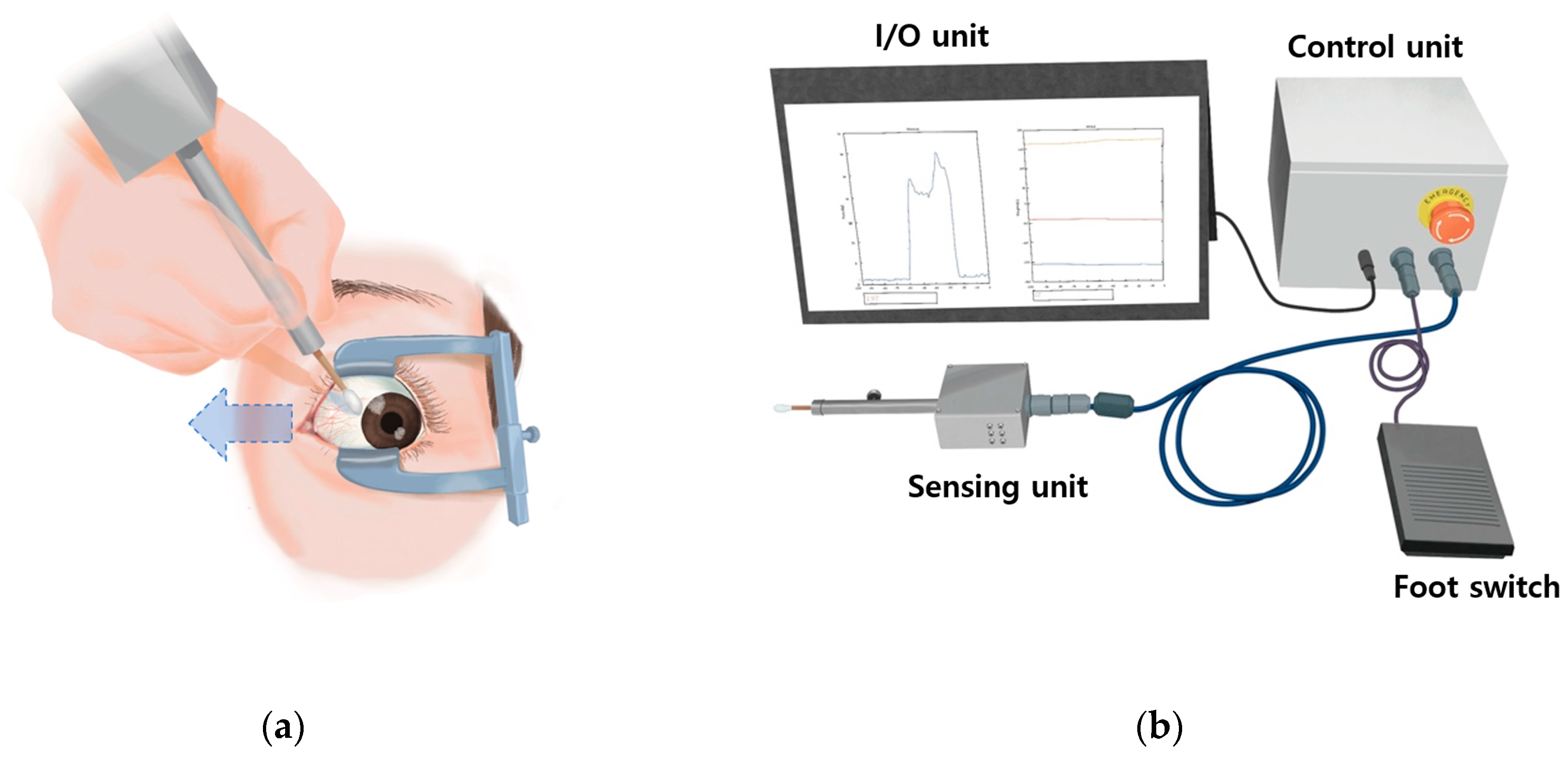
Figure 1. The proposed active extraocular muscle (EOM) tension measuring device (ATMD). (a) Initial position for the force generation test using the ATMD. (b) The ATMD comprised sensing, control, input–output (I/O) units, a display, and a foot switch.
Applsci 13 11431 g001
Figure 2. Details of the sensing unit, which comprised a medical swab, linear bushing, linear motion (LM) guide, pin, initializing bolt, and sensory printed circuit board (PCB).
Applsci 13 11431 g002
Figure 3. Photographs of the PCBs for the control (a) and sensing (b) units.
Applsci 13 11431 g003
Figure 4. Real-time waveforms of the force on the cotton-tipped swab (a) and the tilting angles of the three axes for the ATMD sensing unit: yellow for the x-axis angle, orange for the y-axis angle, and blue for the z-axis angle (b).
Applsci 13 11431 g004
Figure 5. Summary of ATMD structural analysis: (a) three-dimensional (3D) modelling of the sensing unit, (b) simulated boundary conditions, (c) external forces on the top of the shaft and the axial force through the initializing bolt, (d) mesh generation, and (e) stress analysis of assembled components.
Applsci 13 11431 g005
Figure 6. Maximum von Mises stresses measured using the sensing unit as a function of axial forces: (a) 0.98 N, (b) 1.96 N, (c) 4.9 N, and (d) 9.8 N.
Applsci 13 11431 g006
Figure 7. Calibration of ATMD between mass and calculated output (a) and schematic of calibration process (b).
Applsci 13 11431 g007
Figure 8. (a) Summary data of the active EOM tensions for the lateral rectus (LR), medial rectus (MR), superior rectus (SR), and inferior rectus (IR). Data are mean and SD values. (b) Representative active EOM tension records for the MR of the right eye. The other rectus muscles presented similar patterns.
Applsci 13 11431 g008
Table 1. Comparison between conventional method and novel device for measuring active muscle tension.
Conventional Force Generation TestNovel Device in the Current Study
TechniqueActive force felt with the forceps or cotton tipLoad cell (force transducer) used to measure pushing force of cotton tip
Sensing unitHand of examinerElectro-mechanical sensor
AccuracyMay yield less precise results due to subjective factorsOffers higher precision and accuracy in tension measurement
Data collectionRequires manual recording and analysis, potentially introducing human errorAutomated continuous data collection reduces the risk of human error
InterpretationSubjectiveObjective
Inter-examiner variabilityHighly variableLess variable
FeedbackLimited ability to assess muscle active muscle tension dynamicallyEnables dynamic assessment of active muscle tension, aiding in treatment optimization
Disclaimer/Publisher’s Note: The statements, opinions and data contained in all publications are solely those of the individual author(s) and contributor(s) and not of MDPI and/or the editor(s). MDPI and/or the editor(s) disclaim responsibility for any injury to people or property resulting from any ideas, methods, instructions or products referred to in the content.