Article

Establishment of a Periprosthetic Acetabular Bone Defect in an In Vivo Model

by
Frank Sebastian Fröschen
1,*,
Thomas Martin Randau
1,2,
El-Mustapha Haddouti
1,
Frank Alexander Schildberg
1,
Jacques Dominik Müller-Broich
3,
Werner Götz
4,
Susanne Reimann
5,
Dieter Christian Wirtz
1 and
Sascha Gravius
1,6
1
Department of Orthopaedics and Trauma Surgery, University Hospital Bonn, 53127 Bonn, Germany
2
Augustinian Sisters Hospital, Clinic for Orthopedics, Special Orthopedic Surgery and Sports Medicine, 51109 Cologne, Germany
3
Independent Researcher, 53125 Bonn, Germany
4
Department of Oral Medical Technology, School of Dentistry, University of Bonn, 53127 Bonn, Germany
5
Department of Medical Engineering, University of Applied Sciences Bremerhaven, 27568 Bremerhaven, Germany
6
Orthopaedic and Trauma Surgery Centre, University Hospital Mannheim, Medical Faculty Mannheim of the University of Heidelberg, 68167 Mannheim, Germany
*
Author to whom correspondence should be addressed.
Appl. Sci. 2024, 14(8), 3375; https://doi.org/10.3390/app14083375
Submission received: 25 February 2024 / Revised: 11 April 2024 / Accepted: 15 April 2024 / Published: 17 April 2024
(This article belongs to the Special Issue Advances in Orthopedic Diseases Treatment)

Abstract

:
The biological reconstruction of periprosthetic acetabular defects is essential for the success of revision total hip arthroplasty. However, a standardized in vivo defect model with good analogy to the human situation is still lacking, which has significantly limited the research and development of this highly important clinical entity. A defined animal defect model might be a possible solution as it offers the possibility to evaluate different biomaterials for periacetabular bone reconstruction in a reproducible setting. In an ovine periacetabular defect model (n = 27), a defined bone defect (1.5 × 1.5 × 1.5 cm/3.375 cm3) in the cranial load-bearing area of the acetabulum was augmented with two different biomaterials as well as autologous cancellous bone in an ovine periprosthetic defect model and bridged with a Ganz reinforcement ring (n = 9 animals per group). Eight months after implantation, radiological and macroscopic examination was performed. The operation with the establishment of a defined periacetabular defect could be performed in all cases. There were no intraoperative complications in the three groups. During the course of the experiment, three sheep had to be excluded due to complications. A macroscopic evaluation after 8 months showed a firm neocapsula surrounding the hip joint with macroscopic consolidation of the bony defect and a stable inlying implant. There were no detectable differences between the three groups in the macroscopic or radiological evaluation. In summary, the presented ovine model might offer the possibility to create a defined bone defect and investigate bone defect reconstruction with different materials.

1. Introduction

To date, biological defect reconstruction with the downsizing of a bony defect reconstruction is a relevant topic due to the increase in acetabular revision total hip arthroplasty (RTHA). In acetabular RTHA, the right treatment of periacetabular bony defects is a keystone for good long-term outcome. Nevertheless, defect reconstruction might be highly dependent on the used material. The number of studies evaluating the use of cancellous bone or alternative materials—incorporated in the impaction bone grafting technique—in patients with periacetabular defects in RTHA is limited [1,2].
A comparative prospective study evaluating different materials and their material characteristics in vivo is not possible in humans. In this context, an animal model with the best possible analogy to acetabular revision in humans is a possible solution. To date, several established models have been proposed. Unfortunately, these models mainly use dogs, which leads to several disadvantages due to the postoperative load bearing/kennel keeping. Often, the reported results could not be confirmed in humans as dogs are able to avoid postoperative load bearing after all [3,4].
Here, the sheep animal model offers several advantages as sheep are not able to restrain or relieve the operated extremity, while free-range husbandry enables a physiological postoperative weight-bearing pattern in this context. This higher postoperative activity level is relevant to the detection of an implant loosening, while the bony situation with a smooth cortical acetabular surface can be compared very well with the sclerotic periacetabular bone in patients with a need for acetabular RTHA [5,6]. In addition, dogs have a higher bone turnover than humans, while the bone turnover in sheep is similar to that in humans [7].
The aim of this manuscript was to describe an ovine animal model and the surgical method in sheep to create a defined bone defect in the weight-bearing part of the acetabulum as well as to analyze postoperative complications and intraoperative pitfalls.

2. Materials and Methods
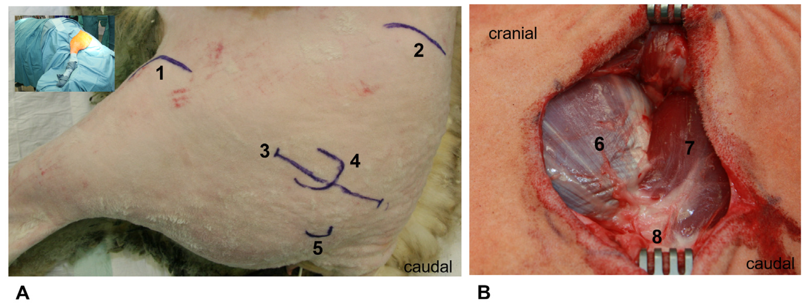
This study was approved by the official state animal care and use committee (State Agency for Nature, Environment and Consumer Protection North Rhine-Westphalia; Düsseldorf, Germany (LANUV NRW), 8.87–50.10.35.08.308). The experiments were performed in accordance with the German federal law regarding the protection of animals, institutional guidelines, and the criteria in “Guide for the Care and Use of Laboratory Animals” (National Institutes of Health publication 8th Edition, 2011).

2.1. Animal Model

For the main experiment, fully grown female, non-pregnant sheep of the Merino breed, with ossified growth plates, were selected (n = 27). At the start of the experiment, the animals were between three and five years old (4.1 ± 0.8 years). The body weight was 90 ± 11 kg.
The animals were provided by the Oberer Hardthof teaching and research facility of the Institute for Animal Breeding and Pet Genetics at the Justus Liebig University in Giessen (Giessen, Germany). Five animals were randomly selected and operated upon one by one in a preliminary experiment to optimize the main experiment (see Table 1). The aim was to clarify operation-specific issues (optimization of the operative access route in the lateral position, selection of suitable surgical tools, choice of the optimal size of the stem prosthesis, positioning and dimensioning of the periprosthetic bone defect, and positioning of the Ganz reinforcement ring and the screw anchoring) as well as the peri- and postoperative management of animal care (surgical preparation, surgical positioning and sterile cover, suitable form of anesthesia, and postoperative positioning and analgesia). These animals were not included in the later evaluation after 8 months. We planned to end the preliminary experiment after one sheep if the perioperative and direct postoperative phases showed feasibility of the surgery and mobilization as planned, with absence of any complication. Unfortunately, a periprosthetic femur fracture at the tip of the stem occurred during the immediate postoperative mobilization of sheep no. 1 without trauma and with a normal implant positioning in the previous postoperative X-ray. As we suspected a non-visible femoral fissure as a possible explanation, we performed the surgery again with the same technique, while taking extensive care of the femur. Unfortunately, the result was the same. As we suspected a mechanical reason directly related to the cementation, a prolongation of the cemented zone was distally performed. This treatment, however, did not show a different result.
Only the cementation of the total femur established a stable situation for postoperative weight bearing.
The sheep (n = 27) were randomly allocated to three groups (Table 1). Two operations were conducted per day on the subsequent fourteen working days. The group allocation was only known to the surgeon and not to any other member involved.

2.2. Livestock Farming

The animals were examined by the animal experiment department and looked after by a veterinarian and an animal care specialist. Fourteen days before the start of the experiment, the sheep were housed in four groups and kept in a closed playpen with straw litter in order to create a stress-free environment. Each sheep was provided with hay and water ad libitum as well as 300–400 g of supplementary feed for sheep and goats (V5103–000 ssniff SF/ZG, ssniff Spezialdiäten GmbH, Soest, Germany) per day. The light–dark rhythm was 12/12 h, the ambient temperature was 20 ± 2 °C, and the relative humidity was 50% ± 10%. Each animal had at least 2 m2 of space available.
Twenty-four hours before the planned operation, the animals were transferred to boxes with no food, but with free access to water. Postoperatively, the animals were kept singly on straw litter for 5 days, afterwards in groups of two animals for further 7 days. The sheep were then moved to external stables and kept indoors in groups of two animals (external laboratories). The external stables held around 700 sheep at the same time. From the 21st postoperative day onward, the sheep were kept on a pasture under the supervision of a shepherd in a large group until the end of the experiment.

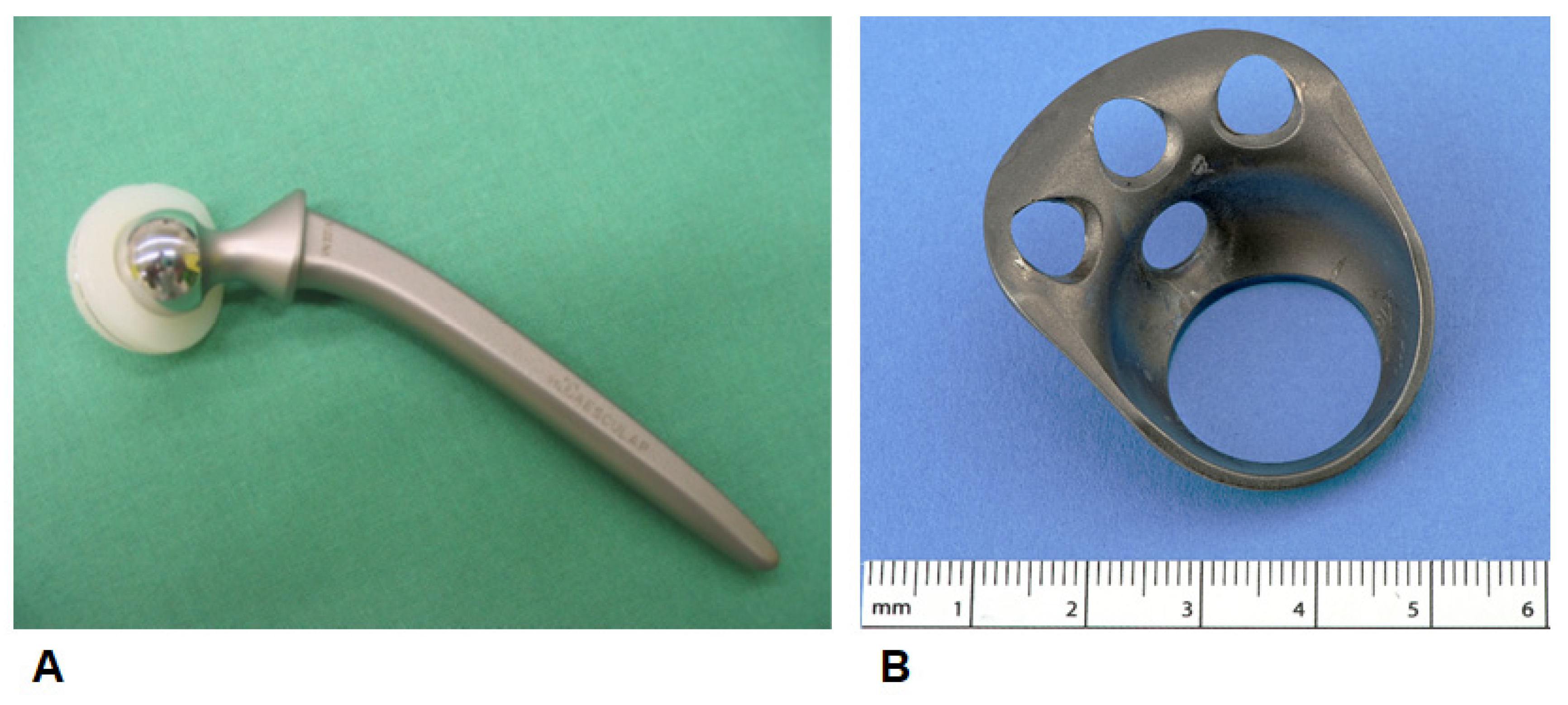
2.3. Implants/Bone Substitutes

We used a Ganz acetabular reinforcement ring made of titanium manufactured according to the ISO standards (Internation Organization for Standardization; Geneva, Switterland; ISO 5832-2/ASTM F67), with a cemented polyethylene cup made with an identical design to the acetabular revision ring (ARR; Fa. Brehm, Weisendorf, Germany), available for acetabular RTHA (outside diameter: 28 mm; inside diameter: 17 mm; 3 holes on acetabular rim; and 1 hole in the cavity) for defect reconstruction. These implants were specially manufactured for this study (Figure 1).
A cemented monobloc implant, made of CoCrMo, prepared for dogs (Aesculap, Tuttlingen, Germany) was used at the femoral side (ISO 5832/V—Head: 17 mm, collum: 21 mm, shaft length: 93 mm, shaft diameter: 9–10 mm, and VN 001K). NanoBone® (group 1), as a representative of the hydroxyapatite-silicate composites, was manufactured by Artoss GmbH (Rostock, Germany). It is produced in a low temperature range (<700 °C) using the sol-gel process [8,9]. The crystal size of the hydroxyapatite contained in NanoBone® corresponds roughly to that found in human bones [10]. The NanoBone® blocks were fitted in the prepared bone beds, while the remaining cavities were filled with granulate (1 × 2 mm) (NanoBone® granulate, Artoss GmbH, Rostock, Germany). In group two, the allogenic bone chips were processed according to the Tutoplast® method (Tutogen Medical GmbH, Neunkirchen am Brand, Germany). In the control group, the autologous sheep cancellous bone was obtained from the previously resected femoral heads of the operated sheep and prepared using a rongeur forceps. In all groups, impaction was performed using hemispherical impactors.

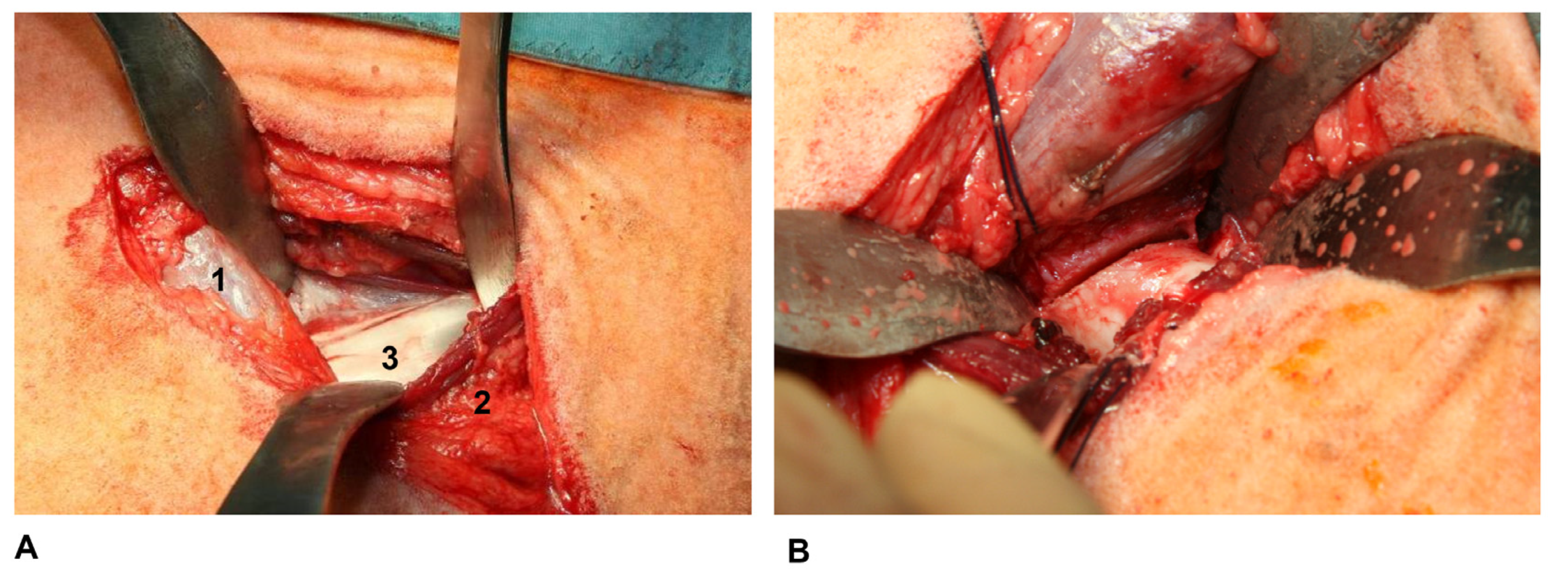
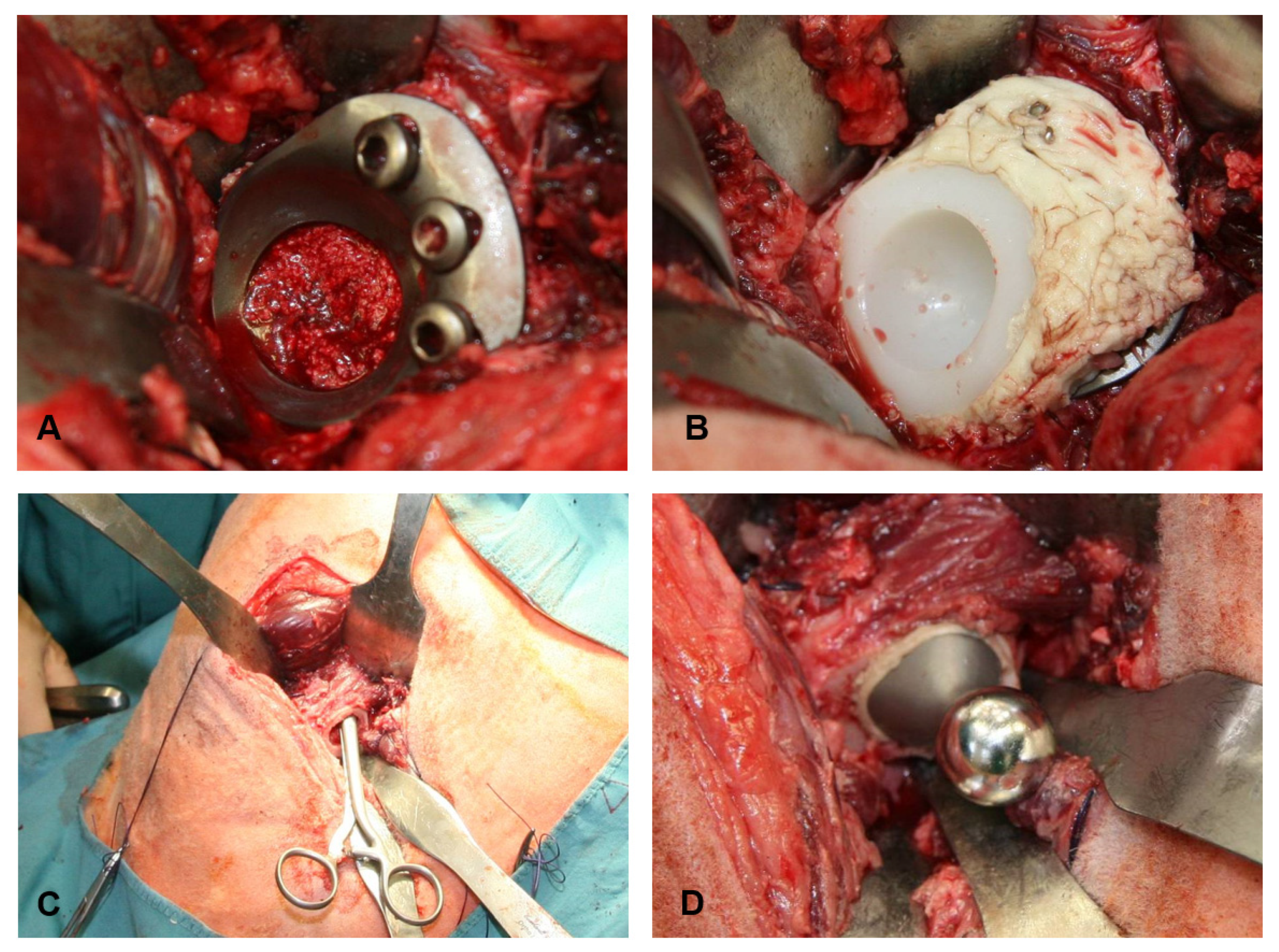
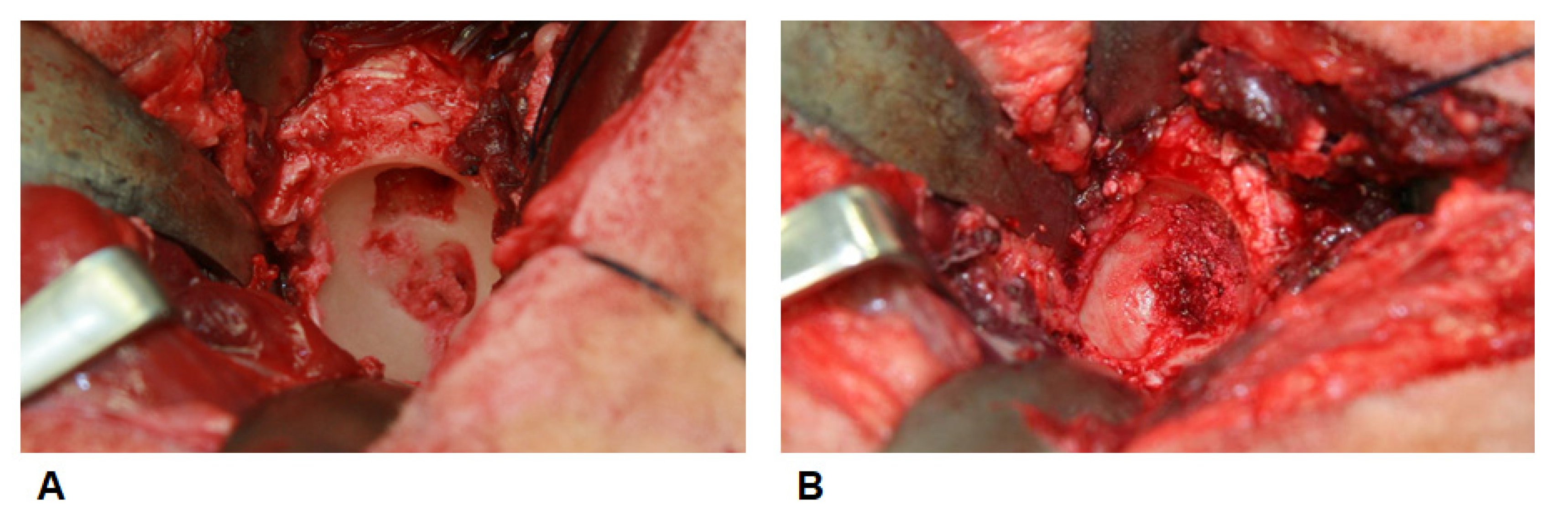
2.4. Anaesthesia and Surgical Technique

The premedication was carried out by an intramuscular injection of 0.1 mg of Ketamin® 10% (Serumwerk Bernburg AG, Bernburg, Germany)/10 kg body weight and 0.1–0.3 mg of xylazine 2% (Serumwerk Bernburg AG, Bernburg, Germany)/kg body weight, followed by an immediate induction of anesthesia with 3–8 mg of pentobarbital/kg body weight (Narcoren®, Merial GmbH, Halbergmoos, Germany). The endotracheal intubation was performed with a laryngoscope (endotracheal tube, Hi-Lo Lanz®, Mallinckrodt medical inc., Athlone, Ireland, diameter 8.5–9.5 mm). The inhalation anesthesia was carried out with isoflurane (Alfa Aesar GmbH & Co., KG, Karlsruhe, Germany) and an oxygen–nitrogen oxide mixture at a ratio of 1:3.
In order to achieve a sufficient depth of anesthesia, isoflurane was initially flooded in at a concentration of 1.5–3.0% by volume; to maintain anesthesia, it was reduced to an average of 0.8–1.0% by volume. For perioperative antibiotic therapy, an intravenous dose of 2.5 mg of Baytril® (Bayer Vital GmbH, Leverkusen, Germany)/kg body weight was administered. Intraoperative analgesia was performed through the intramuscular application of 6 µg of buprenophrine (Merck Healthcare, Darmstadt, Germany)/kg body weight.
The animals were placed in a lateral position on the right side of the body. We always performed surgery of the left hip joint. The lateral surgical approach to the hip joint was performed after taking into account the specific anatomy of the sheep [6,11]. In detail, a 10-cm-long incision centered over the greater trochanter was performed (Figure 2A), followed by a parallel incision of the fascia lata between the tensor fasciae lata muscle and the gluteus medius muscle (Figure 2B).
Afterwards, the gap between those two was used to access and visualize the gluteus profundus muscle (Figure 3A), which was subsequently split in line of its fibers. Two Hohmann retractors were used to visualize the joint capsule. Afterwards, a T-shaped dissection of the ventral capsule was performed, followed by a careful preparation of the acetabulum to gain a good overview of the cup. Here, it is essential to resect the ventral aspect of the joint capsule, including its femoral and acetabular insertion for the best possible overview. After establishing an overview of the situs, osteotomy of the femoral neck (Figure 3B) can be performed with an oscillating saw 1.0–1.5 cm proximal from the lesser trochanter. Two Hohmann retractors can be placed around the femoral neck to protect the surrounding tissue. To visualize the fossa acetabuli, a sharp curette can be used to remove remaining soft tissue or remnants of the ligamentum capitis femoris. For the initial bony preparation of the acetabulum, we used a 28-mm reamer. Reaming was always performed perpendicular to the body’s longitudinal axis. Subsequently, reamers up to 32 mm in diameter were used for the final preparation (Peter Brehm, Weisendorf, Germany). Special attention was paid to retain the subchondral bone lamella.
Afterwards, a defined bone defect, measuring 1.5 × 1.5 × 1.5 cm (~3.375 cm3), was created in the load-bearing cranial area of the acetabular cavity (Figure 4A) using a custom-made drilling template with a depth stop. For this augmentation (Figure 4B, Table 1), the prepared autologous or allogeneic bone chips were impacted in the bone defect step by step using the impaction bone grafting technique analogous to the surgical procedure in humans [12,13,14].
After augmentation of the bony defect, the revision implant was implanted to bridge the defect. A primarily stable situation was achieved with the help of three 6.5 × 25 mm cancellous screws (Peter Brehm, Weisendorf, Germany) at the acetabular rim (Figure 5A). Finally, the inlay was cemented into the acetabular implant according to the anatomical specifications. Until complete curing of the cement (for around 10 min), axial pressure was applied. During this time, access cement had to be removed (Figure 5B).
After the implantation of the cup, the preparation of the femur must be performed. Therefore, the leg was placed in adduction and maximal external rotation. Hohmann retractors were used to elevate the bone above the facia lata. A blunt holding forceps was used to open the femoral medullary cavity (Figure 5C). This is a useful method for the surgeon to palpate the direction in which broaching must be performed. In an ascending series of sizes, rasps are inserted into the femur, up to a diameter of up to 11 mm (Aesculap, Tuttlingen, Germany). The rasps must be inserted until the proximal part of it is flush with the performed femoral osteotomy. Afterwards, cleaning of the bone medullary canal must be performed by jet lavage (Stryker InterPulse® Jet Lavage, Stryker, Duisburg, Germany) before the application of the bone cement (Palacos® R cement, Merck Healthcare, Darmstadt, Germany). Here, it is essential to fill the whole femur with bone cement. We did not use a cement stop. The cementation of, e.g., only the proximal part of the diaphysis might lead to a fracture of the femur at the transition zone without trauma during weight bearing due to the creation of a predetermined fracture zone. After the application of the bone cement, the prothesis can be inserted. During curing and while applying axial pressure on the implant, access cement has to be removed (Figure 5D). After repositioning, the stability of the hip must be verified to exclude instability. Afterward, wound closure can be performed layer by layer. For the closure of the split of the gluteus profundus muscle, the fascia, and the subcutaneous suture, we used absorbable sutures (Vicryl® 0 and 2–0; Johnson & Johnson Medical GmbH, Norderstedt, Germany). Skin closure was performed with non-absorbable sutures (Prolene® 2–0; Johnson & Johnson Medical GmbH, Norderstedt, Germany). At last, sterile compresses were fixated with stitches on the skin followed by the application of a layer of aluminum spray (Aluminium Spray Albrecht, Dechra Veterinary Products Germany GmbH, Aulendorf, Germany).
X-ray imaging was carried out directly postoperatively while the sheep was still in narcosis and after 8 months to evaluate the implant positioning. X-ray imaging was performed with the sheep in a supine position (anterior–posterior plane in 30° abduction with maximal internal rotation and 30° abduction and external rotation).
Postoperatively, the antibiotic and pain therapy were continued up to the fifth day. Therefore, Baytril® (2.5 mg/kg body weight; Bayer Vital GmbH, Leverkusen, Germany) and Rimadyl® (2 mg/kg body weight; Bayer Vital GmbH, Leverkusen, Germany) were applied intramuscularly and subcutaneously, respectively, once per day.

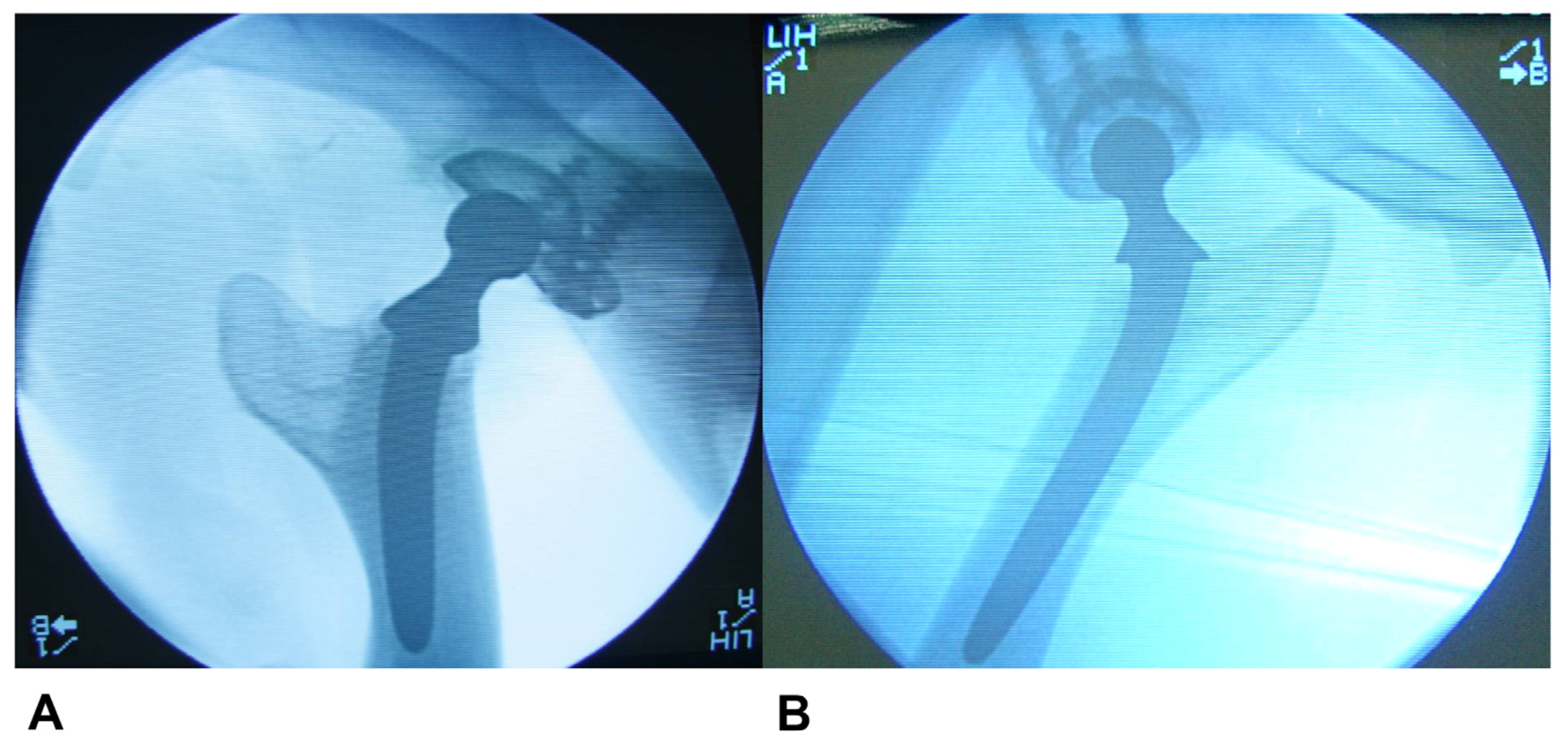
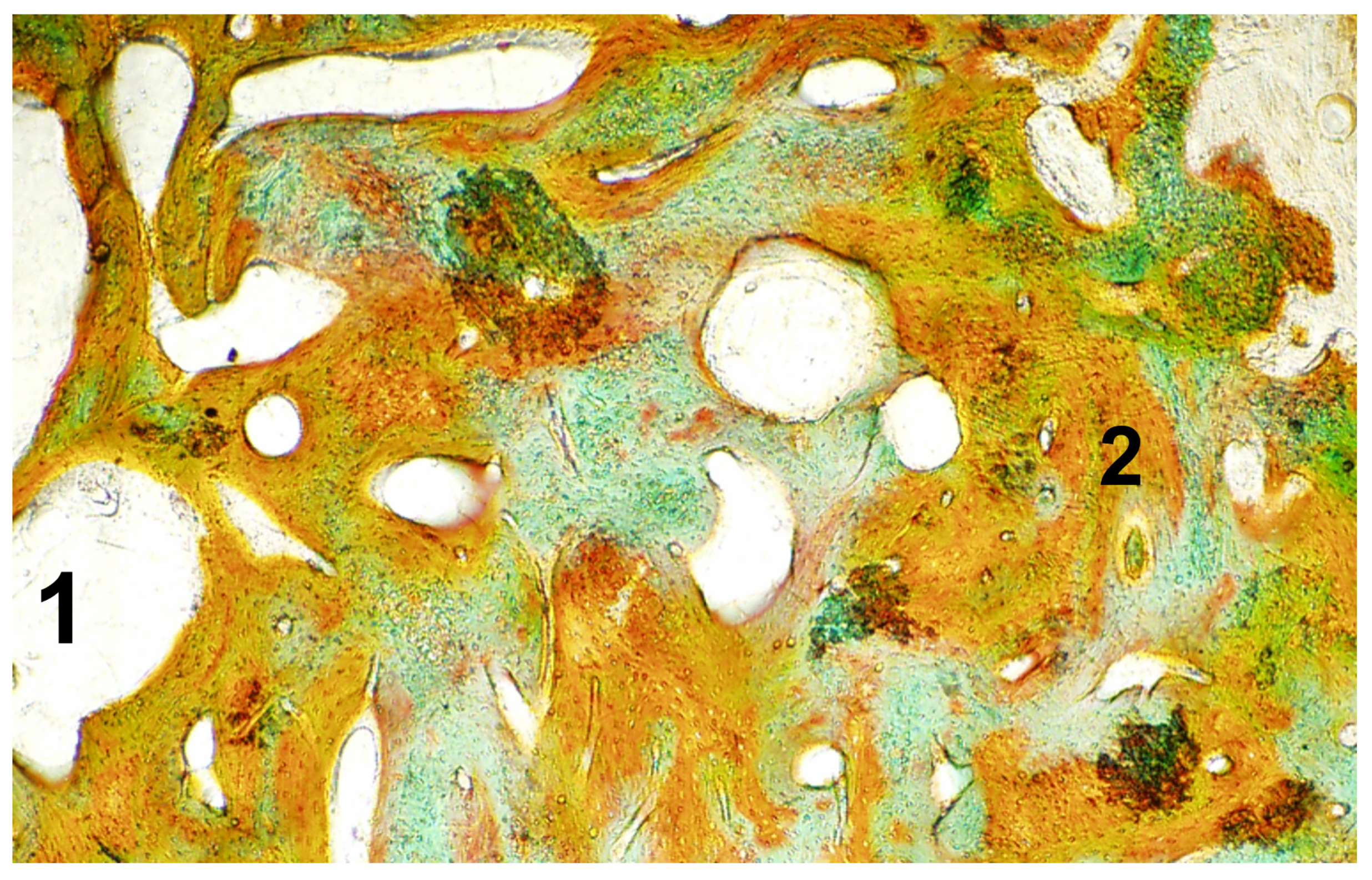
2.5. Sample Retrieval and Radiological, Macroscopic and Microscopic Evaluation

After a standing time of 8 months, euthanasia was carried out immediately after narcotization of the sheep by the rapid application of embutramide (T61, Intervet Deutschland GmbH, Unterschleißheim, Germany) intravenously though a peripheral venous catheter with a dose of 10–15 mL. Afterwards, a renewed radiological evaluation of the operated hip in 2 planes was performed. An evaluation of the X-rays was performed according to the criteria defined by Kavanagh et al. 1985 [15]. For the evaluation of a loosening of the femoral component, all images were evaluated for signs of loosening in the Gruen zones (1–7) [16] for axial migration of the component, change of rotation, and varus or valgus dislocation. In detail, a change of >5° of the longitudinal axis through the femur diaphysis and the longitudinal axis of the femoral stem was counted as a loosening. To access the rotation, the neck of the prosthesis was compared against anatomical landmarks (greater and lesser trochanter). Periarticular ossifications were evaluated according to Brooker et al. (class 1–4), while acetabular radiolucent lines were accessed according to the criteria described by Engh et al. [17,18]. We used the classification according to DeLee et al. to classify periacetabular radiolucent lines [19].
For the macroscopic evaluation, we performed the preparation of the femoral bone and the pelvis. In detail, we performed the resection of all muscles surrounding the hip and the femur, followed by the resection of the ligaments and the joint capsule of the hip before the femur was disarticulated at the level of the knee. We used an oscillating bone saw to cut through the iliac column and both ischial bones at the level of the obturator foramen to explant the hip joint. Afterwards, a purely descriptive macroscopic evaluation was performed. Special attention was paid for signs of a loosening of the femoral stem or the cup, osteolysis, macroscopic visible neoplasia, or inflammatory reactions. For the macroscopic evaluation of the bony consolidation of the reconstructed acetabular bone stock, a small hook was used, and compression was performed to evaluate the stability. An oscillating saw was used to generate 3 × 3 × 5 cm bone samples, which included the augmented defect in its center. We paid special attention to maintaining a safe distance in order to prevent heat damage of the augmented area.
The exclusion criterion of animals of one of the three treatment groups was a standing time of <8 months.
For the microscopic evaluation, the samples were preserved in 4% formaldehyde, washed out, and then dehydrated in an ascending alcohol series (70%, 90%, 96%, and 100% ethanol). Afterwards, the samples were placed in a synthetic material (Technovit 7200 VLC; EXAKT Vertriebs GmbH, Norderstedt, Germany), and polymerization was performed with the EXAKT 520 light polymerization device (EXAKT Vertriebs GmbH, Norderstedt, Germany), with an average total polymerization time of 6 h. For fixation on the microscope slide, Technovit 7230 VLC (EXAKT Vertriebs GmbH, Norderstedt, Germany), in combination with a vacuum adhesive press (EXAKT Vertriebs GmbH, Norderstedt, Germany), was used. To create thin sections, a water-cooled diamond band saw (E 400CS, EXAKT Vertriebs GmbH, Norderstedt, Germany) and a grinding machine (Exakt 4000, EXAKT Vertriebs GmbH, Norderstedt, Germany) were used. The surface of the samples was subsequently polished. After final preparation with 2-Methoxyethyl acetate (3 times for 20 min) and rehydration with a descending alcohol series (100%, 100%, 96%, 80%, and 70% for 2 min each), the samples were rinsed with distilled water. Finally, a Masson–Goldner staining (Carl Roth GmbH and Co., KG, Karlsruhe, Germany) and a toluidine blue staining (Carl Roth GmbH and Co. KG, Karlsruhe, Germany) were performed for the histological evaluation.

2.6. Statistical Analysis

Before beginning the study, a sample size calculation was performed according to Charan et al. using the resource equation method as we lacked any previous findings [20]. Data are presented as means ± standard deviation using Microsoft Excel v.12.0 (Microsoft Corporation, Richmond, CA, USA).

3. Results

Using the abovementioned technique, surgery could be performed in all cases (n = 27). The implantation of the available acetabular and femoral implants was possible in all cases. No operation had to be cancelled intraoperatively due to the inability to implant the femoral or acetabular components. No intraoperative complication occurred. The mean operation time was 178 ± 32 min. There was no difference in operation time between our three groups. X-ray imaging before extubation confirmed the correct positioning of the implant in all cases. There were no signs of a periprosthetic femoral or acetabular fracture. The vital signs were continuously monitored until the first attempt of the sheep to get up. The postoperative analgetic and antibiotic medication could be administered until day 5 after surgery. Except for two sheep, all animals showed a pain-related/protective limping for 3 days, which they suspended spontaneously until day 5 after surgery. Those two animals showed clinical evidence of damage to the sciatic nerve, with a complete regression after 5 and 7 days postoperatively. In no case was it necessary to extend the application of the analgetic and antibiotic medication. The wound dressing, including the suture, was removed on the 17th day after surgery. We could not detect a wound healing disorder in any case. After transferal to the external stables (12th day) and before transferal to the pasture (21st day), a clinical examination was performed by a veterinarian. Only in the absence of any signs of lameness and in the presence of a physiological gait pattern, transferal to the pasture was planned, which could be performed in all cases. We did not perform any kind of further clinical assessment in defined intervals until the end of the experiments, 8 months after surgery.
During the course of the experiment, three postoperative complications (one complication per group; autologous cancellous bone group: periprosthetic fracture of the femur 10 days after surgery; Tutoplast® group: pneumonia 21 days after surgery; and NanoBone® group: cervical abscess with septic circulatory instability 22 days after surgery) occurred before the end of the standing time, which led to the termination of the animal experiment in those cases. This resulted in eight animals per individual group, which were available for test evaluation after a standing time of 8 months.
For the remaining 24 animals, X-ray imaging after surgery and after 8 months did not show any sign of a postoperative heterotopic ossification. We could not detect a postoperative dislocation or migration of the acetabular implant. In no case were radiolucent lines visible in zones 1–3 according to DeLee nor was a migration ≥ 2 mm of the acetabular component detectable.
For the evaluation of the femoral component, we could not detect a secondary varus or valgus dislocation of the femoral stem in comparison to the longitudinal femoral axis; in addition, we could not detect a rotation or axial migration of the femoral component. An osteolysis surrounding the femoral stem or the cement was not visible.

Macroscopic, Microscopic, and Radiological Evaluation

In all cases, the joint was clinically stable without any tendency for (sub)-luxation or a restriction in the passive range of motion. Each of the joints was surrounded by a firm neocapsula, which held the head of the prosthesis firmly and centered in the acetabular component. None of the acetabular components showed macroscopic signs of loosening, with a stable integrated implant in all cases. We did not perform mechanical testing according to a defined mechanical protocol to evaluate loosening of the inlying implant to avoid damaging the newly formed bone stock and allow a subsequent analysis. No obvious signs of neoplasia in the surrounding bone stock could be detected in any of the animals. In all cases, the macroscopic evaluation displayed a vital surrounding bone stock and soft tissue. Signs of local necrosis could not be detected. In no case was a periacetabular ossification or an osteolysis of the surrounding bone stock detectable macroscopically or radiologically. A total macroscopic consolidation of the former bony defect was evident in all cases. During the macroscopic evaluation, we could not detect differences between the three groups. The compression of the newly formed bone stock was not possible. A radiological demarcation of the artificial defect was not possible 8 months after surgery (Figure 6). There was no evidence of a periprosthetic joint infection of the soft tissue or the bone stock. We could not detect an implant failure in any case.
The microscopic evaluation showed a uniform image of cell-rich periprosthetic connective tissue. Round cell infiltrates from cells of the monocytic phagocyte system, especially, macrophages and multinucleated giant cells of the foreign body type, could not be detected, and a chronic inflammatory granulation tissue could not be detected either. Connective tissue between the bone and the implant could not be observed. The microscopic evaluation of the augmented defect showed a complete bone building throughout the defect in the entire defect zone (Figure 7). At the border zone, a near-complete bone remodeling was visible.

4. Discussion

Our most important finding was that the ovine periacetabular defect model is a promising alternative to evaluate bony periacetabular defect reconstruction.
To evaluate the clinical suitability of a bone substitute material, animal studies on bone defect models are of crucial importance. In order to select the appropriate defect model, the choice of species, the age of the animals used, and the size and location of the bone defects should be considered. Furthermore, the macro- and micro-architecture, the composition, and the remodeling properties of the bone of the species must be taken into account [7,21,22].
The most frequently established experimental animals in the field of hip arthroplasty are dogs, followed by animal models of sheep, pigs, rabbits, or rats [22,23]. Nevertheless, large animal models should be preferred for better comparability of the surgical technique and the postoperative care. Therefore, sheep and dog models should be chosen [7,23,24]. Previous studies could already outline—beside improved economic efficiency in comparison to the dog model—the suitability of the sheep model as animal model for in vivo studies [24,25,26,27]. Nunamaker et al., as well as consecutive studies, could already prove its advantages with a bone remodeling rate comparable to the human species [25,27,28,29]. Despite structural differences between the ovine and human acetabulum, with a flatter shape and an oval femoral head, the anatomical and physiological properties of the sheep hip imitate the situation in humans well in the case of acetabular RTHA [5,6,24]. The main reason for this is the thick subchondral sclerotic bone lamella so that after the rimming of the cup, the defect is still contained by sclerotic bone, comparable to acetabular RTHA in humans [5]. Beside this advantage of sheep over dog or pig animal models, a comparable postoperative mobilization—to avoid results that are too good due to a missing postoperative weight bearing—is essential for the analysis of the results. Here, this model benefits from the lacking possibility of the sheep, in comparison to, e.g., dogs, to avoid loading the operated extremity, with consecutive similar loading on all four legs. Furthermore, a free-range husbandry under the supervision of a shepherd where the sheep exercise a physiological load pattern with a body mass that is higher compared to dogs, yet comparable to humans, is an appropriate imitation of physical stress on the joint in humans [21,27,30]. In particular, this free-range husbandry is an advantage of the sheep animal model, while dog models often use a postoperative kennel keeping. Nevertheless, we surely have to admit that immediate postoperative full weight bearing does not fully correspond to the situation in humans as postoperative mobilization after acetabular RTHA often includes partial weight bearing for 6 weeks, followed by a period of 2–4 weeks, in which the load is slowly increased until full weight bearing is reached. On the other hand, direct postoperative full weight bearing is still one of the aims of acetabular RTHA as some groups of patients (e.g., older patients or patients with neurological disorders) are not fully capable of following the prescribed aftercare. In summary, after immediate full weight bearing, a loosening or implant failure might occur earlier and within a short period of time, which is an additional advantage of the ovine model in comparison to the dog model [30]. To avoid disproportionately good results due to the higher osteogenic potential of young animals, we only included adult female sheep [21].
To avoid a spontaneous bone healing, we used the definitions and results for “critical size defect“ of previous studies for sheep [31,32,33,34]. With an average defect size of 3.375 cm3, all defects fulfilled the given definitions and can be graded as “critical size defects“. These sizes prevent self-healing and create a comparable situation to the one found in humans, with need for revision total hip arthroplasty. Here, the main problem is an extensive bone loss in the load-bearing area of the acetabulum with need for reconstruction. Due to ethical concerns, we were not able to establish a fourth group (n = 8; arthroplasty without any kind of defect reconstruction). Nevertheless, we have to admit that pelvic bone defects have a higher regenerative capacity caused by the mechanical stress in comparison to cranial bone defects [35].
Some authors recommend preoperative induction of an osteoarthritis for a more reality-based experimental setup [26,27]. As our study was designed to create a setting in which defect reconstruction is possible with the use of different materials, we did not chemically induce an osteoarthritis. To date, the significance of possible cross-reactions caused by chemical drug-induced osteoarthritis, aseptic periarticular inflammatory reactions, necrotic bone, and materials for bone defect reconstruction are not clear. In addition, inflammatory periprosthetic bone and soft tissue reactions in the present animal model are clearly caused by the interaction of the substitute materials with the surrounding bone and soft tissue. In this context, we have to admit that our primary aim was not the detailed comparison of different materials but to create a setting, which allows evaluation in subsequent experiments.
Our study has limitations, the most relevant being the limited number of animals at the time of evaluation and the duration/standing time until the evaluation of the bone defect. As previous studies already outlined, economical aspects always play a role in animal models. As this study was planned to evaluate a new model, only a limited number of animals were operated, and a limited period for postoperative evaluation was chosen. Further studies might be helpful to provide long-term results.
Another point that must be discussed is the necessary surgical technique for the implantation of the femoral component with the need for cementation of the whole femur to avoid a periprosthetic femur fracture at the tip of the stem. Here, preoperatively, we found no data in the literature that urge the surgeon to perform the cementation of the whole femur. As the fractures occurred at the tip of the stem/end of the cement, we suspect a mechanical problem. Additional studies with the sole purpose of evaluating the different mechanical probabilities might be helpful. In this context, we would like to outline that a limitation of this study is the low number of animals per group so that the postoperative exclusion of three animal led to an overall reduction of 10%. We tried to evaluate our three excluded sheep, and we think that the reason for the femoral fracture was the result of a spontaneous aggressive behavior of the other sheep while being in a group of two. The most likely explanation for the cervical abscess with septic circulatory instability 22 days after surgery of another sheep was a bite. In contrast, we are not able to determine the genesis of pneumonia three weeks after surgery.
In summary, with the presented setup and surgical technique, we were able to create a standardized acetabular bone defect model and perform augmentation of the defect and implantation of a revision cup. This in vivo model might thus be a possible foundation to evaluate different methods for biological or augmentation-based defect reconstruction and gain new insights for further treatment strategies.

Author Contributions

Conceptualization, S.G., D.C.W. and W.G.; methodology, S.G., J.D.M.-B., T.M.R., F.S.F. and F.A.S.; software, E.-M.H.; validation, S.G., T.M.R., E.-M.H. and F.S.F.; formal analysis, S.R., J.D.M.-B., D.C.W. and S.G.; investigation, S.G.; resources, F.S.F., D.C.W., W.G., S.G. and S.R.; data curation, S.R., J.D.M.-B. and S.G.; writing—original draft preparation, F.S.F. and S.G.; writing—review and editing, S.G., F.A.S. and F.S.F.; visualization, S.G. and F.S.F.; supervision, S.G.; project administration, F.S.F., F.A.S., W.G., D.C.W. and S.G. All authors have read and agreed to the published version of the manuscript.

Funding

The used implants were provided for free by Aesculap (Tuttlingen, Germany) and Brehm (Weisendorf, Germany). Materials for bone reconstruction were provided for free by Artoss GmbH (Rostock, Germany) and Tutogen Medical GmbH (Neunkirchen am Brand, Germany).

Institutional Review Board Statement

The animal study protocol was approved by the official state animal care and use committee (LANUV NRW, 8.87–50.10.35.08.308).

Informed Consent Statement

Not applicable.

Data Availability Statement

The original contributions presented in the study are included in the article, further inquiries can be directed to the corresponding author.

Conflicts of Interest

The authors declare no conflicts of interest.

References

  1. Oonishi, H.; Iwaki, Y.; Kin, N.; Kushitani, S.; Murata, N.; Wakitani, S.; Imoto, K. Hydroxyapatite in Revision of Total Hip Replacements with Massive Acetabular Defects: 4- to 10-Year Clinical Results. J. Bone Joint Surg. Br. 1997, 79, 87–92. [Google Scholar] [CrossRef]
  2. Tanaka, C.; Shikata, J.; Ikenaga, M.; Takahashi, M. Acetabular Reconstruction Using a Kerboull-Type Acetabular Reinforcement Device and Hydroxyapatite Granules. J. Arthroplasty 2003, 18, 719–725. [Google Scholar] [CrossRef]
  3. Schulz, K.S. Application of Arthroplasty Principles to Canine Cemented Total Hip Replacement. Vet. Surg. 2000, 29, 578–593. [Google Scholar] [CrossRef]
  4. Hummel, D. Zurich Cementless Total Hip Replacement. Vet. Clin. North Am. Small Anim. Pract. 2017, 47, 917–934. [Google Scholar] [CrossRef]
  5. Lelgemann, B.V. Optimierung der Verbundfestigkeit Zwischen Knochenzement und Azetabulärem Knochen bei Künstlichem Hüftgelenkersatz; Aachener Beiträge zur Medizin: Aachen, Germany, 2006; ISBN 3-86130-696-4. [Google Scholar]
  6. Mumme, T. Verbesserung der Verbundfestigkeit Zwischen Hydrophobem Knochenzement und Hydrophilem Knochen Durch Einen Amphiphilen Knochenhaftvermittler—Am Beispiel der Zementierten Hüftgelenksendoprothetik; Wissenschaftsverlag Mainz: Aachen, Germany, 2007; Volume 46. [Google Scholar]
  7. Gabriele Sommer, N.; Hahn, D.; Okutan, B.; Marek, R.; Weinberg, A.-M. Animal Models in Orthopedic Research: The Proper Animal Model to Answer Fundamental Questions on Bone Healing Depending on Pathology and Implant Material. In Animal Models in Medicine and Biology; Tvrdá, E., Chandra Yenisetti, S., Eds.; IntechOpen: Rijeka, Croatia, 2020; ISBN 978-1-83880-011-6. [Google Scholar]
  8. Gerber, T.; Traykova, T. Development and In Vivo Test of Sol-Gel Derived Bone Grafting Materials. J. Sol-Gel Sci. Technol. 2003, 26, 1173–1178. [Google Scholar] [CrossRef]
  9. Traykova, T.; Bötcher, R.; Neumann, H.G.; Henkel, K.-O.; Bienengräber, V.; Gerber, T. Silica/Calcium Phosphate Sol-Gel Derived Bone Grafting Material—From Animal Tests to First Clinical Experience. Key Eng. Mater. 2003, 254–256, 679–682. [Google Scholar] [CrossRef]
  10. Henkel, K.-O.; Lenz, J.-H.; Gerber, T.; Bienengräber, V. Ein qualitativ neuartiges Knochenaufbaumaterial auf Hydroxylapatit-Xerogel-Basis. ZWR Dtsch. Zahnärztebl. 2005, 114, 416–418. [Google Scholar] [CrossRef]
  11. Müller-Rath, R.; Wirtz, D.C.; Siebert, C.H.; Andereya, S.; Gravius, S.; Hermanns-Sachweh, B.; Marx, R.; Mumme, T. Amphiphilic Bonder Improves Adhesion at the Acrylic Bone Cement–Bone Interface of Cemented Acetabular Components in Total Hip Arthroplasty: In Vivo Tests in an Ovine Model. Arch. Orthop. Trauma Surg. 2008, 128, 701–707. [Google Scholar] [CrossRef]
  12. Schreurs, B.W.; Slooff, T.J.; Buma, P.; Verdonschot, N. Basic Science of Bone Impaction Grafting. Instr. Course Lect. 2001, 50, 211–220. [Google Scholar] [PubMed]
  13. Walter, S.G.; Randau, T.M.; Gravius, N.; Gravius, S.; Fröschen, F.S. Monoflanged Custom-Made Acetabular Components Promote Biomechanical Restoration of Severe Acetabular Bone Defects by Metallic Defect Reconstruction. J. Arthroplasty 2019, 35, 831–835. [Google Scholar] [CrossRef] [PubMed]
  14. Fröschen, F.S.; Randau, T.M.; Hischebeth, G.T.R.; Gravius, N.; Gravius, S.; Walter, S.G. Mid-Term Results after Revision Total Hip Arthroplasty with Custom-Made Acetabular Implants in Patients with Paprosky III Acetabular Bone Loss. Arch. Orthop. Trauma Surg. 2020, 140, 263–273. [Google Scholar] [CrossRef]
  15. Kavanagh, B.F.; Ilstrup, D.M.; Fitzgerald, R.H. Revision Total Hip Arthroplasty. J. Bone Joint Surg. Am. 1985, 67, 517–526. [Google Scholar] [CrossRef] [PubMed]
  16. Gruen, T.A.; McNeice, G.M.; Amstutz, H.C. “Modes of Failure” of Cemented Stem-Type Femoral Components: A Radiographic Analysis of Loosening. Clin. Orthop. 1979, 141, 17–27. [Google Scholar] [CrossRef]
  17. Brooker, A.F.; Bowerman, J.W.; Robinson, R.A.; Riley, L.H. Ectopic Ossification Following Total Hip Replacement. Incidence and a Method of Classification. J. Bone Joint Surg. Am. 1973, 55, 1629–1632. [Google Scholar] [CrossRef]
  18. Engh, C.; Griffin, W.; Marx, C. Cementless Acetabular Components. J. Bone Joint Surg. Br. 1990, 72, 53–59. [Google Scholar] [CrossRef]
  19. DeLee, J.G.; Charnley, J. Radiological Demarcation of Cemented Sockets in Total Hip Replacement. Clin. Orthop. 1976, 121, 20–32. [Google Scholar] [CrossRef]
  20. Charan, J.; Kantharia, N. How to Calculate Sample Size in Animal Studies? J. Pharmacol. Pharmacother. 2013, 4, 303–306. [Google Scholar] [CrossRef]
  21. Pearce, A.; Richards, R.; Milz, S.; Schneider, E.; Pearce, S. Animal Models for Implant Biomaterial Research in Bone: A Review. Eur. Cell. Mater. 2007, 13, 1–10. [Google Scholar] [CrossRef]
  22. Martini, L.; Fini, M.; Giavaresi, G.; Giardino, R. Sheep Model in Orthopedic Research: A Literature Review. Comp. Med. 2001, 51, 292–299. [Google Scholar]
  23. Animal Models in Orthopaedic Research; An, Y.H.; Friedman, R.J. (Eds.) CRC Press: Boca Raton, FL, USA, 1999; ISBN 978-0-8493-2115-3. [Google Scholar]
  24. Phillips, T.W.; Johnston, G.; Wood, P. Selection of an Animal Model for Resurfacing Hip Arthroplasty. J. Arthroplasty 1987, 2, 111–117. [Google Scholar] [CrossRef]
  25. David, A.; Eitenmüller, J.; Muhr, G.; Pommer, A.; Bär, H.F.; Ostermann, P.A.W.; Schildhauer, T.A. Mechanical and Histological Evaluation of Hydroxyapatite-Coated, Titanium-Coated and Grit-Blasted Surfaces under Weight-Bearing Conditions. Arch. Orthop. Trauma Surg. 1995, 114, 112–118. [Google Scholar] [CrossRef]
  26. Phillips, T.W.; Gurr, K.R.; Rao, D.R. Hip Implant Evaluation in an Arthritic Animal Model. Arch. Orthop. Trauma Surg. 1990, 109, 194–196. [Google Scholar] [CrossRef]
  27. Phillips, T.W.; Gurr, K. A Preconditioned Arthritic Hip Model. J. Arthroplasty 1989, 4, 193–200. [Google Scholar] [CrossRef]
  28. Nunamaker, D.M. Experimental Models of Fracture Repair. Clin. Orthop. 1998, 355, S56–S65. [Google Scholar] [CrossRef]
  29. Turner, A.S.; Alvis, M.; Myers, W.; Stevens, M.L.; Lundy, M.W. Changes in Bone Mineral Density and Bone-Specific Alkaline Phosphatase in Ovariectomized Ewes. Bone 1995, 17, S395–S402. [Google Scholar] [CrossRef]
  30. Bergmann, G.; Graichen, F.; Rohlmann, A. Hip Joint Forces in Sheep. J. Biomech. 1999, 32, 769–777. [Google Scholar] [CrossRef]
  31. Gugala, Z.; Gogolewski, S. Regeneration of Segmental Diaphyseal Defects in Sheep Tibiae Using Resorbable Polymeric Membranes: A Preliminary Study. J. Orthop. Trauma 1999, 13, 187–195. [Google Scholar] [CrossRef]
  32. Heckman, J.D.; Boyan, B.D.; Aufdemorte, T.B.; Abbott, J.T. The Use of Bone Morphogenetic Protein in the Treatment of Non-Union in a Canine Model. J. Bone Joint Surg. Am. 1991, 73, 750–764. [Google Scholar] [CrossRef]
  33. den Boer, F.C.; Patka, P.; Bakker, F.C.; Wippermann, B.W.; van Lingen, A.; Vink, G.Q.M.; Boshuizen, K.; Haarman, H.J.T.M. New Segmental Long Bone Defect Model in Sheep: Quantitative Analysis of Healing with Dual Energy X-ray Absorptiometry. J. Orthop. Res. 1999, 17, 654–660. [Google Scholar] [CrossRef]
  34. Aaron, J.E.; Skerry, T.M. Intramembranous Trabecular Generation in Normal Bone. Bone Miner. 1994, 25, 211–230. [Google Scholar] [CrossRef]
  35. Nickel, R.; Schummer, A.; Seiferle, E.; Frewein, J.; Nickel, R. Bewegungsapparat; Lehrbuch der Anatomie der Haustiere; Parey [u.a.]: Berlin, Germany, 1992; ISBN 978-3-489-58016-4. [Google Scholar]
Figure 1. Display of the used implants: cemented monobloc implant made of CoCrMo (Aesculap, Tuttlingen, Germany) (A) and acetabular reinforcement ring out of pure titanium (ARR; Fa. Brehm, Weisendorf, Germany) (B).
Applsci 14 03375 g001
Figure 2. Lateral approach to the hip. Skin incision centered above the greater trochanter (A), followed by subcutaneous preparation to visualize the muscle interval (B) between no. 6 and 7 ((1) knee joint; (2) crista iliaca; (3) 10-cm-long skin incision; (4) great trochanter; (5) tuber ischiadicum; (6) m. tensor fascia latae; (7) m. gluteus medius; and (8) m. gluteobiceps).
Applsci 14 03375 g002
Figure 3. Lateral approach to the hip joint II. Visualization of m. gluteus profundus (A) and osteotomy of the femur (B) ((1) m. gluteus medius; (2) m. tensor fascia latae; and (3) m. gluteus profundus).
Applsci 14 03375 g003
Figure 4. Defined bone defect, measuring 1.5 × 1.5 × 1.5 cm (~ 3.375 cm3) in the load-bearing area of the acetabular cavity before (A) and after (B) augmentation.
Applsci 14 03375 g004
Figure 5. Implantation of the revision cup (A) and inlay (B), followed by the preparation of the femur (C) and implantation of the femoral component (D).
Applsci 14 03375 g005
Figure 6. Exemplary X-ray in 2 planes at follow-up after 8 months (A,B): no lysis margins around the acetabular roof shell (DeLee zones 1–3) and the femoral shaft (green zones 1–7); no secondary migration/rotation of the components; and no periarticular ossifications.
Applsci 14 03375 g006
Figure 7. Exemplary Masson–Goldner staining (original magnification: 4×) of the border zone of the augmented defect (control group). A near-complete remodeling with the reconstruction of the trabecular bone structure is visible at the border of the defect ((1) left part of the image). In the central defect area, dense lamellar bone is visible, with newly formed bone at different stages of mineralization ((2) right part of the image). No residual bone chips can be found.
Applsci 14 03375 g007
Table 1. Study design with the definition of the control as well as the study groups 1 and 2.
Number of Animals [n]Groups
5Preliminary experiment to optimize the main experiment
9Control group: Impaction bone grafting with autologous sheep cancellous bone
9Group 1: NanoBone® [Artoss GmbH, Rostock, Germany]
9Group 2: Ovine Tutoplast®-processed cancellous bone chips [Tutogen® Medical GmbH, Neunkirchen am Brand, Germany]
Disclaimer/Publisher’s Note: The statements, opinions and data contained in all publications are solely those of the individual author(s) and contributor(s) and not of MDPI and/or the editor(s). MDPI and/or the editor(s) disclaim responsibility for any injury to people or property resulting from any ideas, methods, instructions or products referred to in the content.

