Article

High-Pressure Processing of Reduced Salt Pangasius Catfish (Pangasianodon hypophthalmus) Minced Muscle: The Effects on Selected Quality Properties of Its Gels

1
Faculty of Fisheries, Nong Lam University, Ho Chi Minh City 720100, Vietnam
2
School of Agriculture, Biomedicine and Environment, La Trobe Institute for Sustainable Agriculture & Food (LISAF), La Trobe University, Bundoora, VIC 3086, Australia
3
Faculty of Chemical and Food Technology, HCMC University of Technology and Education, Ho Chi Minh City 720100, Vietnam
*
Author to whom correspondence should be addressed.
Appl. Sci. 2025, 15(9), 4727; https://doi.org/10.3390/app15094727 (registering DOI)
Submission received: 26 March 2025 / Revised: 18 April 2025 / Accepted: 22 April 2025 / Published: 24 April 2025
(This article belongs to the Special Issue Innovative Technologies for Food Preservation and Processing)

Abstract

:

Featured Application

High-pressure processing (HPP) is a promising technology for developing healthier fish products with reduced sodium levels and improved or similar gel quality compared to conventional heat-induced products.

Abstract

This study investigated the effects of high-pressure processing (HPP) on selected physicochemical properties of pangasius gels at reduced salt concentrations. Minced pangasius muscle was treated at different HPP conditions (300–500 MPa/10 °C/10 min) with 1% or 2% added salt, then cooking (90 °C/30 min) and compared to heat-induced gels (HIGs) with 2% added salt and cooking (90 °C/30 min). The results showed that HPP significantly improved the texture, whiteness, and water-holding capacity of gels added salt 2% as compared to HIGs. At a reduced salt concentration of 1%, the gel texture, whiteness, and water-holding capacity of pangasius gels are similar to HIGs. SDS-PAGE showed a reduction in myosin band intensity of both SDS-soluble and sarcoplasmic proteins with pressure treatment. Sarcoplasmic actin protein was undetectable in all treatments. Fourier transform infrared spectroscopy (FTIR) analysis revealed higher α-helix content in HPP-treated samples, while SEM images confirmed the formation of a denser, more uniform gel network, particularly at 2% salt. These findings suggested that HPP improved the quality of gel with 2% salt added, while producing gels with similar quality compared to HIGs at reduced salt concentration.

Graphical Abstract

1. Introduction

The use of high-pressure processing (HPP) for the gelation of fish muscle has been studied in various fish species such as barramundi and golden threadfin bream [1,2]. HPP followed by heating resulted in gels with improved texture, glossier, and smoother appearance compared to those formed by heat-only gelation [3]. In addition, HPP has been applied to produce reduced salt surimi/gels from fish muscle with comparable quality to heat-treated gels containing 2% added salt [4,5].
Conventionally, thermal gels are prepared with 2% added salt to enhance functionalities such as improved texture, better water-holding capacity and fat retention, and inhibition of microbial growth during storage [6]. Under salt-added conditions, more proteins responsible for the physical stability and functionality of the product are extracted and solubilized, leading to an increase in the connection between protein and protein-lipid structures. Upon heating, these structures denature and aggregate, forming a gel matrix [7]. The reduction in salt concentration in gels may lead to lower quality due to inadequate solubilization of gel proteins. However, the addition of a high salt concentration of 2% could cause many adverse effects on the gel product, such as oxidative reactions in the gel product or high blood pressure in susceptible individuals [8]. Thus, there is a high demand to find alternative methods to reduce salt in gel products.
HPP has recently emerged as a promising method for producing high-quality gels under low salt conditions. Several mechanisms have been proposed to explain the improved quality of reduced salt gelation induced by HPP. One explanation is that high-pressure processing (HPP) strengthens interactions between myofibrillar proteins, enhancing the solubilization of muscle proteins for gel formation under low-salt conditions [9,10]. Iwasaki et al. [11] also suggested that HPP disrupts the myofibrillar structure and depolymerizes the thin filament, contributing to increased gel strength in low salt conditions. Additionally, HPP is believed to promote the formation of ionic, hydrogen, and hydrophobic interactions, which reinforce the gel structure [6]. Several studies have also reported that hydrogen bonding occurs at pressures below 150 MPa, while ionic and hydrophobic interactions dominate at pressures above 200 MPa [12,13].
Pangasius catfish (Pangasianodon hypophthalmus) is one of the commercially important aquaculture species in several Asian countries and is commonly farmed in Vietnam, Thailand, Indonesia, India, and Bangladesh [14]. It is traded globally, particularly in the United States, European Union, China, Australia, and other markets, primarily in the form of frozen white filets [14,15]. Pangasius fish muscle has also been utilized to produce various value-added products, such as fish fingers, fish cakes, fish sausages, surimi, and fish patties [16]. However, the application of novel processing technologies like HPP on pangasius fish muscle products remains limited. Therefore, the primary objective of this study is to investigate the effects of HPP on the quality of pangasius gels, particularly in the salt-reduced condition.

2. Materials and Methods

2.1. Materials and Fish Gel Preparation

Pangasius fish weighing around 2–3 kg was purchased from a local farm in Chiang Mai, Thailand. After filleting, each filet’s weight is approximately 500–700 g. The filet was stored in an air blast freezer for 4 h, then minced with 1% and 2% NaCl (w/w) for about 3 min. During the mincing step, the temperature of the fish paste was kept below 4 °C. The moisture of the fish paste was adjusted to 78%. After mincing, the fish paste was stuffed into collagen cases (24 mm in diameter × 100 mm in length). Six sausage samples were prepared for each treatment. Salt concentrations of 1% and 2% were chosen because 2% is considered a conventional level in surimi and gel-based products for achieving optimal gel characteristics [5,17]. Meanwhile, several studies have also used 2% salt as the control and reduced it to 1% for low-salt concentration [18,19,20].

2.2. High-Pressure and Thermal Treatment of Barramundi Paste

High-pressure treatment of pangasius minced muscle was conducted in a 5 L chamber HPP system (Baotou Kefa High Pressure Technology Co., Ltd., Baotou, China) with a maximum pressure of 600 MPa. Pangasius samples (1% and 2% added salt) were subjected to high-pressure treatments with the following conditions: 300 MPa/10 °C/10 min (PG300), 400 MPa/10 °C/10 min (PG400), and 500 MPa/10 °C/10 min (PG500). Pressure levels of 300, 400, and 500 MPa were selected based on preliminary trials and previous studies, as they are commonly applied in the gelation of fish proteins while maintaining structural integrity. After pressurization, the pressure-induced pangasius gels were cooked at 90 °C for 30 min. Heat-induced pangasius gels (HIGs) were prepared by adding 2% salt to the minced muscle, followed by cooking at 90 °C for 30 min, and these were used as control samples.

2.3. Proximate Analysis

The composition of the pangasius muscle, including ash, protein, and fat content, was determined before salt addition. Crude protein was analyzed using the Kjeldahl method, crude fat was determined by the Soxhlet method, and ash content was measured following AOAC (2000) procedures.

2.4. Total Plate Count (TPC)

TPC of Pangasius gel was conducted by homogenizing 10 g of fish muscle with 90 mL of sterile diluent solution for 60 s with a homogenizer (HG-15 A, Daihan, Wonju, Republic of Korea). To prepare a series of decimal dilutions, 1 mL of the homogenized solution was transferred into a test tube containing 9 mL of sterile diluent solution, and this process was repeated to achieve dilutions up to 10−5. Microbial enumeration was performed by spreading 0.1 mL of each dilution onto the surface of a sterilized Petri dish containing Plate Count Agar (PCA, HiMedia Laboratories Pvt. Ltd., Mumbai, India). The total plate count was determined after incubation for 48 h at 37 °C [21].

2.5. Color Analysis

Color is an important sensory attribute for seafood products that greatly influences consumer perception and marketability of the products. In surimi or fish gels, whiteness is often associated with freshness, purity, and high quality. The color of the pagasius gels was measured by a Konica Minolta Chroma meter (CR-400, Konica Minolta, Tokyo, Japan). L (brightness), a (+a, red; −a, green), and b (+b, yellow; −b, blue) values were recorded, then whiteness and ∆E were calculated from the following formula [22,23]:
Whitenes   = 100 [ 100 L 2 + a 2 + b 2 ] 1 / 2 E = [ L 2 + a 2 + b 2 ]
where △L, △a, and △b are the differences in the L, a, and b values between pressure and heat-induced gels.

2.6. Mechanical Properties

Hardness, springiness, and gel strength are among the most important parameters for assessing the structural quality of fish gels. For analysis of gel strength, pangasisus gels (2.4 cm × 3 cm) were removed from their cases, and then tempered at 25 °C for 2 h before measuring. Gel strength was investigated by a cylindrical plunger (TA-XT plus, Stable Micro Systems Ltd., Godalming, Surrey, UK) with a diameter = 40 mm at a speed of 50 mm/min with a 200 N load cell. Gel strength was calculated using breaking force × breaking deformation [24].
For analysis of hardness and springiness, the case of pangasisus gels (2.4 cm × 3 cm) was also removed and tempered at 25 °C for 2 h. Hardness and springiness were measured with a texture profile analyzer (TA-XT plus, Stable Micro Systems Ltd., Godalming, Surrey, UK) using a cylindrical plunger (diameter = 40 mm) at a deformation rate of 2 mm/min and compressing 50% of the gel’s height.

2.7. Water-Holding Capacity

WHC is an important parameter of fish gels, reflecting the quality of the formed gel and affecting both sensory properties and the economic value of the final product. About 2 g of pagasius gel was wrapped in filter paper and then placed inside a centrifuge tube. The water-holding capacity was determined by calculating the amount of water retained per 100 g of the initial water content in the sample. Centrifugation was performed using a universal centrifuge (Universal 320 R, Hettich GmbH & Co. KG, Beverly, MA, USA) at 9000× g for 20 min at 20 °C [25].

2.8. Protein Solubility

Pangasius gel (about 4 g) was ground with 40 mL of a 30 g/L NaCl solution for 1 min. The mixture was then centrifuged at 1500× g for 15 min at 5 °C. A 1 mL aliquot of the resulting supernatant was homogenized with 10 mL of the same 30 g/L NaCl solution. Protein concentration in the supernatant was measured using the Lowry method [26] at an absorbance of 550 nm, using bovine serum albumin as the protein standard. The results were expressed as protein solubility [4].

2.9. SDS-Polyacrylamide Gel Eminlectrophoresis

Isolation of Total SDS-Soluble and Sarcoplasmic Protein Fractions:
The extraction process was performed following the method described by Pazos et al. [27]. Two fractions of proteins were prepared: total SDS-soluble proteins and sarcoplasmic proteins. For the total SDS-soluble proteins, 0.5 g of pangasius gels was homogenized with eight volumes of Tris buffer (10 mM Tris-HCl, pH 7.2) containing 2% SDS as a denaturing agent and 5 mM PMSF as a protease inhibitor. The homogenate was boiled, then processed with an Ultra-Turrax homogenizer, and centrifuged at 40,000× g for 12 min at 4 °C. The supernatant was collected as the total SDS-soluble protein fraction. Sarcoplasmic proteins were extracted by homogenizing 0.5 g of the fish gels in eight volumes of non-denaturing Tris buffer (10 mM Tris-HCl, pH 7.2) containing 5 mM PMSF for 2 min. After centrifugation at 40,000× g for 12 min at °C, the supernatant was collected and labeled as the sarcoplasmic protein fraction. Both protein fractions were stored at −80 °C until electrophoretic analysis.
SDS-Polyacrylamide gel electrophoresis:
SDS-PAGE was performed using laboratory-prepared 10% (v/v) polyacrylamide gels with an acrylamide:N,N’-ethylene bis-acrylamide ratio of 200:1. The stacking gel (4% polyacrylamide) was run at 60 V for 30 min, followed by the separating gel at 100 V for 90 min in a Mini-PROTEAN 3 cell system (Bio-Rad, Hercules, CA, USA). Each well was loaded with 30 µg of protein. The running buffer contained 1.44% (w/v) glycine, 0.67% Tris-base, and 0.1% SDS. After electrophoresis, gels were stained overnight with Coomassie PhastGel Blue R-350 dye (GE Healthcare, Uppsala, Sweden). The stained gels were scanned and analyzed by the LabImage 1 D software (Kapelan Bio-Imaging Solutions GmbH, Halle, Germany).
SDS-PAGE provides information on molecular weight distribution and solubility of proteins; it does not reveal changes in secondary structure. Therefore, FTIR analysis was used to investigate changes in the secondary structure of proteins, providing complementary insights into pressure-induced protein denaturation and gelation mechanisms.

2.10. Fourier Transform Infrared Spectroscopy (FTIR)

FTIR was used to investigate conformational changes in protein, particularly within the amide I region (1600–1700 cm−1), which is sensitive to the secondary structure elements such as α-helix, β-sheet, β-turn, and random coil. These structural components determine the functional behavior of proteins during gelation.
Approximately 1 mg of the pangasius gel sample was analyzed at room temperature by placing it on the ATR crystal surface and applying pressure with a flat-tip plunger until spectra with clear peaks were achieved. Infrared spectra ranging from 4000 to 650 cm−1 were obtained with an FT-IR 4700 (JASCO Corporation, Tokyo, Japan) equipped with an ATR prism crystal accessory. The spectral resolution was set to 4 cm−1. All measurements were conducted in triplicate. To enhance the resolution of the spectra, a second-derivative analysis was applied, where the sharp minima corresponded to the regions of maximum intensity in the original spectrum. A second-derivative spectrum was obtained to enhance the resolution of the spectral data. All absorbance versus wavelength spectra were analyzed after background subtraction using OriginPro 2024.

2.11. Scanning Electron Microscope

The scanning electron microscopy procedure was carried out following the method of Iwasaki et al. [28] with minor modifications. The pangasius gel was cut from the center of the fish gel and cut into 1–2 mm square blocks. These blocks were fixed in 2.5% glutaraldehyde containing 0.1 M Na phosphate buffer (pH 7.3) and then dehydrated through a graded ethanol series (50%, 70%, 90%, and 100%). The dehydrated samples were immersed in 2-methyl-2-propanol to replace ethanol and subsequently freeze-dried. The specimens were coated under vacuum with platinum-palladium using a sputter coater. Observations were conducted using a Hitachi scanning electron microscope (TM 4000 plus, Hitachi High-Tech Corporation, Tokyo, Japan) at an accelerating voltage of 10 kV.

2.12. Statistical Analysis

All experiments were performed in quadruplicate, and the results are presented as mean values ± standard deviation. Data were evaluated using one-way analysis of variance (ANOVA) followed by the least significant difference (LSD) test to identify variations in gel properties across different treatments. Statistical analysis was conducted using SPSS software version 21.0 (IBM Corp., Armonk, NY, USA), with the significance level set at p < 0.05.

3. Results and Discussion

3.1. Proximate Analysis

Pangasius minced muscle was analyzed for protein, fat, ash, and moisture content in triplicate. Pangasius minced muscle contained crude protein: 16.46 ± 0.28%, crude fat: 1.15 ± 0.02%, moisture 80.28 ± 0.3%, and ash 1.73 ± 0.6%.

3.2. Total Plate Count (TPC)

The results of TPC (Figure 1) show that the TPC of high-pressure-treated samples is significantly lower than HIGs (p < 0.05). TPC of untreated raw pangasius minced muscle was 5.24 ± 0.21 log CFU/g. All treatments resulted in a reduction in microbial load from 3.02 to 4 logs CFU/g, depending on the treatment conditions. Studies on the effects of high pressure on microorganisms have shown that, at pressure levels of 300 MPa or higher for a few minutes and room temperature (RT), it generally inactivates viable microorganisms in various food products [29]. HPP was known to effectively inactivate heat-resistant fungal spores, such as Talaromyces flavus and Byssochlamys nivea, which can survive conventional thermal processing [30]. Moreover, the combination of HPP and cooking has been shown to improve the inactivation of spore-forming bacteria such as Bacillus cereus and Bacillus subtilis [31]. Thus, these findings are in agreement with previous research on the antimicrobial effects of high-pressure processing. The lowest TPC was found at a pressure level of 500 MPa, ranging from 1.38 to 1.45 log/mL, and significantly lower than samples treated at 300 MPa and 400 MPa (p < 0.05). This result is also similar to the findings of Kunnath et al. [32], in which TPC decreased as the pressure level increased. TPC results show that high-pressure processing effectively reduces microbial numbers in pangasius gel, particularly at 500 MPa.

3.3. Color Analysis

The whiteness of pangasius gel at different pressure treatments and salt concentrations is shown in Table 1. PG gels exhibited whiteness values ranging from 63.17 ± 0.26 to 64.56 ± 2.04 and were higher than those of HIGs (60.52 ± 0.10) (p < 0.05). This result corresponds with the study by Cando et al. (2015) [5] on Alaska Pollock gel in which brightness increased with higher pressure levels.
It is hypothesized that HPP can induce greater protein denaturation, aggregation, and cross-linking, resulting in structural changes in a way that increases whiteness [4,5]. In addition, HPP may disrupt the heme group and break down globin in fish muscle proteins, reducing pigment intensity and enhancing the whiteness of fish gels [33]. It has been suggested that the increase in light scattering due to compact protein structures and the formation of a homogenous gel matrix also enhances the brightness [34]. In this study, all HPP-treated samples showed significantly increased whiteness regardless of salt concentration and pressure level, as compared to HIGs, which could be caused by protein structure changes and pigment disruption. This result also aligns with other research reporting that pressures above 200 MPa significantly altered the color of white fish meat [35].
ΔE represents the perceptual difference in color between fish samples, with higher ΔE values indicating a greater color difference [23]. ΔE values ranging from 3.0 to 6.0 correspond to very distinctive differences, while ΔE values ranging from 6.0 to 12 indicate strong differences [36]. In this study, the ΔE value ranging from 6.15 to 6.59 indicates a strong color difference between the pressure-induced gels and HIGs (Figure 2). These results confirm that HPP changed the appearance of pangasius gels compared to HIGs in both statistically and visually significant ways. The effect was apparent at both 1% and 2% salt concentrations, suggesting that pressure treatment resulted in dominant effects on gel color as compared to salt concentration.

3.4. Mechanical Properties

The results of the mechanical properties of pangasius gel under different salt concentrations and HPP conditions are presented in Table 2. The hardness of the pangasius gel ranged from 7.93 N to 11.56 N. HPP increased the hardness of the pangasius gel with 2% added salt compared to HIGs (p < 0.05). Notably, HPP pangasius gels with 1% added salt showed comparable hardness to the HIGs with 2% added salt (p ≥ 0.05). Thus, the hardness of the pangasius gel under reduced salt conditions remained equivalent to that of the HIG control with 2% added salt. Furthermore, treatment at 500 MPa/10 °C/10 min with 1% added salt exhibited hardness comparable to treatments at 300 MPa/10 °C/10 min with 2% salt and 400 MPa/10 °C/10 min with 2% salt. These findings align with previous studies demonstrating that HPP can reduce the required salt concentrations in barramundi [4] and Alaska Pollock surimi [5].
The increase in hardness of PC gels as compared to HIGs could be explained by the combined effects of pressure followed by heat treatment on protein denaturation and aggregation. HPP causes the partial unfolding of myofibrillar proteins, exposing hydrophobic groups and reactive sites for gelation, resulting in stronger protein–protein interactions after heating and the formation of a denser and more compact gel network [5]. HPP also dissociates actomyosin, increasing the content of myosin monomer for better gelation [11]. Furthermore, the formation of non-covalent bonds such as hydrophobic bonds, hydrogen bonds, and electrostatic interactions is also facilitated under high pressure. This could contribute to the improved hardness of the gel matrix [9]. In certain conditions, HPP also enhances the formation of disulfide bonds, especially with conditions of heat following pressure treatment [34]. The compacted network formed through these interactions could exhibit higher hardness and gel strength. The increase in hardness of PC gels as compared to HIGs could be explained by the combined effects of pressure followed by heat treatment on protein denaturation and aggregation. HPP causes the partial unfolding of myofibrillar proteins, exposing hydrophobic groups and reactive sites for gelation, resulting in stronger protein–protein interactions after heating and the formation of a denser and more compact gel network [5]. HPP also dissociates actomyosin, increasing the content of myosin monomer for better gelation [11]. Furthermore, the formation of non-covalent bonds such as hydrophobic bonds, hydrogen bonds, and electrostatic interactions is also facilitated under high pressure. This could contribute to the improved hardness of the gel matrix [9]. In certain conditions, HPP also enhances the formation of disulfide bonds, especially with conditions of heat following pressure treatment [34]. The compacted network formed through these interactions could exhibit higher hardness and gel strength.
The springiness of pangasius gels did not show statistically significant differences in all treatments (p ≥ 0.05). This finding is consistent with previous studies on the effects of high-pressure treatments on Nemipterus japonicus gels [32] or barramundi gels [2].
In contrast, the gel strength of all HPP samples was significantly higher than that of HIGs (p < 0.05), ranging from 346.21 N.mm to 464.92 N.mm (Table 3). Among the HPP samples, the gel strength was comparable in all treatments (p ≥ 0.05), with the highest average value (464.92 ± 13.56 N.mm) observed in samples with 2% added salt subjected to 500 MPa/10 °C/10 min, followed by cooking at 90 °C for 30 min.
The comparable hardness and higher gel strength of high-pressure-induced pangasius gel with 1% added salt, compared to heat-induced gels (HIGs) with 2% salt, may be attributed to the ability of HPP to enhance interactions among myofibrillar proteins, promoting protein solubility at low salt concentrations [9]. Additionally, pressure causes the disruption of myofibrillar structures, accompanied by the depolymerization of protein polymers, resulting in higher gel strength of myofibrillar proteins under low salt conditions [11]. Furthermore, Ramirez-Suarez and Morrissey [22] reported that under reduced salt conditions, pressure can promote the formation of high molecular weight polypeptides, potentially through disulfide bonding, thereby increasing gel strength. HPP enhances protein bonding reactions and induces the formation of dense gel network structures, contributing to increased gel strength under reduced salt conditions [4]. As a result, HPP has been extensively studied for reducing the amount of salt added to protein gels. Studies have shown that the presence of salt improves protein solubility, enhances bonding, and maintains the denaturation/aggregation pattern of major proteins under thermal conditions [37]. However, using high salt concentrations in protein gels is undesirable due to their negative impact on product quality and consumer health. Therefore, HPP can be beneficial for producing high-quality pangasius gel at low salt concentrations. In this experiment, high-pressure treatment resulted in pangasius gel at a lower salt concentration of 1% with comparable mechanical properties to HIGs.

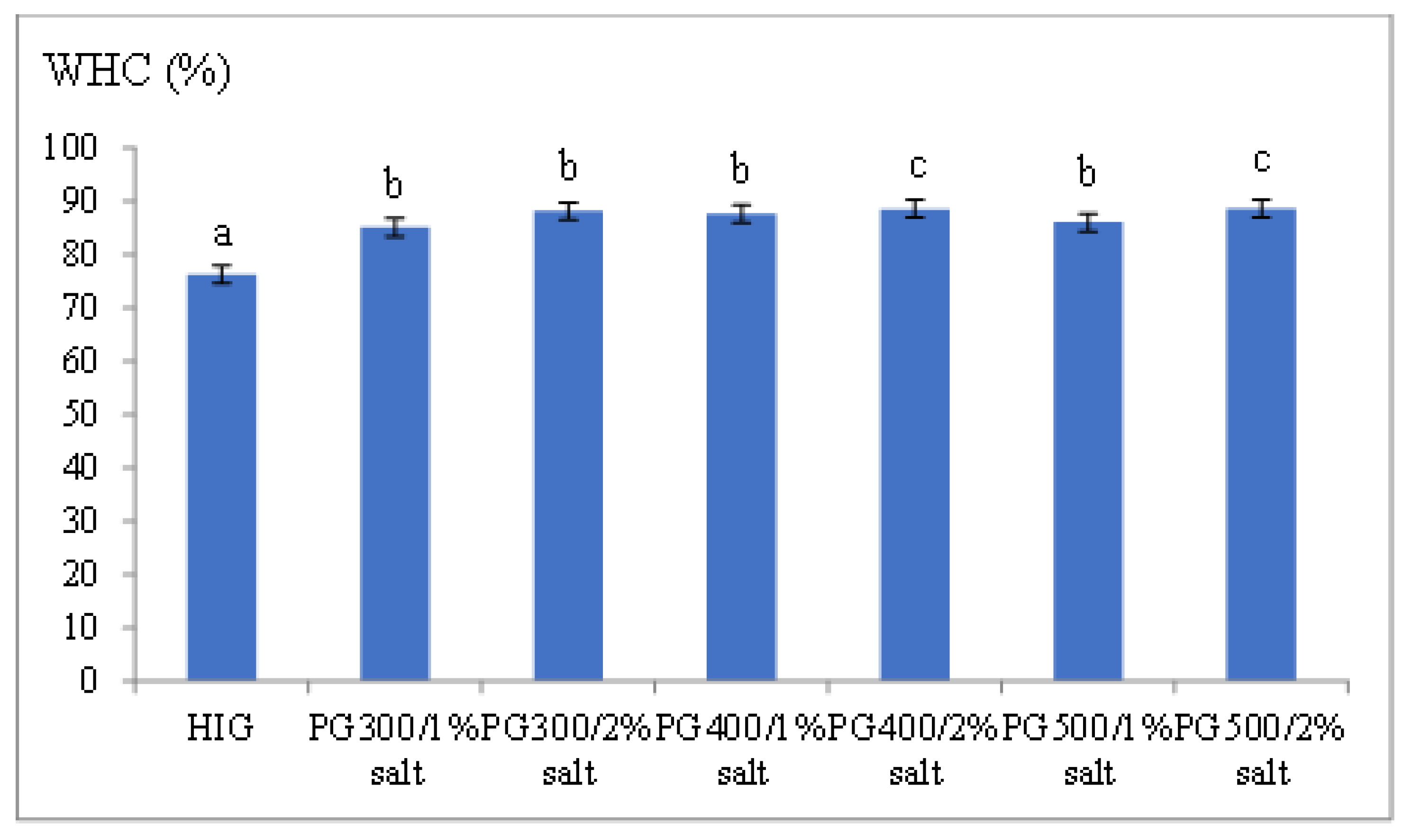
3.5. Water-Holding Capacity

Figure 3 presents the water-holding capacity (WHC) of pangasius gel at different salt concentrations. HPP improved the WHC of all pangasius gel treatments compared to HIGs (p < 0.05). Pangasius gels with 2% added salt and treated at 500 MPa/10 °C/10 min achieved the highest WHC at 88.62%, while HIGs had the lowest WHC at 76.28%. HPP pangasius gels with 1% added salt exhibited similar WHC to those with 2% added salt (p ≥ 0.05). The improved mechanical properties of pangasius gel under low salt conditions could be attributed to the enhanced interactions among myofibrillar proteins by HPP, which promote protein solubility and gel formation at low salt concentrations, resulting in a gel with better WHC [9]. Additionally, the myofibrillar structure is disrupted and degraded into smaller protein fragments, leading to an increased solubility of proteins under low-salt conditions. This also facilitates the binding of free water and fish proteins, resulting in gels with high strength and water-holding capacity under low-salt conditions [38]. Furthermore, HPP creates a stable and compact spatial structure of the gel, which helps immobilize and retain water within the gel structure [1]. HPP also exposes hydrophobic groups, enhancing hydrophobic interactions and stabilizing the water/protein system [5]. These findings suggested that the improvement in WHC by HPP treatment could be caused by several mechanisms, including protein unfolding by HPP, which can promote protein–water interactions; exposing polar and hydrophobic groups that help to bind water molecules more effectively; and better formation of gel structure, which better holds water within the gel matrix as reported by Ma et al. [1]. These mechanisms also explain HPP HPP-treated pangasius gels at 1% salt concentration have comparable WHC to those with 2% salt, thereby supporting the potential of HPP in reducing salt without reducing WHC.

3.6. Protein Solubility

Protein solubility is an important parameter of the functional structure of muscle proteins, particularly myofibrillar proteins, which are responsible for in-gel formation. Protein solubility exhibits the extent of protein denaturation and aggregation induced by processing treatments such as heat or HPP. The decrease in solubility shows the formation of insoluble protein aggregates or network structures, which may contribute to better gelation. In contrast, high solubility indicates that proteins are insufficiently denatured and therefore, less able to form a well-structured gel matrix. Therefore, protein solubility provides important mechanistic insight into how HPP influences the gelation behavior and structural quality of fish protein gels. The protein solubility of HIGs and HPP gels ranged from 7.75 mg/L to 8.52 mg/L and showed no significant differences (p ≥ 0.05). The reduction in protein solubility indicates protein denaturation and the precipitation of myofibrillar proteins, particularly myosin, following the denaturation process [39]. These findings show that both heat treatment and high-pressure processing result in complete protein denaturation, facilitating the formation of a well-structured gel network at both 1% and 2% salt conditions.

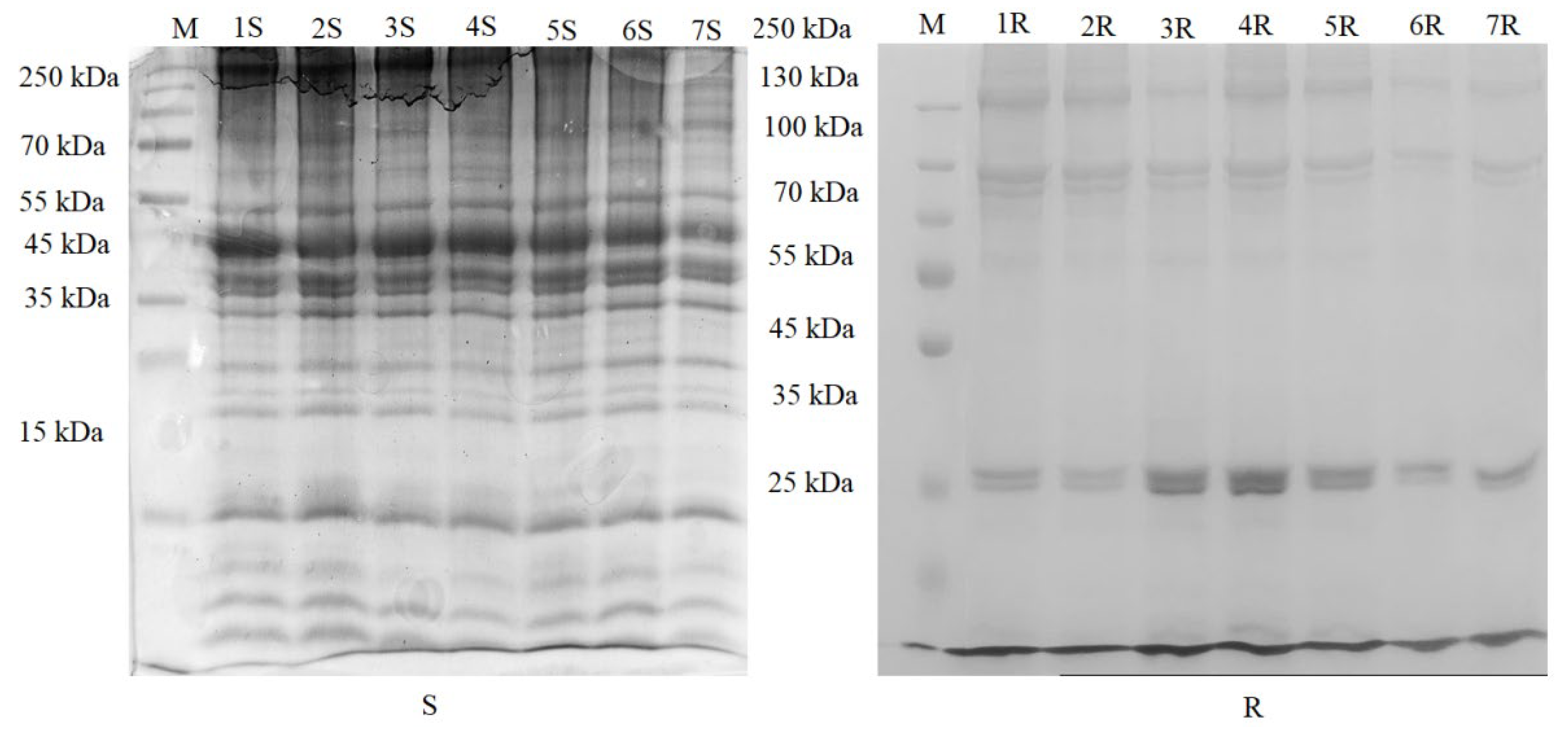
3.7. SDS-Polyacrylamide Gel Electrophoresis
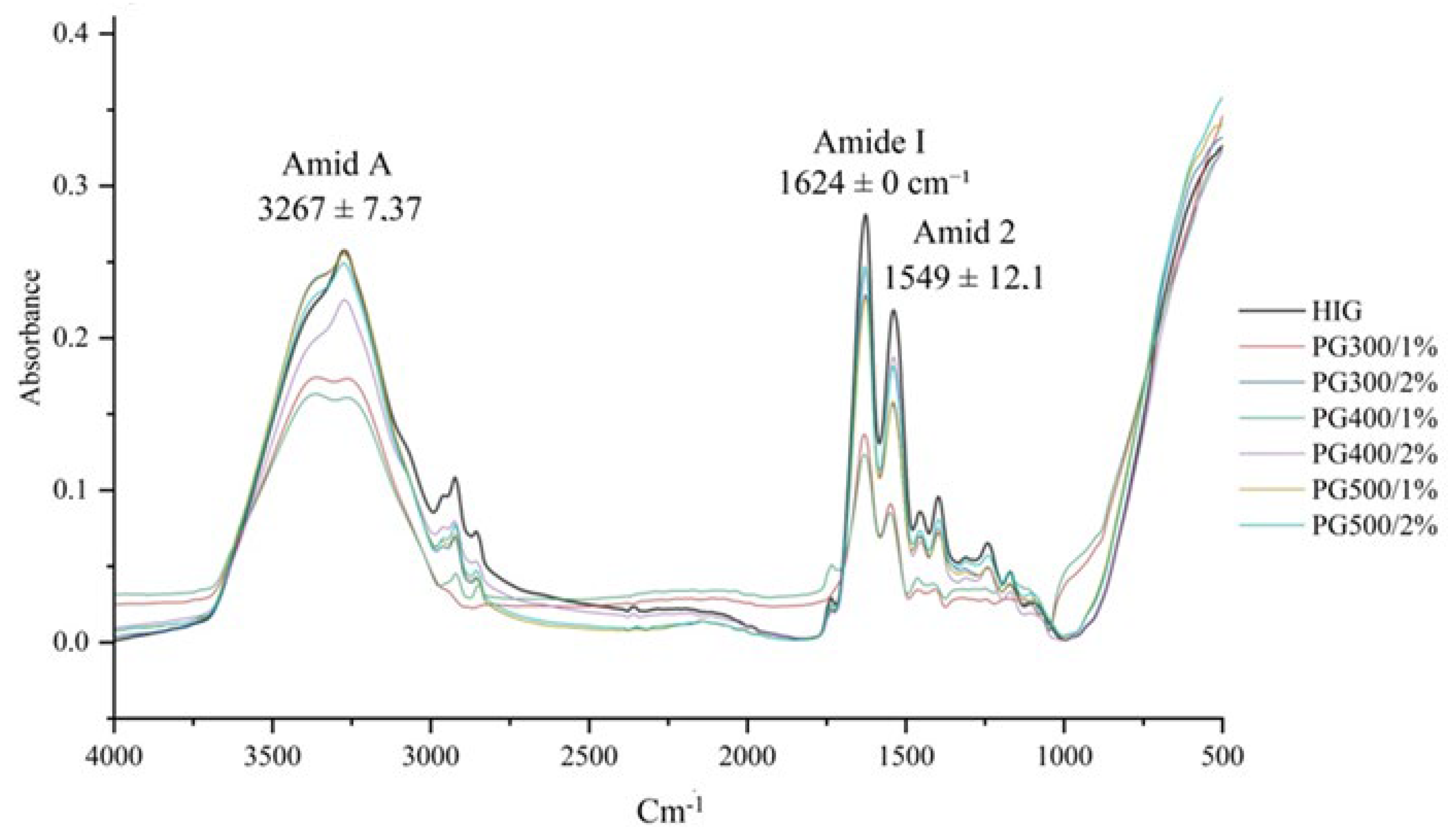
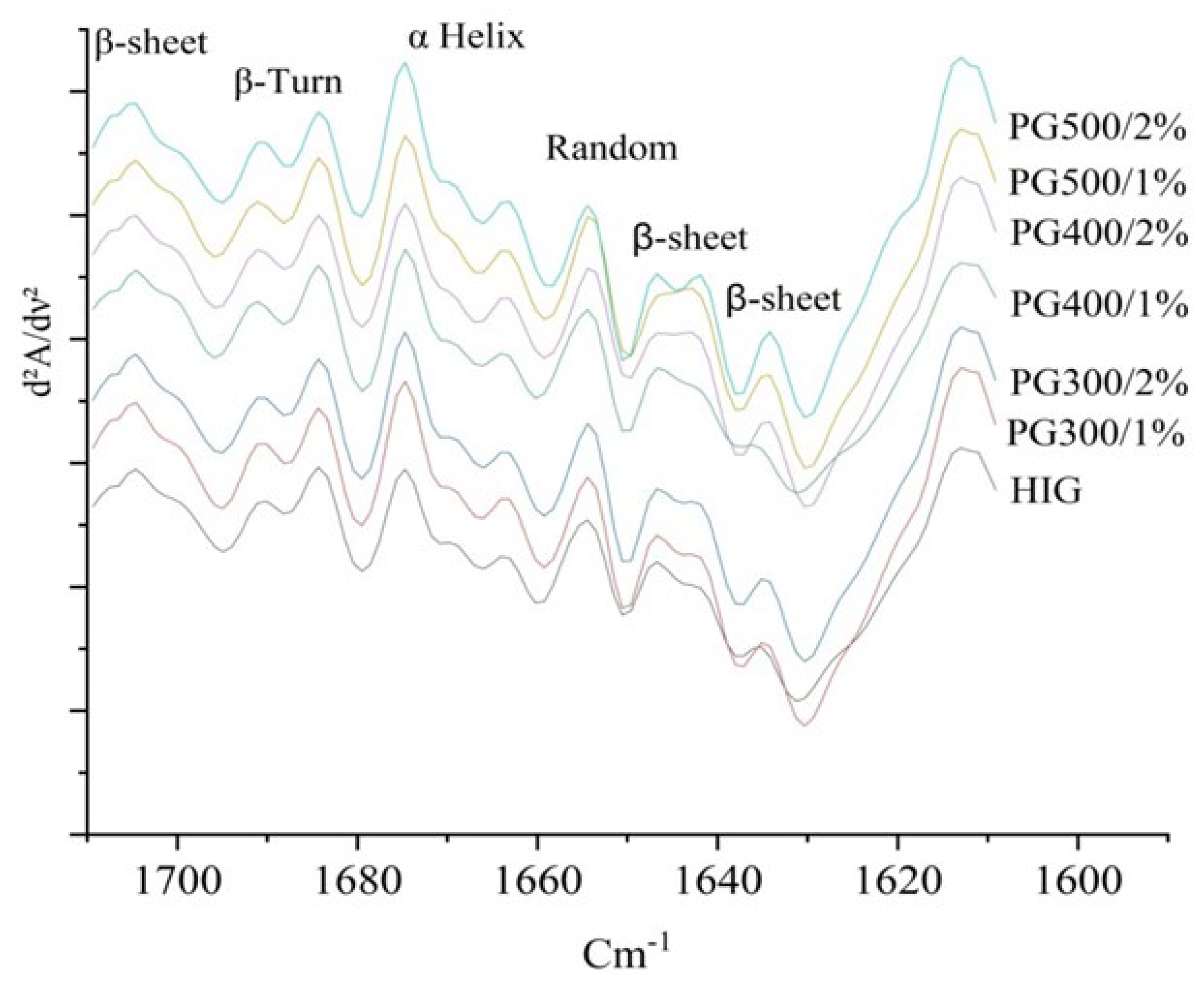
For SDS-soluble proteins, the electrophoresis profile (Figure 4S) showed that HMW proteins, including myosin heavy chain (MHC), were present at 250 kDa and actin at 45 kDa. As pressure increased, the intensity of the 250 kDa band decreased, and this effect was more remarkable at 2% salt compared to 1% salt. At PG500/2%, the 250 kDa band was nearly undetectable. The reduction in high molecular weight proteins, particularly with 2% salt, is mainly attributed to pressure-induced aggregation, which decreases their SDS solubility. Salt could increase this effect by promoting aggregation and reducing the extractable fraction of HMW proteins in the gel [40]. In contrast, no significant differences were observed in the actin bands among the treatments.
In general, the electrophoresis pattern of sarcoplasmic proteins exhibited a similar trend to SDS-soluble proteins, with a reduction in high molecular weight bands, including myosin heavy chain (MHC), as pressure increased (Figure 4R). Particularly, the band intensity in pressure-induced gels with 2% salt was lower than in those with 1% salt, suggesting that higher salt concentration can promote more protein aggregation. A similar observation was found in arrowtooth flounder gels, where the MHC band intensity decreased with increasing pressure [34]. The actin band of sarcoplasmic proteins disappeared in all treatments, indicating that actin proteins were extensively denatured and aggregated.

3.8. FTIR Analysis

The vibrational spectra of PG and HIGs at different salt concentrations were analyzed by FTIR to gain insights into their secondary structure. Spectral analysis within the frequency range of 4000–500 cm−1 revealed peaks at 435 ± 8.6 cm−1, 1391 ± 9.4 cm−1, 1455 ± 2.2 cm−1, 1549 ± 12.1 cm−1, 1624 ± 0 cm−1, 2921 ± 3.37 cm−1, and 3267 ± 7.37 cm−1. Among these bands, the amide I band was observed at 1624 ± 0 cm−1, amide II at 1549 ± 12.1 cm−1, and amide A at 3267 ± 7.37 cm−1 (Figure 5).
The amide I region, characterized by C=O stretching vibrations, typically consists of overlapping component bands derived from structural elements such as α-helices, β-sheets, turns, and disordered or irregular conformations [41]. Amide II (1480–1575 cm−1) primarily corresponds to CN stretching and NH bending, while the amide A band (3100–3600 cm−1) is mainly linked to OH and NH stretching vibrations, with a broadened absorbance reflecting the muscle’s high water content [42,43].
Within the myofiber spectrum, the amide I band (ranging from 1700 to 1600 cm−1) is particularly significant due to its high sensitivity to hydrogen-bonding, dipole–dipole interactions, and the structural configuration of the protein’s polypeptide backbone [5]. Like the study of Herranz et al. [41], the application of HPP resulted in a reduction in the absorbance of the amid I band in pangasius protein as compared to HIGs. This reduction indicates structure alterations, due to the disruption or formation of protein interactions. However, the amide I frequencies were similar in all treatments, suggesting that high pressure caused only minor changes in the protein structure.
To enhance spectral resolution and investigate the changes in the secondary structure of myofibrillar proteins, the amide I region (1700–1600 cm−1) was analyzed using second-derivative spectra (Figure 6). This analysis found nine different bands including 1604 ± 0.58 cm−1, 1625 ± 0.73 cm−1, 1637 ± 0.35 cm−1, 1648 ± 1.3 cm−1, 1654 ± 0.84 cm−1, 1665 ± 1.32 cm−1, 1675 ± 0.26 cm−1, 1680 ± 0.66 cm−1 and 1696 ± 0.66 cm−1. The bands at 1625 ± 0.73 cm−1, 1637 ± 0.35 cm−1, and 1696 ± 0.66 cm−1 were attributed to β-sheet structures; the band at 1654 ± 0.84 cm−1 and 1665 ± 1.32 cm−1 corresponded to α-helices; the bands at 1675 ± 0.26 and 1680 ± 0.66 cm−1 were assigned to β-turns [43]. On the other hand, the band at 1648 ± 1.3 cm−1 was attributed to random structures, and the band at 1604 ± 0.58 cm−1 could be aromatic side chain vibrations of amino acids such as tyrosine, phenylalanine, or tryptophan [44,45,46].
The results in Table 3 show the percentage of the secondary structure of the pangasius protein treated at different HPP conditions and HIGs. The α-helix content was significantly higher in the HPP samples compared to the HIG control (p < 0.05). The stabilization of α-helix under high-pressure conditions can be attributed to the ability of HPP to maintain intramolecular hydrogen bonds, preventing thermal denaturation. Moreno et al. [41] showed that high pressure also increased α-helix content in flying fish surimi that was pressurized and then cooked. Moreover, He et al. [47] suggested that high pressure helps preserve α-helix structures in proteins by restricting the unfolding process and minimizing heat-induced transitions to β-sheet or random coil structures. In contrast, Cando et al. [5] reported that the α-helix structure of isolated hake myofibrils remained similar between heated samples (90 °C for 20 min) and those subjected to pressure treatment (150–250 MPa at 10 °C for 10 min) followed by heating (90 °C for 20 min).
Unlike α-helix structures, β-sheet and β-turn contents showed no significant differences among all treatments, including HIGs. The β-sheet content remained relatively stable, ranging from 29.22% to 31.18%, while β-turn content fluctuated minimally between 19.64% and 20.14%. This suggests that these structures are stable under the thermal and high-pressure conditions. These findings align with previous reports suggesting that β-sheets are stabilized by strong inter-chain hydrogen bonds, which are resistant to both thermal and pressure-induced conformational changes [48,49].
At a 2% salt concentration, the random structure content was similar between pressure-induced gels and HIGs. However, random structure content was significantly lower in high-pressure-treated samples compared to the HIG control (p < 0.05) at reduced salt concentration (1%). This suggests that the less random structure of pressure-induced gels with 1% added salt was denatured compared to HIGs with 2% salt. Thus, HPP can result in molecular reorganization and influence the degree of protein denaturation in different patterns at different salt concentrations. A similar observation was also reported in flying fish surimi in which random structure content was reduced compared to unpressurized samples [50]. Chen et al. [51] also reported a decrease in random coil content in Nemipterus virgatus surimi when pressure increased.

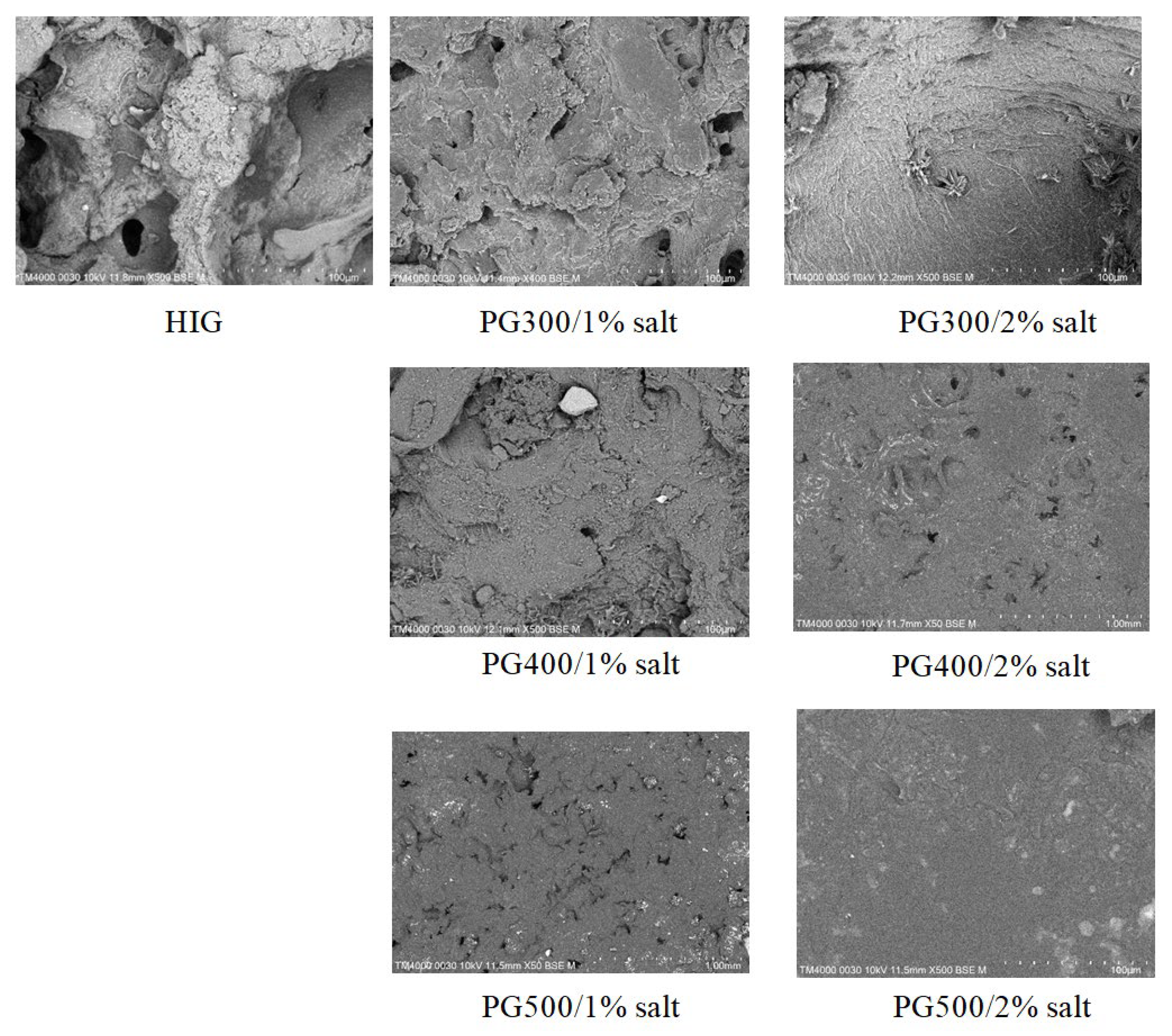
3.9. Scanning Electron Microscope (SEM) Analysis

SEM images showed that the microstructure of pangasius gels changed with varying salt concentrations and processing conditions (Figure 7), corresponding to the mechanical properties.
The HIG sample exhibited a relatively loose and irregular structure, with visible gaps and pores. In contrast, gels treated with high pressure (especially at 400–500 MPa) showed a denser, more homogeneous, and compact structure as compared to HIGs. This indicates that HPP treatment facilitates the formation of a well-developed gel network in pangasius gels. PG with 2% salt treated with high pressure formed a more compact microstructure with fewer voids and a smoother surface compared to PG with 1% salt, exhibiting characteristics typical of pressure-treated gels [3]. In contrast, PG with 1% salt displayed a spongier structure compared to PG with 2% salt due to the lower salt content, which limited protein solubility and resulted in a weaker gel. However, HPP still facilitated the formation of a relatively compact gel with fewer voids and a smoother surface than heat-treated gels. SEM observations also confirm the correspondence between microstructure and the mechanical properties of pangasius gels, in which HPP-treated samples exhibit improved hardness, gel strength, and WHC compared to HIGs. Similar observations in fish gels and surimi treated with HPP have been reported in previous studies, showing that pressure-induced gelation can produce finer and more stable networks compared to heat-only treatments [4,5].

4. Conclusions

The results of this study show that HPP prior to heat treatment is an effective method for producing high-quality pangasius gels at reduced salt conditions. HPP improved gel texture, whiteness, and water-holding capacity in pangasius gels with 2% salt while maintaining these properties of gels with 1% salt comparable to heat-induced gels (HIGs) with 2% salt. SDS-PAGE results showed that increasing pressure reduced the intensity of high molecular weight proteins in both SDS-soluble and sarcoplasmic proteins, likely due to pressure-induced aggregation. Additionally, actin from sarcoplasmic proteins was undetectable in all treatments, indicating that the effect of thermal treatment is predominant over-pressure treatment on sarcoplasmic actin protein.
HPP also facilitated the formation of a more compact gel network, as confirmed by SEM analyses. FTIR spectra showed that HPP increased α-helix content, stabilizing intramolecular hydrogen bonds and reducing protein unfolding. At 1% salt, PG had lower random structure content than HIGs, suggesting reduced protein denaturation. These structural modifications contributed to enhanced gel strength and stability, even at a lower salt concentration (1%). These results indicate that HPP can be applied to develop salt-reduced pangasius gel products with improved textural and structural properties. This approach can satisfy the growing consumer demand for healthier, lower-sodium foods and may contribute to improved product quality and greater acceptability in commercial applications. However, this study has some limitations, as it was conducted using a single fish species (pangasius) under fixed HPP conditions, including temperature and holding time. Therefore, more studies are needed to examine the changes in pangasius gels under different HPP conditions. Future research should explore the long-term storage stability and sensory attributes of high-pressure-induced pangasius gels to further validate their commercial viability. In addition, the investigation of the molecular mechanisms of protein interactions, including bond formation such as disulfide, hydrophobic, and hydrogen bonds, should be conducted for a deeper understanding of the structural changes induced by HPP and their impact on gelation behavior and functional properties.

Author Contributions

Conceptualization, B.Q.T. and V.C.H.; methodology, B.Q.T., R.B. and T.N.A.H.; software, B.Q.T., K.N.H.N., L.T.N. and T.N.A.H. validation, B.Q.T., R.B., K.N.H.N., L.T.N., T.N.A.H. and V.C.H.; formal analysis, B.Q.T., T.N.A.H. and V.C.H.; investigation, B.Q.T., K.N.H.N., T.N.A.H. and V.C.H.; resources, B.Q.T. and R.B.; data curation, B.Q.T., K.N.H.N. and V.C.H.; writing—original draft preparation, B.Q.T. and V.C.H.; writing—review and editing, R.B. and V.C.H.; visualization, B.Q.T. and V.C.H.; supervision, R.B. and V.C.H.; project administration, B.Q.T. and V.C.H.; funding acquisition, B.Q.T. and R.B. All authors have read and agreed to the published version of the manuscript.

Funding

This research was funded by the Vietnam Ministry of Education and Training under the project titled ‘Research on high-pressure processing technology applied to pangasius (Pangasianodon hypophthalmus) muscle and its application in developing value-added products’, grant No. B2023-NLS-02.

Informed Consent Statement

Not applicable.

Data Availability Statement

The original contributions presented in the study are included in the article, further inquiries can be directed to the corresponding author.

Acknowledgments

The corresponding author acknowledges support from Ho Chi Minh City University of Technology and Education (HCMUTE), Vietnam.

Conflicts of Interest

The authors declare no conflicts of interest.

Abbreviations

The following abbreviations are used in this manuscript:
HPP High-pressure processing
HIGsHeat-induced gels
FTIRFourier transform infrared spectroscopy
SEMScanning electron microscope
TPCTotal plate count
WHCWater-holding capacity
MHCMyosin heavy chain

References

  1. Ma, X.-S.; Yi, S.-M.; Yu, Y.-M.; Li, J.-R.; Chen, J.-R. Changes in gel properties and water properties of Nemipterus virgatus surimi gel induced by high-pressure processing. LWT-Food Sci. Technol. 2015, 61, 377–384. [Google Scholar] [CrossRef]
  2. Truong, B.Q.; Buckow, R.; Nguyen, M. Mechanical and Functional Properties of Unwashed Barramundi (Lates calcarifer) Gels as Affected by High-Pressure Processing at three Different Temperatures and Salt Concentrations. J. Aquat. Food Prod. Technol. 2020, 29, 373–382. [Google Scholar] [CrossRef]
  3. Truong, B.Q.; Buckow, R.; Stathopoulos, C.; Nguyen, M. Advances in High-Pressure Processing of Fish Muscles. Food Eng. Rev. 2015, 7, 109–129. [Google Scholar] [CrossRef]
  4. Truong, B.Q.; Buckow, R.; Nguyen, M.H.; Furst, J. Effect of high-pressure treatments prior to cooking on gelling properties of unwashed protein from barramundi (Lates calcarifer) minced muscle. Int. J. Food Sci. Technol. 2017, 52, 1383–1391. [Google Scholar] [CrossRef]
  5. Cando, D.; Herranz, B.; Borderías, A.J.; Moreno, H.M. Effect of high pressure on reduced sodium chloride surimi gels. Food Hydrocoll. 2015, 51, 176–187. [Google Scholar] [CrossRef]
  6. Sikes, A.L.; Tobin, A.B.; Tume, R.K. Use of high pressure to reduce cook loss and improve texture of low-salt beef sausage batters. Innov. Food Sci. Emerg. Technol. 2009, 10, 405–412. [Google Scholar] [CrossRef]
  7. Gordon, A.; Barbut, S. Effect of chloride salts on protein extraction and interfacial protein film formation in meat batters. J. Sci. Food Agric. 1992, 58, 227–238. [Google Scholar] [CrossRef]
  8. Pilar, M.; Gomez-Guillen, M.C. High-Pressure Applications on Myosystems. In Novel Food Processing Technologies; Food Science and Technology; CRC Press: Boca Raton, FL, USA, 2004; pp. 311–342. [Google Scholar]
  9. Macfarlane, J.J.; McKenzie, I.J.; Turner, R.H.; Jones, P.N. Binding of comminuted meat: Effect of high pressure. Meat Sci. 1984, 10, 307–320. [Google Scholar] [CrossRef]
  10. Xu, M.; Ni, X.; Liu, Q.; Chen, C.; Deng, X.; Wang, X.; Yu, R. Ultra-high pressure improved gelation and digestive properties of Tai Lake whitebait myofibrillar protein. Food Chem. X 2024, 21, 101061. [Google Scholar] [CrossRef]
  11. Iwasaki, T.; Noshiroya, K.; Saitoh, N.; Okano, K.; Yamamoto, K. Studies of the effect of hydrostatic pressure pretreatment on thermal gelation of chicken myofibrils and pork meat patty. Food Chem. 2006, 95, 474–483. [Google Scholar] [CrossRef]
  12. Huppertz, T.; Fox, P.F.; Kelly, A.L. High pressure-induced denaturation of α-lactalbumin and β-lactoglobulin in bovine milk and whey: A possible mechanism. J. Dairy Res. 2004, 71, 489–495. [Google Scholar] [CrossRef] [PubMed]
  13. Pérez-Mateos, M.; Lourenço, H.; Montero, P.; Borderías, A.J. Rheological and Biochemical Characteristics of High-Pressure- and Heat-Induced Gels from Blue Whiting (Micromesistius poutassou) Muscle Proteins. J. Agric. Food Chem. 1997, 45, 44–49. [Google Scholar] [CrossRef]
  14. Nguyen, H.T.; Bao, H.N.D.; Dang, H.T.T.; Tómasson, T.; Arason, S.; Gudjónsdóttir, M. Protein Characteristics and Bioactivity of Fish Protein Hydrolysates from Tra Catfish (Pangasius hypophthalmus) Side Stream Isolates. Foods 2022, 11, 4102. [Google Scholar] [CrossRef]
  15. Dang, H.T.T.; Gudjónsdóttir, M.; Tómasson, T.; Nguyen, M.V.; Karlsdóttir, M.G.; Arason, S. Influence of processing additives, packaging and storage conditions on the physicochemical stability of frozen Tra catfish (Pangasius hypophthalmus) fillets. J. Food Eng. 2018, 238, 148–155. [Google Scholar] [CrossRef]
  16. Rathod, N.B.; Pagarkar, A.U.; Pujari, K.H.; Shingare, P.; Satam, S.; Phadke, G.G.; Gaikwad, B. Status of valuable components from pangasius: A review. Int. J. Curr. Microbiol. Appl. Sci. 2018, 7, 2106–2120. [Google Scholar] [CrossRef]
  17. Li, Q.; Chen, S.; You, J.; Hu, Y.; Yin, T.; Liu, R. A comprehensive unraveling of the mystery of reduced-salt surimi gels: From molecular mechanism to future prospects. Trends Food Sci. Technol. 2024, 154, 104783. [Google Scholar] [CrossRef]
  18. Wu, D.; Xiong, J.; Li, P.; Zhang, Y.; Li, F.; Yin, T.; Huang, Q. Dual enhancement effects of different yeast extract on gel properties and saltiness perception of low-salt surimi gel from silver carp. Food Hydrocoll. 2024, 152, 109925. [Google Scholar] [CrossRef]
  19. Zhang, C.; Chen, L.; Lu, M.; Ai, C.; Cao, H.; Xiao, J.; Teng, H. Effect of cellulose on gel properties of heat-induced low-salt surimi gels: Physicochemical characteristics, water distribution and microstructure. Food Chem. X 2023, 19, 100820. [Google Scholar] [CrossRef]
  20. Zhang, X.; Guo, Q.; Shi, W. Ultrasound-assisted processing: Changes in gel properties, water-holding capacity, and protein aggregation of low-salt Hypophthalmichthys molitrix surimi by soy protein isolate. Ultrason. Sonochem. 2023, 92, 106258. [Google Scholar] [CrossRef]
  21. Nelce Mailoa, M.; Marthina Tapotubun, A.; Matrutty, T.E.A.A. Analysis Total Plate Counte (TPC) On Fresh Steak Tuna Applications Edible Coating Caulerpa sp During Stored at Chilling Temperature. IOP Conf. Ser. Earth Environ. Sci. 2017, 89, 012014. [Google Scholar] [CrossRef]
  22. Ramirez-Suarez, J.C.; Morrissey, M.T. Effect of high pressure processing (HPP) on shelf life of albacore tuna (Thunnus alalunga) minced muscle. Innov. Food Sci. Emerg. Technol. 2006, 7, 19–27. [Google Scholar] [CrossRef]
  23. Cruz-Romero, M.; Kelly, A.L.; Kerry, J.P. Effects of high-pressure and heat treatments on physical and biochemical characteristics of oysters (Crassostrea gigas). Innov. Food Sci. Emerg. Technol. 2007, 8, 30–38. [Google Scholar] [CrossRef]
  24. Pérez-Mateos, M.; Montero, P. High-pressure-induced gel of sardine (Sardina pilchardus) washed mince as affected by pressure-time-temperature. J. Food Sci. 1997, 62, 1183–1188. [Google Scholar] [CrossRef]
  25. Uresti, R.M.; Velazquez, G.; Vázquez, M.; Ramírez, J.A.; Torres, J.A. Effect of sugars and polyols on the functional and mechanical properties of pressure-treated arrowtooth flounder (Atheresthes stomias) proteins. Food Hydrocoll. 2005, 19, 964–973. [Google Scholar] [CrossRef]
  26. Lowry, O.H.; Rosebrough, N.J.; Farr, A.L.; Randall, R.J. Protein measurement with the Folin phenol reagent. J. Biol. Chem. 1951, 193, 265–275. [Google Scholar] [CrossRef]
  27. Pazos, M.; Méndez, L.; Vázquez, M.; Aubourg, S.P. Proteomics analysis in frozen horse mackerel previously high-pressure processed. Food Chem. 2015, 185, 495–502. [Google Scholar] [CrossRef]
  28. Iwasaki, T.; Washio, M.; Yamamoto, K.; Nakamura, K. Rheological and Morphological Comparison of Thermal and Hydrostatic Pressure-Induced Filamentous Myosin Gels. J. Food Sci. 2005, 70, e432–e436. [Google Scholar] [CrossRef]
  29. Muntean, M.; Ovidiu, M.; Barbieru, V.; Catunescu, G.; Ranta, O.; Drocas, I.; Terhes, S. High Pressure Processing in Food Industry–Characteristics and Applications. Agric. Agric. Sci. Procedia 2016, 10, 377–383. [Google Scholar] [CrossRef]
  30. Pinto, C.A.; Moreira, S.A.; Fidalgo, L.G.; Inácio, R.S.; Barba, F.J.; Saraiva, J.A. Effects of high-pressure processing on fungi spores: Factors affecting spore germination and inactivation and impact on ultrastructure. Compr. Rev. Food Sci. Food Saf. 2020, 19, 553–573. [Google Scholar] [CrossRef]
  31. Yu, L.; Muralidharan, S.; Lee, N.A.; Lo, R.; Stokes, J.R.; Fitzgerald, M.A.; Turner, M.S. The impact of variable high pressure treatments and/or cooking of rice on bacterial populations after storage using culture-independent analysis. Food Control 2018, 92, 232–239. [Google Scholar] [CrossRef]
  32. Kunnath, S.; Jaganath, B.; Panda, S.K.; Ravishankar, C.N.; Gudipati, V. Modifying textural and functional characteristics of fish (Nemipterus japonicus) mince using high pressure technology. J. Food Sci. Technol. 2022, 59, 4122–4133. [Google Scholar] [CrossRef] [PubMed]
  33. de Oliveira, F.A.; Neto, O.C.; dos Santos, L.M.R.; Ferreira, E.H.R.; Rosenthal, A. Effect of high pressure on fish meat quality—A review. Trends Food Sci. Technol. 2017, 66, 1–19. [Google Scholar] [CrossRef]
  34. Uresti, R.M.; Velazquez, G.; Ramírez, J.A.; Vázquez, M.; Torres, J.A. Effect of high-pressure treatments on mechanical and functional properties of restructured products from arrowtooth flounder (Atheresthes stomias). J. Sci. Food Agric. 2004, 84, 1741–1749. [Google Scholar] [CrossRef]
  35. Buckow, R.; Bull, M. Advanced food preservation technologies. Microbiol. Aust. 2013, 34, 108–111. [Google Scholar] [CrossRef]
  36. Cui, Y.; Zhu, L.; Shang, H.; Xuan, X.; Lin, X. Effects of Combined ε-Polylysine and High Hydrostatic Pressure Treatment on Microbial Qualities, Physicochemical Properties, Taste, and Volatile Flavor Profile of Large Yellow Croaker (Larimichthys crocea). Food Bioprocess Technol. 2024, 18, 3610–3627. [Google Scholar] [CrossRef]
  37. Zhou, Y.; Watkins, P.; Oiseth, S.; Cochet-Broch, M.; Sikes, A.L.; Chen, C.; Buckow, R. High pressure processing improves the sensory quality of sodium-reduced chicken sausage formulated with three anion types of potassium salt. Food Control 2021, 126, 108008. [Google Scholar] [CrossRef]
  38. Tabilo-Munizaga, G.; Barbosa-Cánovas, G.V. Ultra high pressure technology and its use in surimi manufacture: An overview. Food Sci. Technol. Int. 2004, 10, 207–222. [Google Scholar] [CrossRef]
  39. Ramírez, J.A.; Martín-Polo, M.O.; Bandman, E. Fish myosin aggregation as affected by freezing and initial physical state. J. Food Sci. 2000, 65, 556–560. [Google Scholar] [CrossRef]
  40. Sagné, C.; Isambert, M.-F.; Gasnier, B. SDS-resistant aggregation of membrane proteins: Application to the purification of the vesicular monoamine transporter. Biochem. J. 1996, 316 Pt 3, 825–831. [Google Scholar] [CrossRef]
  41. Herranz, B.; Tovar, C.A.; Borderias, A.J.; Moreno, H.M. Effect of high-pressure and/or microbial transglutaminase on physicochemical, rheological and microstructural properties of flying fish surimi. Innov. Food Sci. Emerg. Technol. 2013, 20, 24–33. [Google Scholar] [CrossRef]
  42. Ojagh, S.M.; Núñez-Flores, R.; López-Caballero, M.E.; Montero, M.P.; Gómez-Guillén, M.C. Lessening of high-pressure-induced changes in Atlantic salmon muscle by the combined use of a fish gelatin–lignin film. Food Chem. 2011, 125, 595–606. [Google Scholar] [CrossRef]
  43. Kong, J.; Yu, S. Fourier Transform Infrared Spectroscopic Analysis of Protein Secondary Structures. Acta Biochim. Biophys. Sin. 2007, 39, 549–559. [Google Scholar] [CrossRef] [PubMed]
  44. Hernández, B.; Pflüger, F.; Adenier, A.; Kruglik, S.G.; Ghomi, M. Vibrational Analysis of Amino Acids and Short Peptides in Hydrated Media. VIII. Amino Acids with Aromatic Side Chains: L-Phenylalanine, l-Tyrosine, and l-Tryptophan. J. Phys. Chem. B 2010, 114, 15319–15330. [Google Scholar] [CrossRef] [PubMed]
  45. Sadat, A.; Joye, I.J. Peak Fitting Applied to Fourier Transform Infrared and Raman Spectroscopic Analysis of Proteins. Appl. Sci. 2020, 10, 5918. [Google Scholar] [CrossRef]
  46. Venyaminov, S.Y.; Kalnin, N.N. Quantitative IR spectrophotometry of peptide compounds in water (H2O) solutions. II. Amide absorption bands of polypeptides and fibrous proteins in α-, β-, and random coil conformations. Biopolymers 1990, 30, 1259–1271. [Google Scholar] [CrossRef]
  47. He, J.-S.; Mu, T.-H.; Guo, X.; Zhu, S.; Azuma, N.; Kanno, C. Comparison of the gel-forming ability and gel properties of α-lactalbumin, lysozyme and myoglobin in the presence of β-lactoglobulin under high pressure. Food Hydrocoll. 2013, 33, 415–424. [Google Scholar] [CrossRef]
  48. Bolumar, T.; Middendorf, D.; Toepfl, S.; Heinz, V. Structural Changes in Foods Caused by High-Pressure Processing; Springer: New York, NY, USA, 2016; pp. 509–537. [Google Scholar]
  49. Knorr, D.; Heinz, V.; Buckow, R. High pressure application for food biopolymers. Biochim. Biophys. Acta (BBA)-Proteins Proteom. 2006, 1764, 619–631. [Google Scholar] [CrossRef]
  50. Moreno, H.M.; Bargiela, V.; Tovar, C.A.; Cando, D.; Borderias, A.J.; Herranz, B. High pressure applied to frozen flying fish (Parexocoetus brachyterus) surimi: Effect on physicochemical and rheological properties of gels. Food Hydrocoll. 2015, 48, 127–134. [Google Scholar] [CrossRef]
  51. Chen, H.; Zhou, A.; Benjakul, S.; Zou, Y.; Liu, X.; Xiao, S. The mechanism of low-level pressure coupled with heat treatment on water migration and gel properties of Nemipterus virgatus surimi. LWT 2021, 150, 112086. [Google Scholar] [CrossRef]
Figure 1. TPC of pangasius gels at 1% and 2% salt concentrations at different high-pressure processing conditions. HIGs: conventional heat-induced gels; PG300/1%: salt concentration 1% and 300 MPa/10 °C/10 min; PG300/2%: salt concentration 1% and 300 MPa/10 °C/10 min; PG400/1%: salt concentration 1% and 400 MPa/10 °C/10 min; PG400/2%: salt concentration 1% and 400 MPa/10 °C/10 min; PG500/1%: salt concentration 1% and 500 MPa/10 °C/10 min; PG500/2%: salt concentration 1% and 500 MPa/10 °C/10 min. Different letters (a–c) indicate significant differences (p < 0.05).
Applsci 15 04727 g001
Figure 2. Visual observation of pangasius fish gel color under varying salt concentrations and HPP treatments. HIGs: conventional heat-induced gels; PG300/1%: salt concentration 1% and 300 MPa/10 °C/10 min; PG300/2%: salt concentration 1% and 300 MPa/10 °C/10 min; PG400/1%: salt concen-tration 1% and 400 MPa/10 °C/10 min; PG400/2%: salt concentration 1% and 400 MPa/10 °C/10 min; PG500/1%: salt concentration 1% and 500 MPa/10 °C/10 min; PG500/2%: salt concen-tration 1% and 500 MPa/10 °C/10 min.
Applsci 15 04727 g002
Figure 3. WHC of pangasius gel was added with 1 and 2% salt under different HPP treatments. HIGs: conventional heat-induced gels; PG300/1%: salt concentration 1% and 300 MPa/10 °C/10 min; PG300/2%: salt concentration 1% and 300 MPa/10 °C/10 min; PG400/1%: salt concen-tration 1% and 400 MPa/10 °C/10 min; PG400/2%: salt concentration 1% and 400 MPa/10 °C/10 min; PG500/1%: salt concentration 1% and 500 MPa/10 °C/10 min; PG500/2%: salt concen-tration 1% and 500 MPa/10 °C/10 min. Different letters (a–c) indicate significant differences (p < 0.05).
Applsci 15 04727 g003
Figure 4. SDS-PAGE pattern of SDS-soluble protein (S) and sarcoplasmic protein (R) of pangasius gel obtained under various pressure treatment conditions. M: molecular ladder; 1S/1R: HIGs; 2S/2R: PG300/1%; 3S/3R: PG400/1%; 4S/4R: PG500/1%; 5S/5R: PG300/2%; 6S/6R: PG400/2%; 7S/7R: PG500/2%. HIGs: conventional heat-induced gels; PG300/1%: salt concentration 1% and 300 MPa/10 °C/10 min; PG300/2%: salt concentration 1% and 300 MPa/10 °C/10 min; PG400/1%: salt concen-tration 1% and 400 MPa/10 °C/10 min; PG400/2%: salt concentration 1% and 400 MPa/10 °C/10 min; PG500/1%: salt concentration 1% and 500 MPa/10 °C/10 min; PG500/2%: salt concen-tration 1% and 500 MPa/10 °C/10 min.
Applsci 15 04727 g004
Figure 5. FTIR spectra of pangasius gels with 1 and 2% salt under different HPP treatments over the frequency range 4000–500 cm−1. HIGs: conventional heat-induced gels; PG300/1%: salt concentration 1% and 300 MPa/10 °C/10 min; PG300/2%: salt concentration 1% and 300 MPa/10 °C/10 min; PG400/1%: salt concen-tration 1% and 400 MPa/10 °C/10 min; PG400/2%: salt concentration 1% and 400 MPa/10 °C/10 min; PG500/1%: salt concentration 1% and 500 MPa/10 °C/10 min; PG500/2%: salt concen-tration 1% and 500 MPa/10 °C/10 min.
Applsci 15 04727 g005
Figure 6. Second-derivative of Amide I band (1700–1600 cm−1) of infrared spectra of pangasius gels added 1 and 2% salt under different HPP treatments over the frequency range 1700–1600 cm−1. Peaks corresponding to α-helix (~1654 cm−1), β-sheet (~1625, ~1696 cm−1), β-turn (~1680 cm−1), and random coil (~1648 cm−1) indicate structural changes in the protein secondary structure of different treatments. HIGs: conventional heat-induced gels; PG300/1%: salt concentration 1% and 300 MPa/10 °C/10 min; PG300/2%: salt concentration 1% and 300 MPa/10 °C/10 min; PG400/1%: salt concen-tration 1% and 400 MPa/10 °C/10 min; PG400/2%: salt concentration 1% and 400 MPa/10 °C/10 min; PG500/1%: salt concentration 1% and 500 MPa/10 °C/10 min; PG500/2%: salt concen-tration 1% and 500 MPa/10 °C/10 min.
Applsci 15 04727 g006
Figure 7. Scanning electron microscopy (SEM) images of pangasius gel at different salt concentrations and high-pressure processing conditions. HIGs: conventional heat-induced gels; PG300/1%: salt concentration 1% and 300 MPa/10 °C/10 min; PG300/2%: salt concentration 1% and 300 MPa/10 °C/10 min; PG400/1%: salt concen-tration 1% and 400 MPa/10 °C/10 min; PG400/2%: salt concentration 1% and 400 MPa/10 °C/10 min; PG500/1%: salt concentration 1% and 500 MPa/10 °C/10 min; PG500/2%: salt concen-tration 1% and 500 MPa/10 °C/10 min.
Applsci 15 04727 g007
Table 1. Color of pangasius gel at 1% and 2% salt concentrations under different high-pressure processing conditions.
TreatmentWhiteness∆E
HIGs61.21 ± 0.55a
PG300/1% salt64.56 ± 2.04b6.53 ± 0.88
PG300/2% salt63.17 ± 0.26b6.15 ± 0.99
PG400/1% salt63.45 ± 0.91b6.59 ± 0.47
PG400/2% salt64.06 ± 0.50b6.18 ± 0.71
PG500/1% salt63.81 ± 1.43b6.42 ± 0.59
PG500/2% salt63.26 ± 0.67b6.54 ± 0.49
HIGs: conventional heat-induced gels; PG300/1%: salt concentration 1% and 300 MPa/10 °C/10 min; PG300/2%: salt concentration 1% and 300 MPa/10 °C/10 min; PG400/1%: salt concen-tration 1% and 400 MPa/10 °C/10 min; PG400/2%: salt concentration 1% and 400 MPa/10 °C/10 min; PG500/1%: salt concentration 1% and 500 MPa/10 °C/10 min; PG500/2%: salt concen-tration 1% and 500 MPa/10 °C/10 min. Different letters (a, b) in the same column indicate significant differences (p < 0.05).
Table 2. Changes in the mechanical properties of Pegasus gels added 1 and 2% salt under different HPP treatments.
TreatmentHardness
(N)
Springiness (mm)Gel Strength (N.mm)
HIGs7.93 ± 0.64a0.88 ± 0.02a346.21 ± 16.96a
PG300/1% salt8.34 ± 0.52a0.87 ± 0.01a417.65 ± 50.95b
PG300/2% salt10.39 ± 1.68bc0.89 ± 0.02a424.85 ± 25.29b
PG400/1% salt8.29 ± 0.42a0.87 ± 0.01a408.38 ± 12.43b
PG400/2% salt10.32 ± 0.87bc0.88 ± 0.02a436.92 ± 42.25b
PG500/1% salt8.96 ± 0.59ac0.89 ± 0.01a425.30 ± 23.09b
PG500/2% salt11.56 ± 1.76b0.89 ± 0.02a464.92 ± 13.56b
HIGs: conventional heat induced gels; PG300/1%: salt concentration 1% and 300 MPa/10 °C/10 min; PG300/2%: salt concentration 1% and 300 MPa/10 °C/10 min; PG400/1%: salt concen-tration 1% and 400 MPa/10 °C/10 min; PG400/2%: salt concentration 1% and 400 MPa/10 °C/10 min; PG500/1%: salt concentration 1% and 500 MPa/10 °C/10 min; PG500/2%: salt concen-tration 1% and 500 MPa/10 °C/10 min. Different letters (a–c) in the same column indicate significant differences (p < 0.05).
Table 3. The secondary structures (%) determined by FTIR self-deconvolution of HIGs and pressure-treated pangasius gel added 1 and 2% salt.
Treatmentα-Helix (%)β-Sheet (%)β-Turn (%)Random Structure (%)Aromatic Side Chain
HIGs19.41 ± 2.27a31.18 ± 0.73a19.64 ± 1.33a10.57 ± 0.51a19.17 ± 0.37a
PG300/1% salt24.74 ± 1.99b29.55 ± 0.96a20.14 ± 0.81a8.49 ± 0.66b17.05 ± 3.76a
PG300/2% salt24.14 ± 1.07b29.54 ± 1.32a19.84 ± 0.84a8.95 ± 1.36ab17.52 ± 3.89a
PG400/1% salt24.66 ± 1.87b29.22 ± 2.86a19.85 ± 1.08a8.02 ± 1.26b18.21 ± 1.34a
PG400/2% salt23.53 ± 1.11b30.72 ± 1.72a19.89 ± 1.33a9.06 ± 0.45ab16.77 ± 3.73a
PG500/1% salt23.65 ± 0.98b30.18 ± 1.73a20.11 ± 1.03a7.69 ± 1.38b18.32 ± 3.16a
PG500/2% salt23.82 ± 1.76b29.43 ± 0.89a20.16 ± 1.96a9.19 ± 1.12ab17.37 ± 3.81a
HIGs: conventional heat-induced gels; PG300/1%: salt concentration 1% and 300 MPa/10 °C/10 min; PG300/2%: salt concentration 1% and 300 MPa/10 °C/10 min; PG400/1%: salt concen-tration 1% and 400 MPa/10 °C/10 min; PG400/2%: salt concentration 1% and 400 MPa/10 °C/10 min; PG500/1%: salt concentration 1% and 500 MPa/10 °C/10 min; PG500/2%: salt concen-tration 1% and 500 MPa/10 °C/10 min. Different letters (a, b) in the same column indicate significant differences (p < 0.05).
Disclaimer/Publisher’s Note: The statements, opinions and data contained in all publications are solely those of the individual author(s) and contributor(s) and not of MDPI and/or the editor(s). MDPI and/or the editor(s) disclaim responsibility for any injury to people or property resulting from any ideas, methods, instructions or products referred to in the content.

