Article

Silencing of a Cotton Actin-Binding Protein GhWLIM1C Decreases Resistance against Verticillium dahliae Infection

State Key Laboratory of Ecological Pest Control for Fujian and Taiwan Crops, Key Laboratory of Ministry of Education for Genetics, Breeding and Multiple Utilization of Crops, Plant Immunity Center, Fujian Agriculture and Forestry University, Fuzhou 350002, China
*
Author to whom correspondence should be addressed.
These authors contribute equally to the work.
Plants 2022, 11(14), 1828; https://doi.org/10.3390/plants11141828
Submission received: 21 June 2022 / Revised: 9 July 2022 / Accepted: 9 July 2022 / Published: 12 July 2022
(This article belongs to the Special Issue Molecular Regulation of Plant Immunity)

Abstract

:
LIM proteins are widely spread in various types of plant cells and play diversely crucial cellular roles through actin cytoskeleton assembly and gene expression regulation. Till now, it has not been clear whether LIM proteins function in plant pathogen defense. In this study, we characterized a LIM protein, GhWLIM1C, in upland cotton (Gossypium hirsutum). We found that GhWLIM1C could bind and bundle the actin cytoskeleton, and it contains two LIM domains (LIM1 and LIM2). Both the two domains could bind directly to the actin filaments. Moreover, the LIM2 domain additionally bundles the actin cytoskeleton, indicating that it possesses a different biochemical activity than LIM1. The expression of GhWLIM1C responds to the infection of the cotton fungal pathogen Verticillium dahliae (V. dahliae). Silencing of GhWLIM1C decreased cotton resistance to V. dahliae. These may be associated with the down regulated plant defense response, including the PR genes expression and ROS accumulation in the infected cotton plants. In all, these results provide new evidence that a plant LIM protein functions in plant pathogen resistance and the assembly of the actin cytoskeleton are closely related to the triggering of the plant defense response.

1. Introduction

Cotton (Gossypium spp.) is one of the most important crops cultivated worldwide. The soil-borne pathogen Verticillium dahliae (V. dahliae) seriously threatens cotton production. It may cause a severe vascular disease, Verticillium wilt, and results in massive losses in plant mass and lint yield [1,2]. Increasing the plant resistance to Verticillium wilt is a critical challenge worldwide.
Verticillium wilt is characterized that the hyphae of V. dahliae propagating and is spread in the cotton woody vascular tissues, where fungicides cannot efficiently reach, which may account for the difficulty in controlling the devastating disease [3,4,5]. Besides blockage of the plant xylem vessel, V. dahliae could also synthesize toxic materials and secret proteins, which are thought to play critical roles in the pathogenicity of V. dahliae [6,7]. The secreted proteins are usually employed by pathogens, targeting the plant immune system and result in the attenuation of the host PTI (PAMP triggered immunity) or ETI (effector-triggered immunity) [7].
Upon perception of the infection of the V. dahliae, cotton plants activate the innate immune system. Multiple crucial defense-related proteins are synthesized and activated to participate in plant defense. For example, cotton plants could secrete NRX1 and CRR1 proteins, functioning in the apoplastic compartment to regulate ROS metabolism and antagonize a V. dahliae effector VdSSEP1-mediated fungal disease progression [8,9]. GhLYK1 and GhLYK2, two cell membrane LysM-containing proteins, are induced after V. dahliae infection. GhLYK1 and GhLYK2 silencing compromise cotton plant resistance to V. dahliae. GhLYK2 can induce ROS bursts in plants [10]. Several cotton transcription factors, including MYB108, CBP60g, SARD1 and the recently reported MYB6, have been reported to play crucial roles in cotton defense against V. dahliae [11,12,13]. In addition, some V. dahliae-responsive genes, such as MLP28, MKK2, and BAK1, have been shown to work in cotton defense against Verticillium wilt [14,15,16]. Exploring the defense-related genes in cotton would provide candidate targets for breading Verticillium wilt-resistant plants.
In plants, the actin cytoskeleton plays a central role in regulating intracellular transport, trafficking, and signaling events, including those associated with cell division and development, organelle movement, and vesicle trafficking [17]. Besides the crucial roles in plant cell development, the actin cytoskeleton is also a dynamic platform for sensing and responding to a diverse array of biotic and abiotic stresses. As a function of the plant immune system, accumulating evidence supports the roles of the cytoskeleton in at least two key aspects of the immune response. (1) It functions in the establishment and maintenance of signaling competent microenvironments; (2) it works in cellular trafficking (organelle, defense-related proteins, and small signaling molecules). In addition, the actin cytoskeleton is dynamically assembled in response to the activation of the plant’s immune system. An immediate increase in actin filament abundance is a conserved feature in the plant PTI. Blocking the actin filament density increase leads to enhanced susceptibility of host plants to pathogenic and non-pathogenic bacteria in Arabidopsis [18]. The actin-related proteins, such as the actin depolymerization factor4 (ADF4), the actin motor protein XI (Myosin XI), and the actin capping protein (CP), were shown to regulate the actin organization in the plant defense against bacterial infection [19,20,21]. Though this evidence indicated that the actin cytoskeleton is closely related to the plant’s immune system, the molecular mechanism that actin functions in the defense response, especially in the plant-fungal resistance, are known to be limited.
LIN-11, Isl1, and MEC-3 (LIM)-domain proteins are widely distributed in eukaryotes ranging from yeast (Saccharomyces cerevisiae) to humans [22]. Compared with animal genomes that contain multiple LIM genes, plant genomes encode a limited number of LIM genes. Plant LIM proteins have been shown to function in either actin-binding and bundling activity or in DNA binding to regulate gene expression in plant cell wall synthesis [23,24,25]. Tobacco’s WLIM1, WLIM2, and six Arabidopsis LIM proteins are vital to bundle actin filaments [22,25]. They are also key transcriptional factors through the binding of PAL-box DNA motif to regulate the expression of genes involved in phenylpropanoid biosynthesis and cell division [26,27]. Our previous work characterized four LIM genes in upland cotton (G. hirsutum), and of which WLIM1a was shown to have dual roles during fiber development. It can both bind the actin filaments and act as a transcription factor in the secondary cell wall biosynthesis stage [23]. In this study, we characterized another LIM gene in cotton. We found that GhWLIM1C expression was up regulated in response to V. dahliae infection, and GhWLIM1C and its two LIM domains are predominantly localized at the actin cytoskeleton. The silencing of GhWLIM1C in cotton may result in the attenuation of the plant defense response. In all, our results provide new evidence that the LIM protein, GhWLIM1C, could regulate cotton’s fungal defense via regulation assembly of the actin cytoskeleton. These broaden our understanding of the crucial roles of LIM proteins in plant cells.

2. Results

2.1. Expression of Cotton GhWLIM1C in Response to V. dahliae Infection

Four LIM genes were identified in the G. hirsutum genome in the previous work [23]. To test whether the expression of these cotton LIM genes responds to V. dahliae infection, we analyzed their transcript levels in the roots of G. hirsutum cotton. Among them, the expression of GhWLIM1C (DW492950) was significantly up regulated upon V. dahliae infection when compared with H2O treatment (Figure 1A). It was up regulated to the highest level at 24 h post-inoculation with the V. dahliae spores. As V. dahliae is a soil-borne pathogen and the infection was initiated at the cotton root tissue, we then investigated whether GhWLIM1C is expressed in the cotton root. The qRT-PCR experiment showed that GhWLIM1C was expressed at a higher level in cotton root (R), leaf (L), and fiber (F) compared with the stem (S), petal (P), and anther (A) tissues (Figure 1B).

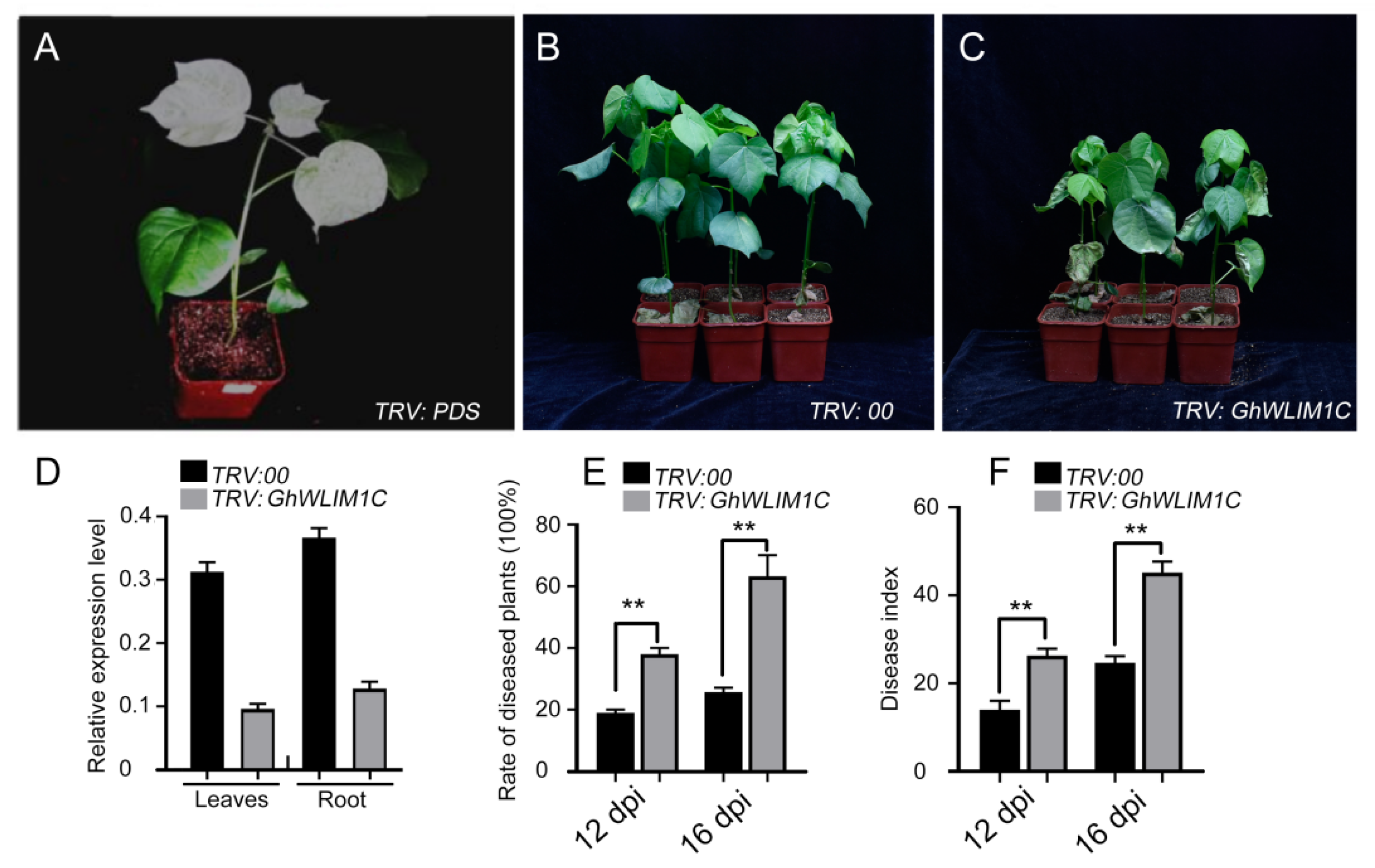
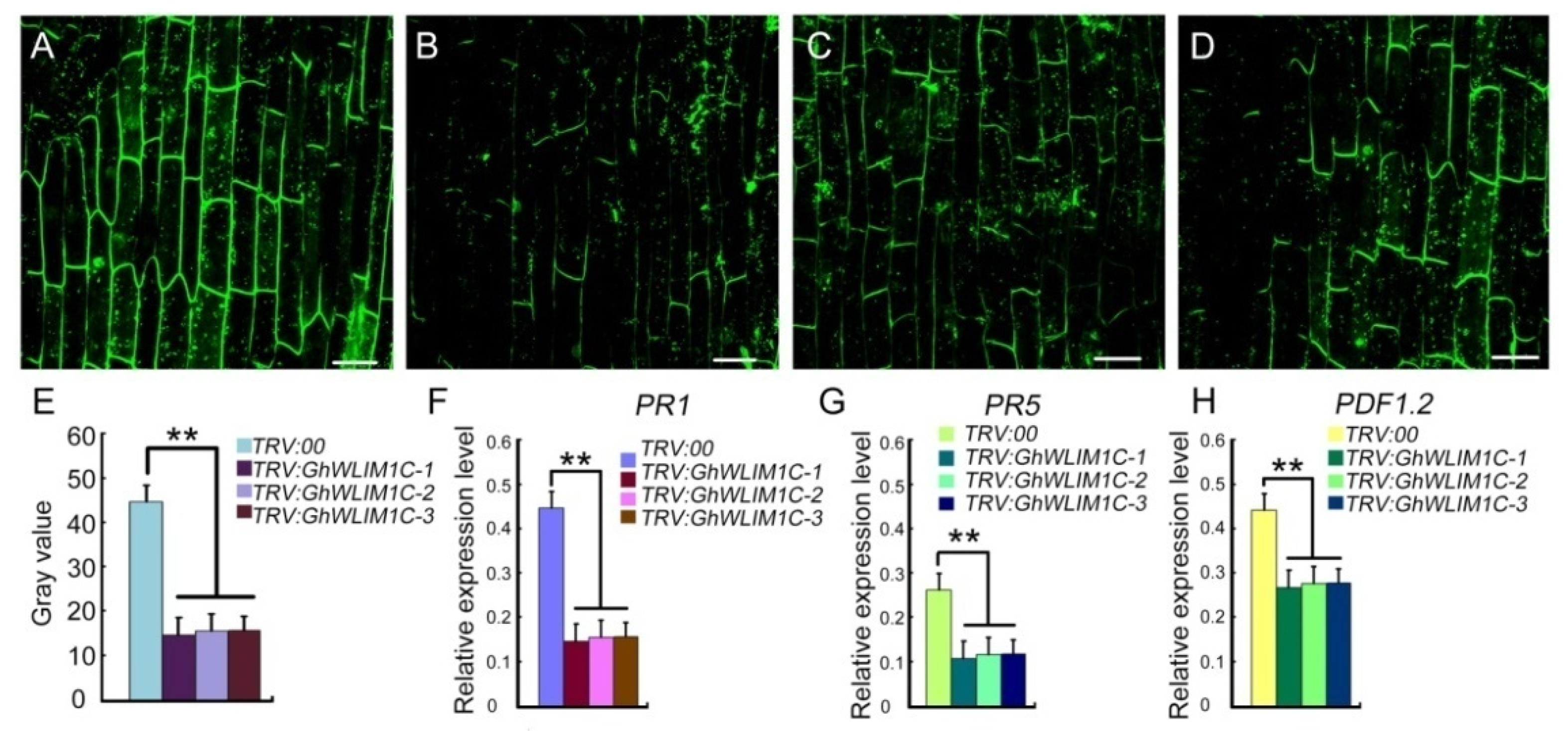
2.2. GhWLIM1C Functions in Defense against V. dahliae Infection

Considering the expression of GhWLIM1C responses to V. dahliae infection, we speculate that it may have a function in fungal disease defense. A virus-induced gene silencing (VIGS) system was employed to investigate the function of GhWLIM1C in defense against V. dahliae infection in G. hirsutum cotton. We preliminary silenced the expression of the cotton phytoene desaturase gene (PDS), which is used as a visible marker to monitor the efficiency of VIGS [28]. As shown in Figure 2A, the new growth of true leaves of plants infiltrated with Agrobacterium carrying PDS exhibited an albino phenotype, indicating that the VIGS system worked efficiently. Next, we then inhibited the expression of GhWLIM1C in G. hirsutum. The qRT-PCR analyses showed that the expression of GhWLIM1C was significantly reduced in the leaves and root tissues in the VIGS plants (Figure 2D). V. dahliae spores were used to infect both the control (TRV:00) and the GhWLIM1C silencing (TRV:GhWLIM1C) plants. Phenotypic analysis indicated that down regulation of GhWLIM1C expression resulted in reduced resistance to the fungal pathogen (Figure 2B,C). The rate of diseased plants and the disease index of GhWLIM1C-silenced plants clearly increased compared with the non-silenced plants (Figure 2E,F).

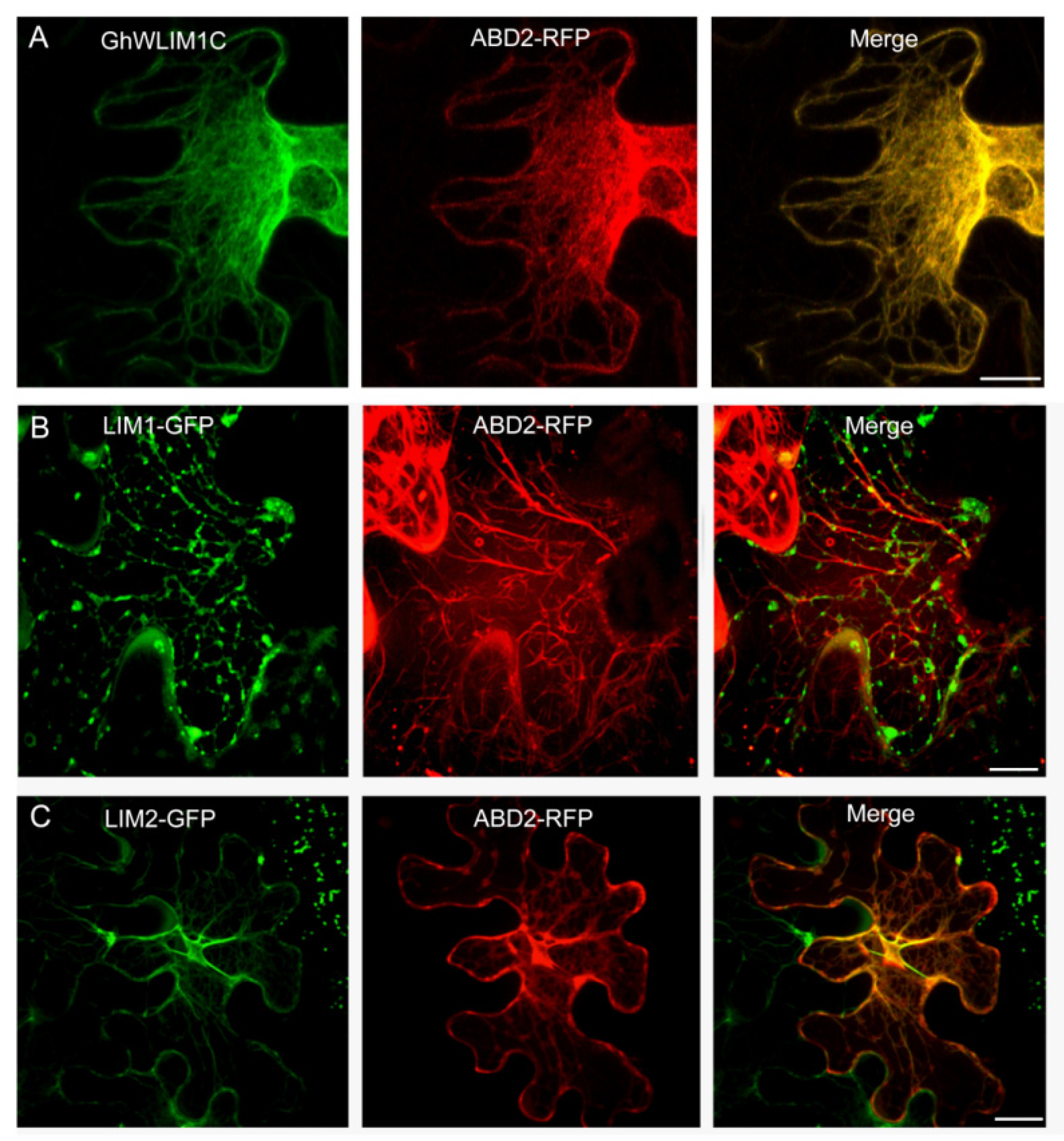
2.3. GhWLIM1C Binds to the Plant Actin Cytoskeleton In Vivo

The subcellular localization of GhWLIM1C was investigated in tobacco cells. We transiently expressed GhWLIM1C fused to GFP in Nicotiana benthamiana epidermal cells. The fluorescence was observed by confocal laser-scanning microscopy. Fluorescence from the GhWLIM1C-GFP fusion protein was detected as filamentous structures similar to the actin filaments. We then additionally expressed ABD2-RFP as the actin cytoskeleton marker to show the filamentous actin. The results showed that GhWLIM1C-GFP could totally co-localize with the ABD2-RFP-labeled actin cytoskeleton (Figure 3A), indicating that GhWLIM1C may function in the actin organization. Further, we also investigated the subcellular localization of the two LIM domains of GhWLIM1C. We constructed LIM1 and LIM2 domain fusion with GFP and expressed them together with ABD2-RFP in tobacco cells. The results showed that the LIM1 domain GFP was visualized as spot-like structures localized on the actin filaments (Figure 3B), and LIM2-GFP could co-localize with the ABD2-RFP-labeled actin cables (Figure 3C). In all, these results suggested that GhWLIM1C and its two LIM domains could bind to the actin cytoskeleton.

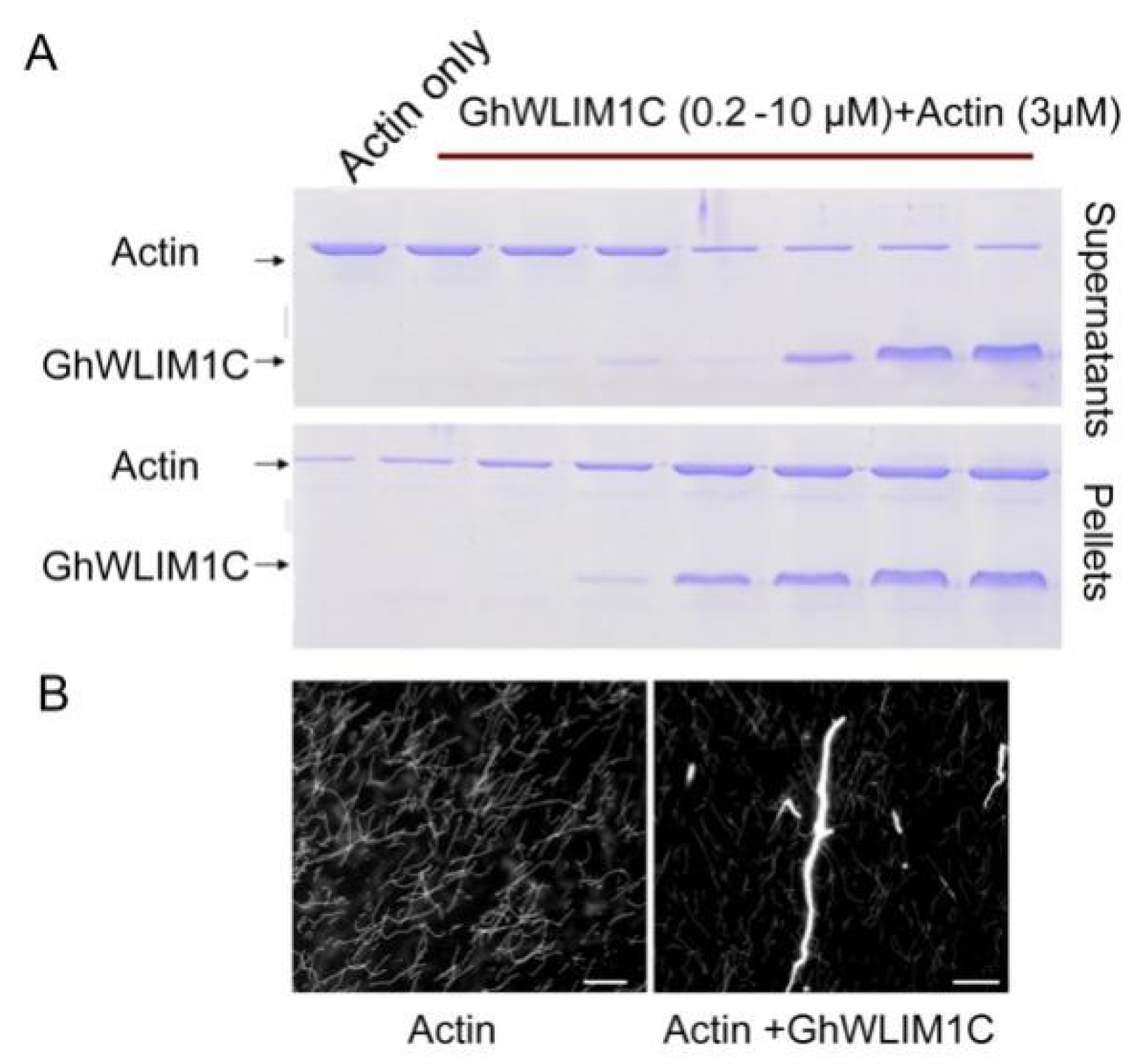
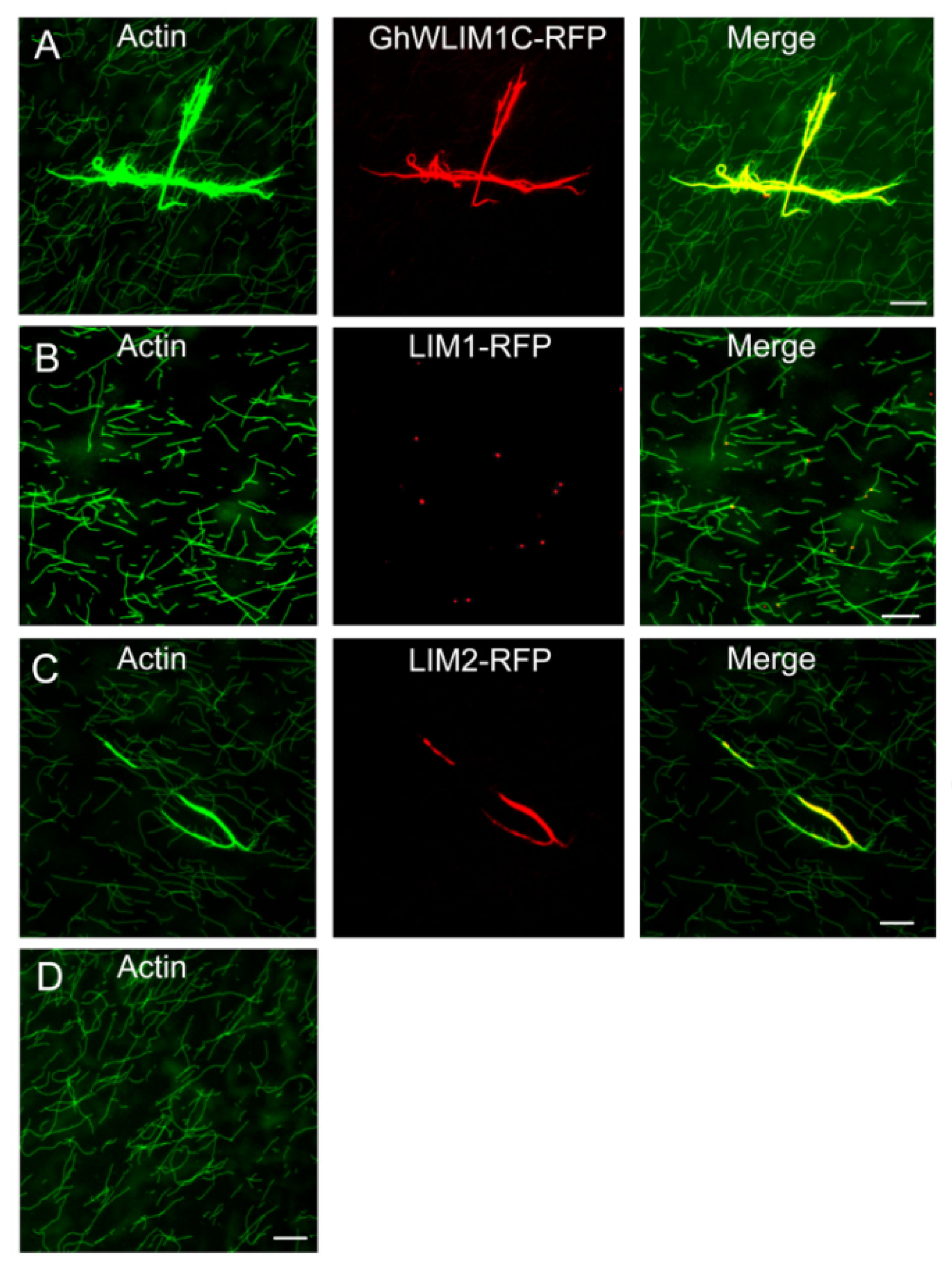
2.4. GhWLIM1C Promotes the Formation of Actin Bundles In Vitro

We performed in vitro experiments to study the biochemical property of GhWLIM1C. We in vitro expressed and purified His-tagged GhWLIM1C from E. coli. A low-speed cosedimentation experiment was conducted to show the property of GhWLIM1C. The rabbit actin was polymerized alone or with an increasing amount of His-GhWLIM1C. After 30 min incubation, the samples were centrifuged at 35,000× g for 30 min. Then, the supernatants and pellets were individually separated by SDS-PAGE. The results showed that only a small portion of actin was detected in the pellets. However, it was gradually increased in the pellets along with the elevated amounts of GhWLIM1C (Figure 4A). We then stained the actin samples with the Alexa488-phalloidin and found that the actin assembles as fine filaments (Figure 4B, left). When GhWLIM1C was added, thick actin cables appeared (Figure 4B, right).
The low-speed cosedimentation experiment shows that GhWLIM1C could bundle the actin cytoskeleton. For further elucidating the mechanism, we in vitro purified His-tagged GhWLIM1C-RFP, LIM1-RFP, and LIM2-RFP. We incubated these proteins with the preassembled actin filaments. Through fluorescence observation, we found that GhWLIM1C formed as robust filamentous structures co-localized with the thick actin bundles (Figure 5A) compared with actin alone forming fine actin filaments (Figure 5D). In addition, we also found that LIM1-RFP was exhibited as spots embedding at the actin filaments (Figure 5B), and LIM2-RFP could form a filamentous structure similar to GhWLIM1C to promote the formation of actin bundles (Figure 5C).

2.5. GhWLIM1C May Function Involving in the Activation of Plant Immune Response

We next explored the mechanism that GhWLIM1C functions in the plant immune response. The experiments above indicated that down regulation of GhWLIM1C in cotton leads to decreased resistance to V. dahliae infection. We then investigated the immune response in the GhWLIM1C-silencing plants. We first analyzed the ROS accumulation in control and the GhWLIM1C-silencing plants. Through H2DCFDA staining and fluorescence observation, we found that at 72 hpi, in the root cells of the GhWLIM1C-silencing plants, less ROS was accumulated compared to the control (Figure 6A–E). Furthermore, we analyzed the expression of several defense-related genes, such as PR1, PR5, and PDF1.,2 in the root tissues of the GhWLIM1C-silencing plants. The results show that the expressions of these genes were significantly down regulated in the GhWLIM1C-silencing plants than in the control (Figure 6F–H).

3. Discussion

Cotton Verticillium wilt is a severe vascular disease that may cause a serious cotton loss in both production and quality. Exploring the key factors involved in the defense against V. dahliae invasion in cotton can provide candidate genes for generating wilt-resistant G. hirsutum cultivars through molecular breeding [29,30]. In this study, we identified a new cotton protein that functions in the plant defense against the fungal pathogen V. dahliae and found that cotton GhWLIM1C protein may positively regulate the fungal defense response.
In eukaryotic cells, the actin-binding proteins (ABPs) are indicated to involve in the actin polymerization, dynamics turnover of actin subunits, organization of the actin filaments to network and bundles, in precisely modulating the dynamic actin cytoskeleton [31]. The LIM domain is a conserved domain found in a variety of different proteins. In animals, LIM proteins differ in their functions due to the additional protein domains and are grouped as LIM-hd, LMO, LIM-kinase, and other LIM proteins [32]. Members of LMO and LIM-hd proteins are usually localized in the cell nucleus to act as transcriptional factors in regulating developmental gene expression [33], while some other LIM proteins (other LIM, CRP1) are indicated in the regulation of F-actin organization [34]. In plants, the LIM protein in tobacco NtWLIM1, cotton WLIM1a, and Arabidopsis’ six LIM proteins was localized at the actin cytoskeleton and in the nucleus [23,27,35]. In addition to binding directly to actin filaments for stabilizing the actin cytoskeleton, tobacco NtWLIM1 was found to be a transcriptional regulator that was able to activate the expression of a b-glucuronidase reporter, and the overexpression of NtWLIM1 led to up regulated expression of PAL-box genes [26]. NtWLIM2 could transcriptionally activate the histone gene, and it shuttles to the nucleus in response to cytoskeletal remodeling [24]. Cotton WLIM1a proteins play dual functions in fiber elongation and secondary cell wall synthesis due to their property of both actin-binding and transcriptional activation of phenolic metabolism [23]. In this study, like the reported plant LIM proteins, GhWLIM1C could also bind to the actin filaments both in vitro and in vivo (Figure 3, Figure 4 and Figure 5). However, in the in vivo experiments, we did not observe an obvious nucleus localization of GhWLIM1C, it seems that it only localized to the actin cytoskeleton (Figure 3A). Considering that GhWLIM1C could both directly bind and bundle the actin filaments in vitro (Figure 4 and Figure 5), we speculate that GhWLIM1C mainly functions in actin remodeling other than gene regulation. A typical plant LIM protein contains two LIM domains, and the two domains were shown to occupy different actin affinities. For example, LIM1 of cotton WLIM1a protein contributed primarily to the actin filament-bundling activity, whereas LIM2 contributed to the DNA-binding activity of the protein [36]. Differently from our experiment, it seems that both LIM1 and LIM2 of GhWLIM1C could bind to the actin filaments (Figure 3B,C; Figure 5B,C); while LIM1 only possesses actin-binding activity (Figure 5B), LIM2 could directly bind and bundle actin filaments (Figure 5C). This indicates that LIM1 and LIM2 may have individual functions though they both contain the same LIM motif.
Emerging evidence showing that the actin cytoskeleton may function crucially in the plant defense response. First, the dynamic rearrangements of the actin cytoskeleton, including the increasing number of actin filamentous and bundling extent elicited after pathogen perception, is a component of plant PTI. Actin filament density and thick actin bundles increase following microbe-associated molecular patterns (MAMP) elf26 treatment, and these dynamic actin changes require pattern recognition receptors sensing the MAMP to trigger the downstream defense response [19]. Secondly, actin could also regulate the strength of the plant’s immunity. The actin cytoskeleton could establish and maintain a signaling-competent microenvironment and traffic plant defense-related organelles, proteins, and small molecules [37]. In our study, we found that GhWLIM1C functions as a pure actin-binding and bundling protein in vivo and in vitro (Figure 3, Figure 4 and Figure 5), and the silencing of this gene leads to decreased resistance to cotton fungal diseases (Figure 2). Thus, we speculate that GhWLIM1C may have a function in the organization of the actin cytoskeleton during plant defense, and its aberrant organization may decrease the downstream immune responses; the generation of ROS in plants cell was reduced, and the expressions of the defense-related genes were weakened (Figure 6). Thus the plant resistances were decreased. Detailed mechanisms about the linking of GhWLIM1C-mediated cytoskeletal rearrangements and plant defense needs further studies. In all, our findings characterized a new actin-related protein, GhWLIM1C, in cotton and provide evidence that GhWLIM1C could regulate plant immune response through regulating actin polymerization.

4. Materials and Methods

4.1. Cotton Growth and V. dahliae Culture and Infection of Cotton Seedlings

Upland cotton seeds TM-1 (Gossypium hirsutum) were grown hydroponically as previously described [38] or cultured in a controlled environment chamber under a 16/8 h photoperiod and at 80% relative humidity. The V. dahliae isolate V991 was used in this study [8]. The fungal strain was grown on potato dextrose agar medium. For suspension spore production, mycelia were cultured in liquid Czapek’s medium at 150 rpm/min, 28 °C for 3–5 days. For cotton inoculation, a spore suspension (1 × 10⁶) conidia/mL with deionized water was used. Then, the suspension was injected into the stem of a cotton seedling (10 days old). Root tissues were harvested at 0, 6, 12, 24, 36, 48, 60, and 72 hpi, frozen in liquid nitrogen, and stored at −80 °C until use.

4.2. RNA Extraction and RT-qPCR Analysis

Total RNA of cotton leaves and root tissues was extracted using a Plant Total RNA Purification Kit (TransGen Biotech) according to the manufacturer’s protocol. The quantitative reverse-transcription PCR assays were performed using SYBR Green Real-Time PCR Master Mix (Toyobo, Japan), with cotton UBI gene as the internal control. All reactions were conducted in triplicate, and primers used in this study are provided in Table S1.

4.3. Virus-Induced Gene Silencing

For silencing of GhWLIM1C in cotton, we employed the virus-induced gene silencing (VIGS) method as described previously [9]. The gene-specific fragments for GhWLIM1C were inserted into pTRV2. Agrobacterium cultures harboring pTRV1 and pTRV2- GhWLIM1C were mixed at a 1:1 ratio and injected into 10-d-old cotton cotyledon.

4.4. Plasmid Construction and Protein Expression

To construct plasmids harboring 35S-GhWLIM1C-GFP, 35S-LIM1-GFP, and 35S-LIM2-GFP, the corresponding cDNA was amplified using gene-specific primers and cloned into the plant expression vectors pCambia1300-GFP, respectively. All constructs were confirmed by sequencing and then transformed into A. tumefaciens strain GV3101. The cDNA fragment contains GhWLIM1C, LIM1, and LIM2 were cloned into the bacterial expression vector PET-28a (Novagen/Merck). The recombinant proteins were expressed in Escherichia coli strain BL21 (DE3). His-tagged LIM proteins were purified using nickel-nitrilotriacetic acid resin following procedures described by the manufacturer (Qiagen). His-tagged GhWLIM1C, LIM1, and LIM2-RFP recombinant proteins were expressed in E. coli and purified following similar procedures as described above. All primers and enzyme sites used in plasmid construction are described in Table S1.

4.5. Low-Speed Cosedimentation Assays

Low-speed cosedimentation assays were conducted as described previously [23]. Rabbit muscle actin (3 µM) was incubated at 22 °C for 60 min either alone or with 3 µM GhWLIM1C in KMEI buffer (10× stock: 500 mM KCl, 10 mM MgCl2, 10 mM EGTA, and 100 mM imidazole, pH 7.0). For the low-speed cosedimentation assay, the samples were centrifuged at 13,500× g for 30 min at 4 °C. The proteins in the supernatants and pellets were separated by SDS-PAGE and visualized with Coomassie Brilliant Blue R250.

4.6. In Vitro Assembly and Observation of the Actin Cytoskeleton

Rabbit muscle actin (3 µM) was incubated at 22 °C for 60 min either alone or with 3 µM GhWLIM1C in KMEI buffer (10× stock: 500 mM KCl, 10 mM MgCl2, 10 mM EGTA, and 100 mM imidazole, pH 7.0). Then, the polymerized F-actin was incubated with LIM proteins (GhWLIM1C, GhWLIM1C-RFP, LIM1-RFP, and LIM2-RFP) for 30 min. After that, these samples were stained with 0.066 µM Alexa488-phalloidin (Molecular Probes) for 10 min. Fluorescence signals were observed under confocal microbiology (Zeiss 880).

4.7. Fluorescent Staining and Confocal Lasers Canning Microscopy

For observation of LIM subcellular localization of GhWLIM1C, LIM1, and LIM2, the GFP-tagged LIM proteins were expressed in N. benthamiana leaves through A. tumefaciens strain GV3101 injection. Then the fluorescent signals were visualized under a confocal microscope after two days of expression.

Supplementary Materials

The following supporting information can be downloaded at: https://www.mdpi.com/article/10.3390/plants11141828/s1, Table S1: Primers used in the study.

Author Contributions

Conceptualization and methodology, validation, formal analysis, investigation, T.C., M.Q. and S.Z.; writing—original draft preparation, T.C. and M.Q.; writing—review and editing, Y.L.; supervision, project administration, funding acquisition Y.L. All authors have read and agreed to the published version of the manuscript.

Funding

The work was supported by grants from the National Natural Science Foundation of China (31900385) and the Distinguished Young Scientists Fund of Fujian Agriculture and Forestry University of China (Kxjq21021).

Institutional Review Board Statement

Not applicable.

Informed Consent Statement

Not applicable.

Data Availability Statement

Not applicable.

Acknowledgments

We thank Yule Liu (Tsinghua University) for kindly providing us with VIGS vector.

Conflicts of Interest

The authors declare no conflict of interest.

References

  1. Bolek, Y.; El-Zik, K.M.; Pepper, A.E.; Bell, A.A.; Magill, C.W.; Thaxton, P.M.; Reddy, O.U.K. Mapping of verticillium wilt resistance genes in cotton. Plant Sci. 2005, 168, 1581–1590. [Google Scholar] [CrossRef]
  2. Cai, Y.F.; He, X.H.; Mo, J.C.; Sun, Q.; Yang, J.P.; Liu, J.G. Molecular research and genetic engineering of resistance to Verticillium wilt in cotton: A review. Afr. J. Biotechnol. 2009, 8, 7363–7372. [Google Scholar]
  3. Vallad, G.E.; Subbarao, K.V. Colonization of resistant and susceptible lettuce cultivars by a green fluorescent protein-tagged isolate of Verticillium dahliae. Phytopathology 2008, 98, 871–885. [Google Scholar] [CrossRef] [PubMed] [Green Version]
  4. Zhao, P.; Zhao, Y.L.; Jin, Y.; Zhang, T.; Guo, H.S. Colonization process of Arabidopsis thaliana roots by a green fluorescent protein-tagged isolate of Verticillium dahliae. Protein Cell 2014, 5, 94–98. [Google Scholar] [CrossRef]
  5. Deng, S.; Wang, C.Y.; Zhang, X.; Wang, Q.; Lin, L. VdNUC-2, the Key Regulator of Phosphate Responsive Signaling Pathway, Is Required for Verticillium dahliae Infection. PLoS ONE 2015, 10, e0145190. [Google Scholar] [CrossRef]
  6. Klosterman, S.J.; Atallah, Z.K.; Vallad, G.E.; Subbarao, K.V. Diversity, Pathogenicity; and Management of Verticillium Species. Annu. Rev. Phytopathol. 2009, 47, 39–62. [Google Scholar] [CrossRef] [Green Version]
  7. Lo Presti, L.; Lanver, D.; Schweizer, G.; Tanaka, S.; Liang, L.; Tollot, M.; Zuccaro, A.; Reissmann, S.; Kahmann, R. Fungal Effectors and Plant Susceptibility. Annu. Rev. Plant Biol. 2015, 66, 513–545. [Google Scholar] [CrossRef]
  8. Li, Y.B.; Han, L.B.; Wang, H.Y.; Zhang, J.; Sun, S.T.; Feng, D.Q.; Yang, C.L.; Sun, Y.D.; Zhong, N.Q.; Xia, G.X. The Thioredoxin GbNRX1 Plays a Crucial Role in Homeostasis of Apoplastic Reactive Oxygen Species in Response to Verticillium dahliae Infection in Cotton. Plant Physiol. 2016, 170, 2392–2406. [Google Scholar] [CrossRef] [Green Version]
  9. Han, L.B.; Li, Y.B.; Wang, F.X.; Wang, W.Y.; Liu, J.; Wu, J.H.; Zhong, N.Q.; Wu, S.J.; Jiao, G.L.; Wang, H.Y.; et al. The Cotton Apoplastic Protein CRR1 Stabilizes Chitinase 28 to Facilitate Defense against the Fungal Pathogen Verticillium dahliae. Plant Cell 2019, 31, 520–536. [Google Scholar] [CrossRef]
  10. Gu, Z.H.; Liu, T.L.; Ding, B.; Li, F.F.; Wang, Q.; Qian, S.S.; Ye, F.; Chen, T.Z.; Yang, Y.W.; Wang, J.Y.; et al. Two Lysin-Motif Receptor Kinases, Gh-LYK1 and Gh-LYK2, Contribute to Resistance against Verticillium wilt in Upland Cotton. Front. Plant Sci. 2017, 8, 2133. [Google Scholar] [CrossRef] [Green Version]
  11. Cheng, H.Q.; Han, L.B.; Yang, C.L.; Wu, X.M.; Zhong, N.Q.; Wu, J.H.; Wang, F.X.; Wang, H.Y.; Xia, G.X. The cotton MYB108 forms a positive feedback regulation loop with CML11 and participates in the defense response against Verticillium dahliae infection. J. Exp. Bot. 2016, 67, 1935–1950. [Google Scholar] [CrossRef] [PubMed] [Green Version]
  12. Qin, J.; Wang, K.; Sun, L.; Xing, H.; Wang, S.; Li, L.; Chen, S.; Guo, H.S.; Zhang, J. The plant-specific transcription factors CBP6Og and SARD1 are targeted by a Verticillium secretory protein VdSCP41 to modulate immunity. Elife 2018, 7, e34902. [Google Scholar] [CrossRef] [PubMed]
  13. Ma, A.F.; Zhang, D.P.; Wang, G.X.; Wang, K.; Li, Z.; Gao, Y.H.; Li, H.C.; Bian, C.; Cheng, J.K.; Han, Y.A.; et al. Verticillium dahliae effector VDAL protects MYB6 from degradation by interacting with PUB25 and PUB26 E3 ligases to enhance Verticillium wilt resistance. Plant Cell 2021, 33, 3675–3699. [Google Scholar] [CrossRef] [PubMed]
  14. Gao, X.Q.; Wheeler, T.; Li, Z.H.; Kenerley, C.M.; He, P.; Shan, L.B. Silencing GhNDR1 and GhMKK2 compromises cotton resistance to Verticillium wilt. Plant J. 2011, 66, 293–305. [Google Scholar] [CrossRef] [Green Version]
  15. Gao, X.Q.; Li, F.J.; Li, M.Y.; Kianinejad, A.S.; Dever, J.K.; Wheeler, T.A.; Li, Z.H.; He, P.; Shan, L.B. Cotton GhBAK1 Mediates Verticillium Wilt Resistance and Cell Death. J. Integr. Plant Biol. 2013, 55, 586–596. [Google Scholar] [CrossRef] [Green Version]
  16. Yang, C.L.; Liang, S.; Wang, H.Y.; Han, L.B.; Wang, F.X.; Cheng, H.Q.; Wu, X.M.; Qu, Z.L.; Wu, J.H.; Xia, G.X. Cotton Major Latex Protein 28 Functions as a Positive Regulator of the Ethylene Responsive Factor 6 in Defense against Verticillium dahliae. Mol. Plant 2015, 8, 399–411. [Google Scholar] [CrossRef] [Green Version]
  17. Porter, K.; Day, B. From filaments to function: The role of the plant actin cytoskeleton in pathogen perception, signaling and immunity. J. Integr. Plant Biol. 2016, 58, 299–311. [Google Scholar] [CrossRef]
  18. Henty-Ridilla, J.L.; Shimono, M.; Li, J.J.; Chang, J.H.; Day, B.; Staiger, C.J. The Plant Actin Cytoskeleton Responds to Signals from Microbe-Associated Molecular Patterns. PLoS Pathog 2013, 9, e1003290. [Google Scholar] [CrossRef] [Green Version]
  19. Henty-Ridilla, J.L.; Li, J.J.; Day, B.; Staiger, C.J. ACTIN DEPOLYMERIZING FACTOR4 Regulates Actin Dynamics during Innate Immune Signaling in Arabidopsis. Plant Cell 2014, 26, 340–352. [Google Scholar] [CrossRef] [Green Version]
  20. Yang, L.; Qin, L.; Liu, G.S.; Peremyslov, V.V.; Dolja, V.V.; Wei, Y.D. Myosins XI modulate host cellular responses and penetration resistance to fungal pathogens. Proc. Natl. Acad. Sci. USA 2014, 111, 13996–14001. [Google Scholar] [CrossRef] [Green Version]
  21. Li, J.J.; Cao, L.Y.; Staiger, C.J. Capping Protein Modulates Actin Remodeling in Response to Reactive Oxygen Species during Plant Innate Immunity. Plant Physiol. 2017, 173, 1125–1136. [Google Scholar] [CrossRef] [PubMed]
  22. Dawid, I.B.; Toyama, R.; Taira, M. Lim Domain Proteins. Cr. Acad. Sci. III-VIE 1995, 318, 295–306. [Google Scholar]
  23. Han, L.B.; Li, Y.B.; Wang, H.Y.; Wu, X.M.; Li, C.L.; Luo, M.; Wu, S.J.; Kong, Z.S.; Pei, Y.; Jiao, G.L.; et al. The Dual Functions of WLIM1a in Cell Elongation and Secondary Wall Formation in Developing Cotton Fibers. Plant Cell 2013, 25, 4421–4438. [Google Scholar] [CrossRef] [PubMed] [Green Version]
  24. Moes, D.; Gatti, S.; Hoffmann, C.; Dieterle, M.; Moreau, F.; Neumann, K.; Schumacher, M.; Diederich, M.; Grill, E.; Shen, W.H.; et al. A LIM Domain Protein from Tobacco Involved in Actin-Bundling and Histone Gene Transcription. Mol. Plant 2013, 6, 483–502. [Google Scholar] [CrossRef] [PubMed] [Green Version]
  25. Zheng, Q.; Zhao, Y. The diverse bilofunctions of LIM domain proteins: Determined by subcellular localization and protein-protein interaction. Biol. Cell 2007, 99, 489–502. [Google Scholar] [CrossRef]
  26. Kawaoka, A.; Kaothien, P.; Yoshida, K.; Endo, S.; Yamada, K.; Ebinuma, H. Functional analysis of tobacco LIM protein Ntlim1 involved in lignin biosynthesis. Plant J. 2000, 22, 289–301. [Google Scholar] [CrossRef]
  27. Thomas, C.; Hoffmann, C.; Dieterle, M.; Van Troys, M.; Ampe, C.; Steinmetza, A. Tobacco WLIM1 is a novel F-actin binding protein involved in actin cytoskeleton remodeling. Plant Cell 2006, 18, 2194–2206. [Google Scholar] [CrossRef] [Green Version]
  28. Pang, J.H.; Zhu, Y.; Li, Q.; Liu, J.Z.; Tian, Y.C.; Liu, Y.L.; Wu, J.H. Development of Agrobacterium-Mediated Virus-Induced Gene Silencing and Performance Evaluation of Four Marker Genes in Gossypium barbadense. PLoS ONE 2013, 8, e73211. [Google Scholar] [CrossRef] [Green Version]
  29. Li, C.; He, X.; Luo, X.Y.; Xu, L.; Liu, L.L.; Min, L.; Jin, L.; Zhu, L.F.; Zhang, X.L. Cotton WRKY1 Mediates the Plant Defense-to-Development Transition during Infection of Cotton by Verticillium dahliae by Activating JASMONATE ZIM-DOMAIN1 Expression. Plant Physiol. 2014, 166, 2179–2194. [Google Scholar] [CrossRef] [Green Version]
  30. Jun, Z.; Zhang, Z.Y.; Gao, Y.L.; Zhou, L.; Fang, L.; Chen, X.D.; Ning, Z.Y.; Chen, T.Z.; Guo, W.Z.; Zhang, T.Z. Overexpression of GbRLK, a putative receptor-like kinase gene, improved cotton tolerance to Verticillium wilt. Sci. Rep. 2015, 5, 15048. [Google Scholar] [CrossRef] [Green Version]
  31. Winder, S.J.; Ayscough, K.R. Actin-binding proteins. J. Cell Sci. 2005, 118, 651–654. [Google Scholar] [CrossRef] [PubMed] [Green Version]
  32. Bach, I. The LIM domain: Regulation by association. Mech. Develop. 2000, 91, 5–17. [Google Scholar] [CrossRef]
  33. Jurata, L.W.; Gill, G.N. Structure and function of LIM domains. Curr. Top Microbiol. 1998, 228, 75–113. [Google Scholar]
  34. Tran, T.C.; Singleton, C.A.; Fraley, T.S.; Greenwood, J.A. Cysteine-rich protein I (CRPI) regulates actin filament bundling. BMC Cell Biol. 2005, 6, 45. [Google Scholar] [CrossRef] [Green Version]
  35. Papuga, J.; Hoffmann, C.; Dieterle, M.; Moes, D.; Moreau, F.; Tholl, S.; Steinmetz, A.; Thomas, C. Arabidopsis LIM Proteins: A Family of Actin Bundlers with Distinct Expression Patterns and Modes of Regulation. Plant Cell 2010, 22, 3034–3052. [Google Scholar] [CrossRef] [Green Version]
  36. Han, L.B.; Li, Y.B.; Sun, Y.D.; Wang, H.Y.; Kong, Z.S.; Xia, G.X. The two domains of cotton WLIM1a protein are functionally divergent. Sci. China Life Sci. 2016, 59, 206–212. [Google Scholar] [CrossRef]
  37. Li, P.; Day, B. Battlefield Cytoskeleton: Turning the Tide on Plant Immunity. Mol. Plant Microbe Interact. 2019, 32, 25–34. [Google Scholar] [CrossRef] [Green Version]
  38. Qu, Z.L.; Wang, H.Y.; Xia, G.X. GhHb1: A nonsymbiotic hemoglobin gene of cotton responsive to infection by Verticillium dahliae. Biochim. Biophys. Acta 2005, 1730, 103–113. [Google Scholar] [CrossRef]
Figure 1. Expression analysis of cotton GhWLIM1C. (A) qRT-PCR analysis of GhWLIM1C expression in cotton roots in response to V. dahliae infection. Total RNA was extracted from cotton roots inoculated with V. dahliae spores (1 × 10⁶ conidia/mL) for 0, 6, 12, 24, 36, 48, 60, and 72 h, respectively, and 0 h indicates cotton plants without inoculation with the fungal spores. Error bars represent SD from three independent experiments (n = 3). Asterisks indicate statistically significant differences, as determined by Student’s t-test (** p < 0.01). The experiments were repeated three times with similar results. (B) qRT-PCR analysis of GhWLIM1C gene expression in root (R), stem (S), leaf (L), petal (P), anther (A), and fiber (F; 12 days post-anthesis) in cotton plants. The expression levels are indicated relative to cotton UBI gene. Error bars represent ± SE of three biological replicates.
Plants 11 01828 g001
Figure 2. GhWLIM1C silencing decreases resistance to V. dahliae infection in cotton. (A) Preliminary assay of the efficiency of VIGS under our experimental conditions. Ten-day-old cotton plants were infiltrated with Agrobacterium carrying TRV:PDS. The photographs were taken 2 weeks after infiltration. (B,C) Phenotypes of control (B) and GhWLIM1C silenced cotton plants (C). Cotton plants were infiltrated with Agrobacterium carrying VIGS-control vector (TRV:00) or (TRV:GhWLIM1C) and inoculated with a suspension of V. dahliae spores. Photographs were taken 2 weeks after V. dahliae infection. (D) qRT-PCR analysis of GhWLIM1C expression in cotton leaves and the root tissues infiltrated with VIGS-control vector (TRV:00) and TRV:GhWLIM1C. Error bars indicate SD from three technical replicates of one biological experiment. The experiments were repeated three times with similar results. (E,F) Rate of diseased plants and disease index of TRV:00 and TRV:GhWLIM1C cotton plants. Error bars represent the SD of three biological replicates (n = 36), and asterisks indicate statistically significant differences, as determined by Student’s t-test (** p < 0.01).
Plants 11 01828 g002
Figure 3. Subcellular localization of GhWLIM1C and its two LIM domains. Colocalization of GhWLIM1C (A), LIM1 (B), LIM2 (C). and ABD2-RFP in the N. benthamiana cells. Agrobacterium cells containing the indicated pair of GhWLIM1C-GFP, LIM1-GFP, LIM2-GFP, and ABD2-RFP plasmids were coinfiltrated into leaves of N. benthamiana. The signal was visualized by confocal microscopy. Bars = 20 µm.
Plants 11 01828 g003
Figure 4. GhWLIM1C bundles actin filaments in vitro. (A) Low-speed cosedimentation assay of the actin-bundling activity of GhWLIM1C. The presence of GhWLIM1C in the pellet indicates its cosedimentation with F-actin. Lane 1, actin alone (3 µM); lanes 2 to 8, actin (3 µM) incubated with 0.2, 0.5, 1, 2, 4, 8, and 10 µM GhWLIM1C, respectively. (B) Actin bundles visualized by fluorescence microscopy. F-actin alone (left); F-actin + 1 µM GhWLIM1C (right). Bars = 1 µm.
Plants 11 01828 g004
Figure 5. Fluorescence microscopy analysis of F-actin-binding/bundling activities of GhWLIM1C and the two LIM domains. Preassembled F-actin incubated with GhWLIM1C-RFP (A), LIM1-RFP (B), and LIM2-RFP (C). Then, the samples were stained with Alexa488-phalloidin and detected with fluorescence microscopy. Actin alone was used as a negative control (D). Bars = 1 µm.
Plants 11 01828 g005
Figure 6. Defense response analysis of GhWLIM1C-silencing plants. (AD) H2DCFDA staining of the ROS in root cells of the TRV:00 (A) and three GhWLIM1C-silencing (BD) cotton plants at 72 hpi. Fluorescence of H2DCFDA was detected with confocal microscopy. Bars = 20 µm. (E) Quantification of the fluorescence in (AD). (FH) qRT-PCR analysis of the expressions of defense-related genes, PR1 (F), PR5 (G) and PDF1.2 (H) in the root tissues of the TRV:00 and three GhWLIM1C silencing cotton plants at 72 hpi. Error bars represent the SD of three biological replicates (n = 36), and asterisks indicate statistically significant differences, as determined by Student’s t-test (** p < 0.01).
Plants 11 01828 g006
Publisher’s Note: MDPI stays neutral with regard to jurisdictional claims in published maps and institutional affiliations.

