1. Introduction
Healthier lifestyles and a healthier environment are new goals of modern civilization. One of the many issues influencing human health is the consumption of less natural and more processed food products. In order to increase the quality, safety, and shelf life of food, industrial processes include the addition of preservatives [
1]. The use of synthetic preservatives for this purpose is extensive, but their impact on human life is under discussion [
2]. One way of dealing with this issue is the replacement of synthetic preservatives with more effective, low-cost ones obtained from natural sources.
Antibiotics represent one of the most important therapies in dealing with infectious diseases. However, extensive use of antibiotics in community settings, hospitals, and agriculture has led to antibiotic resistance. This growing problem has caused a resurgence in the screening of natural products for medicinal uses [
3].
Breast cancer is the second leading cancer-related disease among women. Even though there has been much clinical research, this disease is still the most common cause of cancer death in women worldwide. It has been reported that factors influencing the occurrence of this type of cancer are closely related to estrogens [
4,
5]. Available treatments such as radiotherapy or chemotherapy cause damage to healthy cells as well as to cancer cells and can increase resistance. This can lead to failure of treatment, so the urgent need to discover alternative ways of treating this disease is currently a focus of investigation. Herbal medicine represents one of many ways of dealing with these limitations. It is well known that plants are rich in bioactive compounds that could serve as bases for chemotherapeutic development.
Plants are known to produce wide ranges of secondary metabolites to ensure the survival of their species. One group of plant metabolic products are volatile or essential oils. Essential oils (EOs) have shown increasing potential in the pharmaceutical, food, and cosmetic industries, as they are widely recognized as safe (by the US FDA (Food and Drug Administration) and the EPA (Environmental Protection Agency)) and are already in use in these industries [
6,
7,
8]. Moreover, many studies have characterized EOs as therapeutics (antioxidant, antimicrobial, anti-allergic, antiviral, enzyme inhibitory, insecticidal, anti-tumor, and pro-apoptotic) depending on their chemical compositions, but further studies are still needed to update the current knowledge base.
Commonly known as Spanish marjoram,
Thymus mastichina L. is an endemic species that belongs to the Lamiaceae family and usually inhabits the Iberian Peninsula. It is characterized by leaves arranged in opposite pairs and small zygomorphic and bilabiate flowers [
9,
10]. Traditionally, due to its strong scent of eucalyptus, this plant has been used for treating respiratory, digestive, and rheumatic disorders [
11,
12]. EOs of this aromatic plant consist of a complex mixture of volatile terpenes and are widely used in the perfume and cosmetics industries [
13]. It is well known that the chemical composition of an EO depends on various factors including the plant species, culture, and environmental conditions [
9]. Literature data show that the most abundant EO components of this species are 1,8 cineole and linalool, followed by α-pinene, β-pinene, and α-terpineol [
10,
14]. Previous studies have indicated antibacterial, antifungal, anti-inflammatory, antioxidant, anticancer, antiviral, insecticidal, insect-repellent, and anti-enzymatic (anti-Alzheimer’s, α-amylase, and α-glucosidase) effects of EOs obtained from the aerial parts of this plant, indicating their potential use in the food and pharmaceutical industries [
10,
13,
14,
15].
Green or true cardamom (
Elletaria cardamomum L. Maton), belonging to the Zingiberaceae family, is a plant native to India and Sri Lanka but also cultivated in Guatemala, Nepal, Indonesia, Costa Rica, Mexico, and Tanzania [
16,
17,
18,
19]. Plants of this species can grow up to 8-foot-high shrubs with thick, fleshy, lateral roots [
20]. Traditionally, it is known as the “Queen of Spices”, since its dried fruit is highly priced as a spice around the world, and in India it is considered the second essential “national spice” [
21,
22]. Due to its characteristic aroma, this plant is widely used in the food and cosmetics industries as a flavoring and fragrance agent. Moreover, from the pharmaceutical point of view, it is used to treat various disorders, such as gum infections, asthma, bronchitis, nausea, and cataracts, as well as cardiac, digestive, and kidney diseases [
16,
17,
18,
19,
20,
21]. The biological effects of cardamom are in close relationship with their volatile composition. Cardamom EOs are rich in volatiles that have therapeutic benefits, such as ester α-terpinyl acetate, and monoterpenes 1,8-cineole, limonene, linalool, terpinolene, myrcene, and α-pinene, which are responsible for these EOs’ effectiveness in curing different ailments [
16,
17,
18]. Literature data show that EOs obtained from this plant show various biological effects such as antioxidant, antihypertensive, antidiabetic, gastroprotective, laxative, antispasmodic, antibacterial, anti-platelet-aggregation, and anticancer activities [
16,
17,
18,
19,
23].
Therefore, herein, the chemical composition; effects on cell viability, redox homeostasis, and migratory capacity of the breast cancer MDA-MB-468 cell line; and antibacterial, and antibiofilm activity of Thymus mastichina and Elletaria cardamomum EOs are investigated, clarified, and discussed compared to results from other previously conducted studies.
3. Discussion
There are many reports in the literature of studies involving EOs obtained from
T. mastichina and
E. cardamomum. Previously reported studies of the chemical composition of
T. mastichina indicate different chemotypes, defined by main compounds present in high amounts: 1,8-cineole, linalool, and 1,8-cineole/linalool [
13]. Considering this, our results reveal that the
T. mastichina EO investigated in this study was of the 1,8-cineole chemotype. Studies on
E. cardamomum mainly suggest α-terpinyl acetate and 1,8-cineole as major constituents of this EO, which is in accordance with our results [
16,
24,
25]. Additionally, some reports indicate that these two components are responsible for the fragrance of this EO [
26]. Previously published studies on EOs of
E. cardamomum have reported between 11 and 67 compounds identified in total. Slight differences have been noted throughout the literature regarding other compounds present in high amounts. In some studies, 4-terpinen-4-ol and sabinene were detected; in others, linalool, dihydrocarveol, geraniol,
Z-caryophyllene,
E-nerolidol, eugenol, and terpinen-4-ol, or sabinene, 4-terpinen-4-ol, and myrcene; in still others, linalool acetate, sabinene, and linalool, in line with our present findings [
19,
24,
27,
28]. Reported differences in the chemical profiles of both plant species could affect the observed biological activities of their essential oils, and are observed as a result of different environmental factors influencing plant development as well as the part of the plant used to obtain the EOs.
Medicinal, aromatic plants have been used for centuries for their beneficial properties. Due to their high contents of bioactive secondary metabolites, they have been especially used to treat a variety of human diseases [
29]. The current concentrations of conventional chemotherapeutic drugs used in therapies could potentially be reduced if combined with specific doses of EOs, which could also decrease chemotherapy-associated toxicity. However, there is still a lack of preclinical studies of EOs as effective anticancer agents and protective components, requiring further extensive safety and toxicity studies of EOs prior to their applications in clinical trials [
30]. EOs have been shown to possess a wide range of anticancer properties and mechanisms of action. Breast cancer is the most common cancer in women globally, with an increasing incidence and persistently high level of mortality [
31,
32,
33]. In attempts to solve this problem, EOs are being considered promising agents for novel anticancer therapies to overcome the side effects and the high cost of chemotherapy approaches in dealing with breast cancer [
34]. Considering the number of bioactive components present in EOs, as well as their synergistic actions, it is of high importance to perform further studies regarding content evaluation and the contributions of individual EO components to the overall biological effects of these mixtures. Selective specificity of action against cancer cells is highly required due to a lack of conventional chemotherapeutic strategies [
30].
It has been suggested that the preventive effect of EOs against cancer disorders could be related to the promotion of cell cycle arrest, stimulating cell apoptosis and DNA repair mechanisms while inhibiting cancer cell proliferation, metastasis formation, and multidrug resistance. All the above-mentioned factors make EOs potential candidates for supporting anticancer therapeutic agents [
35].
Reactive oxygen species produced via various metabolic pathways in the tissues damage the cells by binding to cell components such as carbohydrates, proteins, and DNA. The antioxidant activity of
T. mastichina has also been widely explored through different assays, representing an interesting alternative to synthetic antioxidants. In some studies, these effects were related to the sample’s composition and tests were conducted to understand whether some compounds were primarily responsible for the observed activity [
14].
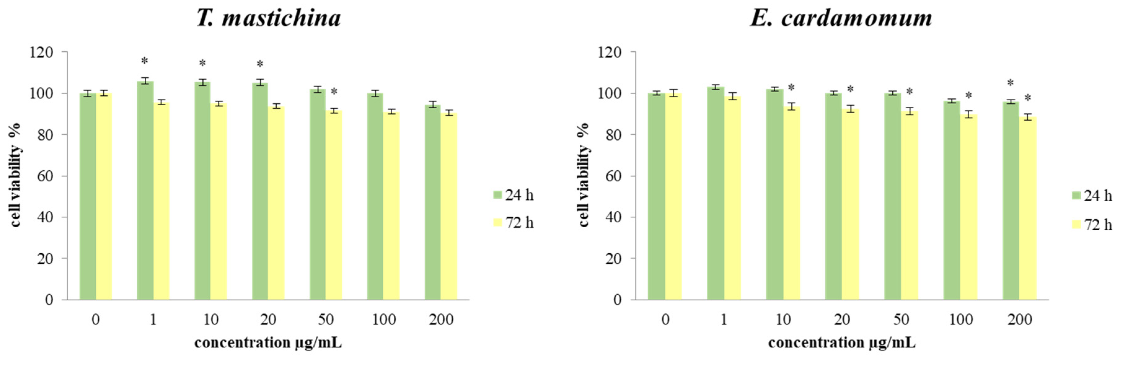
Results obtained after 24 h and 72 h of incubation with various concentrations of both essential oils generally indicated a slight proliferative effect compared to the nontreated cells after 24 h of incubation with
T. mastichina EO, but dose-dependent inhibition of cell viability was detected after 72 h treatment with both EOs, indicating considerable antitumor outcomes. The observed antiviability outcomes could be due to the reduced proliferative potential of the tested cells and to the proapoptotic effects of the main constituents of the EOs. Certain studies have also reported the antiproliferative activity of
T. mastichina EO against human breast carcinoma cell lines, which could be related to 1,8-cineole content [
13,
36]. However, in the case of complex mixtures, it is more likely that a synergy between various components is responsible for the expressed effects.
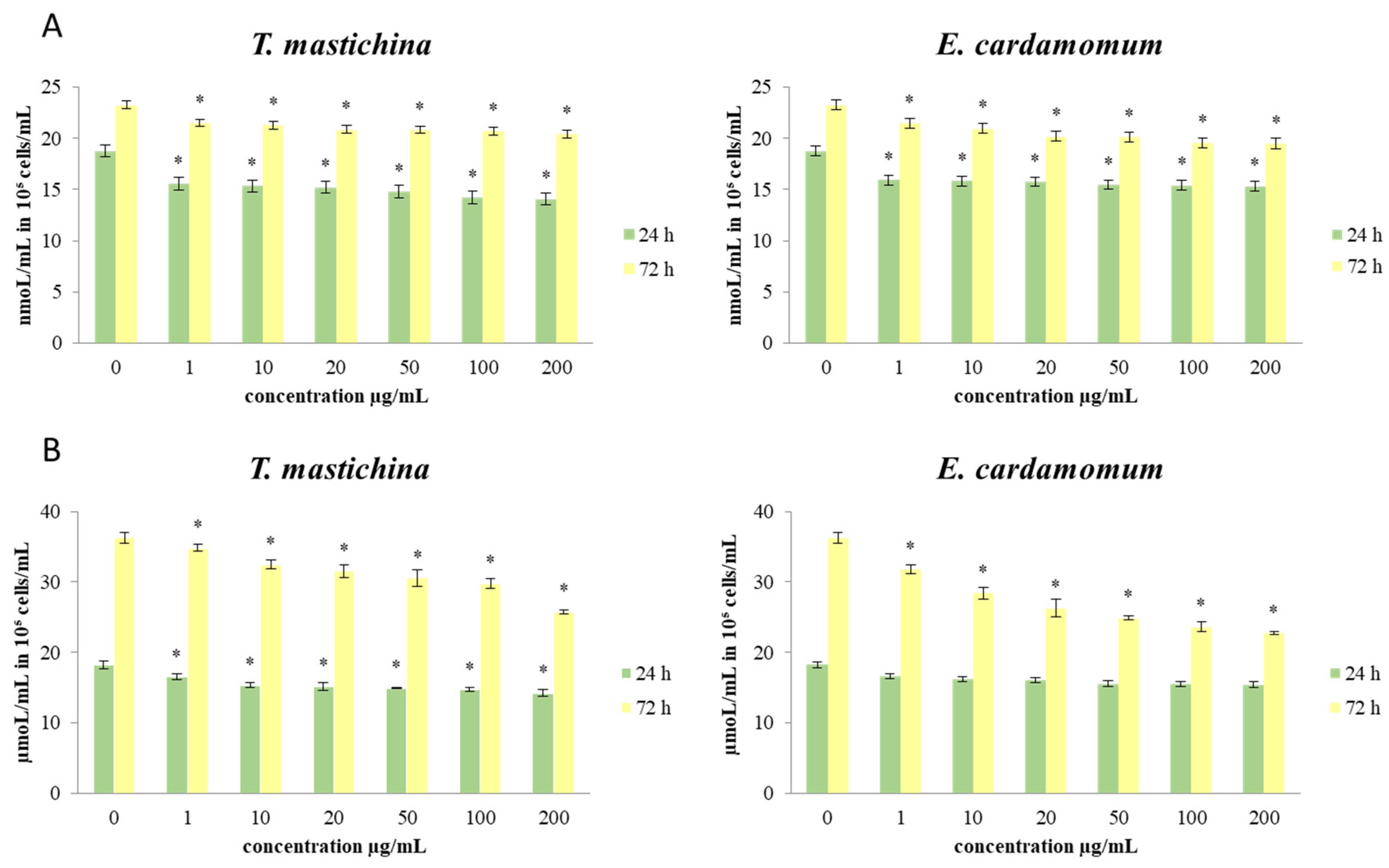
Reactive oxygen species (ROS) appear to be involved in the regulation of various physiological pathways, including signal transduction and differentiation. Recently, emerging evidence has suggested the involvement of ROS and the aberrant activation of redox-sensitive signaling pathways in tumor invasion and migration. In our study, significant dose-dependent and time-dependent antioxidative effects were shown in cells treated both EOs compared to the control cells. Some antioxidants may enhance the effects of cytotoxic regimes, improving the response rate of tumors to chemotherapeutic agents, while some others can ameliorate their antitumor activity [
37]. Disturbance of oxidative homeostasis is one of the major features of cancer cells in general, so our data indicate that the exerted antioxidant impact could be crucial in the breast cancer cell survival and viability detected in the study.
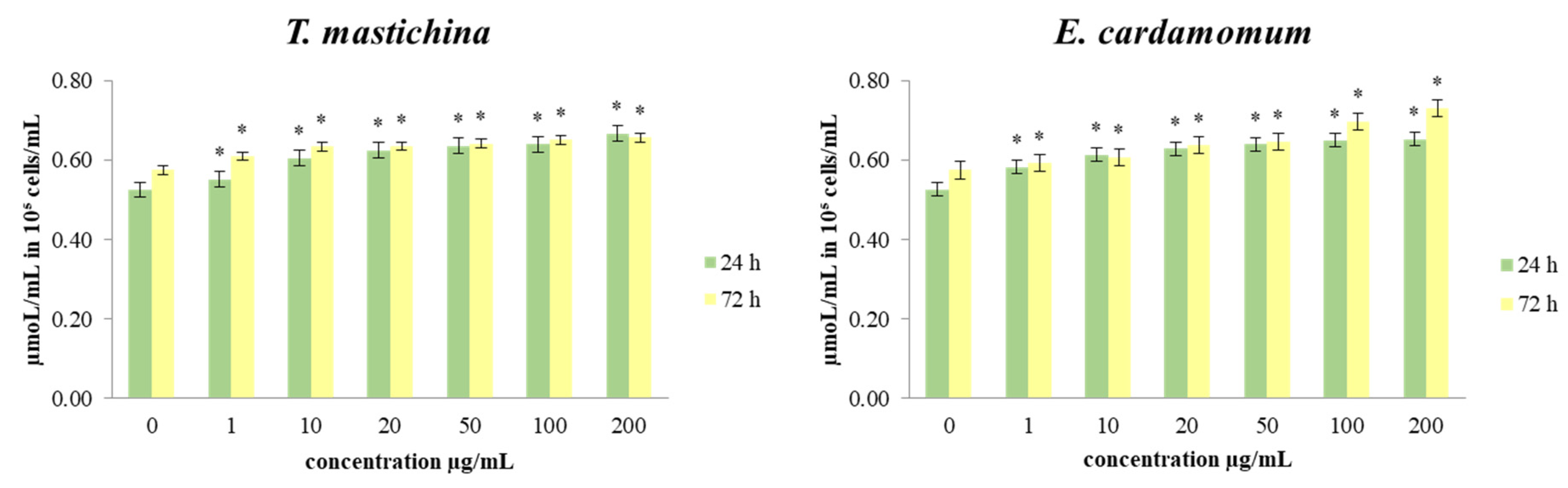
Nitric oxide (NO) has been suggested to possess both antitumor and protumor properties, depending on tissue type and timing [
38]. MDA-MB-468 cells treated with
T. mastichina EO showed a significant decrease in the production of nitrite, the main indicator of NO concentration, compared to the control after both treatment times. However,
E. cardamomum EO only induced a significant drop of NO level after long-term treatment. Changes in the production of NO could affect various signaling pathways that involve nitric oxide, leading to potential antitumor outcomes. The recorded NO decrease could be due to transcriptional and post-translational regulation of iNOS enzyme activity, consequently affecting various protumor signal pathways, but could also inhibit angiogenesis and blood supply in the tumor tissue, thus attenuating tumor growth.
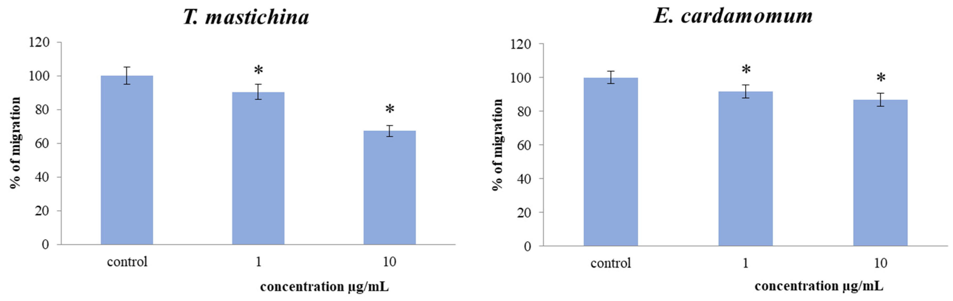
The tested EOs’ actions on migration capacity in breast cancer cells were assessed by 2D transwell migration assay. The results indicated a significant dose-dependent decrease in the cell migration index of MDA-MB-468 cells exposed to both EOs compared to the nontreated cells. The inhibitory effects on NO production detected in the study could be one of the mechanisms of antimigratory potential of the tested EOs, since numerous studies indicate that the reduction of nitric oxide levels can inhibit cell migration [
39,
40].
Previously published studies of the antimicrobial activity of
T. mastichina EO indicate that a higher concentration of linalool is responsible for better antimicrobial properties [
10,
13]. Since the EO tested in the present study was from the 1,8-cineole chemotype, lower amounts of linalool could have contributed to weak antimicrobial effects. The observed effect could also be a product of antagonistic and synergistic effects of the various other constituents present in this EO. However, studies conducted by Faleiro et al. showed moderate antimicrobial activity of Algarve
T. mastichina EOs from the 1,8-cineole chemotype measured by the agar disc diffusion method. In this study, Algarve
T. mastichina EOs showed the best activity against G
+ S. aureus, showing zone of inhibition diameters of 13.7 mm and 15.7 mm for the flower and leaf essential oils [
41]. Using the same method, Ballester-Costa et al. investigated the antimicrobial activity of
T. mastichina EO of the same chemotype against several bacterial strains. They confirmed that this EO had inhibitory activity against eight bacterial strains, but the highest activity was observed against G
+ L. innocua and G
− A. faecalis [
42]. In the same study, the authors observed better activity of this EO when using a microdilution assay, i.e., the less sensitive bacteria strains in the agar disc diffusion method showed higher susceptibility in the microdilution assay [
42]. Cutillas et al. investigated the antimicrobial activities of four different
T. mastichina EOs using a microdilution assay. Their results also suggested weak inhibition of growth of
E. coli,
S. aureus, and
C. albicans by both chemotypes (linalool and 1,8-cineole), with MIC values from 2.3 mg/mL to 9.4 mg/mL [
10].
Literature reports indicate that
E. cardamomum EO varieties exhibit moderate-to-high activity against selected bacteria and fungi using different antimicrobial bioassays. Tarfaoui et al. investigated the antimicrobial activity of
E. cardamomum EOs on several bacterial (
S. aureus,
S. epidermidis,
E. coli,
K. pneumoniae,
P. mirabilis,
P. aeruginosa, and
A. baumannii) and yeast (
C. tropicalis and
C. albicans) strains [
24]. Using a disc diffusion assay, the best antimicrobial effects among the bacterial strains were observed against
S. aureus and
S. epidermidis, with inhibition zones of 20 mm and 14 mm, respectively. The effects of this EO against both yeast strains were similar, showing a zone of inhibition of 13 mm for
C. albicans and 12 mm for
C. tropicalis. The results obtained in this study using a microdilution assay only confirmed the findings of disc diffusion assay. The overall conclusion was that G
+ bacteria are more sensitive to treatment compared to G
− bacterial strains [
24]. The same conclusion was drawn in the more recent study performed by Al-Zereini et al. [
43]. The authors noticed the better antimicrobial activity of
E. cardamomum EOs against G
+ B. subtilis compared to G
− E. coli and
E. aerogenes, which were not inhibited up to the maximum applied concentration in agar diffusion and microbroth dilution assays.
The antibacterial activities of T. mastichina and E. cardamomum EOs were expressed in terms of zones of inhibition in mm and minimal inhibition concentrations in µL/mL. The results obtained in this study using the disc diffusion method showed a weak antimicrobial effect of the EO from T. mastichina against all tested microorganisms. However, the EO from E. cardomomum showed moderate antimicrobial activity against four microorganisms and a weak effect against six microorganisms. The EO obtained from T. mastichina was the most effective against C. glabrata, while E. cardamomum showed the best effect against the biofilm-producing bacterium P. fluorescens. Additionally, the results obtained in this study, unlike previous findings, showed that the E. cardamomum EO displayed better effects against G− bacterial strains compared to G+ ones. However, in line with previously published data, G− E. coli was resistant to treatment with this EO.
The antibiofilm activities of EOs have been the focus of recent investigations [
44,
45,
46]. It is known that bacterial biofilms can protect bacteria against antimicrobial pressures by taking the form of a physical barrier. Biofilm formation makes bacteria highly resistant to environmental stress compared to planktonic bacteria of the same species [
47]. Similarly, it is considered that the planktonic single-celled state is a transitional phase, while the biofilm is in most cases the usual mode of bacterial growth. Various studies have reported abnormalities in protein production associated with biofilm formation and degradation after treatment with EOs [
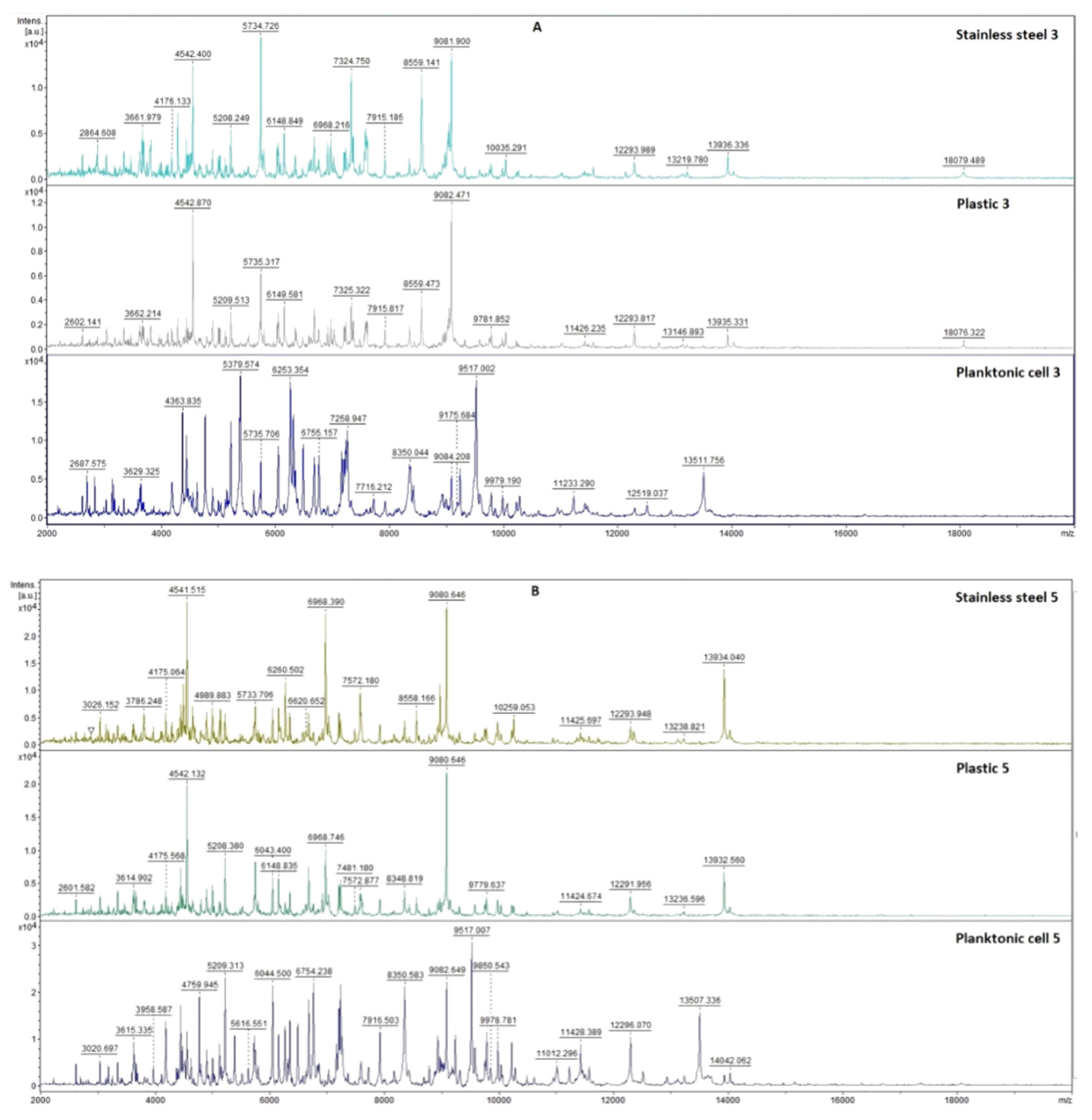
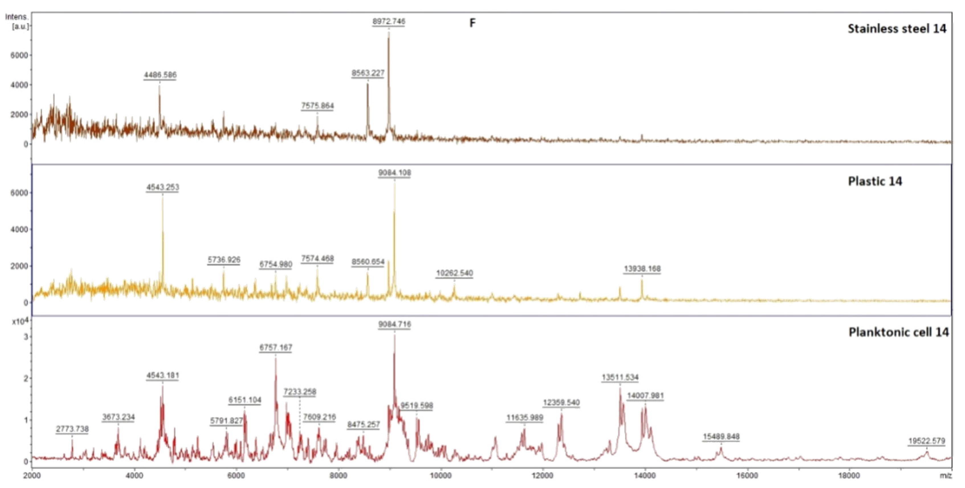
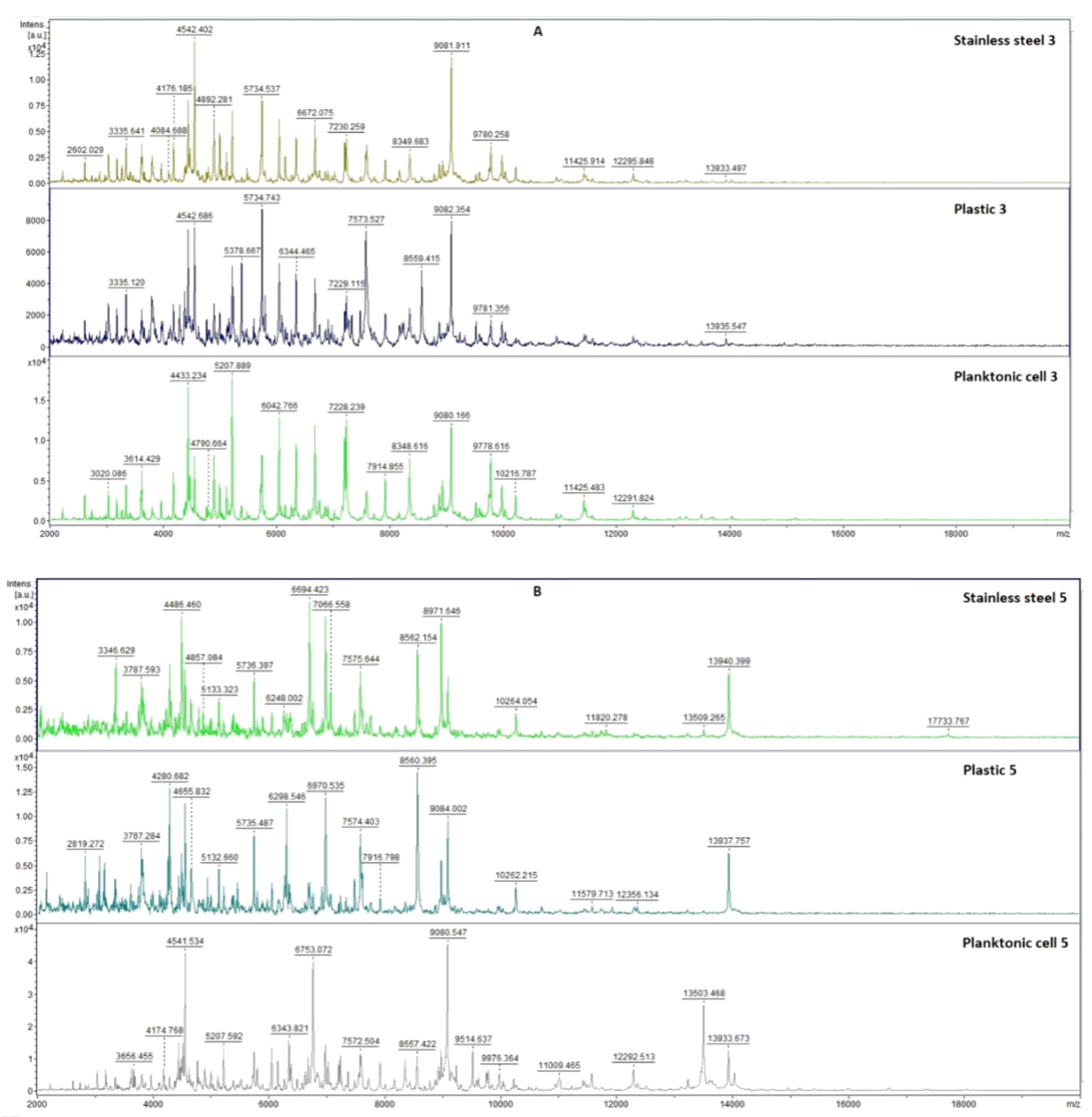
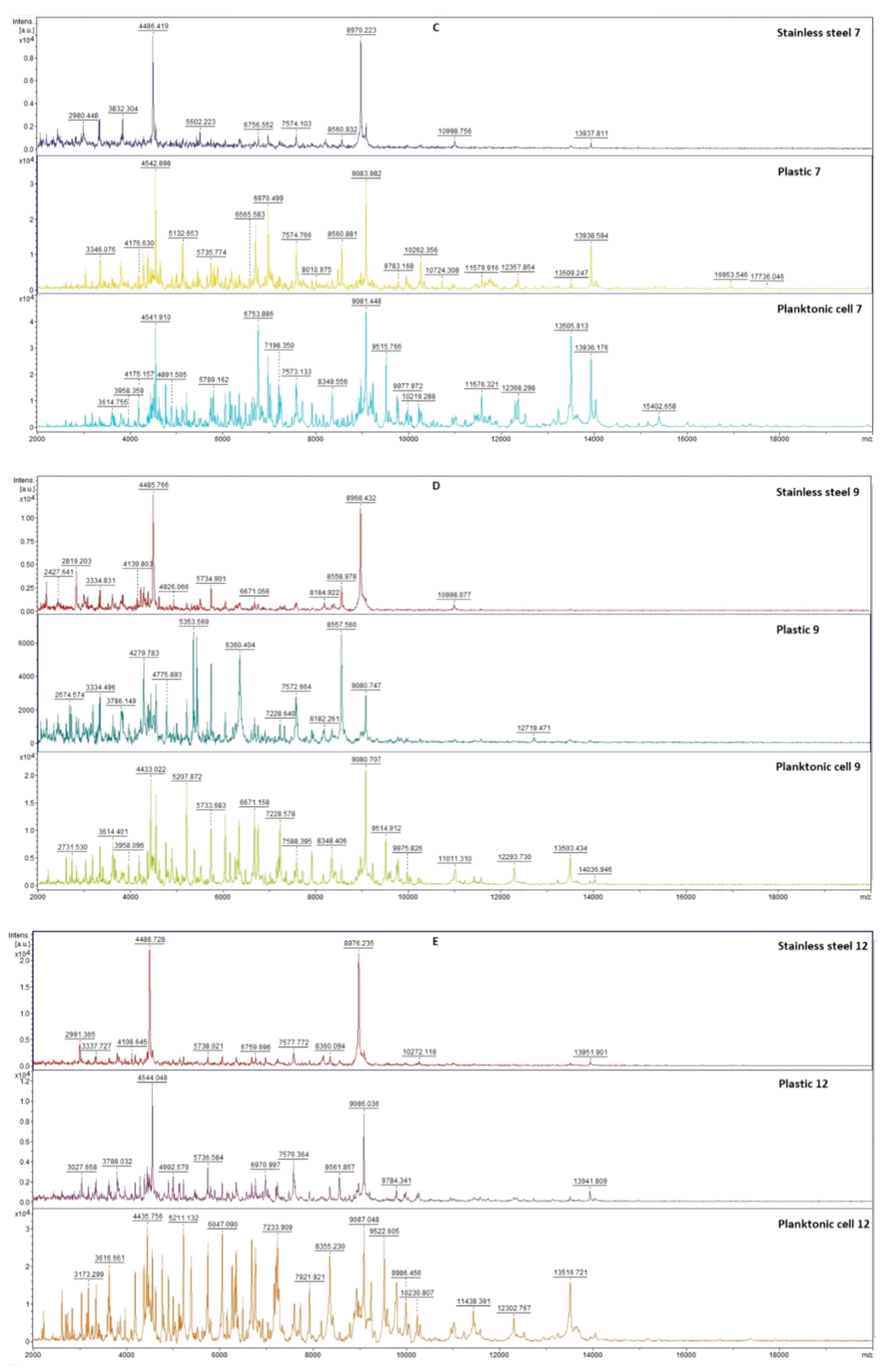
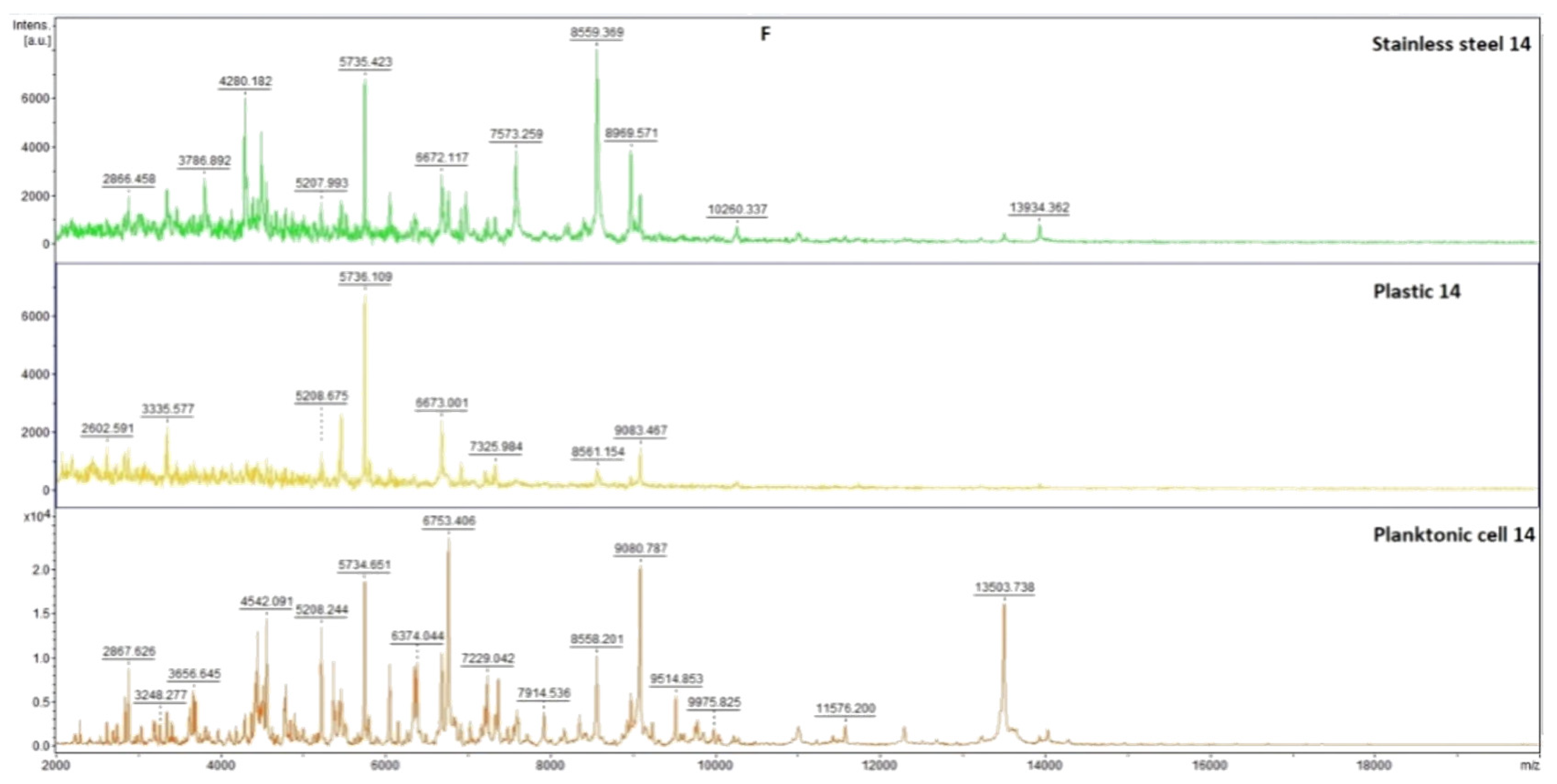
48]. Those abnormalities can be observed using MALDI-TOF mass spectra by comparing untreated samples to treated ones, which was used in this study. Herein, for the first time, we examined the effects of
T. mastichina and
E. cardamomum EOs on biofilm-forming
P. fluorescens bacteria on different surfaces. The results showed changes in the biofilm protein profile of the
P. fluorescens after treatment with these two EOs. It can be concluded that
T. mastichina and
E. cardamomum EOs affected the homeostasis of bacterial biofilms formed on stainless steel and plastic surfaces. Previous studies revealed that treatment with
T. mastichina EO prevented the formation of biofilms by
P. aeruginosa and
S. aureus [
49]. Additionally, E. cardamomum EO showed high antibiofilm potential against biofilms formed by
E. coli and
B. subtilis [
25]. In another study, this EO inhibited biofilm formation inhibition by
E. coli and
S. typhimurium [
50]. Even though many studies have shown an inhibitory activity of EOs on the formation of biofilms, the mechanism behind their antibiofilm activity is generally not understood well to date. The current predictions suggest that the inhibition of some enzymes that are involved in the formation of biofilm could be responsible for EOs’ activity [
25].
4. Materials and Methods
4.1. Essential Oils
Thymus mastichina and Elettaria cardamomum EOs were purchased from Hanus, s.r.o. (Nitra, Slovakia) and were prepared by steam distillation of dried flowering stalks. They were stored in the dark at 4 °C throughout the analysis.
4.2. Gas Chromatography–Mass Spectrometry and Gas Chromatography Analyses of T. mastichina and E. cardamomum
Identification of volatile constituents in EO samples was performed on an Agilent Technologies (Palo Alto, Santa Clara, CA, USA) 6890 N gas chromatograph. The chromatograph was equipped with a quadrupole mass spectrometer 5975 B (Agilent Technologies, Santa Clara, CA, USA), using an HP-5MS capillary column (30 m × 0.25 mm × 0.25 µm). The chromatograph was interfaced and operated by HP Enhanced ChemStation software (Agilent Technologies). The injection volume of the EO sample diluted in hexane (10% solution) was 1 µL. The carrier gas used was helium 5.0, with a flow rate of 1 mL/min. Split/splitless injector temperature was set at 280 °C, MS source and MS quadruple temperature were set at 230 °C and 150 °C, respectively, and the mass scan range was 35–550 amu at 70 eV. The solvent delay time was 3.20 min for EO sample analysis. For n-alkanes (C7–C35), solvent delay time was 2.30 min in order to obtain the retention index for n-heptane.
GC and GC-MS analysis of the T. mastichina sample for Kovats retention index calculations was done under the following chromatographic conditions: temperature program of 50 °C to 90 °C (rate of increase 3 °C/min), held 2 min at 90 °C, 90 °C to 130 °C (rate of increase 4 °C/min), and 130 °C to 290 °C (rate of increase 5 °C/min); the total run time was 57 min, and split ratio was 40.8:1. For the purpose of experimental determination of Van den Dool retention indices, some minor changes were made to the chromatographic conditions. The temperature program was 60 °C to 260 °C with an increasing rate of 3 °C/min, the total run time was 67 min, and the split ratio was 20:1.
The chromatographic conditions for GC and GC-MS analysis of the E. cardamomum sample for Kovats retention index calculations were as follows: temperature program of 50 °C to 70 °C (rate of increase 3 °C/min), held 3 min at 70 °C, 70 °C to 120 °C (rate of increase 4 °C/min), and 120 °C to 290 °C (rate of increase 5 °C/min); total run time was 56 min and split ratio was 40.8:1. Analysis of Van den Dool retention indices included the following chromatographic conditions: temperature program of 60 °C to 260 °C with a rate of increase of 3 °C/min, total run time of 67 min and split ratio of 20:1.
The volatile components of the analyzed EOs were identified by comparison of their retention indices (RI) as well as the reference spectra reported in the literature and the ones stored in the MS library (Wiley7Nist) [
51,
52]. Semiquantification of the components was performed via GC-FID using the same HP-5MS capillary column, taking into consideration amounts higher than 0.1%.
4.3. Cell Culture and Treatment
The human breast cancer cell line MDA-MB-468 was obtained from the American Tissue Culture Collection. The cells were cultivated in DMEM with 10% FBS and an antibiotic mixture (100 IU/mL penicillin and 100 µg/mL streptomycin) to the confluence of 70 to 80%. The cells were placed in a 96-well microplate (10,000 cells per well) and propagated in a humidified atmosphere with 5% CO2 at 37 °C. After 24 h of incubation, 100 μL measures of medium containing different concentrations of EOs of T. mastichina and E. cardamomum (1 µg/mL to 200 µg/mL) were added and the cells were incubated for 24 h and 72 h, after which the measurements of cell viability, redox balance parameters, and migration capacity were conducted. Nontreated cells were used as a control, and all treatment concentrations were obtained by serial dilutions of stock solution. All concentrations were applied in triplicate for all the methods.
4.4. Determination of Cell Viability (MTT Assay)
The viability of the cells was determined using an MTT assay [
53]. The cells were plated at a density of 100,000 cells/mL (100 µL/well) in 96-well plates with DMEM. After an incubation period of 24 h at a temperature of 37 °C in a 5% CO
2 atmosphere, six different concentrations of EOs ranging from 1 to 200 µg/mL were added to the wells at 100 µL volume per well. The untreated cells served as a control. Both treated and control cells were incubated for 24 and 72 h, after which the cell viability was determined via MTT assay. After a period of incubation, 20 µL of MTT (concentration of 5 mg/mL) was added to each well. MTT is a yellow tetrazolium salt that is reduced to purple formazan in the presence of mitochondrial dehydrogenase. During this reaction, which started after approximately three hours, the formed crystals were dissolved in 20 µL of DMSO. The color formed in the reaction was measured using an ELISA reader at a wavelength of 550 nm. The percentage of viable cells was calculated as the ratio between the absorbance at each dose of the treatment and the absorbance of the nontreated control multiplied by 100 to give a percentage.
4.5. Measurement of Superoxide Anion Radical (NBT Test)
The concentrations of superoxide anion radical (O
2•−) in the samples were determined via the spectrophotometric method, which is based on the reduction of nitroblue tetrazolium (NBT) to nitroblue-formazan in the presence of O
2•− [
54]. The assay was performed by adding 20 μL of 5 mg/mL NBT to each well, followed by cell incubation for 1 h at 37 °C in 5% CO
2. To quantify the formazan production, formazan was solubilized in 20 μL DMSO. The absorbances were measured using an ELISA microplate reader at 550 nm. The concentrations of O
2•− were expressed as nanomoles per milliliter (nmoL NBT/mL) in 10
5 cells/mL.
4.6. Measurement of NO Concentration (Griess Method)
The spectrophotometric determination of nitrites (NO
2−) as an indicator of the nitric oxide (NO) level was performed using the Griess method [
55]. The concentration of NO
2− is directly proportional to the intensity of the purple color measured by an ELISA reader at 550 nm. THe Griess reaction is based on the coupling of NO-generated diazonium ion with N-(1-napthyl) ethylenediamine, wherein a chromophoric product is formed. Equal volumes of 0.1% (1 mg/mL)
N-1-napthylethylenediamine dihydrochloride and 1% (10 mg/mL) sulfanilamide solution in 5% phosphoric acid were mixed to form the Griess reagent immediately prior to application to the plate. Over 10 min of incubation (at room temperature, protected from the light sources), a purple color developed. After incubation, absorbances were measured and the nitrite concentrations were expressed in μmoL NO
2−/mL in 10
5 cells/mL.
4.7. Total Glutathione Concentration
The determination of total glutathione was based on the oxidation of reduced glutathione using the DTNB reagent to form a yellow TNB product [
56]. The method was performed on cells seeded in a microtiter plate (10,000 cells in 100 μL). After a period of incubation with treatment, the supernatant was removed, 150 μL of 2.5% sulfosalicylic acid was added, and the plate was sonicated. After sonication, 50 μL of supernatant was reacted with 50 μL of the reaction mixture (1 mM DTNB, 1 mM NADPH, and 0.7 U glutathione reductase in 100 mM phosphate buffer). After 5 min of incubation in the dark at room temperature, absorbances were read using an ELISA reader, and the concentration of total glutathione was expressed as μmoL/mL in 10
5 cells/mL.
4.8. Transwell Assay for Cell Migration
The cell migration capacity was determined by the ability of cells to pass through the pores of polycarbonate membranes (pore size 8 µm; Greiner Bio-One, Gallen, Switzerland) at the bottom of transwell chambers. The migration test was performed according to the protocol described by Chen [
56]. The cells were exposed to 1 µg/mL and 10 µg/mL concentrations of both essential oils for 72 h. The control cells were cultured in DMEM only. After the treatment exposures, all groups of treated cells were trypsinized and placed in the upper chambers at a density of 100,000 cells/well in 500 µL of DMEM with 10% FBS. The lower chambers of the control cells contained 750 µL of DMEM supplemented with 10% FBS, whereas the lower chambers with treated cells were filled with 1 µg/mL and 10 µg/mL concentration of both treatments. After 6 h of incubation at 37 °C, the cells from the upper surface of the filter were completely removed with gentle swabbing. The remaining migrated cells were fixed for 20 min at room temperature in 4% paraformaldehyde and stained with 0.1% crystal violet in 200 mM 2-(N-Morpholino) ethanesulfonic acid (pH 6.0) for 10 min. Next, 10% acetic acid was used to dissolve the dye and the absorbance was measured at 595 nm. The migration index was calculated as the ratio of absorbance of the treated samples divided by the absorbance of the nontreated control cell value and multiplied by 100 to give a percentage.
4.9. Tested Microorganisms
Microorganisms (Escherichia coli CCM 3954, Haemophilus influenzae CCM 4454, Streptococcus pneumoniae CCM 4501, Listeria monocytogenes CCM 4699, Staphylococcus aureus subsp. aureus CCM 2461, Yersinia enterocolitica CCM 7204, Candida glabrata CCM 8270, Candida tropicalis CCM 8264, Candida albicans CCM 8261) were obtained from the Czech Microorganism Collection. The biofilm-forming bacterium Pseudomonas fluorescens was obtained from fish. Identification of Pseudomonas fluorescens was performed using 16 S rRNA sequencing and a MALDI-TOF MS Biotyper.
4.10. Antimicrobial Activity—Disc Diffusion Method
The susceptibility of a bacterial strain to treatment with the
T. mastichina and
E. cardamomum EOs was determined using the disc diffusion method. The microbial inocula were cultivated over a period of 24 h on Tryptone soya agar (TSA, Oxoid, Basingstoke, UK) at 37 °C for bacteria, and on Sabouraud dextrose agar (SDA, Oxoid, Basingstoke, UK) at 25 °C for yeasts. The inoculum density was set at 0.5 McFarland density standard (1.5 × 10
8 CFU/mL). A volume of 100 μL of inoculum was added to plates with Mueller Hinton agar (MHA, Oxoid, Basingstoke, UK). Sterile discs with a diameter of 6 mm were saturated with 10 μL of
T. mastichina and
E. cardamomum EOs. Discs prepared in this way were placed on the layer of agar with a microbial suspension. Incubation of samples lasted for 24 h at a temperature of 37 °C for bacteria and 25 °C for yeasts. Two antibiotics (cefoxitin, gentamicin, Oxoid, Basingstoke, UK) were used as positive controls for G
+ and G
− bacterial strains. For yeast, the antifungal fluconazole (Oxoid, Basingstoke, UK) was used as a positive control. A disc impregnated with 0.1% DMSO (dimethylsulfoxide, Centralchem, Bratislava, Slovak) served as the negative control. After 24 h incubation, the radius of the inhibition zone (from the edge of the disc to the edge of the zone) was measured. The experiment was performed in three repetitions and the average inhibition zone was calculated [
57].
Criteria for detection of inhibitory activity were as follows: an inhibition zone diameter above 5 mm—weak inhibitory activity, above 10 mm—moderate inhibition, and above 15 mm—very strong inhibition. Each test was done in triplicate.
4.11. Minimum Inhibitory Concentration (MIC)
In order to determine MIC values, the microorganisms were aerobically cultured. Cultivation was performed in Mueller Hinton Broth (MHB, Oxoid, Basingstoke, UK) at 37 °C for bacteria and in Sabouraud dextrose broth (SDB, Oxoid, Basingstoke, UK) at 25 °C for yeasts over a period of 24 h. The microbial suspension was applied in a 96-well microtiter plate at a volume of 50 µL (optical density of 0.5 McFarland standard). MHB (100 μL) containing EO in a concentration range of 400 μL/mL to 0.2 μL/mL was added to the sample. The concentration ranges were obtained by serial dilution. The contents of each well were thoroughly mixed by pipetting. MHB and SDB with inocula were used as positive controls. The negative controls were MHB, SDB, and EOs [
58].
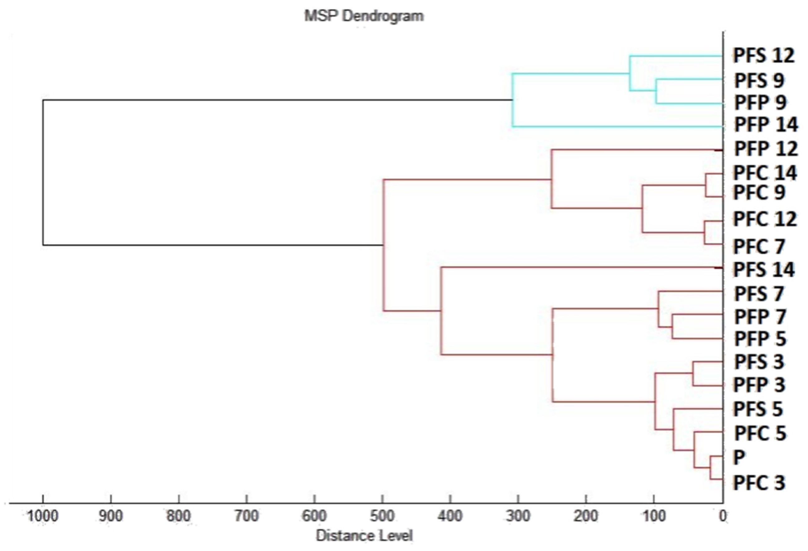
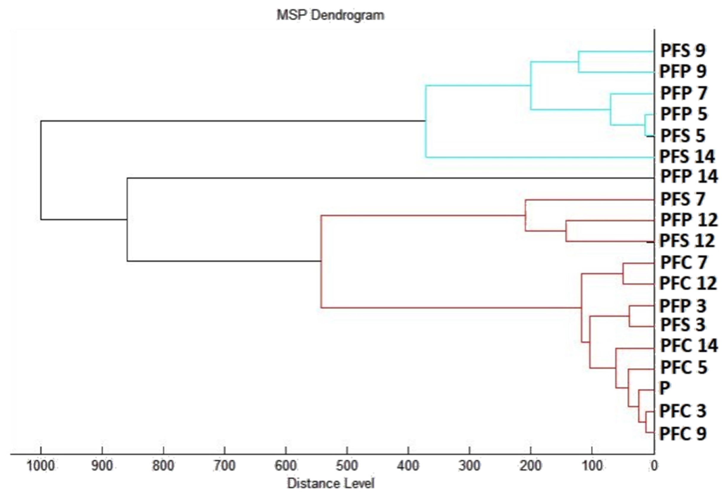
4.12. Analysis of Differences in Biofilm Development with MALDI-TOF MS Biotyper
The representative G
− biofilm-forming bacterium
P. fluorescens, obtained from fish, was used in this experiment. Using a MALDI-TOF MS Biotyper, the various phases of change in the protein structure of the biofilms developing on plastic and stainless steel surfaces were estimated after treatment with the tested EOs. The samples (experimental and control) were prepared in polypropylene tubes (50 mL) using 20 mL of MHB and a plastic and a stainless steel slide. The experimental groups used were MHB enriched with 0.5% of the EOs. The inoculated experimental groups were incubated at 37 °C on a 45° slope shaker at 170 rpm. Before analysis, samples of biofilm were taken using a cotton swab from the plastic and stainless steel slides. Analysis was performed by imprinting samples onto a MALDI-TOF metal target plate. Samples of biofilm and planktonic cells were analyzed on the 3rd, 5th, 7th, 9th, 12th, and 14th days. The planktonic cells were obtained by removing 300 µL of culture medium, which was centrifuged at 12,000 rpm for 1 min. The obtained supernatant was removed, and the planktonic cells were washed three times using ultrapure water. Washed cells were applied to a target plate in a suspension volume of 1 μL. Next, 1 μL of α-cyano-4-hydroxycinnamic acid matrix (10 mg/mL) was applied to the biofilm and planktonic cell samples and dried at room temperature. Using a MALDI-TOF MicroFlex (Bruker Daltonics, Billerica, MA, USA), the samples were processed in the range of
m/
z 200–2000 after crystallization (in linear and positive mode). Spectral data were obtained via automatic analysis. The same sample similarities were used to generate a standard global spectrum (MSP). The dendrograms were obtained using Euclidean distance. For this purpose, 19 MSP from the spectra generated by the MALDI-TOF Biotyper 3.0 were grouped [
59].
4.13. Statistical Analyses
All data were evaluated using IBM-SPSS 23 software for Windows (SPSS Inc., Chicago, IL, USA). The data were presented as a mean ± standard error (S.E.M). The statistical significance was determined using a paired-sample T test. The level of statistical significance was set at p < 0.05. One-way analysis of variance (ANOVA) was performed using Prism 8.0.1 (GraphPad Software, San Diego, CA, USA), followed by Tukey’s test at p < 0.05.


 ,
, 


