Article

Adding New Scientific Evidences on the Pharmaceutical Properties of Pelargonium quercetorum Agnew Extracts by Using In Vitro and In Silico Approaches

by
Annalisa Chiavaroli
1,
Maria Loreta Libero
1,
Simonetta Cristina Di Simone
1,
Alessandra Acquaviva
1,
Nilofar
1,
Lucia Recinella
1,
Sheila Leone
1,
Luigi Brunetti
1,
Donatella Cicia
2,
Angelo Antonio Izzo
2,
Giustino Orlando
1,*,
Gokhan Zengin
3,*,
Abdullahi Ibrahim Uba
4,
Ugur Cakilcioğlu
5,
Muzaffer Mukemre
6,
Omer Elkiran
7,
Luigi Menghini
1 and
Claudio Ferrante
1
1
Department of Pharmacy, “G. d’Annunzio” University of Chieti-Pescara, 66100 Chieti, Italy
2
Department of Pharmacy, School of Medicine, University of Naples Federico II, 80131 Naples, Italy
3
Physiology and Biochemistry Laboratory, Department of Biology, Science Faculty, Selcuk University, 42130 Konya, Turkey
4
Department of Molecular Biology and Genetics, Istanbul AREL University, 34537 Istanbul, Turkey
5
Pertek Sakine Genç Vocational School, Munzur University, 62500 Pertek, Turkey
6
Department of Plant and Animal Production, Yuksekova Vocational School, Hakkari University, 30100 Hakkari, Turkey
7
Department of Environmental Health, Vocational School of Health Services, Sinop University, 57000 Sinop, Turkey
*
Authors to whom correspondence should be addressed.
Plants 2023, 12(5), 1132; https://doi.org/10.3390/plants12051132
Submission received: 13 January 2023 / Revised: 27 February 2023 / Accepted: 28 February 2023 / Published: 2 March 2023

Abstract

:
Pelargonium quercetorum is a medicinal plant traditionally used for treating intestinal worms. In the present study, the chemical composition and bio-pharmacological properties of P. quercetorum extracts were investigated. Enzyme inhibition and scavenging/reducing properties of water, methanol, and ethyl acetate extracts were assayed. The extracts were also studied in an ex vivo experimental model of colon inflammation, and in this context the gene expression of cyclooxygenase-2 (COX-2) and tumor necrosis factor α (TNFα) were assayed. Additionally, in colon cancer HCT116 cells, the gene expression of transient receptor potential cation channel subfamily M (melastatin) member 8 (TRPM8), possibly involved in colon carcinogenesis, was conducted as well. The extracts showed a different qualitative and quantitative content of phytochemicals, with water and methanol extracts being richer in total phenols and flavonoids, among which are flavonol glycosides and hydroxycinnamic acids. This could explain, at least in part, the higher antioxidant effects shown by methanol and water extracts, compared with ethyl acetate extract. By contrast, the ethyl acetate was more effective as cytotoxic agent against colon cancer cells, and this could be related, albeit partially, to the content of thymol and to its putative ability to downregulate TRPM8 gene expression. Additionally, the ethyl acetate extract was effective in inhibiting the gene expression of COX-2 and TNFα in isolated colon tissue exposed to LPS. Overall, the present results support future studies for investigating protective effects against gut inflammatory diseases.

1. Introduction

Pelargonium quercetorum Agnew, belonging to the Gerianaceae family, is a medicinal plant traditionally used by the local population in the Hakkari region of Turkey and in the Kurdistan region of Iraq [1]. The species is characterized by herbaceous or semi-woody shrub plants, with rhizomes, schizocarpic fruits, and orange-red or pink flowers [2]. In Turkey, it is used for treating sore throats and as a wound healing agent, as well as for the management of migraines and pain [1]. The aerial portion is used as antiparasitic; indeed, a recent paper demonstrated the antiparasitic properties of the water extract against Leishmania major [3]. The methanol extract of the whole plant was reported to induce a cytotoxic activity against lung cancer cells [4], and was also effective as anti-inflammatory agent, in macrophages challenged with Escherichia coli lipopolysaccharide (LPS) [5]. Similarly, other Pelargonium species, especially P. sidoides, have been used for the treatment of gastrointestinal and liver diseases [6], but also for treating respiratory disorders [7].
Regarding the chemical composition, Pelargonium species are rich in essential oils, mainly constituted by monoterpenes, which can play antimicrobial and cytotoxic activities [4]. In the methanol extract of P. quercetorum the hydrocarbons tetracosane, heneicosane, and 2-methyleicosane were the phytochemicals present in higher concentration [4]; whereas rutin and shikimic acid were reported to be the main phenolics present in the methanol extract [8]. Ferda et al. [9] described cytotoxic and pro-apoptotic effects by P. quercetorum, and scavenging/reducing properties that could be partially related to the total phenol content of the methanol extract.
In the present study, the chemical composition and biological properties of different polarity extracts of P. quercetorum were investigated. Specifically, phenolic compounds were assayed through colorimetric and chromatographic analysis. Intrinsic enzyme inhibition (against α-glucosidase, α-amylase, cholinesterases, and tyrosinase) and scavenging/reducing properties were assayed as well. The extracts were also studied in an ex vivo experimental model of colon inflammation, constituted by isolated colon specimens exposed to LPS, in order to reproduce the burden of inflammation and oxidative stress occurring during colitis, in vivo [10,11,12,13]. In this context, the gene expression of pro-inflammatory biomarkers, including cyclooxygenase-2 (COX-2) and tumor necrosis factor α (TNFα) were assayed. Additionally, the gene expression of vascular endothelial growth factor A, hypoxia-inducible factor 1α (HIF-1α), deeply involved in the so-called inflammatory-to-cancer transition [14], was also measured in human colon cancer HCT116 cells. In the same cell model, the gene expression of transient receptor potential cation channel subfamily M (melastatin) member 8 (TRPM8), possibly involved in colon carcinogenesis [15,16], was conducted as well.
Finally, in silico studies were carried out for predicting the pharmacokinetics of the main phytochemicals present in the extracts, as well as direct interactions with protein targets assayed through the abovementioned biological models, among which the enzymes tyrosinase, α-amylase, α-glucosidase, cholinesterases, and the TRPM8 receptor.

2. Results and Discussion

2.1. Total Bioactive Compounds

Phenolic compounds are considered to be one of the most important players among the specialized metabolites and therefore the determination of their content is an important parameter to evaluate the pharmacological potential of plant extracts. In the present study, the total contents of phenolics and flavonoids were determined, and the results are summarized in Table 1. The highest total phenolic content (TPC) was found in the water extract with 62.52 mg GAE/g extract, followed by methanol (45.77 mg GAE/g extract), and ethyl acetate (21.94 mg GAE/g extract). For total flavonoid content (TFC), the order was methanol (40.12 mg RE/g extract), water (16.15 mg RE/g extract), and ethyl acetate (2.78 mg RE/g extract). In a previous study by Karatoprak, et al. [17], the total content of bioactive compounds of P. quercetorum extracts depended on the solvents used, and the highest content of them was detected in the methanolic extracts (especially from roots). In addition to P. quercetorum, several studies have reported significant levels of total bioactive compounds in the extracts of some Pelargonium species [18,19,20]. However, the observed results from spectrophotometric assays need to be confirmed by chromatographic techniques including liquid chromatography coupled with different type of detectors, for example, photodiode array or mass spectrometry.
Therefore, we conducted a chromatographic analysis of the extracts for the quantitative determination of phenolic compounds, as shown in Table 2 and Figure 1A–C. Specifically, the methanolic extract was the richest in flavonol glycosides and hydroxycinnamic acids, among which rutin, rosmarinic acid, and caffeic acid were the main phytochemicals (Table 2). A similar pattern of phenolic composition, but at lower concentration, was shown by P. quercetorum water extract. While the EA extract showed a higher concentration of the phenolic monoterpene thymol.
The content of such phytochemicals could indicate scavenging/reducing and enzyme inhibition properties [21,22], thus substantiating the rationale for exploring innovative health-promoting applications, as described below.

2.2. Antioxidant Properties

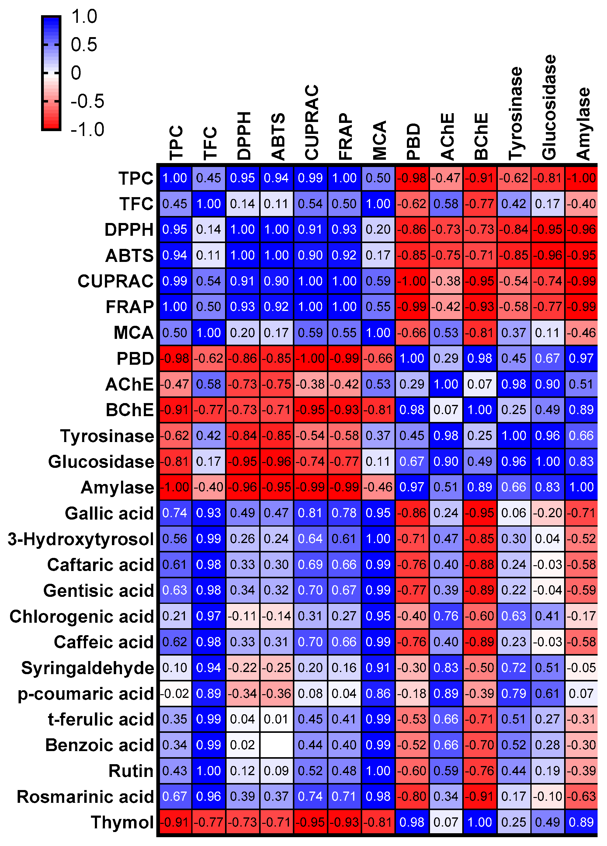
According to the latest scientific findings, oxidative stress is an important trigger for the progression of several chronic and degenerative diseases such as cancer, myocardial problems, and diabetes. In particular, increasing the production of free radicals leads to oxidative stress [23,24]. In this sense, antioxidants are the key players in controlling free radical levels. To that end, scientists are looking for new and safe sources of antioxidants, and phytochemicals are believed to be among the most powerful antioxidants. In the present study, we determined antioxidant properties of P. quercoutum extracts using different methods such as radical scavenging (2,2′-azino-bis(3-ethylbenzothiazoline) 6-sulfonic acid (ABTS) and 1,1-diphenyl-2-picrylhydrazyl (DPPH)), reducing power (cupric ion reducing antioxidant capacity (CUPRAC) and ferric ion reducing antioxidant power (FRAP)), metal chelating, and phosphomolybdenum assay. The results are summarized in Table 3. In radical scavenging assays (DPPH and ABTS), the best activity was found for the water extract (DPPH: 140.40 mg trolox equivalent (TE)/g extract; ABTS: 212.31 mg TE/g extract), followed by methanol and ethyl acetate extracts. The reducing abilities of antioxidant compounds are very close to their electron-donation ability. Cu2+ to Cu and Fe3+ to Fe2+ conversions were measured using CUPRAC and FRAP assays, respectively. As can be seen from Table 3, the water extract showed the strongest reducing abilities, followed by methanol and ethyl acetate extracts. The results obtained from both free radical scavenger and reducing power assays are consistent with the total phenolic content of the tested extracts. This fact was also confirmed by Pearson’s correlation analysis, and we observed a linear correlation between these parameters (Figure 2). In a literature review, several researchers also reported a strong correlation between total phenolic content and antioxidant properties [25,26,27]. In addition to total phenolic content, some phytochemicals including rosmarinic, ferulic, caffeic, and gallic acids can be attributed to the observed antioxidant properties and are known to be powerful antioxidants [28,29,30,31,32]. In contrast to the radical scavenging ability and reducing power, the order of the tested extracts in the phosphomolybdenum assay was ethyl acetate (1.64 mmol TE/g extract) > methanol (1.54 mmol TE/g extract) > water (1.51 mmol TE/g extract). The phosphomolybdenum assay is known for determining total antioxidant capacity and also non-phenolics could play a role in the assay. Consistent with the presented results, a weak correlation between total phenols and phosphomolybdenum assay were reported by several investigators [33,34]. Finally, we investigated the metal chelating ability of P. quercetorum extracts, which is associated with controlling the production of hydroxyl radicals in the Fenton reaction. The methanol extract exhibited the best metal chelating ability with 29.80 mg EDTA equivalent (EDTAE)/g extract, followed by water extract (12.28 mg EDTAE/g extract). However, the ethyl acetate extract showed no metal chelating ability. In a previous study by Karatoprak, Fırat and Koşar [17], the antioxidant properties of P. quercetorum aerial parts and root extracts were investigated by DPPH, ABTS, ferric reducing power and metal chelating assays. In their study, the hydroalcoholic root extract in particular showed better potency compared with aerial parts. In addition to P. quercetorum, several Pelargonium species including P. hispidum [35], P. graveolens [18], P. hybrid [36], and P. sidoides [37] were investigated, and their extracts displayed antioxidant properties. With this in mind, the genus Pelargonium could be considered a valuable source of natural antioxidants.

2.3. Enzyme Inhibitory Properties

Currently, most scientific efforts are focused both on improving the quality of public health and reducing the prevalence of certain diseases such as obesity, diabetes, and cardiovascular diseases [38]. Enzymes play a crucial role at this point and it is known that blocking a specific enzyme is an effective way to treat the abovementioned diseases. In this sense, some enzymes are selected therapeutic targets. For example, acetylcholinesterase is a main target to alleviate the observed symptoms of Alzheimer’s disease [39]. As another example, the carbohydrate-hydrolyzing enzymes, namely α-amylase and α-glucosidase, are checkpoints to control blood glucose level in diabetic patients [40]. For this purpose, several compounds are chemically produced as enzyme inhibitors, but most of them have side effects such as toxicity and gastrointestinal disturbances. At this point, we need to replace the synthetic drugs with natural and safe drugs, and plants are considered a promising in this sense.
In view of the above facts, we investigated the enzyme inhibitory properties of P. quercetorum extracts, and the results are summarized in Table 4. The highest acetylcholinesterase (AChE) inhibitory effect was obtained by the methanol extract with 2.31 mg GALAE/g extract, followed by ethyl acetate and water extracts. However, butyrylcholinesterase (BChE) was only inhibited by the ethyl acetate extract and others were not active on the enzyme. Tyrosinase is the main enzyme involved in the synthesis of melanin, and its inhibition is one of the most effective ways to treat hyperpigmentation problems. As can be seen from Table 4, methanol and ethyl acetate extracts were more active on tyrosinase compared with water. Similar to tyrosinase, the best inhibitory effects on α-amylase and α-glucosidase were recorded in the ethyl acetate and methanol extracts. The water extract exhibited the weakest ability in all enzyme inhibitory assays. As can be seen in Figure 2, the obtained enzyme inhibitory effects could be explained by the presence of some compounds. For example, AChE and tyrosinase inhibitory effects were positively correlated with p-coumaric acid, syringaldehyde, and chlorogenic acid, and these compounds have been described by some researchers as anti-Alzheimer and dermatoprotective agents [41,42,43,44,45]. In addition to these components, thymol had a good interaction with anti-diabetic enzymes, namely α-amylase and α-glucosidase. However, the interactions of specialized metabolites such as synergetic and antagonistic effects should not be forgotten. Thus, in future studies, we can suggest that the compounds could be isolated and then tested for their enzyme inhibitory properties. The enzyme inhibition potential of some Pelargonium species has been reported in the literature by several researchers. For example, Iancu, et al. [46] investigated the α-amylase and α-glucosidase inhibitory effects of two Pelargonium species (P. hispidum and P. grandiflorum) and found that the methanolic extracts had more potent inhibitory properties than hydroalcholic extracts. In another study conducted by Ahamad and Uthirapathy [47], significant α-amylase and α-glucosidase inhibitory properties were reported for P. graveolens essential oil. The IC50 value was reported as 10.5 mg/mL in the AChE inhibitory effect of the essential oil of P. hybrid [36]. To the best of our knowledge, there are no data on the enzyme inhibitory properties of P. quercetorum extracts. Thus, the presented results are the first and they provide new information about the genus Pelargonium.

2.4. In Vitro Study

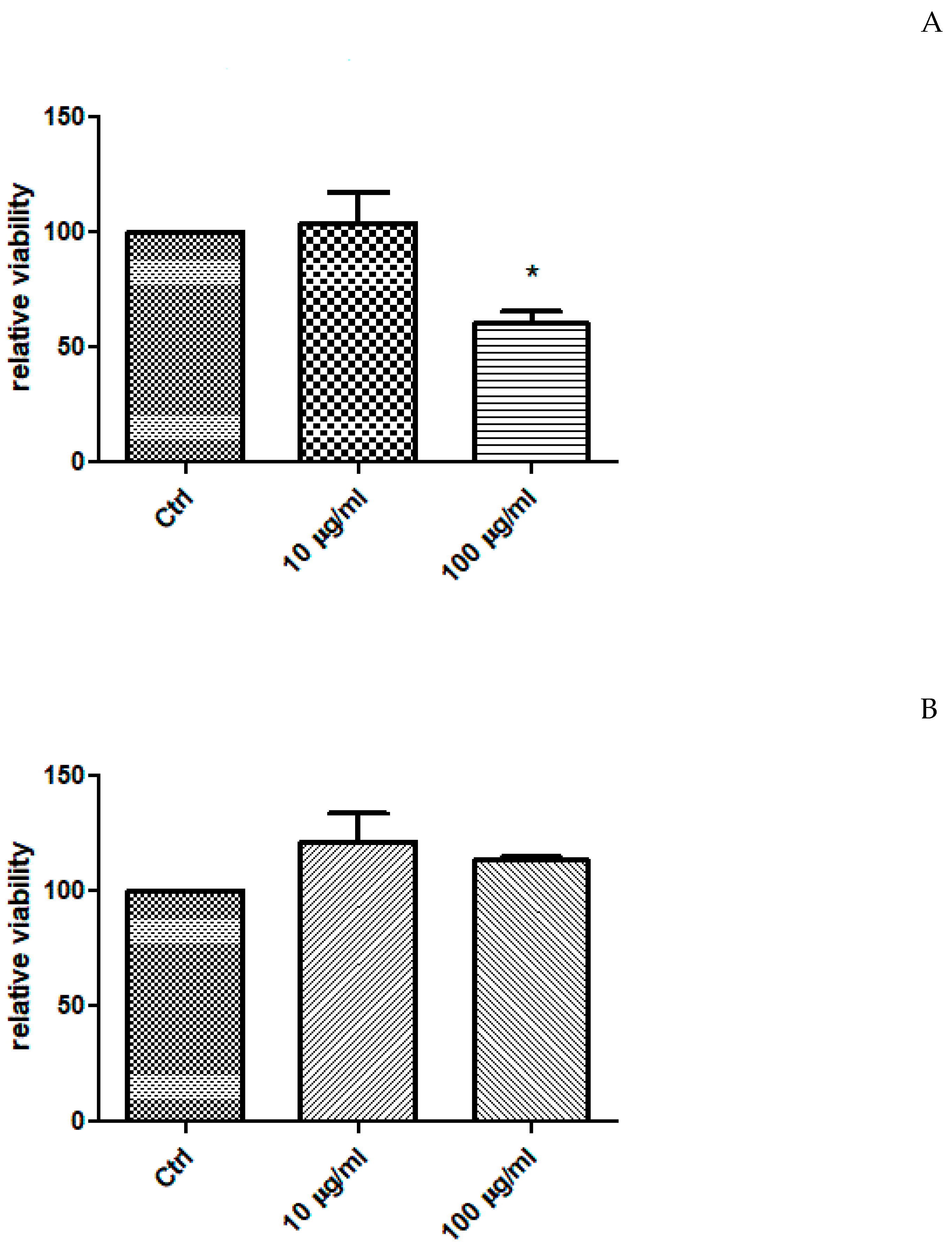
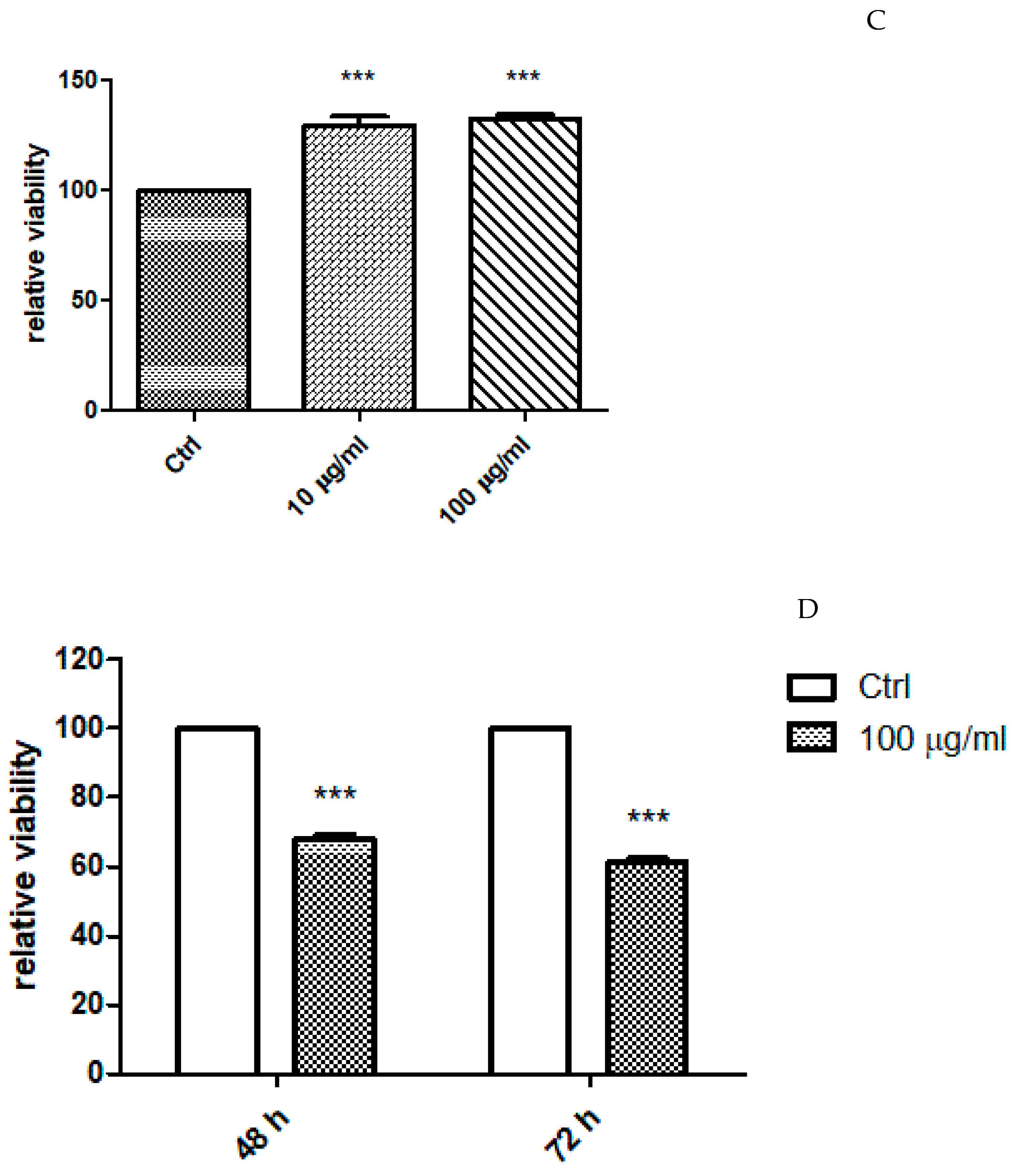
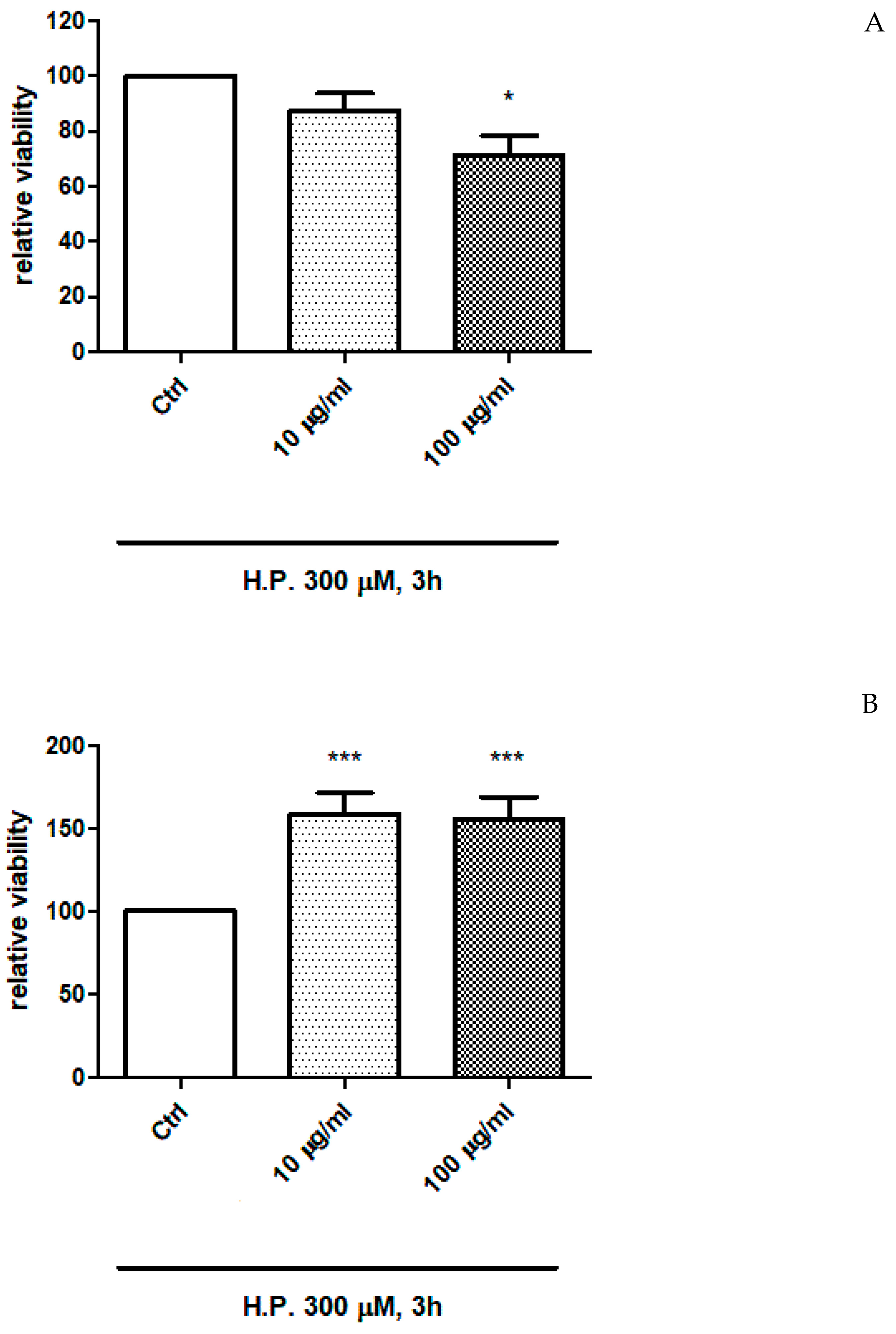
The extracts of P. quercetorum were also investigated in order to evaluate effects on the viability of human colon cancer HCT116 cells. The cells were exposed at different concentrations of the extracts (10–100 µg/mL), finding a significant modulation of cell viability (Figure 3 and Figure 4). Specifically, the ethyl acetate extract was found to be the most effective as cytotoxic agent, with a significant reduction of HCT116 cell viability, at the concentration of 100 µg/mL, also after 48 and 72 h of cell exposure to the extract (Figure 3D and Figure 4D). The effect was more evident in cells not exposed to hydrogen peroxide. While the water and methanol extract stimulated HCT116 cell viability in both basal condition and after challenging with hydrogen peroxide.
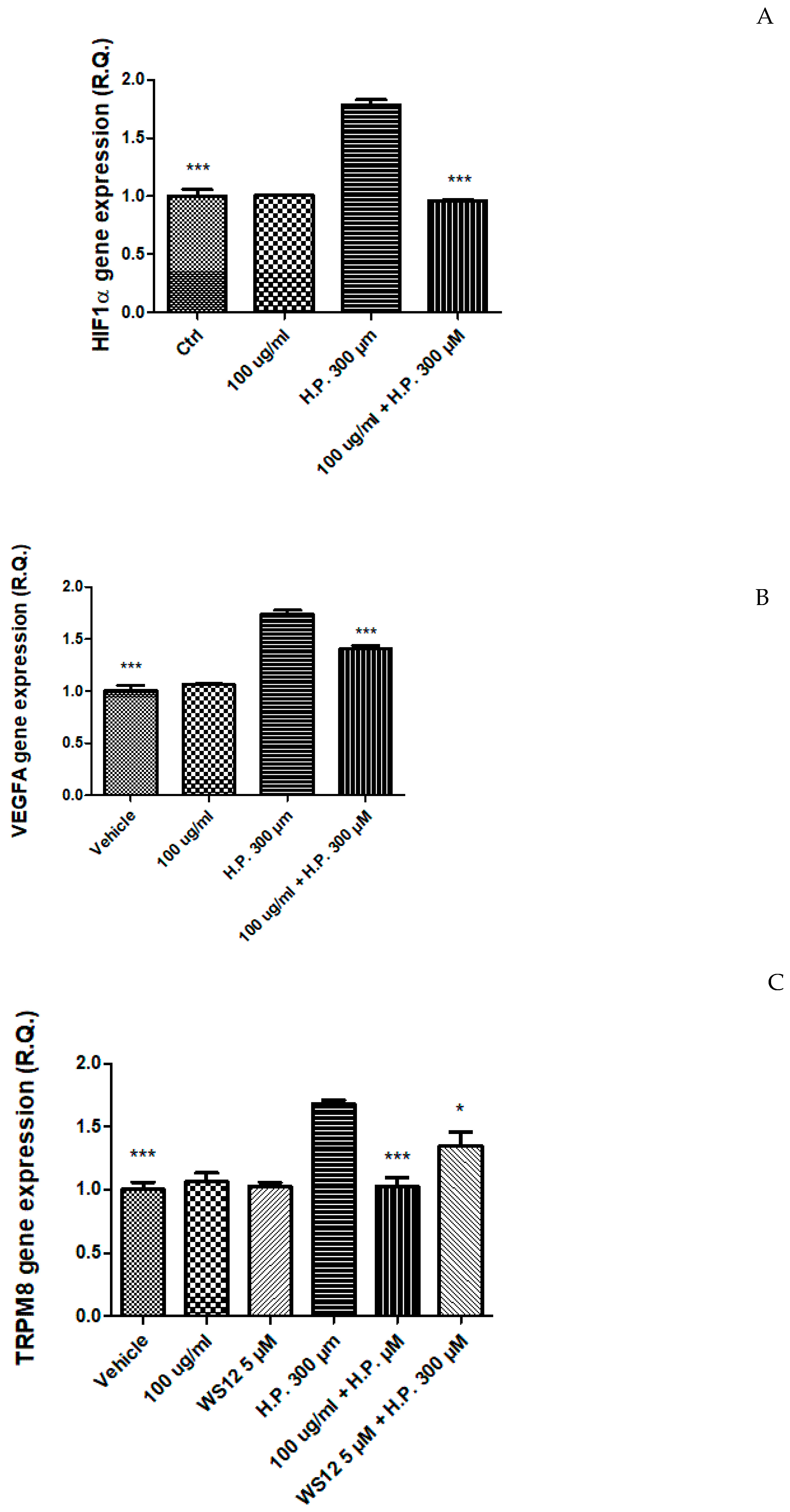
Therefore, in a second set of experiments, cells were exposed to the EA extract (100 µg/mL) in both basal and oxidative stress conditions [hydrogen peroxide (H.P.) 300 µM], in order to explore the mechanism underlying the cytotoxic effect induced by the extract. In this context, the gene expression of HIF1α and VEGFA, two angiogenic factors deeply involved in the so-called inflammatory-to cancer transition [14] were assayed. Hydrogen peroxide was able to upregulate the gene expression of both HIF1α and VEGFA. This is consistent, albeit partially, with literature [48,49]. The extract was capable to blunt the hydrogen peroxide-induced up-regulation of both factors (Figure 5A,B), and this could be related, albeit partially, to the content in phenolic compounds [50,51].
In the same cell model, the gene expression of transient receptor potential (TRP) M8 (TRPM8) was evaluated as well. Indeed, there is evidence of the involvement of TRPM8 receptor in colon cancer [15]. In HCT116 cells, P. quercetorum EA extract reduced the hydrogen peroxide-induced upregulation of TRPM8 receptor (Figure 5C), and this effect was analog to the one induced by WS12 ((1R,2S,5R)-2-isopropyl-N-(4-methoxyphenyl)-5-methylcyclohexanecarboxamide), a selective TRPM8 agonist that was used at the concentration of 5 µM, consistent with its putative affinity towards the receptor [52].
Regarding the inhibition of the TRPM8 gene expression induced by the extract, the content of thymol could play a pivotal role in mediating this effect [53]. However, we cannot exclude that phenolic compounds present in the extract can mediate, albeit partially, such effect. Indeed, a previous study of ours demonstrated the capability of catechin to reduce the gene expression of TRPM8 [52]. Additionally, flavones, isoflavones, non-prenylated chalcones, and glycocoumarins have been suggested to exert antagonistic effects towards TRPM8 receptor [54]. Therefore, a docking approach was conducted to evaluate the putative affinities of extract phenolics towards TRPM8 receptor, as follows.

2.5. Ex Vivo Study

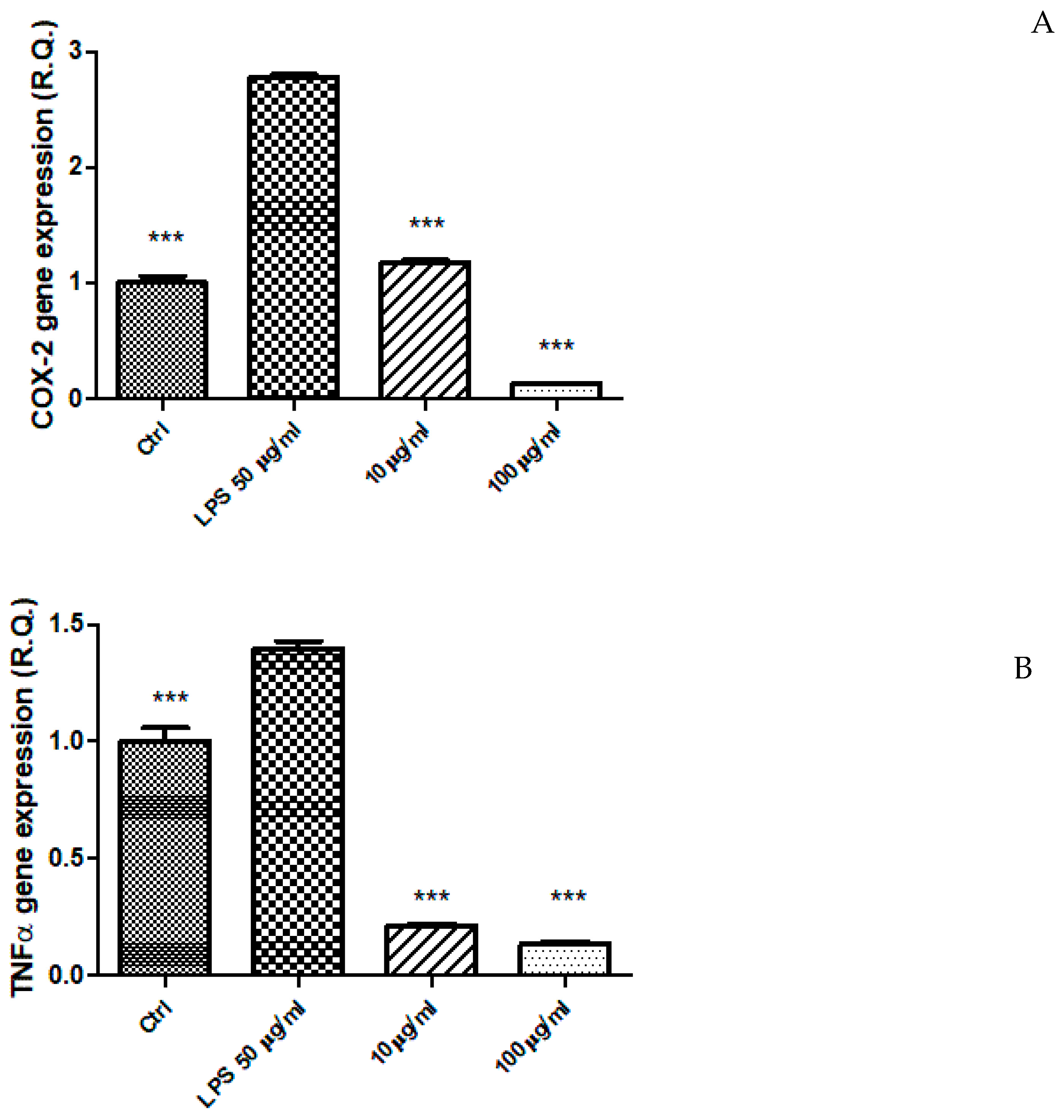
The EA extract (10–100 µg/mL) was also tested in an ex vivo experimental model constituted by isolated mouse colon specimens exposed to LPS, in order to induce acute inflammation [8]. The protective effects were evaluated as inhibition of LPS-induced upregulation of the gene expression of pro-inflammatory mediators, namely COX-2 and TNFα (Figure 6A,B). In this context, the extract was effective in reducing the gene expression of both COX-2 and TNFα. This could be partly related to the content in thymol, that was effective in inhibiting the expression of TNFα and COX-2 in LPS-stimulated macrophages [55]. Additionally, our findings of anti-inflammatory effects in the colon are consistent, albeit partially, with the anti-inflammatory effects induced by P. quercetorum methanol extract in macrophages exposed to LPS [5]. Being the methanol extract rich in phenolic compounds, particularly rutin, as shown by the present study and by the work of Akkemik et al. [8], it could be hypothesized that different specialized metabolites, including monoterpenes and flavonoids may be involved in the anti-inflammatory effects exerted by P. quercetorum extracts.

2.6. Molecular Docking

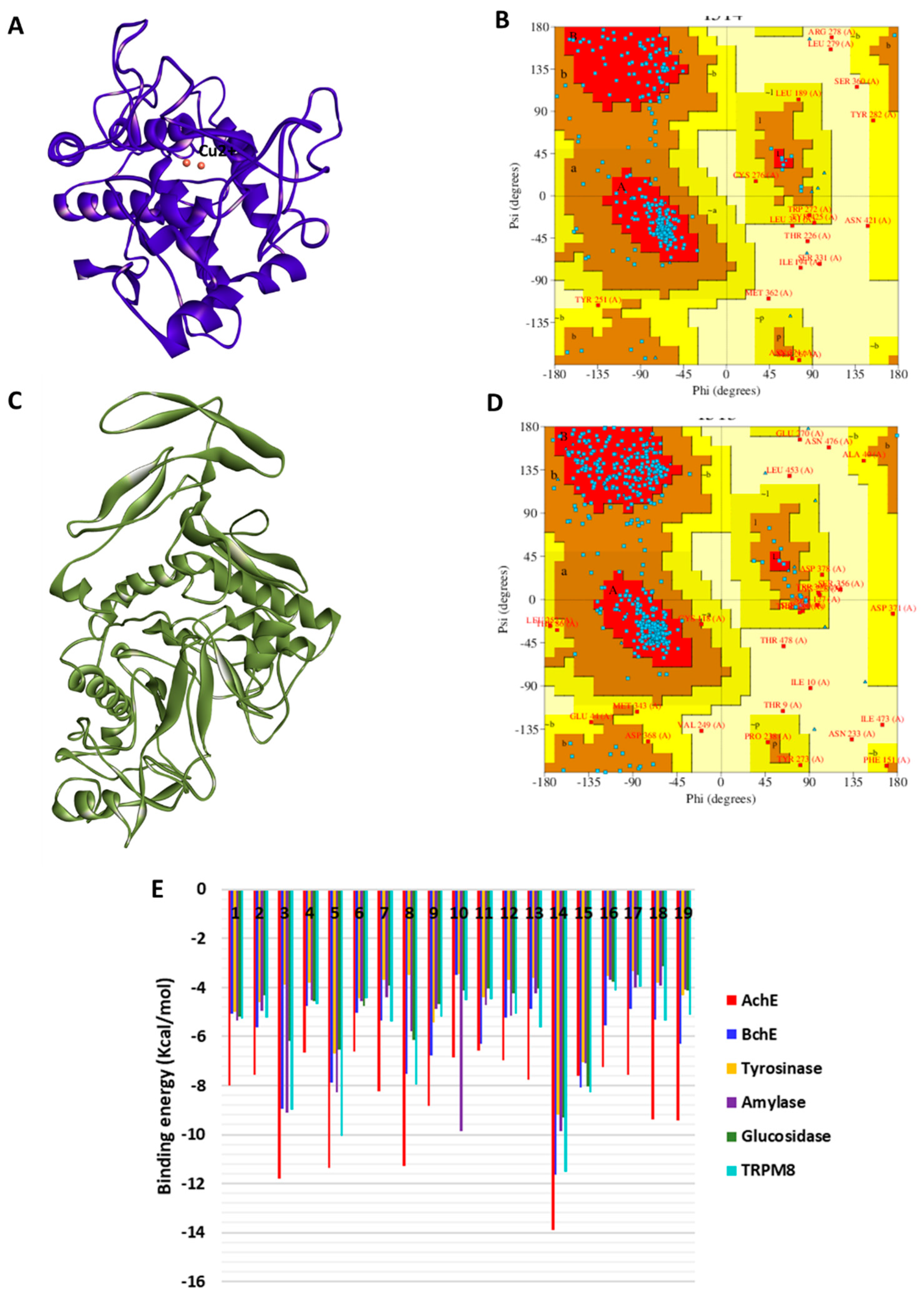
The homology modeling procedure for building human tyrosinase and α-glucosidase models is described in the Section 3. The 3D model of tyrosinase is shown in Figure 7A, and its validation in form of a Ramachandran plot showing energetically allowed regions for backbone dihedral angles ψ against φ of amino acid residues is shown in Figure 7B. The corresponding 3D structure of α-glucosidase is shown in Figure 7C, and its Ramachandran plot, which agreed with those of the known experimental structures, is shown in Figure 7D. The binding energy scores of the bioactive compounds are shown in Figure 7E. All the study compounds exhibited the strongest binding to AChE, and moderate binding to the other enzymes, especially, BChE and α-amylase.
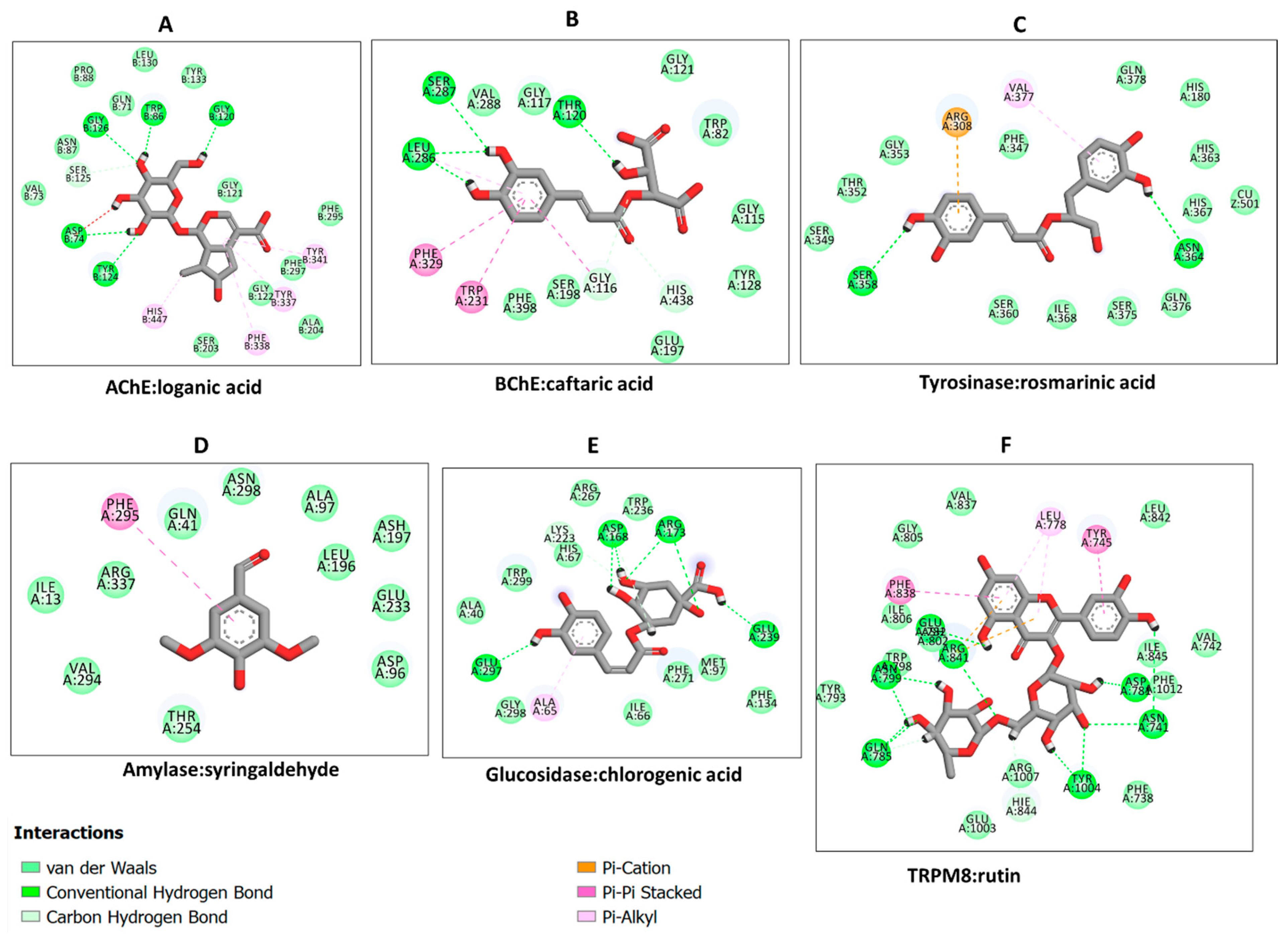
Hence, the protein–ligand interactions were examined for some selected compounds. Loganic acid and caftaric acid demonstrated a similar binding pattern to AChE and BChE, respectively. Both compounds were completely buried in the active sites of the enzymes and formed a couple of H-bonds and several van der Waals interactions. In addition, loganic acid formed hydrophobic interactions with AChE (Figure 8A), whereas caftaric acid formed π–π stacked interactions with BChE (Figure 8B). Likewise, even though rosmarinic acid shares similar interactions with the enzymes, it uniquely formed π–anion interaction and engaged active site copper metal ion via van der Waals interaction (Figure 8C). On the other hand, syringaldehyde was weakly bound to α-amylase, mainly via van der Waals interactions (Figure 8D). Finally, the interaction between α-glucosidase and chlorogenic acid was demonstrated. Chlorogenic acid formed, mainly, a couple of H-bonds and several van der Waals interactions with residues in the active site of α-glucosidase (Figure 8E). In addition, all the constituent compounds were docked into the binding site of transient receptor potential melastatin member 8 (TRPM8)—an ion channel for cold and menthol sensor. All the compounds were found to bind to the TRPM8, with the strongest binding demonstrated by rutin via multiple interactions with the binding site amino acid residues (Figure 8F); thus, suggesting putative abilities of rutin as regulator of TRPM8 activity. In summary, H-bonds are the major contributor to the protein interaction in all the selected docking complexes, except for that of α-amylase and syringaldehyde, in which van der Waals interactions were predominant. This is due to the multiple hydroxyl groups on the phenolic compounds forming H-bonds with the polar amino acid residues in the active site of the enzymes, corroborating with the binding energy scores. H-bonds are known to contribute greatly to protein–ligand interaction. Other interactions such as hydrophobic and van der Waals interactions reinforce the binding, especially in the case of active sites lined by polar amino acids.
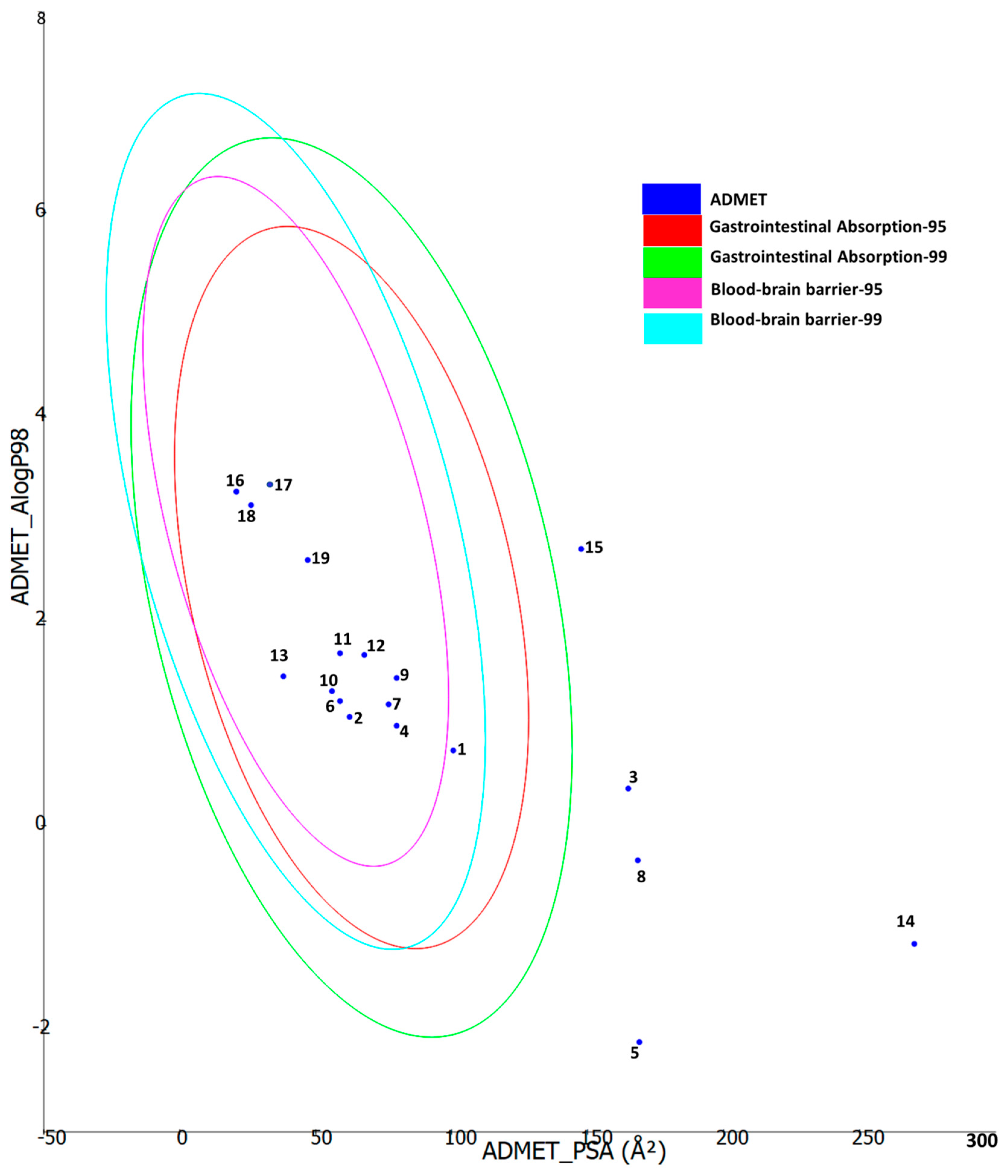
Furthermore, ADMET properties of the bioactive constituents were predicted using Biovia DS ADMET prediction toolkit. The four ellipses define the areas where well-absorbed molecules are expected to be located. At 95 and 99% confidence levels of gastrointestinal absorption (red and green), and 95 and 99% blood-brain barrier penetration probability (magenta and aqua). The compounds are shown according to their serial number (Figure 9). Low-molecular-weight compounds associated with low polarity were found to fall in one or more ellipses, suggesting high absorption and high blood-brain barrier penetration possibility, whereas those with high molecular weight and high polarity fell outside the ellipses, suggesting low absorption and low chance of crossing the blood-brain barrier.

3. Materials and Methods

3.1. Plant Material and Preparation of Extracts

Pelargonium quercetorum samples were collected at the flowering season in June 2020 (Şemdinli, Hakkari, Turkey). The plants were identified by one of the authors (Dr. Ugur Cakilcioglu), and voucher specimens (UC-15-21) were deposited at the herbarium of Munzur University, Turkey. The aerial parts (as mixed) were dried in the shade at room temperature for about 7 days, and then ground into a powder using a mill. All of the samples were kept in a dark place.
In this study the extracts were prepared using three solvents (ethyl acetate, methanol, and water). Maceration was employed as the extraction method to obtain ethyl acetate and methanol extracts., The plant materials (5 g) were macerated overnight at room temperature with 100 mL of these solvents. Finally, the solvents were evaporated from the mixtures. To obtain water extracts, the plant materials (10 g) were kept with 200 mL of boiled water for 15 min, and then the extracts were filtered and lyophilized. All extracts were stored at 4 °C until further analysis was required.

3.2. Chemicals and Reagents

The chemicals were purchased from Sigma-Aldrich (Darmstadt, Germany). They were ABTS, DPPH, gallic acid, rutin, electric eel acetylcholinesterase (AChE) (type-VI-S, EC 3.1.1.7), horse serum butyrylcholinesterase (BChE) (EC 3.1.1.8), galantamine, acetylthiocholine iodide (ATChI), butyrylthiocholine chloride (BTChI) 5,5-dithio-bis(2-nitrobenzoic) acid (DTNB), tyrosinase (EC1.14.18.1, mushroom), α-glucosidase (EC. 3.2.1.20, from Saccharomyces cerevisiae), α-amylase (EC. 3.2.1.1, from porcine pancreas), sodium molybdate, sodium nitrate, sodium carbonate, Folin-Ciocalteu reagent, hydrochloric acid, sodium hydroxide, trolox, ethylenediaminetetraacetate (EDTA), neocuproine, cupric chloride, ammonium acetate, ferric chloride, 2,4,6-Tris(2-pyridyl)-s-triazine (TPTZ), ammonium molybdate, ferrozine, ferrous sulphate hexahydrate, kojic acid, and acarbose. All chemicals were of analytical grade. Chemical standards for HPLC analysis: gallic acid, hydroxytyrosol, caftaric acid, gentisic acid, 4-hydroxybenzoic acid, loganic acid, chlorogenic acid, caffeic acid, syringic acid, syringaldehyde, p-coumaric acid, t-ferulic acid, benzoic acid, rutin, rosmarinic acid, carvacrol, thymol, flavone, and 3-hydroxyflavone were purchased from Merk Life Science S.r.l. (Milan, Italy).

3.3. Determination of Total Polyphenol and Flavonoids Contents

Total phenolic and flavonoid contents were calculated with the Folin-Ciocalteu and AlCl3 assays, respectively [56]. Gallic acid equivalents (mg GAEs/g dry extract) and rutin equivalents (mg REs/g dry extract) were used to describe the outcomes of the two tests. Details are reported as Supplementary Materials.

3.4. HPLC Determination of Phenolic Compounds

The extract was analyzed for phenol quantitative determination using a reversed-phase HPLC-DAD in gradient elution mode [57]. The separation was conducted within 60 min of the chromatographic run, starting from the following separation conditions: 97% water with 0.1% formic acid, 3% methanol with 0.1% formic acid (Table 5). The separation was performed on an Infinity lab Poroshell 120-SB reverse phase column (C18, 150 × 4.6 mm i.d., 2.7 μm; Agilent, Santa Clara, CA, USA). Column temperature was set at 30 °C. Quantitative determination of phenolic compounds was performed via a DAD detector. The selected wavelengths are reported in Table 2. Quantification was done through 7-point calibration curves, with linearity coefficients (R2) > 0.999, in the concentration range 2–140 μg/mL. All standards were purchased from Sigma Aldrich (Milan, Italy), and have a purity ≥95%. The limits of detection were lower than 1 μg/mL for all assayed analytes. The area under the curve from HPLC chromatograms was used to quantify the analyte concentrations in the extract [57].

3.5. Antioxidant and Enzyme Inhibitory Assays

The antioxidant and enzyme inhibitory activity of the extracts was assessed according to methods presented previously [58]. Details are also reported as supplementary materials. Data were expressed as mg Trolox equivalents (TE)/g extract in FRAP, CUPRAC, ABTS, and DPPH radical scavenging activity; mg EDTA equivalents (EDTAE)/g extract in the metal chelating ability, mmol TE/g extract in the phosphomolybdenum assay; mg galanthamine equivalents (GALAE)/g extract in AChE and BChE assays; mg kojic acid equivalents (KAE)/g extract in tyrosinase inhibitory assay; and mmol acarbose equivalents (ACAE)/g extract in α-amylase and α-glucosidase assays.

3.6. In Vitro Study

Human colon cancer-derived HCT116 cell were cultured in DMEM (Euroclone) supplemented with 10% (v/v) heat-inactivated fetal bovine serum and 1% (v/v) penicillin G/streptomycin in a 75 cm2 cell culture flasks. The cultured cells were maintained in a humidified incubator with 5% CO2 at 37 °C. When the confluency reached 80%, a viability test was performed using a 3-(4,5-dimethylthiazol-2-yl)-2,5-diphenyltetrazolium bromide (MTT) test to assess the basal cytotoxicity of the P. quercetorum extracts under investigation. For this assay cells were seeded (5 × 10³ cells/well) onto flat-bottomed 96-well culture plates and incubated overnight. After 24 h, extracts from the aerial parts of P. quercetorum at different concentrations were added, and the plates were incubated for 24, 48, and 72 h. After this time, a total of 10 μL of MTT (5 mg/mL in PBS) was added to each well and incubated for 3 h. The formazan dye formed was solubilized with dimethyl sulfoxide, and the absorbance was recorded at 540 nm as previously described [59]. Effects on cell viability were evaluated in comparison to the vehicle (untreated control group) and expressed as a percentage of the control culture value.
In a second set of experiments, cells were treated with either vehicle or extract for 24, 48, and 72 h and subsequently post-treated with 300 µM of hydrogen peroxide (H.P.) for 3 h. After this time, cell survival was determined by MTT assay as described above.
Each condition was run in triplicate, including untreated control and blank cell-free control.

3.7. Ex Vivo Study

Adult C57/BL6 male mice (3-month-old, weight 20–25 g) were housed in plexiglass cages (2–4 animals per cage; 55 cm × 33 cm × 19 cm) and maintained under standard laboratory conditions (21 ± 2 °C; 55 ± 5% humidity) on a 14/10 h light/dark cycle, with ad libitum access to water and food.
Isolated colon specimens were collected form euthanized mice [Project no. F4738.N.5QP] and maintained in a humidified incubator with 5% CO2 at 37 °C for 4 h (incubation period), in RPMI buffer with added bacterial LPS (10 µg/mL), as previously described [52]. During the incubation period, the tissues were exposed to the extract (10–100 μg/mL).

3.8. RNA Extraction, Reverse Transcription Andreal-Time Reverse Transcription Polymerase Chain Reaction (RT-PCR)

Total RNA was extracted from both HCT116 cells and colon specimens using TRI reagent (Sigma-Aldrich, St. Louis, MO, USA), according to the manufacturer’s protocol, and reverse transcribed using a High Capacity cDNA Reverse Transcription Kit (Thermo Fischer Scientific, Waltman, MA, USA). Gene expression of COX-2, TNF-α, TRPM8, HIF-1α, and VEGF-A were determined by quantitative real-time PCR using TaqMan probe-based chemistry, as previously described [60]. PCR primers and TaqMan probes were purchased from Thermo Fisher Scientific Inc. The Assays-on-Demand Gene Expression Products used for gene expression evaluations in the mouse colon specimens were Mm00478374_m1 for COX-2 gene, Mm00443258_m1 for TNF-α gene, Mm00607939_s1 for β-actin gene. The Assays-on-Demand Gene Expression Products used for gene expression evaluations in the HCT116 cells were: Hs01066596_m1 for TRPM8 gene, Hs00153153_m1 for HIF-1α, Hs00900055_m1 for VEGFA, Hs99999903_m1 for β-actin gene. β-actin was used as the house-keeping gene. The elaboration of data was conducted with the Sequence Detection System (SDS) software version 2.3 (Thermo Fischer Scientific). Relative quantification of gene expression was performed by the comparative 2−ΔΔCt method [61].

3.9. Molecular Modelling

AutodockTools program [62] was used to generate docking grid files using the coordinates of the cocrystal ligand in each crystal. The details of the docking procedure were given in our previous works [63,64]. The binding energy of the ligand poses was estimated, and protein–ligand interaction diagrams were generated using the Biovia DS Visualizer.
The following crystal structures of the target enzymes were retrieved from the protein data bank (https://www.rcsb.org/, accessed on 12 January 2023): AChE (PDB ID: 6O52) [65], BChE (PDB ID: 6EQP) [66], tyrosinase (PDB ID: 6QXD) [67] α-amylase (PDB ID: 2QMK) [68], α-glucosidase (PDB ID: 7KBJ) [69], and transient receptor potential melastatin member 8 (TRPM8)(PDB ID: 6NR3) [70]. However, crystal structures of human tyrosinase and α-glucosidase are not available. Therefore, their human sequences: tyrosinase (UniProt ID: P14679) and α-glucosidase (Uniprot ID: P0DUB6) were retrieved from the Uniprot database (https://www.uniprot.org/, accessed on 12 January 2023) and used to build their homology models using the aforementioned PDB structures as templates. ITASSER web-based tool (https://zhanggroup.org/I-TASSER/, accessed on 12 January 2023) [71] was used to build the model and validated using the PROCHECK server (https://www.ebi.ac.uk/thornton-srv/software/PROCHECK/, accessed on 12 January 2023) [72].
Protein preparation was performed using an online server “Playmolecule proteinPrepare” (https://playmolecule.com/proteinPrepare/, accessed on 12 January 2023) [73], on which each protein was protonated using the predicted pKa of the titratable residues at physiological pH of 7.4. The 3D structure of each ligand was downloaded from the PubChem database (https://pubchem.ncbi.nlm.nih.gov/, accessed on 12 January 2023), and an optimized conformation was generated using Frog2 [74].

3.10. Statistical Analysis

In the antioxidant and enzyme inhibitory assays, the values are expressed as mean ± SD of three parallel experiments. To determine the differences between tested extracts in terms of antioxidant and enzyme inhibitory capacities, one-way analysis of variance (ANOVA) with Tukey test was performed. The statistical analysis was performed using XlStat 16.0 software.
In ex vivo and in vitro studies, the software GraphPad Prism version 5.01 (Graphpad Software Inc., San Diego, CA, USA) was used to perform data analysis. Means ± SEM were determined for each experimental group and analyzed by ANOVA, followed by Newman-Keuls multiple comparison post hoc test. The limit of statistically significant differences between mean values was set at p-value < 0.05. The number of animals randomized for each experimental group was calculated based on the “Resource Equation” N = (E + T)/T (10 ≤ E ≤ 20).

4. Conclusions

The present study explored the composition and biological properties of extracts of different polarity from the aerial parts of P. quercetorum, a plant traditionally used by the local population in the Hakkari region of Turkey. The extracts showed a different qualitative and quantitative content of phytochemicals, with water and methanol extracts being richer in total phenols and flavonoids, especially rutin and hydroxycinnamic acids, namely caffeic acid and rosmarinic acid. This could explain, albeit partially, the higher antioxidant effects showed by methanol and water extracts, compared with ethyl acetate extract. By contrast, the ethyl acetate was more effective as cytotoxic agent against colon cancer cells, and this could be related, albeit partially, to the content of thymol and to its ability to downregulate the gene expression of TRPM8, an endovanilloid receptor possibly involved in colon carcinogenesis. Intriguingly, docking runs also suggest a high putative affinity of rutin towards TRPM8. This could suggest a potential involvement of rutin in mediating, albeit partially, the cytotoxic effects induced by the ethyl acetate extract of P. quercetorum, in human colon cancer HCT116 cells. Finally, the ethyl acetate extract reduced the gene expression of COX-2 and TNFα gene expression, in isolated colon specimens challenged with LPS; thus, supporting further in vitro and vivo studies for investigating protective effects against gut inflammatory diseases. Additionally, further toxicological investigations are needed for defining the limits of biocompatibility in vivo.

Supplementary Materials

The following supporting information can be downloaded at: https://www.mdpi.com/article/10.3390/plants12051132/s1, Assays for Total Phenolic and Flavonoid Contents; Determination of Antioxidant and Enzyme Inhibitory Effects. Refs. [58,75] cited in Supplementary Materials.

Author Contributions

Conceptualization, C.F., L.M., G.O., A.C. and G.Z.; methodology, C.F., L.M., G.O., A.C. and G.Z.; software, C.F., L.M., G.O., A.C. and G.Z.; validation, C.F., L.M., G.O., A.C. and G.Z.; formal analysis, C.F., L.M., G.O., A.C. and G.Z.; investigation, M.L.L., A.A., S.C.D.S., N., D.C., L.R., S.L., A.I.U., U.C., M.M., O.E.; resources, C.F., L.M., G.O., A.C. and G.Z.; data curation, C.F., L.M., G.O., A.C. and G.Z.; writing—original draft preparation, C.F., L.M., G.O., A.C. and G.Z.; writing—review and editing, C.F., L.M., G.O., A.C. and G.Z.; visualization, A.A.I., L.B.; supervision, C.F., L.M., G.O., A.C. and G.Z.; project administration, C.F., L.M., G.O., A.C. and G.Z.; funding acquisition, C.F. All authors have read and agreed to the published version of the manuscript.

Funding

This work was supported by the National Grant Progetti di Rilevante Interesse Nazionale (PRIN) 2017 [project number 2017XC73BW] from MIUR.

Institutional Review Board Statement

Animal experimental procedures were approved by the local ethical committee (University “G. d’Annunzio” of Chieti-Pescara) and Italian Health Ministry (Italian Health Ministry authorization N. F4738.N.5QP).

Informed Consent Statement

Not applicable.

Data Availability Statement

The data that support the findings of this study are available from the corresponding author.

Acknowledgments

The present study is also part of the third mission activities of the botanical garden “Giardino dei Semplici” of “G. d’Annunzio” University. The authors would like to express their gratitude to Christian Antonio Di Paolo, IELTS and Academic English Instructor at the VUS—Anh Văn Hội Việt Mỹ (Ho Chi Minh, Vietnam), for the careful revision of the manuscript.

Conflicts of Interest

The authors declare no conflict of interest.

References

  1. Taherpour, A.A.; Maroofi, H.; Kheradmand, K. Chemical composition of the essential oil of Pelargonium quercetorum Agnew. of Iran. Nat. Prod. Res. 2007, 21, 24–27. [Google Scholar] [CrossRef] [PubMed]
  2. Agnew, A.D.Q. Contributions to the Flora of Iraq: IV: Notes on the Geraniaceae of Iraq, with a New Species of Pelargonium. Kew Bull. 1967, 21, 225–227. [Google Scholar] [CrossRef]
  3. Maroufi, Y.; Hoseini, S.R.; Alavi, M. Antiparasitic Effect of Leaf Extract and Major Metabolites of Pelargonium quercetorum Agnew. against Leishmania Major: In Vitro and In Silico Studies. J. Appl. Biotechnol. Rep. 2022, 9, 817–830. [Google Scholar] [CrossRef]
  4. Aztopal, N.; Cevatemre, B.; Sarimahmut, M.; Ari, F.; Dere, E.; Ozel, M.Z.; Firat, M.; Ulukaya, E. Pelargonium quercetorum Agnew induces apoptosis without PARP or cytokeratin 18 cleavage in non-small cell lung cancer cell lines. Oncol. Lett. 2016, 12, 1429–1437. [Google Scholar] [CrossRef] [PubMed] [Green Version]
  5. Dumlupınar, B.; Karatoprak, G.Ş.; Fırat, M.; Akkol, E.K. Appraisal of the Antimicrobial and Cytotoxic Potentials of Nanoparticles Biosynthesized from the Extracts of Pelargonium quercetorum Agnew. Front. Biosci. 2021, 26, 1089–1096. [Google Scholar] [CrossRef]
  6. Kolodziej, H. Pelargonium reniforme and Pelargonium sidoides: Their botany, chemistry and medicinal use. In Geranium and Pelargonium; CRC Press: Boca Raton, FL, USA, 2002; pp. 274–302. [Google Scholar]
  7. Kolodziej, H. Fascinating Metabolic Pools of Pelargonium Sidoides and Pelargonium Reniforme, Traditional and Phytomedicinal Sources of the Herbal Medicine Umckaloabo. Phytomedicine 2007, 14 (Suppl. 6), 9–17. [Google Scholar] [CrossRef]
  8. Akkemik, E.; Fidan, M.; Balaban, M.; İnal, B. ICP-OES and LC-ESI-MS/MS analyses, enzyme inhibition and dna protection potential of Pelargonium quercetorum Agnew. Stud. Univ. Babes-Bolyai Chem. 2022, 67, 197–213. [Google Scholar] [CrossRef]
  9. Ferda, A.; Çelikler, S.; Karakaş, D. Total phenolic content, antioxidant and cyto-/genotoxic activities of Pelargonium quercetorum agnew in human breast cancer cells. J. Clin. Exp. Investig. 2017, 8, 22–30. [Google Scholar]
  10. Ferrante, C.; Recinella, L.; Ronci, M.; Orlando, G.; Di Simone, S.; Brunetti, L.; Chiavaroli, A.; Leone, S.; Politi, M.; Tirillini, B. Protective effects induced by alcoholic Phlomis fruticosa and Phlomis herba-venti extracts in isolated rat colon: Focus on antioxidant, anti-inflammatory, and antimicrobial activities in vitro. Phytother. Res. 2019, 33, 2387–2400. [Google Scholar] [CrossRef]
  11. Locatelli, M.; Ferrante, C.; Carradori, S.; Secci, D.; Leporini, L.; Chiavaroli, A.; Leone, S.; Recinella, L.; Orlando, G.; Martinotti, S. Optimization of aqueous extraction and biological activity of Harpagophytum procumbens root on ex vivo rat colon inflammatory model. Phytother. Res. 2017, 31, 937–944. [Google Scholar] [CrossRef]
  12. Menghini, L.; Ferrante, C.; Leporini, L.; Recinella, L.; Chiavaroli, A.; Leone, S.; Pintore, G.; Vacca, M.; Orlando, G.; Brunetti, L. An hydroalcoholic chamomile extract modulates inflammatory and immune response in HT29 cells and isolated rat colon. Phytother. Res. 2016, 30, 1513–1518. [Google Scholar] [CrossRef]
  13. Recinella, L.; Chiavaroli, A.; Ronci, M.; Menghini, L.; Brunetti, L.; Leone, S.; Tirillini, B.; Angelini, P.; Covino, S.; Venanzoni, R. Multidirectional pharma-toxicological study on Harpagophytum procumbens DC. ex Meisn.: An IBD-focused investigation. Antioxidants 2020, 9, 168. [Google Scholar] [CrossRef] [Green Version]
  14. Chen, X.; Xu, C.; Hong, S.; Xia, X.; Cao, Y.; McDermott, J.; Mu, Y.; Han, J.-D.J. Immune cell types and secreted factors contributing to inflammation-to-cancer transition and immune therapy response. Cell Rep. 2019, 26, 1965–1977. [Google Scholar] [CrossRef] [Green Version]
  15. Borrelli, F.; Pagano, E.; Romano, B.; Panzera, S.; Maiello, F.; Coppola, D.; de Petrocellis, L.; Buono, L.; Orlando, P.; Izzo, A.A. Colon carcinogenesis is inhibited by the TRPM8 antagonist cannabigerol, a Cannabis-derived non-psychotropic cannabinoid. Carcinogenesis 2014, 35, 2787–2797. [Google Scholar] [CrossRef] [PubMed] [Green Version]
  16. Liu, J.J.; Li, L.Z.; Xu, P. Upregulation of TRPM8 can promote the colon cancer liver metastasis through mediating Akt/GSK-3 signal pathway. Biotechnol. Appl. Biochem. 2022, 69, 230–239. [Google Scholar] [CrossRef] [PubMed]
  17. Karatoprak, G.Ş.; Fırat, M.; Koşar, M. Pelargonium quercetorum Agnew. bitkisinin antioksidan aktivitesinin belirlenmesi. Mersin Üniversitesi Sağlık Bilim. Derg. 2018, 11, 174–183. [Google Scholar] [CrossRef]
  18. El Aanachi, S.; Gali, L.; Nacer, S.N.; Bensouici, C.; Dari, K.; Aassila, H. Phenolic contents and in vitro investigation of the antioxidant, enzyme inhibitory, photoprotective, and antimicrobial effects of the organic extracts of Pelargonium graveolens growing in Morocco. Biocatal. Agric. Biotechnol. 2020, 29, 101819. [Google Scholar] [CrossRef]
  19. Said, E.; Hicham, E.; Naima, Z.; Naji, A.; Hafida, B.; Fatiha, B. Effect of Drying Techniques on the Moroccan Pelargonium graveolens L’Her. Leaves Essential Oil: Yield, Composition, Total Polyphenol Content, Antioxidant Activity, and Hygroscopic Parameters. J. Essent. Oil Bear. Plants 2022, 25, 508–523. [Google Scholar] [CrossRef]
  20. Ennaifer, M.; Bouzaiene, T.; Messaoud, C.; Hamdi, M. Phytochemicals, antioxidant, anti-acetyl-cholinesterase, and antimicrobial activities of decoction and infusion of Pelargonium graveolens. Nat. Prod. Res. 2020, 34, 2634–2638. [Google Scholar] [CrossRef]
  21. Chatatikun, M.; Supjaroen, P.; Promlat, P.; Chantarangkul, C.; Waranuntakul, S.; Nawarat, J.; Tangpong, J. Antioxidant and tyrosinase inhibitory properties of an aqueous extract of Garcinia atroviridis griff. ex. T. Anderson fruit pericarps. Pharmacogn. J. 2020, 12, 71–78. [Google Scholar] [CrossRef] [Green Version]
  22. Meserole, L. Health foods in anti-aging therapy: Reducers of physiological decline and degenerative diseases. In Advances in Phytomedicine; Elsevier: Amsterdam, The Netherlands, 2002; Volume 1, pp. 173–180. [Google Scholar]
  23. Martemucci, G.; Portincasa, P.; Di Ciaula, A.; Mariano, M.; Centonze, V.; D’Alessandro, A.G. Oxidative stress, aging, antioxidant supplementation and their impact on human health: An overview. Mech. Ageing Dev. 2022, 206, 111707. [Google Scholar] [CrossRef] [PubMed]
  24. Silva, A.S.; Nabavi, S.M. Antioxidants effects in health: Concluding remarks and future perspectives. In Antioxidants Effects in Health; Nabavi, S.M., Silva, A.S., Eds.; Elsevier: Amsterdam, The Netherlands, 2022; Chapter 6.3; pp. 851–858. [Google Scholar]
  25. Halim, M.A.; Kanan, K.A.; Nahar, T.; Rahman, M.J.; Ahmed, K.S.; Hossain, H.; Mozumder, N.H.M.R.; Ahmed, M. Metabolic profiling of phenolics of the extracts from the various parts of blackberry plant (Syzygium cumini L.) and their antioxidant activities. LWT 2022, 167, 113813. [Google Scholar] [CrossRef]
  26. Vo, G.T.; Liu, Z.; Chou, O.; Zhong, B.; Barrow, C.J.; Dunshea, F.R.; Suleria, H.A.R. Screening of phenolic compounds in australian grown grapes and their potential antioxidant activities. Food Biosci. 2022, 47, 101644. [Google Scholar] [CrossRef]
  27. Wei, J.; Li, S.; Su, T.; Zhao, J.; Jiang, Y.; Zubarev, Y.A.; Bi, Y. Phenolic compositions and antioxidant activities of Hippophae tibetana and H. rhamnoides ssp. sinensis berries produced in Qinghai-Tibet Plateau. Food Chem. X 2022, 15, 100397. [Google Scholar] [CrossRef] [PubMed]
  28. Marchev, A.S.; Vasileva, L.V.; Amirova, K.M.; Savova, M.S.; Koycheva, I.K.; Balcheva-Sivenova, Z.P.; Vasileva, S.M.; Georgiev, M.I. Rosmarinic acid—From bench to valuable applications in food industry. Trends Food Sci. Technol. 2021, 117, 182–193. [Google Scholar] [CrossRef]
  29. Ojeaburu, S.I.; Oriakhi, K. Hepatoprotective, antioxidant and, anti-inflammatory potentials of gallic acid in carbon tetrachloride-induced hepatic damage in Wistar rats. Toxicol. Rep. 2021, 8, 177–185. [Google Scholar] [CrossRef]
  30. Mahindrakar, K.V.; Rathod, V.K. Ultrasonic assisted aqueous extraction of catechin and gallic acid from Syzygium cumini seed kernel and evaluation of total phenolic, flavonoid contents and antioxidant activity. Chem. Eng. Process. Process Intensif. 2020, 149, 107841. [Google Scholar] [CrossRef]
  31. Spagnol, C.M.; Assis, R.P.; Brunetti, I.L.; Isaac, V.L.B.; Salgado, H.R.N.; Corrêa, M.A. In vitro methods to determine the antioxidant activity of caffeic acid. Spectrochim. Acta Part A Mol. Biomol. Spectrosc. 2019, 219, 358–366. [Google Scholar] [CrossRef]
  32. Zheng, Y.-Z.; Zhou, Y.; Guo, R.; Fu, Z.-M.; Chen, D.-F. Structure-antioxidant activity relationship of ferulic acid derivatives: Effect of ester groups at the end of the carbon side chain. LWT 2020, 120, 108932. [Google Scholar] [CrossRef]
  33. Abdalla, A.A.A.; Yagi, S.; Abdallah, A.H.; Abdalla, M.; Sinan, K.I.; Zengin, G. Phenolic profile, antioxidant and enzyme inhibition properties of seed methanolic extract of seven new Sunflower lines: From fields to industrial applications. Process Biochem. 2021, 111, 53–61. [Google Scholar] [CrossRef]
  34. Benabderrahim, M.A.; Elfalleh, W.; Sarikurkcu, C.; Sarikurkcu, R.B. Biological activities and phytochemical composition of organs from Loranthus europaeus. Ind. Crops Prod. 2019, 141, 111772. [Google Scholar] [CrossRef]
  35. Iancu, C.; Cioancă, O.; Mircea, C.; Mocanu, M.; Hăncianu, M. Pelargonium sp.: Characterization of the polyphenols and their biological potential. Farmacia 2016, 64, 333–338. [Google Scholar]
  36. Fayoumi, L.; Khalil, M.; Ghareeb, D.; Chokr, A.; Bouaziz, M.; El-Dakdouki, M.H. Phytochemical constituents and therapeutic effects of the essential oil of rose geranium (Pelargonium hybrid) cultivated in Lebanon. S. Afr. J. Bot. 2022, 147, 894–902. [Google Scholar] [CrossRef]
  37. Panara, A.; Aalizadeh, R.; Thomaidis, N.S. Chemical characterisation of Pelargonium sidoides root based on LC-QToF-MS non-target screening strategies. Phytochem. Anal. 2022, 33, 40–56. [Google Scholar] [CrossRef]
  38. Kalt, W.; Cassidy, A.; Howard, L.R.; Krikorian, R.; Stull, A.J.; Tremblay, F.; Zamora-Ros, R. Recent research on the health benefits of blueberries and their anthocyanins. Adv. Nutr. 2020, 11, 224–236. [Google Scholar] [CrossRef]
  39. Taqui, R.; Debnath, M.; Ahmed, S.; Ghosh, A. Advances on plant extracts and phytocompounds with acetylcholinesterase inhibition activity for possible treatment of Alzheimer’s disease. Phytomed. Plus 2022, 2, 100184. [Google Scholar] [CrossRef]
  40. Papoutsis, K.; Zhang, J.; Bowyer, M.C.; Brunton, N.; Gibney, E.R.; Lyng, J. Fruit, vegetables, and mushrooms for the preparation of extracts with α-amylase and α-glucosidase inhibition properties: A review. Food Chem. 2021, 338, 128119. [Google Scholar] [CrossRef]
  41. Nugroho, A.; Choi, J.S.; Hong, J.-P.; Park, H.-J. Anti-acetylcholinesterase activity of the aglycones of phenolic glycosides isolated from Leonurus japonicus. Asian Pac. J. Trop. Biomed. 2017, 7, 849–854. [Google Scholar] [CrossRef]
  42. Varela, M.T.; Ferrarini, M.; Mercaldi, V.G.; Sufi, B.d.S.; Padovani, G.; Nazato, L.I.S.; Fernandes, J.P.S. Coumaric acid derivatives as tyrosinase inhibitors: Efficacy studies through in silico, in vitro and ex vivo approaches. Bioorg. Chem. 2020, 103, 104108. [Google Scholar] [CrossRef]
  43. Ahammed, S.; Afrin, R.; Uddin, N.; Al-Amin, Y.; Hasan, K.; Haque, U.; Islam, K.M.M.; Alam, A.H.M.K.; Tanaka, T.; Sadik, G. Acetylcholinesterase Inhibitory and Antioxidant Activity of the Compounds Isolated from Vanda roxburghii. Adv. Pharmacol. Pharm. Sci. 2021, 2021, 5569054. [Google Scholar] [CrossRef]
  44. Li, H.R.; Habasi, M.; Xie, L.Z.; Aisa, H.A. Effect of chlorogenic acid on melanogenesis of B16 melanoma cells. Molecules 2014, 19, 12940–12948. [Google Scholar] [CrossRef] [PubMed] [Green Version]
  45. Oboh, G.; Agunloye, O.M.; Akinyemi, A.J.; Ademiluyi, A.O.; Adefegha, S.A. Comparative study on the inhibitory effect of caffeic and chlorogenic acids on key enzymes linked to Alzheimer’s disease and some pro-oxidant induced oxidative stress in rats’ brain-in vitro. Neurochem. Res. 2013, 38, 413–419. [Google Scholar] [CrossRef] [PubMed]
  46. Iancu, C.; Mircea, C.; Petrariu, F.; Cioancă, O.; Stan, C.; Corciovă, A.; Murărașu, A.; Filip, N.; Hăncianu, M. The evaluation of normo-glycemic and cyto-regenerative effects of Pelargonium species extracts. FARMACIA 2020, 68, 135–141. [Google Scholar] [CrossRef]
  47. Ahamad, J.; Uthirapathy, S. Chemical characterization and antidiabetic activity of essential oils from Pelargonium graveolens leaves. ARO-Sci. J. Koya Univ. 2021, 9, 109–113. [Google Scholar]
  48. Seo, S.; Seo, K.; Ki, S.H.; Shin, S.M. Isorhamnetin inhibits reactive oxygen species-dependent hypoxia inducible factor (HIF)-1α accumulation. Biol. Pharm. Bull. 2016, 39, 1830–1838. [Google Scholar] [CrossRef] [Green Version]
  49. Zhu, J.-W.; Yu, B.-M.; Ji, Y.-B.; Zheng, M.-H.; Li, D.-H. Upregulation of vascular endothelial growth factor by hydrogen peroxide in human colon cancer. World J. Gastroenterol. 2002, 8, 153. [Google Scholar] [CrossRef]
  50. Gong, J.; Zhou, S.; Yang, S. Vanillic acid suppresses HIF-1α expression via inhibition of mTOR/p70S6K/4E-BP1 and Raf/MEK/ERK pathways in human colon cancer HCT116 cells. Int. J. Mol. Sci. 2019, 20, 465. [Google Scholar] [CrossRef] [Green Version]
  51. Taleb Agha, M.; Baharetha, H.M.; Al-Mansoub, M.A.; Tabana, Y.M.; Kaz Abdul Aziz, N.H.; Yam, M.F.; Abdul Majid, A.M.S. Proapoptotic and antiangiogenic activities of Arctium lappa L. on breast cancer cell lines. Scientifica 2020, 2020, 7286053. [Google Scholar] [CrossRef]
  52. Recinella, L.; Chiavaroli, A.; Veschi, S.; Cama, A.; Acquaviva, A.; Libero, M.L.; Leone, S.; Di Simone, S.C.; Pagano, E.; Zengin, G. A grape (Vitis vinifera L.) pomace water extract modulates inflammatory and immune response in SW-480 cells and isolated mouse colon. Phytother. Res. 2022, 36, 4620–4630. [Google Scholar] [CrossRef]
  53. Wang, W.; Wang, H.; Zhao, Z.; Huang, X.; Xiong, H.; Mei, Z. Thymol activates TRPM8-mediated Ca2+ influx for its antipruritic effects and alleviates inflammatory response in Imiquimod-induced mice. Toxicol. Appl. Pharmacol. 2020, 407, 115247. [Google Scholar] [CrossRef]
  54. Sanechika, S.; Shimobori, C.; Ohbuchi, K. Identification of Herbal Components as TRPA1 Agonists and TRPM8 Antagonists. J. Nat. Med. 2021, 75, 717–725. [Google Scholar] [CrossRef] [PubMed]
  55. Wu, H.; Jiang, K.; Yin, N.; Ma, X.; Zhao, G.; Qiu, C.; Deng, G. Thymol mitigates lipopolysaccharide-induced endometritis by regulating the TLR4-and ROS-mediated NF-κB signaling pathways. Oncotarget 2017, 8, 20042. [Google Scholar] [CrossRef] [PubMed] [Green Version]
  56. Zengin, G.; Aktumsek, A. Investigation of antioxidant potentials of solvent extracts from different anatomical parts of Asphodeline anatolica E. Tuzlaci: An endemic plant to Turkey. Afr. J. Tradit. Complement. Altern. Med. 2014, 11, 481–488. [Google Scholar] [CrossRef] [PubMed] [Green Version]
  57. Acquaviva, A.; Cristina Di Simone, S.; Canini, A.; Braglia, R.; Di Marco, G.; Campana, C.; Angelini, P.; Angeles Flores, G.; Venanzoni, R.; Loreta Libero, M.; et al. Phytochemical and Biological Investigations on the Pollen from Industrial Hemp Male Inflorescences. Food Res. Int. 2022, 161, 111883. [Google Scholar] [CrossRef] [PubMed]
  58. Grochowski, D.M.; Uysal, S.; Aktumsek, A.; Granica, S.; Zengin, G.; Ceylan, R.; Locatelli, M.; Tomczyk, M. In vitro enzyme inhibitory properties, antioxidant activities, and phytochemical profile of Potentilla thuringiaca. Phytochem. Lett. 2017, 20, 365–372. [Google Scholar] [CrossRef]
  59. Zengin, G.; Uba, A.I.; Ocal, M.; Sharifi-Rad, M.; Caprioli, G.; Angeloni, S.; Altunoglu, Y.C.; Baloglu, M.C.; Yıldıztugay, E. Integration of in vitro and in silico approaches to assess three Astragalus species from Turkey flora: A novel spotlight from lab bench to functional applications. Food Biosci. 2022, 49, 101858. [Google Scholar] [CrossRef]
  60. Menghini, L.; Leporini, L.; Vecchiotti, G.; Locatelli, M.; Carradori, S.; Ferrante, C.; Zengin, G.; Recinella, L.; Chiavaroli, A.; Leone, S. Crocus sativus L. stigmas and byproducts: Qualitative fingerprint, antioxidant potentials and enzyme inhibitory activities. Food Res. Int. 2018, 109, 91–98. [Google Scholar] [CrossRef] [PubMed]
  61. Orlando, G.; Leone, S.; Ferrante, C.; Chiavaroli, A.; Mollica, A.; Stefanucci, A.; Macedonio, G.; Dimmito, M.P.; Leporini, L.; Menghini, L. Effects of kisspeptin-10 on hypothalamic neuropeptides and neurotransmitters involved in appetite control. Molecules 2018, 23, 3071. [Google Scholar] [CrossRef] [Green Version]
  62. Livak, K.J.; Schmittgen, T.D. Analysis of relative gene expression data using real-time quantitative PCR and the 2−ΔΔCT method. Methods 2001, 25, 402–408. [Google Scholar] [CrossRef]
  63. Gerlits, O.; Ho, K.-Y.; Cheng, X.; Blumenthal, D.; Taylor, P.; Kovalevsky, A.; Radić, Z. A new crystal form of human acetylcholinesterase for exploratory room-temperature crystallography studies. Chem.-Biol. Interact. 2019, 309, 108698. [Google Scholar] [CrossRef]
  64. Morris, G.M.; Huey, R.; Lindstrom, W.; Sanner, M.F.; Belew, R.K.; Goodsell, D.S.; Olson, A.J. AutoDock4 and AutoDockTools4: Automated docking with selective receptor flexibility. J. Comput. Chem. 2009, 30, 2785–2791. [Google Scholar] [CrossRef] [PubMed] [Green Version]
  65. Kurt-Celep, I.; Zheleva-Dimitrova, D.; Gevrenova, R.; Uba, A.I.; Zengin, G.; Yıldıztugay, E.; Picot-Allain, C.M.N.; Lorenzo, J.M.; Mahomoodally, M.F.; Montesano, D. An In-Depth Study on the Metabolite Profile and Biological Properties of Primula auriculata Extracts: A Fascinating Sparkle on the Way from Nature to Functional Applications. Antioxidants 2022, 11, 1377. [Google Scholar] [CrossRef] [PubMed]
  66. Rosenberry, T.; Brazzolotto, X.; Macdonald, I.; Wandhammer, M.; Trovaslet-Leroy, M.; Darvesh, S.; Nachon, F. Comparison of the Binding of Reversible Inhibitors to Human Butyrylcholinesterase and Acetylcholinesterase: A Crystallographic, Kinetic and Calorimetric Study. Molecules 2017, 22, 2098. [Google Scholar] [CrossRef] [PubMed] [Green Version]
  67. Ielo, L.; Deri, B.; Germanò, M.P.; Vittorio, S.; Mirabile, S.; Gitto, R.; Rapisarda, A.; Ronsisvalle, S.; Floris, S.; Pazy, Y.; et al. Exploiting the 1-(4-fluorobenzyl)piperazine fragment for the development of novel tyrosinase inhibitors as anti-melanogenic agents: Design, synthesis, structural insights and biological profile. Eur. J. Med. Chem. 2019, 178, 380–389. [Google Scholar] [CrossRef] [PubMed]
  68. Maurus, R.; Begum, A.; Williams, L.K.; Fredriksen, J.R.; Zhang, R.; Withers, S.G.; Brayer, G.D. Alternative Catalytic Anions Differentially Modulate Human α-Amylase Activity and Specificity. Biochemistry 2008, 47, 3332–3344. [Google Scholar] [CrossRef] [PubMed]
  69. Karade, S.S.; Hill, M.L.; Kiappes, J.L.; Manne, R.; Aakula, B.; Zitzmann, N.; Warfield, K.L.; Treston, A.M.; Mariuzza, R.A. N-Substituted Valiolamine Derivatives as Potent Inhibitors of Endoplasmic Reticulum α-Glucosidases I and II with Antiviral Activity. J. Med. Chem. 2021, 64, 18010–18024. [Google Scholar] [CrossRef]
  70. Yin, Y.; Le, S.C.; Hsu, A.L.; Borgnia, M.J.; Yang, H.; Lee, S.-Y. Structural basis of cooling agent and lipid sensing by the cold-activated TRPM8 channel. Science 2019, 363, eaav9334. [Google Scholar] [CrossRef]
  71. Yang, J.; Zhang, Y. I-TASSER server: New development for protein structure and function predictions. Nucleic Acids Res. 2015, 43, W174–W181. [Google Scholar] [CrossRef] [Green Version]
  72. Laskowski, R.A.; MacArthur, M.W.; Moss, D.S.; Thornton, J.M. PROCHECK: A program to check the stereochemical quality of protein structures. J. Appl. Crystallogr. 1993, 26, 283–291. [Google Scholar] [CrossRef]
  73. Martínez-Rosell, G.; Giorgino, T.; de Fabritiis, G. PlayMolecule ProteinPrepare: A Web Application for Protein Preparation for Molecular Dynamics Simulations. J. Chem. Inf. Model. 2017, 57, 1511–1516. [Google Scholar] [CrossRef]
  74. Miteva, M.A.; Guyon, F.; Tuffery, P. Frog2: Efficient 3D conformation ensemble generator for small compounds. Nucleic Acids Res. 2010, 38, W622–W627. [Google Scholar] [CrossRef] [PubMed] [Green Version]
  75. Uysal, S.; Zengin, G.; Locatelli, M.; Bahadori, M.B.; Mocan, A.; Bellagamba, G.; Aktumsek, A. Cytotoxic and enzyme inhibitory potential of two Potentilla species (P. speciosa L. and P. reptans Willd.) and their chemical composition. Front. Pharmacol. 2017, 8, 290. [Google Scholar] [CrossRef] [PubMed]
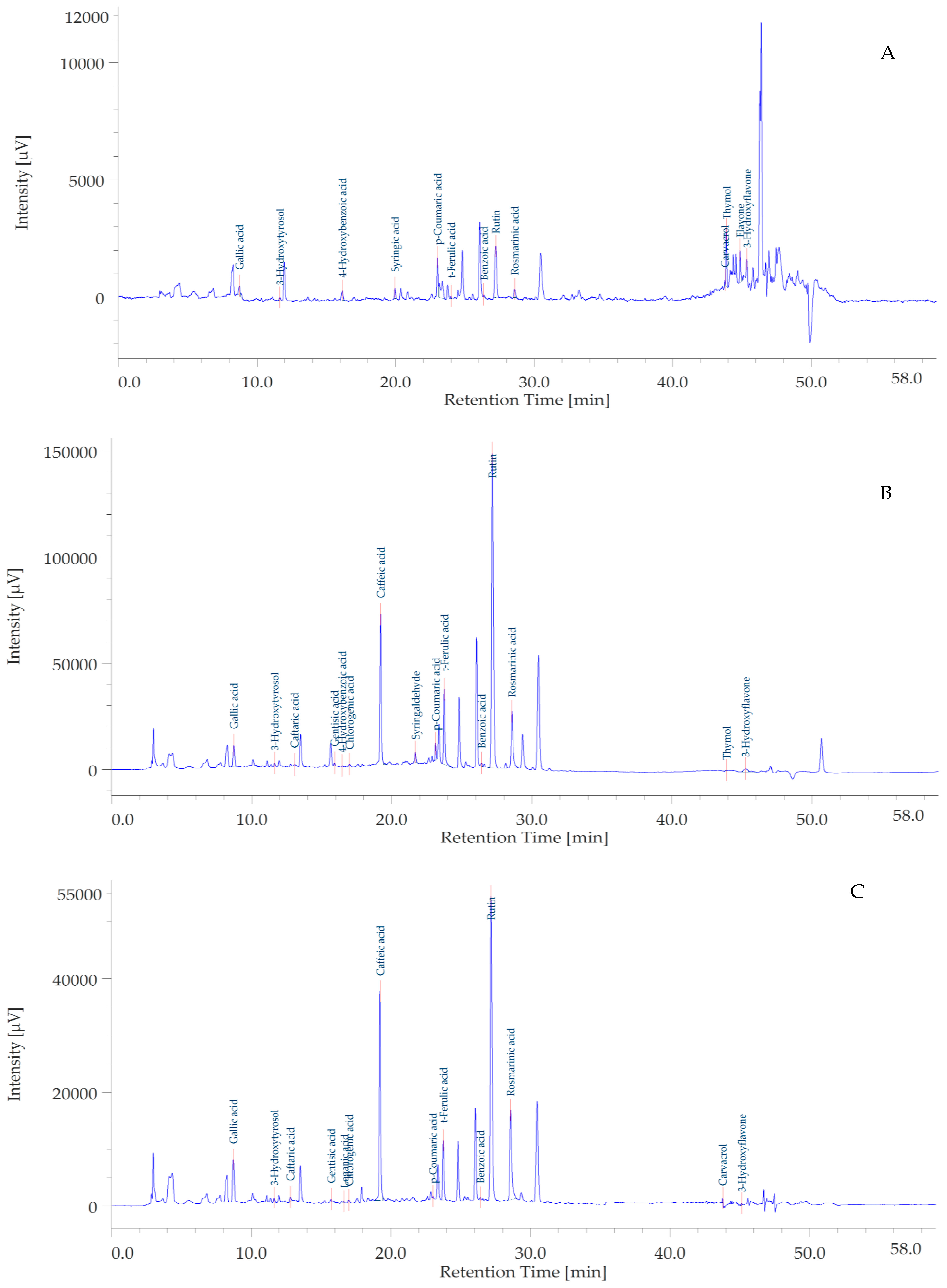
Figure 1. Representative chromatograms of the ethyl acetate (A), methanol (B), and water (C) extracts from the aerial portions of Pelargonium quercetorum. The integration of the present chromatograms has been carried out at 254 nm.
Plants 12 01132 g001
Figure 2. Pearson’s correlations between total/individual bioactive compounds and biological activity assays (p < 0.05). TPC: Total phenolic content; TFC: Total flavonoid content; ABTS, 2,2′-azino-bis(3-ethylbenzothiazoline) 6-sulfonic acid; CUPRAC, cupric ion reducing antioxidant capacity; DPPH, 1,1-diphenyl-2-picrylhydrazyl; FRAP, ferric ion reducing antioxidant power; MCA, metal chelating activity; PBD, phosphomolybdenum activity; AChE: Acetylcholinesterase; BChE: Butrylcholinesterase.
Plants 12 01132 g002
Figure 3. (A) Inhibitory effect of the ethyl acetate extract 10–100 µg/mL from the aerial parts of Pelargonium quercetorum on human colon cancer HCT116 cell viability. * p < 0.05 vs. Ctrl group. (B) Null effect of the methanol extract 10–100 µg/mL from the aerial parts of Pelargonium quercetorum on human colon cancer HCT116 cell viability. (C) Stimulatory effect of the water extract 10–100 µg/mL from the aerial parts of Pelargonium quercetorum on human colon cancer HCT116 cell viability. *** p < 0.001 vs. Ctrl group. (D) Inhibitory effect of the ethyl acetate extract 100 µg/mL from the aerial parts of Pelargonium quercetorum on human colon cancer HCT116 cell viability, after 48 and 72 h of exposure to the extract (*** p < 0.001).
Plants 12 01132 g003aPlants 12 01132 g003b
Figure 4. (A) Inhibitory effect of the ethyl acetate extract 10–100 µg/mL from the aerial parts of Pelargonium quercetorum on the viability of human colon cancer HCT116 cells exposed to hydrogen peroxide (H.P.) 300 µM. * p < 0.05 vs. Ctrl group. (B) Stimulatory effect of the methanol extract 10–100 µg/mL from the aerial parts of Pelargonium quercetorum on the viability of human colon cancer HCT116 cells exposed to hydrogen peroxide (H.P.) 300 µM. *** p < 0.001 vs. Ctrl group. (C) Stimulatory effect of the water extract 10–100 µg/mL from the aerial parts of Pelargonium quercetorum on the viability of human colon cancer HCT116 cells exposed to hydrogen peroxide (H.P.) 300 µM. *** p < 0.001 vs. Ctrl group. (D) Inhibitory effect of the ethyl acetate extract 100 µg/mL from the aerial parts of Pelargonium quercetorum on the viability of human colon cancer HCT116 cells exposed to hydrogen peroxide (H.P.) 300 µM, after 48 and 72 h of exposure to the extract (*** p < 0.001).
Plants 12 01132 g004aPlants 12 01132 g004b
Figure 5. (A) Inhibitory effect of the ethyl acetate extract 100 µg/mL from the aerial parts of Pelargonium quercetorum on HIF1α gene expression in human colon cancer HCT116 cells exposed to hydrogen peroxide (H.P.) 300 µM. *** p < 0.001 vs. H.P. group. (B) Inhibitory effect of the ethyl acetate extract 100 µg/mL from the aerial parts of Pelargonium quercetorum on VEGFA gene expression in human colon cancer HCT116 cells exposed to hydrogen peroxide (H.P.) 300 µM. *** p < 0.001 vs. H.P. group. (C) Inhibitory effect of the ethyl acetate extract 100 µg/mL from the aerial parts of Pelargonium quercetorum on TRPM8 gene expression in human colon cancer HCT116 cells exposed to hydrogen peroxide (H.P.) 300 µM. *** p < 0.001, * p < 0.05 vs. Ctrl group.
Plants 12 01132 g005
Figure 6. (A) Inhibitory effect of the ethyl acetate extract 10–100 µg/mL from the aerial parts of Pelargonium quercetorum on COX-2 gene expression in isolated mouse colon specimens exposed to LPS (50 µg/mL). *** p < 0.001 vs. LPS group. (B) Inhibitory effect of the ethyl acetate extract 10–100 µg/mL from the aerial parts of Pelargonium quercetorum on TNFα gene expression in isolated mouse colon specimens exposed to LPS (50 µg/mL). *** p < 0.001 vs. LPS group.
Plants 12 01132 g006
Figure 7. (A) Modeled structures of tyrosinase, and (B) its Ramachandran plot showing the energetically allowed regions for backbone dihedral angles ψ against φ of amino acid residues. (C) Modeled structures of α-glucosidase and (D) its Ramachandran plot showing the energetically allowed regions for backbone dihedral angles ψ against φ of amino acid residues. (E) Binding energy (docking score) values of the top bioactive compounds from Pelargonuim quercetorum. The compounds are displayed based on their serial number in Table 2.
Plants 12 01132 g007
Figure 8. Protein-ligand interactions: (A) AChE:loganic acid, (B) BChE:caftaric acid, (C) tyrosinase:rosmarinic acid, (D) α-amylase:syringaldehyde, (E) α-glucosidase:chlorogenic acid, and (F) TRPM8:rutin.
Plants 12 01132 g008
Figure 9. ADMET properties of the bioactive compounds extracted from Pelargonium quercetorum predicted using Biovia DS ADMET prediction toolkit. The four ellipses enclose areas where well-absorbed compounds should be found: at 95 and 99% confidence levels for gastrointestinal absorption (red and green), and blood-brain barrier penetration (magenta and aqua). The compounds are shown according to their serial number in Table 2.
Plants 12 01132 g009
Table 1. Total phenolic (TPC) and flavonoid content (TFC) of the tested extracts.
SolventsTPC (mg GAE/g Extract)TFC (mg RE/g Extract)
Ethyl acetate21.94 ± 0.72 c2.78 ± 0.58 c
Methanol45.77 ± 1.02 b40.12 ± 0.20 a
Water62.52 ± 0.43 a16.15 ± 0.17 b
Values are reported as mean ± SD of three parallel experiments. TPC: Total phenolic content; TFC: Total flavonoid content; GAE: Gallic acid equivalent; RE: Rutin equivalent. Different letters indicate significant differences in the tested extracts (p < 0.05).
Table 2. Content in specialized metabolites (µg/mg) of the tested P. quercetorum extracts. All identified phytochemicals have been identified through comparison with pure standards. Most of them have been quantified in at least one extract. Quantitative determination of the compounds was performed via DAD detector at 254–340 nm wavelength.
ComponentsChemical ClassEAMEOHWaterRetention Time (Min)Wavelenghts
1Gallic acidBenzoic acidsnq12.55 ± 0.028.53 ± 0.038.723271
2HydroxytyrosolPhenylethanoidsnq13.78 ± 0.036.18 ± 0.1411.620280
3Caftaric acidHydroxycinnamic acidsnq4.70 ± 0.153.20 ± 0.0413.060310
4Gentisic acidBenzoic acidsnq5.03 ± 0.021.70 ± 0.2115.907325
54-Hydroxybenzoic acidBenzoic acidsnqnqnq16.450256
6Loganic acidIridoidsnqnqnq16.637
7Chlorogenic acidHydroxycinnamic acidsnq1.70 ± 0.200.40 ± 0.0316.960325
8Caffeic acidHydroxycinnamic acidsnq51.75 ± 1.2027.18 ± 0.4419.207325
9Syringic acidBenzoic acidsnqnqnq19.960274
10SyringaldehydeBenzoic aldehydesnq28.35 ± 0.25nq21.667310
11p-Coumaric acidHydroxycinnamic acids1.43 ± 0.1813.53 ± 0.35nq23.140310
12t-Ferulic acidHydroxycinnamic acidsnq33.23 ± 0.218.80 ± 0.0923.747315
13Benzoic acidBenzoic acids1.05 ± 0.028.35 ± 0.263.18 ± 0.0526.400275
14RutinFlavonol glycosides1.05 ± 0.01190.33 ± 0.9263.78 ± 0.9827.170254
15Rosmarinic acidHydroxycinnamic acidsnq45.28 ± 0.4427.13 ± 0.3828.570325
16CarvacrolPhenolic monoterpenesnqnqnq43.757275
17ThymolPhenolic monoterpenes15.68 ± 0.02nqnq43.873275
18FlavoneFlavonesnqnqnq44.840340
193-HydroxyflavoneFlavonesnqnqnq45.327340
nq: not quantified.
Table 3. Antioxidant properties of the tested extracts.
SolventsDPPH (mg TE/g Extract)ABTS (mg TE/g Extract)CUPRAC (mg TE/g Extract)FRAP (mg TE/g Extract)MCA (mg EDTAE/g Extract)PBD (mmol TE/g Extract)
Ethyl acetate9.28 ± 0.52 c27.17 ± 0.04 c96.07 ± 1.11 c32.65 ± 0.70 cna1.64 ± 0.04 a
Methanol48.80 ± 0.01 b78.59 ± 0.10 b172.02 ± 6.72 b102.48 ± 6.20 b29.80 ± 0.79 a1.54 ± 0.07 b
Water140.40 ± 1.16 a212.31 ± 8.88 a207.83 ± 1.36 a142.01 ± 1.08 a12.28 ± 0.23 b1.51 ± 0.01 b
Values are reported as mean ± SD of three parallel measurements. TE: Trolox equivalent; EDTAE: EDTA equivalent; na: not active. Different letters indicate significant differences in the tested extracts (p < 0.05). ABTS, 2,2′-azino-bis(3-ethylbenzothiazoline) 6-sulfonic acid; CUPRAC, cupric ion reducing antioxidant capacity; DPPH, 1,1-diphenyl-2-picrylhydrazyl; FRAP, ferric ion reducing antioxidant power; MCA, metal chelating activity; PBD, phosphomolybdenum activity.
Table 4. Enzyme inhibitory of the tested extracts.
SolventsAChE
(mg GALAE/g Extract)
BChE
(mg GALAE/g Extract)
Tyrosinase (mg KAE/g Extract)α-Amylase (mmol ACAE/g Extract)α-Glucosidase (mmol ACAE/g Extract)
Ethyl acetate1.63 ± 0.11 b2.59 ± 0.0422.68 ± 1.26 b0.91 ± 0.02 a1.37 ± 0.07 a
Methanol2.31 ± 0.29 ana31.24 ± 0.49 a0.45 ± 0.03 b1.38 ± 0.02 a
Water0.76 ± 0.08 cna0.63 ± 0.11 c0.07 ± 0.01 c0.40 ± 0.01 b
Values are reported as mean ± SD of three parallel measurements. GALAE: Galantamine; KAE: Kojic acid; ACAE: Acarbose equivalent; na: not active. Different letters indicate significant differences in the tested extracts (p < 0.05). AChE: Acetylcholinesterase; BChE: Butrylcholinesterase.
Table 5. Gradient Elution Conditions.
Time (Min.)Compasition A% (Water + Formic Acid 0.1%)Compasition B%
(Methanol + Formic Acid 0.1%)
Flow (mL/Min)
1.0097.03.00.600
5.0077.023.00.600
12.0073.027.00.600
18.0057.043.00.600
25.0052.048.00.600
32.0050.050.00.600
34.0050.050.00.600
37.0035.065.00.600
40.005.095.00.600
47.005.095.00.600
48.0097.03.00.600
60.0097.03.00.600
Disclaimer/Publisher’s Note: The statements, opinions and data contained in all publications are solely those of the individual author(s) and contributor(s) and not of MDPI and/or the editor(s). MDPI and/or the editor(s) disclaim responsibility for any injury to people or property resulting from any ideas, methods, instructions or products referred to in the content.