Article

Effects of Flavonoids and Phenols from Moringa oleifera Leaf Extracts on Biofilm Processes in Xanthomonas campestris pv. campestris

by
Riccardo Fontana
1,†,
Anna Caproni
1,†,
Mariaconcetta Sicurella
2,
Stefano Manfredini
3,
Anna Baldisserotto
3 and
Peggy Marconi
1,4,*
1
Department of Chemical, Pharmaceutical and Agricultural Sciences, University of Ferrara, 44121 Ferrara, Italy
2
Department of Environmental Sciences and Prevention, University of Ferrara, 441211 Ferrara, Italy
3
Department of Life Sciences and Biotechnology, University of Ferrara, 44121 Ferrara, Italy
4
Technopole of Ferrara, LTTA Laboratory for Advanced Therapies, Ferrara 44121, Italy
*
Author to whom correspondence should be addressed.
These authors contributed equally to this work.
Plants 2023, 12(7), 1508; https://doi.org/10.3390/plants12071508
Submission received: 5 December 2022 / Revised: 13 March 2023 / Accepted: 21 March 2023 / Published: 30 March 2023
(This article belongs to the Special Issue Plant Extracts as Biological Protective Agents)

Abstract

:
Xanthomonas campestris pv. campestris is the causal agent of black rot in crucifers, a plant disease with significant economic impact. Xanthomonadaceae is a large family of Gram-negative bacteria that cause symptoms by blocking water flow in plants by invading the xylem. To accomplish this, the main mechanism the bacteria use to adapt to environmental changes and colonize tissues is biofilm formation. In recent years, growing interest in natural antimicrobial compounds has led to the study of different phytocomplexes derived from plants. In this work, Moringa oleifera was selected, as its leaves are rich in phenols, essential oils, and vitamins that exert antibacterial activity. X. campestris pv. campestris biofilm, one of its major virulence factors, was studied. Biofilm formation and removal were analyzed on abiotic and biotic surfaces with and without M. oleifera leaf extracts. The data from the analysis show that Moringa oleifera leaf extracts and single phenols were able to inhibit biofilm growth on abiotic surfaces, but the activity of the whole phytocomplex was significantly higher compared to that of individual phenols. The effect of Moringa oleifera extracts on cabbage leaves in vivo was also found to be very important, as scanning electron microscopy showed that treatment with the extracts led to clear unblocking of the xylem, implying many advantages for use in black rot control.

1. Introduction

Xanthomonas campestris pv. campestris (Xcc) is a Gram-negative bacterium that causes black rot in cruciferous plants by invading the xylem and blocking the water flow in plants [1]. It damages plant cells, which causes tissue maceration and blockage of the xylematic vessels. Biofilms are structural communities consisting of aggregates of bacterial cells protected by a polymeric matrix that facilitates their survival in many environments [2]. Once it reaches the xylem, X. campestris pv. campestris colonizes it due to the action of pili and the production of extracellular polysaccharides (EPS), such as xanthan gum, as well as cellulase, protease, polygalacturonase, amylase, hemolysin, and hemagglutinin [3,4]. Furthermore, the type III secretion system participates in the pathogenicity mechanism, as it can interfere with plant defense apparatuses [5].
Black rot is one of the most destructive diseases in the world, with a significant economic impact in many geographic regions [6]. Chemical approaches have been widely used to control X. campestris pv. campestris, but these have led to environmental pollution and the emergence of resistant strains [7,8,9,10]. In particular, copper and streptomycin resistance has been observed in many pathogenic bacteria; copper mixtures are not always able to efficiently control disease, which leads to the selection of resistant bacterial isolates. Furthermore, copper-based compounds can interrupt the microbial activity in soil, as it adversely affects the activity of microorganisms and earthworms, leading to slower decomposition of organic matter [11,12].
In recent years, growing interest in natural antimicrobial compounds that may be bio-safe and eco-friendly have led to the study of different phytocomplexes derived from plants [13]. In this work, Moringa oleifera Lam. (MOL) was selected, which has leaves known to be rich in phenols, flavonols, essential oils, and vitamins that exert antibacterial activity [14]. Previously, we reported high concentrations of flavonols and flavones in M. oleifera Lam. leaves [15,16]; several glycosylated flavonoids have also been identified, including quercetin, kaempferol, and rutin. In the dried leaves, high concentrations of phenolic acids have been detected, such as chlorogenic acid, gallic acid, ferulic acid, ellagic acid, and caffeic acid [14,16,17]. Among them, chlorogenic acid is known to significantly increase membrane permeability in different bacterial pathogens, resulting in the loss of the barrier function, which induces leakage of nucleotide [18].
Previous studies by our group highlighted the effects of antimicrobial activity of different Moringa oleifera extracts on phytopathogens [15,16]. The main components of the phytocomplex detected in our extracts were tested individually in different assays in order to better understand the mechanism of action of each flavonoid and phenol in X. campestris pv. campestris compared to that of the whole phytocomplex. Furthermore, as stated before, biofilm formation is a crucial step in this disease. Biofilms are often studied with scanning electron microscopy, and X. campestris pv. campestris biofilm formation and removal were analyzed on abiotic and biotic surfaces with and without the influence of M. oleifera leaf extracts: M. oleifera hydroalcoholic extract (HA-MOL), M. oleifera methanolic extract (MeOH-MOL), and M. oleifera hydroalcoholic extract with the addition of maltodextrins (HAMD-MOL).
The objectives of this work were to study the phytocomplex and how each individual molecule acts, and to investigate biofilm-associated processes and how they are influenced by the application of M. oleifera extracts.

2. Results

The potential antimicrobial effects of several phytochemical phenolic compounds on membrane permeability, biofilm formation, and swarming motility of X. campestris pv. campestris were evaluated: chlorogenic acid (CA), epigallocatechin (EC), rutin (RU), and quercetin (QU) were selected, as these compounds were found to be the most abundant in the studied extracts from M. oleifera leaves [15,19,20,21]. One of the main results of this study is the discovery of the antimicrobial potential of M. oleifera extracts on X. campestris pv. campestris. The extract effect was retained in all tested strains, without any significant differences, giving us important information on the use of natural phytocomplexes on Xanthomonadaceae. Despite the genetic differences that may exist between bacterial strains, the extract is able to inhibit bacterial growth and alter membrane permeability. Based on analyses of other phytopathogens, this effect is probably extendable to many Gram-negative bacteria.

2.1. Effects of Single Phenols on Membrane Permeability

To evaluate membrane permeability, sub-minimal inhibitory concentrations (MICs) of selected compounds were tested on X. campestris pv. campestris bacterial suspensions for 60 min. To demonstrate possible membrane weathering, a fluorescent intercalating agent, propidium iodide (PI), was added to the bacterial suspensions. The assay showed that the single compounds provided membrane permeability similar to that of the positive control, represented by the bacteria treated with the most effective extract, the hydroalcoholic extract of M. oleifera with maltodextrin (HAMD-MOL) (Figure 1). It was noted that the effects of the phytocomplex were retained and remained higher than those of the single compounds tested (rutin, quercetin, chlorogenic acid, and ellagic acid). However, the experiment also showed that PI could rapidly enter X. campestris pv. campestris cells after chlorogenic acid, ellagic acid, and rutin were added, while the cell membrane was still polarized [18].

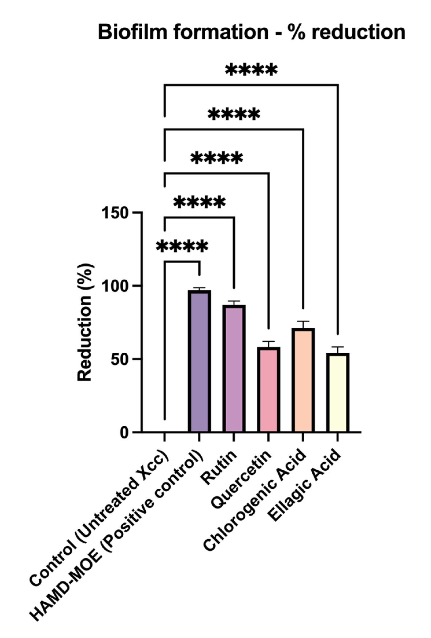
2.2. Effects of Single Phenols on Biofilm Formation

To evaluate whether each isolated compound had anti-biofilm activity, sub-MICs were added to the bacterial suspension. After incubation, biofilm formation was quantified using a spectrophotometer. As shown in Figure 2, compared to the positive control, consisting of only overnight bacterial suspension of X. campestris pv. campestris, there was a marked reduction in biofilm formation for all compounds tested. In this case, rutin seemed to be the most effective, reducing biofilm formation by 91%, compared to the control, followed by chlorogenic acid (70% reduction).

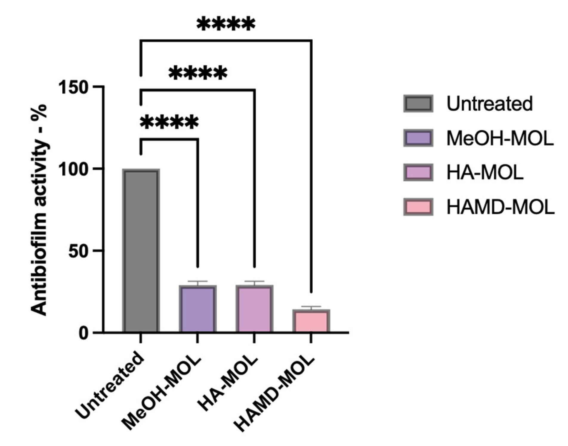
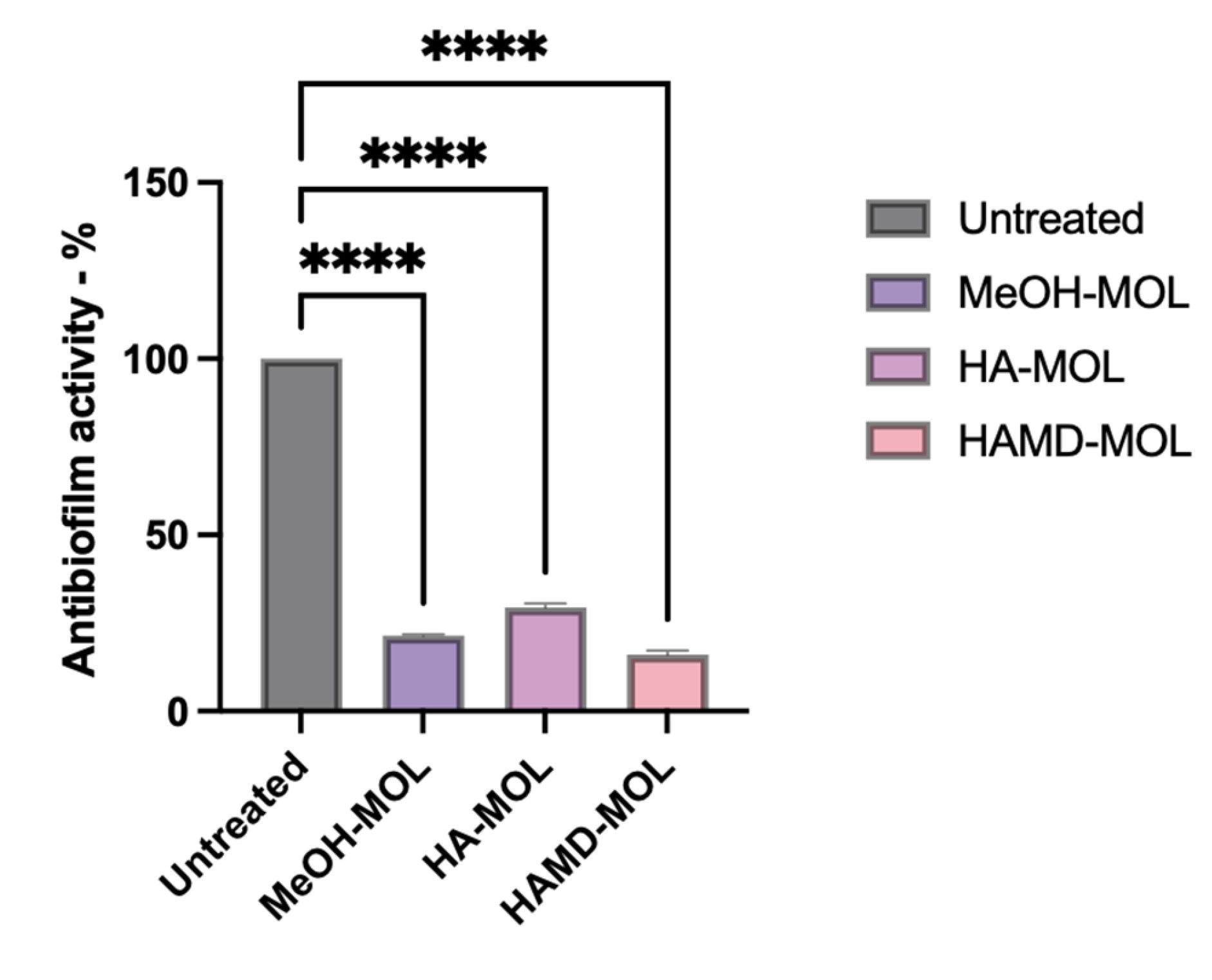
2.3. Assay of Biofilm Formation and Removal on Abiotic Surfaces

To assess whether M. oleifera extracts had anti-biofilm activity, and to visualize which part of the biofilm formation process was affected, biofilm formation on abiotic surfaces (such as a plastic cell strainer) was studied. From the analysis it was possible to confirm the antimicrobial and antibiofilm activity of the alcoholic extracts of M. oleifera. In vitro experiments have shown how extracts can inhibit both the formation and removal of biofilm on inert surfaces. As for the formation assay, MeOH-MOL, HA-MOL, and HAMD-MOL were able to reduce biofilm formation by 71, 70.78, and 85.67%, respectively (Figure 3). Similar activity was detected by the removal assay, with removal of mature biofilm by 78.56, 70.12, and 84%, respectively (Figure 4).
As known from our previous analysis, the three extracts contained comparable quantities of flavonoids and total phenols; in fact, their activity did not differ statistically from one another (Supplementary Tables S1 and S2) [16,22].
Biofilm growth on a plastic cell strainer was also analyzed with scanning electronic microscopy (SEM). This analysis showed that the bacteria were in distress when in contact with the extracts. Compared to the control, the size of planktonic cells was reduced by half (5 µm for untreated vs. ≤2 µm for treated X. campestris pv. campestris), with signs of cell suffering such as folding (Figure 5). Focusing on the three-dimensional development of the biofilm, it was evident that untreated cells (Figure 5a–d) had a clearly defined structure, with abundant and very thick cellular clusters characterized by multilayer distribution. An extracellular matrix was abundantly produced during the maturation of the biofilm, and xanthan gum foils could be observed, as shown in Figure 5a. In addition, thin fibers connecting and covering cells were visualized. Interestingly, X. campestris pv. campestris cells were found in localized groups in the spaces of the cell strainer, where they multiplied to fill the space; the bacteria appeared to be suspended in the air spaces, but they were likely protected by an EPS matrix that dried during sample fixation.
Focusing on the M. oleifera extract treated samples, it was clear that bacteria were not able to produce typical biofilm structures (Figure 5e–j). The bacteria were mainly found to be isolated and in their planktonic state; however, it should be noted that X. campestris pv. campestris started to fill the spaces of the cell strainer even under the effects of MeOH-MOL and HA-MOL, but was not able to cover, adhere to, and colonize the single filaments (Figure 5e,f,i,j). Meanwhile, HAMD-MOL was shown to have a better effect, as it completely inhibited the release of EPS (Figure 5g,h). No extracts allowed the formation of a multilayer biofilm, indicating that quorum sensing (QS) might have been inhibited [23].

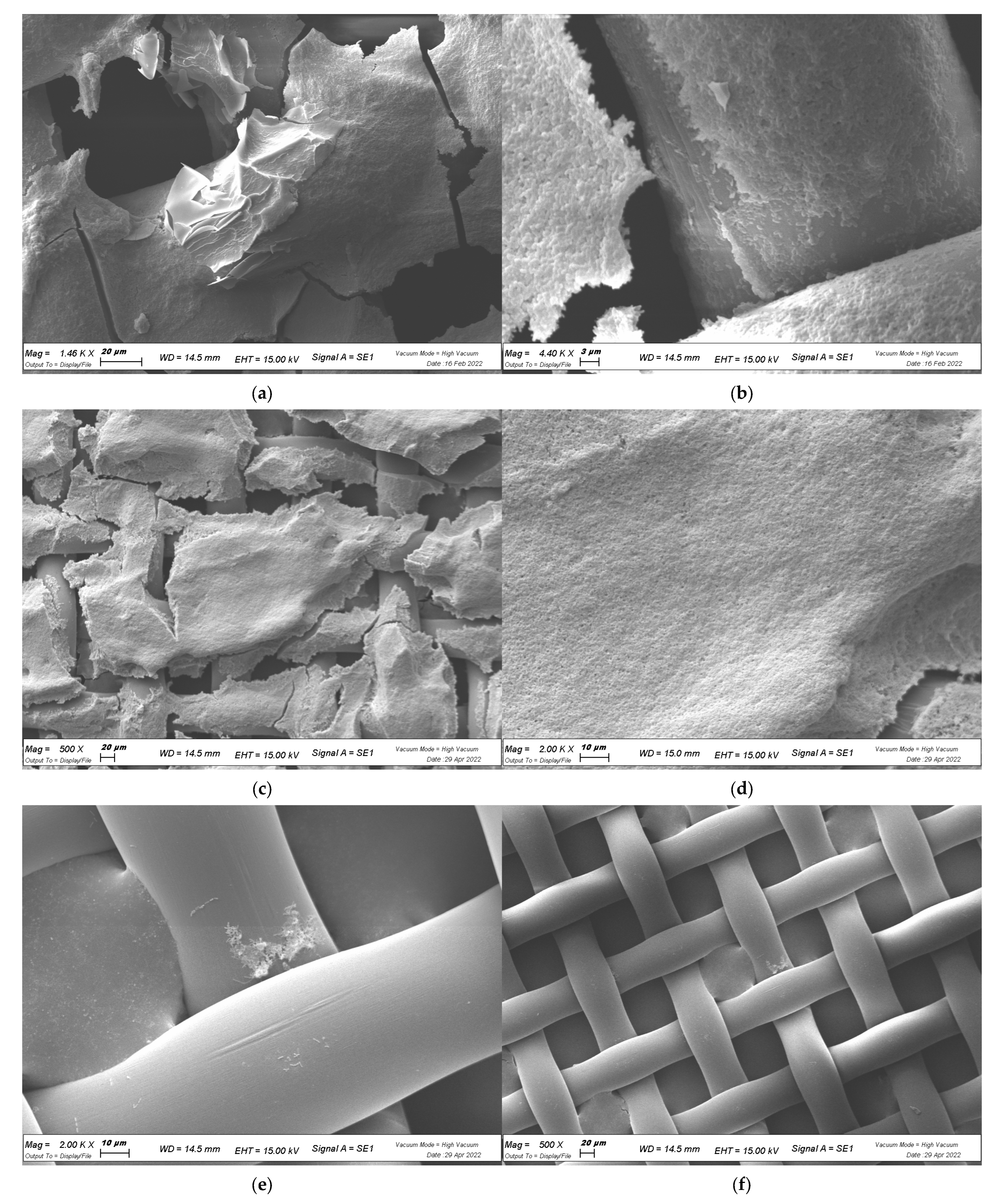
2.4. In Planta Biofilm Formation

To evaluate whether extracts blocked biofilm formation in planta, cabbage leaves, as natural hosts of X. campestris pv. campestris, were used. Colonization of the biotic surface is shown in Figure 6. Cabbage leaves were inoculated with a suspension of X. campestris pv. campestris, followed by M. oleifera extract injection and spraying. The progression of infection was visually monitored at different time points. After the infection was clearly established, cabbages were sliced and visualized with SEM. In the untreated inoculated cabbage, the vessels were completely coated by bacteria and EPS, mainly localized and attached to the inner surface of vessels (Figure 6a,b). When the extracts were applied, the quantity of living bacteria was significantly reduced and localized in a few vessels, with only a few tridimensional structures formed, suggesting that the extracts might not have been able to reach all parts of the cabbage leaf (Figure 6c–h). Again, HAMD-MOL (Figure 6c,d) had the strongest effect in terms of inhibiting bacterial adhesion and growth through the vessel. This effect was also confirmed by the total viable bacteria count from the cabbage leaf core (1 × 1 cm2): the assay showed that untreated cabbage leaves were completely invaded by X. campestris pv. campestris, corresponding to a growth of 2.6 × 10⁸ CFU/mL, while treated leaves showed an average 4- to 5-fold log reduction in bacterial growth, corresponding to 3.03 × 10⁴, 2.59 × 10⁴, and 6.02 × 10³ CFU/mL for MeOH-MOL, HA-MOL, and HAMD-MOL, respectively, as shown in Table 1.
In conclusion, SEM images showed complete colonization by X. campestris pv. campestris on abiotic and biotic surfaces when not under the effects of extracts; in fact, the control showed that the fibers of the cell strainer were coated with EPS, and cabbage xylems were completely invaded. Furthermore, the production of xanthan gum (one of the main components of X. campestris pv. campestris EPS and virulence factors) layers was observed. The antibiofilm properties of HAMD-MOL and MeOH-MOL were proven and confirmed by the presence of just a few solitary planktonic bacteria and no observed complex or organized structures.

3. Discussion

Global research on bacterial biofilms continues on many fronts, with particular emphasis on specifically expressed genes and the role of biofilm in acquiring resistance, as well as the evaluation of control measures and the development of innovative strategies [24,25].
It is known that many natural bioactive molecules have antibiofilm (as well as antimicrobial) activity. This is particularly important in the case of xylematic pathogens; thus, biofilm formation during X. campestris pv. campestris growth was investigated, as it plays a key role in the infection process. The extracts used in this study showed significant antibacterial activity against X. campestris pv. campestris in the biofilm matrix, apparently limiting the adhesion of bacteria to the surface. The extracts also displayed effective biofilm-dissolving ability, supporting their possible use in therapeutic treatments, and the activity could not be completely expressed by the individual molecules detected in MOL leaves. These findings support the assumption that phenolic compounds such as quercetin, rutin, ellagic acid, and others can act cooperatively as antibiofilm compounds, as none of the individual phenols reached the same activity as that of the whole phytocomplex [26].
One of the main differences found was the higher activity of the HAMD-MOL extract over time. The activity was more pronounced when maltodextrins were added, as the tested extracts are comparable in terms of polyphenol percentage. The maltodextrin (MD) hydroalcoholic extract had the highest effect because maltodextrins act as coating agents integrating bioactive molecules, which extends their lifespan and prevents them from losing their activity. A study by Sri Harsha et al., found that the addition of maltodextrins significantly enhanced the stability of certain polyphenolic compounds, particularly chlorogenic acid [22]. Recently, a study found that combined treatment with electrochemically generated H2O2 and maltodextrin was more effective at reducing the density of viable biofilm cells than any single treatment, and another study showed that gum arabic and maltodextrin microencapsulation resulted in significant preservation of pepper seed oil against oxidation during storage [27,28,29].
Based on our previous studies and a literature review, we know that the phytocomplex consists of polyphenolic molecules that can alter the permeability of the bacterial membrane, leading to a shutdown of ATP synthesis, causing the cessation of all ATP-dependent functions and a subsequent reduction in selectivity of compounds that can penetrate the bacterial cytoplasm [14,30,31]. In fact, some of these molecules act by modifying membrane permeability and integrity by promoting the passage through the cytoplasm of compounds capable of inhibiting the enzymatic complexes involved in bacterial replication [32].
Specifically, among the simple phenols, flavonoids, and phenolic acids detected in the leaves of M. oleifera Lam., naringenin, ferulic acid, rutin, chlorogenic acid, and ellagic acid may complex with the bacterial cell walls, affecting the fluidity, structure, and function of the phospholipid double layer. In particular, the antibacterial activity of rutin and quercetin has been linked to their solubility and interaction with the bacterial cell membrane, which is largely determined by quercetin’s hydroxyl groups [33,34,35]. Recent studies have shown that flavonoids (quercetin in particular) can effectively disrupt the integrity of the bacterial membrane, resulting in inhibition of bacterial growth [26,32,36]. Using TEM analysis, Wang et al., observed that in treated E. coli, there were numerous structural anomalies in damaged cells: wall lysis, deformation, leakage of cellular material, and unequal endochyleme density. Eventually, cell cavitation and cell death were evident. Similarly, in treated X. campestris pv. campestris, membrane shrinking and cavitation were noted [18,36]. In a recent study, flavonoid activity was investigated, comparing ceftriaxone and imipenem, since both are bacterial cell wall inhibitors. Alnour et al., suggested that the tested antibiotics caused the cell wall damage, while lesions in the cytoplasmic membrane were inflicted by flavonoids, with greater damage observed when a combination of flavonoids was used [37].
Some molecules, such as robinetin, myricetine, and epigallocatechin, act by creating hydrogen bonds with nucleic bases, inhibiting both DNA and RNA synthesis. Flavonoids with a hydroxyl central ring, such as rutin, bind to the β-subunit of DNA gyrase (GyrB) and block the binding site for ATP, thereby compromising processes such as cell division and chromosome replication, leading to inhibition of bacterial growth, as observed by Plaper et al. [38]. According to Liu et al., quercetin inhibited the supercoiling activity of gyrase in Lactobacillus spp., ultimately leading to the disruption of bacterial DNA replication [39,40]. In addition, rutin is able to inhibit the type II topoisomerase enzyme, promoting DNA cleavage, while myricetin appears to inhibit a number of enzymes of fundamental importance, such as dihydrofolate reductase and different DNA and RNA polymerases [2,41]. In this work, the experiments on membrane permeability showed that PI could rapidly enter X. campestris pv. campestris cells after chlorogenic acid was added, whereas the cell membrane was still polarized, in accordance with a study by Lou at al. [18]. These results suggest that chlorogenic acid might first disrupt the bacterial membrane permeability, and subsequently depolarize it. This condition then leads to X. campestris pv. campestris being exposed to other molecules in the complex, resulting in bacterial cell death.
The formation of biofilms is a crucial mechanism in the pathogenesis of black rot on crucifers. Biofilms allow bacterial colonies to express resistance in a homogeneous and cohesive way, through improved inter-cell communication in stressful environmental conditions [42,43,44,45]. Flavonoids, especially quercetin, are also able to interfere in the pathways involved in quorum sensing, thereby preventing bacterial adhesion and biofilm formation. A recent study determined that quercetin interferes with the functioning of proteins involved in glycolytic pathways, protein folding, and protein elongation, while Manner et al., reported that quercetin inhibited the production of violacein pigment, an indicator of bacterial QS in Chromobacterium violaceum [46,47].
Focusing on phytopathogens, our most recent work showed how the extracts inhibit the production of amylovoran (one of the main virulence factors of Erwinia amylovora, involved in QS mechanisms) in Erwinia amylovora cells, confirming its activity in the inhibition of QS signaling [15]. The modifying of membrane integrity and permeability and the action of flavonoids on GyrB result in a considerable dissipation of energy. Therefore, this membrane condition has implications for a variety of ATP-dependent mechanisms, such as motility. In fact, an analysis carried out in our recent work showed that X. campestris pv. campestris treated with M. oleifera extracts, though subjected to energy shortages, was not able to swarm on soft agar plates [15,16]. In the previous analysis, X. campestris pv. campestris was unable to completely attach to either an abiotic or biotic surface and form a mature biofilm.
The dispersal of bacteria into plant vessels is a process finely regulated by a detection system that perceives and records the characteristics of the surrounding environment, indicating the direction toward a more favorable environment conducive to survival [48,49,50,51]. In this sense, the bacteria try to limit their movement as much as possible to reduce contact with the extracts. Focusing on the cabbage infection, we observed that X. campestris pv. campestris did not move freely along the xylem, but created small masses of bacterial cells that were not able to form a complete biofilm and start the infection. The reduced effectiveness of ATP-dependent mechanisms also means a slowing down, if not a complete cessation, of the processes involved in the secretion of molecules necessary for cell-to-cell communication and, finally, biofilm formation. This may explain the analyzed antibiofilm effect of M. oleifera Lam. extracts on X. campestris pv. campestris isolates; in fact, the ability of some catechins and flavonols to effectively counteract cell adhesion, the initial stage of biofilm formation, was found in E. coli and S. aureus [52]. Ellagic acid, tannic acid, and epigallocatechin gallate, on the other hand, appear to inhibit biofilm maturation, an effect likely associated with wall damage from peptidoglycan cleavage [53,54,55].
As the anti-biofilm activity of the extracts has been defined, more in-depth studies will be needed to evaluate the stage of biofilm formation that is most compromised.

4. Materials and Methods

4.1. Bacterial Strains and Media

The X. campestris pv. campestris isolates used in this study were given by the Emilia Romagna Plant Health Agency (Servizio Fitosanitario Regione Emilia-Romagna), and strain 3586 (ATCC 33913) was used as a control. Strains were isolated from brassica seeds in Bologna, Ravenna, and Forlì-Cesena provinces, as listed in Table 2. Bacterial strains were conserved at −80 °C in Luria–Bertani (LB) broth (Liofilchem, Roseto degli Abruzzi, Italy) with 50% glycerol. During the study, bacteria were cultured in LB broth [56] and plated on R2A agar (18.1 g/L; Scharlab, Lodi, Italy) or LB agar (30 g/L; Liofilchem) and incubated at 25 and 28 °C.
The bacteria listed in Table 2 were isolated from different plant samples in different parts of the region. The tests were run with all strains listed in the table, but no statistical differences in the response to the extracts were detected between strains; therefore, all results were used and are reported as a mean. The analysis performed on the activity of all single strains can be found in Supplementary Materials.

4.2. Plant Material and Extraction Methods

Leaves of M. oleifera were collected in August 2019, and were subsequently dried by Evra S.r.l. (Loc. Galdo, Lauria, Italy). Dried samples were packaged and sent to the laboratory. Upon arrival, dried leaves were ground to a fine powder with a mortar and stored at −80 °C. The extraction process was carried out by adapting a previously described method briefly discussed below [13]. The extracts were stored at −18 °C until use. Quantification results can be found in Supplementary Tables S1 and S2.
The extracts were formulated as follows:
-
Hydroalcoholic extract (HA-MOL): 10 g of powder was mixed with 200 mL of hydroalcoholic solution (ethanol:water, 70:30) at room temperature for 1 h under magnetic stirring, then filtered and concentrated in vacuum to provide the desired dry hydroalcoholic extract.
-
Methanolic extract (MeOH-MOL): 5 g of powder was mixed with 100 mL of methanol and subjected to two sonication cycles (40 °C, 60 min, 80%) with subsequent centrifugation. The supernatant was concentrated under vacuum to obtain the desired product.
-
Hydroalcoholic extract with maltodextrin (HAMD-MOL): dried M. oleifera Lam. leaves were extracted with 50% ethanol (raw material:solvent, 1:10) for 45 min at 45 °C. After filtration, concentration, and pasteurization, the extract was spray-dried using maltodextrin, obtaining a fine powder.

4.3. Effects of Single Phenols on Membrane Permeability

The effects of individual phenols on membrane permeability were assessed with propidium iodine (PI) staining. PI does not penetrate membranes under normal circumstances, but when the integrity and permeability of the structure changes, it enters bacterial cells and intercalates between DNA bases; this intercalation can be read with a spectrofluorometer, allowing differentiation between healthy and functional cells within damaged cells that have a non-functional membrane structure [57,58]. The experiment was conducted as previously established [16]. Bacterial suspensions were grown overnight in Luria Broth (LB) for 24 h at 25 °C. After incubation, 1 × 10⁵ CFU/mL of bacteria was placed in 4 sterile Eppendorf tubes containing M. oleifera Lam. HAMD extract at concentrations corresponding to 2× MIC (0.2 mg/mL), 1× MIC (0.1 mg/mL), 1/2× MIC (0.05 mg/mL), and 1/4× MIC (0.025 mg/mL), as previously described [16], with sub-MICs of different phenols (chlorogenic acid, 10 µg/mL; rutin, 100 µg/mL; quercetin, 100 µg/mL; ellagic acid, 250 µg/mL). Rutin (Sigma Aldrich, St. Louis, MO, USA, CAS 207671-50-9), quercetin (Sigma Aldrich CAS 117-39-5), and ellagic acid (Sigma Aldrich CAS 476-66.4) were suspended in sterile water, while chlorogenic acid (Sigma Aldrich CAS 327-97-9) was dissolved in 1× PBS. The procedure was repeated by varying the incubation time, so the bacterial suspensions containing different concentrations of single phenols were incubated for 180, 120, 60, and 5 min in a static incubator at 25 °C. After incubation, the suspensions were centrifuged for 5 min at 14,000 rpm and washed with 1× PBS (137 mM NaCl, 2,7 mM KCl, 8 mM Na2HPO4, 2 mM KH2PO4). The pellet was then resuspended with propidium iodide (0.5%) and incubated for 15 min, avoiding exposing the suspension to the light. Then, the suspensions were plated on 96-well plates, and the values were read through a fluorescence microplate reader (Tecan-Fluoroscan, Tecan Italia, Cernusco sul Naviglio, Italy) [57]. All analyses were performed using data obtained from 3 different experiments in triplicate.

4.4. Effects of Single Phenols on Biofilm Formation

The effects on biofilm formation were determined by the crystal violet method, as described by Wilson et al. [59]. According to the cited protocol, higher concentrations of bacteria are needed to perform biofilm assays. X. campestris pv. campestris suspensions containing 106 CFU/mL were inoculated in Luria Broth with M. oleifera extracts and single phenols at non-lethal concentrations in a 96-well U-bottom microplate for 72 h at 25 °C. The non-lethal concentrations of single phenols were as follows: chlorogenic acid, 10 µg/mL; rutin, 100 µg/mL; quercetin, 100 µg/mL; and ellagic acid, 250 µg/mL. After incubation, the growth media, extracts, phenols, and planktonic cells were removed from the plate and washed with sterile deionized water. Crystal violet 1% was added to each well and incubated for 30 min at room temperature, and the dye solution was washed off the plate several times with deionized water. Then 200 μL of decoloring solution (90% ethanol) was added to each well and incubated for 15 min at room temperature to increase crystal violet solubility. The contents of the 96-well plate were then transferred to a new clean microplate and biofilm formation was quantified by reading the absorbance at 570 nm through a microplate reader (Tecan-Sunrise, Tecan Italia, Cernusco sul Naviglio, MI, Italy). All analyses were performed using data obtained from 3 different experiments in triplicate.
In order to obtain information on reduced bacterial biofilm after exposure to M. oleifera extracts, bacterial colonization and biofilm matrix synthesis were examined by biofilm formation assays. Biofilm formation was tested on an abiotic surface, cell strainer fibers, and a biotic surface, cabbage leaves, a natural host of X. campestris pv. campestris.

4.5. Assay of Biofilm Formation and Removal on Abiotic Surface

Bacterial adhesion to an inert surface was assayed in a 6-well polypropylene plate. X. campestris pv. campestris suspensions containing 106 CFU/mL were inoculated in LB in a 6-well plate with a cell strainer insert (BD Italia, Milano, Italy). For biofilm formation assay, MOEs were added along with the bacteria; for biofilm removal assay, MOEs were added after 72 h.
For the biofilm formation assay, bacteria (106 CFU/mL) were incubated for 72 h in Luria Broth medium with M. oleifera extract at non-lethal concentrations. The medium was changed every 24 h, and then tested with crystal violet (CV) staining.
For the biofilm removal assay, X. campestris pv. campestris suspensions containing 106 CFU/mL were inoculated in a cell-strainer insert for 72 h and then treated with the extracts for 24 h. Biofilms, generated as described above, were removed and processed by crystal violet staining.
CV 1% was added to each well and incubated for 30 min at room temperature [59,60]. The dye solution was removed by washing the plates 3 times with deionized water for biofilm formation assay, and until a clear solution was obtained for biofilm removal. Then 200 µL of decoloring solution (90% ethanol) was added to each well and incubated for 15 min at room temperature to increase CV solubility. The contents of the 6-well plate were then transferred to a new clean 96-well microplate, and the biofilm was quantified by reading the absorbance at 570 nm through a microplate reader (Tecan-Sunrise, Tecan Italia, Cernusco sul Naviglio, Italy). All analyses were performed using data obtained from 3 different experiments in triplicate.

4.6. Biofilm Formation in Planta and Total Viable Count

X. campestris pv. campestris strains were grown overnight in 5 mL of LB broth at 25 °C for 24 h, centrifuged, and then resuspended with 1× PBS to reach a bacterial concentration of 5 × 10⁷ CFU/mL. In order to colonize plant tissues, bacteria must be highly concentrated with respect to in vitro experiments [61]. Cabbage leaves were superficially sterilized with a 0.7% solution of sodium hypochlorite and then washed with sterile water, delicately perforated with a sterile 10 µL tip, and inoculated with 5 µL of the bacterial suspension. After the drop was absorbed within 30 min, the MIC of each active extract (HA-MOL, 0.5 mg/mL; MeOH-MOL, 0.5 mg/mL; HAMD-MOL, 0.1 mg/mL) was inoculated in the same hole. The cabbage leaves were then incubated at room temperature (ranging from 20 to 22 °C) for 6 days.
After the incubation time, infected and treated cabbage leaves were cut into small pieces, corresponding to an area of 1 × 1 cm2, and soaked in 10 mL of 1× PBS for 1 h. Then 1 mL of PBS was diluted up to 10−9, plated on R2A agar plates, and incubated for 48 h at 25 °C. After incubation, colonies were counted to record bacterial cell viability.
In addition, the leaves were cut in order to record the area of necrosis around the inoculation point and treated for scanning electron microscopy (SEM) observation as described below.

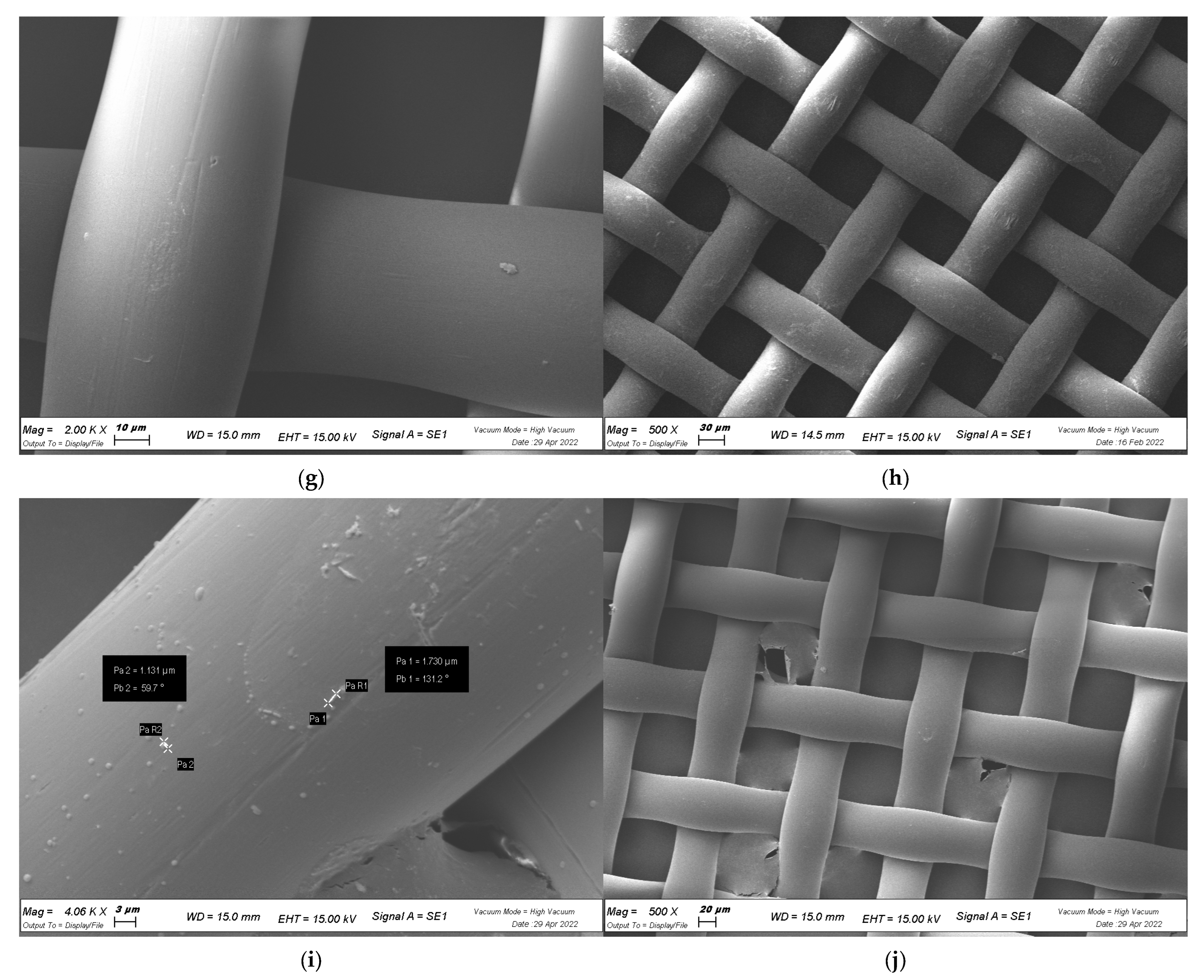
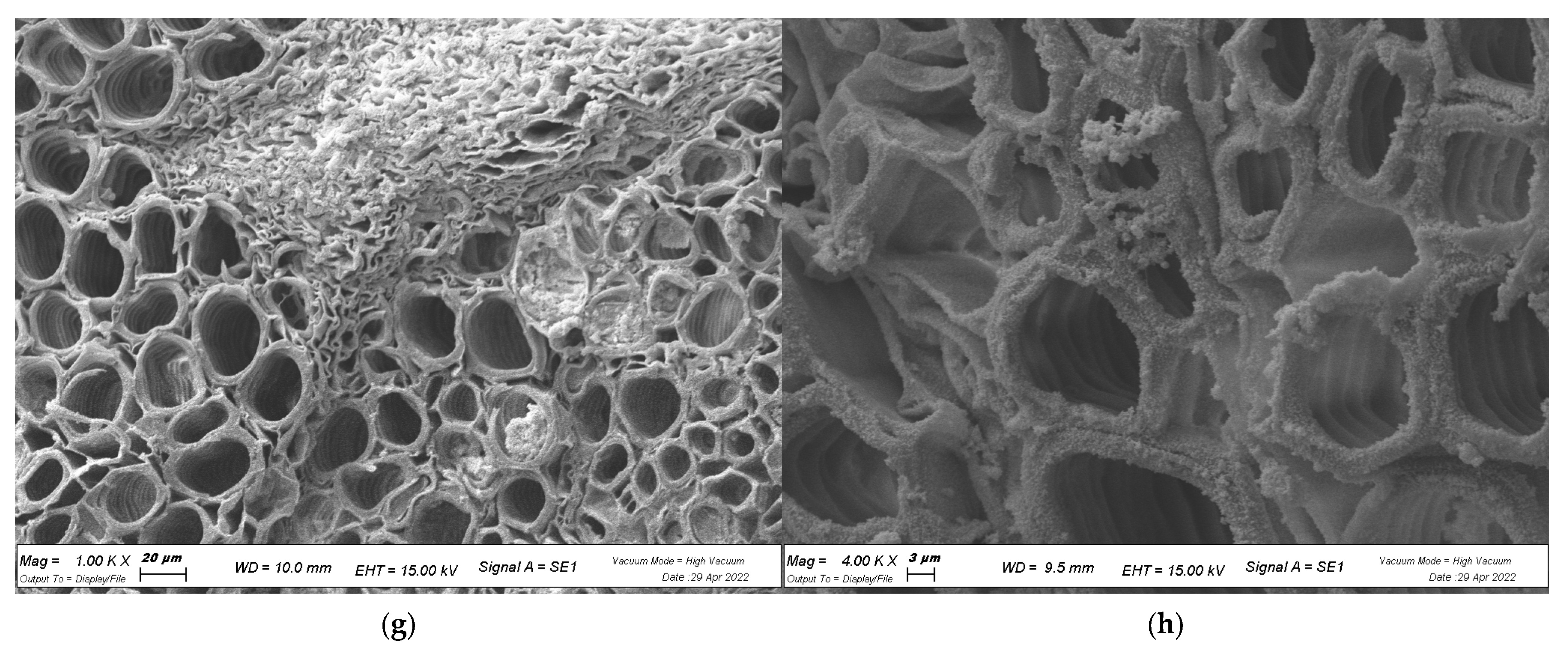
4.7. Visualization of Biofilm with SEM

Ultrastructural analysis was performed by SEM to reveal the cell interaction traits. X. campestris pv. campestris was grown on cell strainers for 72 h and then fixed in 2.5% glutaraldehyde in 0.1 M KPO4 buffer. After being washed with sterile distilled water (SDW), samples were dehydrated by increasing concentrations of ethanol and dried with a critical point dryer. The samples were mounted on metal stubs and coated in gold/palladium. Samples were examined with a Zeiss Evo40 SEM with an electron high tension (EHT) accelerating voltage of 15.00 KV [62].
Visualization of X. campestris pv. campestris on biological surfaces was also achieved by SEM. Previously sterilized cabbage leaves were inoculated with X. campestris pv. campestris as described above and treated with the extracts (HA-MOL, 0.5 mg/mL; MeOH-MOL, 0.5 mg/mL; HAMD-MOL, 0.1 mg/mL). Pieces of inoculated leaves (<6 mm) were fixed in 3% glutaraldehyde in 0.1 M KPO4 buffer. After being washed with SDW, samples were dehydrated by increasing concentrations of ethanol and dried with a critical point dryer. The samples were prepared and observed as described above.

4.8. Statistical Analysis

All tests were performed 3 times in triplicate, and statistical analysis was performed using one-way ANOVA followed by Dunnett’s multiple comparisons test with GraphPad Prism version 9.0.0 for MacOS (GraphPad Software, San Diego, CA, USA, www.graphpad.com; accessed on 8 January 2021), with p ≤ 0.05 indicating significant differences. The detailed statistical analysis is available in the Supplementary Materials, accompanied by a detailed extraction and chemical quantification methodology, as performed, and described, in previous works [63,64,65,66,67].

5. Conclusions

The problematic phenomenon of drug resistance to conventional therapies due to the abuse and misuse of antibiotics in the agro-food sector has led to the enhanced development of new alternative antibacterial agents. Furthermore, newly implemented green policies are increasingly aimed at combating the use of agrochemicals due to their risk to humans, animals, and the environment. In the last few decades, in fact, extracts from plants rich in flavonoids and phenols have been demonstrated to have powerful antibacterial and antibiofilm activity and other pharmacological actions. Moringa oleifera Lam. is marketed as a health supplement primarily for its antibacterial, antioxidant, and anti-inflammatory properties. In the present study, M. oleifera was found to inhibit biofilm formation by X. campestris pv. campestris. As the symptoms observed in the plants are the result of xylem vessel occlusion, unblocking the vessels has recently been investigated. For example, a recent study used a bacteriophage-derived protein that was able to hydrolyze EPS in combination with N-acetylcysteine, leading to the unblocking of xylem in a tree infected with Xylella fastidiosa, whereas X. fastidiosa was not able to adhere to previously treated plants [68].
From this work, it is possible to confirm that the bacterium can adhere to both abiotic and biotic surfaces, and allow biofilm to be developed on them. In addition, while under the effects of M. oleifera extracts, the bacterium changes its cell shape and interrupts the ultrastructural organization that characterizes a developed biofilm, in the adhesion part of the process in particular, and this may have important implications for applications in disease control.

Supplementary Materials

The following supporting information can be downloaded at: https://www.mdpi.com/article/10.3390/plants12071508/s1, Table S1: Total phenol and flavonoid content of leaf extracts of M. oleifera Lam.; Table S2: Percentages of chlorogenic acid, rutin, ellagic acid, ferulic acid, and quercetin in the five different dried extracts of M. oleifera Lam. leaves. Each value was obtained from three analyses (mean ± SD); Table S3: Statistical analysis of membrane permeability alteration by single phenols; Table S4: Statistical analysis of single strains differences on biofilm formation by extracts activity.; Table S5: Statistical analysis of single strains differences on biofilm removal by extracts activity; Figure S1: In vitro membrane permeability assay on single strains by single phenols; Figure S2: In vitro biofilm formation assay on single strains by single phenols; Figure S3: In vitro biofilm formation assay on single strains; Figure S4: In vitro biofilm removal assay on single strains.

Author Contributions

Conceptualization, R.F., S.M. and P.M.; resources, S.M. and P.M.; methodology, R.F., A.C., M.S. and P.M.; software, R.F. and M.S.; investigation, R.F., M.S., A.C. and A.B.; writing—original draft preparation, R.F. and M.S.; writing—review and editing, P.M.; supervision, P.M.; project administration, P.M.; funding acquisition, P.M. All authors have read and agreed to the published version of the manuscript.

Funding

This work was supported by the University of Ferrara Research Funds (P.M., FAR 2021, FAR 2022). We kindly acknowledge the Phytosanitary Department of the Emilia-Romagna region for their support, for the isolation and free concession of the Xcc isolates, and for their precious help. We kindly acknowledge Evra S.r.l. (Loc. Galdo, 85044 Lauria (PZ)) for supplying the material.

Institutional Review Board Statement

Not applicable.

Informed Consent Statement

Not applicable.

Data Availability Statement

Not applicable.

Conflicts of Interest

The authors declare no conflict of interest.

References

  1. Vicente, J.G.; Holub, E.B. Xanthomonas campestris pv. campestris (cause of black rot of crucifers) in the genomic era is still a worldwide threat to brassica crops. Mol. Plant Pathol. 2012, 14, 2–18. [Google Scholar] [CrossRef] [PubMed]
  2. Crossman, L.; Dow, J. Biofilm formation and dispersal in Xanthomonas campestris. Microbes Infect. 2004, 6, 623–629. [Google Scholar] [CrossRef] [PubMed]
  3. He, Y.-W.; Cao, X.-Q.; Poplawsky, A.R. Chemical Structure, Biological Roles, Biosynthesis and Regulation of the Yellow Xanthomonadin Pigments in the Phytopathogenic Genus Xanthomonas. Mol. Plant-Microbe Interact. 2020, 33, 705–714. [Google Scholar] [CrossRef] [Green Version]
  4. He, Y.-W.; Zhang, L.-H. Quorum sensing and virulence regulation in Xanthomonas campestris. FEMS Microbiol. Rev. 2008, 32, 842–857. [Google Scholar] [CrossRef] [Green Version]
  5. Gudesblat, G.E.; Torres, P.S.; Vojnov, A.A. Xanthomonas campestris Overcomes Arabidopsis Stomatal Innate Immunity through a DSF Cell-to-Cell Signal-Regulated Virulence Factor. Plant Physiol. 2008, 149, 1017–1027. [Google Scholar] [CrossRef] [Green Version]
  6. Singh, S.; Dey, S.S.; Bhatia, R.; Batley, J.; Kumar, R. Molecular breeding for resistance to black rot [Xanthomonas campestris pv. campestris (Pammel) Dowson] in Brassicas: Recent advances. Euphytica 2018, 214, 196. [Google Scholar] [CrossRef]
  7. Ryan, R.P.; Ryan, D.J.; Sun, Y.-C.; Li, F.-M.; Wang, Y.; Dowling, D.N. An acquired efflux system is responsible for copper resistance in Xanthomonas strain IG-8 isolated from China. FEMS Microbiol. Lett. 2007, 268, 40–46. [Google Scholar] [CrossRef] [Green Version]
  8. Thapa, S.; Babadoost, M. Effectiveness of Chemical Compounds and Biocontrol Agents for Management of Bacterial Spot of Pumpkin Caused by Xanthomonas cucurbitae. Plant Health Prog. 2016, 17, 106–113. [Google Scholar] [CrossRef] [Green Version]
  9. Kotchoni, O.S.; Torimiro, N.; Gachomo, E.W. Control of Xanthomonas campestris pv. Vignicoia in cowpea following seed and seedling treatment with hydrogen peroxide and N-heterocyclic pyridinium chlorochromate. J. Plant Pathol. 2007, 89, 361–367. Available online: https://www.jstor.org/stable/41998414 (accessed on 9 January 2023).
  10. Liu, Z.; Wang, H.; Wang, J.; Lv, J.; Xie, B.; Luo, S.; Wang, S.; Zhang, B.; Li, Z.; Yue, Z.; et al. Physical, chemical, and biological control of black rot of brassicaceae vegetables: A review. Front. Microbiol. 2022, 13, 1023826. [Google Scholar] [CrossRef]
  11. Shabbir, Z.; Sardar, A.; Shabbir, A.; Abbas, G.; Shamshad, S.; Khalid, S.; Natasha; Murtaza, G.; Dumat, C.; Shahid, M. Copper uptake, essentiality, toxicity, detoxification and risk assessment in soil-plant environment. Chemosphere 2020, 259, 127436. [Google Scholar] [CrossRef] [PubMed]
  12. La Torre, A.; Iovino, V.; Caradonia, F. Copper in plant protection: Current situation and prospects. Phytopathol. Mediterr. 2018, 57, 201–236. [Google Scholar] [CrossRef]
  13. Suteu, D.; Rusu, L.; Zaharia, C.; Badeanu, M.; Daraban, G.M. Challenge of Utilization Vegetal Extracts as Natural Plant Protection Products. Appl. Sci. 2020, 10, 8913. [Google Scholar] [CrossRef]
  14. Kou, X.; Li, B.; Olayanju, J.B.; Drake, J.M.; Chen, N. Nutraceutical or Pharmacological Potential of Moringa oleifera Lam. Nutrients 2018, 10, 343. [Google Scholar] [CrossRef] [PubMed] [Green Version]
  15. Fontana, R.; Macchi, G.; Caproni, A.; Sicurella, M.; Buratto, M.; Salvatori, F.; Pappadà, M.; Manfredini, S.; Baldisserotto, A.; Marconi, P. Control of Erwinia amylovora Growth by Moringa oleifera Leaf Extracts: In Vitro and in Planta Effects. Plants 2022, 11, 957. [Google Scholar] [CrossRef]
  16. Fontana, R.; Caproni, A.; Buzzi, R.; Sicurella, M.; Buratto, M.; Salvatori, F.; Pappadà, M.; Manfredini, S.; Baldisserotto, A.; Marconi, P. Effects of Moringa oleifera Leaf Extracts on Xanthomonas campestris pv. campestris. Microorganisms 2021, 9, 2244. [Google Scholar] [CrossRef]
  17. Chhikara, N.; Kaur, A.; Mann, S.; Garg, M.; Sofi, S.A.; Panghal, A. Bioactive compounds, associated health benefits and safety considerations of Moringa oleifera L.: An updated review. Nutr. Food Sci. 2020, 51, 255–277. [Google Scholar] [CrossRef]
  18. Lou, Z.; Wang, H.; Zhu, S.; Ma, C.; Wang, Z. Antibacterial Activity and Mechanism of Action of Chlorogenic Acid. J. Food Sci. 2011, 76, M398–M403. [Google Scholar] [CrossRef]
  19. Rocchetti, G.; Pamplona Pagnossa, J.; Blasi, F.; Cossignani, L.; Hilsdorf Piccoli, R.; Zengin, G.; Montesano, D.; Cocconcelli, P.S.; Lucini, L. Phenolic profiling and in vitro bioactivity of Moringa oleifera leaves as affected by different extraction solvents. Food Res. Int. 2020, 127, 108712. [Google Scholar] [CrossRef]
  20. Rani, N.Z.A.; Husain, K.; Kumolosasi, E. Moringa Genus: A Review of Phytochemistry and Pharmacology. Front. Pharmacol. 2018, 9, 108. [Google Scholar] [CrossRef] [Green Version]
  21. Kumar, S.; Bhattacharya, A.; Tiwari, P.; Sahu, P. A review of the phytochemical and pharmacological characteristics of Moringa oleifera. J. Pharm. Bioallied Sci. 2018, 10, 181–191. [Google Scholar] [CrossRef]
  22. Harsha, P.S.S.; Lavelli, V. Effects of Maltodextrins on the Kinetics of Lycopene and Chlorogenic Acid Degradation in Dried Tomato. Molecules 2019, 24, 1042. [Google Scholar] [CrossRef] [PubMed] [Green Version]
  23. Guerra, M.L.; Malafaia, C.B.; Macedo, A.J.; Silva, M.V.; Mariano, R.L.R.; Souza, E.B. Biofilm formation by Xanthomonas campestris pv. viticola affected by abiotic surfaces and culture media. Trop. Plant Pathol. 2017, 43, 146–151. [Google Scholar] [CrossRef]
  24. Roy, R.; Tiwari, M.; Donelli, G.; Tiwari, V. Strategies for combating bacterial biofilms: A focus on anti-biofilm agents and their mechanisms of action. Virulence 2018, 9, 522–554. [Google Scholar] [CrossRef] [PubMed] [Green Version]
  25. Jiang, Z.; Nero, T.; Mukherjee, S.; Olson, R.; Yan, J. Searching for the Secret of Stickiness: How Biofilms Adhere to Surfaces. Front. Microbiol. 2021, 12, 686793. [Google Scholar] [CrossRef] [PubMed]
  26. Amin, M.U.; Khurram, M.; Khattak, B.; Khan, J. Antibiotic additive and synergistic action of rutin, morin and quercetin against methicillin resistant Staphylococcus aureus. BMC Complement. Altern. Med. 2015, 15, 59. [Google Scholar] [CrossRef] [PubMed] [Green Version]
  27. Karaaslan, M.; Şengün, F.; Cansu, Ü.; Başyiğit, B.; Sağlam, H.; Karaaslan, A. Gum arabic/maltodextrin microencapsulation confers peroxidation stability and antimicrobial ability to pepper seed oil. Food Chem. 2020, 337, 127748. [Google Scholar] [CrossRef] [PubMed]
  28. Sultana, S.T.; Call, D.R.; Beyenal, H. Maltodextrin enhances biofilm elimination by electrochemical scaffold. Sci. Rep. 2016, 6, 36003. [Google Scholar] [CrossRef] [Green Version]
  29. Papoutsis, K.; Golding, J.B.; Vuong, Q.; Pristijono, P.; Stathopoulos, C.E.; Scarlett, C.J.; Bowyer, M. Encapsulation of Citrus By-Product Extracts by Spray-Drying and Freeze-Drying Using Combinations of Maltodextrin with Soybean Protein and ι-Carrageenan. Foods 2018, 7, 115. [Google Scholar] [CrossRef] [PubMed] [Green Version]
  30. Neupane, P.; Bhuju, S.; Thapa, N.; Bhattarai, H.K. ATP Synthase: Structure, Function and Inhibition. Biomol. Concepts 2019, 10, 1–10. [Google Scholar] [CrossRef]
  31. Sreelatha, S.; Padma, P.R. Antioxidant Activity and Total Phenolic Content of Moringa oleifera Leaves in Two Stages of Maturity. Plant Foods Hum. Nutr. 2009, 64, 303–311. [Google Scholar] [CrossRef]
  32. Nguyen, T.L.A.; Bhattacharya, D. Antimicrobial Activity of Quercetin: An Approach to Its Mechanistic Principle. Molecules 2022, 27, 2494. [Google Scholar] [CrossRef]
  33. Osonga, F.J.; Akgul, A.; Miller, R.M.; Eshun, G.B.; Yazgan, I.; Akgul, A.; Sadik, O.A. Antimicrobial Activity of a New Class of Phosphorylated and Modified Flavonoids. ACS Omega 2019, 4, 12865–12871. [Google Scholar] [CrossRef] [Green Version]
  34. Hooda, H.; Singh, P.; Bajpai, S. Effect of quercitin impregnated silver nanoparticle on growth of some clinical pathogens. Mater. Today Proc. 2020, 31, 625–630. [Google Scholar] [CrossRef]
  35. Cushnie, T.P.T.; Lamb, A.J. Antimicrobial activity of flavonoids. Int. J. Antimicrob. Agents 2005, 26, 343–356. [Google Scholar] [CrossRef]
  36. Wang, S.; Yao, J.; Zhou, B.; Yang, J.; Chaudry, M.T.; Wang, M.; Xiao, F.; Li, Y.; Yin, W. Bacteriostatic Effect of Quercetin as an Antibiotic Alternative In Vivo and Its Antibacterial Mechanism In Vitro. J. Food Prot. 2018, 81, 68–78. [Google Scholar] [CrossRef]
  37. Alnour, T.M.; Ahmed-Abakur, E.H.; Elssaig, E.H.; Abuduhier, F.M.; Ullah, M.F. Antimicrobial synergistic effects of dietary flavonoids rutin and quercetin in combination with antibiotics gentamicin and ceftriaxone against E. coli (MDR) and P. mirabilis (XDR) strains isolated from human infections: Implications for food–medicine interactions. Ital. J. Food Sci. 2022, 34, 34–42. [Google Scholar] [CrossRef]
  38. Plaper, A.; Golob, M.; Hafner, I.; Oblak, M.; Šolmajer, T.; Jerala, R. Characterization of quercetin binding site on DNA gyrase. Biochem. Biophys. Res. Commun. 2003, 306, 530–536. [Google Scholar] [CrossRef]
  39. Liu, H.N.; Liu, Y.; Hu, L.L.; Suo, Y.L.; Zhang, L.; Jin, F.; Feng, X.A.; Teng, N. Effects of dietary supplementation of quercetin on performance, egg quality, cecal microflora populations, and antioxidant status in laying hens. Poult. Sci. 2014, 93, 347–353. [Google Scholar] [CrossRef]
  40. Lin, E.-S.; Luo, R.-H.; Huang, C.-Y. A Complexed Crystal Structure of a Single-Stranded DNA-Binding Protein with Quercetin and the Structural Basis of Flavonol Inhibition Specificity. Int. J. Mol. Sci. 2022, 23, 588. [Google Scholar] [CrossRef]
  41. Ivanov, M.; Novović, K.; Malešević, M.; Dinić, M.; Stojković, D.; Jovčić, B.; Soković, M. Polyphenols as Inhibitors of Antibiotic Resistant Bacteria—Mechanisms Underlying Rutin Interference with Bacterial Virulence. Pharmaceuticals 2022, 15, 385. [Google Scholar] [CrossRef]
  42. Høiby, N.; Ciofu, O.; Johansen, H.K.; Song, Z.-J.; Moser, C.; Jensen, P.Ø.; Molin, S.; Givskov, M.; Tolker-Nielsen, T.; Bjarnsholt, T. The clinical impact of bacterial biofilms. Int. J. Oral Sci. 2011, 3, 55–65. [Google Scholar] [CrossRef] [Green Version]
  43. Penesyan, A.; Paulsen, I.T.; Kjelleberg, S.; Gillings, M.R. Three faces of biofilms: A microbial lifestyle, a nascent multicellular organism, and an incubator for diversity. Npj Biofilms Microbiomes 2021, 7, 80. [Google Scholar] [CrossRef]
  44. Esertaş, Ü.Z.; Kara, Y.; Kiliç, A.O.; Kolayli, S. A comparative study of antimicrobial, anti-quorum sensing, anti-biofilm, anti-swarming, and antioxidant activities in flower extracts of pecan (Carya illinoinensis) and chestnut (Castanea sativa). Arch. Microbiol. 2022, 204, 589. [Google Scholar] [CrossRef]
  45. Ribeiro, S.M.; Felício, M.R.; Boas, E.V.; Gonçalves, S.; Costa, F.F.; Samy, R.P.; Santos, N.C.; Franco, O.L. New frontiers for anti-biofilm drug development. Pharmacol. Ther. 2016, 160, 133–144. [Google Scholar] [CrossRef]
  46. Manner, S.; Fallarero, A. Screening of Natural Product Derivatives Identifies Two Structurally Related Flavonoids as Potent Quorum Sensing Inhibitors against Gram-Negative Bacteria. Int. J. Mol. Sci. 2018, 19, 1346. [Google Scholar] [CrossRef] [Green Version]
  47. Hamrita, B.; Noumi, E.; Hafi, F.; Nazzaro, F.; Snoussi, M. Phytochemical composition and antimicrobial, and anti-quorum sensing activities of Punica granatum L. methanolic extract. Iran. J. Microbiol. 2022, 14, 373–382. [Google Scholar] [CrossRef]
  48. Gianni, D.; Galli, E.; Bernardini, M.L. Biologia dei Microrganismi. 2019. Available online: https://online.universita.zanichelli.it/deho-3ed/- (accessed on 2 August 2022).
  49. He, Y.W.; Wu, J.; Cha, J.S.; Zhang, L.H. Rice bacterial blight pathogen Xanthomonas oryzae pv. oryzae produces multiple DSF-family signals in regulation of virulence factor production. BMC Microbiol. 2010, 10, 187. [Google Scholar] [CrossRef] [Green Version]
  50. Sabuquillo, P.; Cubero, J. Biofilm Formation in Xanthomonas arboricola pv. pruni: Structure and Development. Agronomy 2021, 11, 546. [Google Scholar] [CrossRef]
  51. Dunger, G.; Guzzo, C.R.; Andrade, M.O.; Jones, J.B.; Farah, C.S. Xanthomonas citri subsp. citri Type IV Pilus Is Required for Twitching Motility, Biofilm Development, and Adherence. Mol. Plant-Microbe Interact. 2014, 27, 1132–1147. [Google Scholar] [CrossRef] [Green Version]
  52. Cui, Q.; Du, R.; Liu, M.; Rong, L. Lignans and Their Derivatives from Plants as Antivirals. Molecules 2020, 25, 183. [Google Scholar] [CrossRef] [PubMed] [Green Version]
  53. Mickymaray, S. Efficacy and Mechanism of Traditional Medicinal Plants and Bioactive Compounds against Clinically Important Pathogens. Antibiotics 2019, 8, 257. [Google Scholar] [CrossRef] [Green Version]
  54. Cowan, M.M. Plant Products as Antimicrobial Agents. Clin. Microbiol. Rev. 1999, 12, 564–582. [Google Scholar] [CrossRef] [PubMed] [Green Version]
  55. Quideau, S.; Deffieux, D.; Douat-Casassus, C.; Pouységu, L. Plant Polyphenols: Chemical Properties, Biological Activities, and Synthesis. Angew. Chem. Int. Ed. 2011, 50, 586–621. [Google Scholar] [CrossRef] [PubMed]
  56. Bertani, G. Studies on lysogenesis I. J. Bacteriol. 1951, 62, 293–300. [Google Scholar] [CrossRef] [PubMed] [Green Version]
  57. Crowley, L.C.; Scott, A.P.; Marfell, B.J.; Boughaba, J.A.; Chojnowski, G.; Waterhouse, N.J. Measuring Cell Death by Propidium Iodide Uptake and Flow Cytometry. Cold Spring Harb. Protoc. 2016, 2016, pdb-prot087163. [Google Scholar] [CrossRef] [PubMed]
  58. Kirchhoff, C.; Cypionka, H. Propidium ion enters viable cells with high membrane potential during live-dead staining. J. Microbiol. Methods 2017, 142, 79–82. [Google Scholar] [CrossRef]
  59. Wilson, C.; Lukowicz, R.; Merchant, S.; Valquier-Flynn, H.; Caballero, J.; Sandoval, J.; Okuom, M.; Huber, C.; Brooks, T.D.; Wilson, E.; et al. Quantitative and Qualitative Assessment Methods for Biofilm Growth: A Mini-review. Res. Rev. J. Eng. Technol. 2017, 6. [Google Scholar]
  60. Sena-Vélez, M.; Redondo, C.; Gell, I.; Ferragud, E.; Johnson, E.; Graham, J.H.; Cubero, J. Biofilm formation and motility of Xanthomonas strains with different citrus host range. Plant Pathol. 2014, 64, 767–775. [Google Scholar] [CrossRef]
  61. Ozrenk, K.; Balta, F.; Çelik, F. Levels of fire blight (Erwinia amylovora) susceptibility of native apple, pear and quince germplasm from Lake Van Basin, Turkey. Eur. J. Plant Pathol. 2011, 132, 229–236. [Google Scholar] [CrossRef]
  62. Allan-Wojtas, P.; Hildebrand, P.; Braun, P.; Smith-King, H.; Carbyn, S.; Renderos, W. Low temperature and anhydrous electron microscopy techniques to observe the infection process of the bacterial pathogen Xanthomonas fragariae on strawberry leaves. J. Microsc. 2010, 239, 249–258. [Google Scholar] [CrossRef] [PubMed]
  63. Singleton, V.L.; Rossi, J.A. Colorimetry of Total Phenolics with Phosphomolybdic-Phosphotungstic Acid Reagents. Am. J. Enol. Vitic. 1965, 16, 144. Available online: http://www.ajevonline.org/content/16/3/144.abstract (accessed on 4 September 2022).
  64. Singh, V.; Guizani, N.; Musthafa, M.E.; Hakkim, L. Comparative Analysis of Total Phenolics, Flavonoid Content and Antioxidant Profile of Different Date Varieties (Phoenix dactylifera L.) From Sultanate of Oman. Available online: https://www.researchgate.net/publication/277309552 (accessed on 4 September 2022).
  65. Azieana, J.; Zainon, M.N.; Noriham, A.; Rohana, M.N. Total Phenolic and Flavonoid Content and Antioxidant Activities of Ten Malaysian Wild Mushrooms. OALib 2017, 4, e3987. [Google Scholar] [CrossRef]
  66. Nouman, W.; Anwar, F.; Gull, T.; Newton, A.; Rosa, E.; Domínguez-Perles, R. Profiling of polyphenolics, nutrients and antioxidant potential of germplasm’s leaves from seven cultivars of Moringa oleifera Lam. Ind. Crops Prod. 2016, 83, 166–176. [Google Scholar] [CrossRef]
  67. Baldisserotto, A.; Buso, P.; Radice, M.; Dissette, V.; Lampronti, I.; Gambari, R.; Manfredini, S.; Vertuani, S. Moringa oleifera leaf extracts as multifunctional ingredients for ‘natural and organic’ sunscreens and photoprotective preparations. Molecules 2018, 23, 664. [Google Scholar] [CrossRef] [Green Version]
  68. Ahern, S.; Das, M.; Bhowmick, T.S.; Young, R.; Gonzalez, C.F. Characterization of Novel Virulent Broad-Host-Range Phages of Xylella fastidiosa and Xanthomonas. J. Bacteriol. 2013, 196, 459–471. [Google Scholar] [CrossRef]
Figure 1. In vitro membrane permeability assay. X. campestris pv. campestris permeability was assessed by PI intake compared to untreated control. Data represent average of three independent experiments on three copies (mean ± standard deviation), and values are given as percentages; **** p < 0.001. Complete statistical analysis can be found in Supplementary Table S3. The analysis performed on the activity of single strains can be found in Supplementary Figure S1.
Plants 12 01508 g001
Figure 2. In vitro biofilm formation assay. X. campestris pv. campestris biofilm formation was assessed with the crystal violet method, presented as a percentage compared to untreated control. Data represent the average of three independent experiments on three copies (mean ± standard deviation), and values are given as percentages; **** p < 0.001. The analysis performed on the activity of single strains can be found in Supplementary Figure S2.
Plants 12 01508 g002
Figure 3. In vitro biofilm formation assay. X. campestris pv. campestris biofilm was measured by OD600, presented as a percentage compared to untreated control. Data represent the average of three independent experiments on three copies (mean ± standard deviation), and values are given as percentages; **** p < 0.001. The analysis performed on the activity of single strains can be found in Supplementary Figure S3 and Table S4.
Plants 12 01508 g003
Figure 4. In vitro biofilm removal assay. X. campestris pv. campestris biofilm was measured by OD600, presented as a percentage compared to untreated control. Data represent the average of three independent experiments on three copies (mean ± standard deviation), and values are given as percentages; **** p < 0.001. The analysis performed on the activity of single strains can be found in Supplementary Figure S4 and Table S5.
Plants 12 01508 g004
Figure 5. Effects of biofilm formation on abiotic surface by SEM visualization. (ad) Control (CTRL) samples; (e,f) HA-MOL; (g,h) HAMD-MOL; (i,j) MeOH-MOL. Controls are shown at magnification of 1.46 K, 4.4 K, 500, and 2 K×. Treated samples are shown at magnification of 2 K and 500×, except for (i), which, in order to visualize bacterial size, is shown at magnification of 4.06 K×.
Plants 12 01508 g005aPlants 12 01508 g005b
Figure 6. Effects of biofilm formation on biotic surface, SEM visualization: (a,b) CTRL samples; (c,d) HAMD-MOL; (e,f) HA-MOL; (g,h); MeOH-MOL. All images are shown at 1 K and 4 K× magnification.
Plants 12 01508 g006aPlants 12 01508 g006b
Table 1. Effects of biofilm formation on biotic surface. Total viable colony count data are reported as the mean of three different experiments performed in triplicate ± standard deviation.
ControlMeOH-MOLHA-MOLHAMD-MOL
Total Viable Count2.6 ± 1.44 × 10⁸ CFU/mL3.03 ± 1.52 × 10⁴ CFU/mL2.59 ± 2.1 × 10⁴ CFU/mL6.02 ± 3.65 × 10³ CFU/mL
Table 2. X. campestris pv. campestris bacterial isolates.
BacteriaSample IDHostProvince of IsolationYear of Isolation
Xanthomonas campestris pv. campestris10863Brassica seedsBologna2011
Xanthomonas campestris pv. campestris11043Brassica seedsBologna2011
Xanthomonas campestris pv. campestris15616Brassica seedsForlì-Cesena2011
Xanthomonas campestris pv. campestris15619Brassica seedsForlì-Cesena2012
Xanthomonas campestris pv. campestris15622Brassica seedsForlì-Cesena2012
Xanthomonas campestris pv. campestris30788Brassica seedsRavenna2014
Xanthomonas campestris pv. campestris3586BrassicaDSMZ1995
Disclaimer/Publisher’s Note: The statements, opinions and data contained in all publications are solely those of the individual author(s) and contributor(s) and not of MDPI and/or the editor(s). MDPI and/or the editor(s) disclaim responsibility for any injury to people or property resulting from any ideas, methods, instructions or products referred to in the content.

