Article

A First Approach for the In Vitro Cultivation, Storage, and DNA Barcoding of the Endangered Endemic Species Euonymus koopmannii

1
Plant Genetic Engineering Laboratory, National Center for Biotechnology, Astana 010000, Kazakhstan
2
Faculty of Natural Sciences, L.N. Gumilyov Eurasian National University, Astana 010000, Kazakhstan
*
Author to whom correspondence should be addressed.
Plants 2024, 13(16), 2174; https://doi.org/10.3390/plants13162174 (registering DOI)
Submission received: 18 June 2024 / Revised: 31 July 2024 / Accepted: 1 August 2024 / Published: 6 August 2024
(This article belongs to the Special Issue Plant Tissue Culture IV)

Abstract

:
Euonymus koopmannii is a rare and protected species in Kazakhstan, valued for its ecological role in soil stabilization and its ornamental properties. This study presents the first use of micropropagation and phylogenetic analysis for the endemic plant E. koopmannii. Seedlings of E. koopmannii proved to be more effective than internodes as primary explants for plant micropropagation of in vitro culture, with a multiplication coefficient of 28.5 from seedlings and 6.1 from internodes. On MSR I medium supplemented with 0.5 mg/L IBA and 0.05 mg/L IAA, a higher success rate of 67% was achieved for root formation of test tube-grown E. koopmannii plants. Using mannitol as an osmotic agent at a concentration of 8 mg/L prolonged the storage time of E. koopmannii under slow growth conditions when compared to CCC and abscisic acid. Phylogenetic relationships and species identification were analyzed using four DNA-barcoding markers, comparing E. koopmannii with species from NCBI. All candidate barcoding markers showed sufficient levels of interspecific genetic variation among Euonymus species. In addition, ITS region and rbcL gene sequences effectively distinguished E. koopmannii from other species. These results provide fundamental information that will be valuable for future biotechnological and molecular studies.

1. Introduction

Euonymus L. of the family Celastraceae and genus Euonymus includes 220 species [1] of shrubs, woody climbers, and small trees, both deciduous and perennial. They are primarily native to East Asia (about ninety-five species), extending to the Himalayas, and are also found in Southeast Asia (twelve species), Europe (four species), Australasia (two species), America (seven species), and Madagascar [2]. Euonymus koopmannii Lauche, also known as Euonymus nanus var. turkestanicus, is a species first described in Flora Taurico-Caucasica (1819) and later in Gart.-Zeitung (1883) [3]. E. koopmannii is native to Kazakhstan and grows in the Sary-Chelek and Aksu-Zhabagly reserves. It is listed as a rare protected relict species in the Red Book of Kazakhstan [4], although it is classified as Least Concern in the 2007 IUCN assessment (https://www.iucnredlist.org; accessed on 19 February 2024). E. koopmannii grows on shady, rocky mountain slopes, in forests, river valleys, and among bushes. This shrub, ranging from 2 to 3 m in height, features underground stems, upright shoots, thin polygonal branches, and fleshy leaves of various shapes. The solitary or clustered flowers bloom in June and pear- or heart-shaped fruits ripen to pink in July and August. E. koopmannii is propagated by seed and vegetatively. It plays an important role in soil stabilization and is valued for its ornamental properties [5,6]. Euonymus species have been traditionally used as a medicinal plant in many Asian countries to treat injuries, inflammation, cancer, diabetes, and hyperglycemia. The phenolic profile of Euonymus was investigated using liquid chromatography–mass spectrometry, and 32 compounds with a high flavonoid contents were identified, including quercitin and kaempferol glycosides [7,8]. The efficacy of E. alatus extracts as immunostimulatory agents has been evaluated in macrophages, splenocytes, and an immunosuppressed rat model [9], and scientific evidence supports its role as an anticancer agent [10]. However, the large-scale production of pharmaceutical metabolites from Euonymus spp. is challenging due to seasonal variations, and these plants are not typically grown in plantations. Zhu et. al. reviewed phytochemical and bioactivity studies on Euonymus, highlighting over 230 compounds, such as sesquiterpenoids, diterpenoids, triterpenoids, flavonoids, phenylpropanoids, lignans, steroids, and alkaloids [11].
With up to two-thirds of the world’s plant species being potentially threatened with extinction by the mid-21st century, the establishment of biobanks is critical. Popova E.V. et al. reviewed global biobanking practices, emphasizing in vitro culture methods for the conservation of ornamental, medicinal, and endangered plants [12]. In vitro culture is effective for synthesizing desired compounds as seen in E. maximoviczianus, where the triacylglycerol saturation index and anthocyanin content increase in cultured biomass compared to seedling tissues [13]. Establishing an efficient in vitro micropropagation and plant organogenesis protocol is critical to improving the conservation and utilization of plant resources. This method allows for the large-scale production of genetically uniform plants, which facilitates their conservation and micropropagation. It also provides a platform to study and manipulate biochemical and physiological pathways responsible for production of bioactive compounds with potential applications in medicine, agriculture, and industry. Efficient direct and indirect plant regeneration systems have been established for E. alatus facilitating mass micropropagation and the production of pharmaceutical metabolites [14,15,16]. In addition, Ning et al. [17] reported a rapid leaf regeneration system for E. bungeanus, and Shang et al. achieved successful regeneration of adventitious shoots from hypocotyl explants of E. japonicus [18]. However, there is no information on the in vitro regeneration system of E. koopmannii.
In vitro, slow growth storage (SGS) is effective in conserving plant biodiversity, especially endemic plants. Although SGS protocols have been optimized for some species, and many require further research, SGS also protects species from quarantine infections, such as the Xylella fastidiosa outbreak in Italy [19]. Many plant growth retardants, such as abscisic acid (ABA), chlorocholine chloride (CCC), and mannitol have been found to be effective in extending the storage life of in vitro-grown tissues [20,21].
Recent studies have investigated the genome sequences of several Euonymus species including E. japonicas [22,23,24], E. schensianus [25], E. hamiltonianus [23,26], E. maackii, E. fortunei, E. phellomanus [1,27], E. szechuanensis [28], E. microcarpus [29], E. europaeus [23], and E. alatus [30]. However, the chloroplast and mitochondrial genomes of E. koopmannii from Kazakhstan remain to be sequenced, and there is no biotechnology and molecular research on this species.
E. koopmannii requires micropropagation, conservation [31], and species identification using universal markers as a relict plant. DNA barcoding, widely used in biodiversity conservation, uses standardized universal DNA barcodes such as rbcL, matK, trnH-psbA, and ITS for plant species identification [32,33,34,35]. This is the first biotechnological and molecular research on E. koopmannii. The objectives were (1) to develop a rapid and efficient protocol for the vegetative micropropagation of E. koopmannii by in vitro culture, and (2) to determine the phylogenetic position of E. koopmannii through phylogenomic analysis using barcode sequences of Euonymus species from the NCBI database.

2. Results

2.1. Explant Sterilization

During the investigation of the sterilizing agents’ effects, it became apparent that not all explants could withstand extended exposure times under sterilizing conditions, as shown by the data presented in Table 1.
The results of these studies indicated that up to 87.5% of sterile shoots can be obtained using seeds as explants in the second stage with 5% sodium hypochlorite and Tween 20 for five minutes. However, it was noted that using 5% sodium hypochlorite and Tween 20 in the second stage of sterilization for 10 min can cause burns and death of axillary buds of E. koopmannii. In addition, sterilization with 1% KMnO4 solution was inefficient, reducing the yield of sterile shoots to 43.3%.

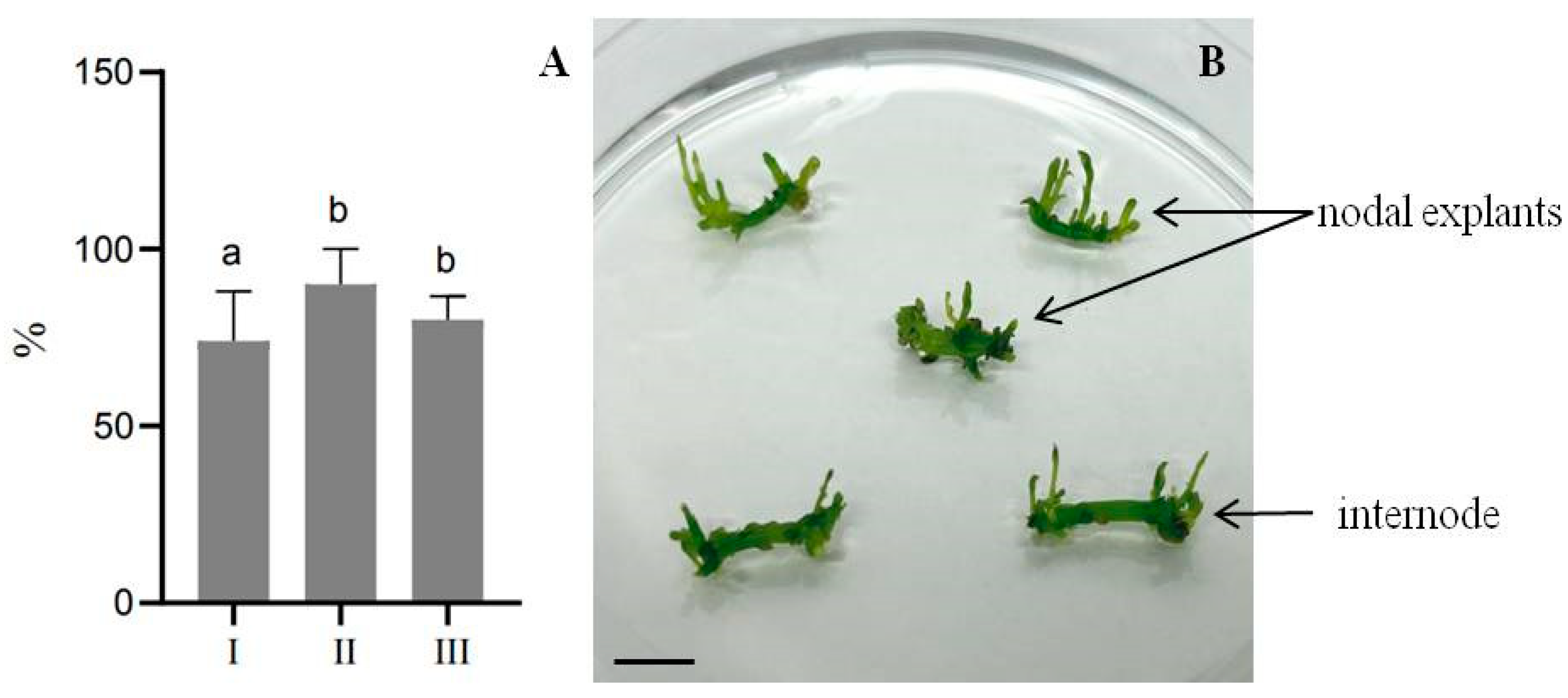
2.2. Direct Shoot Regeneration from Internodes and Nodal Explants
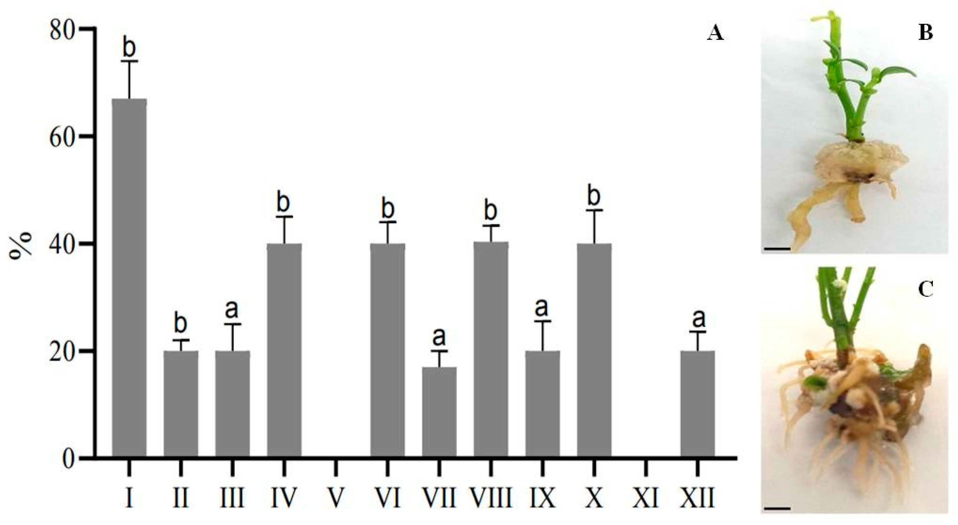
A series of experiments on direct shoot regeneration from internodes and nodal explants showed no significant differences in the regeneration potential between different variants of auxin and cytokinin concentrations, as shown in Figure 1. For example, on the MSDR II medium, the regeneration potential reached 90% when plant growth regulators (PGRs) such as 1.0 mg/L BAP, 0.5 mg/L NAA, and 0.2 mg/L kinetin were added, while the regeneration processes did not show any significant variation in the MSDR I and MSDR III media. However, it was observed that these regenerants were unsuitable for micropropagation, which was confirmed in subsequent experiments. Therefore, it was decided to first induce rhizogenesis before proceeding with micropropagation. Primary regenerants with a height of 2–3 cm were transferred to media designed to induce rhizogenesis, using full- and half-strength MS medium supplemented with auxins such as IBA, IAA, and NAA.

2.3. Root Formation

According to the results presented in Figure 2 and Supplementary Table S1, it was clear that during the initial stage of rhizogenesis, calluses were formed from which the roots subsequently developed. The MSR I medium was effective for root formation in E. koopmannii, with a success rate of 67% when supplemented with 0.5 g/L IBA and 0.05 mg/L NAA. Conversely, no root formation was observed when adding 2 mg/L IBA and 0.5 mg/L NAA. The cultivation of plants on a nutrient medium containing a full strength of MS salts along with varying concentrations of IBA ranging from 0.5 to 2 mg/L, NAA and ranging from 0.05 to 0.5 mg/L resulted in a root formation rate of 40%. In addition, Figure 2 highlights the morphogenic processes of the plants, particularly the intense root formation observed on a half-strength MS medium.

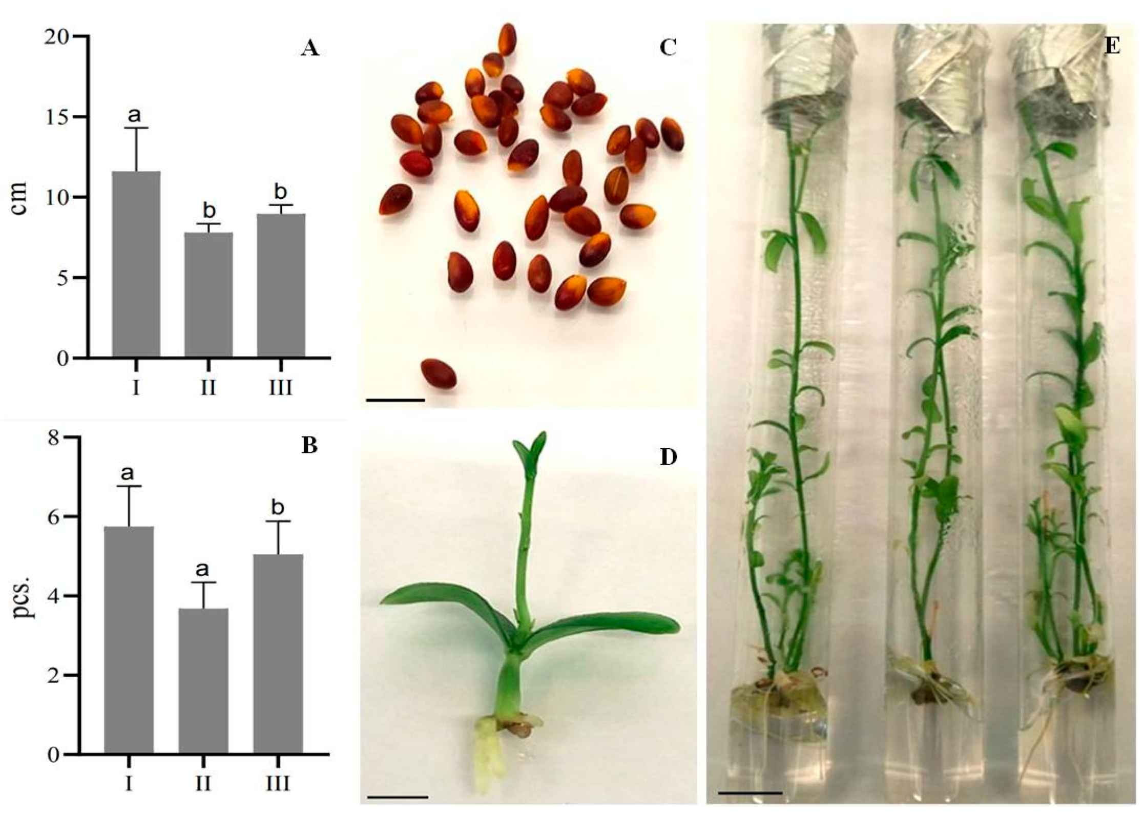
2.4. Micropropagation of Plants from Internode Explants and Seedlings

Figure 3 presents data on the micropropagation of E. koopmannii regenerants induced from seeds (Supplementary Table S2). In the second passage, the microclones showed a significant development delay, with fewer additional shoots than in the first passage. However, in the third passage, an increase in shoot length was observed, up to 9 cm, in contrast to the 7.8 cm length observed in the second passage.
In Figure 3, the number of shoots per explant was higher in the third passage than in the second passage, possibly because the plants were exposed to stress factors after the first passage that initially slowed growth. This phenomenon was evident in two key parameters: average shoot length and number of shoots per explant. After the plant tissues had adapted to the culture conditions, there was a transition from slow growth to intense division and morphogenesis. Micropropagation of E. koopmannii from cuttings taken directly from nodal explants proved difficult due to necrosis of more than 90% of newly planted shoots. However, more than five shoots per explant were again obtained when regenerated plants were propagated through the rooting cycle. Notably, when nodal explants of germinated seedlings were used as explants in the micropropagation process of E. koopmannii, the plants exhibited intense development, resulting in a higher rate of additional shoot induction compared to regenerants obtained from internodes (Figure 4). Thus, after much experimental work, we increased the number of shoots to three per one explant, including the rooting stages. In addition, longer shoots were obtained in the third passage than in the second passage, where the shoot length decreased significantly compared to the first passage.
The experimental data confirm that an effective method for E. koopmannii micropropagation involved plant regeneration followed by rooting and subsequent cutting. Although this method was time consuming, it significantly increased the multiplication rate of E. koopmannii. Notably, the fastest and most efficient method for the micropropagation of E. koopmannii was the third method described above, which uses germinated seedlings as primary explants (Table 2). The multiplication coefficient of germinated seedlings was 28.5, while that of internodes was 6.1.

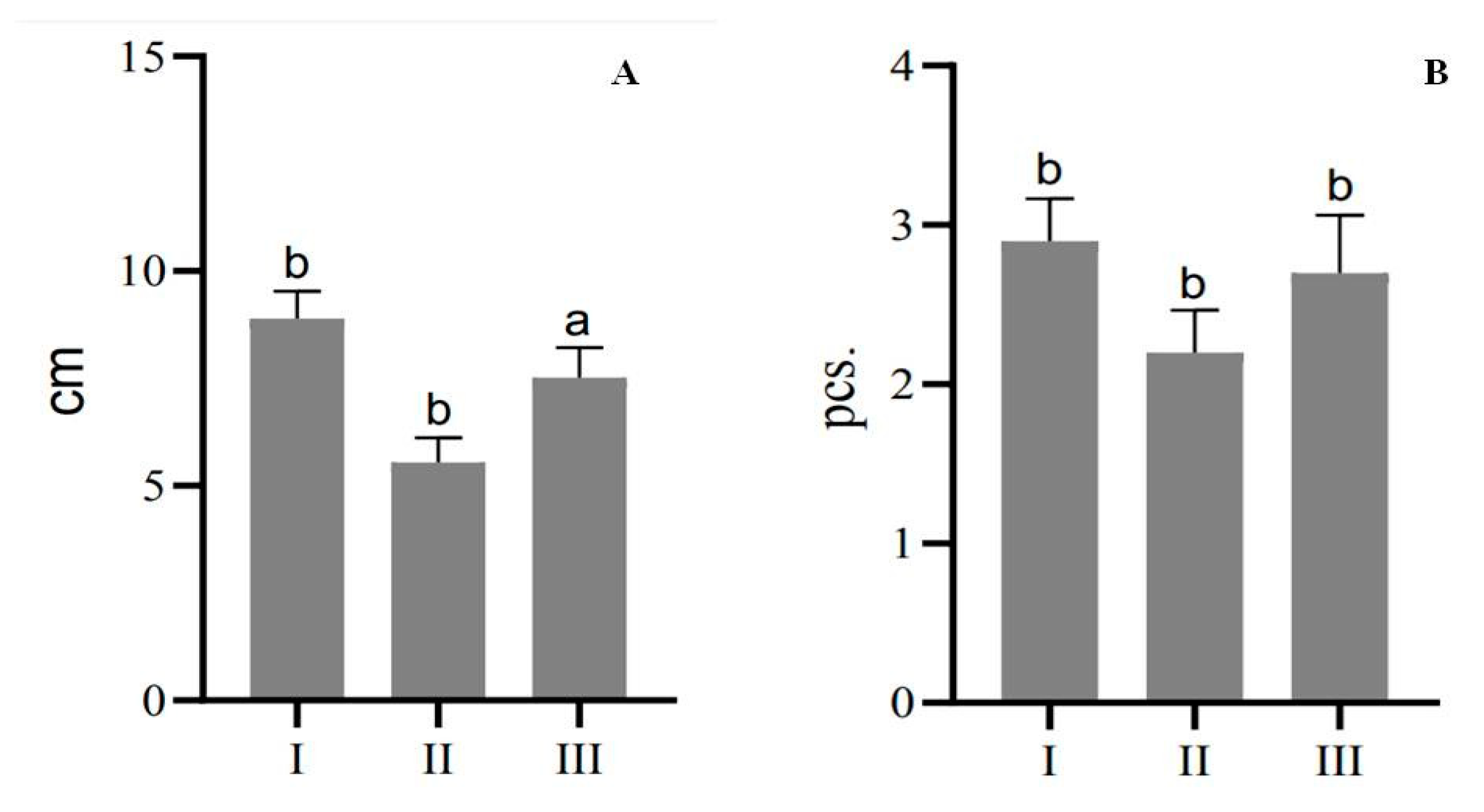
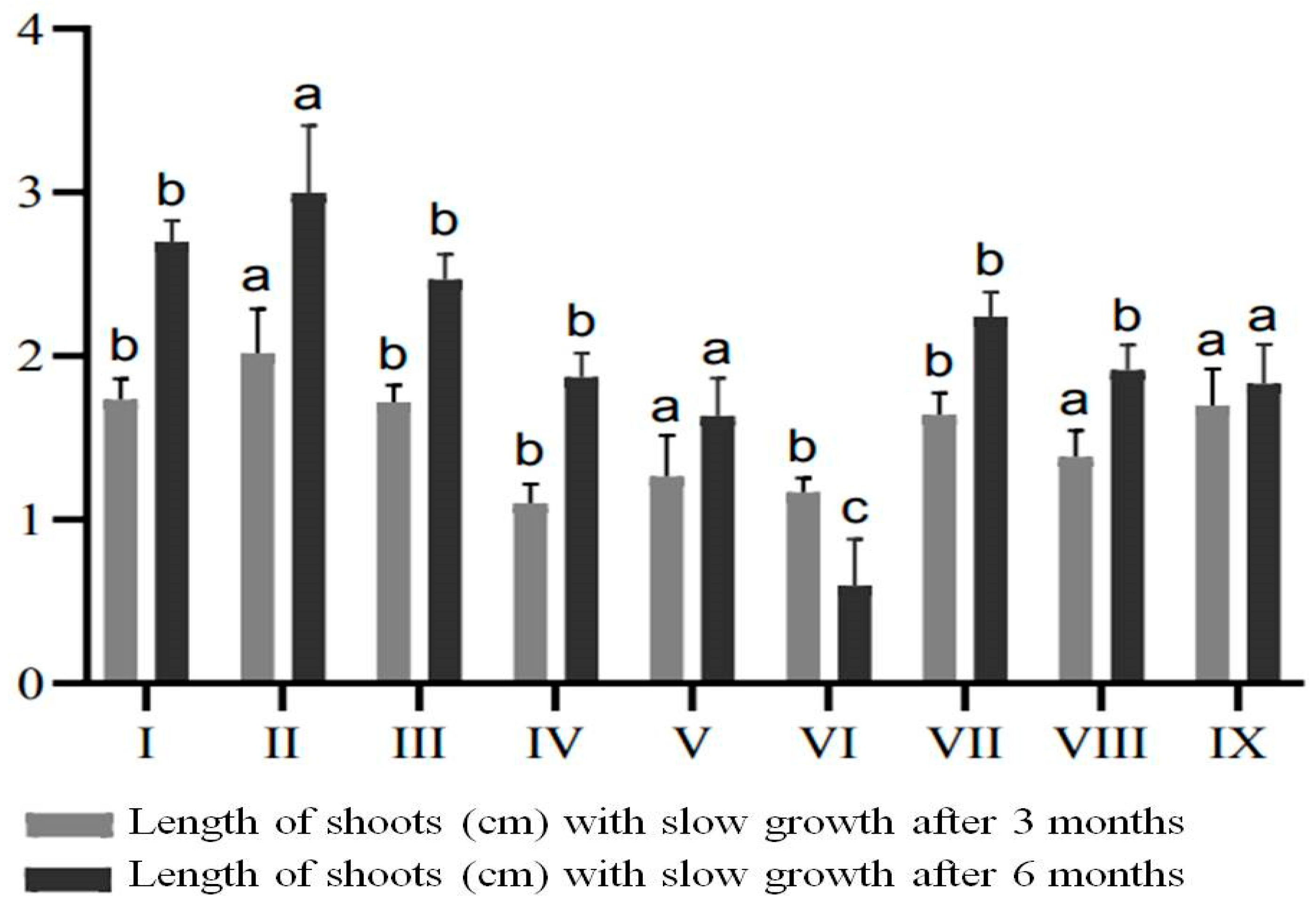
2.5. In vitro Preservation of Plants by Slow Growth Storage

For SGS experiments, 157 in vitro micropropagated E. koopmannii shoots were used, grown in MSDR II medium supplemented with the hormones 1.0 mg/L BAP, 0.5 mg/L NAA, and 0.2 mg/L kinetin (Supplementary Table S3). The data presented in Figure 5 show the efficacy of growth inhibitors during in vitro storage, evaluated at three and six months. It was observed that the use of ABA, CCC, and mannitol resulted in variations in shoot length in nine variants of media with varying concentrations of retardants. The data showed that, in variants of media with different concentrations of ABA, the length of shoots increased from 0.75 to 0.98 cm with increasing concentrations. Similarly, when the retardant CCC was used, a slight increase in length from 0.19 to 0.6 cm was observed. The addition of ABA and CCC at different concentrations significantly increased plant length. In contrast, when mannitol was added at concentrations of 5, 8, and 10 mg/L, the shoot length was considerably lower compared to the treatments with ABA and CCC.
After six months of cultivation, it was also noteworthy that, when mannitol was added at a concentration of 10 mg/L, a decrease in growth and necrosis was observed, with the shoot length decreasing by 0.57 cm after six months of cultivation. When CCC was used at 1 mg/L, the increase in shoot length was only 0.13 cm, possibly due to the more prolonged exposure to this retardant concentration.

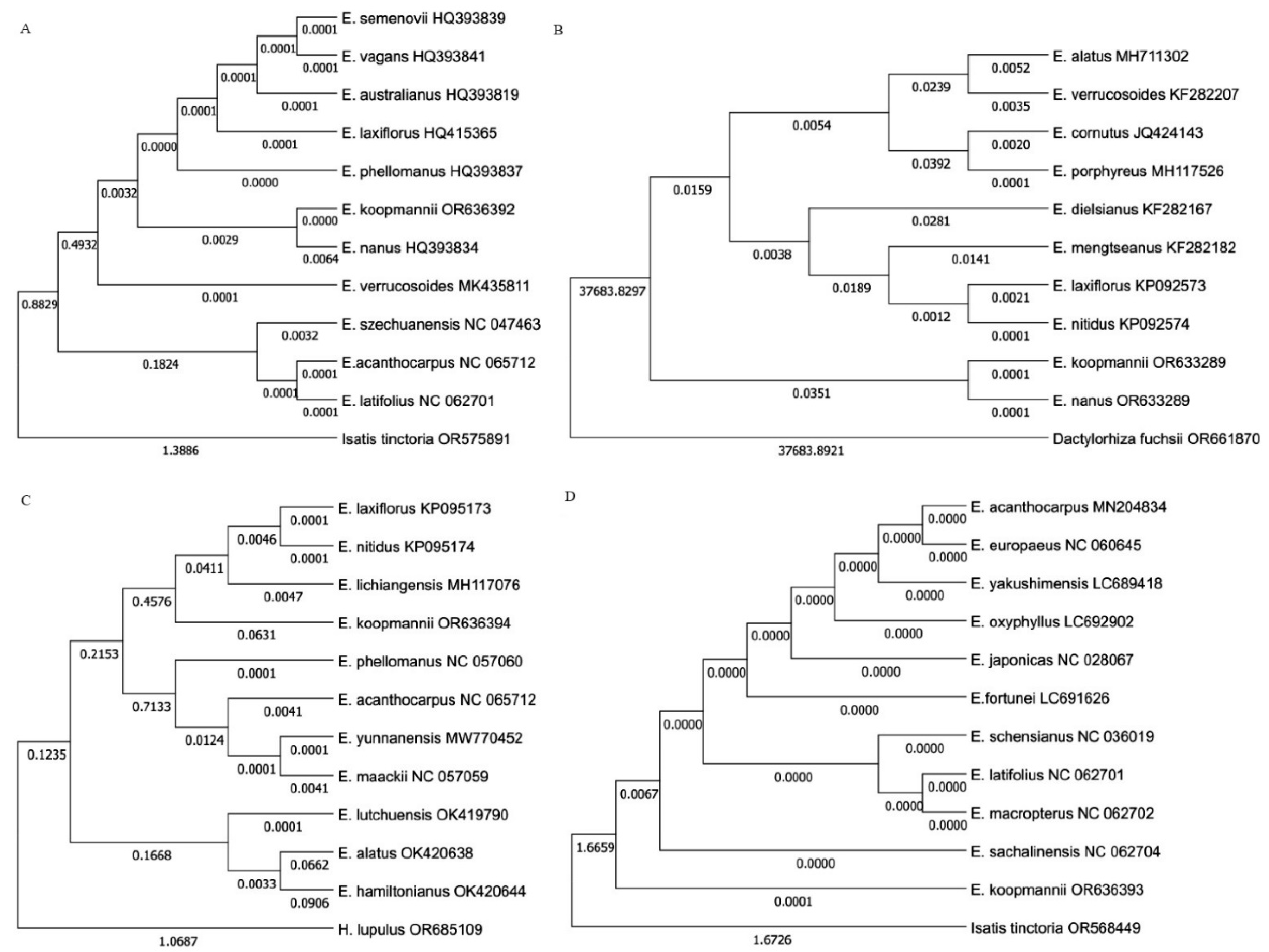
2.6. Phylogenetic Analysis

In this study, four barcoding markers were used for the identification of phylogenetic relationships and species. The sequences of E. koopmannii, together with 10 matK genes, nine ITS regions, ten psbA-trnH regions, and ten recall gene sequences of Euonymus species from the NCBI database, were used to infer the phylogenetic relationships. The length of the sequences was 334 bp for the matK gene, 501 bp for the ITS region, 242 bp for the psbA-trnH region, and 444 bp for the rbcL gene sequences after alignment. The monomorphic sites were arranged at 38–441 bp, and polymorphic sites were 3–139 bp. Genetic variation in the sequences was verified by two neutrality tests, such as nucleotide diversity (Pi) and Tajima’s neutrality test ( π ). Pi ranged from 0.0012 for the rbcL gene to 0.4282 for psbA-trnH region 0.19308, while π ranged from 0.0012 for the rbcL gene to 0.3833 for the psbA-trnH region. A total of 307 bp, 498 bp, 217 bp, and 444 bp nucleotide pairs were analyzed for nucleotide pair frequency analysis in matK gene, ITS region, psbA-trnH region, and rbcL gene sequences, respectively (Table 3). The results of nucleotide pair frequency analysis and the maximum likelihood estimation of substitution of the four barcoding regions are shown in Supplementary Tables S4 and S5. The sequences of matK gene (accession number OR636392), rbcL gene (OR636393), psbA-trnH (OR636394), and ITS (OR633289) regions of E. koopmannii are available in the NCBI database.
The phylogenetic analysis divided Euonymus species into two or three main groups based on all candidate barcoding markers (Figure 6). The matK gene patterns showed that E. koopmannii and E. nanus were closely related to E. semenovii, E. vagans, E. vagans, E. laxiflorus, E. phellomanus, and E. verrucosoides with average pairwise distances of 0.00. The psbA-trnH region showed that E. koopmannii had the same patterns as E. laxiflorus, E. nitidus, and E. lichiangensis with average pairwise distances of 0.06. In contrast, the ITS region and rbcL gene sequences of E. koopmannii were identified from other Euonymus species. Based on the ITS region, sequences E. koopmannii and E. nanus sequences were grouped in one cluster with average pairwise distances of 0.00 and showed 100% identity. Based on the rbcl gene region, only E. koopmannii was included in a group with an average pairwise distance of 0.0068 compared to other species.

3. Discussion

Micropropagation is widely used in the commercialization of ornamental and fruit crops [36] to conserve disappearing and rare plant species under minimal growth conditions. In addition, micropropagation is essential in the propagation of medicinal plants to select cell lines with maximal production of pharmaceutical metabolites [37]. All plant growth regulators have their own specific effects, and their different combinations cause a specific responce in plants [38,39,40]. This study represents a comprehensive effort to establish the initial stages of tissue culture, define the research strategy for the endemic species E. koopmannii, and identify effective types of explants for micropropagation. For example, our results showed that, although the frequency of additional shoot emergence was low when propagating Euonymus from stem segments, it was possible to increase the multiplication coefficient by rooting regenerants first and then propagating them again. Protocols for micropropagation and the induction of root-forming potential were developed, demonstrating the versatility of our approach. In addition, the use of germinated E. koopmannii seedlings provided sufficient material for further experiments, thus ensuring the sustainability of our research efforts. Direct plant regeneration from stem explants has been successfully achieved in several woody plants such as Fraxinus pennsylvanica [41], Ficus religiosa L. [42], Toona ciliata [43], and Lycium barbarum L. [44]. However, studies on the introduction of Euonymus species into in vitro culture have not been completed. For example, in the case of E. bungeanus, an effective regeneration system was developed using leaf explants induced by 750 nm white light and on half MS medium supplemented with BAP and NAA to form axillary buds [45]. Similarly, the study by A. Shang et. al. showed a high regeneration frequency (63.64%) of E. japonicus adventitious shoots induced by culturing hypocotyl segments close to seeds on MS medium supplemented with 1.5 mg/L BAP and 0.05 mg/L NAA [18]. In addition, a highly efficient protocol for shoot regeneration from E. fortunei var. radicans stem explants was established, achieving a regeneration frequency of 5.1 shoots per explants [46]. E. alatus shoots from axillary buds were rooted in a medium containing 3.0 mg/L IBA [14]. These experiments are consistent with other studies demonstrating the efficacy of half MS medium for root formation. However, in our study we used IAA and IBA at concentrations of 0.05 mg/L and 0.5 mg/L, respectively, instead of NAA. Our results partially confirm previous research, where a similar combination of hormones achieved 77% root formation, albeit with slightly different auxin concentrations than those used in our study [47]. In the species E. alatus, the optimization of conditions for the induction of embryogenic suspensions was achieved using the synthetic auxin analog 2,4-D [16]. Based on our research using different types of explants and micropropagation methods, such as direct micropropagation from internodes and nodal segments, it became clear that significant increases in the multiplication coefficient of this endemic plant were achieved only by the process of obtaining regenerates with the root system, followed by their propagation.
Plant genetic resources are conserved using in vitro techniques such as SGS, which regulates plantlet development and reduces handling risks such as contamination and genetic instability. SGS protocols vary based on temperature, light, and media composition, with well-established methods for some fruit species and others requiring further research. Plants respond differently to SGS and micropropagation, being influenced by taxonomic affiliation, growth retardant concentrations, and stress response mechanisms. For example, SGS studies have shown promising results with the addition of CCC at concentrations of 0.2–0.4 mg/L in media for Chrysanthemum and Syringa L. [48]. Many studies over the last decade have investigated the in vitro conservation of fruit species through the SGS technique and this was reviewed by Benelli et al. in 2022 [20]. The study conducted by Pan et al. found that the most effective method for the slow growth conservation of Vitis heyneana was to use specific media formulations, adjust the osmotic pressure with mannitol (10 g/L), and control environmental factors such as the area of air-breathing film, concentration of CCC (5.0 g/L), and light intensity [49]. Many genotypes of fruit and berry crops were well preserved at 4 °C in bags for cultivation in mannitol (2%), with or without plant growth regulators [50]. When discussing the effect of retardants under slow plant storage conditions in our research, it can be inferred that there is a residual effect from the initial concentrations of mannitol and CCC prior to the six-month storage period. CCC inhibited shoot growth under slow storage conditions, whereas mannitol at 8 mg/L supported optimal growth and did not affect shoot development. Thus, mannitol proved to be the most effective in slowing the height growth of E. koopmannii, allowing in vitro storage of shoots for six months at 10 °C with a photoperiod of 16 h of light and 8 h of darkness.
DNA barcoding technology is a powerful tool that uses a short genetic sequence from a standardized genomic region to identify species. This technology has wide applications in various fields, such as species identification, resource management, phylogenetic studies, and evolutionary biology [51,52,53,54,55]. The results of our barcoding study revealed that the average GC content of the ITS region and the rbcL gene was higher than the AT content, while the average GC content of the matk genes and the psbA-trnH region was lower than the AT content. This is a common phenomenon in plants [56]. All candidate barcoding markers satisfied the interspecific genetic variation of Euonymus species. However, E. koopmannii could not be distinguished from other Euonymus species based on the matK gene and the psbA-trnH region. In contrast, the ITS region and rbcL gene sequences were suitable for the molecular identification of E. koopmannii at the molecular level from other species. The molecular markers used in this study proved to be valuable in delineating the primary phylogenetic lineages of Euonymus. In the comparative analysis using the average pairwise distance, E. koopmannii was most similar to E. nanus, E. lichiangensis, E. semenovii, E. vagans, E. laxiflorus, and E. phellomanus. These species belong to the subgenus Euonymus section Euonymus. On the other hand, E. koopmannii was most different from E. szechuanensis, E. cornutus which belong to subgenus Kalonymus and E. alatus which belongs to the subgenus Euonymus, section Melanocarya. In the research of Yan-Nan LI [2], ITS and ETS markers provided substantial information for understanding the phylogeny of Euonymus, and the molecular patterns strongly supported the idea that subgenera and sections differentiated Euonymus species.
In conclusion, our research involves a comprehensive study of an endemic plant, E. koopmannii, including its introduction into in vitro culture, the intricacies of micropropagation, storage under in vitro conditions, and determination of temporal growth parameters during non-transplanted culture storage. These results highlight the potential for further research and optimization of tissue culture techniques for Euonymus species, including E. koopmannii, to contribute to the conservation and sustainable use of these valuable plant resources. The logical continuation of this work was the assignment of a DNA barcode to E. koopmannii, which will further enhance our understanding and conservation efforts for this species.

4. Materials and Methods

4.1. Plant Material and Explant Preparation and Disinfection

All plant materials were collected in the Aksu-Zhabagly Reserve, Maloe Kayindy Gorge (42°24′25″ N, 70°34′49″ E), in the Turkestan region of Kazakhstan, under the guidance of State Reserve botanists. The Forestry and Wildlife Committee of the Ministry of Ecology, Geology and Natural Resources of the Republic of Kazakhstan obtained permission to collect endangered species. Seeds and axillary buds (1.0–1.5 cm) were isolated from the shoots of an actively growing E. koopmannii, approximately 30–35 years old and 1.70 m tall, and used as research objects. The experiments were repeated three times independently. In total, 142 axillary buds and 183 seeds of E. koopmannii were sterilized in two steps, as detailed in Supplementary Table S6. First, seeds and axillary buds of wild-type E. koopmannii were washed in 1.0% sodium hypochlorite and 1% KMnO4 solutions for 30 min, followed by thorough rinsing under running water. Then, in the second stage of sterilization, 3% and 5% sodium hypochlorite solutions were used for 5 and 10 min, respectively, with the addition of Tween 20. The explants were then rinsed 3–4 times with sterile distilled water and dried on sterile filter paper before being planted on nutrient media.

4.2. Direct Regeneration, Rooting and Micropropagation

Murashige and Skoog (MS) mineral salt-based nutrient media with vitamins (M519, PhytoTech) containing 3% sucrose were used for tissue culture. PGRs were added to the nutrient media for micropropagation (Table 4). The stock solutions of PGRs were prepared individually. For example, 20 mg of auxins were dissolved in a few drops of ethanol, while cytokinins were dissolved in a few drops of 1 N NaOH and made up to 20 mL using distilled water, then stored in the refrigerator at 20 °C. The PGR solutions were sterile filtered through a 0.2 µm filter and added to the chilled media under sterile conditions.
Phytogel at 2 g/L was used as the gelling agent. Axillary buds and seeds of E. koopmannii were cultured at 26–28 °C under a 16 h photoperiod, illuminated by fluorescent lamps with an intensity of 2000 lx.
For in vitro rooting of adventitious shoots, auxins such as indolylacetic acid (IAA), naphthylacetic acid (NAA), and indolylbutyric acid (IBA) were used as inducers of rooting processes in different combinations and concentrations as listed in Table 5. The media in the table consisted of half (½) and full-strength MS media supplemented with MS vitamins.

4.3. Slow Growth Storage

Half-strength MS medium supplemented with plant growth retardants, such as ABA, CCC, and mannitol was used to regulate the growth of E. koopmannii at different concentrations indicated in Table 6. The plants were kept for 6 months under light conditions with an intensity of 1200–1500 lux at a reduced temperature of +10 °C, with a photoperiod of 16 h light and 8 h dark. Observations were made after three and six months when the plants were transferred to standard culture conditions.

4.4. DNA Barcoding

For DNA barcoding, young leaves were stored at −80 °C until DNA extraction. DNA was extracted from fresh E. koopmannii leaves using the CTAB method [57] with slight modifications. The extracted DNA was checked for its intactness, homogeneity, and purity by 1% agarose gel electrophoresis and run at 120 V for 30 min. The DNA was stored in a freezer (−20 °C) until used in the next step of the experiment. The selection of universal barcode primers was guided by the relevant literature and was fully detailed in Supplementary Table S7. They were synthesized by the Organic Synthesis Laboratory of the National Center for Biotechnology (NCB) (Astana, Kazakhstan). Amplification and sequencing methods were performed according to the standard protocol of the Plant Genetic Engineering Laboratory of NCB [58].

4.5. Statistical Analyses

The multiplication rate was calculated according to the following formula:
Multiplication rate = b/a
where
  • a—number of shoots after the first passage
  • b—number of shoots after the third passage
For data analysis, the data represent the mean ± standard deviation (SD) of the replicates was analyzed using MS Excel 2021. ANOVA was used to evaluate significant differences, and Tukey’s honestly significant difference (HSD) test (p < 0.05) was analyzed using GraphPad Prism 8 software.
Sequence alignment of Euonymus species was performed using Vector NTI Advance 11.0 [59] and then used for further analyses. The nucleotide diversity score (Pi) was calculated using DnaSP v5.10 [60]. Phylogenetic analyses were performed using the maximum likelihood method and the Kimura 2-parameter model, and evolutionary divergence between sequences was estimated by the software MEGA 11 [61].

5. Conclusions

The present study represents a comprehensive investigation of the endemic plant E. koopmannii, including micropropagation, in vitro storage and growth characteristics of E. koopmannii under slow growth conditions. The barcoding markers for all candidates corresponded to the interspecific genetic variation of Euonymus species. The ITS region and rbcL gene sequences were suitable for distinguishing E. koopmannii from other species at the genetic level. These results provide fundamental helpful information for future biotechnological and molecular research.

Supplementary Materials

The following supporting information can be downloaded at: https://www.mdpi.com/article/10.3390/plants13162174/s1, Table S1: Rhizogenesis of E. koopmannii regenerants on MSR I–XII media. Table S2: Micropropagation E. koopmannii on MSDR II medium containing 1.0 mg/L BAP, 0.5 mg/L NAA and 0.2 mg/L kinetin. Table S3: Slow growing storage data. Table S4: The nucleotide pair frequency analysis of the four bar-coding sequences. Table S5: Maximum likelihood estimate of substitution of the four bar-coding sequences. Table S6: Effect of sterilizing agents and exposure time on obtaining aseptic culture of E. koopmannii. Table S7: Nucleotide sequences of PCR primers used for the four bar-coding sequences. References [62,63,64,65] are cited in the supplementary materials.

Author Contributions

Conceptualization, S.M.; methodology, B.K.; formal analysis, S.B. and D.T.; investigation, B.K., S.B. and D.T.; resources, S.M.; writing—original draft preparation, B.K., S.B. and D.T.; writing—review and editing, S.M.; project administration, S.M.; funding acquisition, S.M. All authors have read and agreed to the published version of the manuscript.

Funding

This research was carried out within the framework of the Program BR21882180, “Strategy creation for conserving and developing medicinal and veterinary plant resources in Kazakhstan amid Climate Change”, supported by the Science Committee of the Ministry of Science and Higher Education of the Republic of Kazakhstan.

Data Availability Statement

All relevant data have been provided as Tables and Figures in the text and Supplementary Materials.

Acknowledgments

We thank the Aksy-Zhabagly Nature Reserve and its employee, Dzhumanov S.D., for providing biological material and the Ministry of Science and Higher Education of the Republic of Kazakhstan for financial support.

Conflicts of Interest

The authors declare no conflicts of interest.

References

  1. Li, Y.; Dong, Y.; Liu, Y.; Yu, X.; Yang, M.; Huang, Y. Comparative Analyses of Euonymus Chloroplast Genomes: Genetic Structure, Screening for Loci with Suitable Polymorphism, Positive Selection Genes, and Phylogenetic Relationships within Celastrineae. Front. Plant Sci. 2021, 11, 593984. [Google Scholar] [CrossRef] [PubMed]
  2. Li, Y.-N.; Xie, L.; Li, J.; Zhang, Z. Phylogeny of Euonymus L. inferred from molecular and morphological data. J. Syst. Evol. 2013, 52, 1–12. [Google Scholar] [CrossRef]
  3. Wittmack, L. Garten-Zeitung; P. Parey: Berlin, Germany, 1883; Volume 2. [Google Scholar]
  4. Shukurov, E.D. Central Asian transboundary project for the conservation of biodiversity of the Western Tien Shan. In Atlas of Biological Diversity of the Western Tien Shan Book; Bismarck LLC: Bishkek, Kyrgyzstan, 2005. (In Russian) [Google Scholar]
  5. Ivashchenko, A.A. Reserves and National Parks of Kazakhstan; Almatykitap: Almaty, Kazakhstan, 2006; p. 284. (In Russian) [Google Scholar]
  6. Available online: https://e-history.kz/ru/kazakhstanika/show/11175 (accessed on 22 February 2024).
  7. Ruiz-Riaguas, A.; Fernandez-de Cordova, M.L.; Llorent-Martinez, E.J. Phenolic profile and antioxidant activity of Euonymus japonicus Thunb. Nat. Prod. Res. 2022, 36, 3445–3449. [Google Scholar] [CrossRef] [PubMed]
  8. Kukula-Koch, W.; Widelski, J.; Koch, W.; Głowniak, K. HPLC, Two-Dimensional TLC Determination of Phenolic Content, and an In Vitro Perspective to Antioxidant Potential of Euonymus verrucosus Scop. Extracts. Acta Chromatogr. 2015, 27, 743–754. [Google Scholar] [CrossRef]
  9. Shin, D.Y.; Kim, B.S.; Lee, H.Y.; Park, Y.M.; Kim, Y.W.; Kim, M.J.; Yang, H.J.; Kim, M.S.; Bae, J.S. Euonymus alatus (Thunb.) Siebold leaf extract enhanced immunostimulatory effects in a cyclophosphamide-induced immunosuppressed rat model. Food Nutr. Res. 2023, 67, 1–11. [Google Scholar] [CrossRef] [PubMed]
  10. Lin, X.L.; Li, K.; Yang, Z.; Chen, B.; Zhang, T. Dulcitol suppresses proliferation and migration of hepatocellular carcinoma via regulating SIRT1/p53 pathway. Phytomedicine 2020, 66, 153112. [Google Scholar] [CrossRef] [PubMed]
  11. Zhu, J.; Qin, J.; Chang, R.; Zeng, Q.; Cheng, X.; Zhang, F.; Jin, H.; Zhang, W. Chemical Constituents of Plants from the Genus Euonymus. Chem. Biodivers. 2012, 9, 1055–1076. [Google Scholar] [CrossRef] [PubMed]
  12. Popova, E.V.; Nosov, A.V.; Titova, M.V.; Kochkina, D.V.; Fomenkov, A.A.; Kulichenko, I.E.; Nosov, A.M. Promising biotechnologies: Collections of cell cultures of higher plants as the basis for the development and production of drugs. J. Plant Physiol. 2021, 68, 227–244. (In Russian) [Google Scholar]
  13. Fomenkov, A.A.; Sidorov, R.A.; Nosov, A.V. Suspension cell culture of Euonymus maximoviczianus is a new object in studies of the biosynthesis of lower alkyl esters of fatty acids. Cell Biol. Plant Biotechnol. 2013, 61. (In Russian) [Google Scholar]
  14. Smith, C.C.; Jernstedt, J.A. In vitro development of adventitious shoots in Euonymus alatus (Celastraceae). Sci. Hortic. 1989, 41, 161–169. [Google Scholar] [CrossRef]
  15. Chen, Y.; Lu, L.; Deng, W.; Yang, X.; McAvoy, R.; Zhao, D.; Pei, Y.; Luo, K.; Duan, H.; Smith, W.; et al. In vitro regeneration and Agrobacterium-mediated genetic transformation of Euonymus alatus. Plant Cell Rep. 2006, 25, 1043–1051. [Google Scholar] [CrossRef]
  16. Woo, H.-A.; Ku, S.S.; Jie, E.Y.; Kim, H.; Kim, H.-S.; Cho, H.S.; Jeong, W.-J.; Park, S.U.; Min, S.R.; Kim, S.W. Efficient plant regeneration from embryogenic cell suspension cultures of Euonymus alatus. Sci. Rep. 2021, 11, 15120. [Google Scholar] [CrossRef]
  17. Ning, K.; Zhou, T.; Jiang, C.Z.; Wu, H.M.; Jiang, J.L.; Chen, J.; El-kassaby, Y.A.; Ma, Y. Rapid and efficient leaf regeneration propagation system for Euonymus bungeanus. Biol. Plant. 2021, 65, 118–125. [Google Scholar] [CrossRef]
  18. Shang, A.-Q.; Cai, H.; Yan, X.-J.; Hu, H.-Z.; Zhao, L.-J. Plant Regeneration from In Vitro Cultured Hypocotyl Explants of Euonymus japonicus Cu Zhi. Agric. Sci. China 2006, 5, 196–201. [Google Scholar] [CrossRef]
  19. Levy, L.; Damsteegt, V.; Welliver, R. First Report of Plum pox virus (Sharka Disease) in Prunus persica in the United States. Plant Dis. 2000, 84, 202. [Google Scholar] [CrossRef]
  20. Benelli, C.; Tarraf, W.; Izgu, T.; De Carlo, A. In Vitro Conservation through Slow Growth Storage Technique of Fruit Species: An Overview of the Last 10 Years. Plants 2022, 11, 3188. [Google Scholar] [CrossRef]
  21. Bautista-Aguilar, J.R.; Iglesias-Andreu, L.G.; Martínez-Castillo, J.; Ramírez-Mosqueda, M.A.; Ortiz-García, M.M. In Vitro Conservation and Genetic Stability in Vanilla planifolia Jacks. HortScience 2021, 56, 1494–1498. [Google Scholar] [CrossRef]
  22. Choi, K.S.; Park, S. The complete chloroplast genome sequence of Euonymus japonicus (Celastraceae). Mitochondrial DNA Part A 2016, 27, 3577–3578. [Google Scholar] [CrossRef]
  23. Park, Y.S.; Kang, J.-S.; Park, J.Y.; Shim, H.; Yang, H.O.; Kang, J.H.; Yang, T.-J. Analysis of the complete plastomes and nuclear ribosomal DNAs from Euonymus hamiltonianus and its relatives sheds light on their diversity and evolution. PLoS ONE 2022, 17, e0275590. [Google Scholar] [CrossRef]
  24. Cai, H.; Gu, X.; Li, Y.; Ren, Y.; Yan, S.; Yang, M. Cold Resistance of Euonymus japonicus Beihaidao Leaves and Its Chloroplast Genome Structure and Comparison with Celastraceae Species. Plants 2022, 11, 2449. [Google Scholar] [CrossRef]
  25. Wang, W.-C.; Chen, S.-Y.; Zhang, X.-Z. Characterization of the complete chloroplast genome of the golden crane butterfly, Euonymus schensianus (Celastraceae). Conserv. Genet. Resour. 2017, 9, 545–547. [Google Scholar] [CrossRef]
  26. Lee, J.; Kang, S.J.; Shim, H.; Lee, S.C.; Kim, N.H.; Jang, W.; Park, J.Y.; Kang, J.H.; Lee, W.H.; Lee, T.J.; et al. Characterization of Chloroplast Genomes, Nuclear Ribosomal DNAs, and Polymorphic SSR Markers Using Whole Genome Sequences of Two Euonymus hamiltonianus Phenotypes. Plant Breed. Biotechnol. 2019, 7, 50–61. [Google Scholar] [CrossRef]
  27. Hua, W.; Chen, C. The complete chloroplast genome sequence of the plant Euonymus fortunei (Celastraceae). Mitochondrial DNA Part B 2018, 3, 637–639. [Google Scholar] [CrossRef] [PubMed]
  28. Wang, X.; Li, H.; Zheng, M.; Jiang, J. The complete chloroplast genome of Euonymus szechuanensis. Mitochondrial DNA Part B 2020, 5, 1130–1131. [Google Scholar] [CrossRef]
  29. Li, H.; Chen, M.; Wang, Z.; Hao, Z.; Zhao, X.; Zhu, W.; Liu, L.; Guo, W. Characterization of the Complete Chloroplast Genome and Phylogenetic Implications of Euonymus microcarpus (Oliv.) Sprague. Genes 2022, 13, 2352. [Google Scholar] [CrossRef] [PubMed]
  30. Zhang, T.-T.; Sun, J.; Xu, L.; Yang, Y.Y.; Zhan, Z.L.; Xing, Y.P.; Zhao, R.; Li, S.N.; Zhang, D.C.; Kang, T.G. The complete mitochondrial genome of Euonymus alatus (celastraceae, Euonymus L.). Mitochondrial DNA Part B 2021, 6, 182–184. [Google Scholar] [CrossRef] [PubMed]
  31. Gubaidullin, N.; Kali, B.; Tussipkan, D.; Manabayeva, S.A. A promising strategy for conservation of Central Asian endemic plant Euonymus koopmannii. Biodivers. J. Biol. Divers. 2024, 25, 3114–3120. [Google Scholar]
  32. Linh, N.N.; Hang, P.L.B.; Hue, H.T.T.; Ha, N.H.; Hanh, H.H.; Ton, N.D.; Hien, L.T.T. Species discrimination of novel chloroplast DNA barcodes and their application for identification of Panax (Aralioideae, Araliaceae). PhytoKeys 2022, 188, 1–18. [Google Scholar] [CrossRef] [PubMed]
  33. Veldman, S.; Ju, Y.; Otieno, J.N.; Abihudi, S.; Posthouwer, C.; Gravendeel, B.; van Andel, T.R.; de Boer, H.J. DNA barcoding augments conventional methods for identification of medicinal plant species traded at Tanzanian markets. J. Ethnopharmacol. 2020, 250, 112495. [Google Scholar] [CrossRef]
  34. Ma, H.-L.; Zhu, Z.-B.; Zhang, X.-M.; Miao, Y.-Y.; Guo, Q.-S. Species identification of the medicinal plant Tulipa edulis (Liliaceae) by DNA barcode marker. Biochem. Syst. Ecol. 2014, 55, 362–368. [Google Scholar] [CrossRef]
  35. Jones, L.; Twyford, A.D.; Ford, C.R.; Rich, T.C.G.; Davies, H.; Forrest, L.L.; Hart, M.L.; McHaffie, H.; Brown, M.R.; Hollingsworth, P.M.; et al. Barcode UK: A complete DNA barcoding resource for the flowering plants and conifers of the United Kingdom. Mol. Ecol. Resour. 2021, 21, 2050–2062. [Google Scholar] [CrossRef]
  36. Singh, V.; Deen, B.; Singh, S. Micropropagation of minor fruit crops of India: A review. Agric. Rev. 2023, 44, 259–263. [Google Scholar] [CrossRef]
  37. Chokheli, V.A.; Dmitriev, P.A.; Rajput, V.D. Recent Development in Micropropagation Techniques for Rare Plant Species. Plants 2020, 9, 1733. [Google Scholar] [CrossRef] [PubMed]
  38. Abeuova, L.S.; Kali, B.R.; Rakhimzhanova, A.O.; Bekkuzhina, S.S.; Manabayeva, S.A. High frequency direct shoot regeneration from Kazakh commercial potato cultivars. PeerJ 2020, 8, e9447. [Google Scholar] [CrossRef]
  39. Zhumabek, A.T.; Rakhimzhanova, A.O.; Bekkuzhina, S.S.; Ramankulov, Y.M.; Manabayeva, S.A. Somatic embryogenesis and plant regeneration from upland switchgrass cultivars. Res. Crops 2020, 21, 349–354. [Google Scholar]
  40. Pasternak, T.P.; Steinmacher, D. Plant Growth Regulation in Cell and Tissue Culture In Vitro. Plants 2024, 13, 327. [Google Scholar] [CrossRef]
  41. Beasley, R.; Pijut, P. Regeneration of Plants from Fraxinus nigra Marsh. Hypocotyls. HortSci. A Publ. Am. Soc. Hortic. Sci. 2013, 48, 887–890. [Google Scholar] [CrossRef]
  42. Hesami, M.; Daneshvar, M.H. In Vitro Adventitious Shoot Regeneration through Direct and Indirect Organogenesis from Seedling-derived Hypocotyl Segments of Ficus religiosa L.: An Important Medicinal Plant. HortScience 2018, 53, 55–61. [Google Scholar] [CrossRef]
  43. Li, P.; Shang, Y.; Zhou, W.; Hu, X.; Mao, W.; Li, J.; Li, J.; Chen, X. Development of an efficient regeneration system for the precious and fast-growing timber tree Toona ciliata. Plant Biotechnol. 2018, 35, 51–58. [Google Scholar] [CrossRef]
  44. Verma, S.K.; Gantait, S.; Mukherjee, E.; Gurel, E. Enhanced somatic embryogenesis, plant regeneration and total phenolic content estimation in Lycium barbarum L.: A highly nutritive and medicinal plant. J. Crop. Sci. Biotechnol. 2022, 25, 547–555. [Google Scholar] [CrossRef]
  45. Ning, X.; Liu, Y.; Jia, M.; Wang, Q.; Sun, Z.; Ji, L.; Mayo, K.H.; Zhou, Y.; Sun, L. Pectic polysaccharides from Radix Sophorae Tonkinensis exhibit significant antioxidant effects. Carbohydr. Polym. 2021, 262, 117925. [Google Scholar] [CrossRef] [PubMed]
  46. Shang, A.; Sun, Z.; Zhao, L. High Efficient Regeneration In Vitro from Hypocotyl of Euonymus fortunei var. radicans. Sci. Silvae Sin. 2009, 45, 136–141. [Google Scholar]
  47. Chen, Y.; Lu, L.; Duan, H.; Deng, W.; McAvoy, R.; Smith, W.; Thammina, C.; von Bodman, S.; Li, Y.; Ye, D.; et al. Biotech approach to neutralize the invasiveness of burning bush (Euonymus alatus), a progress report on development of its genetic transformation system and functional analysis of sterile genes. Acta Hortic. 2008, 769, 21–29. [Google Scholar] [CrossRef]
  48. Mitrofanova, O.V.; Lesnikova-Sedoshenko, N.P.; Ivanova, N.N.; Smykova, N.V.; Mitrofanova, I.V. In vitro propagation and preservation of promising chrysanthemum cultivars and hybrid forms. In Proceedings of the XXX International Horticultural Congress IHC2018: II International Symposium on Micropropagation and In Vitro Techniques 1285, Istanbul, Turkey, 12–16 August 2018. [Google Scholar]
  49. Pan, X.; Zhang, W.; Li, X. In vitro conservation of native Chinese wild grape (Vitis heyneana Roem. & Schult) by slow growth culture. VITIS J. Grapevine Res. 2014, 53, 207–214. [Google Scholar]
  50. Turdiyev, T.T.; Kovalchuk, I.Y.; Kabylbekova, B.Z.; Chukanova, N.I.; Frolov, S.N. In vitro germplasm cold storage of fruit and berry plants of Kazakhstan. Eurasian J. Biosci. 2020, 14, 1213–1219. [Google Scholar]
  51. Li, H.; Xiao, W.; Tong, T.; Li, Y.; Zhang, M.; Lin, X.; Zou, X.; Wu, Q.; Guo, X. The specific DNA barcodes based on chloroplast genes for species identification of Orchidaceae plants. Sci. Rep. 2021, 11, 1424. [Google Scholar] [CrossRef] [PubMed]
  52. Mishra, M.; Jingade, P.; Huded, A.K.C. DNA barcoding analysis and phylogenetic relationships of Indian wild coffee species. Turk. J. Bot. 2022, 46, 109–122. [Google Scholar] [CrossRef]
  53. Hajdari, A.; Pulaj, B.; Schmiderer, C.; Mala, X.; Wilson, B.; Lluga-Rizani, K.; Mustafa, B. A phylogenetic analysis of the wild Tulipa species (Liliaceae) of Kosovo based on plastid and nuclear DNA sequence. Adv. Genet. 2021, 2, e202100016. [Google Scholar] [CrossRef] [PubMed]
  54. Jamdade, R.; Mosa, K.A.; El-Keblawy, A.; Al Shaer, K.; Al Harthi, E.; Al Sallani, M.; Al Jasmi, M.; Gairola, S.; Shabana, H.; Mahmoud, T. DNA barcodes for accurate identification of selected medicinal plants (Caryophyllales): Toward barcoding flowering plants of the United Arab Emirates. Diversity 2022, 14, 262. [Google Scholar] [CrossRef]
  55. Liu, Y.; Zhang, M.; Chen, X.; Chen, X.; Hu, Y.; Gao, J.; Pan, W.; Xin, Y.; Wu, J.; Du, Y.; et al. Developing an efficient DNA barcoding system to differentiate between Lilium species. BMC Plant Biol. 2021, 21, 465. [Google Scholar] [CrossRef]
  56. Chinnkar, M.; Jadhav, P. Evaluating DNA barcoding using five loci (matK, ITS, trnH-psbA, rpoB, and rbcL) for species identification and phylogenetic analysis of Capsicum frutescens. J. Appl. Biol. Biotechnol. 2023, 11, 97–103. [Google Scholar] [CrossRef]
  57. Doyle, J. A rapid DNA isolation procedure for small quantities of fresh leaf tissue. Phytochem. Bull. 1987, 19, 11–15. [Google Scholar]
  58. Sutula, M.; Kakanay, A.; Tussipkan, D.; Dzhumanov, S.; Manabayeva, S. Phylogenetic Analysis of Rare and Endangered Tulipa Species (Liliaceae) of Kazakhstan Based on Universal Barcoding Markers. Biology 2024, 13, 365. [Google Scholar] [CrossRef]
  59. Lu, G.; Moriyama, E.N. Vector NTI, a balanced all-in-one sequence analysis suite. Brief. Bioinform. 2005, 5, 378–388. [Google Scholar] [CrossRef]
  60. Librado, P.; Rozas, J. DnaSP v5: A software for comprehensive analysis of DNA polymorphism data. Bioinformatics 2009, 25, 1451–1452. [Google Scholar] [CrossRef]
  61. Tamura, K.; Stecher, G.; Kumar, S. MEGA11: Molecular Evolutionary Genetics Analysis Version 11. Mol. Biol. Evol. 2021, 38, 3022–3027. [Google Scholar] [CrossRef]
  62. Zhang, C.-Y.; Wang, F.-Y.; Yan, H.-F.; Hao, G.; Hu, C.-M.; Ge, X.-J. Testing DNA barcoding in closely related groups of Lysimachia L. (Myrsinaceae). Mol. Ecol. Resour. 2011, 12, 98–108. [Google Scholar] [CrossRef]
  63. Kuzmina, M.L.; Johnson, K.L.; Barron, H.R.; Hebert, P.D. Identification of the vascular plants of Churchill, Manitoba, using a DNA barcode library. BMC Ecol. 2012, 12, 25. [Google Scholar] [CrossRef]
  64. Parmentier, I.; Duminil, J.; Kuzmina, M.; Philippe, M.; Thomas, D.W.; Kenfack, D.; Chuyong, G.B.; Cruaud, C.; Hardy, O.J. How effective are DNA barcodes in the identification of African rainforest trees? PLoS ONE 2013, 8, e54921. [Google Scholar] [CrossRef]
  65. Khan, A.M.; Bhadauria, S. Molecular characterization of keratin degrading fungi isolated from semi-arid soil by PCR using ITS4 and ITS5 primers. J. King Saud Univ.-Sci. 2019, 31, 1418–1423. [Google Scholar] [CrossRef]
Figure 1. Effect of different concentrations and combinations of cytokinins and auxins on shoot regeneration from E. koopmannii in tissue culture. (A) Frequency of direct regeneration on MSDR I, II, and III medium; (B) Direct shoot regeneration from internodes and nodal explants. Data represent three independent experiments’ mean ± standard deviation (SD) Different letters on the bar indicate statistically significant differences at p < 0.05 Tukey’s (HSD), scale bar 1 cm.
Plants 13 02174 g001
Figure 2. Dynamics of growth and development of E. koopmannii roots during in vitro culture. (A) Frequency of rhizogenesis of E. koopmannii regenerants on MSR I–XII media; (B) 8-week-old plants; (C) 14-week-old plants. Data represent three independent experiments’ mean ± standard deviation (SD). Different letters on the bar indicate statistically significant differences at p < 0.05 Tukey’s (HSD), scale bar 1 cm.
Plants 13 02174 g002
Figure 3. Effect of subculture on elongation of shoot length development and shoot formation of E. koopmannii on MSDR I, II, and III medium: (A) average shoot length, cm; (B) number of shoots per explants, pcs.; (C) seeds; (D) 4-week-old seedlings; (E) 12-week-old plants. Data represent three independent experiments’ mean ± standard deviation (SD). Different letters on the bar indicate statistically significant differences at p < 0.05 Tukey’s (HSD), scale bar 1 cm.
Plants 13 02174 g003
Figure 4. Micropropagation of regenerants obtained from E. koopmannii internodes on MSDR I, II, and III medium: (A) average shoot length, cm; (B) number of shoots per explants, pcs. Data represent three independent experiments’ mean ± standard deviation (SD). Different letters on the bar indicate statistically significant differences at p < 0.05 Tukey’s (HSD).
Plants 13 02174 g004
Figure 5. Development of shoot length (cm) during six months of slow growth on MSP I–IX medium. Data represent the mean ± standard deviation (SD) of three independent experiments. Different letters on the bar represent statistically significant differences at p < 0.05 Tukey’s (HSD).
Plants 13 02174 g005
Figure 6. The phylogenetic tree of E. koopmannii is based on the maximum likelihood method and the Kimura 2-parameter model. (A) matK gene. (B) ITS region. (C) psbA-trnH region. (D) rbcl gene.
Plants 13 02174 g006
Table 1. Effect of sterilizing agents and exposure time on obtaining aseptic culture of E. koopmannii.
No of VariantsSterilization OptionsType of ExplantShoot Formation In Vitro
(%)
Step 1Exposure TimeStep 2Exposure Time
I1% sodium hypochlorite30 min one time5% sodium hypochlorite + Tween 2010 minaxillary buds67.5 ± 0.12
seeds63.3 ± 0.10
II5 minaxillary buds85.2 ± 0.09
seeds87.5 ± 0.07
III1% KMnO4
solution
3% sodium hypochlorite + Tween 2010 minaxillary buds49.4 ± 0.14
seeds43.3 ± 0.51
Table 2. Multiplication coefficient of E. koopmannii after three rounds of micropropagation.
Explant TypeNumber of Shoots after the First Passage, PcsNumber of Shoots after the Second Passage, PcsNumber of Shoots after the Third Passage, PcsMultiplication Rate
Seedlings43111428.5
Internodes720436.1
Table 3. Genetic diversity of E. koopmannii based on the candidate barcoding sequences.
matK GeneITS RegionpsbA-trnH Region rbcL Gene
Total aligned length (bp)334501242444
GC content (%)32.3859.626.445.7
Codon count101-70148
Number of monomorphic sites16540338441
Number of polymorphic sites 132911393
Total number of InDels sites377650
Overlapping InDels sites02380
Number of singleton variables sites24153
Total number of mutations (Eta)1341071963
Parsimony informative sites (PIC)13050830
Nucleotide diversity (Pi)0.19310.06250.42820.0012
Tajima’s neutrality test ( π )0.17230.04940.38330.0012
Mean nucleotide difference (k)57.34530.88975.7820.545
Number of Haplotypes (h)59102
Haplotype diversity (Hd)0.7090.9780.9820.182
Variance of Haplotype diversity0.018650.002920.002150.02061
Sequence conservation (C)0.5810.8140.1990.993
Conservation threshold (CT)0.680.910.291
Table 4. Variations of nutrient media for E. koopmannii.
PGRs, mg/LMSDR IMSDR IIMSDR III
BAP0.51.02.0
NAA0.10.51.0
Kinetin0.2
Table 5. Media options for rhizogenesis of regenerated E. koopmannii plants.
OptionsMSIBA, mg/LIAA, mg/LNAA, mg/L
MSR I½0.50.05
MSR IIfull
MSR III½1.00.1
MSR IVfull
MSR V½2.00.5
MSR VIfull
MSR VII½0.50.1
MSR VIIIfull
MSR IX½1.00.5
MSR Xfull
MSR XI½2.00.05
MSR XIIfull
Table 6. Variations in MSP medium with retardants for storage of E. koopmannii under slow growth conditions.
Variations of MS MediumABA, mg/LMannitol, mg/LCCC, mg/L
MSP I2.0
MSP II5.0
MSP III10.0
MSP IV5.0
MSP V8.0
MSP VI10.0
MSP VII0.2
MSP VIII0.5
MSP IX1.0
Disclaimer/Publisher’s Note: The statements, opinions and data contained in all publications are solely those of the individual author(s) and contributor(s) and not of MDPI and/or the editor(s). MDPI and/or the editor(s) disclaim responsibility for any injury to people or property resulting from any ideas, methods, instructions or products referred to in the content.

