Review

Fungi for Sustainable Pharmaceutical Remediation: Enzymatic Innovations, Challenges, and Applications—A Review

UCD School of Agriculture and Food Science, University College Dublin, Belfield, Dublin 4, Ireland
Processes 2025, 13(4), 1034; https://doi.org/10.3390/pr13041034
Submission received: 10 February 2025 / Revised: 27 March 2025 / Accepted: 27 March 2025 / Published: 31 March 2025
(This article belongs to the Special Issue Feature Review Papers in Section “Pharmaceutical Processes”)

Abstract

:
The extensive use of pharmaceuticals in human and veterinary medicine has led to their persistent environmental release, posing ecological and public health risks. Major sources include manufacturing effluents, excretion, aquaculture, and improper disposal, contributing to bioaccumulation and ecotoxicity. Mycoremediation is the fungal-mediated biodegradation of pharmaceuticals, offers a promising and sustainable approach to mitigate pharmaceutical pollution. Studies have reported that certain fungal species, including Trametes versicolor and Pleurotus ostreatus, can degrade up to 90% of pharmaceutical contaminants, such as diclofenac, carbamazepine, and ibuprofen, within days to weeks, depending on environmental conditions. Fungi produce a range of extracellular enzymes, such as laccases and peroxidases, alongside intracellular enzymes like cytochrome P450 monooxygenases, which catalyze the transformation of complex pharmaceutical compounds. These enzymes play an essential role in modifying, detoxifying, and mineralizing xenobiotics, thereby reducing their environmental persistence and toxicity. The effectiveness of fungal biotransformation is influenced by factors such as substrate specificity, enzyme stability, and environmental conditions. Optimal degradation typically occurs at pH 4.5–6.0 and temperatures of 20–30 °C. Recent advancements in enzyme engineering, immobilization techniques, and bioreactor design have improved catalytic efficiency and process feasibility. However, scaling up fungal-based remediation systems for large-scale applications remains a challenge. Addressing these limitations with synthetic biology, metabolic engineering, and other biotechnological innovations could further enhance the enzymatic degradation of pharmaceuticals. This review highlights the enzymatic innovations, applications, and challenges of pharmaceutical mycoremediation, emphasizing the potential of fungi as a transformative solution for sustainable pharmaceutical waste management.

1. Introduction

Pharmaceuticals play an essential role in healthcare as therapeutic agents and veterinary drugs, posing significant environmental challenges when improperly managed. Active pharmaceutical ingredients (APIs) are the biologically active components of pharmaceuticals, being designed for specific pharmacological effects, but can adversely impact ecosystems when released into the environment [1]. Major sources of pharmaceutical pollution include emissions from manufacturing processes, excretion by humans and animals, aquafarming activities, and the disposal of expired or unused medications [1,2]. These residues contaminate aquatic systems, soil, and agricultural ecosystems, leading to bioaccumulation in crops and biomagnification through food webs [3,4].
A particular concern is environmentally persistent pharmaceutical pollutants (EPPPs), which are engineered for biological stability but present serious ecological risks. These include endocrine disruption in non-target organisms and the exacerbation of antimicrobial resistance (AMR), a global health emergency [5]. Despite the identification of over 600 pharmaceutical compounds in aquatic systems worldwide, critical knowledge gaps remain concerning their environmental persistence, bioavailability, and ecotoxicological impacts [6]. The problem is compounded by insufficient waste management infrastructure, inadequate pharmaceutical disposal practices, and the rapidly growing pharmaceutical industry. Conventional wastewater treatment plants (WWTPs) often fail to fully remove pharmaceutical contaminants, leading to their continuous discharge into natural water bodies [7]. These factors contribute to ecosystem contamination, disrupt trophic dynamics, promote bioaccumulation, and amplify antimicrobial resistance (AMR), highlighting the urgent need for more efficient, scalable, and sustainable mitigation strategies that address these knowledge gaps [3,5].
Fungi have emerged as powerful agents for addressing pharmaceutical contamination due to their exceptional enzymatic diversity and metabolic adaptability. Mycoremediation is a fungal-driven bioremediation strategy that offers an eco-friendly and cost-effective solution with minimal chemical and energy inputs, effectively transforming pollutants into less harmful forms [8]. White-rot fungi, such as Phanerochaete chrysosporium, Trametes versicolor, and Pleurotus ostreatus, excel in degrading persistent organic pollutants like polycyclic aromatic hydrocarbons (PAHs), polychlorinated biphenyls (PCBs), pharmaceuticals, and dyes through extracellular enzymes like laccases, manganese peroxidases, and lignin peroxidases [9,10]. Filamentous fungi, including Aspergillus niger, Penicillium chrysogenum, and Cunninghamella elegans, further contribute via the biodegradation, biosorption, and bioaccumulation of pharmaceutical compounds [11,12,13,14].
Fungi play a crucial role in detoxifying pharmaceutical residues across soil, water, and air, offering effective in situ and ex situ remediation strategies. However, despite their potential, fungal-based remediation is not yet widely implemented on an industrial scale due to challenges related to process optimization, enzyme stability, and regulatory considerations [15,16]. Knowledge gaps exist in understanding the interactions between fungi and complex pharmaceutical mixtures, as well as in developing strategies to enhance fungal enzyme activity under real-world environmental conditions. Advancements in molecular and biochemical techniques are essential to elucidate fungal metabolic pathways and enhance key enzymes, such as laccases, peroxidases, and cytochrome P450 monooxygenases, to improve degradation efficiency [9]. Enzyme immobilization and biotechnological innovations are critical for scaling up mycoremediation. This review explores enzymatic mechanisms, current challenges, and emerging strategies to improve fungal biocatalysis for sustainable pharmaceutical pollution management.

2. Pharmaceuticals in the Environment

Pharmaceuticals, veterinary medicines, and growth-promoting drugs are widely present in the environment on a global scale due to their increased consumption for treating or preventing diseases in humans and animals and their use in agricultural practices [17]. Pharmaceutical pollution varies regionally due to drug consumption, industrial discharge regulations, and wastewater treatment efficiency. High-income countries detect more residues due to extensive drug use and advanced monitoring, while low- and middle-income nations struggle with rising contamination from inadequate treatment and unregulated disposal [5]. The first detection of the pharmaceutical compound clofibric acid in treated wastewater in the USA, at concentrations ranging from 0.8 to 2.0 μg/dm3, was reported by Garrison and colleagues in 1976 [18]. Since then, numerous studies have documented the occurrence of drugs and their ingredients in various environmental matrices, including lakes, rivers, seawater, soil, and sediments. However, the low concentrations of these compounds pose significant challenges for detection and analysis [17]. Even at minute concentrations, pharmaceutical residues can severely affect aquatic life, terrestrial animals, and human health. These compounds can bioaccumulate and magnify through food chains, spreading from plankton to higher trophic levels. Active drug metabolites contribute to environmental pollution, impacting entire ecosystems [3,4].

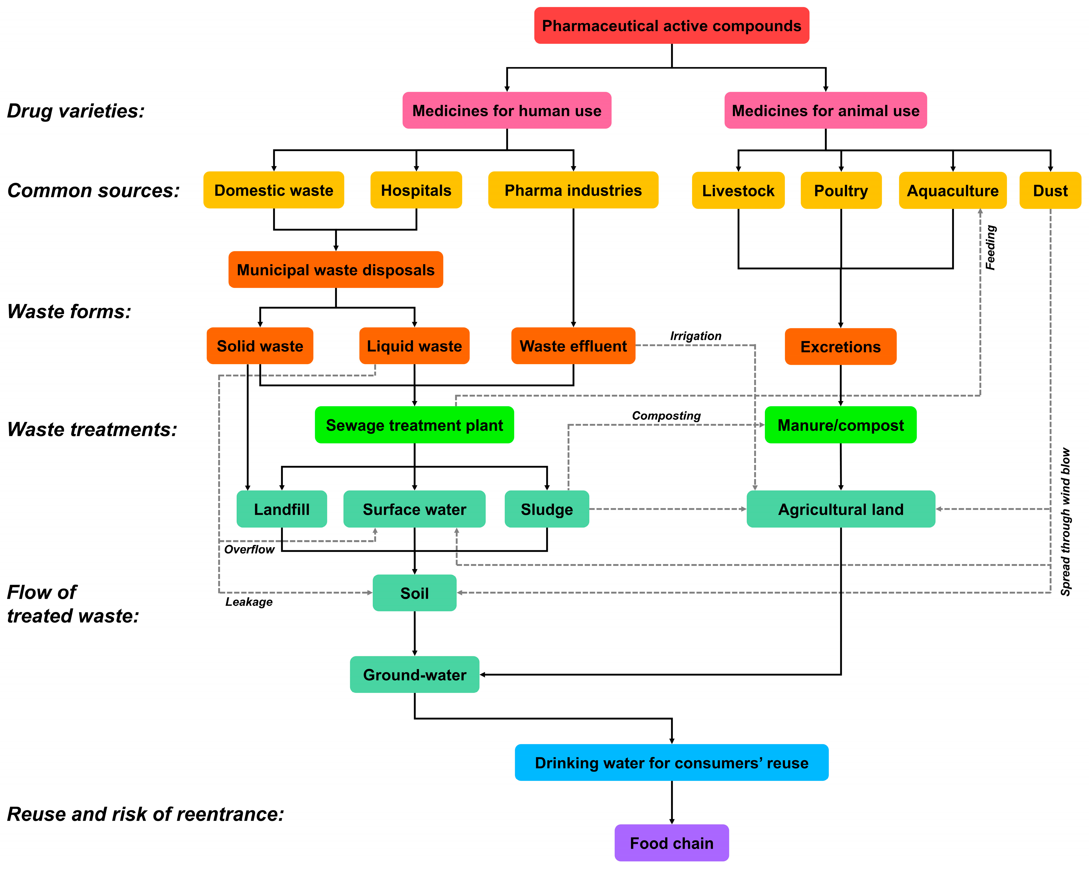
2.1. Sources of Pharmaceuticals in the Environment

Pharmaceutical active compounds (PACs) are introduced into the environment through several pathways, as illustrated in Figure 1. The figure provides a comprehensive overview of the sources, pathways, and risks associated with PACs. These compounds originate primarily from two categories: medicines for human use and medicines for animal use. Human-derived PACs arise from domestic waste, hospitals, and pharmaceutical industries, while animal-derived PACs come from livestock, poultry, aquaculture, and environmental dust due to improper handling or storage. Human sources of PACs include domestic waste, which results from unused or excreted pharmaceuticals from households, hospitals contributing medical waste from healthcare facilities, and pharmaceutical industries where waste is generated during drug manufacturing processes. On the other hand, animal sources include livestock, poultry, and aquaculture, where veterinary medicines are extensively used, as well as dust, which introduces PACs into the environment through improper handling or storage practices [19]. Regional waste management and regulations influence PAC levels, with some nations enforcing strict treatment while others face uncontrolled discharges [5].
The sources of PACs lead to their presence in different waste forms, including solid waste, liquid waste, waste effluent, and animal excretions. Solid waste consists of non-liquid residues from households, industries, or treatment processes, while liquid waste includes water-soluble PACs from effluents or direct discharges. Waste effluent is unprocessed liquid waste released into the environment, and animal excretions, such as urine and feces, are significant contributors to environmental PACs. These waste forms are directed towards treatment systems like sewage treatment plants (STPs) or composting. However, these systems are not always effective in removing PACs entirely. While STPs handle liquid and effluent waste, by-products such as sludge may carry residual PACs [20]. Similarly, animal excretions are often treated as manure or compost and applied to agricultural land, introducing PACs into the soil [21].
The treated waste undergoes various pathways, as depicted in the figure. Solid waste disposed of in landfills can leach PACs into the soil. Treated or untreated effluents may overflow into surface water systems, such as rivers and lakes [22]. Residues from STPs, in the form of sludge, often still contain PACs and are sometimes used on agricultural land. PACs accumulate in the soil from sludge, manure, and direct leakage, eventually leaching into groundwater, creating risks of contamination [21,22].
PACs can re-enter the human water supply and food chain through various routes. Groundwater and surface water contaminated with PACs may be used as drinking water sources, while PACs in agricultural land can accumulate in crops and animals, posing risks of bioaccumulation and human exposure [21,22,23]. Fluoroquinolones and steroid hormones persist and bioaccumulate in plants, aquatic life, and livestock, posing long-term exposure risks, even in low-contamination areas [24]. This cyclic flow emphasizes the need for improved waste treatment methods to mitigate the environmental and health risks posed by PACs.

2.2. Environmental and Ecological Impacts of Pharmaceuticals

Pharmaceutical residues have been widely detected in various environmental compartments, including surface water, groundwater, and river and sea sediments. Studies have reported their presence in biota ranging from algae to fish across the globe [24,25]. Natural and synthetic sex hormones, such as those found in contraceptives, pose significant threats to aquatic ecosystems, with severe ecological consequences [26]. While fluoxetine (FLX) is a widely used antidepressant with billion-dollar sales, it is frequently detected in watercourses due to its persistence in the environment, where it disrupts aquatic ecosystems and poses potential risks to wildlife [27]. Pharmaceuticals, including antibiotics like ciprofloxacin and sulfamethoxazole, promote antimicrobial resistance, contaminate water sources, and accumulate in the food chain, posing risks to ecosystems and human health. Their persistence threatens biodiversity, water quality, and long-term public health [5,28].
The bioaccumulation of pharmaceuticals in aquatic organisms occurs through multiple pathways, including direct absorption from contaminated water, the ingestion of pharmaceutical-laden prey, and uptake from sediment deposits. Long-term exposure to bioaccumulated pharmaceuticals has been linked to reproductive disorders, behavioral changes, and altered metabolic processes in fish and amphibians [3,23]. The persistence of PACs in the environment is driven by multiple processes. Atmospheric deposition facilitates the dispersal of particulate PACs, while irrigation with reclaimed water introduces them into agricultural systems. Additionally, composting can lead to the redistribution of PACs in soil and crops, contributing to their bioaccumulation and prolonged environmental exposure [21,22,23].
Addressing pharmaceutical pollution necessitates the adoption of enhanced waste treatment strategies. Improving the efficiency of sewage treatment plants through advanced technologies, such as ozonation and activated carbon filtration, is essential for minimizing the environmental impact of PACs [29]. Promoting proper disposal practices to reduce domestic pharmaceutical waste is equally important. Given the persistence of PACs, studying their bioaccumulation through the food chain alongside ecotoxicological risk assessments is crucial to understanding long-term exposure risks across trophic levels [23,24]. Stricter regulatory frameworks, advanced analytical methods for monitoring their presence, and global efforts to mitigate their ecological footprint are imperative [30]. These strategies are crucial to understanding and alleviating the adverse impacts of pharmaceuticals on ecosystems and human health. A summary of pharmaceutical pollution worldwide, concentration, contributing countries, and associated health effects is presented in Table 1.

3. Mycoremediation of Pharmaceuticals

Mycoremediation is a biotechnological approach that utilizes fungi to degrade and detoxify pharmaceutical pollutants in the environment, leveraging their metabolic versatility and enzymatic efficiency. Among fungi, filamentous fungi (e.g., Aspergillus and Penicillium) produce powerful enzymes that effectively degrade pharmaceutical contaminants like antibiotics and absorb heavy metals. Mushrooms (e.g., Pleurotus and Ganoderma) excel at breaking down complex pharmaceutical pollutants, including antibiotics, but grow slowly and require solid substrates. Yeasts (e.g., Saccharomyces and Yarrowia) are fast growing, work well in liquids, and are used in wastewater treatment for antibiotic removal through biosorption and biotransformation [7].
While fungi and mushrooms offer strong degradation abilities, yeasts are easier to scale for industrial applications. However, yeasts have limitations, including producing fewer extracellular enzymes, having a lower capacity for degrading complex pharmaceuticals, and being better suited to liquid environments rather than solid substrates [55]. Their metabolic pathways are also less versatile compared to those of filamentous fungi and mushrooms. Nevertheless, yeasts are more frequently used as recombinant hosts for expressing enzymes involved in environmental cleaning, owing to their ease of genetic manipulation and rapid growth [9]. For instance, Pichia pastoris X33 was used by Khan et al. to express the Cunninghamella elegans CYP-CPR system, enabling the biodegradation of pharmaceuticals such as flurbiprofen, diclofenac, and ibuprofen, highlighting the potential of yeast-based expression systems in enhancing the enzymatic breakdown of environmental pollutants [56].
Fungi stand out in pharmaceutical bioremediation because of their ability to break down complex pollutants like drugs through enzymatic processes and biosorption [19]. However, their slow growth and sensitivity to environmental conditions can be a drawback. Bacteria, on the other hand, offer fast and efficient degradation of organic pollutants, including pharmaceuticals, although some strains may need genetic tweaks to perform at their best [27]. Plants, via phytoremediation, absorb and stabilize contaminants from soil and water, providing a long-term, sustainable solution, but their effectiveness is limited by factors such as root uptake and seasonal changes [21]. Each approach has its benefits—bacteria are fast, fungi are great for tough pharmaceuticals, and plants are best for long-term remediation—so the right method depends on the type of pollution, site conditions, and goals [57,58]. This variability is evident in the degradation of fluoroquinolone antibiotics (FQs) like norfloxacin (NOR), ciprofloxacin (CIP), and ofloxacin (OFX). Bacteria such as Microbacterium sp. and Labrys portucalensis show limited degradation, with Labrys removing 60% of OFX but showing incomplete defluorination [59,60]. Fungi like Trametes versicolor are more efficient, degrading up to 80% of OFX [61]. Plants like Cyperus papyrus achieve FQ removal rates of 46–69%, with microbial degradation in the rhizosphere accounting for most of the process [58]. While fungi and bacteria directly transform FQs, plants primarily rely on rhizosphere microorganisms for degradation, with the efficiency varying by species.
Figure 2 illustrates the process of mycoremediation of environmental pharmaceuticals through enzymatic pathways and biocatalysis, where contaminants are broken down into less or non-toxic substances while simultaneously providing fungi with energy and nutrients [7].
Fungi employ multiple degradation pathways to break down complex compounds. These include ligninolytic and non-ligninolytic degradation pathways, aromatic ring cleavage pathways, and the tricarboxylic acid (TCA) cycle, which integrates degradation products into central metabolism. Such pathways transform recalcitrant pharmaceutical compounds into simpler, more biodegradable forms, mitigating their environmental persistence and toxicity [24]. Fungal degradation is mediated through various biochemical reactions, collectively referred to as biocatalysis. These reactions include hydroxylation, esterification, demethylation, epoxidation, oxidation, and denitrification, which alter the chemical structure of pharmaceuticals, rendering them less toxic and more amenable to further degradation [24]. These transformations are catalyzed by a diverse array of extracellular and intracellular fungal enzymes, including cytochrome P450 monooxygenases, laccases (Lacs), lignin peroxidases (LiPs), manganese peroxidases (MnPs), versatile peroxidases (VPs), dye-decolorizing peroxidases (DyPs), and chloroperoxidases (CPOs). Each of these enzymes plays a crucial role in the degradation of various pharmaceutical compounds, with their unique catalytic properties enabling the breakdown of complex organic molecules. The functional diversity of these fungal enzymatic systems allows for the effective processing of a wide range of contaminants, highlighting the adaptability of fungi in environmental bioremediation processes [8,62].
Fungi have shown the capacity to degrade a broad range of pharmaceutical classes, including analgesics, antibiotics, anti-convulsants, antidepressants, antihypertensive agents, non-steroidal anti-inflammatory drugs (NSAIDs), β-blockers, and central nervous system (CNS) stimulants [63]. Fungi utilize these pharmaceuticals as sources of carbon, nitrogen, and energy during degradation, integrating them into their metabolic networks. This metabolic flexibility not only enhances the survival of fungi in polluted environments but also facilitates the conversion of hazardous compounds into non-toxic metabolites, minimizing their ecological impact [64].
The advantages of mycoremediation are profound. It offers an eco-friendly and cost-effective strategy for the degradation of persistent pharmaceutical pollutants, converting them into simpler and less harmful substances. This reduces the bioaccumulation and persistence of contaminants in the environment, addressing a critical challenge in pollution control. Furthermore, the ability of fungi to degrade a wide spectrum of pharmaceutical compounds highlights their potential as a sustainable solution for mitigating environmental pollution [6]. Thus, mycoremediation represents a major advancement in bioremediation, offering an efficient solution for managing pharmaceutical waste and protecting environmental health.
The fungal degradation pathways depicted in Figure 3 highlight the remarkable metabolic adaptability of fungi in degrading quinolone antibiotics. These processes not only convert the parent compounds into less persistent and potentially less toxic metabolites but also elucidate the diverse enzymatic mechanisms employed by fungi, including oxidation, reduction, hydroxylation, and structural rearrangement [19]. The ability of fungi to target a broad spectrum of antibiotics establishes them as promising biological agents for mitigating pharmaceutical pollution in the environment.
Figure 3 illustrates the metabolic versatility of various fungal species in degrading quinolone antibiotics and the corresponding metabolites generated during these transformations. The degradation pathways involve a range of enzymatic reactions, such as hydroxylation, amination, methylation, and ring cleavage, underscoring the potential of fungi to alleviate the environmental impact of these pharmaceuticals [64].
For instance, Wetzstein et al. studied the degradation of pradofloxacin by Gloeophyllum striatum, which occurs primarily through hydroxylation and amine modification (Figure 3A). The resulting metabolites feature the introduction of hydroxyl (-OH) groups at specific positions on the quinolone ring and the addition of amine (-NH2) groups, demonstrating the fungus’s biocatalytic diversity in modifying the antibiotic structure [65]. Similarly, the biotransformation of enrofloxacin by Gloeophyllum striatum and Mucor ramannianus (Figure 3B). These fungi catalyze multiple reactions, including N-dealkylation, leading to the removal of the ethyl group, hydroxylation (-OH) at specific sites, and the formation of an oxo-derivative through oxygen incorporation, significantly altering the antibiotic’s chemical properties [66,67]. These modifications alter the quinolone ring structure, reducing its environmental persistence.
Furthermore, Phanerochaete chrysosporium and Mucor ramannianus metabolize sarafloxacin, producing derivatives that incorporate methyl groups and amine modifications (Figure 3C). These transformations suggest enzymatic mechanisms such as O-methylation and N-dealkylation, which significantly alter the molecular framework of the antibiotic, resulting in structurally distinct metabolites that may affect the biological activity of the compound [68,69]. Xylaria longipes demonstrates its ability to degrade danofloxacin, generating a metabolite with a cyclic oxygen-containing structure (Figure 3D). This metabolic transformation reflects the fungal ability to introduce oxygen-containing functional groups into fluoroquinolone antibiotics [70].
Various fungi have shown promising capabilities in the biodegradation of pharmaceutical compounds, as presented in Table 2. Aspergillus niger is effective in degrading a range of pharmaceuticals, including sulfamethoxazole, metoprolol, acetaminophen, diclofenac, naproxen, ranitidine, and carbamazepine [71]. Bjerkandera adusta also plays a significant role in the biodegradation of diclofenac, sulfamethoxazole, diazepam, ibuprofen, naproxen, citalopram, fluoxetine, and carbamazepine [63,72]. Additionally, Phanerochaete chrysosporium has been shown to degrade sulfamethoxazole, diazepam, ibuprofen, naproxen, diclofenac, citalopram, fluoxetine, and carbamazepine, with significant effects on various pharmaceuticals [72]. Buchicchio et al. demonstrated that Trichoderma harzianum can biodegrade carbamazepine and clarithromycin, with the identified metabolites being pharmacologically inactive and environmentally safe. This study, comparing T. harzianum with Pleurotus ostreatus, highlights the potential of both fungi for pharmaceutical bioremediation, using high-resolution mass spectrometry for precise metabolite identification [73]. Trichoderma pubescens strain DAOM 166162 effectively removed amoxicillin from wastewater, with a >98% removal efficiency, primarily through biodegradation (70.01%) and biosorption (28.44%). The degradation mechanism involved the uncoupling of the lactam ring via hydroxyl group activity in an alkaline environment, and extracellular polymeric substances (EPSs) produced by the fungus played a crucial role in the process [74]. Furthermore, Trametes versicolor has demonstrated biodegradation of various antibiotics, including azithromycin, ciprofloxacin, tetracycline, and cephalexin, as well as ibuprofen, diclofenac, codeine, and acetaminophen and endocrine disruptors like bisphenol A and phthalates [75].

4. Fungal Enzymes in Drug Metabolisms and Biodegradation

Fungal enzymes, especially oxidoreductases like laccases, lignin peroxidases, manganese peroxidases, and cytochrome P450, are essential for breaking down pharmaceutical pollutants. These enzymes enable fungi to transform complex drug compounds through oxidation, hydrolysis, and structural cleavage, making them highly effective in degrading xenobiotics [9]. Both intracellular and extracellular fungal enzymes contribute to detoxification, with extracellular enzymes playing a key role in bioconversion and defense against harmful substances [85]. Due to their efficiency, stability, and broad applicability, fungal enzymes are valuable for industrial processes, wastewater treatment, and pharmaceutical pollution management.

4.1. Laccases in Fungal Biodegradation of Pharmaceuticals

Laccases are multi-copper oxidases that degrade organic compounds by oxidizing substrates and reducing oxygen to water. Electrons transfer through a four-copper center, generating radicals that undergo further degradation. Their broad specificity makes them essential for breaking down xenobiotics, pharmaceuticals, and dyes, with applications in bioremediation and industry [86]. Basidiomycete fungi are the primary producers of laccases essential for lignin degradation, unlike bacteria that break down only small lignin fragments. These fungi secrete ligninases, enabling efficient lignin decomposition, whereas ascomycetes primarily oxidize phenolic compounds. These enzymes aid in delignification, morphogenesis, pigmentation, and pathogenesis. Major laccase producers include Trametes versicolor, Phanerochaete chrysosporium, and Pleurotus ostreatus [87].
Trametes versicolor laccase is a well-studied enzyme that is known for its ability to catalyze the oxidation of various organic substrates. Previous studies have used T. versicolor laccase with a higher redox potential (E°) for more efficient oxidation of substrates like phenols, anilines, and polyphenols, offering the potential for improved environmental clean-up and biotechnological applications. T. versicolor laccase was used to degrade aspirin and ketoprofen at concentrations of 25 mg/L each, with enzyme activity of 40 U/L at 35 °C and pH 4.0 for 6 h, resulting in degradation efficiencies of 72% and 70%, respectively [88]. Ibuprofen (515.7 mg/L) was treated under different conditions, with 29 U/L of laccase activity at 40 °C and pH 7.0 for 8 h, achieving a transformation efficiency of 76% [89]. While laccase is effective in degrading a wide range of pharmaceutical compounds, its degradation efficiency can vary greatly depending on the specific chemical structure of the drug and the operational conditions (pH and temperature).
The presence of Cu2+ significantly boosted laccase activity from Trametes versicolor, accelerating triclosan transformation through self-polymerization into dimers, trimers, and tetramers. Cu2+ enhanced enzymatic reaction kinetics (0.28 to 0.73 h−1) by stabilizing the laccase catalytic center, leading to increased oligomer production compared to systems without Cu2+. The proposed mechanism involved laccase-mediated oxidation of triclosan into phenoxy radicals, which then self-coupled to form larger molecules. Toxicity assays indicated that polymerized triclosan was less harmful, highlighting Cu2+-assisted laccase as a promising approach for triclosan detoxification in wastewater treatment [90]. However, the efficiency of laccases can be limited by their optimal pH and temperature ranges, and some pharmaceuticals may require mediators for effective degradation, as observed in certain studies [89,90].
Figure 4 illustrates the biodegradation of four distinct sulfonamide drugs by Trametes versicolor laccase to degrade sulfonamide drugs into various metabolites [91,92].
In the case of sulfanilamide, laccase catalyzes the cleavage of the sulfonamide bond, leading to the formation of aniline as a primary metabolite. This transformation occurs through oxidative cleavage, where the sulfonyl (-SO2-) group is removed, leaving the aniline moiety (Figure 4A). For sulfadimethoxine, enzymatic oxidation results in the breakdown of the sulfonamide bond, forming aniline as well as a substituted pyrimidine derivative. The two methoxy (-OCH3) groups remain attached to the pyrimidine ring, suggesting oxidation-driven cleavage and polymerization reactions that lead to new aromatic structures (Figure 4B). Sulfapyridine undergoes oxidative coupling, forming a variety of polymeric structures through the action of laccase. The reaction primarily involves the amine group on the benzene ring, leading to dimerization and trimerization products with pyridine-containing linkages. This polymerization is likely facilitated by radical intermediates generated during laccase oxidation (Figure 4C). In sulfathiazole, laccase oxidation results in hydroxylation and the formation of polymeric products, including hydroxylated aromatic amines and thiazole-containing derivatives. The presence of multiple polymeric products suggests that oxidation occurs at various reactive sites, leading to complex rearrangements and coupling reactions (Figure 4D). Overall, T. versicolor laccase initiates biodegradation by oxidizing sulfonamide drugs, primarily through cleavage of the sulfonamide bond and radical-mediated coupling reactions. These processes result in a range of transformation products, potentially reducing the environmental persistence and toxicity of sulfonamide antibiotics.
Phanerochaete chrysosporium effectively degraded sulfamethoxazole, achieving 74% removal at 10 mg L−1 after 10 days, with degradation enhanced by crude laccase activity and reaction time [93]. Similarly, Mn2+ enhanced P. chrysosporium laccase activity and norfloxacin adsorption but inhibited cytochrome P450-mediated intracellular degradation, reducing overall norfloxacin removal efficiency [94]. This suggests that Mn2+ shifts norfloxacin degradation towards extracellular laccase while suppressing intracellular pathways, highlighting the crucial role of P. chrysosporium laccase in the degradation of pharmaceutical contaminants. The Mn2+ dependency of laccases and other peroxidases can limit their activity in certain environments, making it necessary to optimize metal ion concentrations.
Laccase efficiently degrades diclofenac, with complete removal achieved by Myceliophthora thermophila laccase (expressed in Aspergillus sp.) within 8 h or within 1 h using HBT, SYR, or VA as mediators [95]. Y. enterocolitica laccase (expressed in E. coli) achieved 100% biotransformation in 24 h, while Moniliophthora roreri laccase (expressed in P. pastoris) was the least efficient, removing only 58% in 24 h [96]. Laccase also effectively degrades natural estrogens like estradiol and estriol, as well as synthetic ethinylestradiol, while estrone requires a laccase-mediator system for complete removal. M. thermophila laccase removed estradiol in 3 h and estrone with 65% efficiency in 24 h, achieving 100% removal with a mediator system, while M. roreri laccase degraded estradiol, estriol, and ethinylestradiol in 30 min [95,96,97]. Purified laccase from Paraconiothyrium variabile partially degraded nitrazepam, alprazolam, diazepam, and oxazepam at 10 mg/L over 48 h, with enhanced removal using a laccase-mediator system (LMS), while clobazam, chlordiazepoxide, and lorazepam remained resistant [98].
While laccase shows significant potential for pharmaceutical degradation, its efficiency depends on both the enzyme structure and the structure of the target compound. The lack of consistency in degradation efficiency for certain compounds indicates the need for further investigation into enzyme kinetics, optimal environmental conditions, and potential mediator systems to improve degradation rates. Advances in biotechnology can improve laccase performance, but further understanding of structural modifications is essential. Successful implementation of immobilized laccase systems for wastewater treatment will require the optimization of immobilization techniques and an evaluation of their economic viability [96].

4.2. Peroxidases in Fungal Biodegradation of Pharmaceuticals

Heme peroxidases are classified into three main classes: Class I (intracellular peroxidases in bacteria, archaea, and eukaryotic organelles, involved in oxidative stress response), Class II (fungal secreted peroxidases like lignin peroxidase (LiP), versatile peroxidase (VP), and manganese peroxidase (MnP), essential for lignin degradation), and Class III (plant peroxidases involved in lignification and stress response). Class II heme peroxidases play a crucial role in lignin decomposition by cleaving non-phenolic ether bonds and oxidizing Mn2+ to Mn3+, which acts as a diffusible redox mediator. Biochemical and phylogenomic studies confirm their essential role, showing that the diversification of these enzymes correlates with the evolution of white-rot fungi, while their loss or reduction is linked to shifts towards brown rot or ectomycorrhizal lifestyles [99].
Fungal peroxidases catalyze oxidative reactions via a two-electron transfer mechanism. The resting enzyme [(Fe3+)Pox] reacts with H2O2, forming Compound I, which has an oxyl–ferryl iron [Fe(IV) = O] and a cation radical [Pox*]. Compound I undergoes two sequential one-electron reductions via Compound II. Fungal peroxidases differ in substrate specificity: lignin peroxidases oxidize aromatic non-phenolic lignin compounds, with veratryl alcohol (VA) acting as a redox mediator. Manganese peroxidases oxidize Mn2+ to Mn3+, which, stabilized by organic acids, diffuses to oxidize lignin. Chloroperoxidases catalyze oxidative dehydrogenation, oxygen transfer, and oxidative chlorination reactions [100].
Fungal peroxidases are crucial for biodegradation due to their broad substrate range, including phenols, pharmaceuticals, pesticides, melanin, and xenobiotics [101]. However, their large-scale application is hindered by their sensitivity to the pH, temperature, inhibitors, high redox potential, Mn2+ dependency, and limited reusability. Enzyme engineering, such as the creation of a triple mutant (S49C/A67C/H239E) of LiPH8, has improved its thermostability at an acidic pH, increasing its half-life tenfold at pH 2.5 and 25 °C [102]. The need for further studies on enzyme kinetics, optimal reaction conditions, and degradation rates is critical to optimize these enzymes for industrial applications.
Lignin peroxidase (LiP) from Phanerochaete chrysosporium has been effective in degrading various pollutants. Auriol et al. achieved complete degradation of synthetic 17-alpha-ethinylestradiol (400 nM) in 1 h using 0.017 U/mL enzyme and 800 nM H2O2 [103]. Zhang and Geißen reported 100% degradation of diclofenac (5 mg/L) and 15% degradation of carbamazepine (5 mg/L) under similar conditions with veratryl alcohol [104]. Wen et al. optimized conditions for LiP to degrade tetracycline and oxytetracycline (50 mg/L) at pH 4.2 and 37 °C, achieving 95% removal of both antibiotics in 5 min [105]. Despite these successes, the enzymatic degradation rates vary significantly across different substrates and conditions, underscoring the need for a deeper understanding of enzyme kinetics under varying environmental parameters.
Manganese peroxidase (MnP) from Phanerochaete chrysosporium has shown effectiveness in degrading various pollutants. In one study, crude MnP (40 U/L) degraded tetracycline (50 mg/L) by 72.5% under optimal conditions (MnSO4·2H2O 1 mM, H2O2 0.4 mM, 100 mM tartrate buffer, 37 °C, 120 rpm) [106]. The same conditions achieved 84.3% degradation of oxytetracycline. Additionally, MnP, in combination with Tween 80, was used to degrade miconazole and sertraline. The MnP-Tween 80 system degraded miconazole by 88% and sertraline by 85% under conditions including glucose and glucose oxidase for H2O2 generation [107]. MnP’s reliance on Mn2+ for effective catalytic activity highlights the enzyme’s sensitivity to metal ion availability, which may limit its performance in certain environments.
Chloroperoxidase from Caldaromyces fumago effectively oxidizes pharmaceutical micropollutants, including trazodone, sulfamethoxazole, naproxen, tetracycline, estradiol, ketoconazole, ketorolac, and diclofenac, with all products containing at least one chlorine atom. Most oxidation products showed increased biodegradability [108]. In effluent from a water treatment facility, the enzyme catalyzed high-rate transformations of micropollutants. When immobilized in chitosan macrospheres, chloroperoxidase was active for up to three cycles, although its capacity to oxidize ketorolac decreased by 50% after the third cycle. Immobilization also extended the enzyme’s active pH range to pH 5. García-Zamora et al. reported over 80% oxidation for several pollutants under standard conditions (260 nM enzyme, 5 μM pollutant, 0.1 mM H2O2, pH 3.0, 25 °C) [108]. The decrease in activity after multiple cycles of immobilization suggests that enzyme stability, particularly in continuous systems, remains a significant challenge.
Figure 5 illustrates the fungal peroxidase-mediated biodegradation of three fluorinated quinolone antibiotics: ciprofloxacin, ofloxacin, and norfloxacin. The transformation reactions are catalyzed by fungal peroxidases such as lignin peroxidase, manganese peroxidase, or Mn-independent peroxidase, leading to various metabolites, with fungi responsible for each transformation indicated below the respective products [109,110,111].
For ciprofloxacin, the metabolic transformations primarily involve oxidation, hydroxylation, and amide bond modifications. The structural modifications include N-dealkylation, oxidation of the piperazine ring to form hydroxylated or ketone derivatives, and the introduction of oxygen functionalities at different positions of the molecule. The fungi involved in these transformations include Irpex lacteus, Pleurotus tigrinus, Dichomitus squalens, Trametes versicolor, Pleurotus ostreatus, Ganoderma striatum, and Mucor ramannianus (Figure 5A). In ofloxacin, similar enzymatic modifications occur, including hydroxylation of the nitrogen-containing ring system, the oxidation of amine groups, and the cleavage of specific bonds, leading to altered ring structures. The key modifications involve oxidation of the methylated nitrogen moiety and hydroxylation of the aromatic system, which significantly impact the antibiotic’s bioactivity. Fungi involved in these transformations include I. lacteus, P. tigrinus, D. squalens, T. versicolor, and P. ostreatus (Figure 5B). For norfloxacin, enzymatic oxidation and hydroxylation reactions lead to a variety of metabolites, many of which involve N-dealkylation, oxygen incorporation into the piperazine ring, and the cleavage of the ether bond. The degradation pathway suggests a systematic breakdown of the parent molecule by fungal peroxidases, ultimately leading to less complex metabolites. The fungi facilitating these biotransformations include I. lacteus, P. tigrinus, D. squalens, T. versicolor, and P. ostreatus (Figure 5C). Chemically, the observed modifications involve radical-based oxidation catalyzed by peroxidases, where electron-rich sites such as the piperazine or quinolone moieties are the primary targets. The oxidation of amine groups and hydroxylation of aromatic rings result from peroxidase-mediated electron-transfer mechanisms, leading to the cleavage or modification of functional groups. The introduction of hydroxyl or carbonyl groups alters the physicochemical properties of the parent compounds, potentially reducing their antibiotic activity and facilitating further microbial degradation. This fungal-mediated transformation highlights the potential for bioremediation of persistent fluoroquinolone antibiotics in the environment [112].
Fungal peroxidases offer an effective and sustainable solution for degrading pharmaceutically active compounds (PACs) in wastewater, providing a greener alternative to chemical treatments. Studies show that peroxidases, such as lignin peroxidase (LiP) immobilized on silica-coated magnetite nanocomposites, can efficiently degrade drugs like diclofenac and carbamazepine, with enhanced stability over multiple cycles [113]. Compared to chemical methods like UV/H2O2, peroxidases generate smaller molecular fragments and hydroxylated metabolites, highlighting their superior degradation capability [114]. However, achieving consistency in their degradation efficiency and long-term stability remains a challenge, with factors like enzyme inhibition and reusability still requiring optimization for large-scale applications. These findings position peroxidases as a promising biocatalyst for removing PACs from the environment.

4.3. Cytochrome P450 in Fungal Biodegradation of Pharmaceuticals

Cytochrome P450s (CYPs or P450) are a diverse superfamily of heme-containing monooxygenases found across all domains of life, playing essential roles in metabolizing endogenous and xenobiotic compounds. In fungi, they contribute to secondary metabolite biosynthesis, pathogenicity, and environmental adaptation. Filamentous fungi produce bioactive compounds—such as aflatoxins and lovastatin—modified by CYPs [115]. In white-rot fungi, CYPs are increasingly recognized for their role in pollutant degradation. While extracellular ligninolytic enzymes were traditionally considered the main agents of white-rot fungi-mediated biodegradation, intracellular CYPs catalyze key oxidative reactions, including hydroxylation, dealkylation, and dehalogenation [116]. The CYP system in Phanerochaete chrysosporium, a model white-rot fungus, is involved in lignin breakdown and the degradation of various environmental contaminants [117].
Fungal CYPs exhibit significant functional diversity and are classified based on amino acid sequence identity into families (CYP51–CYP69, CYP501–CYP699, and CYP5001–CYP6999) and higher-order clans. However, the rapid expansion of sequenced fungal genomes presents classification challenges. Evolutionary diversification has enabled fungal CYPs to adapt to different ecological niches, particularly in pathogenic and saprophytic species [115]. With a broad substrate range and catalytic versatility, CYPs hold great potential for bioremediation. Studies show that inhibiting fungal CYP activity reduces the degradation of pesticides, herbicides, PAHs, and pharmaceutically active compounds [118,119]. However, their application in large-scale bioremediation processes is limited by enzyme stability, substrate specificity, and slow reaction rates under certain environmental conditions. Despite their significance, fungal CYPs remain underexplored in biodegradation research.
Fungal CYP450s, primarily from class II, play crucial roles in xenobiotic metabolism, secondary metabolite biosynthesis, and sterol production. Electrons from NAD(P)H are transferred via cytochrome P450 reductase (CPR), which contains FAD and FMN, to reduce heme-Fe3+ to heme-Fe2+. Oxygen binding forms a peroxo-ferric intermediate, which, upon protonation, generates a reactive ferryl species (Compound I). This species abstracts a hydrogen atom from the substrate, facilitating hydroxylation and product release, enabling fungi to degrade pollutants and synthesize bioactive compounds [8]. However, this process is dependent on optimal levels of NAD(P)H and molecular oxygen, and the efficiency of these reactions can vary depending on the specific fungal species and environmental factors, such as the pH and temperature.
Cytochrome P450 plays a significant role in the biodegradation of various antibiotics, as demonstrated in several studies. Prieto et al. found that cytochrome P450 contributed to the degradation of ciprofloxacin and norfloxacin by Trametes versicolor, with inhibitor experiments confirming its involvement in oxidation reactions like monohydroxylation and piperazinyl substituent oxidation [109]. Rodríguez-Rodríguez et al. highlighted CYP’s role in the degradation of sulfonamides, specifically sulfathiazole, by Trametes versicolor, where the inhibition of CYP suppressed sulfathiazole degradation but had no effect on sulfapyridine [120]. Gao et al. (2018) further confirmed CYP’s involvement in the degradation of ciprofloxacin, norfloxacin, and sulfamethoxazole by Phanerochaete chrysosporium and Pycnoporus sanguineus, with P450 contributing to oxidative transformations like monohydroxylation and demethylation [111]. These findings collectively underscore cytochrome P450’s essential role in the biotransformation of a range of antibiotics, often in cooperation with other enzymes like laccase, to enhance the efficiency and sustainability of bioremediation processes.
Figure 6 depicts the fungal CYP-mediated biodegradation of various non-fluorinated and fluorinated pharmaceuticals, highlighting the structural modifications introduced by different fungal species. The biotransformation of ibuprofen by T. versicolor CYPs results in hydroxylated derivatives [121]. The aromatic ring undergoes hydroxylation, introducing polar hydroxyl groups, which enhance water solubility and reduce pharmacological activity (Figure 6A). The metabolism of naproxen by T. versicolor CYPs leads to the formation of multiple metabolites, including hydroxylated and oxidized forms [122]. The oxidative demethylation and aromatic hydroxylation steps introduce functional groups that increase polarity (Figure 6B). Carbamazepine is metabolized by P. ostreatus CYPs, resulting in several hydroxylated and epoxide derivatives [123]. The epoxidation of the central aromatic ring is a key transformation, creating highly reactive intermediates that can further rearrange or conjugate, altering the drug’s bioactivity (Figure 6C). The metabolism of flurbiprofen has been demonstrated using Cunninghamella elegans and Aspergillus oryzae. The primary modifications include aromatic hydroxylation and ether cleavage [124,125]. The addition of hydroxyl groups on the aromatic ring by C. elegans CYPs significantly increases the compound’s hydrophilicity [56], while A. oryzae induces O-dealkylation, removing alkyl side chains (Figure 6D). The CYPs of C. elegans metabolize flumequine, leading to hydroxylation of the aromatic ring and subsequent oxidation [126]. These modifications affect the electron density around the quinolone core, potentially altering its antibacterial activity and enhancing biodegradability (Figure 6E). The transformation of flutamide has been demonstrated using C. elegans, Rhodotorula mucilaginosa, and Beauveria bassiana. The major metabolic pathways include hydroxylation of the aromatic ring and amide bond cleavage, forming anilines and hydroxylated derivatives [127,128]. The hydroxylation reactions increase hydrophilicity, while amide bond cleavage significantly alters the molecular structure and pharmacokinetic properties of flutamide (Figure 6F). While these transformations demonstrate the versatility of CYPs in pharmaceutical biodegradation, challenges remain in optimizing the reaction conditions to achieve complete mineralization of these compounds in the environment. Overall, the cytochrome P450-catalyzed reactions primarily involve hydroxylation, oxidative dealkylation, and epoxidation, which introduce polar functional groups, enhance solubility, and facilitate further microbial degradation, ultimately reducing the environmental persistence of these pharmaceutical compounds.

4.4. Enzymatic Pathways and Toxicity in Pharmaceutical Degradation

Understanding enzymatic pathways in pharmaceutical degradation is essential, as these processes can produce either less or more toxic metabolites. Therefore, toxicity evaluation plays a critical role in assessing the environmental impact of enzymatic treatments [129]. Kózka et al. identified three primary enzymatic pathways for antidepressants and immunosuppressants using fungal ligninolytic enzymes: (1) oxidation (e.g., clomipramine, sertraline, and fluoxetine), (2) demethylation, often coupled with oxidation or deamination (e.g., venlafaxine and mianserin), and (3) oxidative cleavage (e.g., fluoxetine and paroxetine) [130]. Similarly, Kasonga et al. mapped enzymatic pathways for ibuprofen and carbamazepine (CBZ) using fungal laccase, lignin peroxidase, and Mn-peroxidase enzymes. Ibuprofen underwent hydroxylation and carboxylation, while CBZ followed four oxidation and hydrolysis routes [131]. Naghdi et al. confirmed that CBZ oxidation products lacked estrogenic activity, demonstrating the need for toxicity evaluation to ensure environmental safety [132]. Laccase-mediated enzymatic pathways also transformed diclofenac into hydroxylated derivatives [133], while Aspergillus sp. laccase facilitated tetracycline detoxification through oxidation, hydroxylation, demethylation, dehydration, deamination, deamidation, and ring opening [134]. Tian et al. identified oxytetracycline as the primary tetracycline transformation product by the laccase-mediated oxidation in Pycnoporus sp. SYBC-L10 [135].
Toxicity evaluation has shown that many enzymatic pathways effectively reduce pharmaceutical toxicity. For instance, the laccase-HBT (hydroxybenzotriazole)-mediated ketoconazole treatment reduced toxicity by decreasing lipophilicity [136], while the laccase-mediated system transformed chloramphenicol into a less toxic aldehyde derivative. Laccase–syringaldehyde (SA) effectively removed chloramphenicol antibiotics but induced unspecific toxicity [137]. Similarly, laccase/TEMPO-mediated atenolol transformation reduced zebrafish egg mortality but retained ~40% toxicity [138]. These findings highlight the necessity of thorough toxicity evaluation to assess the suitability of enzymatic treatments. While enzymatic pathways offer promising solutions for pharmaceutical degradation, comprehensive toxicity evaluation is crucial to ensure their environmental safety.

4.5. Enzyme Immobilization for the Pharmaceutical Degradation

Enzyme immobilization enhances the stability and reusability of biocatalysts, addressing limitations such as the low stability and high production costs associated with free enzymes [139]. Immobilization methods include physical approaches (e.g., adsorption and entrapment) and chemical strategies (e.g., covalent binding and cross-linking), with the latter providing stronger enzyme-support attachment but potentially affecting enzyme conformation [140,141]. The choice of support material, including inorganic, organic, and hybrid matrices, significantly influences the immobilization efficiency and catalytic performance [139]. Immobilized enzymes have demonstrated remarkable potential in the degradation of pharmaceutical compounds, with reported removal efficiencies reaching up to 100% in optimized conditions [129]. However, the economic feasibility and scalability of different immobilization techniques remain critical factors for real-world applications.
Several enzyme immobilization strategies have been explored for pharmaceutical degradation, primarily using laccases from fungal sources. Table 3 enlists various strategies for immobilizing fungal laccases that degrade pharmaceuticals. Polymeric nanofibers, such as poly(l-lactic acid)-co-poly(ε-caprolactone) and polyacrylonitrile–biochar composites, have achieved degradation efficiencies ranging from 63% to 90% through encapsulation or covalent bonding [142]. Polyvinylidene fluoride membranes functionalized with multi-walled carbon nanotubes have demonstrated variable efficiency, depending on the target PAC, e.g., 27% for carbamazepine and 95% for diclofenac [143]. However, their commercial viability depends on membrane longevity and regeneration efficiency. Covalent bonding of laccase from Pycnoporus sanguineus CS43 on titania nanoparticles achieved degradation efficiencies ranging from 50% to 90%, depending on the target PAC, with diclofenac (10 mg/L) degraded by 50% in 4 h and acetaminophen (10 mg/L) by 90% in the same duration [144]. Covalent binding improves enzyme stability but may reduce catalytic efficiency due to conformational constraints.
In contrast, Trametes versicolor laccase immobilized on micro-biochar derived from pine wood and pig manure exhibited an exceptionally high degradation efficiency, reaching 99% [145]. Biochar-based immobilization offers a cost-effective and sustainable alternative, although enzyme leaching and long-term performance require further assessment. Adsorption-based methods, employing magnetite nanoparticles, pristine graphene, mesostructured foams, and polypropylene beads, achieved complete (100%) PAC removal under optimized conditions [129]. These methods are relatively simple and cost efficient but may suffer from enzyme desorption, limiting reusability.
Cross-linking techniques, such as CLEA and M-CLEA, demonstrated high efficiency (90–100%), with M-CLEA-immobilized laccase from Cerrena unicolor achieving 100% degradation of tetracycline (100 mg/L) in 48 h, while CLEA-immobilized laccase from Trametes versicolor degraded 90% of diclofenac (0.001 mg/L) in 24 h [146,147]. Cross-linking improves enzyme stability and reusability but may involve high reagent costs and additional processing steps.
While immobilized biocatalysts offer a promising and sustainable approach for pharmaceutical waste and wastewater treatment, scalability, cost effectiveness, and operational stability in real wastewater treatment applications remain key challenges. Further research is needed to optimize immobilization techniques that balance efficiency, durability, and economic feasibility.
Table 3. Strategies for immobilizing fungal laccases for pharmaceutical degradation.
Immobilization Method and MaterialLaccase SourcePharmaceuticalConcentration (mg/L)Incubation Time (h) at ~25 °CEfficiency (%)Reference
Adsorption
Polypropylene beadsMyceliophthora thermophilaMorphine10.5100[148]
Pinewood-derived nanobiocharTrametes versicolorCarbamazepine0.022480[149]
Mesoporous cellular foamTrametes versicolorTetracycline11100[150]
Bentonite-based mesoporous materialTrametes versicolorTetracycline10360[151]
Pristine few-layer grapheneTrametes versicolorLabetalol hydrochloride11.5100[152]
Adsorption/Entrapment
Graphene oxide–alginate matrixAspergillus nigerCetirizine dihydrochloride201100[153]
Covalent bonding
Polyamide/polyethylenimine nanofibersTrametes versicolorTriclosan102074[154]
Titania nanoparticlesPycnoporus sanguineusDiclofenac10450[144]
Titania nanoparticlesPycnoporus sanguineusAcetaminophen10490[144]
Polyvinylidene fluoride membrane with multi-walled carbon nanotubesTrametes hirsutaDiclofenac5495[143]
Polyvinylidene fluoride membrane with multi-walled carbon nanotubesTrametes hirsutaCarbamazepine54827[143]
Micro-biochar from pine wood (PW) and pig manure (PM)Trametes versicolorDiclofenac0.55 (PW)/2 (PM)99[145]
Chitosan macro-beadsTrametes versicolorDiclofenac50490[155]
Polyacrylonitrile–biochar composite nanofibrous membraneTrametes versicolorDiclofenac0.2873[156]
Polyacrylonitrile−biochar composite nanofibrous membraneTrametes versicolorChlortetracycline0.2863[156]
Polyimide aerogelsTrametes versicolorCarbamazepine0.022474[157]
Commercial silica gel particlesTrametes versicolorSulfamethoxazole200.553[158]
Commercial silica gel particlesTrametes versicolorAmoxicillin20480[158]
Cross-linking
M-CLEACerrena unicolorTetracycline10048100[146]
CLEATrametes versicolorDiclofenac0.0012490[147]
Encapsulation
Poly(l-lactic acid)-co-poly(ε-caprolactone) nanofibersTrametes versicolorNaproxen12490[142]
Poly(l-lactic acid)-co-poly(ε-caprolactone) nanofibersTrametes versicolorDiclofenac12490[142]

4.6. Enzymatic Bioreactors for Pharmaceutical Degradation

Enzymatic degradation in batch reactors is common, but enzyme washout in continuous systems remains challenging. Enzymatic membrane reactors (EMRs), which integrate a membrane separation process with enzymatic degradation, allow enzyme retention and reuse, enhancing long-term efficiency [159]. De Cazes et al. used a ceramic membrane to retain laccase, achieving a degradation rate of 0.34 mg tetracycline per hour over 10 days [160]. EMRs provide advantages such as improved enzyme retention, minimized mass transfer limitations, and easy enzyme replenishment. However, challenges such as enzyme leaching, membrane fouling due to biofilm formation, and high operational costs can impact their large-scale viability.
Ultrafiltration EMRs (UF-EMR) have been explored for continuous micropollutant removal. Nguyen et al. concentrated crude laccase using ultrafiltration, forming an enzymatic layer that facilitated pharmaceutical oxidation [161]. EMRs have been used to treat pharmaceutical wastewater contaminated with compounds like amitriptyline, salicylic acid, triclosan, and gemfibrozil. Enzymes such as laccases were immobilized on ultrafiltration membranes, effectively degrading the pharmaceuticals. These compounds were adsorbed and oxidized by the enzymatic layer on the membrane [162]. Asif et al. compared UF and nanofiltration EMRs (NF-EMR), showing 15–30% improved micropollutant degradation with NF-EMR due to prolonged enzyme–substrate contact [159]. However, membrane fouling in continuous processes remains a challenge, necessitating periodic flushing.
A large-scale study by Abejón et al. explored the use of enzymatic membrane reactors (EMRs) for tetracycline removal from wastewater treatment plant effluents. The study utilized a mathematical model to assess the performance of laccase immobilized on ceramic membranes. The findings emphasized the need for enhanced enzyme properties, such as greater enzyme attachment and improved kinetics, to optimize the process’s feasibility. The research proposed employing series of EMRs for large-scale treatment of effluents from various WWTPs, showing promising results for tetracycline degradation [163].
Hybrid bioreactors (HBRs) integrating membrane filtration with enzyme aggregates have emerged as a cost-effective solution. Ba et al. combined microfiltration membranes with CLEA-laccase, effectively limiting membrane fouling and maintaining continuous operation [164]. Another study using crosslinked tyrosinase and laccase aggregates in a hollow fiber microfiltration system achieved 90% pharmaceutical removal from municipal wastewater [165]. While EMRs and HBRs offer promising solutions, their scalability depends on reducing enzyme costs, optimizing membrane performance, and integrating automated cleaning mechanisms to prevent fouling. Future advancements should focus on scalable, cost-effective designs for real-world wastewater treatment [129].

5. Challenges and Potentials of Pharmaceutical Mycoremediation

Fungi present significant potential for pharmaceutical mycoremediation due to their exceptional enzymatic capabilities and cost effectiveness. They can transform or detoxify hazardous environmental pollutants efficiently, playing a crucial role in the degradation and removal of pharmaceuticals from contaminated environments [12,166]. Fungi can also translocate contaminants across their cell membranes, sequestering or metabolizing them within their cells. Additionally, many fungal species engage in co-metabolism, utilizing environmental nutrients to support the breakdown of pharmaceutical compounds [64].
However, despite these promising properties, several challenges hinder the effective application of fungi in pharmaceutical mycoremediation. One critical limitation is the isolation of fungal strains with optimal biotransformation capacities. Environmental and technical constraints, such as complex ecosystems, fluctuating environmental factors, and limited knowledge of fungal diversity, complicate access to diverse or highly active species [167]. Another significant obstacle is the difficulty in conducting functional analyses of biotransforming enzymes, which complicates their optimization for recombinant production [87]. The intricate, labor-intensive processes involved—such as total RNA extraction, cDNA synthesis, and real-time quantitative PCR—demand specialized expertise, hindering efficient enzyme analysis and optimization.
Several challenges hinder the practical application of fungi in pharmaceutical mycoremediation. These challenges can be broadly classified into regulatory, economic, and technical barriers:
Regulatory barriers: Regulatory approval for using fungal treatments in environmental remediation is often limited. This can hinder the widespread adoption of fungal bioremediation technologies. Stringent regulations around the safety of environmental interventions and potential risks associated with the introduction of fungi into natural ecosystems pose significant obstacles [8].
Economic barriers: The economic challenges associated with pharmaceutical mycoremediation are significant. The costs of isolating and cultivating fungal strains, producing enzymes, and maintaining the ideal environmental conditions for optimal performance are considerable. Additionally, large-scale operations would require substantial financial investment in infrastructure and maintenance. The ongoing costs of ensuring a steady supply of cofactors, such as NADPH for cytochrome P450s, are also economically demanding [8].
Technical barriers: The technical limitations include the difficulty in isolating fungal strains with optimal biotransformation capacities. Environmental factors such as fluctuating temperatures, complex ecosystems, and limited knowledge of fungal diversity make it challenging to access strains that are both diverse and highly active [167]. Furthermore, performing functional analyses of biotransforming enzymes is complex and labor-intensive, requiring specialized expertise, which slows the optimization of these enzymes for recombinant production [87].
To overcome these regulatory, economic, and technical barriers, innovative approaches leveraging advanced molecular and biotechnological tools are essential, with omics technologies and enzyme-related advancements playing a pivotal role in addressing these challenges and enhancing fungal bioremediation efficiency.

5.1. The Role of Omics Technologies

Addressing these challenges necessitates advancements in omics technologies, including transcriptomics and proteomics, to comprehensively decode fungal metabolic pathways. This knowledge could drive efforts to enhance the activity of key enzymes, such as laccases, peroxidases, and cytochrome P450s, that play crucial roles in pharmaceutical degradation.
Metabolomics plays a critical role by providing insights into the small molecules involved in fungal metabolism, such as secondary metabolites and cofactors essential for enzymatic degradation processes. This approach enables the detection of metabolites produced during pharmaceutical biodegradation, offering a direct measure of fungal activity and pollutant transformation [168]. Transcriptomics enables the identification and quantification of gene expression profiles in response to pharmaceutical exposure, revealing the upregulation or downregulation of specific metabolic pathways. Proteomics, on the other hand, allows for the identification of enzyme profiles and their functional states, helping to uncover key enzymes, such as laccases, peroxidases, and cytochrome P450s, that play vital roles in pharmaceutical degradation [169]. By integrating data from all three omics platforms—transcriptomics, proteomics, and metabolomics—researchers can better characterize the enzymes involved, identify novel biotransformation pathways, and discover synergistic interactions between different fungal enzymes [170].
These advancements in omics technologies could revolutionize the bioremediation field by improving our understanding of fungal responses to pharmaceutical pollutants, ultimately enabling the development of more efficient and sustainable remediation strategies [168,169,170].

5.2. Enzyme-Related Challenges in Pharmaceutical Biodegradation

Fungal enzymes such as laccases, peroxidases, and cytochrome P450s play essential roles in the biodegradation of pharmaceutical compounds. However, their effectiveness is hindered by several challenges related to enzyme purity, activity, and stability, which significantly compromise their potential in large-scale bioremediation applications. A major obstacle is the chemical and physical properties of many pharmaceutical pollutants, including low solubility, hydrophobicity, and high polarity [21]. These properties limit the bioavailability of pharmaceuticals, reducing their accessibility to fungal enzymes and impeding degradation efficiency [171].
Additionally, fungal enzymes often require specific co-substrates, electron donors, and redox partners (e.g., NADPH for cytochrome P450s) to sustain their activity. The dependency on such cofactors presents logistical and economic challenges, particularly in large-scale operations, where maintaining a continuous supply of these compounds can be costly and complex [8]. Moreover, the rate of electron transfer in these enzymatic reactions can become a significant bottleneck, further limiting the overall efficiency of the biodegradation process [172].
The enzymatic breakdown of pharmaceutical compounds by fungi is intricately influenced by both environmental and biochemical factors. Key intrinsic factors, such as the pH, temperature, oxygen levels, and nutrient availability, directly impact fungal growth and the stability and activity of the enzymes involved [173]. The optimization of these parameters is crucial for enhancing biocatalytic efficiency and overcoming the limitations of fungal biodegradation systems. Table 4 summarizes the key factors influencing fungal enzyme activity and their effects on pharmaceutical degradation.
Furthermore, the physicochemical properties of pharmaceuticals, including solubility, polarity, ionization potential, and redox characteristics, play a significant role in determining the bioavailability and degradability of pollutants [21]. Drugs with low solubility or high hydrophobicity are particularly challenging, as they are not easily accessible to fungal enzymes [171]. In addition, the presence of functional groups, ions, and humic substances in aqueous environments can either facilitate or hinder the enzymatic breakdown of pharmaceuticals [138,177]. The complexity of these interactions requires a thorough understanding to optimize enzymatic activity.
The fungal species selected for pharmaceutical biodegradation plays a pivotal role in determining the efficiency of the process. Different fungal strains exhibit distinct enzyme profiles, with each species producing a unique set of enzymes that vary in their ability to degrade specific pharmaceutical compounds [65]. Therefore, selecting the appropriate fungal strain, one that possesses the right metabolic capabilities and enzyme complement, is essential for effective pharmaceutical biodegradation. A more in-depth understanding of fungal metabolic pathways and enzyme interactions can facilitate the identification of strains with superior biotransformation capacities, further enhancing the overall biodegradation process.

5.3. Strategies to Enhance Fungal Enzyme Efficiency in Pharmaceutical Biodegradation

Fungal enzyme application in pharmaceutical biodegradation is limited by challenges in activity, stability, and substrate availability, being further hindered by enzyme denaturation, inhibitors, and poor enzyme–substrate interaction. Overcoming these limitations requires a multifaceted approach, including environmental optimization (e.g., pH, temperature, and aeration control), biotechnological advancements (such as enzyme engineering and immobilization), and process engineering strategies to enhance enzymatic performance and scalability [15]. Figure 7 provides a comprehensive framework outlining the key factors influencing fungal biodegradation and proposes targeted strategies to improve efficiency.

5.3.1. Metabolic Engineering and Strain Optimization

Metabolic engineering of fungal strains represents a powerful strategy to enhance enzyme expression, activity, and specificity towards pharmaceutical pollutants [15]. By modifying metabolic pathways through genetic engineering, fungi can be tailored to produce higher levels of key enzymes such as laccases, peroxidases, and cytochrome P450s, improving their capacity for pharmaceutical degradation [178,179,180,181,182]. Moreover, strain selection plays a crucial role in determining biodegradation efficiency, as different fungal species exhibit distinct enzyme profiles with varying degradation capabilities.

5.3.2. Bioreactor Systems and Environmental Optimization

Controlled bioreactor systems provide an effective platform for optimizing environmental conditions that regulate fungal growth and enzymatic activity. Key parameters, such as the temperature, pH, oxygen levels, and nutrient supply, can be fine-tuned to create an ideal environment for fungal-mediated biodegradation [13]. Process optimization, including the regulation of substrate concentration and enzyme loading, further enhances biodegradation efficiency by ensuring optimal enzyme–substrate interactions [183].

5.3.3. Recombinant Enzyme Production and Immobilization

Advancements in recombinant enzyme technology allow for the production of purified enzymes with enhanced catalytic efficiency and stability [56,101]. Genetic engineering techniques enable the development of enzymes with improved resistance to environmental fluctuations, broadening their applicability in pharmaceutical degradation [169]. Additionally, enzyme immobilization on solid supports extends enzyme lifespan, increases reusability, and enhances catalytic performance, making biodegradation processes more economically and environmentally viable [141].

5.3.4. Multi-Enzymatic Systems and Redox Partner Integration

Employing multi-enzymatic systems that combine enzymes with complementary activities can significantly enhance the degradation of complex pharmaceutical compounds [184]. The synergistic action of multiple enzymes enables a more comprehensive breakdown of recalcitrant pollutants, improving overall biodegradation rates. Furthermore, the integration of heterologous redox partners and a stable supply of electron donors, such as NADPH, ensures sustained enzymatic activity and electron-transfer efficiency, addressing a major bottleneck in fungal-mediated degradation processes [12,56].

5.3.5. Process Optimization for Enhanced Biodegradation

Achieving efficient pharmaceutical biodegradation requires systematic process optimization, including the fine-tuning of reaction parameters such as substrate concentration, enzyme loading, and retention time [185]. By refining these conditions, the catalytic efficiency of fungal enzymes can be maximized, leading to more effective degradation of pharmaceutical pollutants.
By integrating metabolic engineering, controlled bioreactor systems, recombinant enzyme technology, and multi-enzymatic approaches, the potential of fungal-mediated biodegradation can be significantly enhanced. These advancements offer a promising pathway for mitigating pharmaceutical pollution and promoting sustainable waste management practices.

6. Conclusions and Future Outlooks

The pervasive contamination of environmental matrices by pharmaceutically active compounds (PACs) constitutes a critical ecological and public health concern. These bioactive xenobiotics, originating from human and veterinary applications, persist in aquatic and terrestrial ecosystems, driving the emergence of antimicrobial resistance, endocrine disruption, and biodiversity decline. Mitigating this global challenge necessitates the development of innovative and sustainable remediation strategies.
Mycoremediation, harnessing the enzymatic capabilities of fungi, emerges as a promising biotechnological intervention for PAC degradation. Fungal oxidoreductases, including laccases, peroxidases, and cytochrome P450 monooxygenases, catalyze key redox reactions that facilitate the structural breakdown of complex pharmaceutical pollutants. However, limitations related to enzyme stability, substrate specificity, and scalability present ongoing challenges to its widespread application.
Advancements in multi-omics technologies—encompassing transcriptomics, proteomics, and metabolomics—will be instrumental in identifying robust fungal strains and elucidating metabolic pathways involved in PAC biodegradation. Cutting-edge innovations in AI-driven enzyme engineering and machine learning-assisted pathway modeling can further accelerate the discovery of high-efficiency biocatalysts tailored for PAC degradation. Synthetic biology and metabolic engineering offer transformative potential for enhancing enzymatic efficiency, optimizing substrate selectivity, and enabling heterologous expression of high-performance biocatalysts. Additionally, enzyme immobilization techniques and nanobiotechnology-based enhancements could improve catalytic stability, reusability, and integration into large-scale remediation systems.
For successful real-world deployment, interdisciplinary research must focus on optimizing bioprocess parameters, designing scalable bioreactor configurations, and incorporating fungal-based remediation into existing wastewater treatment infrastructures. The development of cost-effective, field-deployable bioreactors and modular treatment units will be critical for translating laboratory-scale findings into practical, industrial-scale solutions. Future investigations should prioritize comprehensive ecotoxicological assessments of degradation by-products to ensure complete mineralization and to mitigate secondary contamination risks. Moreover, regulatory frameworks and standardized guidelines must be established to facilitate the commercialization and safe implementation of fungal-based remediation technologies. Strategic collaboration among environmental microbiologists, bioprocess engineers, and policymakers will be essential to advancing mycoremediation into a viable, scalable solution for pharmaceutical pollution mitigation. By leveraging cutting-edge biotechnological innovations, mycoremediation has the potential to evolve into an efficient, sustainable, and globally applicable strategy for environmental restoration.

Funding

This research received no external funding.

Data Availability Statement

Not applicable.

Acknowledgments

M.F.K. acknowledges University College Dublin for providing excellent research facilities and the School of Agriculture and Food Science for their support with laboratory resources. He also expresses gratitude to his parents, Mohd Waseem Khan and Shamim Rabbani, and Atufa Khanam for their unwavering support and encouragement.

Conflicts of Interest

The author declares no conflicts of interest.

References

  1. Puhlmann, N.; Vidaurre, R.; Kümmerer, K. Designing greener active pharmaceutical ingredients: Insights from pharmaceutical industry into drug discovery and development. Eur. J. Pharm. Sci. 2024, 192, 106614. [Google Scholar]
  2. Malmqvist, E.; Fumagalli, D.; Munthe, C.; Larsson, D.J. Pharmaceutical pollution from human use and the polluter pays principle. Public Health Ethics 2023, 16, 152–164. [Google Scholar] [PubMed]
  3. Zenker, A.; Cicero, M.R.; Prestinaci, F.; Bottoni, P.; Carere, M. Bioaccumulation and biomagnification potential of pharmaceuticals with a focus on the aquatic environment. J. Environ. Manag. 2014, 133, 378–387. [Google Scholar]
  4. Kock, A.; Glanville, H.C.; Law, A.C.; Stanton, T.; Carter, L.J.; Taylor, J.C. Emerging challenges of the impacts of pharmaceuticals on aquatic ecosystems: A diatom perspective. Sci. Total Environ. 2023, 878, 162939. [Google Scholar] [PubMed]
  5. Miettinen, M.; Khan, S.A. Pharmaceutical pollution: A weakly regulated global environmental risk. Rev. Eur. Comp. Int. Environ. Law 2022, 31, 75–88. [Google Scholar] [CrossRef]
  6. aus der Beek, T.; Weber, A.; Bergmann, A.; Grüttner, G.; Carius, A. Pharmaceuticals in the environment: Global occurrence and potential cooperative action under the strategic approach to international chemicals management (SAICM). IWW Rhein.-Westfälisches Inst. Wasser 2015, 35, 823–835. [Google Scholar]
  7. Zhang, H.; Wang, X.C.; Zheng, Y.; Dzakpasu, M. Removal of pharmaceutical active compounds in wastewater by constructed wetlands: Performance and mechanisms. J. Environ. Manag. 2023, 325, 116478. [Google Scholar]
  8. Dinakarkumar, Y.; Gnanasekaran, R.; Reddy, G.K.; Vasu, V.; Balamurugan, P.; Murali, G. Fungal bioremediation: An overview of the mechanisms, applications, and future perspectives. Environ. Chem. Ecotoxicol. 2024, 6, 293–302. [Google Scholar]
  9. Torres-Farradá, G.; Thijs, S.; Rineau, F.; Guerra, G.; Vangronsveld, J. White rot fungi as tools for the bioremediation of xenobiotics: A review. J. Fungi 2024, 10, 167. [Google Scholar] [CrossRef]
  10. Khan, M.F.; Hof, C.; Niemcová, P.; Murphy, C.D. Recent advances in fungal xenobiotic metabolism: Enzymes and applications. World J. Microbiol. Biotechnol. 2023, 39, 296. [Google Scholar]
  11. Khan, M.F.; Murphy, C.D. Nitroreduction of flutamide by Cunninghamella elegans NADPH: Cytochrome P450 reductase. Biochem. Biophys. Rep. 2022, 29, 101209. [Google Scholar]
  12. Khan, M.F.; Hof, C.; Niemcova, P.; Murphy, C.D. Biotransformation of fluorinated drugs and xenobiotics by the model fungus Cunninghamella elegans. Methods Enzymol. 2024, 696, 251–285. [Google Scholar] [PubMed]
  13. Khan, M.F.; Murphy, C.D. Application of microbial biofilms in biocatalysis and biodegradation. In Enzymes for Pollutant Degradation; Springer Nature: Singapore, 2022; pp. 93–118. [Google Scholar]
  14. Silva, A.; Delerue-Matos, C.; Figueiredo, S.A.; Freitas, O.M. The use of algae and fungi for removal of pharmaceuticals by bioremediation and biosorption processes: A review. Water 2019, 11, 1555. [Google Scholar] [CrossRef]
  15. Garg, S.; Kim, M.; Romero-Suarez, D. Current advancements in fungal engineering technologies for Sustainable Development Goals. Trends Microbiol. 2024, 33, 285–301. [Google Scholar]
  16. Lakhani, S.; Acharya, D.; Sakariya, R.; Sharma, D.; Patel, P.; Shah, M.; Prajapati, M. A comprehensive study of bioremediation for pharmaceutical wastewater treatment. Clean. Chem. Eng. 2022, 4, 100073. [Google Scholar]
  17. Boxall, A.B.; Rudd, M.A.; Brooks, B.W.; Caldwell, D.J.; Choi, K.; Hickmann, S.; Innes, E.; Ostapyk, K.; Staveley, J.P.; Verslycke, T.; et al. Pharmaceuticals and personal care products in the environment: What are the big questions? Environ. Health Perspect. 2012, 120, 1221–1229. [Google Scholar]
  18. Szymonik, A.; Lach, J.; Malińska, K. Fate and removal of pharmaceuticals and illegal drugs present in drinking water and wastewater. Ecol. Chem. Eng. S 2017, 24, 65–85. [Google Scholar]
  19. Amobonye, A.; Aruwa, C.E.; Aransiola, S.; Omame, J.; Alabi, T.D.; Lalung, J. The potential of fungi in the bioremediation of pharmaceutically active compounds: A comprehensive review. Front. Microbiol. 2023, 14, 1207792. [Google Scholar]
  20. Park, J.; Kim, C.; Hong, Y.; Lee, W.; Chung, H.; Jeong, D.H.; Kim, H. Distribution and removal of pharmaceuticals in liquid and solid phases in the unit processes of sewage treatment plants. Int. J. Environ. Res. Public Health 2020, 17, 687. [Google Scholar] [CrossRef]
  21. Mosharaf, M.K.; Gomes, R.L.; Cook, S.; Alam, M.S.; Rasmussen, A. Wastewater reuse and pharmaceutical pollution in agriculture: Uptake, transport, accumulation and metabolism of pharmaceutical pollutants within plants. Chemosphere 2024, 364, 143055. [Google Scholar]
  22. Ortúzar, M.; Esterhuizen, M.; Olicón-Hernández, D.R.; González-López, J.; Aranda, E. Pharmaceutical pollution in aquatic environments: A concise review of environmental impacts and bioremediation systems. Front. Microbiol. 2022, 13, 869332. [Google Scholar] [CrossRef]
  23. Xie, Z.; Lu, G.; Yan, Z.; Liu, J.; Wang, P.; Wang, Y. Bioaccumulation and trophic transfer of pharmaceuticals in food webs from a large freshwater lake. Environ. Pollut. 2017, 222, 356–366. [Google Scholar] [CrossRef] [PubMed]
  24. Patel, M.; Kumar, R.; Kishor, K.; Mlsna, T.; Pittman, C.U., Jr.; Mohan, D. Pharmaceuticals of emerging concern in aquatic systems: Chemistry, occurrence, effects, and removal methods. Chem. Rev. 2019, 119, 3510–3673. [Google Scholar] [PubMed]
  25. Grabicová, K.; Grabic, R.; Fedorova, G.; Kolářová, J.; Turek, J.; Brooks, B.W.; Randák, T. Psychoactive pharmaceuticals in aquatic systems: A comparative assessment of environmental monitoring approaches for water and fish. Environ. Pollut. 2020, 261, 114150. [Google Scholar] [CrossRef]
  26. Weerasinghe, C.; Akhtar, N.; Uddin, M.H.; Rachamalla, M.; Sumon, K.A.; Islam, M.J.; Bhandari, R.K.; Rashid, H. Contraceptive-Pill-Sourced Synthetic Estrogen and Progestogen in Water Causes Decrease in GSI and HSI and Alters Blood Glucose Levels in Climbing Perch (Anabas testudineus). Hydrobiology 2022, 2, 19–35. [Google Scholar] [CrossRef]
  27. Khan, M.F.; Murphy, C.D. Bacterial degradation of the anti-depressant drug fluoxetine produces trifluoroacetic acid and fluoride ion. Appl. Microbiol. Biotechnol. 2021, 105, 9359–9369. [Google Scholar] [PubMed]
  28. Ifedinezi, O.V.; Nnaji, N.D.; Anumudu, C.K.; Ekwueme, C.T.; Uhegwu, C.C.; Ihenetu, F.C.; Obioha, P.; Simon, B.O.; Ezechukwu, P.S.; Onyeaka, H. Environmental antimicrobial resistance: Implications for food safety and public health. Antibiotics 2024, 13, 1087. [Google Scholar] [CrossRef]
  29. Zahmatkesh, S.; Bokhari, A.; Karimian, M.; Zahra, M.M.; Sillanpää, M.; Panchal, H.; Alrubaie, A.J.; Rezakhani, Y. A comprehensive review of various approaches for treatment of tertiary wastewater with emerging contaminants: What do we know? Environ. Monit. Assess. 2022, 194, 884. [Google Scholar]
  30. Desai, M.; Njoku, A.; Nimo-Sefah, L. Comparing environmental policies to reduce pharmaceutical pollution and address disparities. Int. J. Environ. Res. Public Health 2022, 19, 8292. [Google Scholar] [CrossRef]
  31. Lin, A.Y.; Tsai, Y.T. Occurrence of pharmaceuticals in Taiwan’s surface waters: Impact of waste streams from hospitals and pharmaceutical production facilities. Sci. Total Environ. 2009, 407, 3793–3802. [Google Scholar]
  32. Li, D.; Yang, M.; Hu, J.; Ren, L.; Zhang, Y.; Li, K. Determination and fate of oxytetracycline and related compounds in oxytetracycline production wastewater and the receiving river. Environ. Toxicol. Chem. 2008, 27, 80–86. [Google Scholar]
  33. Babić, S.; Mutavdžić, D.; Ašperger, D.; Horvat, A.J.; Kaštelan-Macan, M. Determination of veterinary pharmaceuticals in production wastewater by HPTLC-videodensitometry. Chromatographia 2007, 65, 105–110. [Google Scholar]
  34. Fick, J.; Söderström, H.; Lindberg, R.H.; Phan, C.; Tysklind, M.; Larsson, D.J. Contamination of surface, ground, and drinking water from pharmaceutical production. Environ. Toxicol. Chem. 2009, 28, 2522–2527. [Google Scholar] [PubMed]
  35. Sim, W.J.; Lee, J.W.; Lee, E.S.; Shin, S.K.; Hwang, S.R.; Oh, J.E. Occurrence and distribution of pharmaceuticals in wastewater from households, livestock farms, hospitals and pharmaceutical manufacturers. Chemosphere 2011, 82, 179–186. [Google Scholar] [PubMed]
  36. Khan, G.A.; Berglund, B.; Khan, K.M.; Lindgren, P.E.; Fick, J. Occurrence and abundance of antibiotics and resistance genes in rivers, canal and near drug formulation facilities—A study in Pakistan. PLoS ONE 2013, 8, e62712. [Google Scholar]
  37. Rutgersson, C.; Fick, J.; Marathe, N.; Kristiansson, E.; Janzon, A.; Angelin, M.; Johansson, A.; Shouche, Y.; Flach, C.F.; Larsson, D.J. Fluoroquinolones and qnr genes in sediment, water, soil, and human fecal flora in an environment polluted by manufacturing discharges. Environ. Sci. Technol. 2014, 48, 7825–7832. [Google Scholar]
  38. Klosterhaus, S.L.; Grace, R.; Hamilton, M.C.; Yee, D. Method validation and reconnaissance of pharmaceuticals, personal care products, and alkylphenols in surface waters, sediments, and mussels in an urban estuary. Environ. Int. 2013, 54, 92–99. [Google Scholar]
  39. Yan, C.; Yang, Y.; Zhou, J.; Liu, M.; Nie, M.; Shi, H.; Gu, L. Antibiotics in the surface water of the Yangtze Estuary: Occurrence, distribution and risk assessment. Environ. Pollut. 2013, 175, 22–29. [Google Scholar]
  40. Zou, S.; Xu, W.; Zhang, R.; Tang, J.; Chen, Y.; Zhang, G. Occurrence and distribution of antibiotics in coastal water of the Bohai Bay, China: Impacts of river discharge and aquaculture activities. Environ. Pollut. 2011, 159, 2913–2920. [Google Scholar]
  41. Liang, X.; Chen, B.; Nie, X.; Shi, Z.; Huang, X.; Li, X. The distribution and partitioning of common antibiotics in water and sediment of the Pearl River Estuary, South China. Chemosphere 2013, 92, 1410–1416. [Google Scholar]
  42. Yang, Y.; Fu, J.; Peng, H.; Hou, L.; Liu, M.; Zhou, J.L. Occurrence and phase distribution of selected pharmaceuticals in the Yangtze Estuary and its coastal zone. J. Hazard. Mater. 2011, 190, 588–596. [Google Scholar]
  43. McEneff, G.; Barron, L.; Kelleher, B.; Paull, B.; Quinn, B. A year-long study of the spatial occurrence and relative distribution of pharmaceutical residues in sewage effluent, receiving marine waters and marine bivalves. Sci. Total Environ. 2014, 476, 317–326. [Google Scholar]
  44. Li, Y.W.; Wu, X.L.; Mo, C.H.; Tai, Y.P.; Huang, X.P.; Xiang, L. Investigation of sulfonamide, tetracycline, and quinolone antibiotics in vegetable farmland soil in the Pearl River Delta area, southern China. J. Agric. Food Chem. 2011, 59, 7268–7276. [Google Scholar]
  45. Gibson, R.; Durán-Álvarez, J.C.; Estrada, K.L.; Chávez, A.; Cisneros, B.J. Accumulation and leaching potential of some pharmaceuticals and potential endocrine disruptors in soils irrigated with wastewater in the Tula Valley, Mexico. Chemosphere 2010, 81, 1437–1445. [Google Scholar] [PubMed]
  46. Spongberg, A.L.; Witter, J.D.; Acuña, J.; Vargas, J.; Murillo, M.; Umaña, G.; Gómez, E.; Perez, G. Reconnaissance of selected PPCP compounds in Costa Rican surface waters. Water Res. 2011, 45, 6709–6717. [Google Scholar]
  47. Pintado-Herrera, M.G.; González-Mazo, E.; Lara-Martín, P.A. Environmentally friendly analysis of emerging contaminants by pressurized hot water extraction–stir bar sorptive extraction–derivatization and gas chromatography–mass spectrometry. Anal. Bioanal. Chem. 2013, 405, 401–411. [Google Scholar] [PubMed]
  48. Lester, Y.; Mamane, H.; Zucker, I.; Avisar, D. Treating wastewater from a pharmaceutical formulation facility by biological process and ozone. Water Res. 2013, 47, 4349–4356. [Google Scholar]
  49. Collado, N.; Rodriguez-Mozaz, S.; Gros, M.; Rubirola, A.; Barceló, D.; Comas, J.; Rodriguez-Roda, I.; Buttiglieri, G. Pharmaceuticals occurrence in a WWTP with significant industrial contribution and its input into the river system. Environ. Pollut. 2014, 185, 202–212. [Google Scholar]
  50. Lin, A.Y.; Yu, T.H.; Lin, C.F. Pharmaceutical contamination in residential, industrial, and agricultural waste streams: Risk to aqueous environments in Taiwan. Chemosphere 2008, 74, 131–141. [Google Scholar]
  51. Wille, K.; Noppe, H.; Verheyden, K.; Vanden Bussche, J.; De Wulf, E.; Van Caeter, P.; Janssen, C.R.; De Brabander, H.F.; Vanhaecke, L. Validation and application of an LC-MS/MS method for the simultaneous quantification of 13 pharmaceuticals in seawater. Anal. Bioanal. Chem. 2010, 397, 1797–1808. [Google Scholar]
  52. Hu, P.; Guo, C.; Zhang, Y.; Lv, J.; Zhang, Y.; Xu, J. Occurrence, distribution and risk assessment of abused drugs and their metabolites in a typical urban river in north China. Front. Environ. Sci. Eng. 2019, 13, 1. [Google Scholar]
  53. Prasse, C.; Schlüsener, M.P.; Schulz, R.; Ternes, T.A. Antiviral drugs in wastewater and surface waters: A new pharmaceutical class of environmental relevance? Environ. Sci. Technol. 2010, 44, 1728–1735. [Google Scholar]
  54. Phillips, P.J.; Smith, S.G.; Kolpin, D.W.; Zaugg, S.D.; Buxton, H.T.; Furlong, E.T.; Esposito, K.; Stinson, B. Pharmaceutical formulation facilities as sources of opioids and other pharmaceuticals to wastewater treatment plant effluents. Environ. Sci. Technol. 2010, 44, 4910–4916. [Google Scholar]
  55. Yang, Q.; Zhang, H.; Li, X.L.; Wang, Z.; Xu, Y.; Ren, S.; Chen, X.; Xu, Y.; Hao, H.; Wang, H. Extracellular enzyme production and phylogenetic distribution of yeasts in wastewater treatment systems. Bioresour. Technol. 2013, 140, 264–273. [Google Scholar]
  56. Khan, M.F.; Murphy, C.D. Cytochrome P450 5208A3 is a promiscuous xenobiotic biotransforming enzyme in Cunninghamella elegans. Enzyme Microb. Technol. 2022, 161, 110102. [Google Scholar]
  57. Kayal, A.; Mandal, S. Microbial degradation of antibiotic: Future possibility of mitigating antibiotic pollution. Environ. Monit. Assess. 2022, 194, 639. [Google Scholar] [PubMed]
  58. Chen, J.; Liu, S.S.; Wu, Q.; Huang, W.J.; Yang, F.; Wang, Y.J.; He, L.X.; Ying, G.G.; Chen, W.L.; Chen, C.E. Removal, fate, and bioavailability of fluoroquinolone antibiotics in a phytoremediation system with four wetland plants: Combing dynamic DGT and traditional methods. Sci. Total Environ. 2023, 881, 163464. [Google Scholar]
  59. Kim, D.W.; Heinze, T.M.; Kim, B.S.; Schnackenberg, L.K.; Woodling, K.A.; Sutherland, J.B. Modification of norfloxacin by a Microbacterium sp. strain isolated from a wastewater treatment plant. Appl. Environ. Microbiol. 2011, 77, 6100–6108. [Google Scholar]
  60. Amorim, C.L.; Moreira, I.S.; Maia, A.S.; Tiritan, M.E.; Castro, P.M. Biodegradation of ofloxacin, norfloxacin, and ciprofloxacin as single and mixed substrates by Labrys portucalensis F11. Appl. Microbiol. Biotechnol. 2014, 98, 3181–3190. [Google Scholar]
  61. Gros, M.; Cruz-Morato, C.; Marco-Urrea, E.; Longrée, P.; Singer, H.; Sarrà, M.; Hollender, J.; Vicent, T.; Rodriguez-Mozaz, S.; Barceló, D. Biodegradation of the X-ray contrast agent iopromide and the fluoroquinolone antibiotic ofloxacin by the white rot fungus Trametes versicolor in hospital wastewaters and identification of degradation products. Water Res. 2014, 60, 228–241. [Google Scholar]
  62. Ghosh, S.; Rusyn, I.; Dmytruk, O.V.; Dmytruk, K.V.; Onyeaka, H.; Gryzenhout, M.; Gafforov, Y. Filamentous fungi for sustainable remediation of pharmaceutical compounds, heavy metal and oil hydrocarbons. Front. Bioeng. Biotechnol. 2023, 11, 1106973. [Google Scholar]
  63. Dhiman, N.; Chaudhary, S.; Singh, A.; Chauhan, A.; Kumar, R. Sustainable degradation of pharmaceutical waste using different fungal strains: Enzyme induction, kinetics and isotherm studies. Environ. Technol. Innov. 2022, 25, 102156. [Google Scholar]
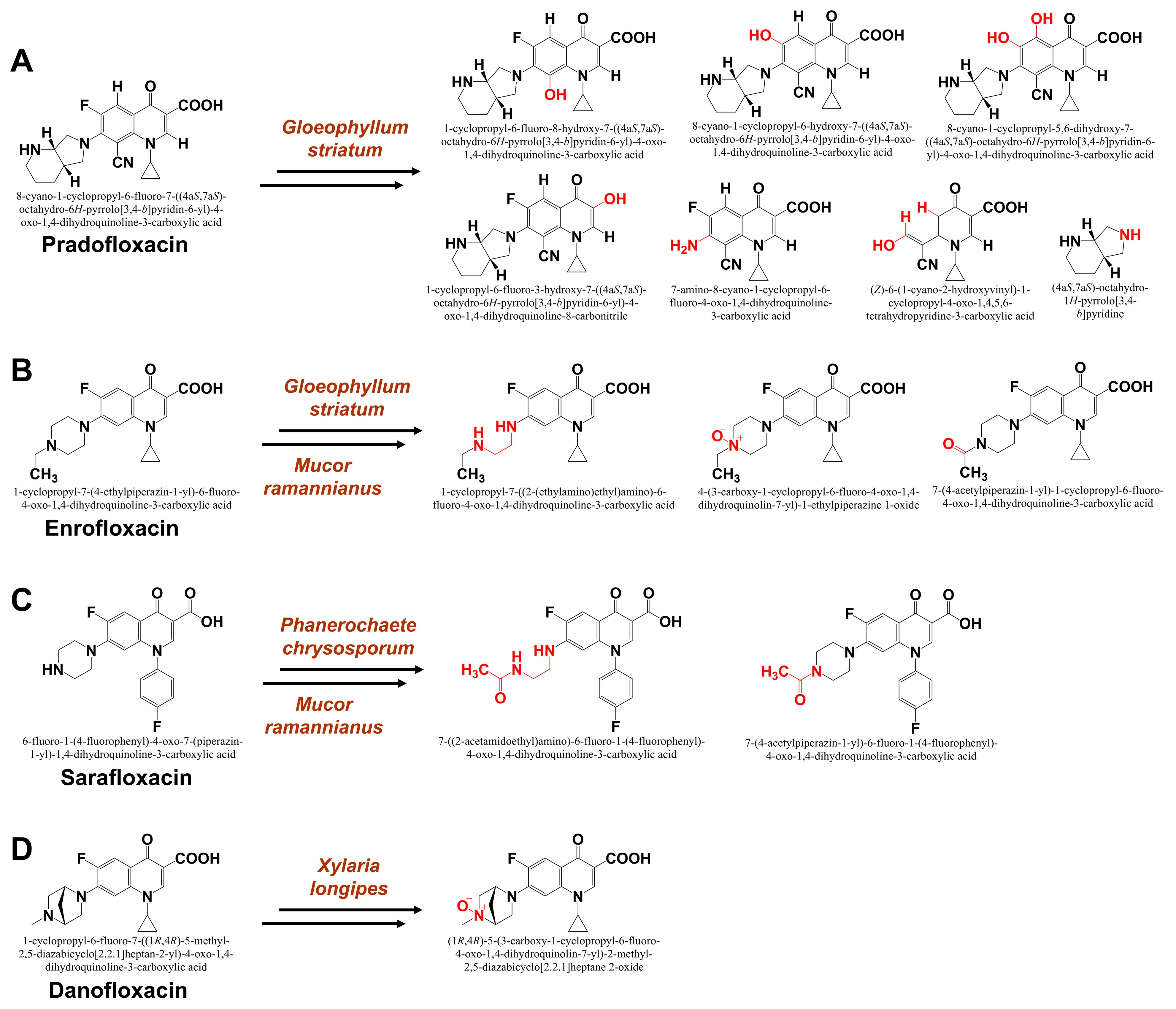
  64. Akrout, I.; Staita, K.; Zouari-Mechichi, H.; Ghariani, B.; Khmaissa, M.; Navarro, D.; Doan, A.; Albert, Q.; Faulds, C.; Sciara, G.; et al. Valorizing fungal diversity for the degradation of fluoroquinolones. Heliyon 2024, 10, e30611. [Google Scholar]
  65. Wetzstein, H.G.; Schneider, J.; Karl, W. Metabolite proving fungal cleavage of the aromatic core part of a fluoroquinolone antibiotic. AMB Express 2012, 2, 3. [Google Scholar] [PubMed]
  66. Parshikov, I.A.; Freeman, J.P.; Lay, J.O., Jr.; Beger, R.D.; Williams, A.J.; Sutherland, J.B. Microbiological transformation of enrofloxacin by the fungus Mucor ramannianus. Appl. Environ. Microbiol. 2000, 66, 2664–2667. [Google Scholar] [PubMed]
  67. Wetzstein, H.G.; Schmeer, N.; Karl, W. Degradation of the fluoroquinolone enrofloxacin by the brown rot fungus Gloeophyllum striatum: Identification of metabolites. Appl. Environ. Microbiol. 1997, 63, 4272–4281. [Google Scholar]
  68. Marengo, J.R.; Kok, R.A.; Burrows, L.A.; Velagaleti, R.R.; Stamm, J.M. Biodegradation of 14C-sarafloxacin hydrochloride, a fluoroquinolone antimicrobial by Phanerochaete chrysosporium. J. Sci. Ind. Res. 2001, 60, 121–130. [Google Scholar]
  69. Parshikov, I.A.; Freeman, J.P.; Lay, J.O., Jr.; Moody, J.D.; Williams, A.J.; Beger, R.D.; Sutherland, J.B. Metabolism of the veterinary fluoroquinolone sarafloxacin by the fungus Mucor ramannianus. J. Ind. Microbiol. Biotechnol. 2001, 26, 140–144. [Google Scholar]
  70. Rusch, M.; Kauschat, A.; Spielmeyer, A.; Römpp, A.; Hausmann, H.; Zorn, H.; Hamscher, G. Biotransformation of the antibiotic danofloxacin by Xylaria longipes leads to an efficient reduction of its antibacterial activity. J. Agric. Food Chem. 2015, 63, 6897–6904. [Google Scholar]
  71. Bodin, H.; Daneshvar, A.; Gros, M.; Hultberg, M. Effects of biopellets composed of microalgae and fungi on pharmaceuticals present at environmentally relevant levels in water. Ecol. Eng. 2016, 91, 169–172. [Google Scholar]
  72. Rodarte-Morales, A.I.; Feijoo, G.; Moreira, M.T.; Lema, J.M. Degradation of selected pharmaceutical and personal care products (PPCPs) by white-rot fungi. World J. Microbiol. Biotechnol. 2011, 27, 1839–1846. [Google Scholar] [CrossRef]
  73. Buchicchio, A.; Bianco, G.; Sofo, A.; Masi, S.; Caniani, D. Biodegradation of carbamazepine and clarithromycin by Trichoderma harzianum and Pleurotus ostreatus investigated by liquid chromatography–high-resolution tandem mass spectrometry (FTICR MS-IRMPD). Sci. Total Environ. 2016, 557, 733–739. [Google Scholar] [CrossRef]
  74. Cai, Y.; Yu, H.; Ren, L.; Ou, Y.; Jiang, S.; Chai, Y.; Chen, A.; Yan, B.; Zhang, J.; Yan, Z. Treatment of amoxicillin-containing wastewater by Trichoderma strains selected from activated sludge. Sci. Total Environ. 2023, 867, 161565. [Google Scholar] [PubMed]
  75. Tormo-Budowski, R.; Cambronero-Heinrichs, J.C.; Durán, J.E.; Masís-Mora, M.; Ramírez-Morales, D.; Quirós-Fournier, J.P.; Rodríguez-Rodríguez, C.E. Removal of pharmaceuticals and ecotoxicological changes in wastewater using Trametes versicolor: A comparison of fungal stirred tank and trickle-bed bioreactors. Chem. Eng. J. 2021, 410, 128210. [Google Scholar] [CrossRef]
  76. Palli, L.; Castellet-Rovira, F.; Pérez-Trujillo, M.; Caniani, D.; Sarrà-Adroguer, M.; Gori, R. Preliminary evaluation of Pleurotus ostreatus for the removal of selected pharmaceuticals from hospital wastewater. Biotechnol. Prog. 2017, 33, 1529–1537. [Google Scholar] [CrossRef]
  77. Badia-Fabregat, M.; Lucas, D.; Pereira, M.A.; Alves, M.; Pennanen, T.; Fritze, H.; Rodríguez-Mozaz, S.; Barceló, D.; Vicent, T.; Caminal, G. Continuous fungal treatment of non-sterile veterinary hospital effluent: Pharmaceuticals removal and microbial community assessment. Appl. Microbiol. Biotechnol. 2016, 100, 2401–2415. [Google Scholar]
  78. Cruz-Morató, C.; Lucas, D.; Llorca, M.; Rodriguez-Mozaz, S.; Gorga, M.; Petrovic, M.; Barceló, D.; Vicent, T.; Sarrà, M.; Marco-Urrea, E. Hospital wastewater treatment by fungal bioreactor: Removal efficiency for pharmaceuticals and endocrine disruptor compounds. Sci. Total Environ. 2014, 493, 365–376. [Google Scholar]
  79. Del Álamo, A.C.; Pariente, M.I.; Vasiliadou, I.; Padrino, B.; Puyol, D.; Molina, R.; Martínez, F. Removal of pharmaceutical compounds from urban wastewater by an advanced bio-oxidation process based on Trametes versicolor immobilised in a continuous RBC system. Environ. Sci. Pollut. Res. 2018, 25, 34884–34892. [Google Scholar]
  80. Cruz-Morató, C.; Ferrando-Climent, L.; Rodriguez-Mozaz, S.; Barceló, D.; Marco-Urrea, E.; Vicent, T.; Sarrà, M. Degradation of pharmaceuticals in non-sterile urban wastewater by Trametes versicolor in a fluidised bed bioreactor. Water Res. 2013, 47, 5200–5210. [Google Scholar] [PubMed]
  81. Marco-Urrea, E.; Pérez-Trujillo, M.; Cruz-Morató, C.; Caminal, G.; Vicent, T. Degradation of the drug sodium diclofenac by Trametes versicolor pellets and identification of some intermediates by NMR. J. Hazard. Mater. 2010, 176, 836–842. [Google Scholar]
  82. Tran, N.H.; Urase, T.; Kusakabe, O. Biodegradation characteristics of pharmaceutical substances by whole fungal culture Trametes versicolor and its laccase. J. Water Environ. Technol. 2010, 8, 125–140. [Google Scholar]
  83. Pezzella, C.; Macellaro, G.; Sannia, G.; Raganati, F.; Olivieri, G.; Marzocchella, A.; Schlosser, D.; Piscitelli, A. Exploitation of Trametes versicolor for bioremediation of endocrine disrupting chemicals in bioreactors. PLoS ONE 2017, 12, e0178758. [Google Scholar]
  84. Becker, D.; Rodriguez-Mozaz, S.; Insa, S.; Schoevaart, R.; Barceló, D.; de Cazes, M.; Belleville, M.P.; Sanchez-Marcano, J.; Misovic, A.; Oehlmann, J.; et al. Removal of endocrine disrupting chemicals in wastewater by enzymatic treatment with fungal laccases. Org. Process Res. Dev. 2017, 21, 480–491. [Google Scholar]
  85. El-Gendi, H.; Saleh, A.K.; Badierah, R.; Redwan, E.M.; El-Maradny, Y.A.; El-Fakharany, E.M. A comprehensive insight into fungal enzymes: Structure, classification, and their role in mankind’s challenges. J. Fungi 2021, 8, 23. [Google Scholar] [CrossRef]
  86. Jones, S.M.; Solomon, E.I. Electron transfer and reaction mechanism of laccases. Cell. Mol. Life Sci. 2015, 72, 869–883. [Google Scholar]
  87. Singh, D.; Gupta, N. Microbial laccase: A robust enzyme and its industrial applications. Biologia 2020, 75, 1183–1193. [Google Scholar]
  88. Al-Sareji, O.J.; Meiczinger, M.; Salman, J.M.; Al-Juboori, R.A.; Hashim, K.S.; Somogyi, V.; Jakab, M. Ketoprofen and aspirin removal by laccase immobilized on date stones. Chemosphere 2023, 311, 137133. [Google Scholar] [PubMed]
  89. Zhang, J.; Cai, Q.; Chen, J.; Lu, Y.; Ren, X.; Liu, Q.; Wen, L.; Mateen, M. Enhanced removal of ibuprofen in water using dynamic dialysis of laccase catalysis. J. Water Process Eng. 2022, 47, 102791. [Google Scholar]
  90. Sun, K.; Li, S.; Yu, J.; Gong, R.; Si, Y.; Liu, X.; Chu, G. Cu2+-assisted laccase from Trametes versicolor enhanced self-polyreaction of triclosan. Chemosphere 2019, 225, 745–754. [Google Scholar]
  91. Schwarz, J.; Aust, M.O.; Thiele-Bruhn, S. Metabolites from fungal laccase-catalysed transformation of sulfonamides. Chemosphere 2010, 81, 1469–1476. [Google Scholar]
  92. Yang, L.H.; Qiao, B.; Xu, Q.M.; Liu, S.; Yuan, Y.; Cheng, J.S. Biodegradation of sulfonamide antibiotics through the heterologous expression of laccases from bacteria and investigation of their potential degradation pathways. J. Hazard. Mater. 2021, 416, 125815. [Google Scholar] [PubMed]
  93. Guo, X.L.; Zhu, Z.W.; Li, H.L. Biodegradation of sulfamethoxazole by Phanerochaete chrysosporium. J. Mol. Liq. 2014, 198, 169–172. [Google Scholar]
  94. Chen, Z.; Li, N.; Lan, Q.; Zhang, X.; Wu, L.; Liu, J.; Yang, R. Laccase inducer Mn2+ inhibited the intracellular degradation of norfloxacin by Phanerochaete chrysosporium. Int. Biodeterior. Biodegrad. 2021, 164, 105300. [Google Scholar]
  95. Lloret, L.; Eibes, G.; Lú-Chau, T.A.; Moreira, M.T.; Feijoo, G.; Lema, J.M. Laccase-catalysed degradation of anti-inflammatories and estrogens. Biochem. Eng. J. 2010, 51, 124–131. [Google Scholar]
  96. Chmelová, D.; Ondrejovič, M.; Miertuš, S. Laccases as effective tools in the removal of pharmaceutical products from aquatic systems. Life 2024, 14, 230. [Google Scholar] [CrossRef]
  97. Bronikowski, A.; Hagedoorn, P.L.; Koschorreck, K.; Urlacher, V.B. Expression of a new laccase from Moniliophthora roreri at high levels in Pichia pastoris and its potential application in micropollutant degradation. AMB Express 2017, 7, 73. [Google Scholar]
  98. Ostadhadi-Dehkordi, S.; Tabatabaei-Sameni, M.; Forootanfar, H.; Kolahdouz, S.; Ghazi-Khansari, M.; Faramarzi, M.A. Degradation of some benzodiazepines by a laccase-mediated system in aqueous solution. Bioresour. Technol. 2012, 125, 344–347. [Google Scholar] [PubMed]
  99. Adamo, M.; Comtet-Marre, S.; Büttner, E.; Kellner, H.; Luis, P.; Vallon, L.; Prego, R.; Hofrichter, M.; Girlanda, M.; Peyret, P.; et al. Fungal dye-decolorizing peroxidase diversity: Roles in either intra-or extracellular processes. Appl. Microbiol. Biotechnol. 2022, 106, 2993–3007. [Google Scholar]
  100. Conesa, A.; Punt, P.J.; van den Hondel, C.A. Fungal peroxidases: Molecular aspects and applications. J. Biotechnol. 2002, 93, 143–158. [Google Scholar]
  101. Sung, H.J.; Khan, M.F.; Kim, Y.H. Recombinant lignin peroxidase-catalyzed decolorization of melanin using in-situ generated H2O2 for application in whitening cosmetics. Int. J. Biol. Macromol. 2019, 136, 20–26. [Google Scholar]
  102. Son, H.; Seo, H.; Han, S.; Kim, S.M.; Khan, M.F.; Sung, H.J.; Kang, S.H.; Kim, K.J.; Kim, Y.H. Extra disulfide and ionic salt bridge improves the thermostability of lignin peroxidase H8 under acidic condition. Enzyme Microb. Technol. 2021, 148, 109803. [Google Scholar] [CrossRef] [PubMed]
  103. Auriol, M.; Filali-Meknassi, Y.; Adams, C.D.; Tyagi, R.D. Natural and synthetic hormone removal using the horseradish peroxidase enzyme: Temperature and pH effects. Water Res. 2006, 40, 2847–2856. [Google Scholar] [CrossRef] [PubMed]
  104. Zhang, Y.; Geißen, S.U. In vitro degradation of carbamazepine and diclofenac by crude lignin peroxidase. J. Hazard. Mater. 2010, 176, 1089–1092. [Google Scholar] [PubMed]
  105. Wen, X.; Jia, Y.; Li, J. Degradation of tetracycline and oxytetracycline by crude lignin peroxidase prepared from Phanerochaete chrysosporium—A white rot fungus. Chemosphere 2009, 75, 1003–1007. [Google Scholar]
  106. Wen, X.; Jia, Y.; Li, J. Enzymatic degradation of tetracycline and oxytetracycline by crude manganese peroxidase prepared from Phanerochaete chrysosporium. J. Hazard. Mater. 2010, 177, 924–928. [Google Scholar] [CrossRef]
  107. Inoue, S.; Igarashi, Y.; Yoneda, Y.; Kawai, S.; Okamura, H.; Nishida, T. Elimination and detoxification of fungicide miconazole and antidepressant sertraline by manganese peroxidase-dependent lipid peroxidation system. Int. Biodeterior. Biodegrad. 2015, 100, 79–84. [Google Scholar]
  108. García-Zamora, J.L.; León-Aguirre, K.; Quiroz-Morales, R.; Parra-Saldívar, R.; Gómez-Patiño, M.B.; Arrieta-Baez, D.; Rebollar-Pérez, G.; Torres, E. Chloroperoxidase-mediated halogenation of selected pharmaceutical micropollutants. Catalysts 2018, 8, 32. [Google Scholar] [CrossRef]
  109. Prieto, A.; Möder, M.; Rodil, R.; Adrian, L.; Marco-Urrea, E. Degradation of the antibiotics norfloxacin and ciprofloxacin by a white-rot fungus and identification of degradation products. Bioresour. Technol. 2011, 102, 10987–10995. [Google Scholar]
  110. Čvančarová, M.; Moeder, M.; Filipová, A.; Cajthaml, T. Biotransformation of fluoroquinolone antibiotics by ligninolytic fungi–metabolites, enzymes and residual antibacterial activity. Chemosphere 2015, 136, 311–320. [Google Scholar]
  111. Gao, N.; Liu, C.X.; Xu, Q.M.; Cheng, J.S.; Yuan, Y.J. Simultaneous removal of ciprofloxacin, norfloxacin, sulfamethoxazole by co-producing oxidative enzymes system of Phanerochaete chrysosporium and Pycnoporus sanguineus. Chemosphere 2018, 195, 146–155. [Google Scholar] [CrossRef]
  112. Sellami, K.; Couvert, A.; Nasrallah, N.; Maachi, R.; Abouseoud, M.; Amrane, A. Peroxidase enzymes as green catalysts for bioremediation and biotechnological applications: A review. Sci. Total Environ. 2022, 806, 150500. [Google Scholar] [PubMed]
  113. Pylypchuk, I.V.; Daniel, G.; Kessler, V.G.; Seisenbaeva, G.A. Removal of diclofenac, paracetamol, and carbamazepine from model aqueous solutions by magnetic sol–gel encapsulated horseradish peroxidase and lignin peroxidase composites. Nanomaterials 2020, 10, 282. [Google Scholar] [CrossRef] [PubMed]
  114. Al-Maqdi, K.A.; Hisaindee, S.; Rauf, M.A.; Ashraf, S.S. Detoxification and degradation of sulfamethoxazole by soybean peroxidase and UV+ H₂O₂ remediation approaches. Chem. Eng. J. 2018, 352, 450–458. [Google Scholar]
  115. Chen, W.; Lee, M.K.; Jefcoate, C.; Kim, S.C.; Chen, F.; Yu, J.H. Fungal cytochrome P450 monooxygenases: Their distribution, structure, functions, family expansion, and evolutionary origin. Genome Biol. Evol. 2014, 6, 1620–1634. [Google Scholar]
  116. Lin, S.; Wei, J.; Yang, B.; Zhang, M.; Zhuo, R. Bioremediation of organic pollutants by white rot fungal cytochrome P450: The role and mechanism of CYP450 in biodegradation. Chemosphere 2022, 301, 134776. [Google Scholar]
  117. Mori, T.; Ohno, H.; Ichinose, H.; Kawagishi, H.; Hirai, H. White-rot fungus Phanerochaete chrysosporium metabolizes chloropyridinyl-type neonicotinoid insecticides by an N-dealkylation reaction catalyzed by two cytochrome P450s. J. Hazard. Mater. 2021, 402, 123831. [Google Scholar]
  118. Khan, M.F.; Murphy, C.D. Cunninghamella spp. produce mammalian-equivalent metabolites from fluorinated pyrethroid pesticides. AMB Express 2021, 11, 101. [Google Scholar]
  119. Khan, M.F.; Murphy, C.D. Fluorotelomer alcohols are efficiently biotransformed by Cunninghamella elegans. Environ. Sci. Pollut. Res. 2023, 30, 23613–23623. [Google Scholar]
  120. Rodríguez-Rodríguez, C.E.; García-Galán, M.J.; Blánquez, P.; Díaz-Cruz, M.S.; Barceló, D.; Caminal, G.; Vicent, T. Continuous degradation of a mixture of sulfonamides by Trametes versicolor and identification of metabolites from sulfapyridine and sulfathiazole. J. Hazard. Mater. 2012, 213, 347–354. [Google Scholar]
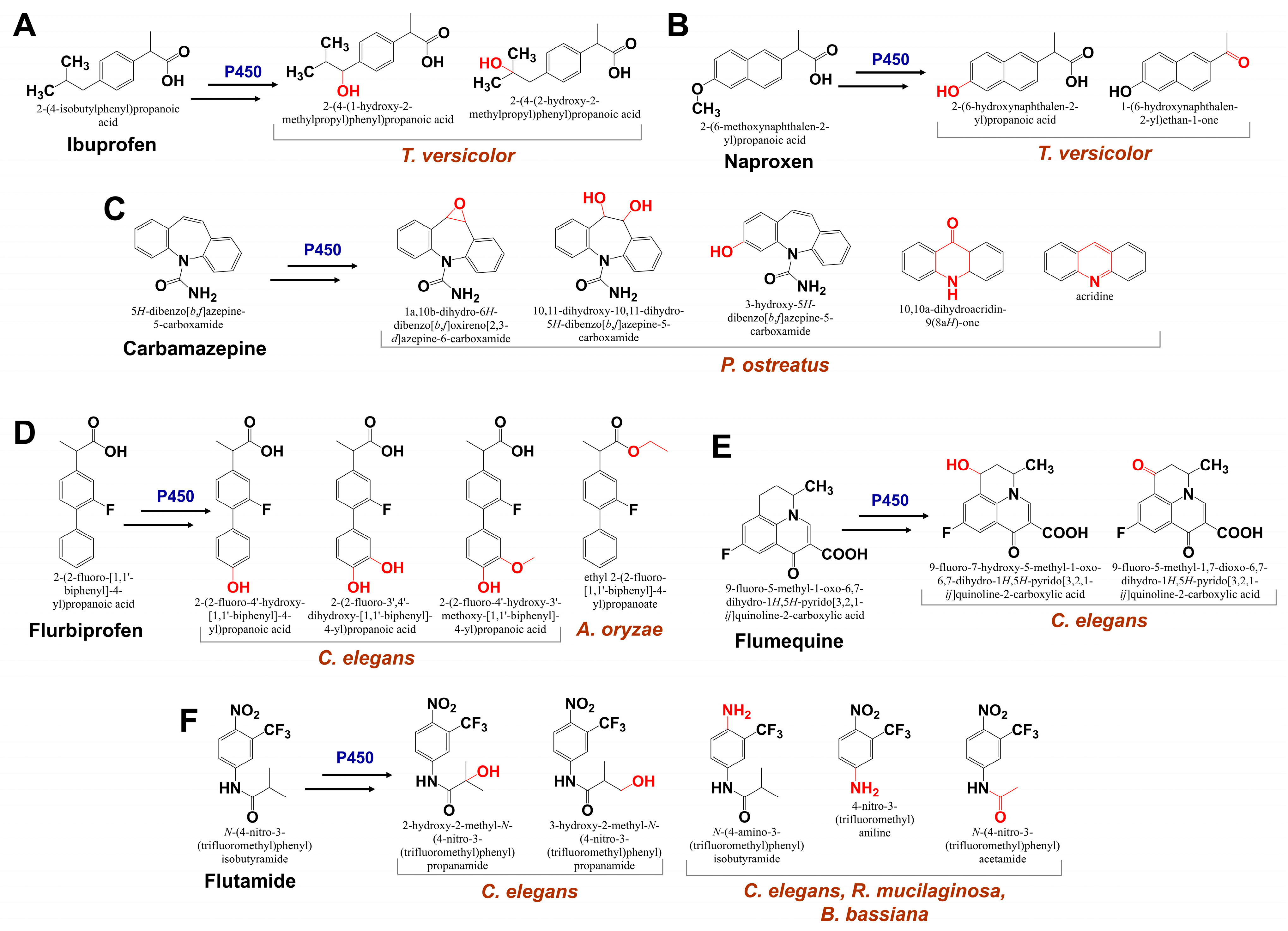
  121. Marco-Urrea, E.; Pérez-Trujillo, M.; Vicent, T.; Caminal, G. Ability of white-rot fungi to remove selected pharmaceuticals and identification of degradation products of ibuprofen by Trametes versicolor. Chemosphere 2009, 74, 765–772. [Google Scholar]
  122. Marco-Urrea, E.; Pérez-Trujillo, M.; Blánquez, P.; Vicent, T.; Caminal, G. Biodegradation of the analgesic naproxen by Trametes versicolor and identification of intermediates using HPLC-DAD-MS and NMR. Bioresour. Technol. 2010, 101, 2159–2166. [Google Scholar] [PubMed]
  123. Golan-Rozen, N.; Chefetz, B.; Ben-Ari, J.; Geva, J.; Hadar, Y. Transformation of the recalcitrant pharmaceutical compound carbamazepine by Pleurotus ostreatus: Role of cytochrome P450 monooxygenase and manganese peroxidase. Environ. Sci. Technol. 2011, 45, 6800–6805. [Google Scholar]
  124. Amadio, J.; Gordon, K.; Murphy, C.D. Biotransformation of flurbiprofen by Cunninghamella species. Appl. Environ. Microbiol. 2010, 76, 6299–6303. [Google Scholar]
  125. Tamborini, L.; Romano, D.; Pinto, A.; Contente, M.; Iannuzzi, M.C.; Conti, P.; Molinari, F. Biotransformation with whole microbial systems in a continuous flow reactor: Resolution of (RS)-flurbiprofen using Aspergillus oryzae by direct esterification with ethanol in organic solvent. Tetrahedron Lett. 2013, 54, 6090–6093. [Google Scholar]
  126. Williams, A.J.; Deck, J.; Freeman, J.P.; Chiarelli, M.P.; Adjei, M.D.; Heinze, T.M.; Sutherland, J.B. Biotransformation of flumequine by the fungus Cunninghamella elegans. Chemosphere 2007, 67, 240–243. [Google Scholar]
  127. Amadio, J.; Murphy, C.D. Production of human metabolites of the anti-cancer drug flutamide via biotransformation in Cunninghamella species. Biotechnol. Lett. 2011, 33, 321–326. [Google Scholar] [PubMed]
  128. Herath, W.; Khan, I.A. Microbial metabolism. Part 11. Metabolites of flutamide. Chem. Pharm. Bull. 2010, 58, 562–564. [Google Scholar]
  129. Sá, H.; Michelin, M.; Tavares, T.; Silva, B. Current challenges for biological treatment of pharmaceutical-based contaminants with oxidoreductase enzymes: Immobilization processes, real aqueous matrices and hybrid techniques. Biomolecules 2022, 12, 1489. [Google Scholar] [CrossRef]
  130. Kózka, B.; Nałęcz-Jawecki, G.; Turło, J.; Giebułtowicz, J. Application of Pleurotus ostreatus to efficient removal of selected antidepressants and immunosuppressants. J. Environ. Manag. 2020, 273, 111131. [Google Scholar]
  131. Kasonga, T.K.; Coetzee, M.A.; Kamika, I.; Momba, M.N. Assessing a co-culture fungal granule ability to remove pharmaceuticals in a sequencing batch reactor. Environ. Technol. 2022, 43, 1684–1699. [Google Scholar]
  132. Naghdi, M.; Taheran, M.; Brar, S.K.; Kermanshahi-pour, A.; Verma, M.; Surampalli, R.Y. Biotransformation of carbamazepine by laccase-mediator system: Kinetics, by-products and toxicity assessment. Process Biochem. 2018, 67, 147–154. [Google Scholar]
  133. Lonappan, L.; Rouissi, T.; Laadila, M.A.; Brar, S.K.; Hernandez Galan, L.; Verma, M.; Surampalli, R.Y. Agro-industrial-produced laccase for degradation of diclofenac and identification of transformation products. ACS Sustain. Chem. Eng. 2017, 5, 5772–5781. [Google Scholar]
  134. Wang, X.; Meng, F.; Zhang, B.; Xia, Y. Elimination of tetracyclines in seawater by laccase-mediator system. Chemosphere 2023, 333, 138916. [Google Scholar] [PubMed]
  135. Tian, Q.; Dou, X.; Huang, L.; Wang, L.; Meng, D.; Zhai, L.; Shen, Y.; You, C.; Guan, Z.; Liao, X. Characterization of a robust cold-adapted and thermostable laccase from Pycnoporus sp. SYBC-L10 with a strong ability for the degradation of tetracycline and oxytetracycline by laccase-mediated oxidation. J. Hazard. Mater. 2020, 382, 121084. [Google Scholar]
  136. Yousefi-Ahmadipour, A.; Bozorgi-Koshalshahi, M.; Mogharabi, M.; Amini, M.; Ghazi-Khansari, M.; Faramarzi, M.A. Laccase-catalyzed treatment of ketoconazole, identification of biotransformed metabolites, determination of kinetic parameters, and evaluation of micro-toxicity. J. Mol. Catal. B Enzym. 2016, 133, 77–84. [Google Scholar]
  137. Navada, K.K.; Kulal, A. Enzymatic degradation of chloramphenicol by laccase from Trametes hirsuta and comparison among mediators. Int. Biodeterior. Biodegrad. 2019, 138, 63–69. [Google Scholar]
  138. Feng, Y.; Shen, M.; Wang, Z.; Liu, G. Transformation of atenolol by a laccase-mediator system: Efficiencies, effect of water constituents, and transformation pathways. Ecotoxicol. Environ. Saf. 2019, 183, 109555. [Google Scholar]
  139. Khan, M.F.; Kundu, D.; Gogoi, M.; Shrestha, A.K.; Karanth, N.G.; Patra, S. Enzyme-Responsive and Enzyme Immobilized Nanoplatforms for Therapeutic Delivery: An Overview of Research Innovations and Biomedical Applications. In Nanopharmaceuticals: Principles and Applications, 3rd ed.; Yata, V., Ranjan, S., Dasgupta, N., Lichtfouse, E., Eds.; Springer: Cham, Switzerland, 2020; Volume 48, pp. 165–200. [Google Scholar]
  140. Kundu, D.; Khan, M.F.; Gogoi, M.; Patra, S. Environmental Impact and Econanotoxicity of Engineered Nanomaterials. In Nanotoxicology and Nanoecotoxicology, 1st ed.; Springer: New York, NY, USA, 2021; Volume 1, pp. 287–312. [Google Scholar]
  141. Khan, M.F.; Murphy, C.D. Environmental remediation by novel nanomaterials and fungi with high-degradation capacity of hazardous contaminants. In Bio and Nanoremediation of Hazardous Environmental Pollutants; CRC Press: Boca Raton, FL, USA, 2023; pp. 283–310. [Google Scholar]
  142. Zdarta, J.; Jankowska, K.; Wyszowska, M.; Kijeńska-Gawrońska, E.; Zgoła-Grześkowiak, A.; Pinelo, M.; Meyer, A.S.; Moszyński, D.; Jesionowski, T. Robust biodegradation of naproxen and diclofenac by laccase immobilized using electrospun nanofibers with enhanced stability and reusability. Mater. Sci. Eng. C 2019, 103, 109789. [Google Scholar]
  143. Masjoudi, M.; Golgoli, M.; Nejad, Z.G.; Sadeghzadeh, S.; Borghei, S.M. Pharmaceuticals removal by immobilized laccase on poly-vinylidene fluoride nanocomposite with multi-walled carbon nanotubes. Chemosphere 2021, 263, 128043. [Google Scholar]
  144. García-Morales, R.; García-García, A.; Orona-Navar, C.; Osma, J.F.; Nigam, K.D.; Ornelas-Soto, N. Biotransformation of emerging pollutants in groundwater by laccase from P. sanguineus CS43 immobilized onto titania nanoparticles. J. Environ. Chem. Eng. 2018, 6, 710–717. [Google Scholar]
  145. Lonappan, L.; Liu, Y.; Rouissi, T.; Pourcel, F.; Brar, S.K.; Verma, M.; Surampalli, R.Y. Covalent immobilization of laccase on citric acid functionalized micro-biochars derived from different feedstock and removal of diclofenac. Chem. Eng. J. 2018, 351, 985–994. [Google Scholar]
  146. Yang, J.; Lin, Y.; Yang, X.; Ng, T.B.; Ye, X.; Lin, J. Degradation of tetracycline by immobilized laccase and the proposed transformation pathway. J. Hazard. Mater. 2017, 322, 525–531. [Google Scholar] [PubMed]
  147. Primožič, M.; Kravanja, G.; Knez, Ž.; Crnjac, A.; Leitgeb, M. Immobilized laccase in the form of (magnetic) cross-linked enzyme aggregates for sustainable diclofenac (bio) degradation. J. Clean. Prod. 2020, 275, 124121. [Google Scholar]
  148. Huber, D.; Bleymaier, K.; Pellis, A.; Vielnascher, R.; Daxbacher, A.; Greimel, K.J.; Guebitz, G.M. Laccase catalyzed elimination of morphine from aqueous systems. New Biotechnol. 2018, 42, 19–25. [Google Scholar]
  149. Naghdi, M.; Taheran, M.; Brar, S.K.; Kermanshahi-Pour, A.; Verma, M.; Surampalli, R.Y. Immobilized laccase on oxygen functionalized nanobiochars through mineral acids treatment for removal of carbamazepine. Sci. Total Environ. 2017, 584, 393–401. [Google Scholar]
  150. Zdarta, J.; Feliczak-Guzik, A.; Siwińska-Ciesielczyk, K.; Nowak, I.; Jesionowski, T. Mesostructured cellular foam silica materials for laccase immobilization and tetracycline removal: A comprehensive study. Microporous Mesoporous Mater. 2020, 291, 109688. [Google Scholar]
  151. Wen, X.; Zeng, Z.; Du, C.; Huang, D.; Zeng, G.; Xiao, R.; Lai, C.; Xu, P.; Zhang, C.; Wan, J.; et al. Immobilized laccase on bentonite-derived mesoporous materials for removal of tetracycline. Chemosphere 2019, 222, 865–871. [Google Scholar]
  152. Dong, S.; Jing, X.; Cao, Y.; Xia, E.; Gao, S.; Mao, L. Non-covalent assembled laccase-graphene composite: Property, stability and performance in beta-blocker removal. Environ. Pollut. 2019, 252, 907–916. [Google Scholar]
  153. Sharifi-Bonab, M.; Rad, F.A.; Mehrabad, J.T. Preparation of laccase-graphene oxide nanosheet/alginate composite: Application for the removal of cetirizine from aqueous solution. J. Environ. Chem. Eng. 2016, 4, 3013–3020. [Google Scholar] [CrossRef]
  154. Maryšková, M.; Schaabová, M.; Tomankova, H.; Novotný, V.; Rysová, M. Wastewater treatment by novel polyamide/polyethylenimine nanofibers with immobilized laccase. Water 2020, 12, 588. [Google Scholar] [CrossRef]
  155. Apriceno, A.; Astolfi, M.L.; Girelli, A.M.; Scuto, F.R. A new laccase-mediator system facing the biodegradation challenge: Insight into the NSAIDs removal. Chemosphere 2019, 215, 535–542. [Google Scholar] [CrossRef] [PubMed]
  156. Taheran, M.; Naghdi, M.; Brar, S.K.; Knystautas, E.J.; Verma, M.; Surampalli, R.Y. Covalent immobilization of laccase onto nanofibrous membrane for degradation of pharmaceutical residues in water. ACS Sustain. Chem. Eng. 2017, 5, 10430–10438. [Google Scholar] [CrossRef]
  157. Simón-Herrero, C.; Naghdi, M.; Taheran, M.; Brar, S.K.; Romero, A.; Valverde, J.L.; Ramirez, A.A.; Sánchez-Silva, L. Immobilized laccase on polyimide aerogels for removal of carbamazepine. J. Hazard. Mater. 2019, 376, 83–90. [Google Scholar] [CrossRef] [PubMed]
  158. Guardado, A.L.; Druon-Bocquet, S.; Belleville, M.P.; Sanchez-Marcano, J. A novel process for the covalent immobilization of laccases on silica gel and its application for the elimination of pharmaceutical micropollutants. Environ. Sci. Pollut. Res. 2021, 28, 25579–25593. [Google Scholar] [CrossRef]
  159. Asif, M.B.; Hai, F.I.; Dhar, B.R.; Ngo, H.H.; Guo, W.; Jegatheesan, V.; Price, W.E.; Nghiem, L.D.; Yamamoto, K. Impact of simultaneous retention of micropollutants and laccase on micropollutant degradation in enzymatic membrane bioreactor. Bioresour. Technol. 2018, 267, 473–480. [Google Scholar] [CrossRef] [PubMed]
  160. De Cazes, M.; Belleville, M.P.; Petit, E.; Llorca, M.; Rodríguez-Mozaz, S.; De Gunzburg, J.; Barceló, D.; Sanchez-Marcano, J. Design and optimization of an enzymatic membrane reactor for tetracycline degradation. Catal. Today 2014, 236, 146–152. [Google Scholar] [CrossRef]
  161. Nguyen, L.N.; Vu, M.T.; Johir, M.A.; Pathak, N.; Zdarta, J.; Jesionowski, T.; Semblante, G.U.; Hai, F.I.; Khanh, D.N.; Nghiem, L.D. A novel approach in crude enzyme laccase production and application in emerging contaminant bioremediation. Processes 2020, 8, 648. [Google Scholar] [CrossRef]
  162. Nguyen, L.N.; Hai, F.I.; Price, W.E.; Kang, J.; Leusch, F.D.; Roddick, F.; van de Merwe, J.P.; Magram, S.F.; Nghiem, L.D. Degradation of a broad spectrum of trace organic contaminants by an enzymatic membrane reactor: Complementary role of membrane retention and enzymatic degradation. Int. Biodeterior. Biodegrad. 2015, 99, 115–122. [Google Scholar] [CrossRef]
  163. Abejón, R.; De Cazes, M.; Belleville, M.P.; Sanchez-Marcano, J. Large-scale enzymatic membrane reactors for tetracycline degradation in WWTP effluents. Water Res. 2015, 73, 118–131. [Google Scholar] [CrossRef]
  164. Ba, S.; Jones, J.P.; Cabana, H. Hybrid bioreactor (HBR) of hollow fiber microfilter membrane and cross-linked laccase aggregates eliminate aromatic pharmaceuticals in wastewaters. J. Hazard. Mater. 2014, 280, 662–670. [Google Scholar] [CrossRef]
  165. Ba, S.; Haroune, L.; Soumano, L.; Bellenger, J.P.; Jones, J.P.; Cabana, H. A Hybrid Bioreactor Based on Insolubilized Tyrosinase and Laccase Catalysis and Microfiltration Membrane Removal of Pharmaceuticals from Wastewater. Chemosphere 2018, 201, 749–755. [Google Scholar] [CrossRef] [PubMed]
  166. Khan, M.F.; Paul Guin, J.; Thampi, R.K.; Sullivan, J.A.; Murphy, C.D. Enhanced Removal of Perfluorooctanoic Acid with Sequential Photocatalysis and Fungal Treatment. Environ. Sci. Pollut. Res. 2023, 30, 91478–91486. [Google Scholar]
  167. Wu, B.; Hussain, M.; Zhang, W.; Stadler, M.; Liu, X.; Xiang, M. Current Insights into Fungal Species Diversity and Perspective on Naming the Environmental DNA Sequences of Fungi. Mycology 2019, 10, 127–140. [Google Scholar] [PubMed]
  168. Li, G.; Jian, T.; Liu, X.; Lv, Q.; Zhang, G.; Ling, J. Application of Metabolomics in Fungal Research. Molecules 2022, 27, 7365. [Google Scholar] [CrossRef]
  169. Damasio, A.; Goldman, G.H.; Silva, R.N.; Segato, F. Advances in the Regulation and Production of Fungal Enzymes by Transcriptomics, Proteomics, and Recombinant Strain Design. Front. Bioeng. Biotechnol. 2019, 7, 157. [Google Scholar]
  170. Sanches, P.H.; de Melo, N.C.; Porcari, A.M.; de Carvalho, L.M. Integrating Molecular Perspectives: Strategies for Comprehensive Multi-Omics Integrative Data Analysis and Machine Learning Applications in Transcriptomics, Proteomics, and Metabolomics. Biology 2024, 13, 848. [Google Scholar] [CrossRef]
  171. Alaidaroos, B.A. Advancing Eco-Sustainable Bioremediation for Hydrocarbon Contaminants: Challenges and Solutions. Processes 2023, 11, 3036. [Google Scholar] [CrossRef]
  172. Marques, H.M. Electron Transfer in Biological Systems. JBIC J. Biol. Inorg. Chem. 2024, 1, 641–683. [Google Scholar]
  173. Corbu, V.M.; Gheorghe-Barbu, I.; Dumbravă, A.Ș.; Vrâncianu, C.O.; Șesan, T.E. Current Insights in Fungal Importance—A Comprehensive Review. Microorganisms 2023, 11, 1384. [Google Scholar] [CrossRef]
  174. Kelbert, M.; Pereira, C.S.; Daronch, N.A.; Cesca, K.; Michels, C.; de Oliveira, D.; Soares, H.M. Laccase as an Efficacious Approach to Remove Anticancer Drugs: A Study of Doxorubicin Degradation, Kinetic Parameters, and Toxicity Assessment. J. Hazard. Mater. 2021, 409, 124520. [Google Scholar]
  175. Frasconi, M.; Favero, G.; Boer, H.; Koivula, A.; Mazzei, F. Kinetic and Biochemical Properties of High and Low Redox Potential Laccases from Fungal and Plant Origin. Biochim. Biophys. Acta (BBA)-Proteins Proteom. 2010, 1804, 899–908. [Google Scholar]
  176. Singh, A.K.; Abellanas-Perez, P.; de Andrades, D.; Cornet, I.; Fernandez-Lafuente, R.; Bilal, M. Laccase-Based Biocatalytic Systems Application in Sustainable Degradation of Pharmaceutically Active Contaminants. J. Hazard. Mater. 2024, 1, 136803. [Google Scholar]
  177. Tinoco-Valencia, R.; Gómez-Cruz, C.; Galindo, E.; Serrano-Carreon, L. Toward an Understanding of the Effects of Agitation and Aeration on Growth and Laccases Production by Pleurotus ostreatus. J. Biotechnol. 2014, 177, 67–73. [Google Scholar] [PubMed]
  178. Sang, B.I.; Kim, Y.H.; Yoo, Y.J. Shear Effects on Production of Lignin Peroxidase by Phanerochaete chrysosporium. Biotechnol. Bioprocess Eng. 1996, 1, 26–31. [Google Scholar]
  179. Li, J.; Liu, Z.; Zhao, J.; Wang, G.; Xie, T. Molecular Insights into Substrate Promiscuity of CotA Laccase Catalyzing Lignin-Phenol Derivatives. Int. J. Biol. Macromol. 2024, 256, 128487. [Google Scholar]
  180. Guengerich, F.P. Mechanisms of Cytochrome P450-Catalyzed Oxidations. ACS Catal. 2018, 8, 10964–10976. [Google Scholar]
  181. Lorenzo, M.; Moldes, D.; Couto, S.R.; Sanromán, M.A. Inhibition of Laccase Activity from Trametes versicolor by Heavy Metals and Organic Compounds. Chemosphere 2005, 60, 1124–1128. [Google Scholar]
  182. Okal, E.J.; Heng, G.; Magige, E.A.; Khan, S.; Wu, S.; Ge, Z.; Zhang, T.; Mortimer, P.E.; Xu, J. Insights into the Mechanisms Involved in the Fungal Degradation of Plastics. Ecotoxicol. Environ. Saf. 2023, 262, 115202. [Google Scholar]
  183. Mitra, S.; Murthy, G.S. Bioreactor Control Systems in the Biopharmaceutical Industry: A Critical Perspective. Syst. Microbiol. Biomanuf. 2022, 1, 91–112. [Google Scholar]
  184. Mendes, S.; Robalo, M.P.; Martins, L.O. Bacterial Enzymes and Multi-Enzymatic Systems for Cleaning-Up Dyes from the Environment. Microb. Degrad. Synth. Dyes Wastewaters 2015, 1, 27–55. [Google Scholar]
  185. Sharma, K.; Kaushik, G.; Thotakura, N.; Raza, K.; Sharma, N.; Nimesh, S. Enhancement Effects of Process Optimization Technique While Elucidating the Degradation Pathways of Drugs Present in Pharmaceutical Industry Wastewater Using Micrococcus yunnanensis. Chemosphere 2020, 238, 124689. [Google Scholar] [CrossRef] [PubMed]
Figure 1. Sources and pathways of pharmaceutical active compounds in the environment, including waste forms (solid and liquid), waste treatment processes, the flow of treated waste into ecosystems, and its potential reuse and re-entry into the food chain, highlighting contamination routes in water matrices and the associated toxicological impacts.
Processes 13 01034 g001
Figure 2. Mycoremediation of environmental pharmaceuticals through enzymatic pathways and biocatalysis, breaking down contaminants into less or non-toxic substances while supplying fungi with energy and nutrients. Various degradation pathways, including ligninolytic degradation, non-ligninolytic degradation, ring cleavage pathways, TCA cycle, etc. Various biocatalytic reactions, including hydroxylation, esterification, dehydration, epoxidation, demethylation, chlorine, oxidation, denitrification, etc. Various degrading enzymes, including cytochrome P450 (monooxygenase), dioxygenase, laccase, lignin peroxidase, manganese peroxidase, versatile peroxidase, etc. Various pharmaceutical classes, including ANL (analgesic), ABX (antibiotic), ACV (anti-convulsant), ADP (antidepressant), AHT (antihypertensive agent), ASP (anti-spasmodic), BB (β-blocker), CNSS (CNS stimulant), HLA (hypolipidemic agent), NSAID (non-steroidal anti-inflammatory drug), and SERM (selective estrogen receptor modulator).
Processes 13 01034 g002
Figure 3. Metabolites produced in fungal degradation of different quinolone antibiotics. (A) Pradofloxacin degradation by Gloeophyllum striatum. (B) Enrofloxacin degradation by Gloeophyllum striatum and Mucor ramannianus. (C) Sarafloxacin degradation by Phanerochaete chrysosporum and Mucor ramannianus. (D) Danofloxacin degradation by Xylaria longipes. The fungal biodegradation forms various metabolites, depicted in red, highlighting the structural transformations. The figure is sourced from references [64,65,66,67,68,69,70].
Processes 13 01034 g003
Figure 4. Trametes versicolor laccase-mediated biodegradation of sulfonamide drugs: (A) sulfanilamide, (B) sulfadimethoxine, (C) sulfayridine, and (D) sulfathiazole. The enzymatic degradation leads to the formation of various metabolites, depicted in red, highlighting the structural transformations. The figure is sourced from references [91,92].
Processes 13 01034 g004
Figure 5. Fungal peroxidase-mediated (lignin peroxidase, Mn peroxidase, or Mn-independent peroxidase) biodegradation of fluorinated quinolone antibiotics: (A) ciprofloxacin, (B) ofloxacin, and (C) norfloxacin. Fungi are labeled under the metabolite produced during biodegradation. The enzymatic degradation leads to the formation of various metabolites, depicted in red, highlighting the structural transformations. The figure is sourced from references [109,110,111].
Processes 13 01034 g005
Figure 6. Fungal cytochrome P450-mediated biodegradation of non-fluorinated and fluorinated drugs: (A) ibuprofen, (B) naproxen, (C) carbamazepine, (D) flurbiprofen, (E) flumequine, and (F) flutamide. Fungi are labeled under the metabolite produced during biodegradation. The enzymatic degradation leads to the formation of various metabolites, depicted in red, highlighting the structural transformations. The figure is sourced from reference [56,121,122,123,124,125,126,127,128].
Processes 13 01034 g006
Figure 7. Factors influencing fungal biodegradation of pharmaceuticals and strategies to address these limitations.
Processes 13 01034 g007
Table 1. Pharmaceuticals found in various environmental sources.
Drug NameEnvironmental SourceConcentration (ng L−1 or ng g−1) *Contributing CountryHarmful Side Effects on Human HealthReferences
Antibiotics
SulfamethoxazoleIndustrial effluent1,340,000China/TaiwanGastrointestinal disturbances and skin irritations[31]
Penicilloic acidsIndustrial effluent44,000,000 ChinaHypersensitivity, angioedema, and anaphylaxis[32]
OxytetracyclineIndustrial effluent19,500,000 ChinaSkin irritations and gastrointestinal disturbances[32]
SulfaguanidineIndustrial effluent>1,100,000CroatiaKidney damage and destroys red blood cells[33]
CiprofloxacinIndustrial effluent14,000,000IndiaTendon problems, nerve damage, and low blood sugar[34]
LincomycinIndustrial effluent43,900,000KoreaNausea, vomiting, swollen tongue, and vaginal itching[35]
CetirizineSurface water28,000IndiaDrowsiness, fatigue, dry mouth, nausea, and vomiting[34]
OxytetracyclineSurface water712,000ChinaSkin irritations and gastrointestinal disturbances[32]
Penicilloic acidsSurface water11,600,000ChinaHypersensitivity, angioedema, and anaphylaxis[32]
SulfamethoxazoleSurface water49,000PakistanGastrointestinal disturbances and skin irritations[36]
CiprofloxacinGroundwater770IndiaTendon problems, nerve damage, and low blood sugar[37]
ClarithromycinSeawater (coastal)17.6USAHearing loss, mood swings, myopathy, and vision issues[38]
ErythromycinSeawater (coastal)5–70ChinaLiver disease, stomach cramps, and diarrhea[39]
SulfadiazineSeawater (coastal)0.6–71.8 ChinaHallucinations, seizure, liver problems, and dark urine[39]
NorfloxacinSeawater (coastal)3.0–6800 ChinaHeadache, dark urine, muscle weakness, and diarrhea[40]
OfloxacinSeawater (coastal)3.5–5100ChinaNausea, headache, insomnia, and vaginitis[40]
RoxithromycinSeawater (coastal)6–630ChinaSkin irritations and gastrointestinal disturbances[40]
SulfadimidineSeawater (coastal)1.3–219ChinaAllergies, gastric issues, anemia, and crystalluria[41]
SulfamethoxazoleSeawater (coastal)4.2–765ChinaGastrointestinal disturbances and skin irritations[42]
TrimethoprimSeawater (coastal)60–870IrelandItching and rash, stomach upset, and headache[43]
CiprofloxacinSoil1900IndiaTendon problems, nerve damage, and low blood sugar[37]
NorfloxacinSoil61.9ChinaHeadache, dark urine, muscle weakness, and diarrhea[44]
SulfamerazineSoil16ChinaNausea, diarrhea, and hypersensitivity reactions[44]
TriclosanSoil0.4–35.5MexicoInterferes with thyroid hormone metabolism[45]
Analgesic
IbuprofenIndustrial effluent1,500,000 ChinaNausea, dyspepsia, and hypertension[31]
AcetaminophenSeawater (coastal)1.9–1952 Costa RicaNausea, vomiting, liver damage, and polyuria[46]
Sea sediments96–100 Spain[47]
IbuprofenSea sediments98–100 SpainNausea, dyspepsia, and hypertension[47]
Anti-convulsant
CarbamazepineSeawater (coastal)50–1400 IrelandAtaxia, dizziness, drowsiness, nausea, and vomiting[43]
Venlafaxine Industrial effluent11,700,000 IsraelDyspepsia, tachycardia, insomnia, and sweating[48]
Industrial effluent2600Spain[49]
NSAID
Diclofenac Surface water27,000China/TaiwanIndigestion, headache, dizziness, and drowsiness[50]
Seawater (coastal)283–843 China[42]
Seawater (coastal)60–550Ireland[43]
KetoprofenSeawater (coastal)185–805 Costa RicaAbdominal pain, diarrhea, edema, and headaches[46]
IndomethacinSea sediments12–164 ChinaHeart attack, stroke, skin changes, weight gain, etc.[42]
Beta-blockers
AtenololSeawater (coastal)80–293 BelgiumConstipation, indigestion, depression, and insomnia[51]
PropranololSeawater (coastal)0.3–142 ChinaConstipation, decreased sex drive, and insomnia[42]
CNS stimulant
MethamphetamineRiver sediments2.6–32.4 ChinaDistractibility, memory loss, and mood disturbances[52]
EphedrineRiver sediments2.6–32.4 ChinaAnxiety, dizziness, headache, and insomnia[52]
Anti-spasmodic
MebeverineSea sediments18–415 ChinaHeartburn, malaise, insomnia, and bradycardia[42]
Antiviral
OseltamivirSurface water160SwitzerlandNausea, vomiting, insomnia, and headache[53]
Fibric acid agent
GemfibrozilSeawater (coastal)77–758 Costa RicaIndigestion, drowsiness, joint pain, and impotence[47]
Muscle relaxant
MetaxaloneIndustrial effluent3,800,000USAGastrointestinal issues, nervousness, and drowsiness[54]
SERM
TamoxifenSea sediments212–431 ChinaIncreased tumor or bone pain, hot flashes, and nausea[42]
* The unit ng L−1 is for wastewater sources, and the unit ng g−1 dry weight is for sediments and soil.
Table 2. Mycoremediation of pharmaceuticals by various fungi.
FungiBiodegrades Pharmaceutical Compound(s)Pharmaceutical CategoryReferences
Phanerochaete chrysosporiumSulfamethoxazoleAntibiotic[72]
DiazepamPsychiatric drug
Ibuprofen, naproxen, and diclofenacAnti-inflammatory
Citalopram and fluoxetineAntidepressant
CarbamazepineAnti-epileptic
Ibuprofen, diclofenac, and naproxenAnti-inflammatory
CarbamazepineAnti-epileptic
Pleurotus ostreatusDiclofenac and ketoprofenAnti-inflammatory[76]
AtenololAntihypertensive
CarbamazepineAnti-epileptic[73]
ClarithromycinAntibiotic
Trametes versicolorAzithromycin, ciprofloxacin, tetracycline, and cephalexinAntibiotic[77]
Metoprolol and carazololβ-blockers
DiazepamPsychiatric drug
Ciprofloxacin and ofloxacinAntibiotic[78]
Acetaminophen, ibuprofen, and ketoprofenAnti-inflammatory
CarbamazepinePsychiatric drug[78,79]
ErythromycinAntibiotic[80]
Salicylic acidKeratolytic agent
Codeine and acetaminophenAnalgesic
Ibuprofen and ketoprofenAnti-inflammatory
SulfamethoxazoleAntibiotic[79]
AntipyrineAnalgesic
Clofibric acidAntilipidemic
AtenololAntihypertensive
HydrochlorothiazideDiuretic
RanitidineHistamine 2 blocker
DiclofenacAnti-inflammatory[81]
PropyphenazoneAnalgesic[82]
Fenoprofen, naproxen, ketoprofen, and indomethacinAnti-inflammatory
Clofibric acidAntilipidemic
GemfibrozilLipid regulation
Bisphenol A, nonylphenol, parabens, and phthalatesEndocrine-disrupting chemicals[83]
17α-ethinyl-estradiol, 17β-estradiol, estriol, and estroneHormones[84]
Trichoderma harzianumCarbamazepineAnti-epileptic[73]
ClarithromycinAntibiotic
Trichoderma pubescensAmoxicillinAntibiotic[74]
Aspergillus nigerSulfamethoxazoleAntibiotic[71]
Metoprololβ-blockers
AcetaminophenAnalgesic
Diclofenac and naproxenAnti-inflammatory
RanitidineHistamine 2 blocker
CarbamazepineAnti-epileptic
Bjerkandera adustaDiclofenacAnti-inflammatory[63]
SulfamethoxazoleAntibiotic[73]
DiazepamPsychiatric drug
Ibuprofen, naproxen, and diclofenacAnti-inflammatory
Citalopram and fluoxetineAntidepressant
CarbamazepineAnti-epileptic
Fomitopsis meliaeDiclofenacAnti-inflammatory[63]
Myceliophthora thermophila17α-ethinyl-estradiol, 17β-estradiol, estriol, and estroneHormones[84]
Table 4. Key factors affecting fungi and their enzyme-catalyzed degradation of pharmaceuticals.
FactorEffect on Enzyme ActivityExampleReference
Fungal strain type-Different fungal species produce different enzyme types
-Some fungi have high oxidative enzyme activity, while others lack key metabolic pathways
-T. versicolor produces laccases that degrade doxorubicin
-P. chrysosporium secretes ligninolytic enzymes that degrade diclofenac
-Cunninghamella elegans CYPs degrade flutamide
[11,104,174]
pH-Affects enzyme structure and substrate binding
-High pH disrupts internal electron transfer in laccases and peroxidases
-T. versicolor laccases degrade doxorubicin at pH 4 but not at pH 3
-Cytochrome P450 enzymes require a neutral pH for optimal drug metabolism
-P. chrysosporium lignin peroxidase degrades melanin at pH 4
[12,101,174]
Temperature-Low temperatures slow reaction rates
-High temperatures can denature enzymes, reducing activity
-Laccase-ABTS system achieves complete ketoprofen and aspirin removal at 35 °C
-Cytochrome P450 enzymes function best at 30 °C for flurbiprofen, diclofenac, and ibuprofen biodegradation but lose stability at higher temperatures
-P. chrysosporium lignin peroxidase LiPH8 isozyme performs optimally at 25 °C in ABTS oxidation
[56,88,102]
Oxygen levels-Oxygen is required for oxidation reactions
-Low oxygen levels limit enzymatic activity, particularly for laccases and cytochrome P450 enzymes
-Low oxygen reduces laccase oxidation capacity
-Cytochrome P450 enzymes require molecular oxygen for pharmaceutical breakdown
[8,175]
Nutrient availability-Limited nutrients can reduce enzyme production
-Carbon and nitrogen sources affect fungal metabolism
-White-rot fungi produce more laccase in nutrient-limited environments, enhancing pharmaceutical degradation[176]
Agitation-Increases oxygen transfer and substrate availability
-Excessive agitation can reduce fungal growth or deactivate enzymes
-Higher agitation increases P. ostreatus and T. versicolor laccase activity but may lower fungal biomass
-Shear sensitivity impacts lignin peroxidase overproduction in P. chrysosporium
[172,173]
Electron-donating and -withdrawing groups-EDGs enhance enzymatic oxidation
-EWGs reduce enzyme affinity for substrates
-DHQ (hydroxyl group as EDG) shows high biotransformation despite iodine (EWG)
-Laccases and cytochrome P450 oxidize EDG-containing substrates efficiently
[174,175]
Influence of Ions-Some ions inhibit enzyme activity by blocking active sites
-Certain metal ions enhance electron transfer and enzymatic oxidation
-Inorganic anions interfere with radical formation and alter pH
-5 mM NaCl inhibits T. versicolor laccase by 20%
-Cu2+ enhances triclosan degradation, while Mn2+ inhibits tetracycline removal
-HCO3 competes with substrates, reducing degradation efficiency
[176]
Humic substances-Humic acids compete with pharmaceuticals for enzyme binding-HA inhibits TEMPO-mediated Lac reactions[138]
Drug properties-Solubility, polarity, and redox potential affect bioavailability to enzymes-Highly hydrophobic drugs resist enzymatic breakdown[21,171]
Disclaimer/Publisher’s Note: The statements, opinions and data contained in all publications are solely those of the individual author(s) and contributor(s) and not of MDPI and/or the editor(s). MDPI and/or the editor(s) disclaim responsibility for any injury to people or property resulting from any ideas, methods, instructions or products referred to in the content.

