Article

Deletion of the Class 1 Histone Deacetylase PsHos2 Induces Secondary Metabolic Perturbations in the Sea Cucumber-Associated Penicillium sclerotiorum

1
Biology Institute, Qilu University of Technology (Shandong Academy of Sciences), Jinan 250103, China
2
State Key Laboratory of Bioreactor Engineering, School of Biotechnology, East China University of Science and Technology, Shanghai 200237, China
3
Key Laboratory for Enzyme and Enzyme-Like Material Engineering of Heilongjiang, College of Life Science, Northeast Forestry University, Harbin 150040, China
4
Center of Edible Fungi, Northwest A&F University, Yangling 712100, China
*
Authors to whom correspondence should be addressed.
These authors contributed equally to this work.
Fermentation 2025, 11(4), 230; https://doi.org/10.3390/fermentation11040230
Submission received: 10 March 2025 / Revised: 18 April 2025 / Accepted: 18 April 2025 / Published: 21 April 2025
(This article belongs to the Special Issue New Research on Fungal Secondary Metabolites, 3rd Edition)

Abstract

:
The long-term coexistence of sea cucumber-associated microorganisms with their host enables them to jointly withstand the unique marine ecological environment, and possess great potential for producing various natural products. However, under conventional laboratory conditions, most biosynthetic gene clusters (BGCs) in these microorganisms remain silent, necessitating the establishment of effective activation strategies for exploring bioactive secondary metabolites (SMs). Histone acetylation status regulates chromatin structure and plays a crucial role in cellular physiology and fungal secondary metabolism. Penicillium sclerotiorum SD-36 was isolated from sea cucumbers in our previous study. Genome sequencing results indicate that this strain harbors as many as 52 BGCs, suggesting it holds a wealth of genetic resources essential for synthesizing diverse SMs. Here, we describe the impact of a class 1 histone deacetylase (HDAC), PsHos2, on secondary metabolism of sea cucumber-associated Penicillium sclerotiorum SD-36. The colony morphology and SM profile of ΔPsHos2 exhibited significant changes, with the emergence of multiple new compound peaks. Six compounds, including five azaphilones, which are characterized by a pyranoquinone core structure, were isolated from ΔPsHos2, and seventeen unreported potential azaphilone-related nodes were obtained using molecular networking based on LC-MS/MS. Transcriptome analysis revealed that PsHos2 influenced the expression of 44 BGC core genes. Specifically, seven genes within cluster 86.1, the putative BGC for azaphilones, were upregulated, including two polyketide synthase (PKS) genes. The results indicate that regulation based on class 1 HDACs is an important strategy for enhancing SM synthesis in sea cucumber-associated fungi and expanding the resources of marine natural products.

1. Introduction

Natural products, with their diverse structures and abundant activities, constitute a significant source for new drug development [1]. Over the past four decades (from 1981 to 2019), natural products or their derivatives have comprised more than 35% of new drugs [1], with a quarter exhibiting biological activity [2,3]. The marine environment, distinguished by its uniqueness and abundance, harbors a wealth of natural products with diverse structural types [4,5]. Examples include cephalosporin antibiotics produced by marine fungi and conotoxin drugs derived from marine cone snails, both of which are important pharmaceuticals [6,7]. Sea cucumbers, which are used for both medicinal and food purposes, contain a variety of biologically active substances [8]. They play a crucial role in marine ecosystems by providing unique habitats for microorganisms such as bacteria and fungi [9]. Many studies have indicated that co-associated microorganisms are the actual producers of many natural products, and the metabolites produced by these microorganisms can promote the growth of their hosts or provide chemical protection to them [10,11,12]. For example, Leopold-Messer et al. (2023) found that sponge-associated Acidobacteria are capable of synthesizing structurally novel natural products, and these metabolites may play a key role in host chemical defense and environmental adaptation [10]. According to the review by Chen et al. (2021), Aspergillus and Penicillium are the dominant genus of sea cucumber-associated fungi and are able to produce compounds with significant antimicrobial and cytotoxic activities, providing important clues for understanding the function of microbial metabolites in the immune defense system of sea cucumbers [11]. Consequently, sea cucumber-associated microorganisms can serve as an important source for the research and development of novel drugs.
As a unique biological resource, more than 140 natural products, including polyketides, alkaloids, and terpenes, have been isolated from sea cucumber-associated microorganisms to date, exhibiting various biological activities such as antibacterial, cytotoxic, and anti-angiogenic properties [11,13]. Genomic analysis reveals that these microorganisms possess a vast array of secondary metabolite (SM) biosynthetic gene clusters (BGCs), indicating significant potential for natural product synthesis. However, due to its complexity, it is difficult to fully simulate their ecological environment in laboratory conditions. As a result, studies show that secondary metabolism in filamentous fungi is controlled by a complex regulatory network influenced by transcription factors and epigenetic regulators, including histone deacetylases (HDACs) [14,15]. HDACs impact DNA processes and are linked to heterochromatin and gene silencing [16]. Based on phylogenetic analysis and enzymatic catalysis mechanisms, HDACs are classified into four major classes: class 1 (Rpd3-like), class 2 (HdaI-like), class 3 (Sirtuins), and class 4 (exclusive to plants and mammals) [17]. Different types of HDACs have been reported to be associated with the discovery of new secondary metabolites [17].
The groundbreaking work belongs to Shwab et al., who activated two gene clusters by disrupting the hdaA gene of HDAC in Aspergillus nidulans [18]. Williams et al. utilized the HDAC inhibitor suberoylanilide hydroxamic acid to induce Cladosporium cladosporioides to produce perylenequinones that could not be obtained in a single fermentation culture [19]. These compounds were only produced when Cladosporium infected Cucumis sativus seedlings, suggesting a link between fungus–host interaction and epigenetic modifications [19]. HdaA knockout pleiotropically activated secondary metabolite genes in Calcarisporium arbuscula [20] and Penicillium chrysogenum gene clusters [21,22]. Pidroni et al. demonstrated that class 1 HDAC HosA plays a pivotal regulatory role in the secondary metabolism of Aspergillus nidulans, effectively surpassing the regulatory functions of other known HDACs, including the class 2 enzyme HdaA [23]. It can be seen that HDACs are important targets for studying the regulatory mechanisms of secondary metabolite expression, discovering new functional and structurally novel compounds, and enhancing the yield of secondary metabolites. Considering the vital function of HosA in regulating secondary metabolism in A. nidulans, the role of its homologous protein Hos2 in Penicillium remains to be elucidated. Further research into Hos2 will provide insights into its distinct metabolic regulation mechanism in Penicillium.
In our previous research, we successfully isolated and identified a fungal strain from sea cucumber, naming it Penicillium sclerotiorum SD-36. Through genome sequencing, we discovered a rich resource of gene clusters within it, yet analysis of its fermentation products revealed no corresponding metabolites, indicating that the unknown compounds it synthesizes await further exploration. Given the genomic potential of P. sclerotiorum SD-36 and the proven utility of HDAC manipulation in other fungi, we hypothesized that deletion of the class 1 HDAC gene would activate silent BGCs, leading to increased production of bioactive secondary metabolites. To test this, we focused on predicting and analyzing HDAC homolog genes in this strain. Specifically, a knockout operation on the unstudied class 1 HDAC gene Hos2 in P. sclerotiorum was conducted, and in-depth research on the impact of the Hos2 gene on secondary metabolism was carried out. In the ΔPsHos2 strain, a series of azaphilones were successfully isolated and identified. Furthermore, several potential new analogs were discovered using molecular networking analysis. The results indicate that HDAC Hos2 is an important target for regulating secondary metabolism. Genetic manipulation of this gene can effectively tap into and utilize the resources of secondary metabolites in fungi.

2. Materials and Methods

2.1. Strains and Media

The strain P. sclerotiorum SD-36 was isolated from Apostichopus japonicus from Chengshantou Island, Weihai City, the Yellow Sea, China, and stored at the Biology Institute, Shandong Academy of Sciences, Jinan, China, and the China Microbiological Culture Collection Center (CGMCC) with CGMCC No. 40419. The strain was cultured on potato dextrose agar (PDA, pH 5.8–6.0, Qingdao Hope Bio-Technology Co., Ltd., Qingdao, China) and potato dextrose broth (PDB, Qingdao Hope Bio-Technology Co., Ltd., China) at 28 °C without additional salt supplementation, consistent with standard laboratory conditions for filamentous fungi. Escherichia coli strain DH5α was grown in Luria–Bertani medium (LB) for general plasmid propagation.

2.2. Bioinformatics Analysis

The genomic sequencing of P. sclerotiorum SD-36 was performed utilizing a PacBio Sequel instrument, following which it was assembled de novo with the Hierarchical Genome Assembly Process 3 (HGAP3) [24]. To predict BGCs, we employed antiSMASH 7.0 and 2ndFind [25]. Gene function prediction was carried out utilizing the NCBI database, while gene prediction was facilitated by AUGUSTUS. To construct sequence similarity networks (SSNs) of proteins, we leveraged the Enzyme Function Initiative–Enzyme Similarity Tool (EFI-EST) [26]. Visual analysis of these networks was performed using Cytoscape v3.10.2 software. Additionally, sequence alignment and phylogenetic analyses were conducted using MEGA 7.0 software, employing the maximum likelihood method. The resulting phylogenetic tree was then visualized utilizing the iTOL platform [27].

2.3. Plasmids Construction

For the knockout of PsHos2 (g10385), a CRISPR-Cas9 system was used developed by Nødvig with minor modifications [28]. gRNAs were designed using Benchling (https://www.benchling.com/, accessed on 7 April 2020) and CHOPCHOP (http://chopchop.cbu.uib.no/, accessed on 7 April 2020). The primer pairs gpdA-F1/g10385-RL and g10385-FL/TtrpC-R were utilized for amplifying the gRNA expression cassette fragments 1 and 2, with pFC332-SD30 serving as the template. Fragments 1 and 2 were fused together as gRNA expression cassettes and inserted into linearized pFC332 (treated by PacI and Nt.BbvCI) using USERTM enzyme mix (New England Biolabs, Ipswich, MA, USA) to generate plasmid pFC332-g10385. The primers are summarized in Table S1.

2.4. P. sclerotiorum SD-36 Transformation

P. sclerotiorum SD-36 was cultured on the PDA plate for a week. The spores were harvested and suspended in 100 mL PDB medium and shaken overnight at 28 °C. Transformations were carried out as described by Liu et al. with minor modifications [29]. Briefly, the mycelium was first collected through filtration, and thoroughly washed three times with 15 mL of osmotic medium. Subsequently, the washed mycelium was suspended in 4 mL of osmotic medium containing 40 mg lywallzyme (Guangdong Microbial Culture Collection Center, Guangzhou, China), and incubated at 32 °C for 4 h. The resulting protoplasts were filtered, added with 15 mL of protoplast trapping buffer, and collected by centrifugation. The collected protoplasts were resuspended in the STC buffer. For the transformation process, approximately 107 protoplasts were mixed with 5 μg plasmid and 50 µL of PEG solution. The mixture was incubated on ice for 30 min. After the initial incubation, 1 mL of PEG solution was added to the mixture and the incubation was continued at room temperature for another 20 min. Finally, the transformed protoplasts were plated on MYG-sorbitol agar medium (10 g/L malt extract powder, 4 g/L glucose, 4 g/L yeast extract, 0.8 M sorbitol, and 10 g/L agar) with 200 μg/mL of hygromycin B. Target-specific genome engineering was analyzed by PCR. Primers for detecting gene editing mutations were designed to bind upstream and downstream from the gene-targeting sequence. The amplified bands by primer pairs g10385-F/g10385-R were subsequently purified and sent for sequencing (General Biology (Anhui) Co., Ltd., Chuzhou, China).

2.5. SM Analysis of P. sclerotiorum SD-36

The wild-type (WT) strain and mutants of P. sclerotiorum SD-36 were grown on PDA plates for a week and inoculated into 30 flasks (300 mL PDB per flask) at 28 °C for 14 d. The fermentation was collected and an equal volume ratio of ethyl acetate (EtOAc) was added for extraction. After rotary evaporation at 40 °C, a residue was procured. The resulting solids were subsequently dissolved in chromatographic-grade methanol (MeOH) and subjected to analysis using Agilent 1260 infinity high-performance liquid chromatography (HPLC) system equipped with a YMC-Pack ODS-A (10 × 250 mm, 5 μm) (YMC Co., Ltd., Kyoto, Japan). Analysis was programmed as follows: 0–60 min, linear gradient from I (5% MeOH) to II (100% MeOH); 60–80 min, isometric elution by II. The column temperature was 38 °C, and the flow rate was 1 mL/min. The compounds were detected at 254 nm.
The crude extract of ΔPsHos2 was further separated using silica gel column chromatography with eluents of petroleum ether: EtOAc (100:0, 90:10, 80:20, 60:40, 40:60, 20:80, 0:100, v/v) to yield seven fractions (Fr.1–7). A second separation of Fr.3 using silica gel column chromatography (petroleum ether: EtOAc, 8:1 and 4:1, v/v) yielded 6 fractions (Fr.3.1–3.6). Fr. 3.2 was identified as compound A1. Fr. 3.5 underwent further fractionation by HPLC (isocratic elution 85% MeOH) to collect compound A2. Fr.4 was eluted under the same conditions as Fr. 3 and yielded five fractions (Fr.4.1–4.5). Fr.4.3 was further fractionated into subfractions by HPLC (0.01% formic acid in H2O (III): acetonitrile (MeCN) 25:75, v/v) to collect compound A3. Fr.4.4 was fractionated by HPLC (III: MeCN 95:5 to 50:50, linear gradient, v/v) to collect compound A4. Fr.5 was subjected to fractionation on a Sephadex LH-20 column using an isocratic elution of dichloromethane (DCM): MeOH (1:1, v/v), followed by separation with HPLC (III: MeCN 40:60, v/v) to collect compound A5. Fr.6 was isolated through silica gel column chromatography (DCM: MeOH, 40:1, v/v), followed by a further purification step with HPLC (H2O: MeOH 25:75, v/v) to collect compound A6. The Nuclear Magnetic Resonance (NMR) spectra of the compounds were recorded using a Bruker Biospin Avance 400 spectrometer. Additionally, High-Resolution Electrospray Ionization Mass Spectrometry (HR-ESI-MS) or ESI-MS data were captured on an Agilent 6230 mass spectrometer (Agilent Technologies, Inc., Santa Clara, CA, USA).

2.6. Molecular Networking Analysis

The crude extracts of WT and ΔPsHos2 were dissolved in MeOH to achieve a concentration of 5 mg/mL. An injection volume of 10 μL was used, and the samples were subsequently analyzed utilizing an LC-MS/MS system. This system comprised a Shimadzu LC-30AD series UPLC (Shimadzu Co., Ltd., Shanghai, China), which was fitted with a Waters ACQUITY BEH C18 column (2.1 × 100 mm, 1.7 μm particles). The analysis ran a gradient of H2O (containing 1‰ HCOOH) and MeCN, interfaced with a Thermo Scientific Q Exactive Orbitrap mass spectrometer (Thermo Fisher Scientific Inc., Shanghai, China) equipped with an ESI source functioning in positive ion mode. The raw data were transformed into the mzMLfile format utilizing the MSConvert software, a component of the ProteoWizard project (version 3.0.4738). Subsequently, the resulting .mzML file underwent processing with MZmine (version 2.53). A feature-based molecular networking (FBMN) analysis was constructed utilizing the web-based workflow (version release_28.2) hosted on GNPS (Global Natural Products Social Molecular Networking, https://gnps.ucsd.edu/, accessed on 21 September 2023) [30]. The resulting FBMN data was then visualized using Cytoscape software (version 3.7.2).

2.7. Transcriptome Analysis

Total RNA was extracted using Trizol and the libraries were constructed using VAHTS Universal V5 RNA-seq Library Prep kit. The transcriptome sequencing and analysis were conducted by OE Biotech Co., Ltd. (Shanghai, China). The library was sequenced using the llumina Novaseq 6000 platform, and the sequencing data were optimized using fastp [31]. The clean reads were mapped to the reference genome using HISAT2 [32]. Fragments per kilobase of exon model per million mapped reads (FPKMs) of each gene were calculated and the read counts were obtained by HTSeq-count [33]. The significance threshold for differentially expressed genes (DEGs) was identified as fold change (FC) ≥ 1.5 or ≤0.67 (p < 0.05) [34]. Hierarchical cluster analysis of DEGs was carried out using R software (version 3.2.0) to visualize the expression patterns of genes in WT and ΔPsHos2 strains. The RNA-seq data were deposited in the NCBI Sequence Read Archive (SRA) database with the accession number PRJNA1215867.

2.8. Real-Time PCR Analysis

Total RNA was extracted using Trizol and cDNA was synthesized using qPCR (+gDNA wiper) HiScript III RT SuperMix reverse transcription (Vazyme Biotech Co., Ltd, Nanjing, China). Real-time PCR amplification was carried out on the LightCycler® 96 Real-Time PCR System from Roche (F. Hoffmann-La Roche AG, Basel, Switzerland). Each cDNA sample was assayed in triplicate and the average threshold cycle (Ct) was calculated. Relative expression levels were subsequently via the 2−∆∆Ct method. β-tubulin gene was chosen as the reference gene. For statistical analysis, Microsoft Excel (version 2019) was utilized.

3. Results

3.1. Gene Sequencing and Bioinformation Analysis

The genome of P. sclerotiorum SD-36 encompasses 108 contigs, with a cumulative length of 37,242,374 base pairs (bp) and a G+C content of 46.74%. Notably, this genome harbors a rich repertoire of 52 BGCs that are responsible for the production of SMs, as outlined in Table S2. Within these clusters, we have discerned 12 type I polyketide synthase (T1PKS) clusters, four non-ribosomal peptide synthetase (NRPS) clusters, three terpene clusters, as well as clusters belonging to other types. Four HDAC homologous genes were screened from P. sclerotiorum SD-36 by local blast analysis using reference HDAC sequences from Penicillium (Table S3). A phylogenetic analysis further revealed that the four HDAC homologous sequences belong to class 1 (g10385 and g3589) and class 2 (g5299 and g1628) HDAC (Figure 1).

3.2. Generation of the HDAC Gene Knockout Mutant Strain

The correctly sequenced plasmid was transformed into the protoplasts of P. sclerotiorum SD-36 using the PEG-mediated protoplast transformation method. The transformants were then screened using a medium containing hygromycin B. The selected transformants underwent preliminary PCR verification, and those with visible bands were subjected to gene sequencing to confirm the gene editing status. For the knockout of the gene PsHos2, the plasmid pFC332-g10385 was constructed (Figure S1). Following the transformation experiment, ten transformants were selected. PCR verification indicated that the target band was successfully amplified in nine transformants. Compared to the WT strain, the target fragment of transformant 8 was slightly longer. Gene sequencing revealed that transformant 8 had a random insertion of 120 bp near the target sequence of the original PsHos2 sequence, while transformant 4 exhibited base mutations (Figure S2). Utilizing the CRISPR-Cas9 gene editing technology, two successfully knocked-out transformants (ΔPsHos2) were obtained (Figure S3).

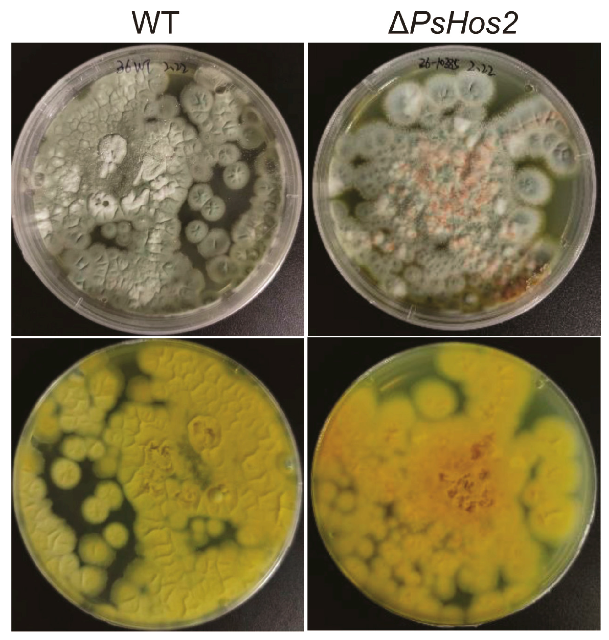
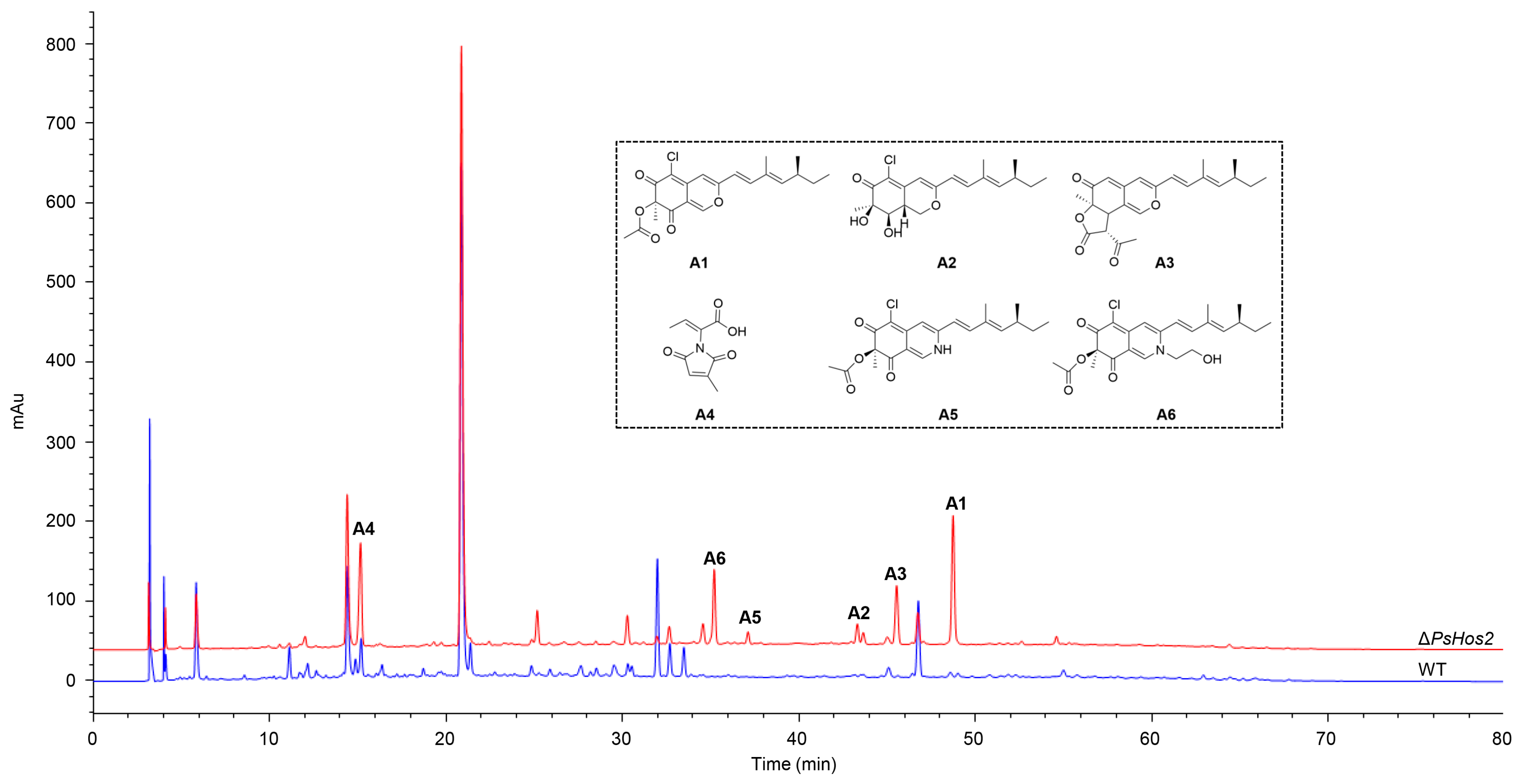
3.3. Effect of HDAC Deletion on the Phenotype, Growth, and SMs of P. sclerotiorum SD-36

As shown in Figure 2, when the mutant strain and WT strain were simultaneously inoculated on PDA medium and observed after 5 days of culturing, it was found that the colony morphology of the mutant strain underwent significant changes. Biological replicates (n = 3) consistently showed that ΔPsHos2 and WT strain were able to fully cover the plate, but ΔPsHos2 exhibited orange-red hyphae on its surface, and the color on the reverse side of the plate was also darker.
The HPLC profiling of SMs from WT and ΔPsHos2, generated from three independent biological replicates, reveals significant differences (Figure 3). Compared to WT, the metabolic profile of ΔPsHos2 exhibits several new peaks. Scale-up fermentation was conducted to characterize the differentially produced compounds between ∆PsHos2 and WT. Compounds 16 (A1A6) from ∆PsHos2 were identified as sclerotiorin (A1, 176 mg) [35], isochromophilone III (A2, 15 mg) [36], ochrephilone (A3, 7 mg) [37], pencolide (A4, 934 mg) [38], sclerotioramine (A5, 8 mg) [39], and isochromophilone VI (A6, 12 mg) [36] by ESI-MS and NMR spectra (Figure 3 and Figure S4–S20). Excluding compound A4, the remaining five compounds all belong to the class of azaphilones, which are a group of compounds characterized by their nitrogen-containing ketone structures. This finding highlights the specificity of the PsHos2-mediated regulatory mechanisms in P. sclerotiorum SD-36, influencing the production of predominantly azaphilone-type secondary metabolites.

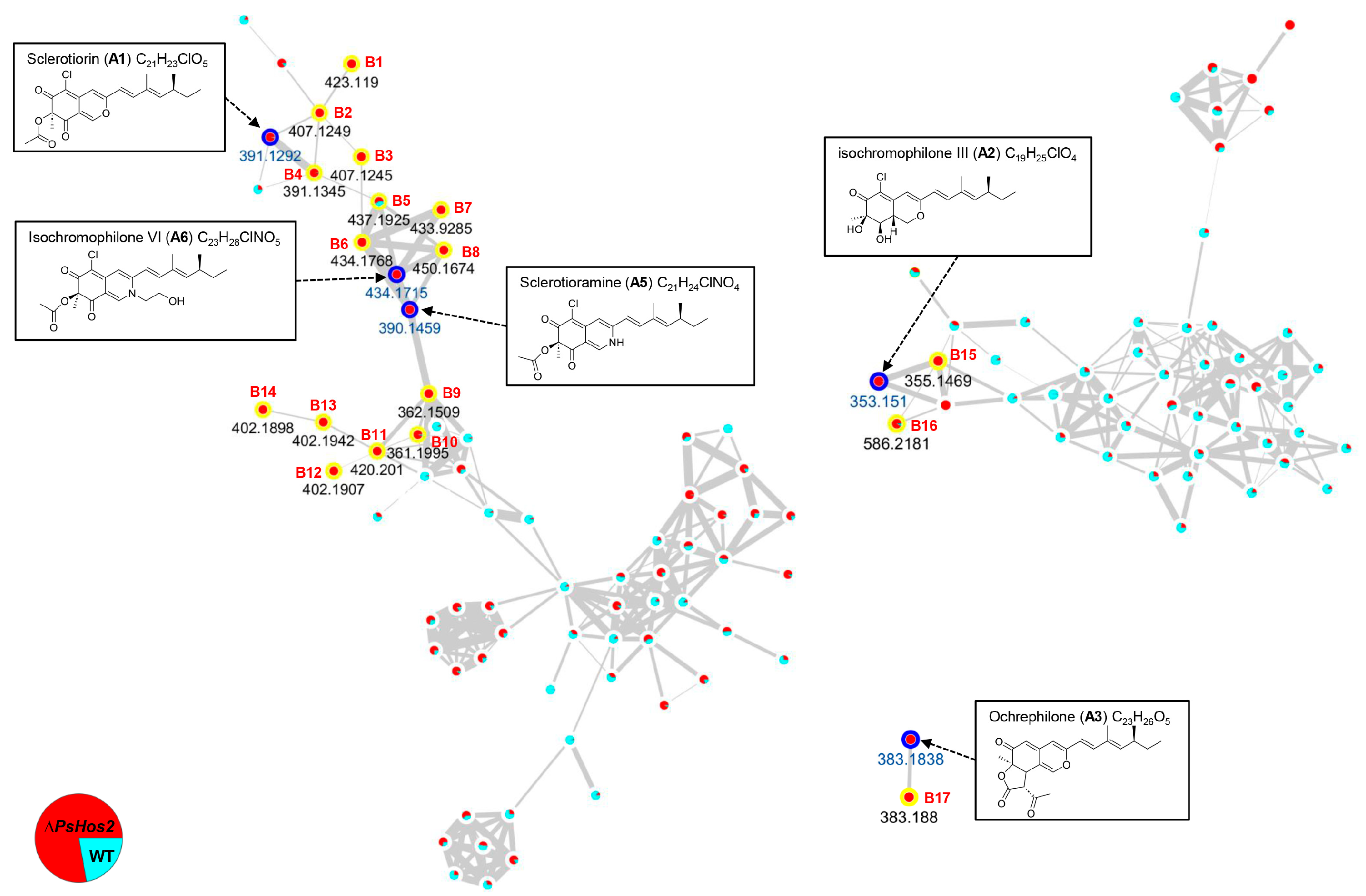
3.4. 17 Potential Azaphilone-Related Nodes Identified by Molecular Networking Analysis

We carried out feature-based molecular networking (FBMN) (workflow version release_28.2) analysis on the extract of ∆PsHos2 and WT to uncover new azaphilone analogs using A1A6 as the seeds (Figure 4 and Figure S21). Further careful annotation using their precursor mass alongside HR-ESI-MS data uncovered 14 potential nodes (B1B14) related to azaphilones A1, A5, and A6, two potential nodes (B15 and B16) related to A2, and one potential node (B17) related to A3. All the 17 potential nodes were up-regulated in ∆PsHos2, and displayed previously unreported m/z values of 423.1190, 407.1249, 407.1245, 391.1345, 437.1925, 434.1768, 433.9285, 450.1674, 362.1509, 361.1995, 420.2010, 402.1907, 402.1942, 402.1898, 355.1469, 586.2181, and 383.1880, respectively (B1B17) (Table S1).

3.5. Putative BGC of Azaphilones in P. sclerotiorum SD-36

Azaphilones possess a series of typical structural features, including a highly oxidized pyranoquinone bicyclic core (commonly referred to as the isoprene moiety) and a quaternary carbon center. Furthermore, most azaphilones carry aliphatic side chains. Within this class of compounds, the hydrogen atom at the fifth carbon position can sometimes be replaced by a chlorine atom, resulting in derivatives with distinct biological activities and pharmacological properties. The biosynthetic pathway of azaphilones is generally complex, usually involving the concerted action of a non-reducing polyketide synthase (NR-PKS) and a highly reducing polyketide synthase (HR-PKS). In the genome of P. sclerotiorum SD-36, we have identified cluster 86.1 (Figure 5, Table S5), which harbors two PKS genes. This cluster spans 24 genes (g11539-g11562), totaling 83,346 base pairs in length, and includes two distinct PKS genes, encoding an HR-PKS (by g11546) and an NR-PKS (by g11555), respectively. Sequence analysis indicated that g11546 and g11555 possessed open reading frames of 7767 bp and 8232 bp, respectively, encoding polypeptides composed of 2588 and 2743 amino acids. These polypeptides have estimated molecular weights of 280.02 kDa and 299.39 kDa, respectively. To enhance visualization and classification, we constructed SSNs for approximately 1000 PKS sequences, using g11546 and g11555 as queries (Figures S22 and S23). The most prevalent PKSs were those from Aspergillus species, with sequences predominantly grouped by genus. Notably, g11555 clustered with Q5BEJ6 (AfoE), Q0CF73 (ATEG_07661), and A0A0KMCJ4 (CazM), while g11546 clustered with Q5BEJ4 (AfoG) and Q0CF75 (ATEG_07659). All five PKSs are involved in the biosynthesis of azaphilones. A phylogenetic analysis, based on complete amino acid sequences, revealed that these PKSs are closely related (Figure S24). A comparison of the amino acid sequences showed that g11555 shared 64.26% sequence identity with AfoE, 65.39% with ATEG_07661, and 66.92% with CazM. Additionally, the gene cluster encodes various other functional proteins, such as four oxidoreductases (encoded by g11549, g11551, g11556, and g11562), one transcriptional regulator (encoded by g11550), one transporter (encoded by g11552), and one O-acetyltransferase (encoded by g11553). These proteins likely play crucial roles in the biosynthesis, regulation, and transport of azaphilones. Through the coordinated action of these proteins, P. sclerotiorum SD-36 may synthesize azaphilones with specific structures and biological activities.

3.6. Effect of PsHos2 on the Transcriptome of P. sclerotiorum SD-36

In the transcriptome analysis of ΔPsHos2 and WT strains cultivated for 14 days, a total of 4795 differentially expressed genes (DEGs) were identified, with 2418 genes upregulated and 2557 genes downregulated in ΔPsHos2 (Figure S25). Notably, 1086 of these DEGs could be annotated to KEGG pathways, indicating significant alterations in gene expression profiles in the absence of PsHos2. Further analysis with antiSMASH revealed changes in the transcription levels of 51 BGCs in ΔPsHos2, among which 44 core genes exhibited altered expression, suggesting a global impact of PsHos2 deletion on the secondary metabolism of P. sclerotiorum SD-36 (Table S6). Specifically, four NRPS genes were upregulated, while two were downregulated; eight T1PKS genes showed increased expression, and five demonstrated decreased expression. Variations in the expression levels of other types of core genes are detailed in Table S6. Additionally, the expression of PsHos3 and PsClr3 genes in ΔPsHos2 was upregulated by 2.722-fold and 1.903-fold, respectively. This compensatory upregulation of other HDAC genes may be attributed to the loss of PsHos2, suggesting a potential regulatory interplay among HDACs within the strain. The transcriptome results revealed that expression levels of 11 genes within the putative cluster 86.1 for azaphilones underwent changes in ΔPsHos2, with seven genes being upregulated, including two PKS genes (g11546 and g11555), a dehydrogenase gene (g11551), an efflux pump gene (g11552), and an O-acetyltransferase gene (g11553) (Table S7).

3.7. Real-Time PCR Analysis

Utilizing real-time PCR assays, we investigated the transcriptional level of cluster 86.1 in different strains. With β-tubulin serving as the internal control, we observed that in the ΔPsHos2 mutant compared to WT, the core PKS genes g11546 and g11555 within cluster 86.1 were significantly upregulated by 24.020-fold and 1.460-fold, respectively. Furthermore, the FAD-dependent monooxygenase gene g11549 exhibited a remarkable 59.625-fold upregulation, while the transcription factor gene g11550 was upregulated by 3.610-fold. Notably, the efflux pump gene g11552 was dramatically upregulated by an astonishing 1111.109-fold. Additionally, the remaining three HDAC genes displayed upregulation ranging from 2.032-fold to 2.623-fold (Figure S26). The above results indicate that the knockout of PsHos2 increases the gene transcription level of cluster 86.1, while also elevating the expression levels of other HDACs.

4. Discussion

Sea cucumbers play a significant role in marine ecosystems and provide unique habitats for a variety of microorganisms [9]. Sea cucumbers are important marine resources for both medicine and food, and compounds isolated from them exhibit multiple biological activities, such as anti-inflammatory, antibacterial, anticancer, antihypertensive, anti-angiogenic, and radioprotective properties [8]. Numerous studies have shown that the microorganisms co-inhabiting with marine animals are the true producers of many marine natural products, such as those found in sponges and ascidians [10,11]. Therefore, the exploration of active natural products from sea cucumber-associated microorganisms is crucial for the development of novel pharmaceuticals. Due to the highly complex native environments of microorganisms, it is challenging to simulate their natural habitats under conventional laboratory conditions. Consequently, the production potential of bioactive natural products from microorganisms remains difficult to effectively stimulate. The metabolism of microorganisms is influenced by a complex and delicate regulatory network, among which epigenetic regulation stands as a significant regulatory mechanism [40].
The inactivation of HDAC is an effective strategy to activate silent secondary metabolite BGCs and enhance the biologically active natural products of filamentous fungi [22]. The knockout of class 2 HDAC gene hdaA increases the transcription level of several NRPS gene clusters in Aspergillus fumigatus AF293 [41] and can activate the production of a series of macrodiolides in Pestalotiopsis fici CGMCC3.15140 [42]. In the study of Penicillium, the inactivation of HdaA has been found to upregulate the production of meleagin/roquefortine in P. chrysogenum [22] and transcriptionally activate the sorbicillinoids BGC [21]. On the other hand, the knockout of another class 2 HDAC, Clr3, suppressed the production of secondary metabolites in Penicillium brasilianum [43]. In our previous work, we discovered that the class 1 HDAC Hos2 affects the morphology, sporulation ability, and secondary metabolism of Chaetomium olivaceum [44]. Specifically, its deletion activates the BGCs for polyketides and asterriquinone. Hos2 plays a significant role in fungal growth and development, pathogenicity, and the regulation of secondary metabolites. In Ustilaginoidea virens and Alternaria alternata, Hos2-deletion mutants exhibit retarded vegetative growth, reduced conidiation and germination, and attenuated virulence [45,46]. Furthermore, Hos2 directly binds to the aflatoxin biosynthesis genes in Aspergillus flavus, regulating its biosynthesis [47]. These results prompted us to delete the Hos2 homologue of P. sclerotiorum SD-36, a so far not studied class 1 HDAC in Penicillium.
Both WT and ΔPsHos2 strains of P. sclerotiorum SD-36 grow normally on PDA plates. However, the colony surface of ΔPsHos2 appears orange-red, and the color on the back of the plate is also deepened, indicating that PsHos2 affects the colony morphology of P. sclerotiorum SD-36. After knocking out PsHos2, the SM profile of P. sclerotiorum SD-36 undergoes significant changes, with six compounds isolated from ΔPsHos2, including five azaphilones. Azaphilones are a class of fungal polyketide pigments featuring a highly oxygenated pyran-quinone bicyclic core, often involving multiple chiral centers and flexible side chains [48]. Due to their structural diversity and excellent biological activities such as antibacterial, antioxidant, anti-inflammatory, cytotoxic, and enzyme inhibitory activities, azaphilones have attracted increasing research interest for applications in the food, agriculture, printing, cosmetics, and pharmaceutical industries [49]. The five azaphilones obtained from ΔPsHos2 all exhibit acetylcholinesterase inhibitory activity, with A2 demonstrating the highest potency [50]. Among them, A1, A2, A5, and A6 exhibit varying cytotoxicity against three cancer cell lines, KB, MCF-7, and NCI-H187, with IC50 values ranging from 2.2 to 35.2 μg/mL [50]. Additionally, A5 and A6 show inhibitory activity against Mycobacterium tuberculosis, and A5 possesses antimalarial activity against Plasmodium falciparum [50]. Besides the purified and isolated compounds mentioned above, molecular networking analysis identified 17 unreported potential azaphilone-related nodes. These findings suggest that PsHos2 negatively regulates the biosynthesis of azaphilones in P. sclerotiorum SD-36, and its deletion activates the BGC for azaphilones.
The biosynthetic pathway of azaphilones has not been fully elucidated. However, by combining fungal genome sequencing with genetic manipulation, some azaphilone biosynthetic pathways have been reported. Pavesi et al. reviewed the recent research progress in azaphilone biosynthesis research, summarizing five main azaphilone biosynthetic pathways: the Monascus azaphilone pathway, the Aspergillus azaphilone pathway, the citrinin pathway, the Chaetomium azaphilone/cochliodone pathway and the Hypoxylon azaphilone pathway [48]. The initiation of azaphilone biosynthesis typically involves the catalysis of a common orcinaldehyde intermediate by either a single NR-PKS or a combination of NR-PKS and HR-PKS [48]. This intermediate can then be further modified by enzymes towards the citrinin pathway or by generating a common pyranoquinone core [48]. The NR-PKS and HR-PKS can function in sequential, convergent, mixed, or collaborative pathways. Using antiSMASH analysis, 12 T1PKS clusters were identified in P. sclerotiorum SD-36, with cluster 86.1 showing a 37% similarity to the azasperpyranone BGC. Cluster 86.1 contains two PKS genes: g11546, which encodes an HR-PKS, and g11555, which encodes an NR-PKS. The transcriptome results revealed that the transcription levels of seven genes in this cluster, including these two PKS genes, were upregulated in ΔPsHos2 (Table S7). Both SSN analysis and phylogenetic analysis revealed that g11555 and g11546 cluster with enzymes involved in azaphilone biosynthesis (Figures S22–S24). Specifically, g11555 shares 64.26–66.92% sequence identity with NR-PKS AfoE, ATEG_07661, and CazM. AfoE and AfoG are core enzymes of asperfuranone BGC in A. nidulans, functioning in sequential pathway [51]. AfoG forms a polyketide chain that is then transferred to the starter acyltransferase (SAT) domain of AfoE for further extension. ATEG_07661 and ATEG_07659 are involved in the biosynthesis of azasperpyranones in Aspergillus terreus, which are produced through collaborative biosynthesis by two independent gene clusters via transcriptional crosstalk [52]. CazM originates from the caz gene cluster in Chaetomium globosum, responsible for the biosynthesis of chaetoviriins and chaetomugilins, which are formed via sequential and convergent pathways [53]. Phylogenetic analysis also identified two other NR-PKSs involved in azaphilone biosynthesis: G3XMC4 (AzaA) and Q0CSA2 (TazA) (Figure S24). These enzymes are responsible for the biosynthesis of azanigerones in Aspergillus niger and azaterrilone A in A. terreus, respectively, and share sequence identities of 44.1% and 42.78% with g11555 [54,55]. All of the aforementioned biosynthetic pathways belong to the Aspergillus azaphilone pathway, and their biosynthetic routes and mechanisms can provide valuable insights into the biosynthesis of A1A6. Moreover, Transcriptome analysis reveals that the deletion of PsHos2 has a broad impact on the secondary metabolism of P. sclerotiorum SD-36, with transcriptional changes observed in 44 core genes, including multiple NRPS and PKS genes. This further confirms that the class 1 HDAC Hos2 is a critical target for regulating secondary metabolism in filamentous fungi. Although the present study has revealed a role for PsHos2 in the regulation of secondary metabolism, some limitations remain. The laboratory conditions used in this study could not fully simulate the natural sea cucumber microenvironment, including host-microbe interactions, immune factors, and symbiotic signaling, potentially affecting the ecological relevance of the metabolite production observed. Nevertheless, the successful isolation of six compounds from ΔPsHos2 provides a foundation for probing their ecological functions. Beyond pharmacological potential, these azaphilones may serve as key mediators in microbial community dynamics, while computational modeling could bridge the gap between lab-based discovery and ecological impact [56]. Future studies could further elucidate the regulatory mechanism of PsHos2 and its ecological significance through multi-omics integration, host-microbe co-culture models, and synthetic biology approaches. Although two successfully edited transformants were confirmed via PCR and Sanger sequencing, the potential off-target effects of CRISPR-Cas9 editing were not fully evaluated in this study. While targeted sequencing verified modifications at the PsHos2 locus, undetected mutations in other genomic regions could theoretically influence secondary metabolism. This limitation highlights the need for future studies to employ whole-genome sequencing or computational off-target prediction tools to ensure the specificity of genetic manipulations. Additionally, expanding the analysis to more independent transformants will validate the consistency of the observed metabolic and transcriptional changes, further strengthening the causal link between PsHos2 deletion and secondary metabolite perturbations.

5. Conclusions

In this study, we explored the impact of the class 1 HDAC PsHos2 on the secondary metabolism of the sea cucumber-associated Penicillium sclerotiorum SD-36. The deletion of PsHos2 significantly changed the colony morphology and the SM profile. Six compounds were isolated from ΔPsHos2. Through identification, five of them belonged to the azaphilones. In addition, 17 previously unreported potential azaphilone-related nodes were obtained by molecular networking. Further analysis with antiSMASH and transcriptome results revealed changes in the transcription levels of 52 BGCs in ΔPsHos2, among which 44 core genes exhibited altered expression, suggesting a global impact of PsHos2 on the secondary metabolism of P. sclerotiorum SD-36. This study demonstrates that the regulation of class 1 HDAC is an important strategy for exploring active substances from sea cucumber-associated fungi, and provides new ideas and methods for the development of marine biological resources. Similar strategies could be applied to other underexplored marine symbionts, expanding the chemical diversity available for bioprospecting.

Supplementary Materials

The following supporting information can be downloaded at: https://www.mdpi.com/article/10.3390/fermentation11040230/s1, Figure S1: Electropherogram of PCR products for the construction of plasmid pFC332-g10385; Figure S2: Electropherogram of PCR products for the verification of ΔPsHos2; Figure S3: Alignment of ΔPsHos2 transformants with WT; Figure S4: Mass spectrum of compound A1; Figure S5: 1H NMR spectrum of compound A1; Figure S6: 13C NMR spectrum of compound A1; Figure S7: Mass spectrum of compound A2; Figure S8: 1H NMR spectrum of compound A2; Figure S9: 13C NMR spectrum of compound A2; Figure S10: Mass spectrum of compound A3; Figure S11: 1H NMR spectrum of compound A3; Figure S12: 13C NMR spectrum of compound A3; Figure S13: 1H NMR spectrum of compound A4; Figure S14: 13C NMR spectrum of compound A4; Figure S15: Mass spectrum of compound A5; Figure S16: 1H NMR spectrum of compound A5; Figure S17: 13C NMR spectrum of compound A5; Figure S18: Mass spectrum of compound A6; Figure S19: 1H NMR spectrum of compound A6; Figure S20: 13C NMR spectrum of compound A6; Figure S21: The entire networking for crude extracts of the ∆PsHos2 and WT; Figure S22: SSNs network analysis based on g11555 and its homologous sequences; Figure S23: SSNs network analysis based on g11546 and its homologous sequences; Figure S24: Phylogenetic analysis of fungal NR-PKS and HR-PKS sequences; Figure S25: Differential gene grouping clustering map; Figure S26: mRNA expression level of genes from cluster 86.1; Table S1: Primers used in this study; Table S2: Gene clusters predicted by antiSMASH; Table S3: HDAC homologous sequences in P. sclerotiorum SD-36; Table S4: Detailed annotation for azaphilone-related nodes from FBMN analysis; Table S5: Predicted gene functions of cluster 86.1; Table S6: Differentially expressed core genes of the BGCs in P. sclerotiorum SD-36; Table S7: DGEs involves in cluster 86.1 in P. sclerotiorum SD-36.

Author Contributions

Conceptualization, P.Z. and L.Z.; Methodology, P.Z., Q.W. and G.Z.; Validation, C.L., M.L. and J.Q.; Formal analysis, C.L.; Investigation, L.Z. and X.X.; Resources, P.Z.; Data curation, P.Z., J.L., Q.Z., T.Z. and Q.W.; Writing—original draft preparation, P.Z. and J.L.; Writing—review and editing, X.X., J.Q. and M.L.; Funding acquisition, P.Z., L.Z. and X.X. All authors have read and agreed to the published version of the manuscript.

Funding

This research was funded by Key R&D Program of Shandong Province (grant number 2022SFGC0105), Natural Science Foundation of Shandong Province (grant numbers ZR2022QC186 and ZR2023QB021), Young Taishan Scholarship to Xuekui Xia (grant number tsqn202103100), Jinan Talent Project for Universities (grant number 202228088), Key Innovation Project and Science, and Education and Industry Integration Innovation Pilot Project Qilu University of Technology (Shandong Academy of Sciences) (grant number 2024ZDZX03), Key Core Technology Research and Development in Shaanxi Province Agriculture (grant number 2024NYGG010), and the National Key Research and Development Program (grant number 2021YFD1600401).

Institutional Review Board Statement

The CRISPR technology and related research involved in this article strictly comply with international biosafety guidelines and the laws and regulations of the respective country. Laboratory procedures adhere to the corresponding Biosafety Level (BSL) requirements to ensure the safety and controllability of the gene editing process.

Informed Consent Statement

Not applicable.

Data Availability Statement

The original contributions presented in this study are included in the article. Further inquiries can be directed to the corresponding author.

Conflicts of Interest

The authors declare that they have no competing financial interests or personal relationships that may have influenced the work reported in this study.

Abbreviations

The following abbreviations are used in this manuscript:
BGCBiosynthetic gene cluster
HDACHistone deacetylase
PKSPolyketide synthase
SMSecondary metabolite
PDAPotato dextrose agar
LBLuria–Bertani medium
SSNSequence similarity network
EtOAcEthyl acetate
MeOHMethanol
HPLCHigh-performance liquid chromatography
MeCNAcetonitrile
DCMDichloromethane
NMRNuclear Magnetic Resonance
HR-ESI-MSHigh-Resolution Electrospray Ionization Mass Spectrometry
FBMNFeature-based molecular networking
NRPSNon-ribosomal peptide synthetase
NR-PKSNon-reducing polyketide synthase
HR-PKSHighly reducing polyketide synthase

References

  1. Newman, D.J.; Cragg, G.M. Natural products as sources of new drugs over the nearly four decades from 01/1981 to 09/2019. J. Nat. Prod. 2020, 83, 770–803. [Google Scholar] [CrossRef] [PubMed]
  2. Meena, S.N.; Wajs-Bonikowska, A.; Girawale, S.; Imran, M.; Poduval, P.; Kodam, K.M. High-throughput mining of novel compounds from known microbes: A boost to natural product screening. Molecules 2024, 29, 3237. [Google Scholar] [CrossRef]
  3. Willems, T.; De Mol, M.L.; De Bruycker, A.; De Maeseneire, S.L.; Soetaert, W.K. Alkaloids from marine fungi: Promising antimicrobials. Antibiotics 2020, 9, 340. [Google Scholar] [CrossRef] [PubMed]
  4. Li, X.; Xu, J.; Wang, P.; Ding, W. Novel indole diketopiperazine stereoisomers from a marine-derived fungus Aspergillus sp. Mycology 2023, 14, 1–10. [Google Scholar] [CrossRef]
  5. Lu, X.-W.; Qin, S.; Wang, Q.; Lu, C.-Y.; Li, W.-J. Functional activity and mechanisms of common marine traditional Chinese medicine. Food Med. Homol. 2025, 2, 9420075. [Google Scholar] [CrossRef]
  6. Liu, L.; Chen, Z.; Liu, W.; Ke, X.; Tian, X.; Chu, J. Cephalosporin C biosynthesis and fermentation in Acremonium chrysogenum. Appl. Microbiol. Biotechnol. 2022, 106, 6413–6426. [Google Scholar] [CrossRef] [PubMed]
  7. Gao, S.; Yao, X.; Yan, N. Structure of human Cav2.2 channel blocked by the painkiller ziconotide. Nature 2021, 596, 143–147. [Google Scholar] [CrossRef]
  8. Hossain, A.; Dave, D.; Shahidi, F. Northern sea cucumber (Cucumaria frondosa): A potential candidate for functional food, nutraceutical, and pharmaceutical sector. Mar. Drugs 2020, 18, 274. [Google Scholar] [CrossRef]
  9. Chen, L.; Du, S.; Qu, W.Y.; Guo, F.R.; Wang, G.Y. Biosynthetic potential of culturable bacteria associated with Apostichopus japonicus. J. Appl. Microbiol. 2019, 127, 1686–1697. [Google Scholar] [CrossRef]
  10. Leopold-Messer, S.; Chepkirui, C.; Mabesoone, M.F.J.; Meyer, J.; Paoli, L.; Sunagawa, S.; Uria, A.R.; Wakimoto, T.; Piel, J. Animal-associated marine Acidobacteria with a rich natural-product repertoire. Chem 2023, 9, 3696–3713. [Google Scholar] [CrossRef]
  11. Chen, L.; Wang, X.-Y.; Liu, R.-Z.; Wang, G.-Y. Culturable microorganisms associated with sea cucumbers and microbial natural products. Mar. Drugs 2021, 19, 461. [Google Scholar] [CrossRef]
  12. Tang, C.; Gao, R.; Tang, X.; Zhang, Y.; Feng, W.; Feng, B.; Lu, X. Metabolites isolated from Penicillium HDS-Z-1E, an endophytic fungal strain isolated from Taxus cuspidata and their activation effect of catalase. Chin. Herb. Med. 2024, 16, 227–230. [Google Scholar] [CrossRef] [PubMed]
  13. Qi, J.; Jiang, L.; Zhao, P.; Chen, H.; Jia, X.; Zhao, L.; Dai, H.; Hu, J.; Liu, C.; Shim, S.H.; et al. Chaetoglobosins and azaphilones from Chaetomium globosum associated with Apostichopus japonicus. Appl. Microbiol. Biotechnol. 2020, 104, 1545–1553. [Google Scholar] [CrossRef] [PubMed]
  14. Zhang, Y.; Yu, W.; Lu, Y.; Wu, Y.; Ouyang, Z.; Tu, Y.; He, B. Epigenetic regulation of fungal secondary metabolism. J. Fungi 2024, 10, 648. [Google Scholar] [CrossRef] [PubMed]
  15. Verma, A.; Tiwari, H.; Singh, S.; Gupta, P.; Rai, N.; Kumar Singh, S.; Singh, B.P.; Rao, S.; Gautam, V. Epigenetic manipulation for secondary metabolite activation in endophytic fungi: Current progress and future directions. Mycology 2023, 14, 275–291. [Google Scholar] [CrossRef]
  16. Millán-Zambrano, G.; Burton, A.; Bannister, A.J.; Schneider, R. Histone post-translational modifications—Cause and consequence of genome function. Nat. Rev. Genet. 2022, 23, 563–580. [Google Scholar] [CrossRef]
  17. Pfannenstiel, B.T.; Keller, N.P. On top of biosynthetic gene clusters: How epigenetic machinery influences secondary metabolism in fungi. Biotechnol. Adv. 2019, 37, 107345. [Google Scholar] [CrossRef]
  18. Shwab, E.K.; Bok, J.W.; Tribus, M.; Galehr, J.; Graessle, S.; Keller, N.P. Histone deacetylase activity regulates chemical diversity in Aspergillus. Eukaryot. Cell 2007, 6, 1656–1664. [Google Scholar] [CrossRef]
  19. Williams, R.B.; Henrikson, J.C.; Hoover, A.R.; Lee, A.E.; Cichewicz, R.H. Epigenetic remodeling of the fungal secondary metabolome. Org. Biomol. Chem. 2008, 6, 1895–1897. [Google Scholar] [CrossRef]
  20. Mao, X.-M.; Xu, W.; Li, D.; Yin, W.-B.; Chooi, Y.-H.; Li, Y.-Q.; Tang, Y.; Hu, Y. Epigenetic genome mining of an endophytic fungus leads to the pleiotropic biosynthesis of natural products. Angew. Chem. Int. Ed. Engl. 2015, 54, 7592–7596. [Google Scholar] [CrossRef]
  21. Guzman-Chavez, F.; Salo, O.; Samol, M.; Ries, M.; Kuipers, J.; Bovenberg, R.A.L.; Vreeken, R.J.; Driessen, A.J.M. Deregulation of secondary metabolism in a histone deacetylase mutant of Penicillium chrysogenum. Microbiologyopen 2018, 7, e00598. [Google Scholar] [CrossRef] [PubMed]
  22. Ding, Z.; Zhou, H.; Wang, X.; Huang, H.; Wang, H.; Zhang, R.; Wang, Z.; Han, J. Deletion of the histone deacetylase HdaA in endophytic fungus Penicillium chrysogenum Fes1701 induces the complex response of multiple bioactive secondary metabolite production and relevant gene cluster expression. Molecules 2020, 25, 3657. [Google Scholar] [CrossRef] [PubMed]
  23. Pidroni, A.; Faber, B.; Brosch, G.; Bauer, I.; Graessle, S. A class 1 histone deacetylase as major regulator of secondary metabolite production in Aspergillus nidulans. Front. Microbiol. 2018, 9, 2212. [Google Scholar] [CrossRef]
  24. Chin, C.S.; Alexander, D.H.; Marks, P.; Klammer, A.A.; Drake, J.; Heiner, C.; Clum, A.; Copeland, A.; Huddleston, J.; Eichler, E.E. Nonhybrid, finished microbial genome assemblies from long-read SMRT sequencing data. Nat. Methods 2013, 10, 563–569. [Google Scholar] [CrossRef] [PubMed]
  25. Blin, K.; Shaw, S.; Augustijn, H.E.; Reitz, Z.L.; Biermann, F.; Alanjary, M.; Fetter, A.; Terlouw, B.R.; Metcalf, W.W.; Helfrich, E.J.N.; et al. antiSMASH 7.0: New and improved predictions for detection, regulation, chemical structures and visualisation. Nucleic Acids Res. 2023, 51, W46–W50. [Google Scholar] [CrossRef]
  26. Oberg, N.; Zallot, R.; Gerlt, J.A. EFI-EST, EFI-GNT, and EFI-CGFP: Enzyme Function Initiative (EFI) web resource for genomic enzymology tools. J. Mol. Biol. 2023, 435, 168018. [Google Scholar] [CrossRef]
  27. Letunic, I.; Bork, P. Interactive Tree Of Life (iTOL) v5: An online tool for phylogenetic tree display and annotation. Nucleic Acids Res. 2021, 49, W293–W296. [Google Scholar] [CrossRef]
  28. Nødvig, C.S.; Nielsen, J.B.; Kogle, M.E.; Mortensen, U.H. A CRISPR-Cas9 system for genetic engineering of filamentous fungi. PLoS ONE 2015, 10, e0133085. [Google Scholar] [CrossRef]
  29. Liu, Y.; Li, P.; Qi, C.; Zha, Z.; Meng, J.; Liu, C.; Han, J.; Zhou, Q.; Luo, Z.; Wang, J.; et al. Cryptic piperazine derivatives activated by knocking out the global regulator LaeA in Aspergillus flavipes. Bioorganic Med. Chem. 2024, 103, 117685. [Google Scholar] [CrossRef]
  30. Nothias, L.-F.; Petras, D.; Schmid, R.; Dührkop, K.; Rainer, J.; Sarvepalli, A.; Protsyuk, I.; Ernst, M.; Tsugawa, H.; Fleischauer, M.; et al. Feature-based molecular networking in the GNPS analysis environment. Nat. Methods. 2020, 17, 905–908. [Google Scholar] [CrossRef]
  31. Deshpande, D.; Chhugani, K.; Chang, Y.; Karlsberg, A.; Loeffler, C.; Zhang, J.; Muszyńska, A.; Munteanu, V.; Yang, H.; Rotman, J.; et al. RNA-seq data science: From raw data to effective interpretation. Front. Genet. 2023, 14, 997383. [Google Scholar] [CrossRef] [PubMed]
  32. Jiang, G.; Zheng, J.Y.; Ren, S.N.; Yin, W.; Xia, X.; Li, Y.; Wang, H.L. A comprehensive workflow for optimizing RNA-seq data analysis. BMC Genom. 2024, 25, 631. [Google Scholar] [CrossRef] [PubMed]
  33. Putri, G.H.; Anders, S.; Pyl, P.T.; Pimanda, J.E.; Zanini, F. Analysing high-throughput sequencing data in Python with HTSeq 2.0. Bioinformatics 2022, 38, 2943–2945. [Google Scholar] [CrossRef]
  34. Love, M.I.; Huber, W.; Anders, S. Moderated estimation of fold change and dispersion for RNA-seq data with DESeq2. Genome Biol. 2014, 15, 550. [Google Scholar] [CrossRef]
  35. Maccurin, T.; Reilly, J. Sclerotiorine, a chlorinated metabolic product of Penicillium Sclerotiorum, Van Beyma. Nature 1940, 146, 335. [Google Scholar] [CrossRef]
  36. Arai, N.; Shiomi, K.; Tomoda, H.; Tabata, N.; Yang, D.J.; Masuma, R.; Kawakubo, T.; Ōmura, S. Isochromophilones III-VI, inhibitors of acyl-CoA:cholesterol acyltransferase produced by Penicillium multicolor FO-3216. J. Antibiot. 1995, 48, 696–702. [Google Scholar] [CrossRef] [PubMed]
  37. Seto, H.; Tanabe, M. Utilization of 13C-13C coupling in structural and biosynthetic studies. III. Ochrephilone—A new fungal metabolite. Tetrahedron Lett. 1974, 15, 651–654. [Google Scholar] [CrossRef]
  38. Birkinshaw, J.H.; Kalyanpur, M.G.; Stickings, C.E. Studies in the biochemistry of micro-organisms. 113. Pencolide, a nitrogen-containing metabolite of Penicillium multicolor Grigorieva-Manilova and Poradielova. Biochem. J. 1963, 86, 237–243. [Google Scholar] [CrossRef]
  39. Fielding, H.C.; Graham, N.B.; Robertson, A.; Travers, R.B.; Whalley, W.B. 988. The chemistry of fungi. Part XXX. apoSclerotioramine and its derivatives. J. Chem. Soc. 1957, 4931–4944. [Google Scholar] [CrossRef]
  40. Bind, S.; Bind, S.; Sharma, A.K.; Chaturvedi, P. Epigenetic Modification: A Key Tool for Secondary Metabolite Production in Microorganisms. Front. Microbiol. 2022, 13, 784109. [Google Scholar] [CrossRef]
  41. Lee, I.; Oh, J.-H.; Keats Shwab, E.; Dagenais, T.R.T.; Andes, D.; Keller, N.P. HdaA, a class 2 histone deacetylase of Aspergillus fumigatus, affects germination and secondary metabolite production. Fungal Genet. Biol. 2009, 46, 782–790. [Google Scholar] [CrossRef] [PubMed]
  42. Wu, G.; Zhou, H.; Zhang, P.; Wang, X.; Li, W.; Zhang, W.; Liu, X.; Liu, H.-W.; Keller, N.P.; An, Z.; et al. Polyketide production of pestaloficiols and macrodiolide ficiolides revealed by manipulations of epigenetic regulators in an endophytic fungus. Org. Lett. 2016, 18, 1832–1835. [Google Scholar] [CrossRef]
  43. Akiyama, D.Y.; Rocha, M.C.; Costa, J.H.; Teles, C.B.; da Silva Zuccoli, G.; Malavazi, I.; Fill, T.P. The Penicillium brasilianum histone deacetylase clr3 regulates secondary metabolite production and tolerance to oxidative stress. J. Fungi 2022, 8, 514. [Google Scholar] [CrossRef] [PubMed]
  44. Zhao, P.; Cao, S.; Wang, J.; Lin, J.; Zhang, Y.; Liu, C.; Liu, H.; Zhang, Q.; Wang, M.; Meng, Y.; et al. Activation of secondary metabolite gene clusters in Chaetomium olivaceum via the deletion of a histone deacetylase. Appl. Microbiol. Biotechnol. 2024, 108, 332. [Google Scholar] [CrossRef] [PubMed]
  45. Long, Z.; Wang, P.; Yu, Q.; Wang, B.; Li, D.; Yang, C.; Liu, L.; Duan, G.; Sun, W. The histone deacetylase UvHOS2 regulates vegetative growth, conidiation, ustilaginoidin synthesis, and pathogenicity in Ustilaginoidea virens. Phytopathol. Res. 2024, 6, 11. [Google Scholar] [CrossRef]
  46. Ma, H.; Li, L.; Gai, Y.; Zhang, X.; Chen, Y.; Zhuo, X.; Cao, Y.; Jiao, C.; Gmitter, F.; Li, H. Histone acetyltransferases and deacetylases are required for virulence, conidiation, DNA damage repair, and multiple stresses resistance of Alternaria alternata. Front. Microbiol. 2021, 12, 783633. [Google Scholar] [CrossRef]
  47. Lan, H.; Wu, L.; Sun, R.; Keller, N.P.; Yang, K.; Ye, L.; He, S.; Zhang, F.; Wang, S. The HosA histone deacetylase regulates aflatoxin biosynthesis through direct regulation of aflatoxin cluster genes. Mol. Plant-Microbe Interact. 2019, 32, 1210–1228. [Google Scholar] [CrossRef]
  48. Pavesi, C.; Flon, V.; Mann, S.; Leleu, S.; Prado, S.; Franck, X. Biosynthesis of azaphilones: A review. Nat. Prod. Rep. 2021, 38, 1058–1071. [Google Scholar] [CrossRef]
  49. Chen, C.; Tao, H.; Chen, W.; Yang, B.; Zhou, X.; Luo, X.; Liu, Y. Recent advances in the chemistry and biology of azaphilones. RSC Adv. 2020, 10, 10197–10220. [Google Scholar] [CrossRef]
  50. Hemtasin, C.; Kanokmedhakul, S.; Moosophon, P.; Soytong, K.; Kanokmedhakul, K. Bioactive azaphilones from the fungus Penicillium multicolor CM01. Phytochem. Lett. 2016, 16, 56–60. [Google Scholar] [CrossRef]
  51. Chiang, Y.-M.; Szewczyk, E.; Davidson, A.D.; Keller, N.; Oakley, B.R.; Wang, C.C.C. A gene cluster containing two fungal polyketide synthases encodes the biosynthetic pathway for a polyketide, asperfuranone, in Aspergillus nidulans. J. Am. Chem. Soc. 2009, 131, 2965–2970. [Google Scholar] [CrossRef] [PubMed]
  52. Huang, X.; Zhang, W.; Tang, S.; Wei, S.; Lu, X. Collaborative biosynthesis of a class of bioactive azaphilones by two separate gene clusters containing four PKS/NPPSs with transcriptional crosstalk in fungi. Angew. Chem. Int. Ed. Engl. 2020, 59, 4349–4353. [Google Scholar] [CrossRef]
  53. Winter, J.M.; Sato, M.; Sugimoto, S.; Chiou, G.; Garg, N.K.; Tang, Y.; Watanabe, K. Identification and characterization of the chaetoviridin and chaetomugilin gene cluster in Chaetomium globosum reveal dual functions of an iterative highly-reducing polyketide synthase. J. Am. Chem. Soc. 2012, 134, 17900–17903. [Google Scholar] [CrossRef] [PubMed]
  54. Zabala, A.O.; Xu, W.; Chooi, Y.-H.; Tang, Y. Characterization of a silent azaphilone gene cluster from Aspergillus niger ATCC 1015 reveals a hydroxylation-mediated pyran-ring formation. Chem. Biol. 2012, 19, 1049–1059. [Google Scholar] [CrossRef] [PubMed]
  55. Sun, W.-W.; Li, C.-Y.; Chiang, Y.-M.; Lin, T.-S.; Warren, S.; Chang, F.-R.; Wang, C.C.C. Characterization of a silent azaphilone biosynthesis gene cluster in Aspergillus terreus NIH 2624. Fungal Genet. Biol. 2022, 160, 103694. [Google Scholar] [CrossRef]
  56. McMullin, R.M.; Wing, S.R.; Sabadel, A.J.M.; Hageman, K.J. Experimentally derived biochemical modelling parameters to improve understanding of aquaculture’s effect on marine food webs. Mar. Environ. Res. 2022, 178, 105645. [Google Scholar] [CrossRef]
Figure 1. Phylogenetic relationship of HDAC homologous genes from P. sclerotiorum SD-36 with the orthologs found in other representative fungi. The maximum likelihood method in MEGA7.0 software was used for the construction of phylogenetic tree. Background colors indicate distinct evolutionary branches within HDAC classes. The g10385 and g3589 belong to Class 1 HDACs (pink and yellow background), while g5299 and g1628 belong to Class 2 HDACs (green and blue background).
Fermentation 11 00230 g001
Figure 2. The phenotype of P. sclerotiorum SD-36 strains.
Fermentation 11 00230 g002
Figure 3. HPLC profiling for the extracts of P. sclerotiorum SD-36 strains, detected under UV absorption at 254 nm.
Fermentation 11 00230 g003
Figure 4. Clustering of azaphilone analogs. In total, 17 unreported potential azaphilone-related nodes (B1B17) were obtained within the network. Node colors: red (∆PsHos2), blue (wild type).
Fermentation 11 00230 g004
Figure 5. Putative BGC of azaphilones in P. sclerotiorum SD-36.
Fermentation 11 00230 g005
Disclaimer/Publisher’s Note: The statements, opinions and data contained in all publications are solely those of the individual author(s) and contributor(s) and not of MDPI and/or the editor(s). MDPI and/or the editor(s) disclaim responsibility for any injury to people or property resulting from any ideas, methods, instructions or products referred to in the content.

