Article

Production, Extraction and Partial Characterization of Natural Pigments from Chryseobacterium sp. kr6 Growing on Feather Meal Biomass

by
Sabrine Gemelli
1,
Silvana Terra Silveira
1,
Maria Elisa Pailliè-Jiménez
1,
Alessandro de Oliveira Rios
2 and
Adriano Brandelli
1,*
1
Laboratório de Bioquímica e Microbiologia Aplicada, Instituto de Ciência e Tecnologia de Alimentos, Universidade Federal do Rio Grande do Sul, Porto Alegre 91501-970, Brazil
2
Laboratório de Compostos Bioativos, Instituto de Ciência e Tecnologia de Alimentos, Universidade Federal do Rio Grande do Sul, Porto Alegre 91501-970, Brazil
*
Author to whom correspondence should be addressed.
Biomass 2024, 4(2), 530-542; https://doi.org/10.3390/biomass4020028
Submission received: 1 February 2024 / Revised: 20 April 2024 / Accepted: 20 May 2024 / Published: 3 June 2024

Abstract

:
Obtaining natural pigments from microorganisms is an alternative with high potential for biotechnological application. The use of agro-industrial wastes as substrate for cultivations enables a reduction of the production cost and may add value to potentially polluting byproducts. In this work, the extraction of pigments produced by the bacterium Chryseobacterium sp. strain kr6 was evaluated, employing feather meal as the sole carbon source for bacterial growth. The maximum production of the yellow pigments was observed for cultivation at 30 °C, during 48 h, with 5 g/L feather meal. The pigment extraction from the bacterial biomass was performed with the aid of physical methods and the testing of different organic solvents. The conditions that provided better extraction were using ultrasound with acetone as the solvent, reaching a yield of 180 μg/g biomass after optimization. The pigment was partially characterized via UV-visible, FTIR and mass spectroscopy and CIELAB color parameters, suggesting the presence of molecules belonging to the flexirubin group (aryl polyenes). The antioxidant capacity of the pigment was confirmed via the scavenging of DPPH radical and thiobarbituric acid reactive substances (TBARS) methodologies. Moreover, the pigment extract showed antimicrobial activity against Staphylococcus aureus and Enterococcus faecalis.

1. Introduction

Pigments of natural occurrence in plants, algae, fungi and bacteria provide the varied range of colors of these organisms. In photosynthetic organisms, pigments act as molecules that absorbs sunlight energy, presenting an important role in energetic metabolism, and as photoprotectors due to their capability to inactivate reactive oxygen species formed via light and air exposition [1]. Some microbial pigments have been gaining increased interest because of their promising effects as preventive therapeutic agents, also presenting important antioxidant activities [2,3]. For instance, the consumption of antioxidant carotenoids reduces the risks of degenerative diseases such as certain cancers and cardiovascular diseases [4].
The growing demand for natural pigments is because of their wide range of applications, highlighted in the food industry, cosmetics, and animal feed. Such increased demand has influenced the research for new sources and processes to obtain natural colorants. Microbial pigments obtained from fungi, microalgae and some bacteria have been studied as alternative sources of natural dyes, which are mostly obtained from plant materials [5,6]. Microorganisms are interesting sources for pigment production due to their advantages over plants in terms of availability, stability, cost efficiency, labor, yield and easy downstream processing [1,6], as well as the possibility of obtaining differentiated products via genetic manipulation and metabolic engineering [7]. A diversity of pigments like carotenoids, melanins, flavins and monascins have been produced using microorganisms [1]. Many of the microbial pigments not only act as coloring agents in various food processing methods and in the cosmetic industry but also possess anticancer, antioxidant, anti-inflammatory and antimicrobial activities [2,6].
However, the development of biotechnological processes may have elevated production costs. Thus, it is interesting to search for microorganisms and growth substrates that allow a cost-effective process. Microorganisms can be cultivated through solid state and submerged cultivation on natural raw materials or industrial organic wastes. Thus, agro-industrial byproducts can be interesting low-cost substrates for pigment production [8]. The poultry industry generates a large amount of byproducts, mostly feathers, the quantity of which reaches a million tons per year [9]. In this regard, Chryseobacterium sp. kr6 is a gold–yellow pigmented bacterium presenting strong keratin-degrading capability when growing on feather meal or raw feathers [10]. Although the production of keratinases by this bacterium on feather waste has been well-established [11], pigment production has been addressed to a limited extent. A preliminary investigation was performed with kr6 cells obtained in BHI medium, indicating that the pigment can be extracted with acetone and presented characteristics of a flexirubin-like molecule [12]. However, the extraction protocol was not optimized, and feather meal was not explored as a growth substrate.
The extraction is a critical step for obtaining natural pigments. Organic solvents have been used for the initial separation of the crude pigment from the starting material, but the extensive extraction times, limited selectivity and damage of labile pigments can be some of the difficulties of the extraction technique. Therefore, the optimization of the extraction protocol aids to improve extraction yields and prevent degradation of pigments, ensuring the production of high-quality natural dyes [1,13,14]. Furthermore, the characterization of crude pigment extracts provides relevant information about physical, chemical and biological properties that are important to designate the potential applications of natural colorants [2,3,6]. Considering the ability of strain kr6 to thrive on feather meal, it appears interesting to investigate this substrate for pigment production and extraction. Thus, the aim of this study was to determine optimal conditions for the extraction of pigments from Chryseobacterium sp. kr6 growing on feather meal. The pigment extract was partially characterized, and its antioxidant and antimicrobial activities were determined.

2. Materials and Methods

2.1. Microorganism and Cultivation Media

The bacterium Chryseobacterium sp. strain kr6, previously isolated from decomposing feathers at a poultry processing plant [10], was retrieved from the culture collection of the Laboratory of Biochemistry and Applied Microbiology (UFRGS, Porto Alegre, Brazil). This strain was propagated in tryptone soy agar (Accumedia, Baltimore, MD, USA) plates at 30 °C. The strain was inoculated in feather meal broth (0.5 g/L NaCl, 0.4 g/L KH2PO4, 0.015 g/L CaCl2 and 50 g/L feather meal). The pH was adjusted to 8.0, and the medium was sterilized via autoclaving. Feather meal used as the basis of cultivation media for the production of pigments and was obtained from Santista (Esteio, Brazil). The composition of feather meal was as follows (d.b., g/100 g): crude protein (85.4), crude fat (8.5), ash (5.3) and crude fiber (0.8).

2.2. Production of Pigments

The strain kr6 was cultivated in 250 mL Erlenmeyer flasks containing 50 mL of feather meal broth, in an orbital shaker at 30 °C and 100 rpm. Samples were removed and centrifuged at 10,000× g for 20 min at 4 °C. The biomass was washed twice with distilled water and used for the extraction of pigments.

2.3. Extraction of Pigments

The conditions for pigment extraction were investigated comparing the ultrasound and mechanical methods, in different time intervals (10, 20, 30, 60 and 90 min). Ultrasound extraction was carried out in an ultrasound bath (USC700, Unique, Indaiatuba, Brazil) operating at 28 °C using a potency of 500 W and a biomass:solvent ratio of 1:3 (w/v). Mechanical extraction was performed in a water bath at 28 °C and 100 rpm using glass beads (ϕ = 2 mm), with a biomass:solvent ratio of 1:6 (w/v). After each time interval, samples were centrifuged at 10,000× g for 5 min, and the pigment rich supernatant was collected. The pigment yield was determined with the following equation, originally described for carotenoids [13]:
Yield (µg/g biomass) = (1000 × A × D × V)/(0.16 × W)
where A = absorbance at 450 nm, D = dilution factor, V = solvent volume (mL), 0.16 = extinction coefficient for carotenoids and W = sample wet weight (g).
After the investigation of physical methods, different solvents were tested to evaluate their capacity to solubilize the pigments produced by Chryseobacterium sp. kr6: acetone, chloroform, DMSO, ethyl ether and hexane. Solvents were tested using a 30 min ultrasound extraction protocol, followed by centrifugation at 10,000× g for 5 min. The influence of the selected solvents on pigment yield was tested using a factorial experiment, with solvent type, volume (3 or 6 mL), number of extractions (3 or 6) and extraction time (10 or 20 min) as variables.
Finally, the pigment extraction was optimized using a 22 factorial design [15,16], taking the extraction volume (X1) and extraction time (X2) as independent variables and pigment yield (Y) as the response (Table S1). The results were analyzed using the software Statistica 7.0 (Statsoft Inc., Tulsa, OK, USA).

2.4. Color Analysis

The optical properties of the pigment were expressed as color measurements determined at five random points with a colorimeter (Minolta CR-400, Osaka, Japan). The following parameters were measured: L (luminosity, ranging from 0 to 100, from dark to light), a (red on positive direction and green in negative direction) and b (yellow in the positive direction and blue in negative direction). The colorimeter was calibrated against a standard white background. The values of Chroma and Hue angle were calculated from the following equations [17]:
Chroma = C = (a2 + b2)0.5
Hue = h = tan−1 (b/a)

2.5. Instrumental Analyses

The pigment extract was subjected to scanning spectrophotometry in the range of 200–600 nm, using a Shimadzu UV-mini 1240 spectrophotometer (Shimadzu, Tokyo, Japan), at 5 nm intervals, using acetone as a blank.
The infrared spectrum of the pigment extract was obtained with a Varian 640-IR FTIR spectrometer (Thermo Fischer, Waltham, MA, USA), analyzed as a KBr pellet. The spectral region investigated was 500–4000 cm−1, with a spectral resolution of 4 cm−1.
The pigment extract was applied to a Nova-Pak C18 column (4 μm; 3.9 × 300 mm), eluted with methanol containing 0.1% (v/v) formic acid at a flow rate of 0.9 mL/min in a HPLC system equipped with a diode-array detector (Shimadzu). The peak detection at 450 nm was subjected to mass spectrometry analysis using the mass spectrometer micrOTOF-Q III (Bruker Daltonics, Billerica, MA, USA), with electrospray ionization nano-ESI operating in negative mode, the capillary voltage at 3500 V, a scan range of m/z 100 to 1500, a dry temperature and gas flow (N2) at 310 °C and 8 L/min and nebulizer gas pressure set at 4 bar.

2.6. Thiobarbituric Acid Reactive Substances (TBARS)

Determination of TBARS was carried out as described elsewhere [18]. Tubes containing ultrapure water and olive oil were incubated with 100 mM ferric sulfate in a water bath at 80 °C for 10 min. Then, the sample containing carotenoid pigment was added with 81 mg/mL SDS in an acetate buffer with pH 3.44 containing 6 mg/mL thiobarbituric acid (TBA). The reaction mixture was incubated for 60 min at 100 °C in a water bath. Samples without pigment or TBA addition were used as controls. The absorbance was measured at 532 nm. The experiment was realized in triplicate. BHA was used as a positive control at 0.5 mg/mL. The TBARS concentration was calculated using a standard curve developed with known concentrations of 1,1,3,3-tetramethoxypropane, and the results were expressed as nmol malonaldehyde (MDA)/mL.

2.7. DPPH Radical Scavenging Assay

The DPPH (2,2′-diphenyl-1-picrylhydrazyl) scavenging activity was determined as described by Brand-Williams et al. [19]. The method is based on the capture of DPPH radicals by antioxidants, resulting in a decrease of the absorbance at 515 nm. The standard curve was performed using DPPH concentrations from 0 to 60 µM. The results were expressed as μM DPPH, corresponding to the minimal antioxidant concentration to reduce 50% of the initial DPPH. The experiment was realized in triplicate.

2.8. Antimicrobial Activity

The antimicrobial activity of the pigment was evaluated using the disk diffusion method [20]. The bacteria tested were Escherichia coli ATCC 25922, Salmonella enterica ATCC 13076, Enterococcus faecalis ATCC 51299, Staphylococcus aureus ATCC 19095, Bacillus cereus ATCC 9634 and Listeria monocytogenes ATCC 7644. The bacterial inoculum was diluted in sterile saline to reach 0.5 McFarland turbidity standard and 100 µL of each bacterial suspension was uniformly seeded onto BHI agar plates using a sterile swab. Sterile filter paper disks (6 mm diameter) were placed on inoculated plates and an aliquot of 20 μL pigment (100 mg/mL solution) was applied on the disks. After incubation at 37 °C for 24 h, plates were evaluated for the presence of zones of inhibition of microbial growth.

3. Results and Discussion

3.1. Production of Pigments

The bacterium Chryseobacterium sp. strain kr6 was capable of growing and producing yellowish-orange pigments in a culture medium containing feather meal as the unique carbon source (Figure 1, inset). The production of total pigments was monitored for up to 72 h of cultivation, and a relationship with microbial growth was observed (Figure 1). The maximum pigment concentration was observed at 48 h during the stationary growth phase, with a decrease at 72 h. This maximum value at 48 h resulted in a pigment yield of 64.5 μg/g, which was coincident with maximum cell counts, suggesting the pigment production by Chryseobacterium sp. kr6 is associated with the accumulation of cellular biomass.
Pigment production by bacteria has been associated with cell growth. For example, the endophytic bacterium Citricoccus parietis started yellow pigment production after 30 h, reaching the highest level of production at the end of the exponential phase at 70 h, coinciding with bacterial growth [21]. Pigment production by Rhodotorula glutinis in medium containing agro-industrial residues was also parallel to cellular growth, with the maximum cell yield and maximum pigment concentration occurring simultaneously [22]. Although poultry byproducts have been poorly investigated for pigment production, the utilization of feathers as substrate for Kocuria rosea resulted in a fermented product for use in animal feed containing an amount of carotenoid pigments that reached 68 μg/g of fermented product [23].
Some physicochemical parameters such as light exposure, temperature and pH should be considered in the bioproduction of pigments. For example, temperatures higher than 30 °C caused a decrease in pigment production and cellular growth of Rhodotorula mucilaginosa [24]. In this study, the temperature was fixed at 30 °C, which was previously determined for optimal growth of strain kr6 [10] and resulted in elevated production of pigments. The cultivation was carried out in the dark, under shaking, and the pH was constantly about 8.0 to 8.5 during bacterial growth. These conditions seem favorable to accumulation of yellow pigments by Chryseobacterium sp. kr6.

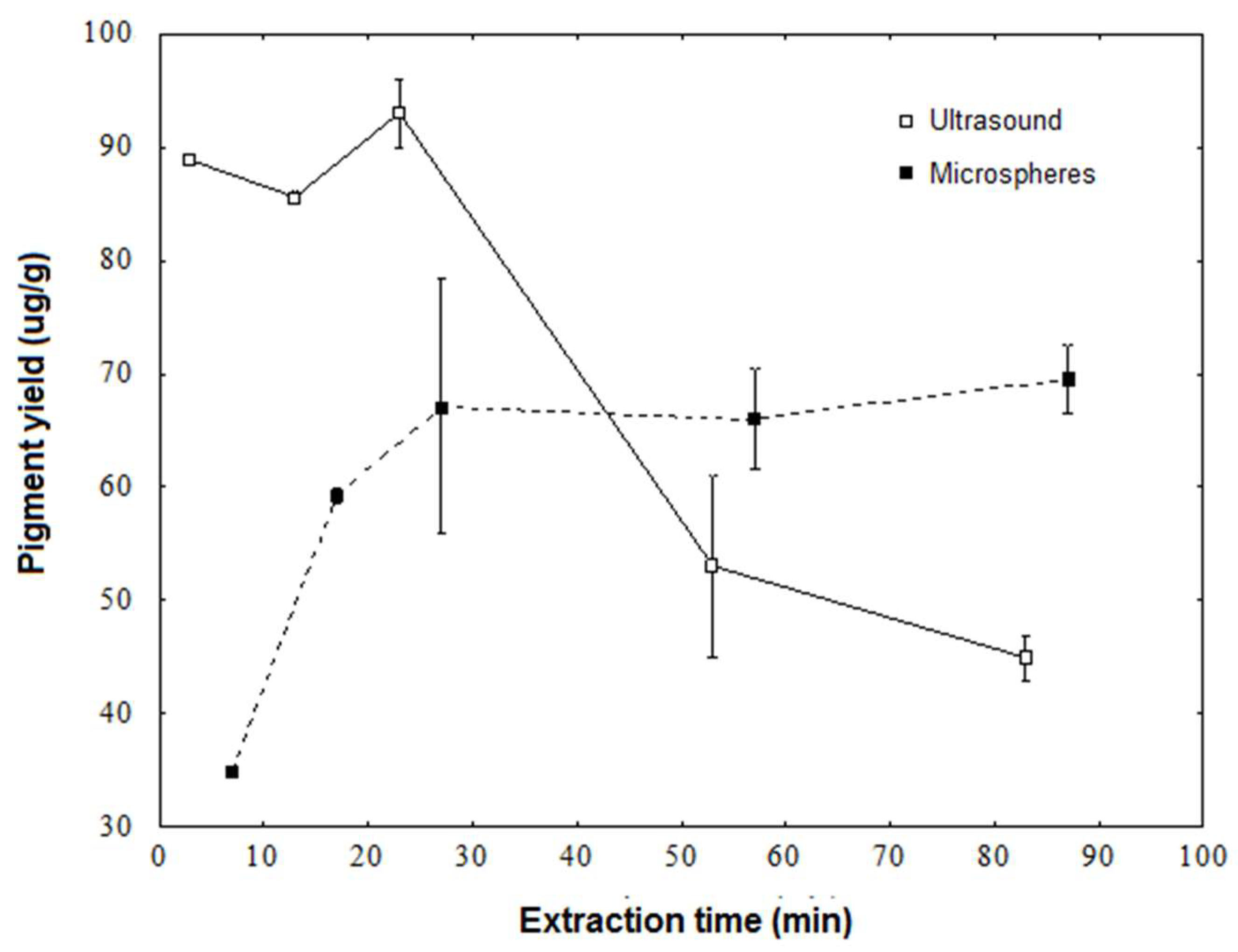
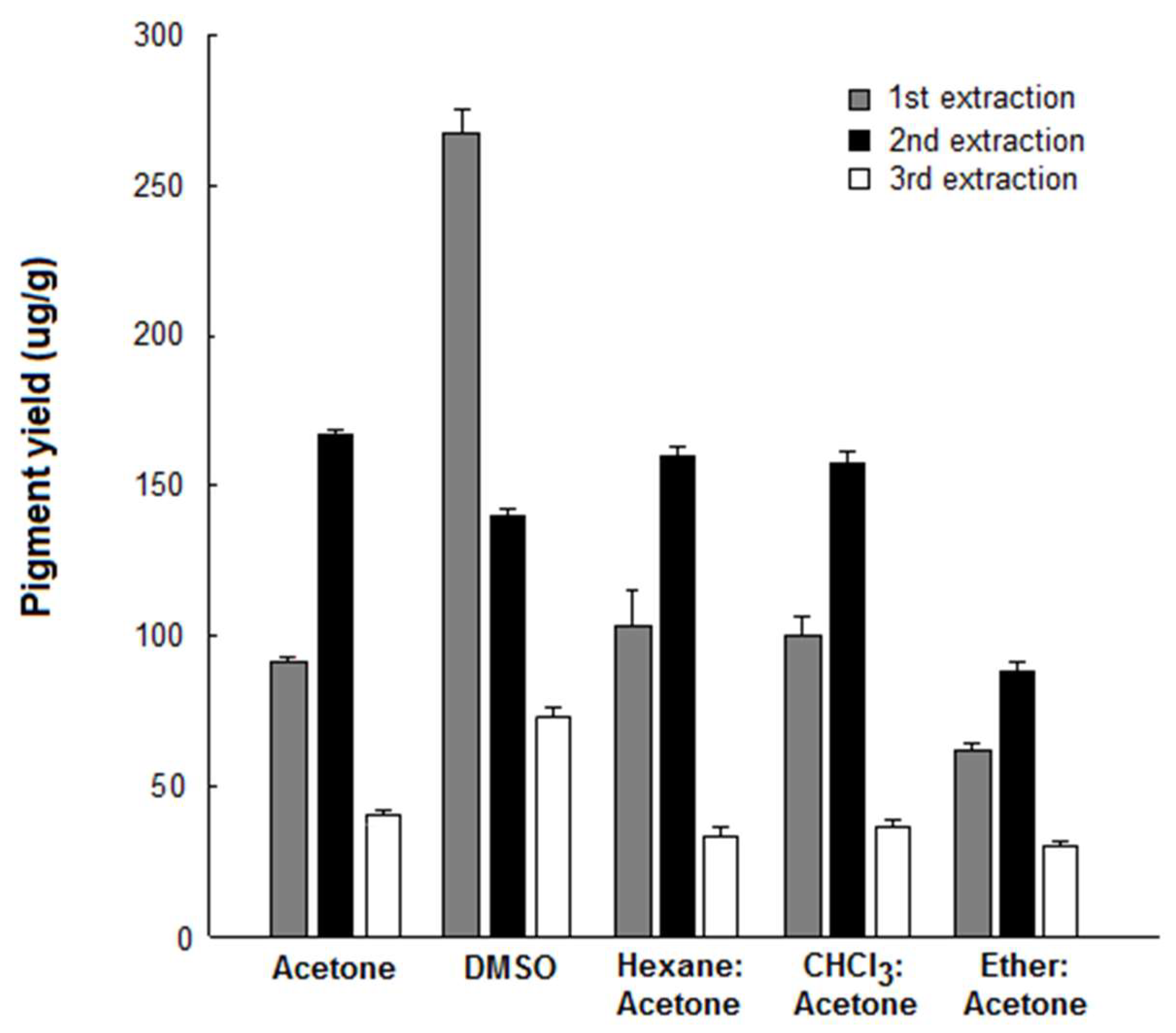
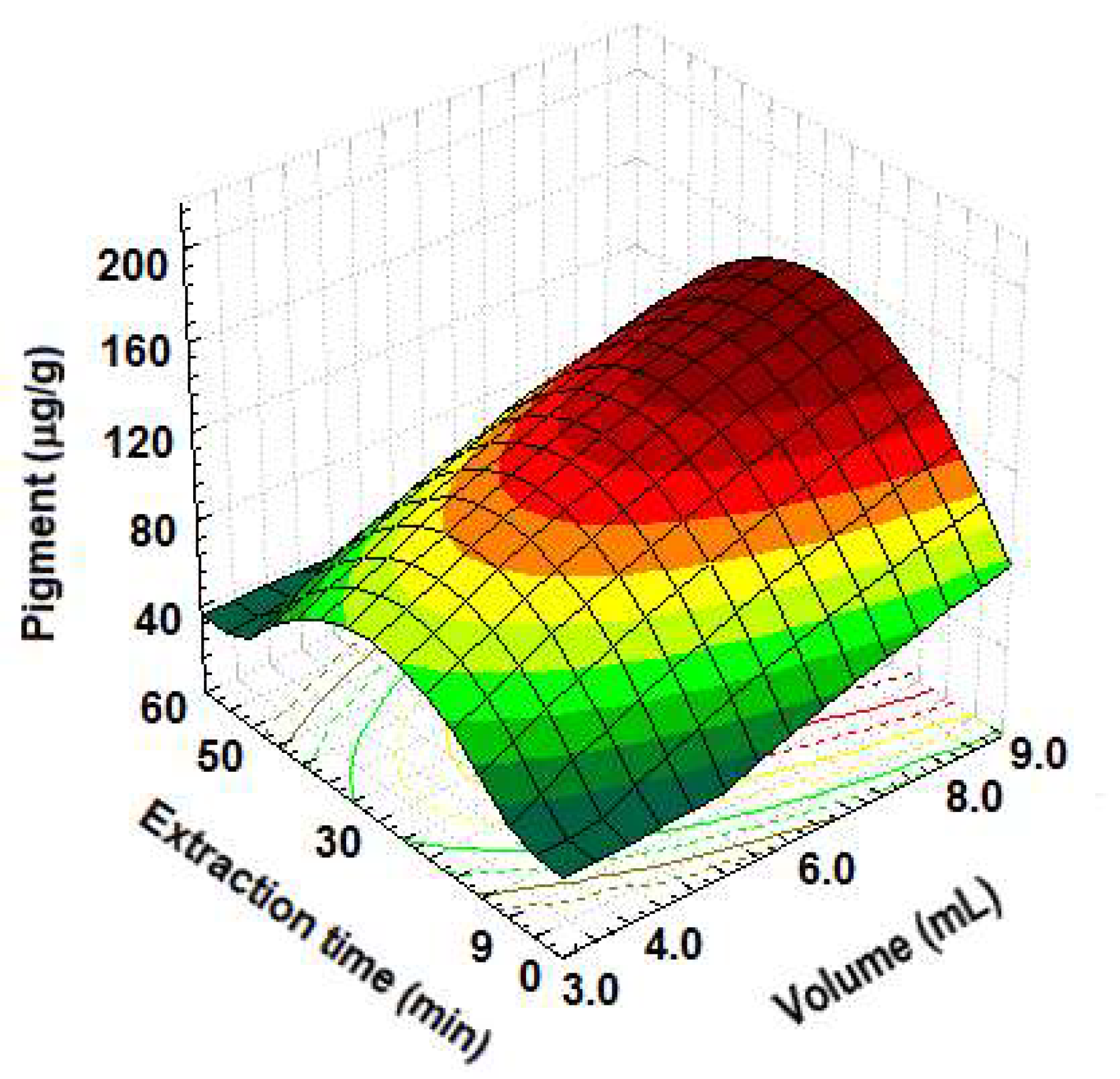
3.2. Pigment Extraction

The production of natural pigments via microbial cultivation has been recognized, but pigment extraction can be limited by elevated costs of some downstream processing. Thus, the development of efficient protocols for the recovery of intracellular pigments from microbial biomass has attracted increased attention [25].
In this study, two extraction methods were initially compared. Through the size reduction of particles (microbial cells) via physical treatments, important gains in extraction efficiency and reduction of extraction time can be reached. The sonication method showed higher yield, when compared with the method using microspheres. The highest yield was reached at 30 min extraction for both methods (Figure 2). Similarly, the maximum amount of pigments from Rhodobacter sphaeroides was obtained using ultrasound [13]. The use of ultrasound may reduce the dependence of a specific solvent and allow for alternative solvents that can be more economical and environmentally attractive, adding safety and health benefits. The efficacy of ultrasound can be attained by the increase in surface mass transfer and the efficacy decrease sharply after the surface solute was totally removed [26,27]. In this study, this effect was also observed after 30 min extraction (Figure 2). Moreover, partial degradation of pigments via extensive ultrasound treatments cannot be ruled out.
The lower yield of pigments obtained via the microspheres method can be associated to the difficulty in disrupting the microbial cell wall, and thus a smaller amount of pigments was released when compared with ultrasound. This result can be associated with the fact that the complex cell envelope of Gram-negative bacteria would require high shearing forces to be broken [28]. Although the intracellular concentration of pigment was not determined, the typical yellow color could be visually observed in the cell debris after the microsphere method, suggesting that the extraction was incomplete. Similarly, depending on the extraction process, the cell debris of Enterobacter sp. PWN1 remained colored, indicating a low extraction efficiency of pigments [29].
The extraction of pigments produced by Chryseobacterium sp. kr6 using organic solvents was then investigated. In a first approach, five solvents, namely, acetone, chloroform, DMSO, ethyl ether and hexane were tested. As the pigments often accumulate in the lipid phase, a variety of organic solvents has been successfully tested to extract pigments from microbial cells [25,29]. Most studies use a single solvent, but the mixture of solvents has not been properly investigated. In this study, it was observed that acetone and their equivolumetric mixtures with hexane and chloroform showed similar results for pigment extraction (Figure 3). Except for DMSO, the highest yield was observed in the second extraction. The mixture of solvents, when used to rupture cells, can present synergic interactions resulting in increased pigment yield in some cases [25]. The solvent DMSO showed a different characteristic in comparison to the other systems, with a higher yield in the first extraction. This fact can be associated with the pigment solubility in the solvent or the efficacy in the rupture of the microbial cell wall in the first extraction step. Based on these results, acetone and DMSO were selected for further evaluation.
To optimize the extraction with the selected solvents, a factorial experiment was carried out, with solvent type, extraction volume, number of extractions and extraction time as variables. Based on the Tukey’s test, the best results were obtained for extractions conducted in the runs 3 and 7, yielding 125 μg/g and 137 μg/g, respectively (Table 1). Such values were not significantly different (p < 0.05). In both experiments, acetone was used as the solvent and the same extraction volume (6 mL) was employed. The extraction condition that presented a lower value for the pigment yield was run 1, which was significantly lower (p < 0.05).
After determining the best method and solvent for extraction, a 22 experimental design was developed to investigate the influence of solvent volume and extraction time on pigment yield. The experimental conditions and the results are shown in Table S1. The highest yield was observed in run 6 (180 μg/g), which was carried out with 9.0 mL and 30 min ultrasound extraction (Table S1). The lowest yields were observed in runs 3 and 7 (about 22 μg/g). The effects of independent variables on the response are presented in Table S2. The results obtained in this experiment suggested that as the extraction time increased, the pigment degradation also increased, and as the solvent volume increased, so did the solubilized pigment quantity. The computed F-value model (15.5) was higher than the F-value in statistic tables at 95% of confidence (Ftab = 4.07), which demonstrates significance for the regression model [15]. Thus, the model is suitable to predict the extraction yield as a function of solvent volume and extraction time.
The effect of solvent volume and extraction time on pigment yield is shown in Figure 4. Solvent volume has a significant linear effect (p < 0.001) and quadratic effect (p = 0.017) on pigment extraction (Table S2). The increase in yield is observed when the solvent volume increases from 3.0 to 9.0 mL, but the pigment extraction is not significantly increased when extraction time is greater than 30 min. In this regard, the dissolution of pigments from Rhodobacter sphaeroides did not reach an equilibrium if the extraction time was lower than 40 min, and part of the pigment remains associated to the microbial biomass, but surpassing 40 min, the yield is slightly lower [13]. This effect could be associated to the long duration of exposure of dissolved pigments to oxygen, light and microbial enzymes, resulting in an increased possibility of pigment degradation [30].

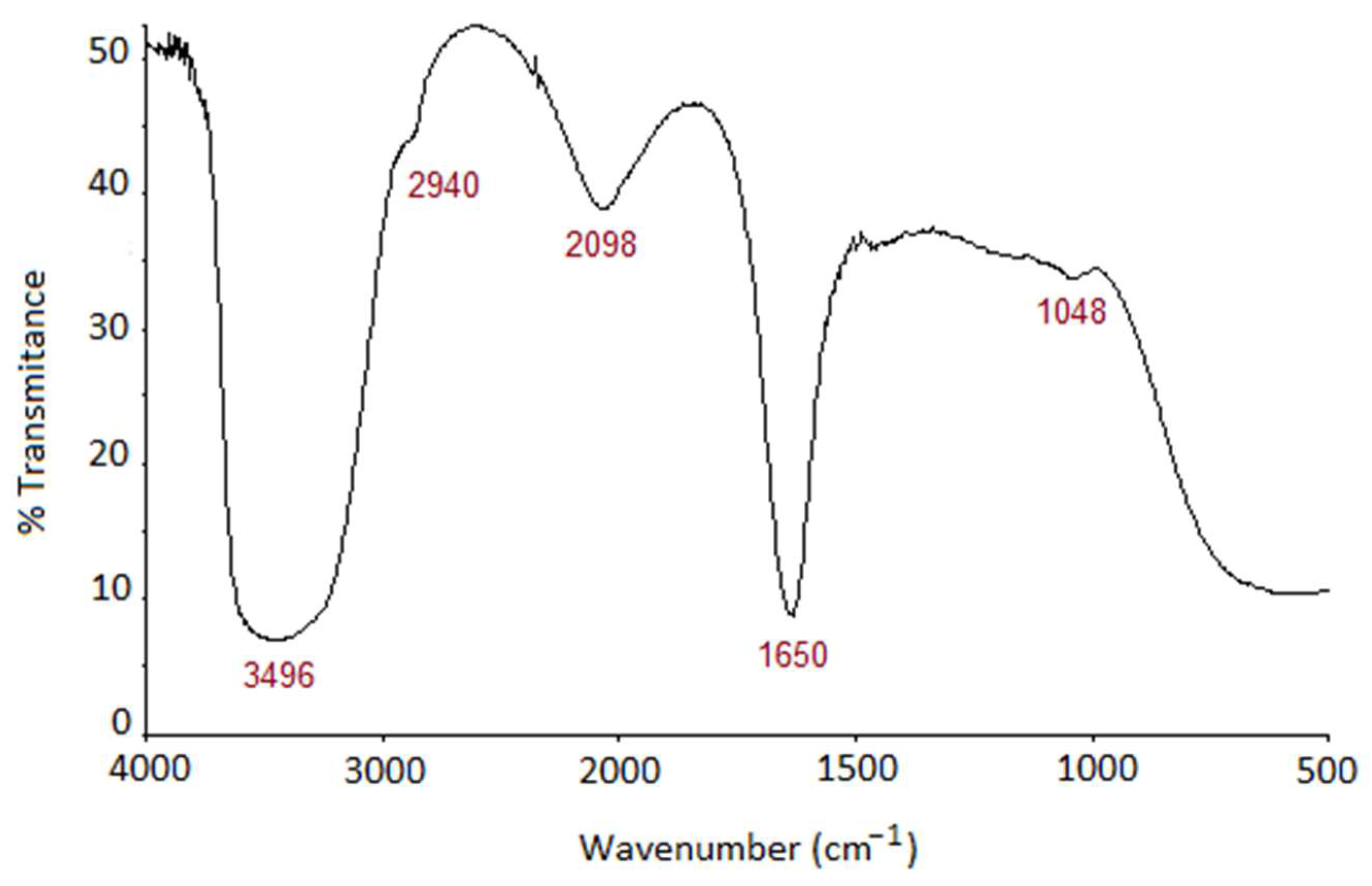
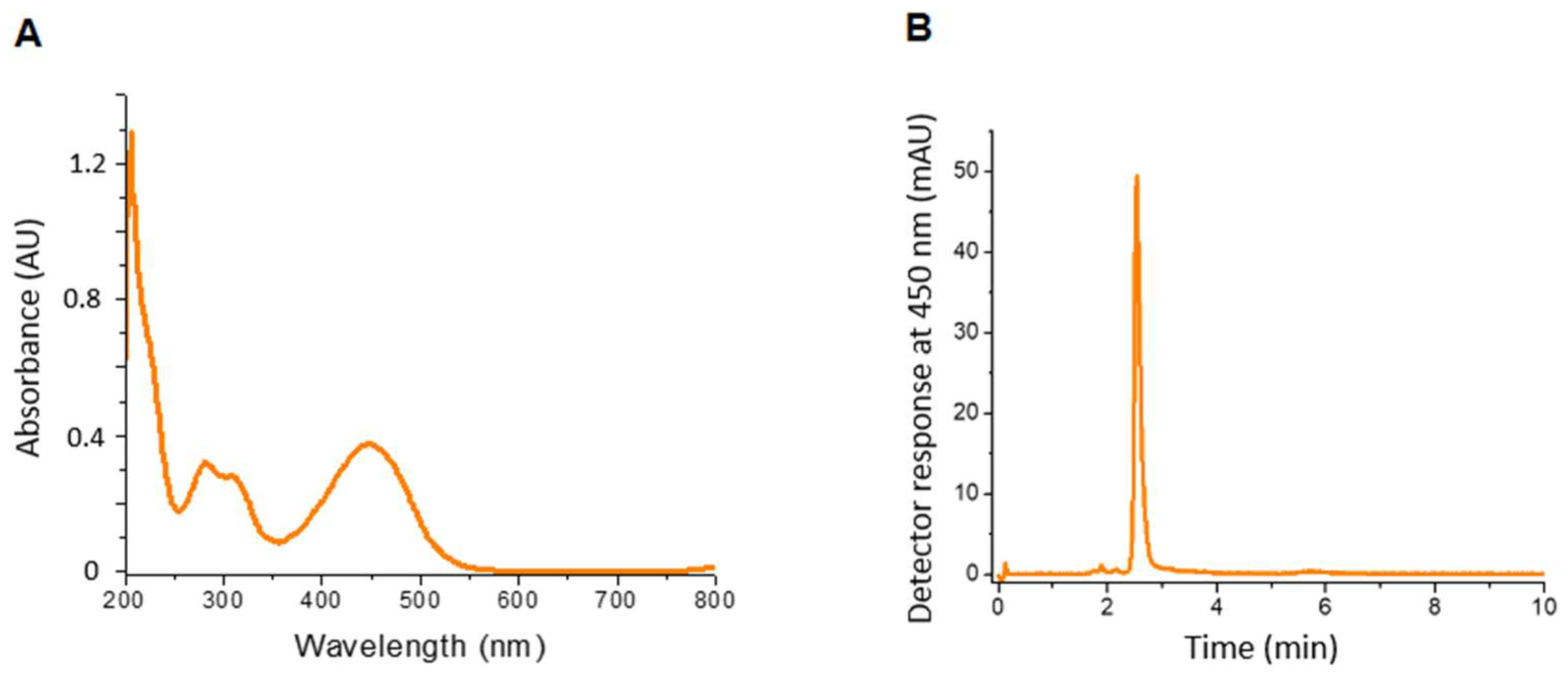
3.3. Characterization of the Pigment Extract

Color analysis of the pigment extract revealed the values of CIELAB parameters L*, a* and b* were 58.3 ± 6.7, 3.9 ± 1.5 and 32.9 ± 3.8, respectively. The two-dimensional plot of chromatic components a* and b* show that the pigment falls in the orange region (Figure S1). Values for Chroma (saturation) and Hue angle were calculated as 33.2 ± 3.9 and 83.2 ± 1.9, respectively. Likewise, previous studies on the colorimetric parameters of flexirubin-type pigments extracted from Chryseobacterium strains also revealed positive b* values and Hue angle values close to 80°, which represent colors in the yellow/orange region [31,32].
The pigment extract obtained from Chryseobacterium sp. kr6 biomass was analyzed with FTIR (Figure 5). The broad band at 3496 cm−1 can be assigned as a characteristic O-H stretching band of hydroxyl groups. The shoulder at 2940 cm−1 usually corresponds to CH and CH2 stretching of aliphatic groups. The combination of bands at 2100 cm−1 and 1650 cm−1 is considered as indicative of conjugated C=C bonds [33]. Carbonyl groups show characteristic sharp peaks in the range 1750–1610 cm−1. The peak at 1650 cm−1 can also be assigned to keto groups, which present typical bands at 1650–1670 cm−1 or lower values in FTIR [34]. The weak band at 1048 cm−1 corresponds to a C-O bond.
The pigment extract was analyzed with scanning spectrophotometry in the range of 200–800 nm. The resulting electromagnetic spectrum is shown in Figure 6A. It was possible to observe the absorbance peaks at the wavelengths 280 nm, 325 nm and 450 nm, the latter corresponding to a λmax value for carotenoid pigments like zeaxanthin, β-cryptoxanthin and β-carotene [34]. However, the absorption spectra of carotenoids have typical profiles between 400 and 500 nm, with λmax at about 450 nm, and two minor bands on each side are usually observed. The exact position of the λmax and companion minor bands are variable according to the pigment and in some cases are sufficiently different for the identification of each carotenoid [35]. In this study, only the maximum absorbance at 450 nm was observed in the extract obtained at 48 h of cultivation. Moreover, the absorbance peaks at 280 nm and 325 nm are typical of aromatic groups and polyenes with more than five C=C alkene groups, respectively, which are typically found in aryl polyene pigments [36].
A single major peak was eluted from the RP-HPLC column when monitored at 450 nm (Figure 6B). Further analysis of the MS spectra revealed a major peak at m/z 619 [M-H]- (Figure S2), analogous to that found for a flexirubin produced by Chryseobacterium sp. isolated from mountain soil [37]. The mass corresponds to a molecular formula C42H5204, suggesting the structure is similar to the known flexirubin of Chitinophaga pinensis [36] and Chryseobacterium artocarpi [38], minus a methyl group.
The Flavobacteriaceae family, which includes the genus Chryseobacterium, is a recognized source of pigments, including rare monocyclic C40 carotenoids, such as the (3R)-saproxanthin and (3R,2′S)-myxol isolated from species of marine origin [39]. The pigment obtained from Chryseobacterium sp. kr6 growing on feather meal showed some similar features of flexirubin pigments described for Chryseobacterium sp. UTM-3T [40] and C. artocarpi CECT 8497 [38]. Although further characterization is necessary to elucidate the exact chemical structure, the pigment extracted from strain kr6 belongs to the flexirubin group.

3.4. Antioxidant Activity

The antioxidant capacity of the pigment was evaluated. In this study, olive oil was used as the substrate in the presence of ferric sulfate as a pro-oxidant. The lipid peroxidation of olive oil was inhibited in the presence of Chryseobacterium pigments, as evaluated by the decrease in the generation of thiobarbituric acid reactive substances (Figure 7A). The differences were significantly different from control (p < 0.05).
Although the TBARS method has been established, other methods are useful to evaluate the antioxidant activity. Scavenging of DPPH radicals is one of the major methods commonly used to determine antioxidant activity. The method is based on the reduction of alcoholic DPPH in the presence of a hydrogen-donating antioxidant resulting in the generation of the nonradical form of DPPH [41]. The pigment extracted from Chryseobacterium biomass also showed antioxidant activity as determined with the DPPH method (Figure 7B). The result was significantly different from control, but higher antioxidant activity was observed for BHA (p < 0.05).
Antioxidant activity of pigments from Chryseobacterium spp. has been described. The yellowish-orange pigments from C. artocarpi were encapsulated via spray-drying using gum arabic or κ-carragenan, and the antioxidant capacity of the resulting powders was confirmed via DPPH assay [40]. Furthermore, a detailed study on the antioxidant activity of this pigment was performed through different assays using Trolox as a standard antioxidant. The pigment showed a similar performance in a TBARS assay, while the DPPH scavenging capability was lower than Trolox [42], in agreement to that observed in this study. Likewise, samples of bacterial cellulose functionalized with flexirubin produced by Chryseobacterium shigense showed antioxidant ability via the ABTS+ assay [31].
Some pigments act as scavengers of singlet oxygen and peroxyl radicals, reducing oxidative damage to DNA and lipids [4]. In this regard, microorganisms can synthesize a large diversity of pigments, which may be highly active against reactive oxygen species [2,6,39]. The scavenging activity is often correlated with the number of double bonds in the molecule. The mechanism by which the aryl polyene pigments like flexirubin protect the biological systems probably depends on the energy transfer from excited oxygen to the molecule, where the energy is dissipated by stretching and vibration of the pigment in the solvent milieu [43]. The antioxidant activity of flexirubins can be attributed to the hydrogen atom transfer from the phenolic hydroxyl group and the long polyene chain, as previously determined through different assays [42]. Moreover, flexirubin pigments produced by Chryseobacterium strains are very stable under UV and sunlight exposure even at high temperatures [32,40], suggesting the importance of these pigments for a broad range of applications. In this regard, polymeric nanofibers incorporating flexirubin showed ABTS radical scavenging activity and the ability to reduce enzymatic browning of apple slices. Thus, the fabricated material has potential for application in active packaging systems [44]. Due to their properties, flexirubins have been proposed as useful dyes in textiles, food and pharma industries [6,36,40].

3.5. Antimicrobial Activity

A preliminary assessment of the antimicrobial activity of pigments extracted from strain kr6 was carried out. The pigment was evaluated against Gram-positive and Gram-negative bacteria via the disk diffusion method. Among the bacteria tested, consistent inhibition was only observed against S. aureus and E. faecalis (Figure S3), although small inhibitory halos were eventually observed against other bacteria such as E. coli.
The antimicrobial activity of yellow/orange pigments extracted from bacteria has been described. Flavobacterium sp. P1 produces a yellow pigment tentatively identified as zeaxanthin. The pigment extract at 5 mg/mL showed a broad range of antibacterial activity via the disk diffusion test, with haloes around 15 mm against S. aureus and E. coli [45]. Chryseobacterium strains present biosynthetic gene clusters responsible for the production of antimicrobials and may produce antibiotics [46]. However, only the flexirubin pigment of C. artocarpi CECT 8497 has been previously associated with antimicrobial activity [47].

4. Conclusions

In this study, Chyseobacterium sp. kr6 was capable of producing a gold–yellow pigment during growth on feather meal. The pigment was easily recovered from bacterial biomass via ultrasound extraction with acetone as the solvent. The pigment belongs to the flexirubin group, which are highly stable pigments with potential applications in food, pharma and textile industries. Antioxidant and antimicrobial activities were observed for the pigment extract, indicating that a highly abundant byproduct from the poultry industry can be used to produce bioactive molecules via microbial cultivation, representing an interesting way to manage this waste material.

Supplementary Materials

The following supporting information can be downloaded at: https://www.mdpi.com/article/10.3390/biomass4020028/s1, Figure S1: Two-dimensional plot for the pigment of Chryseobacterium sp. kr6; Figure S2: Mass spectrum of the pigment extract from Chryseobacterium sp. kr6; Figure S3: Antimicrobial activity of the pigment extract from Chryseobacterium sp. kr6. Table S1: Experimental design and results of 22 factorial design; Table S2: Model coefficients for pigment extraction; Table S3: Analysis of variance (ANOVA) for the regression model.

Author Contributions

Conceptualization, A.B. and S.T.S.; methodology, S.G., S.T.S. and M.E.P.-J.; software, S.T.S.; formal analysis, S.G., S.T.S. and M.E.P.-J.; writing—original draft preparation, S.G. and S.T.S.; writing—review and editing, A.d.O.R. and A.B.; supervision, A.d.O.R. and A.B.; funding acquisition, A.B. All authors have read and agreed to the published version of the manuscript.

Funding

This research was funded by Conselho Nacional de Desenvolvimento Científico e Tecnológico (CNPq, Brazil), grant 306936/2017-8 and the APC was funded by MDPI.

Institutional Review Board Statement

Not applicable.

Informed Consent Statement

Not applicable.

Data Availability Statement

All related data and methods are presented in this paper. Additional inquiries should be addressed to the corresponding author.

Conflicts of Interest

The authors declare no conflicts of interest.

References

  1. Pailliè-Jiménez, M.E.; Stincone, P.; Brandelli, A. Natural pigments of microbial origin. Front. Sustain. Food Syst. 2020, 4, 590439. [Google Scholar] [CrossRef]
  2. Agarwal, H.; Bajpai, S.; Mishra, A.; Kohli, I.; Varma, A.; Fouillaud, M.; Dufossé, L.; Joshi, N.C. Bacterial pigments and their multifaceted roles in contemporary biotechnology and pharmacological applications. Microorganisms 2023, 11, 614. [Google Scholar] [CrossRef] [PubMed]
  3. Orlandi, V.T.; Martegani, E.; Giaroni, C.; Baj, A.; Bolognese, F. Bacterial pigments: A colorful palette reservoir for biotechnological applications. Biotechnol. Appl. Biochem. 2022, 69, 981–1001. [Google Scholar] [CrossRef] [PubMed]
  4. Fiedor, J.; Burda, K. Potential role of carotenoids as antioxidants in human health and disease. Nutrients 2014, 6, 466–488. [Google Scholar] [CrossRef] [PubMed]
  5. Saini, D.K.; Chakdar, H.; Pabbi, S.; Shukla, P. Enhancing production of microalgal biopigments through metabolic and genetic engineering. Crit. Rev. Food Sci. Nutr. 2020, 60, 391–405. [Google Scholar] [CrossRef] [PubMed]
  6. Venil, C.K.; Dufossé, L.; Devi, P.R. Bacterial pigments: Sustainable compounds with market potential for pharma and food industry. Front. Sustain. Food Syst. 2020, 4, 100. [Google Scholar] [CrossRef]
  7. Kim, S.H.; Kim, M.S.; Lee, B.Y.; Lee, P.C. Generation of structurally novel short carotenoids and study of their biological activity. Sci. Rep. 2016, 6, 21987. [Google Scholar] [CrossRef]
  8. Liu, J.; Luo, Y.; Guo, T.; Tang, C.; Chai, X.; Zhao, W.; Bai, J.; Lin, Q. Cost-effective pigment production by Monascus purpureus using rice straw hydrolysate as substrate in submerged fermentation. J. Biosci. Bioeng. 2020, 129, 229–236. [Google Scholar] [CrossRef] [PubMed]
  9. Brandelli, A. Bacterial keratinases: Useful enzymes for bioprocessing agroindustrial wastes and beyond. Food Bioprocess Technol. 2008, 1, 105–116. [Google Scholar] [CrossRef]
  10. Silveira, S.T.; Casarin, F.; Gemelli, S.; Brandelli, A. Thermodynamics and kinetics of heat inactivation of a novel keratinase from Chryseobacterium sp. strain kr6. Appl. Biochem. Biotechnol. 2010, 162, 548–560. [Google Scholar] [CrossRef]
  11. Jiménez, M.E.P.; Pinilla, C.M.B.; Rodrigues, E.; Brandelli, A. Extraction and partial characterisation of antioxidant pigment produced by Chryseobacterium sp. kr6. Nat. Prod. Res. 2019, 33, 1541–1549. [Google Scholar] [CrossRef] [PubMed]
  12. Riffel, A.; Lucas, F.; Heeb, P.; Brandelli, A. Characterization of a new keratinolytic bacterium that completely degrades native feather keratin. Arch. Microbiol. 2003, 179, 258–265. [Google Scholar] [CrossRef] [PubMed]
  13. Gu, Z.; Chen, D.; Han, Y.; Chen, Z.; Gu, F. Optimization of carotenoids extraction from Rhodobacter sphaeroides. LWT Food Sci. Technol. 2008, 41, 1082–1088. [Google Scholar] [CrossRef]
  14. Ngamwonglumlert, L.; Devahastin, S.; Chiewchan, N. Natural colorants: Pigment stability and extraction yield enhancement via utilization of appropriate pretreatment and extraction methods. Crit. Rev. Food Sci. Nutr. 2017, 57, 3243–3259. [Google Scholar] [CrossRef] [PubMed]
  15. Myers, R.H.; Montgomery, R.C. Response Surface Methodology: Process and Product Optimization Using Designed Experiments; Wiley: New York, NY, USA, 2002. [Google Scholar]
  16. Silveira, S.T.; Daroit, D.J.; Brandelli, A. Pigment production by Monascus purpureus in grape waste using factorial design. LWT Food Sci. Technol. 2008, 41, 170–174. [Google Scholar] [CrossRef]
  17. Caivano, J.L.; Buera, M.P. Color in Food: Technological and Psycophysical Aspects; CRC Press: Boca Raton, FL, USA, 2012. [Google Scholar]
  18. Ohkawa, H.; Ohishi, H.; Yagi, K. Assay for lipid peroxide in animal tissues by thiobarbituric acid reaction. Anal. Biochem. 1979, 95, 351–358. [Google Scholar] [CrossRef] [PubMed]
  19. Brand-Williams, W.; Cuveliver, M.E.; Berset, C. Use of free radical method to evaluate antioxidant activity. LWT Food Sci. Technol. 1995, 28, 25–30. [Google Scholar] [CrossRef]
  20. Kimura, H.; Sashihara, T.; Matsusaki, H.; Sonomoto, K.; Ishizaki, A. Novel bacteriocin of Pediococcus sp. ISK-1 isolated from well-aged bed of fermented rice bran. Ann. N. Y. Acad. Sci. 1998, 864, 345–348. [Google Scholar] [CrossRef]
  21. Hagaggi, N.S.; Abdul-Raouf, U.M. Production of bioactive β-carotene by the endophytic bacterium Citricoccus parietis AUCs with multiple in vitro biological potentials. Microb. Cell Fact. 2023, 22, 90. [Google Scholar] [CrossRef]
  22. Tinoi, J.; Rakariyatham, N.; Deming, R.L. Simplex optimization of carotenoid production by Rhodotorula glutinis using hydrolyzed mung bean waste flour as substrate. Process Biochem. 2005, 40, 2551–2557. [Google Scholar] [CrossRef]
  23. Bertsch, A.; Coello, N. A biotechnological process for test and recycling poultry feathers as a feed ingredient. Bioresour. Technol. 2005, 96, 1703–1708. [Google Scholar] [CrossRef] [PubMed]
  24. Aksu, Z.; Eren, A.T. Carotenoid production by the yeast Rhodotorula mucilaginosa: Use of agricultural wastes as carbon source. Process Biochem. 2005, 40, 2985–2991. [Google Scholar] [CrossRef]
  25. Park, P.K.; Kim, E.Y.; Chu, K.H. Chemical disruption of yeast cells for the isolation of carotenoid pigments. Sep. Purif. Technol. 2007, 53, 148–152. [Google Scholar] [CrossRef]
  26. Balachandran, S.; Kentish, E.; Mawson, R.; Ashokkumar, M. Ultrasonic enhancement of the supercritical extraction from ginger. Ultrason. Sonochem. 2006, 13, 471–479. [Google Scholar] [CrossRef] [PubMed]
  27. Zheng, S.; Zhang, G.; Wang, H.; Long, Z.; Wei, T.; Li, Q. Progress in ultrasound-assisted extraction of the value-added products from microorganisms. World J. Microbiol. Biotechnol. 2021, 37, 71. [Google Scholar] [CrossRef] [PubMed]
  28. Harrison, S. Bacterial cell disruption: A key unit operation in the recovery of intracellular products. Biotechnol. Adv. 1991, 9, 217–240. [Google Scholar] [CrossRef] [PubMed]
  29. Poddar, K.; Padhan, B.; Sarkar, D.; Sarkar, A. Purification and optimization of pink pigment produced by newly isolated bacterial strain Enterobacter sp. PWN1. SN Appl. Sci. 2021, 3, 105. [Google Scholar] [CrossRef]
  30. Gang, C.; Zora, D. Carotenoids are degraded by free radicals but do not affect lipid peroxidation in unilamellar liposomes under different oxygen tensions. FEBS Lett. 2001, 505, 151–154. [Google Scholar]
  31. Amorim, L.F.A.; Fangueiro, R.; Gouveia, I.C. Characterization of bioactive colored materials produced from bacterial cellulose and bacterial pigments. Materials 2022, 15, 2069. [Google Scholar] [CrossRef]
  32. Wahidin, M.A.; Aruldass, C.A.; Hamzah, M.A.A.M.; Setu, S.A.; Ahmad, W.A. Flexirubin-type pigment production from Chryseobacterium artocarpi CECT 8497 and its application as natural ink. J. Energy Environ. Sustain. 2017, 3, 62–65. [Google Scholar]
  33. Shutova, V.V.; Tyutyaev, E.V.; Churin, A.A.; Ponomarev, V.Y.; Belyakova, G.A.; Maksimov, G.V. IR and Raman spectroscopy in the study of carotenoids of Caldophora rivularis algae. Biophysics 2016, 61, 601–605. [Google Scholar] [CrossRef]
  34. Butnariu, M. Methods of analysis (extraction, separation, identification and quantification) of carotenoids from natural products. J. Ecosys. Ecograph. 2016, 6, 193. [Google Scholar] [CrossRef]
  35. Ashenafi, E.L.; Nyman, M.C.; Shelly, J.T.; Mattson, N.S. Spectral properties and stability of selected carotenoid and chlorophyll compounds in different solvent systems. Food Chem. Adv. 2023, 2, 100178. [Google Scholar] [CrossRef]
  36. Schöner, T.A.; Fuchs, S.W.; Schönau, C.; Bode, H.B. Initiation of the flexirubin biosynthesis in Chitinophaga pinensis. Microb. Biotechnol. 2014, 7, 232–241. [Google Scholar] [CrossRef]
  37. Park, S.A.; Ahn, S.Y.; Choi, K.Y. Functional microbial pigments isolated from Chryseobacterium and Deinococcus species for bio-paint application. Biotechnol. Bioprocess. Eng. 2020, 25, 394–402. [Google Scholar] [CrossRef]
  38. Aruldass, C.A.; Aziz, A.; Venil, C.K.; Khasim, A.R.; Ahmad, W.A. Utilization of agro-industrial waste for the production of yellowish-orange pigment from Chryseobacterium artocarpi CECT 8497. Int. Biodeter. Biodegrad. 2016, 113, 342–349. [Google Scholar] [CrossRef]
  39. Shindo, K.; Misawa, N. New and rare carotenoids isolated from marine bacteria and their antioxidant activities. Mar. Drugs 2014, 12, 1690–1698. [Google Scholar] [CrossRef]
  40. Venil, C.K.; Khasim, A.R.; Aruldass, C.A.; Ahmad, W.A. Microencapsulation of flexirubin-type pigment by spray drying. Characterization and antioxidant activity. Int. Biodeterior. Biodegrad. 2016, 113, 350–356. [Google Scholar] [CrossRef]
  41. Huang, D.; Ou, B.; Prior, R.L. The chemistry behind antioxidant capacity assays. J. Agric. Food Chem. 2005, 53, 1841–1856. [Google Scholar] [CrossRef]
  42. Mogadem, A.; Almamary, M.A.; Mahat, N.A.; Jemon, K.; Ahmad, W.A.; Ali, I. Antioxidant activity evaluation of flexirubin-type pigment from Chryseobacterium artocarpi CECT 8497 and related docking study. Molecules 2021, 26, 979. [Google Scholar] [CrossRef]
  43. Nimse, S.B.; Pal, D. Free radicals, natural antioxidants, and their action mechanisms. RCS Adv. 2015, 5, 27986–28006. [Google Scholar]
  44. Amorim, L.F.A.; Fangueiro, R.; Gouveia, I.C. Novel functional material incorporating flexirubin in polyvinyl alcohol-kefiran/polycaprolactone nanofibers. J. Appl. Polym. Sci. 2022, 139, e53208. [Google Scholar] [CrossRef]
  45. Patki, J.M.; Singh, S.; Singh, S.; Padmadas, N.; Dasgupta, D. Analysis of the applicative potential of pigments extracted from bacterial isolates of mangrove soil as topical UV protectants. Braz. J. Pharm. Sci. 2021, 57, e19127. [Google Scholar] [CrossRef]
  46. Dahal, R.H.; Chaudhary, D.K.; Kim, D.U.; Pandey, R.P.; Kim, J. Chryseobacterium antibioticum sp. nov. with antimicrobial activity against Gram-negative bacteria, isolated from Arctic soil. J. Antibiot. 2021, 74, 115–123. [Google Scholar] [CrossRef]
  47. Venil, C.K.; Khasim, A.R.; Aruldass, C.A.; Ahmad, W.A. Safety evaluation of flexirubin from Chryseobacterium artocarpi CECT 8497: Acute, subacute toxicity and mutagenicity studies. Process Saf. Environ. Prot. 2017, 112, 362–370. [Google Scholar] [CrossRef]
Figure 1. Growth and pigment production by Chryseobacterium sp. kr6. The strain was grown during 72 h in feather meal broth, and values of viable cell counts (circles), absorbance at 450 nm (squares) and pigment yield (triangles) were monitored every 24 h. Each point corresponds to the mean ± standard deviation of three independent experiments. Inset: Culture of Chryseobacterium sp. kr6 in feather meal broth after 48 h.
Biomass 04 00028 g001
Figure 2. Pigment extraction by using different techniques. Bacterial biomass was subjected to either ultrasound processing (□) or shaking with microspheres (■). Each point corresponds to the mean ± standard deviation of three independent experiments.
Biomass 04 00028 g002
Figure 3. Use of organic solvents to extract pigments from Chryseobacterium sp. kr6 biomass. Values represent the pigment yields obtained after the first, second and third extraction. Bars are the means ± standard deviations of three independent experiments.
Biomass 04 00028 g003
Figure 4. Influence of processing time and solvent volume on the extraction of pigments from Chryseobacterium sp. kr6 biomass.
Biomass 04 00028 g004
Figure 5. FTIR spectrum of the pigment extracted from Chryseobacterium sp. kr6 biomass.
Biomass 04 00028 g005
Figure 6. Electromagnetic spectrum (A) and RP-HPLC elution profile (B) of the pigment extracted from Chryseobacterium sp. kr6 biomass.
Biomass 04 00028 g006
Figure 7. Antioxidant activity of pigment extract from Chryseobacterium sp. kr6. The antioxidant activity was evaluated via thiobarbituric acid reactive substances (TBARS) method (A) and scavenging of DPPH radical method (B). Bars are the means ± standard deviations of three independent experiments. Different letters indicate that values are significantly different at p < 0.05.
Biomass 04 00028 g007
Table 1. Factorial experiment for extraction of pigments from Chryseobacterium sp. kr6 under different conditions.
RunSolventVolume (mL)Extractions (n)Time (min)Yield (μg/g) 1
1Acetone331015.83 d
2DMSO332057.45 c
3Acetone6320125.36 a
4DMSO631090.45 b
5Acetone362070.50 c
6DMSO361061.01 c
7Acetone6620137.29 a
8DMSO6620104.55 b
1 Different letters indicate that values are significantly different at p < 0.05 (Tukey’s test).
Disclaimer/Publisher’s Note: The statements, opinions and data contained in all publications are solely those of the individual author(s) and contributor(s) and not of MDPI and/or the editor(s). MDPI and/or the editor(s) disclaim responsibility for any injury to people or property resulting from any ideas, methods, instructions or products referred to in the content.

