


Neurol. Int. 2025, 17(11), 174; https://doi.org/10.3390/neurolint17110174 - 22 Oct 2025
Abstract
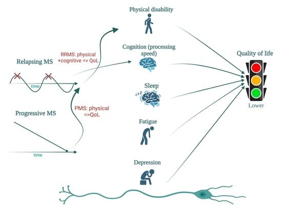
Background: Multiple sclerosis (MS) is a chronic inflammatory disease of the central nervous system, which can lead to physical and cognitive disability, fatigue, depression, and sleep disturbance, all of which may impair quality of life (QoL). While the physical disability is widely known
[...] Read more.
Background: Multiple sclerosis (MS) is a chronic inflammatory disease of the central nervous system, which can lead to physical and cognitive disability, fatigue, depression, and sleep disturbance, all of which may impair quality of life (QoL). While the physical disability is widely known to influence the QoL, the relative contributions of cognitive impairment, fatigue, and sleep quality remain incompletely defined. Objectives: To evaluate the relationship between QoL, physical and cognitive disability, sleep quality, fatigue, and depression in people with MS (PwMS), and to explore phenotype-specific differences between relapsing and progressive forms. Methods: In this monocentric cross-sectional study, 112 PwMS underwent physical assessment (EDSS, MSFC), cognitive testing (SDMT, PASAT, MoCA, MMSE), and QoL evaluation (MSIS-29, EQ-5D, EQ-VAS, MSNQ). A subgroup of 29 patients also completed the Pittsburgh Sleep Quality Index (PSQI), Epworth Sleepiness Scale (ESS), Modified Fatigue Impact Scale (MFIS), and Beck Depression Inventory (BDI). Correlation and group analyses were performed. Results: Progressive MS patients showed greater physical disability (mean EDSS 5.8 vs. 2.6, p < 0.001), poorer cognitive performance, and lower QoL. Across the cohort, QoL strongly correlated with physical disability (EDSS) and cognitive performance (SDMT), with physical measures showing stronger associations. In relapsing MS, physical and cognitive impairment were linked to reduced QoL, whereas in progressive MS, physical disability predominated. In the sleep subgroup, poorer PSQI scores, longer sleep latency, and daytime sleepiness correlated with higher fatigue (MFIS), depressive symptoms (BDI), and reduced QoL (MSIS-29, EQ-5D). Conclusions: QoL in MS reflects the combined burden of physical disability, cognitive impairment, fatigue, depression, and poor sleep quality, with phenotype-specific patterns. While physical disability is the main QoL determinant in progressive MS, cognitive deficits with slowed processing speed play an important role in relapsing MS. Comprehensive, multidimensional assessment, including sleep and mood screening, may support individualized management strategies in MS.
Full article
(This article belongs to the Topic Management of Multiple Sclerosis: Past, Present and Promise)
►
Show Figures
Graphical abstract


