Feature Papers in Biomaterials for Tissue Engineering and Regenerative Medicine

A special issue of Journal of Functional Biomaterials (ISSN 2079-4983). This special issue belongs to the section "Biomaterials for Tissue Engineering and Regenerative Medicine".

Deadline for manuscript submissions: closed (30 November 2022) | Viewed by 20547
Special Issue Editors

Canadian Light Source, University of Saskatchewan, 44 Innovation Boulevard, Saskatoon, SK S7N 2V3, Canada
Interests: biomaterials; synchrotron X-ray imaging; computed tomography; tissue engineering; in vivo imaging
Special Issues, Collections and Topics in MDPI journals

E-Mail Website
Guest Editor
Department of Mechanical Engineering, Cleveland State University, Cleveland, OH 44115, USA
Interests: advanced manufacturing; biomaterials; biomechanics; regenerative medicine; disease modelling
Special Issues, Collections and Topics in MDPI journals
Director of Consumables, Zepto Life Technology, 1000 Westgate Dr, St Paul, MN 55114, USA
Interests: functional biomaterials; advanced biomanufacturing; microfluidic diagnostics

Special Issue Information

Dear Colleagues,

Biomaterials have found wide applications in the emerging field of tissue engineering and regenerative medicine (TERM) for diagnosis, treating, and/or repairing damaged tissue/organs. This Special Issue aims to collect high-quality original and review papers, addressing the most important issues in this field. The topics include, but are not limited to, biomaterial sciences, bioinks for printing, biomaterial processing and bioprinting, microfluid diagnosis, biomaterial-based TERM and cancer therapies, as well as advanced imaging methods/technologies to non-invasively track biomaterial-involved TERM and cancer therapies in living animal models and/or human patients. 

Prof. Dr. Daniel Chen
Dr. Ning Zhu
Dr. Liqun Ning
Dr. Minggan Li
Guest Editors

Manuscript Submission Information

Manuscripts should be submitted online at www.mdpi.com by registering and logging in to this website. Once you are registered, click here to go to the submission form. Manuscripts can be submitted until the deadline. All submissions that pass pre-check are peer-reviewed. Accepted papers will be published continuously in the journal (as soon as accepted) and will be listed together on the special issue website. Research articles, review articles as well as short communications are invited. For planned papers, a title and short abstract (about 100 words) can be sent to the Editorial Office for announcement on this website.

Submitted manuscripts should not have been published previously, nor be under consideration for publication elsewhere (except conference proceedings papers). All manuscripts are thoroughly refereed through a single-blind peer-review process. A guide for authors and other relevant information for submission of manuscripts is available on the Instructions for Authors page. Journal of Functional Biomaterials is an international peer-reviewed open access monthly journal published by MDPI.

Please visit the Instructions for Authors page before submitting a manuscript. The Article Processing Charge (APC) for publication in this open access journal is 2700 CHF (Swiss Francs). Submitted papers should be well formatted and use good English. Authors may use MDPI's English editing service prior to publication or during author revisions.

Keywords

  • biomaterials
  • bioprinting
  • tissue engineering
  • regenerative medicine
  • diagnosis
  • advanced imaging

Published Papers (4 papers)

Order results
Result details
Select all
Export citation of selected articles as:

Research

Jump to: Review

20 pages, 3723 KiB  
Article
Self-Crosslinkable Oxidized Alginate-Carboxymethyl Chitosan Hydrogels as an Injectable Cell Carrier for In Vitro Dental Enamel Regeneration
by Fatemeh Mohabatpour, Zahra Yazdanpanah, Silvana Papagerakis, Xiongbiao Chen and Petros Papagerakis
J. Funct. Biomater. 2022, 13(2), 71; https://doi.org/10.3390/jfb13020071 - 01 Jun 2022
Cited by 13 | Viewed by 3553
Abstract
Injectable hydrogels, as carriers, offer great potential to incorporate cells or growth factors for dental tissue regeneration. Notably, the development of injectable hydrogels with appropriate structures and properties has been a challenging task, leaving much to be desired in terms of cytocompatibility, antibacterial [...] Read more.
Injectable hydrogels, as carriers, offer great potential to incorporate cells or growth factors for dental tissue regeneration. Notably, the development of injectable hydrogels with appropriate structures and properties has been a challenging task, leaving much to be desired in terms of cytocompatibility, antibacterial and self-healing properties, as well as the ability to support dental stem cell functions. This paper presents our study on the development of a novel self-cross-linkable hydrogel composed of oxidized alginate and carboxymethyl chitosan and its characterization as a cell carrier for dental enamel regeneration in vitro. Oxidized alginate was synthesized with 60% theoretical oxidation degree using periodate oxidation and characterized by Fourier Transform Infrared spectroscopy, proton nuclear magnetic resonance spectroscopy, and Ultraviolet-visible absorption spectroscopy. Then, hydrogels were prepared at three varying weight ratios of oxidized alginate to carboxymethyl chitosan (4:1, 3:1, and 2:1) through Schiff base reactions, which was confirmed by Fourier Transform Infrared spectroscopy. The hydrogels were characterized in terms of gelation time, swelling ratio, structure, injectability, self-healing, antibacterial properties, and in vitro characterization for enamel regeneration. The results demonstrated that, among the three hydrogels examined, the one with the highest ratio of oxidized alginate (i.e., 4:1) had the fastest gelation time and the lowest swelling ability, and that all hydrogels were formed with highly porous structures and were able to be injected through a 20-gauge needle without clogging. The injected hydrogels could be rapidly reformed with the self-healing property. The hydrogels also showed antibacterial properties against two cariogenic bacteria: Streptococcus mutans and Streptococcus sobrinus. For in vitro enamel regeneration, a dental epithelial cell line, HAT-7, was examined, demonstrating a high cell viability in the hydrogels during injection. Furthermore, HAT-7 cells encapsulated in the hydrogels showed alkaline phosphatase production and mineral deposition, as well as maintaining their round morphology, after 14 days of in vitro culture. Taken together, this study has provided evidence that the oxidized alginate-carboxymethyl chitosan hydrogels could be used as an injectable cell carrier for dental enamel tissue engineering applications. Full article
Show Figures

Figure 1

Review

Jump to: Research

18 pages, 1291 KiB  
Review
Collagen as a Biomaterial for Skin and Corneal Wound Healing
by Renáta Sklenářová, Naoufal Akla, Meagan Jade Latorre, Jitka Ulrichová and Jana Franková
J. Funct. Biomater. 2022, 13(4), 249; https://doi.org/10.3390/jfb13040249 - 16 Nov 2022
Cited by 21 | Viewed by 5458
Abstract
The cornea and the skin are two organs that form the outer barrier of the human body. When either is injured (e.g., from surgery, physical trauma, or chemical burns), wound healing is initiated to restore integrity. Many cells are activated during wound healing. [...] Read more.
The cornea and the skin are two organs that form the outer barrier of the human body. When either is injured (e.g., from surgery, physical trauma, or chemical burns), wound healing is initiated to restore integrity. Many cells are activated during wound healing. In particular, fibroblasts that are stimulated often transition into repair fibroblasts or myofibroblasts that synthesize extracellular matrix (ECM) components into the wound area. Control of wound ECM deposition is critical, as a disorganized ECM can block restoration of function. One of the most abundant structural proteins in the mammalian ECM is collagen. Collagen type I is the main component in connective tissues. It can be readily obtained and purified, and short analogs have also been developed for tissue engineering applications, including modulating the wound healing response. This review discusses the effect of several current collagen implants on the stimulation of corneal and skin wound healing. These range from collagen sponges and hydrogels to films and membranes. Full article
Show Figures

Graphical abstract

54 pages, 2086 KiB  
Review
Preparation and Use of Decellularized Extracellular Matrix for Tissue Engineering
by Adam D. McInnes, Michael A. J. Moser and Xiongbiao Chen
J. Funct. Biomater. 2022, 13(4), 240; https://doi.org/10.3390/jfb13040240 - 14 Nov 2022
Cited by 28 | Viewed by 6909
Abstract
The multidisciplinary fields of tissue engineering and regenerative medicine have the potential to revolutionize the practise of medicine through the abilities to repair, regenerate, or replace tissues and organs with functional engineered constructs. To this end, tissue engineering combines scaffolding materials with cells [...] Read more.
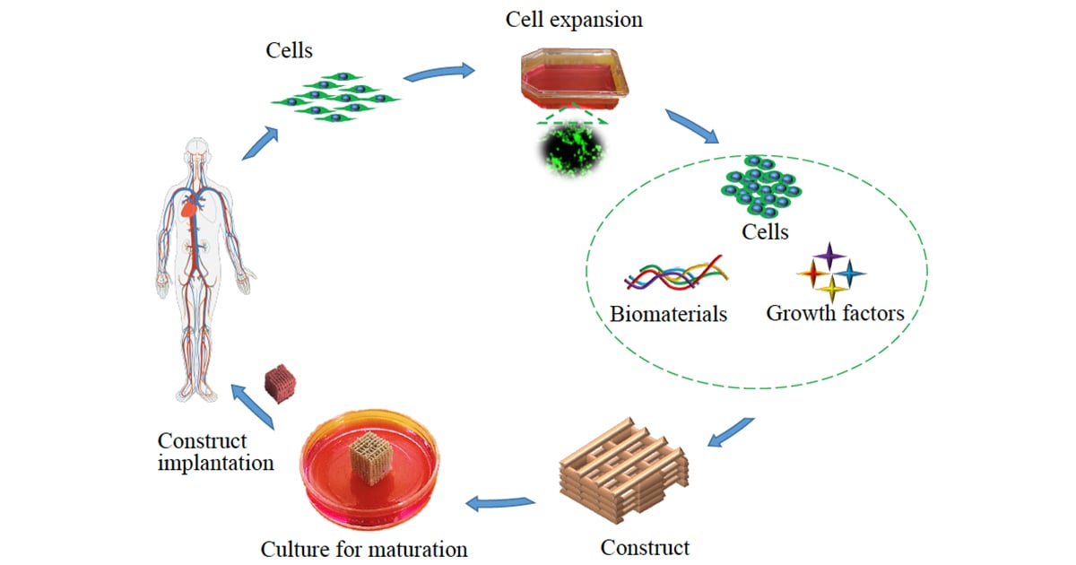
The multidisciplinary fields of tissue engineering and regenerative medicine have the potential to revolutionize the practise of medicine through the abilities to repair, regenerate, or replace tissues and organs with functional engineered constructs. To this end, tissue engineering combines scaffolding materials with cells and biologically active molecules into constructs with the appropriate structures and properties for tissue/organ regeneration, where scaffolding materials and biomolecules are the keys to mimic the native extracellular matrix (ECM). For this, one emerging way is to decellularize the native ECM into the materials suitable for, directly or in combination with other materials, creating functional constructs. Over the past decade, decellularized ECM (or dECM) has greatly facilitated the advance of tissue engineering and regenerative medicine, while being challenged in many ways. This article reviews the recent development of dECM for tissue engineering and regenerative medicine, with a focus on the preparation of dECM along with its influence on cell culture, the modification of dECM for use as a scaffolding material, and the novel techniques and emerging trends in processing dECM into functional constructs. We highlight the success of dECM and constructs in the in vitro, in vivo, and clinical applications and further identify the key issues and challenges involved, along with a discussion of future research directions. Full article
Show Figures

Graphical abstract

20 pages, 2585 KiB  
Review
3D Printing of Bioinert Oxide Ceramics for Medical Applications
by Irene Buj-Corral and Aitor Tejo-Otero
J. Funct. Biomater. 2022, 13(3), 155; https://doi.org/10.3390/jfb13030155 - 17 Sep 2022
Cited by 12 | Viewed by 3717
Abstract
Three-dimensionally printed metals and polymers have been widely used and studied in medical applications, yet ceramics also require attention. Ceramics are versatile materials thanks to their excellent properties including high mechanical properties and hardness, good thermal and chemical behavior, and appropriate, electrical, and [...] Read more.
Three-dimensionally printed metals and polymers have been widely used and studied in medical applications, yet ceramics also require attention. Ceramics are versatile materials thanks to their excellent properties including high mechanical properties and hardness, good thermal and chemical behavior, and appropriate, electrical, and magnetic properties, as well as good biocompatibility. Manufacturing complex ceramic structures employing conventional methods, such as ceramic injection molding, die pressing or machining is extremely challenging. Thus, 3D printing breaks in as an appropriate solution for complex shapes. Amongst the different ceramics, bioinert ceramics appear to be promising because of their physical properties, which, for example, are similar to those of a replaced tissue, with minimal toxic response. In this way, this review focuses on the different medical applications that can be achieved by 3D printing of bioinert ceramics, as well as on the latest advances in the 3D printing of bioinert ceramics. Moreover, an in-depth comparison of the different AM technologies used in ceramics is presented to help choose the appropriate methods depending on the part geometry. Full article
Show Figures

Figure 1

Back to TopTop