Next Issue
Volume 16, November
Previous Issue
Volume 16, September
 
 
ijms-logo

Journal Browser

Journal Browser

Int. J. Mol. Sci., Volume 16, Issue 10 (October 2015) – 145 articles , Pages 23127-25933

Cover Story: 
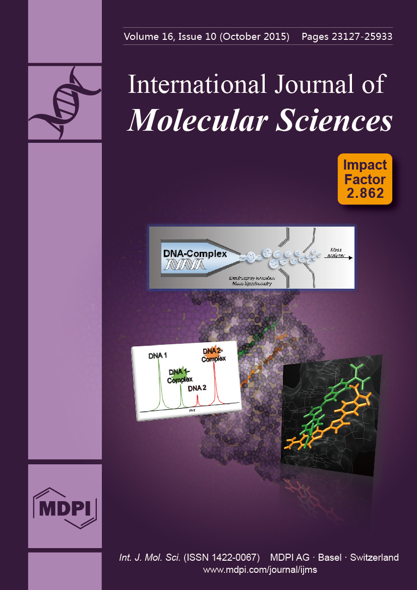
ESI mass spectrometry studies have made enormous contributions with macromolecules and their complexes. Nucleic acids are attractive therapeutic targets for compounds to modulate genetic processes and we have prepared several compound classes to target DNA. Screening for ligand–DNA complexes can prove daunting but a competitive mass spectrometric binding assay can help rapidly identify compounds with DNA sequence specificity. This powerful assay provides stoichiometry, binding mode and relative binding affinity and helps form rationally designed compounds with therapeutic potential. View this article.
  • Issues are regarded as officially published after their release is announced to the table of contents alert mailing list.
  • You may sign up for e-mail alerts to receive table of contents of newly released issues.
  • PDF is the official format for papers published in both, html and pdf forms. To view the papers in pdf format, click on the "PDF Full-text" link, and use the free Adobe Reader to open them.
Order results
Result details
Section
Select all
Export citation of selected articles as:
3515 KiB  
Review
Combined Treatments with Photodynamic Therapy for Non-Melanoma Skin Cancer
by Silvia Rocío Lucena, Nerea Salazar, Tamara Gracia-Cazaña, Alicia Zamarrón, Salvador González, Ángeles Juarranz and Yolanda Gilaberte
Int. J. Mol. Sci. 2015, 16(10), 25912-25933; https://doi.org/10.3390/ijms161025912 - 28 Oct 2015
Cited by 107 | Viewed by 11706
Abstract
Non-melanoma skin cancer (NMSC) is the most common form of cancer in the Caucasian population. Among NMSC types, basal cell carcinoma (BCC) has the highest incidence and squamous cell carcinoma (SCC) is less common although it can metastasize, accounting for the majority of [...] Read more.
Non-melanoma skin cancer (NMSC) is the most common form of cancer in the Caucasian population. Among NMSC types, basal cell carcinoma (BCC) has the highest incidence and squamous cell carcinoma (SCC) is less common although it can metastasize, accounting for the majority of NMSC-related deaths. Treatment options for NMSC include both surgical and non-surgical modalities. Even though surgical approaches are most commonly used to treat these lesions, Photodynamic Therapy (PDT) has the advantage of being a non-invasive option, and capable of field treatment, providing optimum cosmetic outcomes. Numerous clinical research studies have shown the efficacy of PDT for treating pre-malignant and malignant NMSC. However, resistant or recurrent tumors appear and sometimes become more aggressive. In this sense, the enhancement of PDT effectiveness by combining it with other therapeutic modalities has become an interesting field in NMSC research. Depending on the characteristics and the type of tumor, PDT can be applied in combination with immunomodulatory (Imiquimod) and chemotherapeutic (5-fluorouracil, methotrexate, diclofenac, or ingenol mebutate) agents, inhibitors of some molecules implicated in the carcinogenic process (COX2 or MAPK), surgical techniques, or even radiotherapy. These new strategies open the way to a wider improvement of the prevention and eradication of skin cancer. Full article
(This article belongs to the Special Issue Advances in Photodynamic Therapy)
Show Figures

Graphical abstract

1183 KiB  
Article
Identification of Molecular Fingerprints in Human Heat Pain Thresholds by Use of an Interactive Mixture Model R Toolbox (AdaptGauss)
by Alfred Ultsch, Michael C. Thrun, Onno Hansen-Goos and Jörn Lötsch
Int. J. Mol. Sci. 2015, 16(10), 25897-25911; https://doi.org/10.3390/ijms161025897 - 28 Oct 2015
Cited by 39 | Viewed by 8865
Abstract
Biomedical data obtained during cell experiments, laboratory animal research, or human studies often display a complex distribution. Statistical identification of subgroups in research data poses an analytical challenge. Here were introduce an interactive R-based bioinformatics tool, called “AdaptGauss”. It enables a valid identification [...] Read more.
Biomedical data obtained during cell experiments, laboratory animal research, or human studies often display a complex distribution. Statistical identification of subgroups in research data poses an analytical challenge. Here were introduce an interactive R-based bioinformatics tool, called “AdaptGauss”. It enables a valid identification of a biologically-meaningful multimodal structure in the data by fitting a Gaussian mixture model (GMM) to the data. The interface allows a supervised selection of the number of subgroups. This enables the expectation maximization (EM) algorithm to adapt more complex GMM than usually observed with a noninteractive approach. Interactively fitting a GMM to heat pain threshold data acquired from human volunteers revealed a distribution pattern with four Gaussian modes located at temperatures of 32.3, 37.2, 41.4, and 45.4 °C. Noninteractive fitting was unable to identify a meaningful data structure. Obtained results are compatible with known activity temperatures of different TRP ion channels suggesting the mechanistic contribution of different heat sensors to the perception of thermal pain. Thus, sophisticated analysis of the modal structure of biomedical data provides a basis for the mechanistic interpretation of the observations. As it may reflect the involvement of different TRP thermosensory ion channels, the analysis provides a starting point for hypothesis-driven laboratory experiments. Full article
(This article belongs to the Special Issue Molecular and Cellular Mechanisms of Pain)
Show Figures

Graphical abstract

5964 KiB  
Article
Heat Killed Lactobacillus reuteri GMNL-263 Reduces Fibrosis Effects on the Liver and Heart in High Fat Diet-Hamsters via TGF-β Suppression
by Wei-Jen Ting, Wei-Wen Kuo, Dennis Jine-Yuan Hsieh, Yu-Lan Yeh, Cecilia-Hsuan Day, Ya-Hui Chen, Ray-Jade Chen, Viswanadha Vijaya Padma, Yi-Hsing Chen and Chih-Yang Huang
Int. J. Mol. Sci. 2015, 16(10), 25881-25896; https://doi.org/10.3390/ijms161025881 - 28 Oct 2015
Cited by 44 | Viewed by 7390
Abstract
Obesity is one of the major risk factors for nonalcoholic fatty liver disease (NAFLD), and NAFLD is highly associated with an increased risk of cardiovascular disease (CVD). Scholars have suggested that certain probiotics may significantly impact cardiovascular health, particularly certain Lactobacillus species, such [...] Read more.
Obesity is one of the major risk factors for nonalcoholic fatty liver disease (NAFLD), and NAFLD is highly associated with an increased risk of cardiovascular disease (CVD). Scholars have suggested that certain probiotics may significantly impact cardiovascular health, particularly certain Lactobacillus species, such as Lactobacillus reuteri GMNL-263 (Lr263) probiotics, which have been shown to reduce obesity and arteriosclerosis in vivo. In the present study, we examined the potential of heat-killed bacteria to attenuate high fat diet (HFD)-induced hepatic and cardiac damages and the possible underlying mechanism of the positive effects of heat-killed Lr263 oral supplements. Heat-killed Lr263 treatments (625 and 3125 mg/kg-hamster/day) were provided as a daily supplement by oral gavage to HFD-fed hamsters for eight weeks. The results show that heat-killed Lr263 treatments reduce fatty liver syndrome. Moreover, heat-killed Lactobacillus reuteri GMNL-263 supplementation in HFD hamsters also reduced fibrosis in the liver and heart by reducing transforming growth factor β (TGF-β) expression levels. In conclusion, heat-killed Lr263 can reduce lipid metabolic stress in HFD hamsters and decrease the risk of fatty liver and cardiovascular disease. Full article
(This article belongs to the Section Biochemistry)
Show Figures

Graphical abstract

980 KiB  
Review
Aminolevulinic Acid-Based Tumor Detection and Therapy: Molecular Mechanisms and Strategies for Enhancement
by Xue Yang, Pratheeba Palasuberniam, Daniel Kraus and Bin Chen
Int. J. Mol. Sci. 2015, 16(10), 25865-25880; https://doi.org/10.3390/ijms161025865 - 28 Oct 2015
Cited by 127 | Viewed by 8318
Abstract
Aminolevulinic acid (ALA) is the first metabolite in the heme biosynthesis pathway in humans. In addition to the end product heme, this pathway also produces other porphyrin metabolites. Protoporphyrin (PpIX) is one heme precursor porphyrin with good fluorescence and photosensitizing activity. Because tumors [...] Read more.
Aminolevulinic acid (ALA) is the first metabolite in the heme biosynthesis pathway in humans. In addition to the end product heme, this pathway also produces other porphyrin metabolites. Protoporphyrin (PpIX) is one heme precursor porphyrin with good fluorescence and photosensitizing activity. Because tumors and other proliferating cells tend to exhibit a higher level of PpIX than normal cells after ALA incubation, ALA has been used as a prodrug to enable PpIX fluorescence detection and photodynamic therapy (PDT) of lesion tissues. Extensive studies have been carried out in the past twenty years to explore why some tumors exhibit elevated ALA-mediated PpIX and how to enhance PpIX levels to achieve better tumor detection and treatment. Here we would like to summarize previous research in order to stimulate future studies on these important topics. In this review, we focus on summarizing tumor-associated alterations in heme biosynthesis enzymes, mitochondrial functions and porphyrin transporters that contribute to ALA-PpIX increase in tumors. Mechanism-based therapeutic strategies for enhancing ALA-based modalities including iron chelators, differentiation agents and PpIX transporter inhibitors are also discussed. Full article
(This article belongs to the Special Issue Advances in Photodynamic Therapy)
Show Figures

Graphical abstract

2342 KiB  
Review
Site-Specific PEGylation of Therapeutic Proteins
by Jonathan K. Dozier and Mark D. Distefano
Int. J. Mol. Sci. 2015, 16(10), 25831-25864; https://doi.org/10.3390/ijms161025831 - 28 Oct 2015
Cited by 213 | Viewed by 16128
Abstract
The use of proteins as therapeutics has a long history and is becoming ever more common in modern medicine. While the number of protein-based drugs is growing every year, significant problems still remain with their use. Among these problems are rapid degradation and [...] Read more.
The use of proteins as therapeutics has a long history and is becoming ever more common in modern medicine. While the number of protein-based drugs is growing every year, significant problems still remain with their use. Among these problems are rapid degradation and excretion from patients, thus requiring frequent dosing, which in turn increases the chances for an immunological response as well as increasing the cost of therapy. One of the main strategies to alleviate these problems is to link a polyethylene glycol (PEG) group to the protein of interest. This process, called PEGylation, has grown dramatically in recent years resulting in several approved drugs. Installing a single PEG chain at a defined site in a protein is challenging. Recently, there is has been considerable research into various methods for the site-specific PEGylation of proteins. This review seeks to summarize that work and provide background and context for how site-specific PEGylation is performed. After introducing the topic of site-specific PEGylation, recent developments using chemical methods are described. That is followed by a more extensive discussion of bioorthogonal reactions and enzymatic labeling. Full article
(This article belongs to the Special Issue Protein Engineering)
Show Figures

Graphical abstract

1072 KiB  
Article
Interleukins 6 and 15 Levels Are Higher in Subcutaneous Adipose Tissue, but Obesity Is Associated with Their Increased Content in Visceral Fat Depots
by Marta Izabela Jonas, Alina Kurylowicz, Zbigniew Bartoszewicz, Wojciech Lisik, Maurycy Jonas, Zbigniew Wierzbicki, Andrzej Chmura, Piotr Pruszczyk and Monika Puzianowska-Kuznicka
Int. J. Mol. Sci. 2015, 16(10), 25817-25830; https://doi.org/10.3390/ijms161025817 - 28 Oct 2015
Cited by 43 | Viewed by 7536
Abstract
Excess adiposity is associated with chronic inflammation, which takes part in the development of obesity-related complications. The aim of this study was to establish whether subcutaneous (SAT) or visceral (VAT) adipose tissue plays a major role in synthesis of pro-inflammatory cytokines. Concentrations of [...] Read more.
Excess adiposity is associated with chronic inflammation, which takes part in the development of obesity-related complications. The aim of this study was to establish whether subcutaneous (SAT) or visceral (VAT) adipose tissue plays a major role in synthesis of pro-inflammatory cytokines. Concentrations of interleukins (IL): 1β, 6, 8 and 15 were measured at the protein level by an ELISA-based method and on the mRNA level by real-time PCR in VAT and SAT samples obtained from 49 obese (BMI > 40 kg/m2) and 16 normal-weight (BMI 20–24.9 kg/m2) controls. IL-6 and IL-15 protein concentrations were higher in SAT than in VAT for both obese (p = 0.003 and p < 0.0001, respectively) and control individuals (p = 0.004 and p = 0.001, respectively), while for IL-1β this was observed only in obese subjects (p = 0.047). What characterized obese individuals was the higher expression of IL-6 and IL-15 at the protein level in VAT compared to normal-weight controls (p = 0.047 and p = 0.016, respectively). Additionally, obese individuals with metabolic syndrome had higher IL-1β levels in VAT than did obese individuals without this syndrome (p = 0.003). In conclusion, concentrations of some pro-inflammatory cytokines were higher in SAT than in VAT, but it was the increased pro-inflammatory activity of VAT that was associated with obesity and metabolic syndrome. Full article
(This article belongs to the Special Issue Molecular Research on Obesity and Diabetes)
Show Figures

Graphical abstract

1057 KiB  
Review
Hormonal Regulation of Response to Oxidative Stress in Insects—An Update
by Dalibor Kodrík, Andrea Bednářová, Milada Zemanová and Natraj Krishnan
Int. J. Mol. Sci. 2015, 16(10), 25788-25816; https://doi.org/10.3390/ijms161025788 - 27 Oct 2015
Cited by 92 | Viewed by 10129
Abstract
Insects, like other organisms, must deal with a wide variety of potentially challenging environmental factors during the course of their life. An important example of such a challenge is the phenomenon of oxidative stress. This review summarizes the current knowledge on the role [...] Read more.
Insects, like other organisms, must deal with a wide variety of potentially challenging environmental factors during the course of their life. An important example of such a challenge is the phenomenon of oxidative stress. This review summarizes the current knowledge on the role of adipokinetic hormones (AKH) as principal stress responsive hormones in insects involved in activation of anti-oxidative stress response pathways. Emphasis is placed on an analysis of oxidative stress experimentally induced by various stressors and monitored by suitable biomarkers, and on detailed characterization of AKH’s role in the anti-stress reactions. These reactions are characterized by a significant increase of AKH levels in the insect body, and by effective reversal of the markers—disturbed by the stressors—after co-application of the stressor with AKH. A plausible mechanism of AKH action in the anti-oxidative stress response is discussed as well: this probably involves simultaneous employment of both protein kinase C and cyclic adenosine 3′,5′-monophosphate pathways in the presence of extra and intra-cellular Ca2+ stores, with the possible involvement of the FoxO transcription factors. The role of other insect hormones in the anti-oxidative defense reactions is also discussed. Full article
(This article belongs to the Section Biochemistry)
Show Figures

Graphical abstract

1270 KiB  
Article
Rare Titin (TTN) Variants in Diseases Associated with Sudden Cardiac Death
by Oscar Campuzano, Olallo Sanchez-Molero, Irene Mademont-Soler, Helena Riuró, Catarina Allegue, Monica Coll, Alexandra Pérez-Serra, Jesus Mates, Ferran Picó, Anna Iglesias and Ramon Brugada
Int. J. Mol. Sci. 2015, 16(10), 25773-25787; https://doi.org/10.3390/ijms161025773 - 27 Oct 2015
Cited by 17 | Viewed by 8442
Abstract
A leading cause of death in western countries is sudden cardiac death, and can be associated with genetic disease. Next-generation sequencing has allowed thorough analysis of genes associated with this entity, including, most recently, titin. We aimed to identify potentially pathogenic genetic variants [...] Read more.
A leading cause of death in western countries is sudden cardiac death, and can be associated with genetic disease. Next-generation sequencing has allowed thorough analysis of genes associated with this entity, including, most recently, titin. We aimed to identify potentially pathogenic genetic variants in titin. A total of 1126 samples were analyzed using a custom sequencing panel including major genes related to sudden cardiac death. Our cohort was divided into three groups: 432 cases from patients with cardiomyopathies, 130 cases from patients with channelopathies, and 564 post-mortem samples from individuals showing anatomical healthy hearts and non-conclusive causes of death after comprehensive autopsy. None of the patients included had definite pathogenic variants in the genes analyzed by our custom cardio-panel. Retrospective analysis comparing the in-house database and available public databases also was performed. We identified 554 rare variants in titin, 282 of which were novel. Seven were previously reported as pathogenic. Of these 554 variants, 493 were missense variants, 233 of which were novel. Of all variants identified, 399 were unique and 155 were identified at least twice. No definite pathogenic variants were identified in any of genes analyzed. We identified rare, mostly novel, titin variants that seem to play a potentially pathogenic role in sudden cardiac death. Additional studies should be performed to clarify the role of these variants in sudden cardiac death. Full article
(This article belongs to the Special Issue Human Single Nucleotide Polymorphisms and Disease Diagnostics)
Show Figures

Figure 1

2436 KiB  
Article
Identification and Analysis of Regulatory Elements in Porcine Bone Morphogenetic Protein 15 Gene Promoter
by Qianhui Wan, Yaxian Wang and Huayan Wang
Int. J. Mol. Sci. 2015, 16(10), 25759-25772; https://doi.org/10.3390/ijms161025759 - 27 Oct 2015
Cited by 4 | Viewed by 4811
Abstract
Bone morphogenetic protein 15 (BMP15) is secreted by the mammalian oocytes and is indispensable for ovarian follicular development, ovulation, and fertility. To determine the regulation mechanism of BMP15 gene, the regulatory sequence of porcine BMP15 was investigated in this study. The cloned BMP15 [...] Read more.
Bone morphogenetic protein 15 (BMP15) is secreted by the mammalian oocytes and is indispensable for ovarian follicular development, ovulation, and fertility. To determine the regulation mechanism of BMP15 gene, the regulatory sequence of porcine BMP15 was investigated in this study. The cloned BMP15 promoter retains the cell-type specificity, and is activated in cells derived from ovarian tissue. The luciferase assays in combination with a series of deletion of BMP15 promoter sequence show that the −427 to −376 bp region of BMP15 promoter is the primary regulatory element, in which there are a number of transcription factor binding sites, including LIM homeobox 8 (LHX8), newborn ovary homeobox gene (NOBOX), and paired-like homeodomain transcription factor 1 (PITX1). Determination of tissue-specific expression reveals that LHX8, but not PITX1 and NOBOX, is exclusively expressed in pig ovary tissue and is translocated into the cell nuclei. Overexpression of LHX8 in Chinese hamster ovary (CHO) cells could significantly promote BMP15 promoter activation. This study confirms a key regulatory element that is located in the proximal region of BMP15 promoter and is regulated by the LHX8 factor. Full article
(This article belongs to the Section Biochemistry)
Show Figures

Graphical abstract

5751 KiB  
Article
JNK-Bcl-2/Bcl-xL-Bax/Bak Pathway Mediates the Crosstalk between Matrine-Induced Autophagy and Apoptosis via Interplay with Beclin 1
by Jiong Yang and Shukun Yao
Int. J. Mol. Sci. 2015, 16(10), 25744-25758; https://doi.org/10.3390/ijms161025744 - 27 Oct 2015
Cited by 91 | Viewed by 9495
Abstract
Autophagy is associated with drug resistance which has been a threat in chemotherapy of hepatocellular carcinoma (HCC). The interconnected molecular regulators between autophagy and apoptosis serve as switching points critical to the ultimate outcome of the cell. Our study was performed to investigate [...] Read more.
Autophagy is associated with drug resistance which has been a threat in chemotherapy of hepatocellular carcinoma (HCC). The interconnected molecular regulators between autophagy and apoptosis serve as switching points critical to the ultimate outcome of the cell. Our study was performed to investigate the crosstalk between autophagy and apoptosis in HCC after the treatment of matrine. Flow cytometry and TUNEL (terminal dexynucleotidyl transferase (TdT)-mediated dUTP nick end labeling) assay were used to detect apoptosis in vitro and in vivo, respectively. Bax oligomerization and Cytochrome c release assay were performed. Immunoprecipitation and siRNA transfection were used to detect the interplay between Bcl-2/Bcl-xL,Bax, and Beclin 1. Our results showed that: (1) matrine not only activated caspase and PARP (poly ADP-ribose polymerase) cleavage, but also triggered autophagy as shown by the increased levels of LC3II, Beclin 1, and PI3KC3, and the decreased level of p62; (2) matrine treatment promoted the JNK-Bcl-2/ Bcl-xL-Bax/Bak pathway; (3) Bax was oligomerized, the mitochondrial membrane potential altered, and Cytochrome c was released subsequently; (4) Bax interacts with Beclin 1 and inhibits autophagy, which may be a new crosstalk point; and (5) finally, we showed that matrine suppressed the growth of a MHCC97L xenograft in vivo for the first time. In conclusion, the JNK-Bcl-2/Bcl-xL-Bax/Bak pathway mediates the crosstalk between matrine-induced autophagy and apoptosis via interplay with Beclin 1. Full article
(This article belongs to the Section Biochemistry)
Show Figures

Graphical abstract

743 KiB  
Review
Epidemiology of Hepatitis E Virus in European Countries
by Daniele Lapa, Maria Rosaria Capobianchi and Anna Rosa Garbuglia
Int. J. Mol. Sci. 2015, 16(10), 25711-25743; https://doi.org/10.3390/ijms161025711 - 27 Oct 2015
Cited by 94 | Viewed by 8206
Abstract
Over the last decade the seroprevalence of immunoglobulin (IgG) anti hepatitis E virus (HEV) has been increasing in European countries and shows significant variability among different geographical areas. In this review, we describe the serological data concerning the general population and risk groups [...] Read more.
Over the last decade the seroprevalence of immunoglobulin (IgG) anti hepatitis E virus (HEV) has been increasing in European countries and shows significant variability among different geographical areas. In this review, we describe the serological data concerning the general population and risk groups in different European countries. Anti-HEV antibody prevalence ranged from 1.3% (blood donors in Italy) to 52% (blood donors in France). Various studies performed on risk groups in Denmark, Moldova and Sweden revealed that swine farmers have a high seroprevalence of HEV IgG (range 13%–51.1%), confirming that pigs represent an important risk factor in HEV infection in humans. Subtypes 3e,f are the main genotypes detected in the European population. Sporadic cases of autochthonous genotype 4 have been described in Spain, France, and Italy. Although most HEV infections are subclinical, in immune-suppressed and transplant patients they could provoke chronic infection. Fulminant hepatitis has rarely been observed and it was related to genotype 3. Interferon and ribavirin treatment was seen to represent the most promising therapy. Full article
(This article belongs to the Special Issue Viral Hepatitis Research)
2418 KiB  
Article
ClRTL1 Encodes a Chinese Fir RNase III–Like Protein Involved in Regulating Shoot Branching
by Xia Li, Qian Su, Renhua Zheng, Guangxin Liu, Ye Lu, Liming Bian, Jinhui Chen and Jisen Shi
Int. J. Mol. Sci. 2015, 16(10), 25691-25710; https://doi.org/10.3390/ijms161025691 - 26 Oct 2015
Cited by 4 | Viewed by 6217
Abstract
Identification of genes controlling shoot branching is crucial for improving plant architecture and increasing crop yield or biomass. A branching mutant of Chinese fir named “Dugansha” (Cunninghamia lanceolata var. dugan.) has been isolated in our laboratory. We chose the cDNA-AFLP technique [...] Read more.
Identification of genes controlling shoot branching is crucial for improving plant architecture and increasing crop yield or biomass. A branching mutant of Chinese fir named “Dugansha” (Cunninghamia lanceolata var. dugan.) has been isolated in our laboratory. We chose the cDNA-AFLP technique and an effective strategy to screen genes that potentially regulate shoot branching in Chinese fir using this mutant. An RNase III-like1 cDNA fragment named ClRTL1 was identified as a potential positive regulator. To investigate the function of ClRTL1 in regulating shoot branching, we cloned the full-length cDNA sequence from C. lanceolata (Lamb.) Hook, deduced its secondary structure and function, and overexpressed the coding sequence in Arabidopsis. The ClRTL1 cDNA is 1045 bp and comprises an open reading frame of 705 bp. It encodes a protein of 235 amino acids. The deduced secondary structure of the ClRTL1 indicates that it is a mini-RNase III-like protein. The expression analysis and phenotypes of 35S: ClRTL1 in A. thaliana implies that ClRTL1 plays a role in promoting shoot branching in Chinese fir. Full article
(This article belongs to the Special Issue Molecular Research in Plant Secondary Metabolism 2015)
Show Figures

Graphical abstract

2151 KiB  
Article
The Effect of Interferon-γ and Zoledronate Treatment on Alpha-Tricalcium Phosphate/Collagen Sponge-Mediated Bone-Tissue Engineering
by Peiqi Li, Yoshiya Hashimoto, Yoshitomo Honda, Yoshiyuki Arima and Naoyuki Matsumoto
Int. J. Mol. Sci. 2015, 16(10), 25678-25690; https://doi.org/10.3390/ijms161025678 - 26 Oct 2015
Cited by 8 | Viewed by 6141
Abstract
Inflammatory responses are frequently associated with the expression of inflammatory cytokines and severe osteoclastogenesis, which significantly affect the efficacy of biomaterials. Recent findings have suggested that interferon (IFN)-γ and zoledronate (Zol) are effective inhibitors of osteoclastogenesis. However, little is known regarding the utility [...] Read more.
Inflammatory responses are frequently associated with the expression of inflammatory cytokines and severe osteoclastogenesis, which significantly affect the efficacy of biomaterials. Recent findings have suggested that interferon (IFN)-γ and zoledronate (Zol) are effective inhibitors of osteoclastogenesis. However, little is known regarding the utility of IFN-γ and Zol in bone tissue engineering. In this study, we generated rat models by generating critically sized defects in calvarias implanted with an alpha-tricalcium phosphate/collagen sponge (α-TCP/CS). At four weeks post-implantation, the rats were divided into IFN-γ, Zol, and control (no treatment) groups. Compared with the control group, the IFN-γ and Zol groups showed remarkable attenuation of severe osteoclastogenesis, leading to a significant enhancement in bone mass. Histomorphometric data and mRNA expression patterns in IFN-γ and Zol-injected rats reflected high bone-turnover with increased bone formation, a reduction in osteoclast numbers, and tumor necrosis factor-α expression. Our results demonstrated that the administration of IFN-γ and Zol enhanced bone regeneration of α-TCP/CS implants by enhancing bone formation, while hampering excess bone resorption. Full article
(This article belongs to the Special Issue Biomaterials for Tissue Engineering)
Show Figures

Graphical abstract

1856 KiB  
Article
De Novo Sequencing and Analysis of the Safflower Transcriptome to Discover Putative Genes Associated with Safflor Yellow in Carthamus tinctorius L.
by Xiuming Liu, Yuanyuan Dong, Na Yao, Yu Zhang, Nan Wang, Xiyan Cui, Xiaowei Li, Yanfang Wang, Fawei Wang, Jing Yang, Lili Guan, Linna Du, Haiyan Li and Xiaokun Li
Int. J. Mol. Sci. 2015, 16(10), 25657-25677; https://doi.org/10.3390/ijms161025657 - 26 Oct 2015
Cited by 25 | Viewed by 7550
Abstract
Safflower (Carthamus tinctorius L.), an important traditional Chinese medicine, is cultured widely for its pharmacological effects, but little is known regarding the genes related to the metabolic regulation of the safflower’s yellow pigment. To investigate genes related to safflor yellow biosynthesis, 454 [...] Read more.
Safflower (Carthamus tinctorius L.), an important traditional Chinese medicine, is cultured widely for its pharmacological effects, but little is known regarding the genes related to the metabolic regulation of the safflower’s yellow pigment. To investigate genes related to safflor yellow biosynthesis, 454 pyrosequencing of flower RNA at different developmental stages was performed, generating large databases.In this study, we analyzed 454 sequencing data from different flowering stages in safflower. In total, 1,151,324 raw reads and 1,140,594 clean reads were produced, which were assembled into 51,591 unigenes with an average length of 679 bp and a maximum length of 5109 bp. Among the unigenes, 40,139 were in the early group, 39,768 were obtained from the full group and 28,316 were detected in both samples. With the threshold of “log2 ratio ≥ 1”, there were 34,464 differentially expressed genes, of which 18,043 were up-regulated and 16,421 were down-regulated in the early flower library. Based on the annotations of the unigenes, 281 pathways were predicted. We selected 12 putative genes and analyzed their expression levels using quantitative real time-PCR. The results were consistent with the 454 sequencing results. In addition, the expression of chalcone synthase, chalcone isomerase and anthocyanidin synthase, which are involved in safflor yellow biosynthesis and safflower yellow pigment (SYP) content, were analyzed in different flowering periods, indicating that their expression levels were related to SYP synthesis. Moreover, to further confirm the results of the 454 pyrosequencing, full-length cDNA of chalcone isomerase (CHI) and anthocyanidin synthase (ANS) were cloned from safflower petal by RACE (Rapid-amplification of cDNA ends) method according to fragment of the transcriptome. Full article
(This article belongs to the Special Issue Plant Molecular Biology)
Show Figures

Figure 1

790 KiB  
Article
Theoretical Mechanistic and Kinetic Studies on Homogeneous Gas-Phase Formation of Polychlorinated Naphthalene from 2-Chlorophenol as Forerunner
by Fei Xu, Ruiming Zhang, Yunfeng Li, Qingzhu Zhang and Wenxing Wang
Int. J. Mol. Sci. 2015, 16(10), 25641-25656; https://doi.org/10.3390/ijms161025641 - 26 Oct 2015
Cited by 8 | Viewed by 5581
Abstract
Polychlorinated naphthalenes (PCNs) are dioxins-like compounds and are formed along with polychlorinated dibenzo-p-dioxins (PCDDs) and polychlorinated dibenzofurans (PCDFs) in thermal and combustion procedures. Chlorophenols (CPs) are the most important forerunners of PCNs. A comprehensive comprehension of PCN formation procedure from CPs [...] Read more.
Polychlorinated naphthalenes (PCNs) are dioxins-like compounds and are formed along with polychlorinated dibenzo-p-dioxins (PCDDs) and polychlorinated dibenzofurans (PCDFs) in thermal and combustion procedures. Chlorophenols (CPs) are the most important forerunners of PCNs. A comprehensive comprehension of PCN formation procedure from CPs is a precondition for reducing the discharge of PCNs. Experiments on the formation of PCNs from CPs have been hindered by PCN toxicity and short of precise detection methods for active intermediate radicals. In this work, PCN formation mechanism in gas-phase condition from 2-chlorophenol (2-CP) as forerunner was studied by quantum chemistry calculations. Numbers of energetically advantaged formation routes were proposed. The rate constants of key elementary steps were calculated over 600–1200 K using canonical variational transition-state theory (CVT) with small curvature tunneling contribution (SCT) method. This study illustrates formation of PCNs with one chlorine atom loss from 2-CP is preferred over that without chlorine atom loss. In comparison with formation of PCDFs from 2-CP, PCN products are less chlorinated and have lower formation potential. Full article
(This article belongs to the Section Physical Chemistry, Theoretical and Computational Chemistry)
Show Figures

Graphical abstract

1002 KiB  
Review
Perturbation of Brain Oscillations after Ischemic Stroke: A Potential Biomarker for Post-Stroke Function and Therapy
by Gratianne Rabiller, Ji-Wei He, Yasuo Nishijima, Aaron Wong and Jialing Liu
Int. J. Mol. Sci. 2015, 16(10), 25605-25640; https://doi.org/10.3390/ijms161025605 - 26 Oct 2015
Cited by 74 | Viewed by 8976
Abstract
Brain waves resonate from the generators of electrical current and propagate across brain regions with oscillation frequencies ranging from 0.05 to 500 Hz. The commonly observed oscillatory waves recorded by an electroencephalogram (EEG) in normal adult humans can be grouped into five main [...] Read more.
Brain waves resonate from the generators of electrical current and propagate across brain regions with oscillation frequencies ranging from 0.05 to 500 Hz. The commonly observed oscillatory waves recorded by an electroencephalogram (EEG) in normal adult humans can be grouped into five main categories according to the frequency and amplitude, namely δ (1–4 Hz, 20–200 μV), θ (4–8 Hz, 10 μV), α (8–12 Hz, 20–200 μV), β (12–30 Hz, 5–10 μV), and γ (30–80 Hz, low amplitude). Emerging evidence from experimental and human studies suggests that groups of function and behavior seem to be specifically associated with the presence of each oscillation band, although the complex relationship between oscillation frequency and function, as well as the interaction between brain oscillations, are far from clear. Changes of brain oscillation patterns have long been implicated in the diseases of the central nervous system including ischemic stroke, in which the reduction of cerebral blood flow as well as the progression of tissue damage have direct spatiotemporal effects on the power of several oscillatory bands and their interactions. This review summarizes the current knowledge in behavior and function associated with each brain oscillation, and also in the specific changes in brain electrical activities that correspond to the molecular events and functional alterations observed after experimental and human stroke. We provide the basis of the generations of brain oscillations and potential cellular and molecular mechanisms underlying stroke-induced perturbation. We will also discuss the implications of using brain oscillation patterns as biomarkers for the prediction of stroke outcome and therapeutic efficacy. Full article
(This article belongs to the Special Issue Neurological Injuries’ Monitoring, Tracking and Treatment)
Show Figures

Figure 1

2824 KiB  
Review
The Role of Plant–Microbe Interactions and Their Exploitation for Phytoremediation of Air Pollutants
by Nele Weyens, Sofie Thijs, Robert Popek, Nele Witters, Arkadiusz Przybysz, Jordan Espenshade, Helena Gawronska, Jaco Vangronsveld and Stanislaw W. Gawronski
Int. J. Mol. Sci. 2015, 16(10), 25576-25604; https://doi.org/10.3390/ijms161025576 - 26 Oct 2015
Cited by 116 | Viewed by 22212
Abstract
Since air pollution has been linked to a plethora of human health problems, strategies to improve air quality are indispensable. Despite the complexity in composition of air pollution, phytoremediation was shown to be effective in cleaning air. Plants are known to scavenge significant [...] Read more.
Since air pollution has been linked to a plethora of human health problems, strategies to improve air quality are indispensable. Despite the complexity in composition of air pollution, phytoremediation was shown to be effective in cleaning air. Plants are known to scavenge significant amounts of air pollutants on their aboveground plant parts. Leaf fall and runoff lead to transfer of (part of) the adsorbed pollutants to the soil and rhizosphere below. After uptake in the roots and leaves, plants can metabolize, sequestrate and/or excrete air pollutants. In addition, plant-associated microorganisms play an important role by degrading, detoxifying or sequestrating the pollutants and by promoting plant growth. In this review, an overview of the available knowledge about the role and potential of plant–microbe interactions to improve indoor and outdoor air quality is provided. Most importantly, common air pollutants (particulate matter, volatile organic compounds and inorganic air pollutants) and their toxicity are described. For each of these pollutant types, a concise overview of the specific contributions of the plant and its microbiome is presented. To conclude, the state of the art and its related future challenges are presented. Full article
(This article belongs to the Special Issue Plant Microbe Interaction)
Show Figures

Graphical abstract

1607 KiB  
Article
Novel Electrokinetic Microfluidic Detector for Evaluating Effectiveness of Microalgae Disinfection in Ship Ballast Water
by Myint Myint Maw, Junsheng Wang, Fabo Li, Jinhu Jiang, Younan Song and Xinxiang Pan
Int. J. Mol. Sci. 2015, 16(10), 25560-25575; https://doi.org/10.3390/ijms161025560 - 26 Oct 2015
Cited by 15 | Viewed by 5430
Abstract
Ship ballast water treatment methods face many technical challenges. The effectiveness of every treatment method usually is evaluated by using large scale equipment and a large volume of samples, which involves time-consuming, laborious, and complex operations. This paper reports the development of a [...] Read more.
Ship ballast water treatment methods face many technical challenges. The effectiveness of every treatment method usually is evaluated by using large scale equipment and a large volume of samples, which involves time-consuming, laborious, and complex operations. This paper reports the development of a novel, simple and fast platform of methodology in evaluating the efficiency and the best parameters for ballast water treatment systems, particularly in chemical disinfection. In this study, a microfluidic chip with six sample wells and a waste well was designed, where sample transportation was controlled by electrokinetic flow. The performance of this microfluidic platform was evaluated by detecting the disinfection of Dunaliella salina (D. salina) algae in ballast water treated by sodium hypochlorite (NaClO) solution. Light-induced chlorophyll fluorescence (LICF) intensity was used to determine the viability of microalgae cells in the system, which can be operated automatically with the dimension of the detector as small as 50 mm × 24 mm × 5 mm. The 40 µL volume of sample solution was used for each treatment condition test and the validity of detection can be accomplished within about five min. The results show that the viability of microalgae cells under different treatment conditions can be determined accurately and further optimal treatment conditions including concentrations of NaClO and treatment time can also be obtained. These results can provide accurate evaluation and optimal parameters for ballast water treatment methods. Full article
(This article belongs to the Special Issue Microalgal Biotechnology)
Show Figures

Graphical abstract

671 KiB  
Article
Non-Alcoholic Steatohepatitis (NASH): Risk Factors in Morbidly Obese Patients
by Alexandre Losekann, Antonio C. Weston, Angelo A. De Mattos, Cristiane V. Tovo, Luis A. De Carli, Marilia B. Espindola, Sergio R. Pioner and Gabriela P. Coral
Int. J. Mol. Sci. 2015, 16(10), 25552-25559; https://doi.org/10.3390/ijms161025552 - 23 Oct 2015
Cited by 25 | Viewed by 5990
Abstract
The aim was to investigate the prevalence of non-alcoholic steatohepatitis (NASH) and risk factors for hepatic fibrosis in morbidly obese patients submitted to bariatric surgery. This retrospective study recruited all patients submitted to bariatric surgery from January 2007 to December 2012 at a [...] Read more.
The aim was to investigate the prevalence of non-alcoholic steatohepatitis (NASH) and risk factors for hepatic fibrosis in morbidly obese patients submitted to bariatric surgery. This retrospective study recruited all patients submitted to bariatric surgery from January 2007 to December 2012 at a reference attendance center of Southern Brazil. Clinical and biochemical data were studied as a function of the histological findings of liver biopsies done during the surgery. Steatosis was present in 226 (90.4%) and NASH in 176 (70.4%) cases. The diagnosis of cirrhosis was established in four cases (1.6%) and fibrosis in 108 (43.2%). Risk factors associated with NASH at multivariate analysis were alanine aminotransferase (ALT) >1.5 times the upper limit of normal (ULN); glucose ≥ 126 mg/dL and triglycerides ≥ 150 mg/dL. All patients with ALT ≥1.5 times the ULN had NASH. When the presence of fibrosis was analyzed, ALT > 1.5 times the ULN and triglycerides ≥ 150 mg/dL were risk factors, furthermore, there was an increase of 1% in the prevalence of fibrosis for each year of age increase. Not only steatosis, but NASH is a frequent finding in MO patients. In the present study, ALT ≥ 1.5 times the ULN identifies all patients with NASH, this finding needs to be further validated in other studies. Moreover, the presence of fibrosis was associated with ALT, triglycerides and age, identifying a subset of patients with more severe disease. Full article
(This article belongs to the Special Issue Non-Alcoholic Fatty Liver Disease Research 2016)
1106 KiB  
Review
Cell Death Conversion under Hypoxic Condition in Tumor Development and Therapy
by Yu Qiu, Peng Li and Chunyan Ji
Int. J. Mol. Sci. 2015, 16(10), 25536-25551; https://doi.org/10.3390/ijms161025536 - 23 Oct 2015
Cited by 33 | Viewed by 9145
Abstract
Hypoxia, which is common during tumor progression, plays important roles in tumor biology. Failure in cell death in response to hypoxia contributes to progression and metastasis of tumors. On the one hand, the metabolic and oxidative stress following hypoxia could lead to cell [...] Read more.
Hypoxia, which is common during tumor progression, plays important roles in tumor biology. Failure in cell death in response to hypoxia contributes to progression and metastasis of tumors. On the one hand, the metabolic and oxidative stress following hypoxia could lead to cell death by triggering signal cascades, like LKB1/AMPK, PI3K/AKT/mTOR, and altering the levels of effective components, such as the Bcl-2 family, Atg and p62. On the other hand, hypoxia-induced autophagy can serve as a mechanism to turn over nutrients, so as to mitigate the adverse condition and then avoid cell death potentially. Due to the effective role of hypoxia, this review focuses on the crosstalk in cell death under hypoxia in tumor progression. Additionally, the illumination of cell death in hypoxia could shed light on the clinical applications of cell death targeted therapy. Full article
(This article belongs to the Section Biochemistry)
Show Figures

Figure 1

6512 KiB  
Article
Molecular Cloning and Characterization of DXS and DXR Genes in the Terpenoid Biosynthetic Pathway of Tripterygium wilfordii
by Yuru Tong, Ping Su, Yujun Zhao, Meng Zhang, Xiujuan Wang, Yujia Liu, Xianan Zhang, Wei Gao and Luqi Huang
Int. J. Mol. Sci. 2015, 16(10), 25516-25535; https://doi.org/10.3390/ijms161025516 - 23 Oct 2015
Cited by 57 | Viewed by 7840
Abstract
1-Deoxy-d-xylulose-5-phosphate synthase (DXS) and 1-deoxy-d-xylulose-5-phosphate reductoisomerase (DXR) genes are the key enzyme genes of terpenoid biosynthesis but still unknown in Tripterygium wilfordii Hook. f. Here, three full-length cDNA encoding DXS1, DXS2 and DXR were cloned from suspension cells of T. wilfordii with ORF [...] Read more.
1-Deoxy-d-xylulose-5-phosphate synthase (DXS) and 1-deoxy-d-xylulose-5-phosphate reductoisomerase (DXR) genes are the key enzyme genes of terpenoid biosynthesis but still unknown in Tripterygium wilfordii Hook. f. Here, three full-length cDNA encoding DXS1, DXS2 and DXR were cloned from suspension cells of T. wilfordii with ORF sizes of 2154 bp (TwDXS1, GenBank accession no.KM879187), 2148 bp (TwDXS2, GenBank accession no.KM879186), 1410 bp (TwDXR, GenBank accession no.KM879185). And, the TwDXS1, TwDXS2 and TwDXR were characterized by color complementation in lycopene accumulating strains of Escherichia coli, which indicated that they encoded functional proteins and promoted lycopene pathway flux. TwDXS1 and TwDXS2 are constitutively expressed in the roots, stems and leaves and the expression level showed an order of roots > stems > leaves. After the suspension cells were induced by methyl jasmonate, the mRNA expression level of TwDXS1, TwDXS2, and TwDXR increased, and triptophenolide was rapidly accumulated to 149.52 µg·g−1, a 5.88-fold increase compared with the control. So the TwDXS1, TwDXS2, and TwDXR could be important genes involved in terpenoid biosynthesis in Tripterygium wilfordii Hook. f. Full article
(This article belongs to the Special Issue Molecular Research in Plant Secondary Metabolism 2015)
Show Figures

Figure 1

1268 KiB  
Article
The Fab Fragment of a Humanized Anti-Toll Like Receptor 4 (TLR4) Monoclonal Antibody Reduces the Lipopolysaccharide Response via TLR4 in Mouse Macrophage
by Binggang Cai, Maorong Wang, Xuhui Zhu, Jing Xu, Wenkai Zheng, Yiqing Zhang, Feng Zheng, Zhenqing Feng and Jin Zhu
Int. J. Mol. Sci. 2015, 16(10), 25502-25515; https://doi.org/10.3390/ijms161025502 - 23 Oct 2015
Cited by 17 | Viewed by 6605
Abstract
Lipopolysaccharides (LPS) can induce acute inflammation, sepsis, or chronic inflammatory disorders through the Toll receptor 4 (TLR4) signaling pathway. The TLR4/MD2 (myeloid differentiation protein 2) complex plays a major role in the immune response to LPS. However, there is not a good method [...] Read more.
Lipopolysaccharides (LPS) can induce acute inflammation, sepsis, or chronic inflammatory disorders through the Toll receptor 4 (TLR4) signaling pathway. The TLR4/MD2 (myeloid differentiation protein 2) complex plays a major role in the immune response to LPS. However, there is not a good method to suppress the immune response induced by LPS via this complex in macrophages. In this article, we aimed to evaluate the effects of humanized anti-TLR4 monoclonal antibodies on LPS-induced responses in mouse macrophages. The peritoneal macrophages of mice were incubated with anti-TLR4 monoclonal antibodies and stimulated with LPS. The expression levels of cytokines were analyzed by quantitative polymerase chain reaction and enzyme-linked immunosorbent assays. Additionally, activation of various signaling pathways was evaluated by Western blotting. The results showed that the humanized anti-TLR4 monoclonal antibody blocked the inflammatory cytokines expression at both the mRNA and protein level. We also found that the Fab fragment significantly inhibited the nuclear factor kappaB signaling pathway by reducing the phosphorylation of the inhibitor of kappaBalpha and decreasing the translocation of p65, resulting in the suppression of p38, extracellular signal-regulated kinase 1/2, c-Jun N-terminal kinase 1/2, and IFN-β regulatory factor 3 phosphorylation. Therefore, our study showed that this humanized anti-TLR4 monoclonal antibody could effectively protect against LPS-induced responses by blocking the TLR4 signaling pathway in mouse peritoneal macrophages. Full article
(This article belongs to the Section Molecular Recognition)
Show Figures

Graphical abstract

736 KiB  
Review
Stem Cells in Skin Regeneration, Wound Healing, and Their Clinical Applications
by Nkemcho Ojeh, Irena Pastar, Marjana Tomic-Canic and Olivera Stojadinovic
Int. J. Mol. Sci. 2015, 16(10), 25476-25501; https://doi.org/10.3390/ijms161025476 - 23 Oct 2015
Cited by 217 | Viewed by 17204
Abstract
The skin is the largest organ of the body and has an array of functions. Skin compartments, epidermis, and hair follicles house stem cells that are indispensable for skin homeostasis and regeneration. These stem cells also contribute to wound repair, resulting in restoration [...] Read more.
The skin is the largest organ of the body and has an array of functions. Skin compartments, epidermis, and hair follicles house stem cells that are indispensable for skin homeostasis and regeneration. These stem cells also contribute to wound repair, resulting in restoration of tissue integrity and function of damaged tissue. Unsuccessful wound healing processes often lead to non-healing wounds. Chronic wounds are caused by depletion of stem cells and a variety of other cellular and molecular mechanisms, many of which are still poorly understood. Current chronic wound therapies are limited, so the search to develop better therapeutic strategies is ongoing. Adult stem cells are gaining recognition as potential candidates for numerous skin pathologies. In this review, we will discuss epidermal and other stem cells present in the skin, and highlight some of the therapeutic applications of epidermal stem cells and other adult stem cells as tools for cell/scaffold-based therapies for non-healing wounds and other skin disorders. We will also discuss emerging concepts and offer some perspectives on how skin tissue-engineered products can be optimized to provide efficacious therapy in cutaneous repair and regeneration. Full article
(This article belongs to the Special Issue Molecular Research of Epidermal Stem Cells 2015)
1030 KiB  
Article
Photodynamic Therapy (PDT) with Chemotherapy for Advanced Lung Cancer with Airway Stenosis
by Masakazu Kimura, Kuniharu Miyajima, Masakazu Kojika, Takafumi Kono and Harubumi Kato
Int. J. Mol. Sci. 2015, 16(10), 25466-25475; https://doi.org/10.3390/ijms161025466 - 23 Oct 2015
Cited by 59 | Viewed by 7994
Abstract
Intractable advanced lung cancer can be treated palliatively with photodynamic therapy (PDT) combined with chemotherapy to remove central and peripheral (lobar or segmental bronchi) bronchial stenosis and obstruction. We present data for 12 (eight men, four women) consecutive patients with 13 advanced non-small [...] Read more.
Intractable advanced lung cancer can be treated palliatively with photodynamic therapy (PDT) combined with chemotherapy to remove central and peripheral (lobar or segmental bronchi) bronchial stenosis and obstruction. We present data for 12 (eight men, four women) consecutive patients with 13 advanced non-small cell lung carcinomas in whom curative operations were contraindicated, who underwent PDT combined with chemotherapy for local control of the intraluminal lesions. The mean age was 73.3 years (range, 58–80 years), and the stages of cancer were IIA–IV. The median stenosis rates before treatment, one week post-treatment, and one month post-treatment were 60% (range, 30%–100%), 15% (range, 15%–99%), and 15% (range 15%–60%), respectively. The mean and median survival times were 9.3 and 5.9 months, respectively. The overall 1-year survival rate was 30.0%. No PDT-related morbidity or mortality occurred. In this single-institution study, all patients experienced improved symptoms and quality of life at one week after treatment; furthermore, an objective response was evidenced by the substantial increase in the openings of the bronchial lumen and prevention of obstructive pneumonia. Therefore, PDT with chemotherapy was useful and safe for the treatment of bronchial obstruction. Full article
(This article belongs to the Special Issue Advances in Photodynamic Therapy)
Show Figures

Graphical abstract

2310 KiB  
Article
TP53inp1 Gene Is Implicated in Early Radiation Response in Human Fibroblast Cells
by Nikolett Sándor, Boglárka Schilling-Tóth, Enikő Kis, Lili Fodor, Fruzsina Mucsányi, Géza Sáfrány and Hargita Hegyesi
Int. J. Mol. Sci. 2015, 16(10), 25450-25465; https://doi.org/10.3390/ijms161025450 - 23 Oct 2015
Cited by 9 | Viewed by 7009
Abstract
Tumor protein 53-induced nuclear protein-1 (TP53inp1) is expressed by activation via p53 and p73. The purpose of our study was to investigate the role of TP53inp1 in response of fibroblasts to ionizing radiation. γ-Ray radiation dose-dependently induces the expression of TP53inp1 [...] Read more.
Tumor protein 53-induced nuclear protein-1 (TP53inp1) is expressed by activation via p53 and p73. The purpose of our study was to investigate the role of TP53inp1 in response of fibroblasts to ionizing radiation. γ-Ray radiation dose-dependently induces the expression of TP53inp1 in human immortalized fibroblast (F11hT) cells. Stable silencing of TP53inp1 was done via lentiviral transfection of shRNA in F11hT cells. After irradiation the clonogenic survival of TP53inp1 knockdown (F11hT-shTP) cells was compared to cells transfected with non-targeting (NT) shRNA. Radiation-induced senescence was measured by SA-β-Gal staining and autophagy was detected by Acridine Orange dye and microtubule-associated protein-1 light chain 3 (LC3B) immunostaining. The expression of TP53inp1, GDF-15, and CDKN1A and alterations in radiation induced mitochondrial DNA deletions were evaluated by qPCR. TP53inp1 was required for radiation (IR) induced maximal elevation of CDKN1A and GDF-15 expressions. Mitochondrial DNA deletions were increased and autophagy was deregulated following irradiation in the absence of TP53inp1. Finally, we showed that silencing of TP53inp1 enhances the radiation sensitivity of fibroblast cells. These data suggest functional roles for TP53inp1 in radiation-induced autophagy and survival. Taken together, we suppose that silencing of TP53inp1 leads radiation induced autophagy impairment and induces accumulation of damaged mitochondria in primary human fibroblasts. Full article
(This article belongs to the Collection Radiation Toxicity in Cells)
Show Figures

Graphical abstract

909 KiB  
Article
Characterization of Peripheral Immune Cell Subsets in Patients with Acute and Chronic Cerebrovascular Disease: A Case-Control Study
by Peter Kraft, Christiane Drechsler, Michael K. Schuhmann, Ignaz Gunreben and Christoph Kleinschnitz
Int. J. Mol. Sci. 2015, 16(10), 25433-25449; https://doi.org/10.3390/ijms161025433 - 23 Oct 2015
Cited by 7 | Viewed by 5400
Abstract
Immune cells (IC) play a crucial role in murine stroke pathophysiology. However, data are limited on the role of these cells in ischemic stroke in humans. We therefore aimed to characterize and compare peripheral IC subsets in patients with acute ischemic stroke/transient ischemic [...] Read more.
Immune cells (IC) play a crucial role in murine stroke pathophysiology. However, data are limited on the role of these cells in ischemic stroke in humans. We therefore aimed to characterize and compare peripheral IC subsets in patients with acute ischemic stroke/transient ischemic attack (AIS/TIA), chronic cerebrovascular disease (CCD) and healthy volunteers (HV). We conducted a case-control study of patients with AIS/TIA (n = 116) or CCD (n = 117), and HV (n = 104) who were enrolled at the University Hospital Würzburg from 2010 to 2013. We determined the expression and quantity of IC subsets in the three study groups and performed correlation analyses with demographic and clinical parameters. The quantity of several IC subsets differed between the AIS/TIA, CCD, and HV groups. Several clinical and demographic variables independently predicted the quantity of IC subsets in patients with AIS/TIA. No significant changes in the quantity of IC subsets occurred within the first three days after AIS/TIA. Overall, these findings strengthen the evidence for a pathophysiologic role of IC in human ischemic stroke and the potential use of IC-based biomarkers for the prediction of stroke risk. A comprehensive description of IC kinetics is crucial to enable the design of targeted treatment strategies. Full article
(This article belongs to the Special Issue The Immune System and Inflammation in Cerebral Ischemia)
Show Figures

Figure 1

1894 KiB  
Review
Changes in Regenerative Capacity through Lifespan
by Maximina H. Yun
Int. J. Mol. Sci. 2015, 16(10), 25392-25432; https://doi.org/10.3390/ijms161025392 - 23 Oct 2015
Cited by 115 | Viewed by 16002
Abstract
Most organisms experience changes in regenerative abilities through their lifespan. During aging, numerous tissues exhibit a progressive decline in homeostasis and regeneration that results in tissue degeneration, malfunction and pathology. The mechanisms responsible for this decay are both cell intrinsic, such as cellular [...] Read more.
Most organisms experience changes in regenerative abilities through their lifespan. During aging, numerous tissues exhibit a progressive decline in homeostasis and regeneration that results in tissue degeneration, malfunction and pathology. The mechanisms responsible for this decay are both cell intrinsic, such as cellular senescence, as well as cell-extrinsic, such as changes in the regenerative environment. Understanding how these mechanisms impact on regenerative processes is essential to devise therapeutic approaches to improve tissue regeneration and extend healthspan. This review offers an overview of how regenerative abilities change through lifespan in various organisms, the factors that underlie such changes and the avenues for therapeutic intervention. It focuses on established models of mammalian regeneration as well as on models in which regenerative abilities do not decline with age, as these can deliver valuable insights for our understanding of the interplay between regeneration and aging. Full article
(This article belongs to the Special Issue Molecular and Cellular Basis of Regeneration and Tissue Repair)
Show Figures

Graphical abstract

1299 KiB  
Article
MicroRNA-214 and MicroRNA-126 Are Potential Biomarkers for Malignant Endothelial Proliferative Diseases
by Kazuki Heishima, Takashi Mori, Yukie Ichikawa, Hiroki Sakai, Yuki Kuranaga, Takayuki Nakagawa, Yuiko Tanaka, Yasuhiko Okamura, Mikio Masuzawa, Nobuhiko Sugito, Mami Murakami, Nami Yamada, Yukihiro Akao and Kohji Maruo
Int. J. Mol. Sci. 2015, 16(10), 25377-25391; https://doi.org/10.3390/ijms161025377 - 23 Oct 2015
Cited by 33 | Viewed by 6806
Abstract
Malignant endothelial proliferative diseases including human angiosarcoma (AS) and canine hemangiosarcoma (HSA) are serious diseases with a grave prognosis. Establishing liquid biopsy-based biomarkers for screening has definite clinical utility; however, plasma miRNAs up- or down-regulated in these sarcomas have been unclear. For identifying [...] Read more.
Malignant endothelial proliferative diseases including human angiosarcoma (AS) and canine hemangiosarcoma (HSA) are serious diseases with a grave prognosis. Establishing liquid biopsy-based biomarkers for screening has definite clinical utility; however, plasma miRNAs up- or down-regulated in these sarcomas have been unclear. For identifying possible diagnostic plasma miRNAs for these sarcomas, we investigated whether plasma miR-214 and miR-126, which miRNAs play important roles in angiogenesis and tumorigenesis, were elevated in malignant endothelial proliferative diseases. For this investigation, human angiosarcoma and canine hemangiosarcoma cell lines and clinical plasma samples of canine hemangiosarcoma were examined by performing miRNA qRT-PCR. We report here that human angiosarcoma and canine hemangiosarcoma cell lines over-secreted miR-214 and miR-126 via microvesicles; in addition, their levels in the plasma samples from canines with hemangiosarcoma were increased. Moreover, the surgical resection of primary tumors decreased the levels of plasma miR-214 and miR-126. Our findings suggest that these malignant endothelial proliferative diseases over-secreted miR-214 and miR-126, thus suggesting that these miRNAs have potential as diagnostic biomarkers for malignant endothelial proliferative diseases in canine and possible in human angiosarcoma. Full article
(This article belongs to the Special Issue MicroRNA in Various Disease States as Biomarkers)
Show Figures

Figure 1

2536 KiB  
Article
Structure-Functional Study of Tyrosine and Methionine Dipeptides: An Approach to Antioxidant Activity Prediction
by Anna Torkova, Olga Koroleva, Ekaterina Khrameeva, Tatyana Fedorova and Mikhail Tsentalovich
Int. J. Mol. Sci. 2015, 16(10), 25353-25376; https://doi.org/10.3390/ijms161025353 - 23 Oct 2015
Cited by 30 | Viewed by 5556
Abstract
Quantum chemical methods allow screening and prediction of peptide antioxidant activity on the basis of known experimental data. It can be used to design the selective proteolysis of protein sources in order to obtain products with antioxidant activity. Molecular geometry and electronic descriptors [...] Read more.
Quantum chemical methods allow screening and prediction of peptide antioxidant activity on the basis of known experimental data. It can be used to design the selective proteolysis of protein sources in order to obtain products with antioxidant activity. Molecular geometry and electronic descriptors of redox-active amino acids, as well as tyrosine and methionine-containing dipeptides, were studied by Density Functional Theory method. The calculated data was used to reveal several descriptors responsible for the antioxidant capacities of the model compounds based on their experimentally obtained antioxidant capacities against ABTS (2,2′-Azino-bis-(3-ethyl-benzothiazoline-6-sulfonate)) and peroxyl radical. A formula to predict antioxidant activity of peptides was proposed. Full article
(This article belongs to the Section Physical Chemistry, Theoretical and Computational Chemistry)
Show Figures

Figure 1

767 KiB  
Article
A Parallel Biological Optimization Algorithm to Solve the Unbalanced Assignment Problem Based on DNA Molecular Computing
by Zhaocai Wang, Jun Pu, Liling Cao and Jian Tan
Int. J. Mol. Sci. 2015, 16(10), 25338-25352; https://doi.org/10.3390/ijms161025338 - 23 Oct 2015
Cited by 14 | Viewed by 4231
Abstract
The unbalanced assignment problem (UAP) is to optimally resolve the problem of assigning n jobs to m individuals (m < n), such that minimum cost or maximum profit obtained. It is a vitally important Non-deterministic Polynomial (NP) complete problem in operation [...] Read more.
The unbalanced assignment problem (UAP) is to optimally resolve the problem of assigning n jobs to m individuals (m < n), such that minimum cost or maximum profit obtained. It is a vitally important Non-deterministic Polynomial (NP) complete problem in operation management and applied mathematics, having numerous real life applications. In this paper, we present a new parallel DNA algorithm for solving the unbalanced assignment problem using DNA molecular operations. We reasonably design flexible-length DNA strands representing different jobs and individuals, take appropriate steps, and get the solutions of the UAP in the proper length range and O(mn) time. We extend the application of DNA molecular operations and simultaneity to simplify the complexity of the computation. Full article
(This article belongs to the Section Physical Chemistry, Theoretical and Computational Chemistry)
Previous Issue
Next Issue
Back to TopTop