


Cancers, Volume 16, Issue 16 (August-2 2024) – 154 articles
Cover Story (view full-size image):
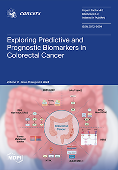
Colorectal cancer (CRC) is the second leading cause of cancer-related deaths globally. This review explores established tissue markers like -RAS/BRAF, HER2, and microsatellite instability focusing on their roles in targeted treatment selection and advances in targeted therapies. It also highlights promising therapeutic targets and the clinical utility of liquid biopsies. By synthesizing evidence and identifying gaps, the review offers insights into the biomarker landscape in CRC and discusses future challenges in translating biomarkers into clinical practice to enhance personalized medicine for CRC patients. View this paper
- Issues are regarded as officially published after their release is announced to the table of contents alert mailing list.
- You may sign up for e-mail alerts to receive table of contents of newly released issues.
- PDF is the official format for papers published in both, html and pdf forms. To view the papers in pdf format, click on the "PDF Full-text" link, and use the free Adobe Reader to open them.
Previous Issue
Next Issue