Next Issue
Volume 13, April
Previous Issue
Volume 13, February


Pathogens, Volume 13, Issue 3 (March 2024) – 90 articles

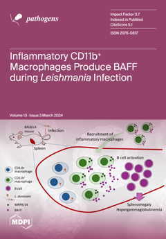
Cover Story (view full-size image): Visceral leishmaniasis (VL), caused by parasitic protozoa of the genus Leishmania, manifests clinical symptoms such as splenomegaly. It has previously been shown that B-cell activating factor (BAFF) is involved in splenomegaly in VL. Although BAFF is known to be expressed by a variety of cells, the mechanism of elevated BAFF expression in VL is not clear. We identified CD11b+ cells as BAFF-producing cells in the spleens of mice infected with Leishmania donovani. The number of CD11b+ cells increased in the spleens during infection, and the increased CD11b+ cells were primarily MRP14+ mononuclear cells. These results suggest that the increased BAFF expression in the spleen of L. donovani-infected mice involves a recruitment of inflammatory macrophages distinct from host macrophages for the parasites. View this paper
  • Issues are regarded as officially published after their release is announced to the table of contents alert mailing list.
  • You may sign up for e-mail alerts to receive table of contents of newly released issues.
  • PDF is the official format for papers published in both, html and pdf forms. To view the papers in pdf format, click on the "PDF Full-text" link, and use the free Adobe Reader to open them.
Order results
Result details
Section
Select all
Export citation of selected articles as:
11 pages, 567 KiB  
Article
Blastocystis spp. and Other Intestinal Parasites in Polish Soldiers Deployed to Lebanon and Iraq
by Danuta Izabela Kosik-Bogacka, Krzysztof Korzeniewski, Natalia Łanocha-Arendarczyk, Joanna Korycińska, Małgorzata Lepczyńska, Ewa Dzika and Małgorzata Marchelek-Myśliwiec
Pathogens 2024, 13(3), 271; https://doi.org/10.3390/pathogens13030271 - 21 Mar 2024
Cited by 1 | Viewed by 1927
Abstract
Intestinal parasitic infections are one of the most common infectious diseases worldwide, particularly in developing countries. A distinct group at increased risk of infection is military personnel deployed overseas for extended periods, typically six months at a time. The aim of this study [...] Read more.
Intestinal parasitic infections are one of the most common infectious diseases worldwide, particularly in developing countries. A distinct group at increased risk of infection is military personnel deployed overseas for extended periods, typically six months at a time. The aim of this study was to determine the prevalence of Blastocystis spp. and other intestinal parasites in Polish military personnel returning from deployments to Lebanon (n = 206) and Iraq (n = 220). In this group of subjects, we found Blastocystis spp. (13.6%), Dientamoeba fragilis (3.3%), Entamoeba coli (0.9%), and Endolimax nana (0.5%). Entamoeba histolytica sensu lato and Chilomastix mesnili infections were detected only in one soldier returning from Lebanon and Iraq, respectively. Blastocystis subtype (ST) 3 was predominant in soldiers returning from Lebanon, followed by ST2 and ST1. ST1 infection was predominant in soldiers returning from Iraq, followed by ST3 and ST2. Our study affirms that, deployment abroad is of no influence of the prevalence of parasitic protozoa. However, it would be worth to monitor parasite infection in military personnel returning from tropical zone even if they have no actual symptoms. In addition, it is very important to determine the subtypes of Blastocystis—this may help to clearly define their pathogenicity, especially considering the scarcity of studies on Blastocystis genotypes in Iraqi and Lebanese residents. Full article
Show Figures

Figure 1

13 pages, 1640 KiB  
Article
Vector-Borne and Zoonotic Pathogens in Raccoon Dogs (Nyctereutes procyonoides) and Raccoons (Procyon lotor) from Schleswig-Holstein, Germany
by Jana C. Klink, Alexandra Rieger, Peter Wohlsein, Ursula Siebert and Anna Obiegala
Pathogens 2024, 13(3), 270; https://doi.org/10.3390/pathogens13030270 - 21 Mar 2024
Cited by 2 | Viewed by 1946
Abstract
Raccoon dogs (Nyctereutes procyonoides) and raccoons (Procyon lotor) are invasive alien species originating from East Asia and North America, respectively. They are discussed as vectors and reservoirs for various infectious diseases, including vector-borne and zoonotic pathogens, and are therefore [...] Read more.
Raccoon dogs (Nyctereutes procyonoides) and raccoons (Procyon lotor) are invasive alien species originating from East Asia and North America, respectively. They are discussed as vectors and reservoirs for various infectious diseases, including vector-borne and zoonotic pathogens, and are therefore a potential threat to human and domestic animal health, as well as to biodiversity and conservation. In the years 2021 and 2022, 110 raccoon dogs (Nyctereutes procyonoides) and 30 raccoons (Procyon lotor) were screened via qPCR for the presence of Leptospira spp., Rickettsia spp. and Borreliella spp. in the German federal state of Schleswig-Holstein as part of a health and risk assessment study. Borreliella spp. were confirmed in one raccoon dog and one raccoon, identified as Borreliella afzelii in the raccoon. Leptospira spp. were found in 21 (19.44%) raccoon dogs and 2 (6.90%) raccoons. In five raccoon dogs, Leptospira spp. were identified as Leptospira borgpetersenii, Leptospira kirschneri and Leptospira interrogans. Full article
(This article belongs to the Section Ticks)
Show Figures

Figure 1

12 pages, 577 KiB  
Review
Fungemia by Wickerhamomyces anomalus—A Narrative Review
by Petros Ioannou, Stella Baliou and Diamantis P. Kofteridis
Pathogens 2024, 13(3), 269; https://doi.org/10.3390/pathogens13030269 - 21 Mar 2024
Cited by 3 | Viewed by 2683
Abstract
Wickerhamomyces anomalus has been previously classified as Hansenula anomala, Pichia anomala, and Candida pelliculosa and was recently reclassified in the genus Wickerhamomyces after phylogenetic analysis of its genetic sequence. An increasing number of reports of human infections by W. anomalus have emerged, [...] Read more.
Wickerhamomyces anomalus has been previously classified as Hansenula anomala, Pichia anomala, and Candida pelliculosa and was recently reclassified in the genus Wickerhamomyces after phylogenetic analysis of its genetic sequence. An increasing number of reports of human infections by W. anomalus have emerged, suggesting that this microorganism is an emerging pathogen. The present review aimed to provide data on the epidemiology, antifungal resistance, clinical characteristics, treatment, and outcomes of fungemia by W. anomalus by extracting all the available information from published original reports in the literature. PubMed/Medline, Cochrane Library, and Scopus databases were searched for eligible articles reporting data on patients with this disease. In total, 36 studies involving 170 patients were included. The age of patients with fungemia by W. anomalus ranged from 0 to 89 years; the mean age was 22.8 years, the median age was 2.2 years, with more than 37 patients being less than one month old, and 54% (88 out of 163 patients) were male. Regarding patients’ history, 70.4% had a central venous catheter use (CVC), 28.7% were on total parenteral nutrition (TPN), 97% of neonates were hospitalized in the neonatal ICU (NICU), and 39.4% of the rest of the patients were hospitalized in the intensive care unit (ICU). Previous antimicrobial use was noted in 65.9% of patients. The most common identification method was the matrix-assisted laser desorption/ionization time-of-flight mass spectrometry (MALDI-TOF MS) in 34.1%, VITEK and VITEK 2 in 20.6%, and ID32 C in 15.3%. W. anomalus had minimal antifungal resistance to fluconazole, echinocandins, and amphotericin B, the most commonly used antifungals for treatment. Fever and sepsis were the most common clinical presentation noted in 95.8% and 86%, respectively. Overall mortality was 20% and was slightly higher in patients older than one year. Due to the rarity of this disease, future multicenter studies should be performed to adequately characterize patients’ characteristics, treatment, and outcomes, which will increase our understanding and allow drawing safer conclusions regarding optimal management. Full article
(This article belongs to the Special Issue Recent Research on Hospital-Acquired Bloodstream Infections)
Show Figures

Figure 1

15 pages, 7202 KiB  
Review
Community-Directed Vector Control to Accelerate Onchocerciasis Elimination
by Benjamin Jacob, Edwin Michael and Thomas R. Unnasch
Pathogens 2024, 13(3), 268; https://doi.org/10.3390/pathogens13030268 - 21 Mar 2024
Cited by 1 | Viewed by 2304
Abstract
Onchocerciasis, or river blindness, has historically been one of the most important causes of blindness worldwide, and a major cause of socio-economic disruption, particularly in sub-Saharan Africa. Its importance as a cause of morbidity and an impediment to economic development in some of [...] Read more.
Onchocerciasis, or river blindness, has historically been one of the most important causes of blindness worldwide, and a major cause of socio-economic disruption, particularly in sub-Saharan Africa. Its importance as a cause of morbidity and an impediment to economic development in some of the poorest countries in the world motivated the international community to implement several programs to control or eliminate this scourge. Initially, these involved reducing transmission of the causative agent Onchocerca volvulus through controlling the vector population. When ivermectin was found to be a very effective drug for treating onchocerciasis, the strategy shifted to mass drug administration (MDA) of endemic communities. In some countries, both vector control and ivermectin MDA have been used together. However, traditional vector control methods involve treating rivers in which the black fly vectors breed with insecticides, a process which is expensive, requires trained personnel to administer, and can be ecologically harmful. In this review, we discuss recent research into alternatives to riverine insecticide treatment, which are inexpensive, ecologically less harmful, and can be implemented by the affected communities themselves. These can dramatically reduce vector densities and, when combined with ivermectin MDA, can accelerate the time to elimination when compared to MDA alone. Full article
(This article belongs to the Special Issue Research on the Epidemiology and Transmission of Filarial Diseases)
Show Figures

Figure 1

14 pages, 1597 KiB  
Article
The Course of COVID-19 and Long COVID: Identifying Risk Factors among Patients Suffering from the Disease before and during the Omicron-Dominant Period
by Mateusz Babicki, Damian Kołat, Żaneta Kałuzińska-Kołat, Joanna Kapusta, Agnieszka Mastalerz-Migas, Piotr Jankowski and Michał Chudzik
Pathogens 2024, 13(3), 267; https://doi.org/10.3390/pathogens13030267 - 20 Mar 2024
Cited by 2 | Viewed by 1956
Abstract
SARS-CoV-2 has acquired many mutations that influence the severity of COVID-19’s course or the risk of developing long COVID. In 2022, the dominant SARS-CoV-2 variant was Omicron. This study aimed to compare the course of COVID-19 in the periods before and during the [...] Read more.
SARS-CoV-2 has acquired many mutations that influence the severity of COVID-19’s course or the risk of developing long COVID. In 2022, the dominant SARS-CoV-2 variant was Omicron. This study aimed to compare the course of COVID-19 in the periods before and during the dominance of the Omicron variant. Risk factors for developing long COVID were also assessed. This study was based on stationary visits of patients after COVID-19 and follow-up assessments after 3 months. Clinical symptoms, comorbidities, and vaccination status were evaluated in 1967 patients. Of the analyzed group, 1308 patients (66.5%) were affected by COVID-19 in the period before the Omicron dominance. The prevalence of long COVID was significantly lower among patients of the Omicron group (47.7% vs. 66.9%, p < 0.001). The risk of long COVID was higher for women (OR: 1.61; 95% CI: 1.31, 1.99]) and asthmatics (OR: 1.46; 95% CI: 1.03, 2.07]). Conclusively, infection during the Omicron-dominant period was linked to a lower risk of developing long COVID. Females are at higher risk of developing long COVID independent of the pandemic period. Individuals affected by COVID-19 in the Omicron-dominant period experience a shorter duration of symptoms and reduced frequency of symptoms, except for coughing, which occurs more often. Full article
(This article belongs to the Special Issue Infectious Diseases and Vaccine Technology Research)
Show Figures

Figure 1

17 pages, 993 KiB  
Review
Regulation of Autophagosome–Lysosome Fusion by Human Viral Infections
by Po-Yuan Ke
Pathogens 2024, 13(3), 266; https://doi.org/10.3390/pathogens13030266 - 20 Mar 2024
Viewed by 2213
Abstract
Autophagy plays a fundamental role in maintaining cellular homeostasis by eliminating intracellular components via lysosomes. Successful degradation through autophagy relies on the fusion of autophagosomes to lysosomes, which leads to the formation of autolysosomes containing acidic proteases that degrade the sequestered materials. Viral [...] Read more.
Autophagy plays a fundamental role in maintaining cellular homeostasis by eliminating intracellular components via lysosomes. Successful degradation through autophagy relies on the fusion of autophagosomes to lysosomes, which leads to the formation of autolysosomes containing acidic proteases that degrade the sequestered materials. Viral infections can exploit autophagy in infected cells to balance virus–host cell interactions by degrading the invading virus or promoting viral growth. In recent years, cumulative studies have indicated that viral infections may interfere with the fusion of autophagosomes and lysosomes, thus benefiting viral replication and associated pathogenesis. In this review, I provide an overview of the current understanding of the molecular mechanism by which human viral infections deregulate autophagosome–lysosome fusion and summarize the physiological significance in the virus life cycle and host cell damage. Full article
(This article belongs to the Special Issue Viral Infections of Humans: Epidemiology and Control)
Show Figures

Graphical abstract

9 pages, 4929 KiB  
Systematic Review
Beau’s Lines and COVID-19; A Systematic Review on Their Association
by Aris P. Agouridis, Christina Mastori-Kourmpani, Polyna Antoniou, Paschalis Konstantinou, Evangelos C. Rizos and Constantinos Tsioutis
Pathogens 2024, 13(3), 265; https://doi.org/10.3390/pathogens13030265 - 20 Mar 2024
Viewed by 3333
Abstract
Background: Beau’s lines are transverse grooves in the nail plate that result from transient interruption of the growth of the proximal nail matrix after severe disease. The aim of this study is to systematically report all evidence on the association of Beau’s lines [...] Read more.
Background: Beau’s lines are transverse grooves in the nail plate that result from transient interruption of the growth of the proximal nail matrix after severe disease. The aim of this study is to systematically report all evidence on the association of Beau’s lines with COVID-19 infection or vaccination against COVID-19. Methods: PubMed and Scopus databases were searched up to January 2024 for articles reporting Beau’s lines associated with COVID-19 infection or vaccination for COVID-19. PROSPERO ID: CRD42024496830. Results: PubMed search identified 299 records while Scopus search identified 18 records. After screening the bibliography, nine studies including 35 cases were included in our systematic review. The studies were reported from different areas around the world. Included studies documented Beau’s lines following COVID-19 vaccination (two studies) or after COVID-19 infection (seven studies). High variability was recorded in onset and resolution times among included cases, averaging 3 months and 6 months after COVID-19 infection, respectively. In the two studies reporting Beau’s lines after vaccination, onset was at 7 days and 6 weeks and resolution occurred after 8 and 17 weeks, respectively. Conclusions: To the best of our knowledge, this is the first systematic review reporting the association of Beau’s lines with COVID-19 infection and vaccination. Severe immune response can result in the formation of these nail disorders. Of importance, Beau’s lines represent a potential indicator of prior severe COVID-19 infection or vaccination for COVID-19, as well as a sign of long COVID-19 syndrome. Full article
Show Figures

Figure 1

13 pages, 6028 KiB  
Article
Metagenomic Detection and Genetic Characterization of Human Sapoviruses among Children with Acute Flaccid Paralysis in Nigeria
by Uwem Etop George, Temitope O. C. Faleye, Lander De Coninck, Sheriff Tunde Agbaje, Ijeoma Maryjoy Ifeorah, Bernard Anyebe Onoja, Elijah Igbekele Oni, Adebowale Oluseyi Olayinka, Toluwani Goodnews Ajileye, Arthur Obinna Oragwa, Toluwanimi Emmanuel Akinleye, Bolutife Olubukola Popoola, Oluwadamilola Gideon Osasona, Olaitan Titilola Olayinka, Oluwadamilola Adefunke George, Ahmed Iluoreh Muhammad, Isaac Komolafe, Adekunle Johnson Adeniji, Jelle Matthijnssens and Moses Olubusuyi Adewumi
Pathogens 2024, 13(3), 264; https://doi.org/10.3390/pathogens13030264 - 19 Mar 2024
Cited by 2 | Viewed by 2033
Abstract
Using a metagenomic sequencing approach on stool samples from children with Acute Flaccid Paralysis (AFP), we describe the genetic diversity of Sapoviruses (SaVs) in children in Nigeria. We identified six complete genome sequences and two partial genome sequences. Several SaV genogroups and genotypes [...] Read more.
Using a metagenomic sequencing approach on stool samples from children with Acute Flaccid Paralysis (AFP), we describe the genetic diversity of Sapoviruses (SaVs) in children in Nigeria. We identified six complete genome sequences and two partial genome sequences. Several SaV genogroups and genotypes were detected, including GII (GII.4 and GII.8), GIV (GIV.1), and GI (GI.2 and GI.7). To our knowledge, this is the first description of SaV infections and complete genomes from Nigeria. Pairwise identity and phylogenetic analysis showed that the Nigerian SaVs were related to previously documented gastroenteritis outbreaks with associated strains from China and Japan. Minor variations in the functional motifs of the nonstructural proteins NS3 and NS5 were seen in the Nigerian strains. To adequately understand the effect of such amino acid changes, a better understanding of the biological function of these proteins is vital. The identification of distinct SaVs reinforces the need for robust surveillance in acute gastroenteritis (AGE) and non-AGE cohorts to better understand SaVs genotype diversity, evolution, and its role in disease burden in Nigeria. Future studies in different populations are, therefore, recommended. Full article
(This article belongs to the Section Viral Pathogens)
Show Figures

Figure 1

5 pages, 177 KiB  
Editorial
Recent Advances in the Epidemiology of Pathogenic Agents
by Wei-Chuan Chen and Yusen Eason Lin
Pathogens 2024, 13(3), 263; https://doi.org/10.3390/pathogens13030263 - 19 Mar 2024
Viewed by 2158
Abstract
The COVID-19 pandemic has underscored the pivotal role of epidemiology in studying pathogenic agents [...] Full article
(This article belongs to the Special Issue Epidemiology of Pathogenic Agents)
14 pages, 2083 KiB  
Article
Genetic Diversity of Trypanosomes Infesting Cattle from Savannah District in North of Côte d’Ivoire Using Conserved Genomic Signatures: rRNA, ITS1 and gGAPDH
by Jean-Yves Ekra, Eliakunda Michael Mafie, Edouard K. N’Goran, Dramane Kaba, Biégo Guillaume Gragnon and Jagan Srinivasan
Pathogens 2024, 13(3), 262; https://doi.org/10.3390/pathogens13030262 - 19 Mar 2024
Viewed by 1836
Abstract
The potential danger to livestock from African animal trypanosomiasis is well known. However, the trypanosome species circulating in cattle and their genetics are poorly understood. After different alignments according to three regions (ITS1, gGAPDH and rRNA gene) of the trypanosome genome, phylogenetic analyses [...] Read more.
The potential danger to livestock from African animal trypanosomiasis is well known. However, the trypanosome species circulating in cattle and their genetics are poorly understood. After different alignments according to three regions (ITS1, gGAPDH and rRNA gene) of the trypanosome genome, phylogenetic analyses were used to show the genetic diversity of the different species that were circulating in the cattle in three regions (Bagoue, Poro and Tchologo) of Côte d’Ivoire. These analyses were performed by alignment of ITS1; by alignment of partial 18S, ITS1, 5.8S, ITS2 and partial 28S rRNA genes; and by alignment of gGAPDH gene with sequences of Trypanosomes found in GenBank. Three species were identified (T. vivax, T. theileri and T. congolense) in the cattle in the three northern regions of Côte d’Ivoire. T. vivax and T. theileri were the most abundant species in the present study. Contrary to the other primers used in this study, the ITS1 primers were not able to amplify T. theileri. We observed mixed infections between T. theileri and the other two species identified (T. vivax and T. congolense). As far as primers are concerned, in some cases, rRNA was able to identify the same species of trypanosomes that the ITS1 and gGAPDH primers were able to identify. Two main distinct groups of T. theileri complex were identified. The T. congolense and T. vivax strains were close to African strains, such as those from Kenya, Nigeria and Cameroon, unlike the T. theileri strain. Three trypanosome species (T. vivax, T. theileri and T. congolense) circulate in cattle in the Savannah district of Côte d’Ivoire. The genetic diversity of the trypanosome species encountered in this study cannot be classified as intraspecies according to geographical area and breed of cattle they infect. Full article
Show Figures

Figure 1

15 pages, 1990 KiB  
Article
Genome Sequencing of Historical Encephalomyocarditis Viruses from South Africa Links the Historical 1993/4 Savanna Elephant (Loxodonta africana) Outbreak to Cryptic Mastomys Rodents
by Vanessa van Meer, Janusz T. Pawęska, Robert Swanepoel, Antoinette Grobbelaar and Armanda D. Bastos
Pathogens 2024, 13(3), 261; https://doi.org/10.3390/pathogens13030261 - 19 Mar 2024
Viewed by 1654
Abstract
From 1993 to 1994, 64 free-ranging elephants (Loxodonta africana) succumbed to encephalomyocarditis in the Kruger National Park, South Africa, of which 83% were adult bulls. Mastomys rodents were implicated as the reservoir host of the Encephalomyocarditis virus (EMCV) based on serology [...] Read more.
From 1993 to 1994, 64 free-ranging elephants (Loxodonta africana) succumbed to encephalomyocarditis in the Kruger National Park, South Africa, of which 83% were adult bulls. Mastomys rodents were implicated as the reservoir host of the Encephalomyocarditis virus (EMCV) based on serology and RT-PCR. However, in the absence of sequence-confirmation of both the virus and the rodent host, definitive links between the elephant outbreak strains and rodent reservoir could not be established. In this study, we generate the first reference genome sequences for three historical EMCVs isolated from two Mastomys rodents and one Mastomys-associated mite, Laelaps muricola, in Gauteng Province, South Africa, in 1961. In addition, near-complete genome sequences were generated for two elephant outbreak virus strains, for which data were previously limited to the P1 and 3D genome regions. The consensus sequence of each virus was determined using a PCR-Sanger sequencing approach. Phylogenetic analysis confirmed the three near-identical (99.95–99.97%) Mastomys-associated viruses to be sister to the two near-identical (99.85%) elephant outbreak strains, differing from each other at 6.4% of sites across the ~7400-nucleotide region characterised. This study demonstrates a link between Mastomys-associated viruses and the historical elephant outbreak strains and implicates Mastomys as reservoirs of EMCV in South Africa. Full article
(This article belongs to the Special Issue Molecular Detection and Characterisation of Viral Pathogens)
Show Figures

Figure 1

15 pages, 884 KiB  
Review
Obtaining Microbiologically Safe Hatching Eggs from Hatcheries: Using Essential Oils for Integrated Sanitization Strategies in Hatching Eggs, Poultry Houses and Poultry
by Gabriel da Silva Oliveira, Concepta McManus, Igor Rafael Ribeiro Vale and Vinícius Machado dos Santos
Pathogens 2024, 13(3), 260; https://doi.org/10.3390/pathogens13030260 - 18 Mar 2024
Cited by 9 | Viewed by 2204
Abstract
Essential oils are liquids containing non-toxic compounds that are unfavorable to the growth of microorganisms. They are sold globally at affordable or very high prices, depending on the availability and type of plant, the scale of production, the extraction method, costs associated with [...] Read more.
Essential oils are liquids containing non-toxic compounds that are unfavorable to the growth of microorganisms. They are sold globally at affordable or very high prices, depending on the availability and type of plant, the scale of production, the extraction method, costs associated with logistics and electricity consumption, among other variables. Each year, the quantity of research dedicated to the antimicrobial potential of essential oils in poultry farming is expanding. Researchers consensually relay that this increase is due to the growing resistance of microorganisms to traditional antimicrobials and concerns about the toxicity of these products. This review proposes an analysis of the antimicrobial feasibility of using essential oils to address microbial challenges in poultry farms, aiming to ensure the production and supply of microbiologically safe hatching eggs. Based on the findings in the literature, in addition to following other necessary precautions in the daily routines of poultry farming practices, developing an antimicrobial control program with essential oils that integrates poultry facilities, poultry and hatching eggs, adapted to the particularities of each context seems to be extremely effective. Full article
(This article belongs to the Special Issue One Health: Pathogenesis of Bacterial Infections in Animals)
Show Figures

Figure 1

17 pages, 325 KiB  
Review
Infections in Disorders of Immune Regulation
by Abarna Thangaraj, Reva Tyagi, Deepti Suri and Sudhir Gupta
Pathogens 2024, 13(3), 259; https://doi.org/10.3390/pathogens13030259 - 17 Mar 2024
Viewed by 2748
Abstract
Primary immune regulatory disorders (PIRDs) constitute a spectrum of inborn errors of immunity (IEIs) that are primarily characterized by autoimmunity, lymphoproliferation, atopy, and malignancy. In PIRDs, infections are infrequent compared to other IEIs. While susceptibility to infection primarily stems from antibody deficiency, it [...] Read more.
Primary immune regulatory disorders (PIRDs) constitute a spectrum of inborn errors of immunity (IEIs) that are primarily characterized by autoimmunity, lymphoproliferation, atopy, and malignancy. In PIRDs, infections are infrequent compared to other IEIs. While susceptibility to infection primarily stems from antibody deficiency, it is sometimes associated with additional innate immune and T or NK cell defects. The use of immunotherapy and chemotherapy further complicates the immune landscape, increasing the risk of diverse infections. Recurrent sinopulmonary infections, particularly bacterial infections such as those associated with staphylococcal and streptococcal organisms, are the most reported infectious manifestations. Predisposition to viral infections, especially Epstein–Barr virus (EBV)-inducing lymphoproliferation and malignancy, is also seen. Notably, mycobacterial and invasive fungal infections are rarely documented in these disorders. Knowledge about the spectrum of infections in these disorders would prevent diagnostic delays and prevent organ damage. This review delves into the infection profile specific to autoimmune lymphoproliferative syndrome (ALPS), Tregopathies, and syndromes with autoimmunity within the broader context of PIRD. Despite the critical importance of understanding the infectious aspects of these disorders, there remains a scarcity of comprehensive reports on this subject. Full article
(This article belongs to the Special Issue Infection in Inborn Errors of Immunity)
17 pages, 3210 KiB  
Article
An Entomological Investigation during a Recent Rift Valley Fever Epizootic/Epidemic Reveals New Aspects of the Vectorial Transmission of the Virus in Madagascar
by Luciano Michaël Tantely, Soa Fy Andriamandimby, Maminirina Fidelis Ambinintsoa, Manou Rominah Raharinirina, Jean Théophile Rafisandratantsoa, Jean-Pierre Ravalohery, Aina Harimanana, Nirina Nantenaina Ranoelison, Judickaelle Irinantenaina, Miamina Fidy Ankasitrahana, Dany Bakoly Ranoaritiana, Laurence Randrianasolo, Rindra Vatosoa Randremanana, Vincent Lacoste, Philippe Dussart and Romain Girod
Pathogens 2024, 13(3), 258; https://doi.org/10.3390/pathogens13030258 - 16 Mar 2024
Cited by 2 | Viewed by 1970
Abstract
A Rift Valley fever (RVF) outbreak occurred in at least five regions of Madagascar in 2021. The aim of this study was to provide an overview of the richness, abundance, ecology, and trophic preferences of mosquitoes in the Mananjary district and to investigate [...] Read more.
A Rift Valley fever (RVF) outbreak occurred in at least five regions of Madagascar in 2021. The aim of this study was to provide an overview of the richness, abundance, ecology, and trophic preferences of mosquitoes in the Mananjary district and to investigate the distribution of mosquitoes that were RT-PCR-positive for RVFV. Three localities were prospected from 26 April to 4 May 2021, using light traps, BG-Sentinel traps baited with an artificial human odor, Muirhead-Thomson pit traps, and indoor pyrethroid spray catches. A total of 2806 mosquitoes belonging to at least 26 species were collected. Of 512 monospecific pools of mosquitoes tested with real-time RT-PCR, RVFV was detected in 37 pools representing 10 mosquito species. The RVFV-positive species were as follows: Aedes albopictus, Ae. argenteopunctatus, Anopheles coustani, An. gambiae s.l., An. mascarensis, An. squamosus/cydippis, Culex antennatus, Cx. decens, Cx. Tritaeniorhynchus, and Uranotaenia spp. Of the 450 tested engorged females, 78.7% had taken a blood meal on humans, 92.9% on cattle, and 71.6% had taken mixed (human–cattle) blood meals. This investigation suggests the potential role of mosquitoes in RVFV transmission within this epizootic/epidemic context and that the human populations at the three study sites were highly exposed to mosquitoes. Therefore, the use of impregnated mosquito nets as an appropriate prevention method is recommended. Full article
(This article belongs to the Special Issue Viral Infections of Humans: Epidemiology and Control)
Show Figures

Figure 1

9 pages, 246 KiB  
Brief Report
The Re-Emergence of Rift Valley Fever in Mananjary District, Madagascar in 2021: A Call for Action
by Aina Nirina Harimanana, Soa Fy Andriamandimby, Dany Bakoly Ranoaritiana, Laurence Randrianasolo, Judickaelle Irinantenaina, Nirina Nantenaina Ranoelison, Jean Théophile Rafisandrantatsoa, Miamina Fidy Ankasitrahana, Antso Hasina Raherinandrasana, Manuela Vololoniaina Andriamahatana, Michael Luciano Tantely, Romain Girod, Philippe Dussart, Vincent Lacoste and Rindra Vatosoa Randremanana
Pathogens 2024, 13(3), 257; https://doi.org/10.3390/pathogens13030257 - 16 Mar 2024
Viewed by 1623
Abstract
An epizootic of rift valley fever (RVF) was suspected on 21 February 2021 in various districts of Madagascar, with a lab confirmation on 1 April 2021. A cross-sectional survey aiming to detect cases of RVF in humans and to study the circulation of [...] Read more.
An epizootic of rift valley fever (RVF) was suspected on 21 February 2021 in various districts of Madagascar, with a lab confirmation on 1 April 2021. A cross-sectional survey aiming to detect cases of RVF in humans and to study the circulation of rift valley fever virus (RVFV) in animals was conducted from 22 April to 4 May 2021 in the district of Mananjary. Blood samples from cattle and humans were tested using serological and molecular techniques. In cattle, the circulation of RVFV was confirmed between 5 February and 4 May 2021. The positivity rates of anti-RVFV IgG and IgM were 60% and 40%, respectively. In humans, the circulation of RVFV was observed from 1 April to 5 May 2021. The positivity rate of RVFV was estimated to be 11.7% by combining the results of the molecular and serological approaches. Of the 103 individuals who agreed to participate in the survey, 3 were determined to be positive by RT-PCR, and 10 had anti-RVFV IgM. Among them, one was positive for both. Given that previous studies have reported the circulation of RVFV during inter-epidemic periods and the occurrence of outbreaks due to imported RVFV in Madagascar, our findings suggest the importance of strengthening RVF surveillance from a “One Health” perspective by conducting syndromic and risk-based surveillance at the national and regional levels. Full article
(This article belongs to the Special Issue Viral Infections of Humans: Epidemiology and Control)
15 pages, 3328 KiB  
Article
Alterations in N-glycosylation of HCV E2 Protein in Children Patients with IFN-RBV Therapy Failure
by Karolina Zimmer, Alicja M. Chmielewska, Paulina Jackowiak, Marek Figlerowicz and Krystyna Bienkowska-Szewczyk
Pathogens 2024, 13(3), 256; https://doi.org/10.3390/pathogens13030256 - 15 Mar 2024
Viewed by 1764
Abstract
The glycosylation of viral envelope proteins plays an important role in virus biology and the immune response of the host to infection. Hepatitis C virus (HCV) envelope proteins E1 and E2, key players in virus entry and spread, are highly N-glycosylated and possess [...] Read more.
The glycosylation of viral envelope proteins plays an important role in virus biology and the immune response of the host to infection. Hepatitis C virus (HCV) envelope proteins E1 and E2, key players in virus entry and spread, are highly N-glycosylated and possess 4 (5 in certain genotypes) to 11 conserved glycosylation sites, respectively. Many published results based on recombinant proteins indicate that the glycan shield can mask the epitopes targeted by neutralizing antibodies. Glycan shifting within the conserved linear E2 region (412–423) could be one of the escape strategies used by HCV. In the present report, we isolated E2 genes from samples (collected before the IFN-RBV therapy) originating from pediatric patients infected with HCV gt 1a. We analyzed the biochemical properties of cloned E2 glycoprotein variants and investigated their glycosylation status. The sequencing of E2 genes isolated from patients who did not respond to therapy revealed mutations at N-glycosylation sites, thus leading to a lower molecular weight and a low affinity to both linear and conformational neutralizing antibodies. The loss of the glycosylation site within the conserved epitope (amino acid 417) impaired the binding with AP33, an antibody that potently neutralizes all genotypes of HCV. Our findings, based on clinical samples, confirm the influence of N-glycosylation aberrations on the antigenic and conformational properties of HCV E1/E2, which may possibly correlate with the outcome of therapy in patients. Full article
Show Figures

Figure 1

14 pages, 3316 KiB  
Article
The Zoonotic Angiostrongylus cantonensis and the Veterinary Parasite Aelurostrongylus abstrusus Infecting Terrestrial Gastropods from Urban Areas of Macapá, Brazilian Amazon Region
by Tatiane Alves Barbosa, Silvana Carvalho Thiengo, Monica Ammon Fernandez, Jucicleide Ramos-de-Souza and Suzete Rodrigues Gomes
Pathogens 2024, 13(3), 255; https://doi.org/10.3390/pathogens13030255 - 15 Mar 2024
Viewed by 2096
Abstract
Metastrongyloidea includes nematodes that parasitize mammals, mainly infecting their respiratory and cardiovascular systems, and are responsible for emerging zoonosis in the world. Terrestrial mollusks are their main intermediate hosts, with few exceptions. Here we present the results of a malacological survey to know [...] Read more.
Metastrongyloidea includes nematodes that parasitize mammals, mainly infecting their respiratory and cardiovascular systems, and are responsible for emerging zoonosis in the world. Terrestrial mollusks are their main intermediate hosts, with few exceptions. Here we present the results of a malacological survey to know the distribution of Angiostrongylus cantonensis in Macapá, Amapá, in the Brazilian Amazon region, after the report of a case of eosinophilic meningitis in 2018. Mollusks were collected in 45 neighborhoods between March 2019 and February 2020. They were identified, parasitologically analyzed, and their nematodes parasites were identified based on the morphology and MT-CO1 sequencing. Infections of An. cantonensis were observed in Achatina fulica, Sarasinula linguaeformis and Subulina octona. These are the first records of the natural infection of the last two species by An. cantonensis in the Brazilian Amazon region. The angiostrongylid Aelurostrongylus abstrusus, which parasitizes cats, was also detected parasitizing A. fulica and Diplosolenodes occidentalis. This is also the first record of the slug D. occidentalis infected by Ae. abstrusus. The highest infection rates were recorded in neighborhoods where the environment conditions favor the proliferation of both mollusks and rodents. The results demonstrate the ample distribution of An. cantonensis in Macapá and the need for surveillance and mollusk vector control in Brazil and other countries. Full article
(This article belongs to the Special Issue Rat Lungworm Disease)
Show Figures

Figure 1

5 pages, 747 KiB  
Case Report
Challenges in Toxocariasis Diagnosis: From Pericarditis, through Hepatic Tumor, to the Detection of Brain Aneurysms: Case Report
by Martyna Biała, Joanna Nieleńczuk, Anna Chodorowska and Bartosz Szetela
Pathogens 2024, 13(3), 254; https://doi.org/10.3390/pathogens13030254 - 15 Mar 2024
Cited by 1 | Viewed by 1891
Abstract
Toxocariasis is the parasitic infection caused by the larvae of Toxocara roundworms species: Toxocara canis from dogs and, less frequently, Toxocara cati from cats. The high proportion of asymptomatic cases of toxocariasis and the uncharacteristic clinical manifestations mimicking other medical conditions make diagnosis [...] Read more.
Toxocariasis is the parasitic infection caused by the larvae of Toxocara roundworms species: Toxocara canis from dogs and, less frequently, Toxocara cati from cats. The high proportion of asymptomatic cases of toxocariasis and the uncharacteristic clinical manifestations mimicking other medical conditions make diagnosis challenging. The main clinical presentations of toxocariasis are visceral and ocular larva migrans. Migration to the central nervous system (neurotoxocariasis) is rare and can cause meningitis, encephalitis, myelitis, cerebral vasculitis, seizures, headache or asymptomatic CNS infection. Neurotoxocariasis is an uncommon diagnosis and it is probably underdiagnosed due to the nonspecific clinical manifestations, low awareness of physicians as well as the lack of standardized diagnostic exams. To date, no causality has been proven between neurotoxocariasis and aneurysms, but due to the character of immune response elicited by the parasites, it remains an important possibility for further research. We present a case report of a woman infected with Toxocara canis highlighting the diagnostic difficulties. We aim to raise the awareness of the clinical symptomatology of neurotoxocariasis. Full article
(This article belongs to the Section Parasitic Pathogens)
Show Figures

Figure 1

16 pages, 4481 KiB  
Article
Epidemiology, Clinical Signs, and Risk Factors Associated with Theileriosis in Australian Cattle (2006–2022)
by Emily Onizawa and Cheryl Jenkins
Pathogens 2024, 13(3), 253; https://doi.org/10.3390/pathogens13030253 - 15 Mar 2024
Cited by 2 | Viewed by 1863
Abstract
For over a decade, bovine anaemia caused by Theileria orientalis Ikeda has been a significant disease in the Australian cattle industry. In this study, we conducted a spatial and temporal analysis of theileriosis in Australia using historic data from submissions to the New [...] Read more.
For over a decade, bovine anaemia caused by Theileria orientalis Ikeda has been a significant disease in the Australian cattle industry. In this study, we conducted a spatial and temporal analysis of theileriosis in Australia using historic data from submissions to the New South Wales Department of Primary Industries (NSW DPI) from 2006 to 2022, where herd history, clinical signs, and PCR results were available. Since the first detections of bovine theileriosis in the Sydney area in 2006, the disease spread north- and southward and is now endemic to the southeast coast of Australia, closely mirroring the distribution of the principal vector Haemaphysalis longicornis. Across all years, the prevalence of the Ikeda genotype was 88%, while the prevalence of the benign Chitose and Buffeli genotypes was 55% and 38%, respectively. The majority of submissions were from beef cattle in coastal NSW, with anaemia, fever, jaundice, abortion, and lethargy the most frequently reported clinical signs. Transportation was identified as the major risk factor for disease. Until 2015, the majority of cases were reported in adult cattle, while in later years, calves made up the majority of cases, most likely due to the widespread acquisition of immunity in adults. Calves were significantly more likely to present with diarrhoea, lethargy, and anaemia, and to suffer mortality, while adults were significantly more likely to present with jaundice. Instances of abortion were observed to be significantly associated with beef cattle. The relationship between the level of parasitaemia and anaemia revealed a strong negative correlation for all animals examined. Full article
(This article belongs to the Section Parasitic Pathogens)
Show Figures

Figure 1

16 pages, 11125 KiB  
Article
The Influence of L-Lysine-Alpha-Oxidase on the Biofilm Formation of Opportunistic Microorganisms Associated with Inflammatory Diseases of the Urinary Tract
by Alexandr Senyagin, Nadezhda Sachivkina, Milana Das, Anna Arsenyuk, Ramziya Mannapova, Alfir Mannapov, Tursumbai Kubatbekov, Dmitriy Svistunov, Olesya Petrukhina, Andrey Zharov and Natallia Zhabo
Pathogens 2024, 13(3), 252; https://doi.org/10.3390/pathogens13030252 - 15 Mar 2024
Cited by 1 | Viewed by 2106
Abstract
Urinary tract infections occupy a special niche among diseases of infectious etiology. Many microorganisms associated with urinary tract infections, such as Klebsiella oxytoca, Enterococcus spp., Morganella morganii, Moraxella catarrhalis, Pseudomonas aeruginosa, Proteus mirabilis, Staphylococcus aureus, Staphylococcus spp., [...] Read more.
Urinary tract infections occupy a special niche among diseases of infectious etiology. Many microorganisms associated with urinary tract infections, such as Klebsiella oxytoca, Enterococcus spp., Morganella morganii, Moraxella catarrhalis, Pseudomonas aeruginosa, Proteus mirabilis, Staphylococcus aureus, Staphylococcus spp., and Candida spp., can form biofilms. The aim of this research was to study the effect of the enzyme L-lysine-Alpha-oxidase (LO) produced by the fungus Trichoderma harzianum Rifai on the biofilm formation process of microorganisms associated with urinary tract infections. Homogeneous LO showed a more pronounced effect than the culture liquid concentrate (cCL). When adding samples at the beginning of incubation, the maximum inhibition was observed in relation to Enterococcus faecalis 5960—cCL 86%, LO 95%; Enterococcus avium 1669—cCL 85%, LO 94%; Enterococcus cloacae 6392—cCL 83%, LO—98%; and Pseudomonas aeruginosa 3057—cCL 70%, LO—82%. The minimum inhibition was found in Candida spp. Scanning electron microscopy was carried out, and numerous morphological and structural changes were observed in the cells after culturing the bacterial cultures in a medium supplemented with homogeneous LO. For example, abnormal division was detected, manifesting as the appearance of joints in places where the bacteria diverge. Based on the results of this work, we can draw conclusions about the possibility of inhibiting microbial biofilm formation with the use of LO; especially significant inhibition was achieved when the enzyme was added at the beginning of incubation. Thus, LO can be a promising drug candidate for the treatment or prevention of infections associated with biofilm formation. Full article
(This article belongs to the Section Bacterial Pathogens)
Show Figures

Figure 1

14 pages, 3934 KiB  
Article
Etiology of Ulcerative Dermal Necrosis (UDN) in Brown Trout (Salmo trutta Morpha trutta)—Preliminary Results
by Marek Matras, Magdalena Stachnik, Anna Kycko, Magdalena Wasiak, Ewa Paździor, Joanna Maj-Paluch, Ewa Borzym and Michał Reichert
Pathogens 2024, 13(3), 251; https://doi.org/10.3390/pathogens13030251 - 15 Mar 2024
Viewed by 1607
Abstract
Every year, ulcerative dermal necrosis (UDN) affects salmonids that spend most of their lives in the sea during their migration to the rivers of northern Poland to spawn. The clinical form of the disease manifests itself in ulcerative skin lesions, which lead to [...] Read more.
Every year, ulcerative dermal necrosis (UDN) affects salmonids that spend most of their lives in the sea during their migration to the rivers of northern Poland to spawn. The clinical form of the disease manifests itself in ulcerative skin lesions, which lead to significant weakening of the fish and, in most cases, result in their death. This study was carried out on samples taken from sea trout in the Słupia River in northern Poland. In order to identify the pathogen, experiments on the transmission of the disease were carried out, and additional histopathological, microbiological and electron microscopic examinations were performed. As a result of these studies, it was possible to experimentally transfer the disease from sick to healthy fish. The results indicate a complex etiology of the disease (lack of a clearly defined pathogen), in which the change in the environment from salty to freshwater triggers the related changes in skin physiology, which are the main causes of increased susceptibility to the development of the disease. Full article
(This article belongs to the Special Issue Emerging Pathogens in Aquaculture)
Show Figures

Figure 1

24 pages, 15946 KiB  
Article
Characterization of a Novel Species of Legionella Isolated from a Healthcare Facility: Legionella resiliens sp. nov
by Sandra Cristino, Maria Rosaria Pascale, Federica Marino, Carlo Derelitto, Silvano Salaris, Massimiliano Orsini, Stefano Squarzoni, Antonella Grottola and Luna Girolamini
Pathogens 2024, 13(3), 250; https://doi.org/10.3390/pathogens13030250 - 14 Mar 2024
Cited by 1 | Viewed by 2433
Abstract
Two Legionella-like isolates, 8cVS16T and 9fVS26, were isolated from a water distribution system (WDS) in a healthcare facility. Cells were Gram- and Ziehl Neelsen-stain-negative, rod-shaped, motile, and exhibited a blue-white fluorescence under Wood’s lamp at 365 nm. The strains grew in [...] Read more.
Two Legionella-like isolates, 8cVS16T and 9fVS26, were isolated from a water distribution system (WDS) in a healthcare facility. Cells were Gram- and Ziehl Neelsen-stain-negative, rod-shaped, motile, and exhibited a blue-white fluorescence under Wood’s lamp at 365 nm. The strains grew in a range of 32–37 °C on BCYE with L-cysteine (Cys+), GVPC, and MWY agar medium, with a positive reaction for oxidase, catalase, and gelatinase. The dominant fatty acids were summed features 3 (C16:1ω7c/C16:1ω6c) (27.7%), C16:0 iso (17.5%), and C16:0 (16.3%), and Q13 as the major ubiquinone. The mip and rpoB gene sequences showed a similarity of 96.7% and 92.4%, with L. anisa (ATCC 35292T). The whole genomes sequencing (WGS) performed displayed a GC content of 38.21 mol% for both. The digital DNA-DNA hybridization (dDDH) analysis demonstrated the separation of the two strains from the phylogenetically most related L. anisa (ATCC 35292T), with ≤43% DNA-DNA relatedness. The Average Nucleotide Identity (ANI) between the two strains and L. anisa (ATCC 35292T) was 90.74%, confirming that the two isolates represent a novel species of the genus Legionella. The name proposed for this species is Legionella resiliens sp. nov., with 8cVS16T (=DSM 114356T = CCUG 76627T) as the type strain. Full article
(This article belongs to the Special Issue Bacterial Biofilm, Genomics and Virulence)
Show Figures

Graphical abstract

13 pages, 611 KiB  
Review
Avian Influenza A Viruses Modulate the Cellular Cytoskeleton during Infection of Mammalian Hosts
by Flora De Conto
Pathogens 2024, 13(3), 249; https://doi.org/10.3390/pathogens13030249 - 14 Mar 2024
Viewed by 1964
Abstract
Influenza is one of the most prevalent causes of death worldwide. Influenza A viruses (IAVs) naturally infect various avian and mammalian hosts, causing seasonal epidemics and periodic pandemics with high morbidity and mortality. The recent SARS-CoV-2 pandemic showed how an animal virus strain [...] Read more.
Influenza is one of the most prevalent causes of death worldwide. Influenza A viruses (IAVs) naturally infect various avian and mammalian hosts, causing seasonal epidemics and periodic pandemics with high morbidity and mortality. The recent SARS-CoV-2 pandemic showed how an animal virus strain could unpredictably acquire the ability to infect humans with high infection transmissibility. Importantly, highly pathogenic avian influenza A viruses (AIVs) may cause human infections with exceptionally high mortality. Because these latter infections pose a pandemic potential, analyzing the ecology and evolution features of host expansion helps to identify new broad-range therapeutic strategies. Although IAVs are the prototypic example of molecular strategies that capitalize on their coding potential, the outcome of infection depends strictly on the complex interactions between viral and host cell factors. Most of the studies have focused on the influenza virus, while the contribution of host factors remains largely unknown. Therefore, a comprehensive understanding of mammals’ host response to AIV infection is crucial. This review sheds light on the involvement of the cellular cytoskeleton during the highly pathogenic AIV infection of mammalian hosts, allowing a better understanding of its modulatory role, which may be relevant to therapeutic interventions for fatal disease prevention and pandemic management. Full article
(This article belongs to the Special Issue Emerging Viruses and Viral Zoonoses)
Show Figures

Figure 1

12 pages, 803 KiB  
Article
Antifungal Susceptibility of Saccharomyces cerevisiae Isolated from Clinical Specimens
by Aleksandra Górzyńska, Kamila Kondracka, Agnieszka Korzeniowska-Kowal and Urszula Nawrot
Pathogens 2024, 13(3), 248; https://doi.org/10.3390/pathogens13030248 - 14 Mar 2024
Cited by 1 | Viewed by 4319
Abstract
(1) Background: Despite being considered a non-pathogenic yeast, recently, a growing occurrence of Saccharomyces cerevisiae infections has been noted. There is little knowledge about the drug susceptibility of this species. Therefore, the objective of this research was to expand it and determine the [...] Read more.
(1) Background: Despite being considered a non-pathogenic yeast, recently, a growing occurrence of Saccharomyces cerevisiae infections has been noted. There is little knowledge about the drug susceptibility of this species. Therefore, the objective of this research was to expand it and determine the drug susceptibility profile of a local collection of clinical isolates of this species. (2) Methods: This study contained 55 clinical isolates identified as Saccharomyces cerevisiae using the MALDI-TOF method. The susceptibility of Saccharomyces cerevisiae was tested to 10 antifungals (amphotericin B, flucytosine, fluconazole, voriconazole, posaconazole, micafungin, anidulafungin, caspofungin, and itraconazole) using MICRONAUT-AT tests and manogepix, a new drug, using the microdilution method according to EUCAST. (3) Results: Overall, most strains were classified as sensitive to amphotericin B and flucytosine (MIC ranges of ≤0.03–1 and ≤0.06–0.125, respectively) and also to echinocandins. However, five isolates expressed high MIC values for all of the tested azoles, indicating cross-resistance. The MIC range for manogepix was 0.001–0.125 mg/L, with an MIC50 of 0.03 mg/L and an MIC90 of 0.06 mg/L. (4) Conclusions: The occurrence of resistance to azoles may be a concerning problem and therefore should be investigated further. However, the new antifungal manogepix appears to be an interesting new therapeutic option for treating such infections. Full article
Show Figures

Figure 1

15 pages, 1215 KiB  
Article
Genetic Characterization of the RAP-1A and SBP-4 Genes of Babesia Species Infecting Cattle from Selangor, Malaysia, and Ribah, Nigeria
by Adamu Isah Gano, Siti Zubaidah Ramanoon, Nor-Azlina Abdul Aziz, Mazlina Mazlan, Mohd Rosly Shaari, Abdullahi Aliyu, Muhammad Bashir Bello, Mustapha Umar Imam and Hazilawati Hamzah
Pathogens 2024, 13(3), 247; https://doi.org/10.3390/pathogens13030247 - 13 Mar 2024
Viewed by 1713
Abstract
Bovine babesiosis has substantial economic implications in the cattle industry, emphasizing the need for a thorough understanding of the genetic diversity of the causative apicomplexan pathogen. Although babesiosis has been extensively studied globally, the genetic diversity of Babesia species in Malaysian and Nigerian [...] Read more.
Bovine babesiosis has substantial economic implications in the cattle industry, emphasizing the need for a thorough understanding of the genetic diversity of the causative apicomplexan pathogen. Although babesiosis has been extensively studied globally, the genetic diversity of Babesia species in Malaysian and Nigerian cattle remains unreported. This study aims to bridge this gap by detecting and characterizing Babesia species in selected cattle herds. Our investigation explores the genetic diversity of Babesia species in cattle from Selangor, Malaysia, and Ribah, Nigeria. Blood samples revealed a 32.9% infection rate via PCR analysis. Further genetic analysis detected variations in Malaysian Babesia bigemina isolates but genetic similarity among Nigerian isolates. Conversely, all Babesia bovis isolates displayed genetic homogeneity. In summary, this research identifies genetic diversity in Babesia species affecting Malaysian and Nigerian cattle, highlighting regional disparities. Full article
(This article belongs to the Section Parasitic Pathogens)
Show Figures

Figure 1

10 pages, 710 KiB  
Article
Refinement of the rKLi8.3-Based Serodiagnostic ELISA Allows Detection of Canine Leishmaniosis in Dogs with Low Antibody Titers
by Henrique C. Teixeira, Giulia P. C. Valle, Rouzbeh Mahdavi, Priscila S. M. Dias, Erick E. de Oliveira, Cristina P. Aira, Daniela Heinz, Andreas Latz, Marta de Lana, Fernanda N. Morgado, Renato Porrozzi and Ulrich Steinhoff
Pathogens 2024, 13(3), 246; https://doi.org/10.3390/pathogens13030246 - 13 Mar 2024
Cited by 1 | Viewed by 2693
Abstract
The diagnosis of canine leishmaniasis (CanL) still represents a challenge due to the variable clinical manifestations and the large number of asymptomatic dogs. Serological tests are most commonly used to detect infected animals, revealing anti-Leishmania antibodies, mainly of the IgG isotype. Recently, [...] Read more.
The diagnosis of canine leishmaniasis (CanL) still represents a challenge due to the variable clinical manifestations and the large number of asymptomatic dogs. Serological tests are most commonly used to detect infected animals, revealing anti-Leishmania antibodies, mainly of the IgG isotype. Recently, a new diagnostic antigen, rKLi8.3, containing 8.3 kinesin tandem repeats (TR) from a Leishmania infantum strain from Sudan, has been shown to provide excellent specificity and sensitivity for the detection of Leishmania-infected humans and dogs. However, asymptomatic animals with very low antibody titers are often difficult to detect by serodiagnosis. Thus, we wondered whether the addition of an anti-IgG-enhancing step in the protein A/G-based rKLi8.3-ELISA will improve the diagnostic performance without decreasing the specificity. For this, parasitologically confirmed CanL cases with low or high clinical scores, uninfected healthy controls and dogs with other infections were tested by rKLi8.3-ELISA as well as two different immunochromatographic rapid tests, rKLi8.3-lateral flow test (LFT) and Dual Path Platform (DPP®) based on the rK28 antigen. Our results show that the diagnostic accuracies of the rKLi8.3-ELISA and LFT were similar to that of DPP, missing several asymptomatic animals. However, the addition of a secondary, amplifying anti-dog IgG antibody in the protein A/G-based rKLi8.3-ELISA enabled the detection of nearly all asymptomatic dogs without compromising its specificity. Full article
(This article belongs to the Section Vaccines and Therapeutic Developments)
Show Figures

Figure 1

16 pages, 6499 KiB  
Article
Canine Schistosomiasis in the West Coast: Heterobilharzia americana in Two Natural Intermediate Hosts Found in the Colorado River, California
by Anil Baniya, Connor J. Goldy, Jiranun Ardpairin, Perla Achi, Yu Wei Chang, Rose C. Adrianza, Apichat Vitta and Adler R. Dillman
Pathogens 2024, 13(3), 245; https://doi.org/10.3390/pathogens13030245 - 13 Mar 2024
Viewed by 16607
Abstract
The emergence of infectious diseases presents a significant global health, economic, and security risk. Climate change can unexpectedly lead to the spread of pathogens, vectors, or hosts into new areas, contributing to the rise of infectious diseases. Surveillance plays a crucial role in [...] Read more.
The emergence of infectious diseases presents a significant global health, economic, and security risk. Climate change can unexpectedly lead to the spread of pathogens, vectors, or hosts into new areas, contributing to the rise of infectious diseases. Surveillance plays a crucial role in monitoring disease trends and implementing control strategies. In this study, we document the first discovery of Heterobilharzia americana, a parasitic schistosome of mammals and its intermediate hosts Galba cubensis and Galba humilis along the banks of the Colorado River in California. We conducted multiple samplings of snails from various locations in the region with a previous history of canine schistosomiasis. Nucleotide sequencing of the multiple regions of the snails’ and parasites’ DNA revealed the coexistence of G. cubensis and G. humilis, both infected with H. americana. Phylogenetic analyses further validate the presence of H. americana in California, suggesting a wider distribution than previously reported. Our findings have implications for public health, veterinary medicine, and biodiversity conservation, contributing to developing effective control strategies to prevent the spread of this emerging infectious disease. Full article
Show Figures

Figure 1

15 pages, 1977 KiB  
Article
Indoor Inactivation of SARS-CoV-2 Virus by Liquid Hyperoxygen
by Giovanni Barco, Zumama Khalid, Alessandra Pulliero, Claudio Angelinetta, Ubaldo Prati and Alberto Izzotti
Pathogens 2024, 13(3), 244; https://doi.org/10.3390/pathogens13030244 - 11 Mar 2024
Viewed by 2094
Abstract
The possible future emergence of new SARS-CoV-2 virus variants pushes the development of new chemoprophylaxis protocols complementary to the unspecific and specific immune-prophylaxis measures currently used. The SARS-CoV-2 virus is particularly sensitive to oxidation, due to the relevant positive electrical charge of its [...] Read more.
The possible future emergence of new SARS-CoV-2 virus variants pushes the development of new chemoprophylaxis protocols complementary to the unspecific and specific immune-prophylaxis measures currently used. The SARS-CoV-2 virus is particularly sensitive to oxidation, due to the relevant positive electrical charge of its spike protein used as a ligand for target cells. The present study evaluated the safety and efficacy of a new oxidant preparation, liquid hyperoxygen (IOL), to neutralize the SARS-CoV-2 virus. IOL was incubated with throat swabs containing a human-type virus. The samples were then incubated with cells expressing the ACE2 receptor and, therefore, very sensitive to SARS-CoV-2 infection. The ability to neutralize SARS-CoV-2 was determined by assessing the amount of viral nucleic acid inside cells by PCR. The results obtained indicate that IOL, even at considerable dilutions, is capable, after incubation times of less than 30 min and even equal to 5 min, of completely inhibiting SARS-CoV-2 infection. This inhibitory effect has been shown to be due to the oxidizing capacity of the IOL. This oxidizing capacity is exerted towards the virus but does not damage eukaryotic cells either in the in vitro or in vivo skin models. Obtained results indicate that the use of IOL, a hydrophilic liquid mixture saturated with highly reactive oxygen and nitrogen species, is a new powerful, safe, and effective tool for preventing possible future outbreaks of the COVID-19 disease. Full article
Show Figures

Graphical abstract

12 pages, 894 KiB  
Review
Unveiling the Dual Threat: How Microbial Infections and Healthcare Deficiencies Fuel Cervical and Prostate Cancer Deaths in Africa
by Sofian Abdul-Nasir, Hyungdon Lee, Md. Habibur Rahman, Johny Bajgai, Kyu-Jae Lee, Cheol-Su Kim and Soo-Ki Kim
Pathogens 2024, 13(3), 243; https://doi.org/10.3390/pathogens13030243 - 10 Mar 2024
Viewed by 2150
Abstract
Cervical and prostate cancer account for 7.1 and 7.3 deaths per 100,000 people globally in 2022. These rates increased significantly to 17.6 and 17.3 in Africa, respectively, making them the second and third leading cause of cancer deaths in Africa, only surpassed by [...] Read more.
Cervical and prostate cancer account for 7.1 and 7.3 deaths per 100,000 people globally in 2022. These rates increased significantly to 17.6 and 17.3 in Africa, respectively, making them the second and third leading cause of cancer deaths in Africa, only surpassed by breast cancer. The human papillomavirus is the prime risk factor for cervical cancer infection. On the other hand, prostate cancer risks include ageing, genetics, race, geography, and family history. However, these factors alone cannot account for the high mortality rate in Africa, which is more than twice the global mortality rate for the two cancers. We searched PubMed, Embase, Scopus, and Web of Science to select relevant articles using keywords related to microorganisms involved in cervical and prostate cancer and the impact of poor healthcare systems on the mortality rates of these two cancers in Africa by carrying out a detailed synopsis of the studies on microbial agents involved and the contributory factors to the deteriorating healthcare system in Africa. It became apparent that the developed countries come first in terms of the prevalence of cervical and prostate cancer. However, more people per capita in Africa die from these cancers as compared to other continents. Also, microbial infections (bacterial or viral), especially sexually transmitted infections, cause inflammation, which triggers the pathogenesis and progression of these cancers among the African population; this has been linked to the region’s deficient health infrastructure, making it difficult for people with microbial infections to access healthcare and hence making infection control and prevention challenging. Taken together, untreated microbial infections, primarily sexually transmitted infections due to the deficient healthcare systems in Africa, are responsible for the high mortality rate of cervical and prostate cancer. Full article
Show Figures

Figure 1

12 pages, 321 KiB  
Article
Hunting for Answers: Assessing Brucella spp. Seroprevalence and Risks in Red Deer and Wild Boar in Central Portugal
by Humberto Pires, Luís Cardoso, Ana Patrícia Lopes, Maria da Conceição Fontes, Sérgio Santos-Silva, Manuela Matos, Cristina Pintado, Natália Roque, Leonardo Filipe Fonseca, Inês Morgado, Ana Sofia Dias, Luís Figueira, Ana Cristina Matos, João Rodrigo Mesquita and Ana Cláudia Coelho
Pathogens 2024, 13(3), 242; https://doi.org/10.3390/pathogens13030242 - 8 Mar 2024
Cited by 2 | Viewed by 2015
Abstract
Between 2016 and 2023, a cross-sectional study was conducted in the central region of Portugal in order to better understand the epidemiology and public health risks resulting from the handling and consumption of game animals infected with Brucella spp. The seroprevalence and risk [...] Read more.
Between 2016 and 2023, a cross-sectional study was conducted in the central region of Portugal in order to better understand the epidemiology and public health risks resulting from the handling and consumption of game animals infected with Brucella spp. The seroprevalence and risk factors for Brucella spp. seropositivity were evaluated. Antibodies against Brucella spp. were determined using a commercial enzyme-linked immunosorbent assay (ELISA) according to the manufacturer’s instructions. Results showed that in the 650 serum samples collected from red deer (n = 298) and wild boars (n = 352) in Portugal, 21.7% (n = 141; 95% CI: 18.6–25.1%) tested positive. Wild boar had a significantly higher prevalence (35.5%; 95% CI: 30.5–40.8%) than red deer (5.4%, 95% CI: 3.1–8.6%; p ≤ 0.001). Risk factors for seropositivity were investigated using multivariable logistic regression models. The odds of being seropositive was 8.39 (95% CI: 4.75–14.84; p ≤ 0.001) times higher in wild boar than in red deer. Correlations between sex, age, body condition, and seropositivity could not be observed. The higher seroprevalence in wild boar suggests that this species may primarily contribute to the Brucella spp. ecology in central Portugal. Full article
(This article belongs to the Special Issue Epidemiology and Outcomes Research in Infectious Diseases)
Previous Issue
Next Issue
Back to TopTop