Abstract
This study aimed to explore whether the defect of visual function is a risk factor of knee anterior cruciate ligament (ACL) sports injury and to provide a theoretical basis for the primary prevention of ACL sports injury. This cross-sectional study included 392 participants divided into two groups: the sports injury group (287 with sports injury of knee) and the control group (105 healthy volunteers). Participants in the sports injury group were further divided into the ACL-Intact group (133) and the ACL-Deficient group (154). Participants in the sports injury group received a questionnaire about the conditions of their injury (including injury action, site condition, weather, contact) and a visual examination by synoptophore (including binocular vision, subjective and objective oblique angle, visual fusion range, stereoacuity). Participants in the control group only received the visual examination. In the end, we found that low visual fusion range (p = 0.003) and injury action, especially quick turn (p = 0.001), sudden stop (p < 0.001) and jump (p = 0.001), are the major risk factors for ACL injury in the analysis of the integrated data. In addition, athletes with low vision fusion range have increased risk of ACL sports injury when they make a sudden stop on wooden floor, plastic floor or cement floor on cloudy days (OR = 13.208). Visual factors, especially low fusion range, significantly increase the risk of ACL sports injury.
1. Introduction
Anterior cruciate ligament (ACL) injury of the knee joint is one of the most common sports injuries for athletes, accounting for nearly 50% of knee joint injuries, which often needs surgical treatment [1,2]. ACL injury causes a series of short-term and long-term adverse effects. The short-term consequences of ACL injury include ligament reconstruction surgery, long-term rehabilitation training, suspension of previous sports, early termination of sports careers and so on. In the long run, whether ACL reconstruction surgery is performed or not, the risk of knee osteoarthritis (OA) and lifelong disability increases significantly. Osteoarthritis secondary to ACL and meniscus injury has brought a huge economic burden to society [3]. Therefore, the prevention of ACL injury is an important goal of public health intervention. In a sense, the research on the prevention of ACL injury is more socially significance than the research on the treatment of ACL injury [4]. Therefore, in order to reduce the incidence of ACL injury in sports, we first need to explore the risk factors of ACL injury.
Current research suggests that the risk factors of ACL can be roughly divided into internal factors and external factors. Among them, internal factors include demographic, neuromuscular, joint relaxation and bone morphological risk factors [5]. External factors are mainly the physical condition of athletes, including environmental factors and sports factors. At the actual injury scene, internal factors and external factors do not exist in isolation and often act together on the athletes. When the athletes themselves have some internal risk factors, the external risk factors in the sports environment further increase the risk of injury in the process of playing the sports.
At the same time, motor balance function is also an important factor [6,7]. Recent studies have shown that visual feedback is essential to determine balance, and the postural stability of individuals with low vision is worse than that with normal vision [8]. The influence of dim light can also cause visual changes in gait speed, playing football and trunk control parameters. Additionally, visual problems can lead to a more cautious and unstable gait [9]. On the other hand, researchers have found that stability in basketball players and other dynamic sports can be enhanced through visual training [10]. The above research shows that gait, balance function and sensory motor response time may be affected by visual function, which is closely related to the risk of sports injury. Therefore, we believe that abnormal visual function may be one of the important risk factors of ACL injury. However, through the search of literature libraries at home and abroad, we did not find any previous literature on the correlation between visual defects and ACL injury risk.
The purpose of this study was to explore whether the defects of visual-related functions are risk factors that lead to or increase the sports injury of the ACL.
2. Materials and Methods
2.1. Statement of Informed Consent and Ethical Board Approval
This study was approved by the medical ethics committee of Sun Yat-sen University (sysec-ky-ks-030) and registered in the China Clinical Research Registry (chictr180015581).
2.2. Study Design and Data Collection
This study included patients with a knee sports injury who underwent knee arthroscopy in our hospital between January 2020 and January 2021. Inclusion criteria for the ACL-D group included patients with a knee sports injury in our department who had a clear history of sports injury and arthroscopic indications and an average exercise time per week in the year before injury of 1 to 10 h. Inclusion criteria for the ACL-I group included imaging data, physical examination and intraoperative arthroscopy which confirmed the existence of meniscus injury and indications of meniscus suture or plastic surgery without ACL injury; other aspects are the same as above. Inclusion criteria for the control group included no previous history of knee fracture or ligament, cartilage and meniscus injury; other aspects are the same as above. Patients with previous history of fractures, ligament, cartilage and meniscus injuries or operation on the knee were excluded.
In this study, a questionnaire survey was used to explore the internal and external risk factors affecting knee sports injury. After signing the informed consent form, participants were asked to fill in questionnaires related to gender, age, weight, height, dominant side, injury behavior, site conditions, weather, contact condition and repeated injuries. These also included basic information about binocular vision and eye diseases.
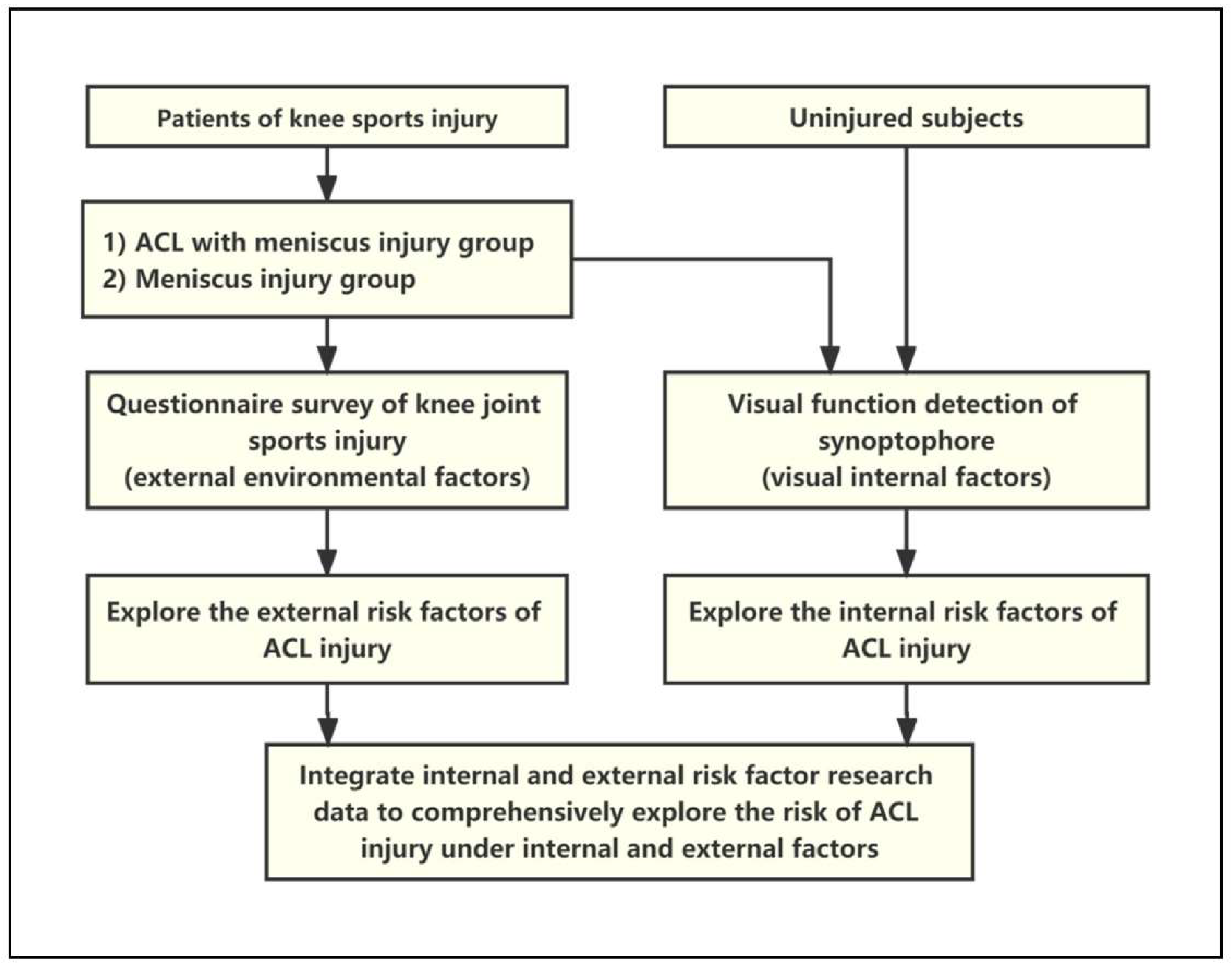
Then, in order to further explore the correlation between visual factors and ACL injury, two experienced ophthalmologists used a synoptophore to check the visual function according to the operation specifications, including dominant eye, pupil distance, binocular vision, subjective and objective oblique angle of view, fusion range and stereo visual sensitivity. This study also included participants without sports injury as the control group to reduce the statistical deviation. The data of visual examination and external factors were used for comprehensive analysis to further confirm whether the combination of visual factors and external factors increases the risk of ACL injury (Figure A1).
2.3. Statistical Analysis
Sample size calculation: the sample size estimation of this study was completed by using software (SAS 9.4 statistical software, NCSU, USA) and the sample size calculation formula of multi-group medical record control studies. We set the α level to 0.05, and the test efficiency was 0.80. The standard deviation of the data was estimated and calculated. The minimum sample size of each group was 94 participants.
Analysis of questionnaire survey results: in order to explore which external factors increase the risk of ACL injury, we used the chi-square test or t-test of two independent samples (SPSS 25.0 statistical software, IBM, USA) to compare the differences between the meniscus injury group and ACL combined with meniscus injury group. Single-factor logistic regression analysis (r-4.0.1) was used as the primary screening with p < 0.2 as the standard. The items that met the primary screening criteria of single-factor logistic regression analysis were continued to carry out multi-factor logistic regression analysis (r-4.0.1) and further explore the environmental external risk factors of ACL injury with p < 0.05 as the standard.
Statistical analysis of visual examination results: in order to explore the visual risk factors of ACL injury, the chi-square test or analysis of variance (ANOVA) (SPSS 25.0 statistical software, IBM, USA) was used to compare the differences among meniscus injury group, ACL combined with meniscus injury group and uninjured control group. Univariate and multivariate logistic regression analysis (r-4.0.1) were used to explore the visual internal risk factors of ACL injury.
Comprehensive analysis of integrated data: in order to explore the risk of ACL injury under internal and external factors, we established a multi-factor logistic regression model to obtain the corrected OR value of each item, in which the OR value of each item approximately reflected the risk of ACL injury. The product of OR values approximately reflected the ACL damage risk when they existed together.
3. Results
3.1. Eye Disease Is a Major Risk Factor of ACL Injury from Questionnaires
A total of 287 patients were included in this part of the study, of which 133 patients had an ACL injury (ACL-D group, 46.34%), and 154 patients had no ACL injury (ACL-I group, 53.66%). The majority of participants in both groups were male; the average age was 29.14 ± 0.88 (years old). There was no significant difference between the two groups among age (p = 0.288), gender (p = 0.095), BMI (p = 0.222) and dominant side (p = 0.976) by t-test or analysis of variance test of the independent samples of the two groups (p > 0.05) (Table 1).

Table 1.
Statistical description of 287 knee injury questionnaires.
The results of the questionnaires showed that, in the ACL-D group, 72.18% of them were non-contact injuries, 63.16% on the dominant side, and 66.17% were single injuries. Regarding injury action, quick turn had the highest proportion, 24.06%; sunny day made up the highest proportion of weather factors (81.95%); and, in the site factors, the cement floor accounted for the most (38.59%).
In terms of visual-related factors, 31.58% (ACL-D) and 37.01% (ACL-I) of the two groups reported that their binocular vision was normal, while other participants had different degrees of ametropia. In addition, most of the participants had no other eye diseases. Among the participants with eye diseases, the proportion of self-reported astigmatism was the highest, accounting for 30.08% (ACL-D) and 37.66% (ACL-I), respectively (Table 1).
In the chi-square test and t-test of the two groups of independent samples, as shown in Table 1, we found that the injury action (p = 0.010) was statistically significant between the two groups (p < 0.05). Although there were differences between the two groups in terms of contact condition (p = 0.702), site conditions (p = 0.125), weather (p = 0.915), right eye vision (p = 0.389), left eye vision (p = 0.391), history of eye diseases (p = 0.069) and multiple injuries (p = 0.312), they were found to be statistically significant (p > 0.05) (Table 1).
In the logistic regression analysis, we screened out the possible risk factors related to ACL injury according to p < 0.2 in the univariate logistic regression analysis. Among them, age (p = 0.129), gender (p = 0.096), weight (p = 0.033), BMI (p = 0.055), eye disease (p = 0.095), injury behavior (p = 0.014) and site conditions (p = 0.141) met the test criteria of p < 0.2 (Table 2). Then, seven items with a p-value of less than 0.2 in univariate analysis were included in the multivariate logistic regression analysis. Through the analysis, it was found that strabismus (p = 0.035) (eye disease) and quick turn (p = 0.008) (injury action) were significantly correlated with ACL injury (Table 3). These results suggest that rapid redirection (injury action) and strabismus (eye disease) are risk factors for ACL injury.
Table 2.
Results of univariate logistic regression analysis of 287 knee injury questionnaires.
Table 3.
Results of multivariate logistic regression analysis of 287 knee injury questionnaires.
3.2. Low Visual Fusion Range Is a Major Risk Factor for ACL Injury from Visual Examination
In this study, a total of 377 people underwent visual function examination; 127 (38.46%) were in the ACL-D group, and 145 (33.69%) were in the ACL-I group.
The participants of the visual examination were all participants that were included in the questionnaire. A total of 154 people in the ACL-D group and 133 people in the ACL-I group were enrolled. Nine people in the ACL-D group and six people in the ACL-I group, a total of 15 people, were lost. The control group included 105 uninjured healthy volunteers (27.85%). There were more male participants in the three groups. The average ages of the three groups were 28.72 ± 8.81 (ACL-I group), 27.71 ± 8.23 (ACL-D group) and 27.61 ± 7.99 (control group) years old (Table 4).
Table 4.
Statistical description of 377 visual examination data.
In the ACL-I, ACL-D and control groups, we found that the main visual eye of most participants was the right eye, accounting for 83.46%, 85.52% and 83.81%, and the pupil distance was 62.06 ± 2.71 mm, 62.31 ± 2.76 mm and 63.23 ± 10.02 mm, respectively. Among the three groups, only about 45% of the participants had normal visual acuity on both sides, and most of the participants had different degrees of ametropia on at least one side.
The difference between subjective and objective squint angles can reflect the first level of stereo visual function. The larger the value, the higher the possibility of a squint. The proportion of strabismus (difference ≥ 5) in the ACL-D group (15.86%) was slightly higher than that in the ACL-I group (9.45%) and control group (9.52%). Compared with the mean value of the subjective and objective oblique angle difference between the three groups, the control group (1.69 ± 1.88°) was slightly lower than the ACL combined with meniscus injury group (1.97 ± 1.91°) and meniscus injury group (1.99 ± 2.19°).
The visual fusion range can reflect the second level of stereo visual function. The higher the value, the better the stereoscopic function. The proportion of people with abnormal visual fusion range (fusion range < 25°) in the ACL-D group (80.00%) was slightly higher than that in the ACL-I group (70.08%) and control group (71.43%). Compared with the average range of visual fusion among the three groups, the ACL-D group (17.82 ± 6.17°) was slightly lower than that of ACL-I group (20.26 ± 7.27°) and control group (22.07 ± 5.16°).
Stereoacuity can reflect the third level of visual function. The higher the value, the worse the stereoacuity. In the ACL-D group (66.90%) and ACL-I group (68.50%), the proportion of people with normal stereoacuity (stereoacuity ≤ 60″) was slightly lower than that in the control group (74.29%). The mean stereoacuity of the ACL-D group (273.93 ± 522.72″) was slightly higher than that of the ACL-I group (221.89 ± 411.15″) and control group (176.76 ± 361.42″).
The chi-square test and analysis of variance showed that the visual fusion range of the three groups was significantly different (p < 0.001), while gender (p = 0.494), age (p = 0.512), dominant eye (p = 0.883), pupil distance (p = 0.280), right visual acuity (p = 0.966) and left visual acuity (p = 0.922) were not. Meanwhile, there was no significant difference in oblique angle difference (p = 0.464) and stereopsis sensitivity (p = 0.233) (Table 4).
In the logistic regression analysis (Table 5 and Table 6), through the preliminary screening of single-factor logistic regression analysis, only the visual fusion range met the screening criteria of a p-value less than 0.2. Further, the multivariate logistic regression analysis results showed that only the visual fusion range was correlated with ACL injury when comparing the meniscus injury group (p < 0.001) and the control group (p < 0.001). These results suggest that low vision fusion range is the most likely internal risk factor for ACL injury.
Table 5.
Results of univariate logistic regression analysis of 377 visual examination results.
Table 6.
Results of multivariate logistic regression analysis of 377 visual examination results.
3.3. Low Visual Fusion Range Combined with Extrinsic Factors Increases the Risk of ACL Injury
ACL injury is caused by internal and external factors. Therefore, in order to evaluate the risk factors of ACL injury, we conducted a comprehensive analysis of the visual fusion range and external factors of injury, including site conditions, weather and action. Based on the analysis results of visual function examination data, we believe that the fusion range is the most likely risk factor affecting the increase of ACL injury risk. Therefore, we replaced the column of “eye disease” in the questionnaire data with the data of fusion range and conducted logistic regression analysis again. The results showed that visual fusion range (p = 0.003) and injury action, especially quick turn (p = 0.001), sudden stop (p < 0.001) and jump (p = 0.001), were independent risk factors for ACL injury (Table 7).
Table 7.
Results of multivariate logistic regression analysis of combined data.
Then, through the establishment of a multi-factor logistic regression model, we corrected the OR values of different items so that the product of their OR values reflected the risk of different factors at the same time. We ranked and superimposed the OR value of the visual fusion range with the first three items with the largest OR value among the external factors, such as site conditions, weather and action of injury. The results showed that the defect of visual fusion range was closely related to contact ACL injury (OR = 10.583) and non-contact ACL injury (OR = 13.208) (Table 8 and Table 9). This indicates that athletes with vision fusion range defects may have an increased risk of ACL non-contact sports injury when they make a sudden stop on wooden floor, plastic floor or cement floor on cloudy days.
Table 8.
Odd ratio (OR) value of combined risk factors (non-contact injury).
Table 9.
Odd ratio (OR) value of combined risk factors (contact injury).
4. Discussion
Through the comprehensive analysis of the external environmental factors of ACL injury collected by the questionnaire and the internal visual factors collected by visual inspection, this study found that athletes with abnormal visual fusion range have increased risk of ACL non-contact and contact injury when they perform a quick turn, sudden stop, jump or other actions on hard ground, such as wooden floor or cement ground, on cloudy days.
In this study, we found that low visual fusion range is a risk factor for ACL sports injury. When one fixes one’s eyes on an object, two slightly different images are formed in one’s retina and then are merged into one image in the cerebral cortex. The range of visual fusion can reflect this ability. It belongs to the second level of stereo visual function [11,12,13]. The larger the range of stereo vision fusion, the better the stereo vision fusion function [14].
We did not find a direct correlation between abnormal vision and ACL sports injury. At present, there is no literature to support that abnormal vision increases the risk of ACL sports injury. Vision and visual fusion range are not the same, but they are related. Some scholars believe that this is due to anisometropia [11,12,13]. This study also indicates that fusion range is closely related to ametropia. People with abnormal vision (ametropia) are more likely to have abnormal stereo vision fusion range (87.6%). Therefore, in this study, we used multivariate logistic regression analysis to eliminate the confounding factors caused by the correlation between items as much as possible.
Questionnaires and visual examination were performed to establish whether participants had strabismus. The statistical significance of the two results was inconsistent, which may be due to the fact that the questionnaire data were biased by the subjective thinking of the participants. Therefore, it cannot be concluded that strabismus is a risk factor for ACL injury.
For the site factors, we found that hard sites, such as cement floor and wood floor, are more likely to cause ACL sports injury, which is consistent with previous studies [15,16,17]. Explained from the mechanism of ACL injury, it is actually the function of ground reactive force disturbances, that is, the loading effect of sheer force on knee joint [18], and sheer force is an important risk factor for knee motion injury, especially non-contact injury of ACL [19].
For weather factors, we found that the athletes with low visual fusion range have the highest risk of ACL injury in the cloudy weather. We believe that this is because insufficient light on cloudy days increases the negative impact of abnormal fusion on proprioception balance.
For the injury action, our results showed that the action during injury is an important risk factor affecting ACL injury. The OR value of quick turn is the highest, which is consistent with the previous biomechanical studies that have shown that the actions that cause the maximum load of ACL in sports often include rapid change of direction with knee torsion [20,21,22,23]. In addition, the factors that may lead to the possibility and severity of ACL injury also include the stress and torque borne by the body, the body posture during landing, the completion of technical actions and the ground after jumping [19]. Therefore, the injury action can, indeed, affect the risk of ACL injury to a great extent. Sports that often involve such high-risk actions include football, basketball and skiing, which are also the sports with the highest incidence of ACL sports injury [24,25].
The results of this study provide a theoretical basis for the correlation between visual function and ACL injury risk and provide some sports protection guidance for athletes. Based on the conclusions of this study, we suggest that athletes, especially those with visual function defects, choose sports venues with sufficient light and soft ground, such as grass and artificial turf, and avoid fierce knee torsion. Meanwhile, this study can help sports lovers to pay more attention to the screening of stereo visual function. For the people who have been found to have stereo visual function defects, especially relating to the visual fusion range, we suggest that they should carry out standardized and targeted visual function training as soon as possible to restore stereoscopic visual function [26,27,28].
5. Conclusions
We found that abnormal visual fusion range is a risk factor for ACL injury. Athletes with abnormal visual fusion have increased risk of ACL non-contact and contact sports injury when they make a quick turn, sudden stop or jump on hard ground, such as wood floor, plastic ground or cement ground, on cloudy days.
Author Contributions
Conceptualization, Z.C. and Y.L.; methodology, Y.L. and Y.Z.; software, J.W. and X.D.; investigation, Z.Z. and C.J.; data curation, Z.C. and Y.L.; writing—original draft preparation, Y.L.; writing—review and editing, Y.L. and B.S.; visualization, C.L.; supervision, N.C.; project administration, W.L. All authors have read and agreed to the published version of the manuscript.
Funding
This study was supported by 1. National Natural Science Foundation of China [82172416, 81802172, 82022046]; 2. Natural Science Foundation of Guangdong Province [2019A1515011684, 2020A1515011322, 2022A1515011714]; 3. Natural Science Foundation of Guangdong Province for Distinguished Young Scholars [2020B1515020014].; and 4. Guangdong Science and Technology Collaborative Innovation Center for Sports [2019B110210004].
Institutional Review Board Statement
This study was approved by the medical ethics committee of Sun Yat-sen University (sysec-ky-ks-030) and registered in the China Clinical Research Registry (chictr180015581).
Informed Consent Statement
Informed consent was obtained from all subjects involved in the study.
Data Availability Statement
Not applicable.
Acknowledgments
Thank all participants of this study for their active participation and cooperation in this study.
Conflicts of Interest
The authors declare no conflict of interest.
Appendix A
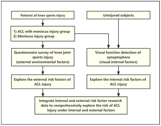
Figure A1.
Flow chart 2: Research design and data analysis process.
Figure A1.
Flow chart 2: Research design and data analysis process.
References
- Janssen, K.W.; Orchard, J.W.; Driscoll, T.R.; van Mechelen, W. High incidence and costs for anterior cruciate ligament reconstructions performed in Australia from 2003–2004 to 2007–2008: Time for an anterior cruciate ligament register by Scandinavian model? Scand. J. Med. Sci. Sports 2012, 22, 495–501. [Google Scholar] [CrossRef] [PubMed]
- von Porat, A.; Roos, E.M.; Roos, H. High prevalence of osteoarthritis 14 years after an anterior cruciate ligament tear in male soccer players: A study of radiographic and patient relevant outcomes. Ann. Rheum. Dis. 2004, 63, 269–273. [Google Scholar] [CrossRef] [PubMed]
- Lohmander, L.S.; Englund, P.M.; Dahl, L.L.; Roos, E.M. The long-term consequence of anterior cruciate ligament and meniscus injuries: Osteoarthritis. Am. J. Sports Med. 2007, 35, 1756–1769. [Google Scholar] [CrossRef] [PubMed]
- Atik, O.S.; Kaya, I. Is it possible to prevent ACL injury? Jt. Dis. Relat. Surg. 2022, 33, 263–264. [Google Scholar] [CrossRef] [PubMed]
- Hodel, S. Introducing the Lateral Femoral Condyle Index as a Risk Factor for Anterior Cruciate Ligament Injury: Response. Am. J. Sports Med. 2020, 48, NP42–NP43. [Google Scholar] [CrossRef]
- O’Connor, S.; McCaffrey, N.; Whyte, E.F.; Fop, M.; Murphy, B.; Moran, K. Can the Y balance test identify those at risk of contact or non-contact lower extremity injury in adolescent and collegiate Gaelic games? J. Sci. Med. Sport 2020, 23, 943–948. [Google Scholar] [CrossRef]
- Horoupian, D.S.; Kress, Y.; Yen, S.H.; Gaskin, F. Nickel-induced changes and reappraisal of Rosenthal fibers in focal CNS lesions. J. Neuropathol. Exp. Neurol. 1982, 41, 664–675. [Google Scholar] [CrossRef]
- Alsubaie, S.F.; Abdelbasset, W.K.; Alkathiry, A.A.; Alshehri, W.M.; Azyabi, M.M.; Alanazi, B.B.; Alomereni, A.A.; Asiri, F.Y. Anterior cruciate ligament injury patterns and their relationship to fatigue and physical fitness levels—A cross-sectional study. Medicine 2021, 100, e24171. [Google Scholar] [CrossRef]
- Tomomitsu, M.S.; Alonso, A.C.; Morimoto, E.; Bobbio, T.G.; Greve, J.M. Static and dynamic postural control in low-vision and normal-vision adults. Clinics 2013, 68, 517–521. [Google Scholar] [CrossRef]
- Helbostad, J.L.; Vereijken, B.; Hesseberg, K.; Sletvold, O. Altered vision destabilizes gait in older persons. Gait Posture 2009, 30, 233–238. [Google Scholar] [CrossRef]
- Adamek, B.; Karczewicz, D. The dependence of the range of fusion on some selected functions of the visual system. Part I: Study on convergent and divergent fusion. Klin Oczna 2006, 108, 163–166. [Google Scholar] [PubMed]
- Parker, A.J.; Smith, J.E.; Krug, K. Neural architectures for stereo vision. Philos. Trans. R. Soc. Lond. B Biol. Sci. 2016, 371, 20150261. [Google Scholar] [CrossRef] [PubMed]
- Bridge, H. Effects of cortical damage on binocular depth perception. Philos. Trans. R. Soc. Lond. B Biol. Sci. 2016, 371, 20150254. [Google Scholar] [CrossRef] [PubMed]
- Banstola, S.; Hanna, K.; O’Connor, A. Changes to Visual Parameters Following Virtual Reality Gameplay. Br. Ir. Orthopt. J. 2022, 18, 57–64. [Google Scholar] [CrossRef]
- Ngatuvai, M.S.; Yang, J.; Kistamgari, S.; Collins, C.L.; Smith, G.A. Epidemiological Comparison of ACL Injuries on Different Playing Surfaces in High School Football and Soccer. Orthop. J. Sports Med. 2022, 10, 23259671221092321. [Google Scholar] [CrossRef]
- Loughran, G.J.; Vulpis, C.T.; Murphy, J.P.; Weiner, D.A.; Svoboda, S.J.; Hinton, R.Y.; Milzman, D.P. Incidence of Knee Injuries on Artificial Turf Versus Natural Grass in National Collegiate Athletic Association American Football: 2004–2005 Through 2013–2014 Seasons. Am. J. Sports Med. 2019, 47, 1294–1301. [Google Scholar] [CrossRef]
- Balazs, G.C.; Pavey, G.J.; Brelin, A.M.; Pickett, A.; Keblish, D.J.; Rue, J.P. Risk of Anterior Cruciate Ligament Injury in Athletes on Synthetic Playing Surfaces: A Systematic Review. Am. J. Sports Med. 2015, 43, 1798–1804. [Google Scholar] [CrossRef]
- Johnson, W.R.; Mian, A.; Donnelly, C.J.; Lloyd, D.; Alderson, J. Predicting athlete ground reaction forces and moments from motion capture. Med. Biol. Eng. Comput. 2018, 56, 1781–1792. [Google Scholar] [CrossRef]
- Cronstrom, A.; Creaby, M.W.; Ageberg, E. Do knee abduction kinematics and kinetics predict future anterior cruciate ligament injury risk? A systematic review and meta-analysis of prospective studies. BMC Musculoskelet. Disord. 2020, 21, 563. [Google Scholar] [CrossRef]
- Arendt, E.; Dick, R. Knee injury patterns among men and women in collegiate basketball and soccer. NCAA data and review of literature. Am. J. Sports Med. 1995, 23, 694–701. [Google Scholar] [CrossRef]
- Arendt, E.A.; Agel, J.; Dick, R. Anterior cruciate ligament injury patterns among collegiate men and women. J. Athl. Train. 1999, 34, 86–92. [Google Scholar] [PubMed]
- Hughes, G. A review of recent perspectives on biomechanical risk factors associated with anterior cruciate ligament injury. Res. Sports Med. 2014, 22, 193–212. [Google Scholar] [CrossRef] [PubMed]
- Bates, N.A.; Schilaty, N.D.; Nagelli, C.V.; Krych, A.J.; Hewett, T.E. Validation of Noncontact Anterior Cruciate Ligament Tears Produced by a Mechanical Impact Simulator Against the Clinical Presentation of Injury. Am. J. Sports Med. 2018, 46, 2113–2121. [Google Scholar] [CrossRef] [PubMed]
- Kaeding, C.C.; Leger-St-Jean, B.; Magnussen, R.A. Epidemiology and Diagnosis of Anterior Cruciate Ligament Injuries. Clin. Sports Med. 2017, 36, 1–8. [Google Scholar] [CrossRef] [PubMed]
- Rai, S.K.; Gupta, T.P.; Singh, V.B.; Kale, A.; Vij, V.; Shaki, O. Retrospective analysis and risk of progression of partial anterior cruciate ligament injuries in a young population. Arch. Orthop. Trauma Surg. 2022. [Google Scholar] [CrossRef] [PubMed]
- Fooken, J.; Lalonde, K.M.; Mann, G.K.; Spering, M. Eye movement training is most effective when it involves a task-relevant sensorimotor decision. J. Vis. 2018, 18, 18. [Google Scholar] [CrossRef]
- Caldani, S.; Delorme, R.; Moscoso, A.; Septier, M.; Acquaviva, E.; Bucci, M.P. Improvement of Pursuit Eye Movement Alterations after Short Visuo-Attentional Training in ADHD. Brain Sci. 2020, 10, 816. [Google Scholar] [CrossRef]
- Sahraie, A.; Cederblad, A.M.H.; Kenkel, S.; Romano, J.G. Efficacy and predictors of recovery of function after eye movement training in 296 hemianopic patients. Cortex 2020, 125, 149–160. [Google Scholar] [CrossRef]
Publisher’s Note: MDPI stays neutral with regard to jurisdictional claims in published maps and institutional affiliations. |
© 2022 by the authors. Licensee MDPI, Basel, Switzerland. This article is an open access article distributed under the terms and conditions of the Creative Commons Attribution (CC BY) license (https://creativecommons.org/licenses/by/4.0/).