Article

Exploring Endophytic Bacteria from Artemisia spp. and Beneficial Traits on Pea Plants

by
Shervin Hadian
1,*,
Donald L. Smith
2,
Stanislav Kopriva
3,
Eglė Norkevičienė
4 and
Skaidrė Supronienė
1
1
Microbiology Laboratory, Lithuanian Research Centre for Agriculture and Forestry, Institute of Agriculture, Instituto Ave. 1, Akademija, LT-58344 Kėdainiai, Lithuania
2
Department of Plant Science, McGill University, Montreal, QC H9X 3V9, Canada
3
Botanical Institute, University of Cologne, 50674 Cologne, Germany
4
Department of Grass Breeding, Lithuanian Research Centre for Agriculture and Forestry, Institute of Agriculture, Instituto Ave. 1, Akademija, LT-58344 Kėdainiai, Lithuania
*
Author to whom correspondence should be addressed.
Plants 2024, 13(12), 1684; https://doi.org/10.3390/plants13121684
Submission received: 14 May 2024 / Revised: 14 June 2024 / Accepted: 15 June 2024 / Published: 18 June 2024

Abstract

:
Endophytic microorganisms represent promising solutions to environmental challenges inherent in conventional agricultural practices. This study concentrates on the identification of endophytic bacteria isolated from the root, stem, and leaf tissues of four Artemisia plant species. Sixty-one strains were isolated and sequenced by 16S rDNA. Sequencing revealed diverse genera among the isolated bacteria from different Artemisia species, including Bacillus, Pseudomonas, Enterobacter, and Lysinibacillus. AR11 and VR24 obtained from the roots of A. absinthium and A. vulgaris demonstrated significant inhibition on Fusarium c.f. oxysporum mycelial growth. In addition, AR11, AR32, and CR25 exhibited significant activity in phosphatase solubilization, nitrogen fixation, and indole production, highlighting their potential to facilitate plant growth. A comparative analysis of Artemisia species showed that root isolates from A. absinthium, A. campestris, and A. vulgaris have beneficial properties for inhibiting pathogen growth and enhancing plant growth. AR11 with 100% similarity to Bacillus thuringiensis, could be considered a promising candidate for further investigation as microbial biofertilizers. This finding highlights their potential as environmentally friendly alternatives to chemical pesticides, thereby contributing to sustainable crop protection practices.

1. Introduction

Modern agriculture encounters numerous challenges, including biotic stresses from pathogens and pests, and abiotic factors such as salinity, drought, and extreme temperatures. These issues are exacerbated by the continuous rise in the global population. The combined effects of extreme climatic events, like droughts, floods, and high temperatures, along with the unsustainable use of water resources, have led to substantial reductions in crop yields. Furthermore, agriculture also faces problems related to the loss of biodiversity and the excessive use of chemical pesticides and fertilizers. The overuse of chemical compounds resulted in pathogen resistance and a polluted environment [1]. Recent research efforts focus on developing alternative methods to reduce reliance on chemical compounds.
Endophytic bacteria colonize and inhabit the internal tissues of plants. The positive impacts of endophytes on improving plant growth and health have been extensively studied [2]. Endophytic bacteria contribute to enhanced plant growth through processes such as nitrogen fixation [3], stress alleviation [4], production of phytohormones [5], and the generation of growth-promoting signal compounds [6]. Endophytic bacteria have the capability to induce tolerance to pollutants, making them potential candidates for application in bioremediation [7]. Beyond terrestrial plants, numerous studies have pointed out that culturable endobacteria in wetland plants can serve various purposes, including bioremediation, enzyme production [8], biocontrol [9], and promotion of plant growth [10].
Artemisia L. plants are known for their many beneficial bioactive compounds, which have antimicrobial, antifungal, antioxidant, and allelopathic effects. Furthermore, endophytic bacteria associated with Artemisia plants have been identified as contributors to various advantages for their host plants. These benefits encompass improved nutrient absorption, regulation of plant phytohormones, and the ability to combat pests and pathogens. Despite extensive research on the bioactive compounds of Artemisia plant extracts, there is a significant gap in understanding the diversity of endophytic bacteria associated with Artemisia spp. and their mechanism of action [11].
The two endophytic root bacteria, Bacillus subtilis and Stenotrophomonas spp. are associated with Artemisia annua. They enhance the host plant’s nitrogen and phosphorus absorption, resulting in increased shoot height, leaf area, and overall plant biomass [12]. Ebu et al. (2023) reported the isolation of various endophytic bacteria, including Bacillus cereus, B. subtilis, Citrobacter freundii, Enterobacter asburiae, E. cloacae, E. kobei, E. ludwigii, Enterococcus faecium, and Pseudomonas monteilli from Artemisia annua, Moringa oleifera, and Ocimum lamiifolium. They found that these endophytic bacteria have the potential to solubilize insoluble forms of phosphate and zinc [13].
Fusarium oxysporum is a soil-borne phytopathogen that causes vascular wilt, root rot, and crown rot diseases on a wide range of economically significant crop plants, including tomato, banana, sweet potato, cotton, pea, and chickpea [14,15]. One of the most serious pea diseases is root rot, which is caused by F. oxysporum [16]. It caused severe damage at all stages of crop growth, with yield losses of up to 97% [17]. Endophytic bacteria isolated from various higher plants, including tomato, guava, bamboo, pine, and cacao, demonstrated significant antifungal activity against Fusarium oxysporum in tomato. Additionally, these bacteria were found to directly enhance the growth of tomato plants [18,19].
The present study aimed to determine the diversity of endophytic bacteria living in different Artemisia spp. plant tissues (A. absinthium, A. campestris, A. dubia, and A. vulgaris) by 16S rDNA gene sequence, followed by phylogenetic analysis. Evaluate their potential to inhibit the mycelial growth of pea root rot causing Fusarium sp. Assess their beneficial traits such as phosphate solubilization, nitrogen fixation, indole production, and seed germination enhancement. We emphasized the role of endophytic bacteria associated with various Artemisia species in the development of biocontrol agents aimed at reducing reliance on chemical fungicides and promoting eco-friendly agricultural practices.

2. Results

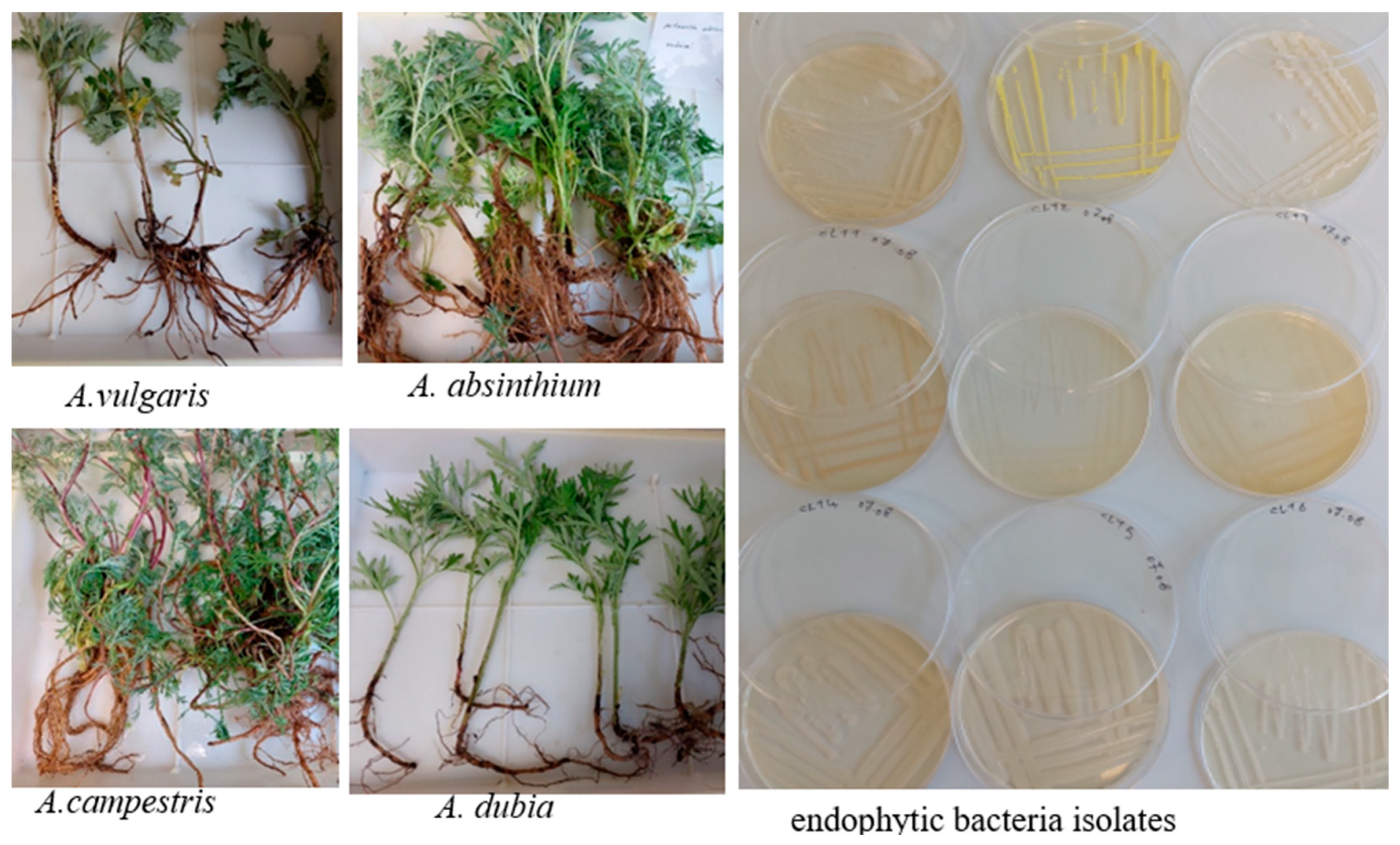
2.1. Isolation Endophytic Bacteria from Artemisia spp.

In total, 61 endophytic bacteria displaying distinct morphologies were isolated from the roots, stems, and leaves of A. absinthium, A. campestris, A. dubia, and A. vulgaris plants. Morphological and biochemical characterization of the individual strains showed that bacteria species isolated from different plant parts of Artemisia species were distinct, though some similarities were noted. Morphologically, the endophytic bacteria isolates displayed a variety of colony shapes, colors, and margins (Figure 1). Gram staining revealed that 83% of isolates were Gram-positive. Sixty-one percent of the isolates showed positive catalase activity.

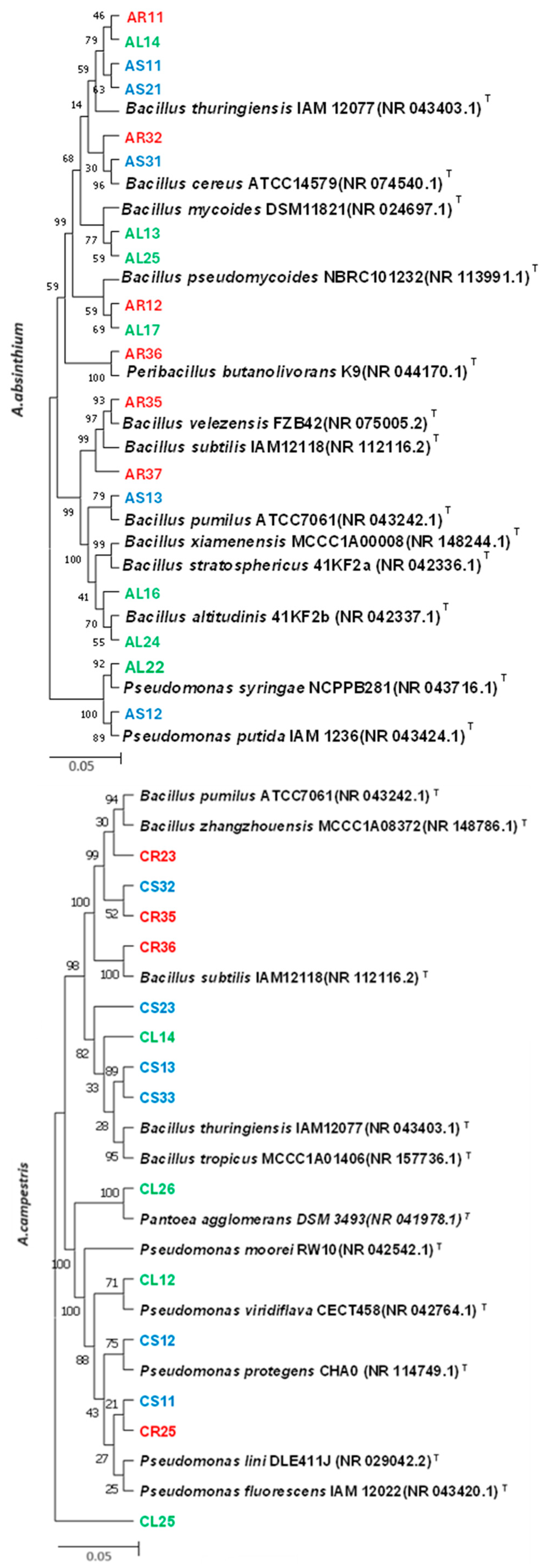
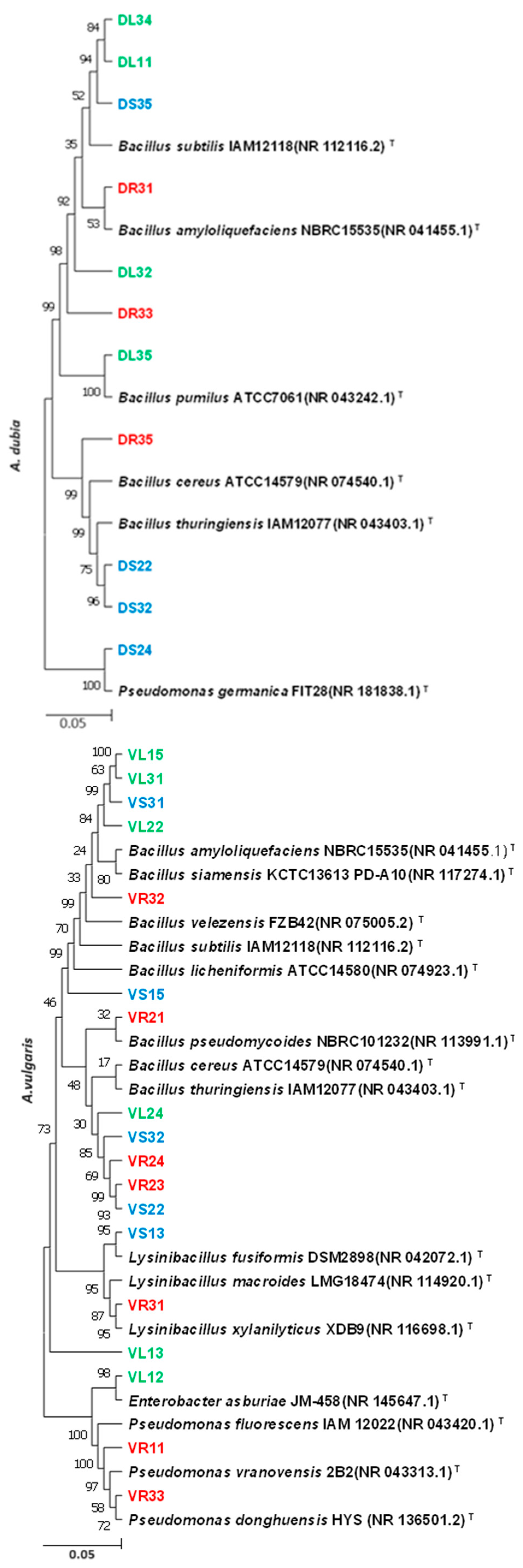
2.2. Molecular Identification of Endophytic Bacteria

Based on 16S rDNA gene sequencing, the majority of strains belonged to the phylum Proteobacteria and Firmicutes. These sequences were subsequently used for phylogenetic analyses, as shown in Figure 2. Specifically, strains isolated from A. absinthium (AR11, AR32, and AR35) showed similarity to Bacillus thuringiensis IAM12077, B. cereus ATCC14579, and B. velezensis FZB42. Strains from A. campestris (CR25 and CL14) exhibited similarity to Pseudomonas fluorescens IAM 12022 and B. thuringiensis IAM12077. Strains from A. dubia (DR31 and DR33) displayed similarity to B. amyloliquefaciens NBRC15535 and B. subtilis IAM12118. Strains from A. vulgaris (VR32 and VR24) showed similarity to B. velezensis FZB42 and B. thuringiensis IAM12077.

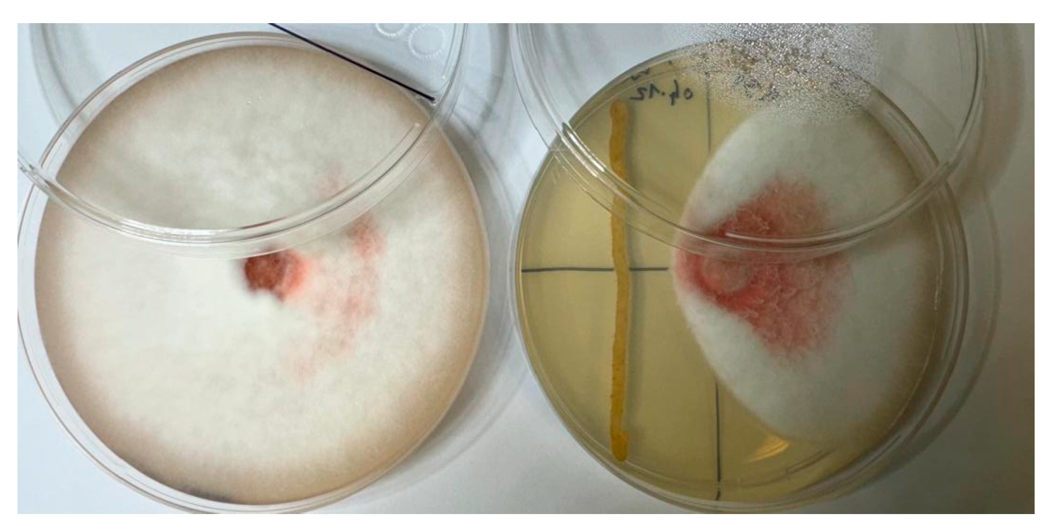
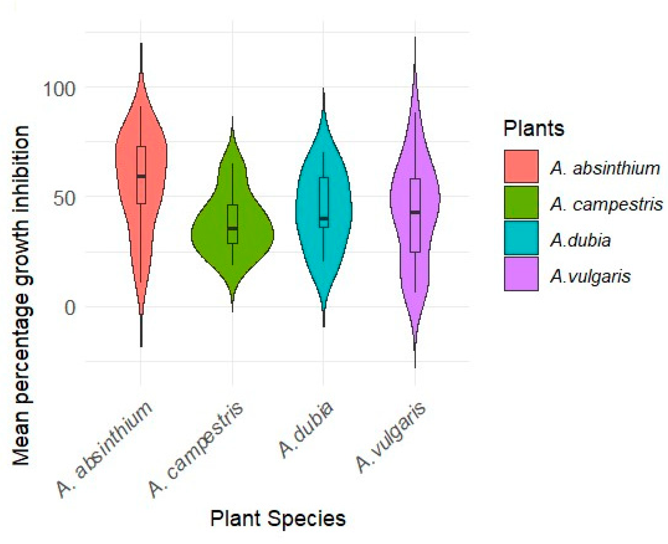
2.3. Inhibitory Effect of Endophytic Bacteria on the Growth of Fusarium c.f. oxysporum

Phytopathogenic fungi were isolated from pea roots exhibiting disease symptoms. The ability of the isolates to cause root rot in pea plants was confirmed by plant pathogenicity assays. Morphological analysis and ITS sequence revealed high sequence similarity to Fusarium oxysporum HDV247. The in vitro growth inhibition activity test F. c.f. oxysporum showed that 61 endophytic bacteria differed in their inhibitory effects on the growth of F. c.f. oxysporum mycelia (Figure 3).
Statistical analysis showed significant differences (p < 0.001) between the growth inhibition activity of 61 endophytic bacteria on F. c.f. oxysporum AR11 (90.83%) and VR24 (88%) strains isolated from the roots of A. absinthium and A. vulgaris, respectively, showed the highest inhibitory effect on F. c.f. oxysporum growth (Figure 4).
The mean growth inhibition activity values across different plant species showed that strains from A. absinthium exhibited the highest levels, followed by strains from A. dubia, A. vulgaris, and A. campestris, respectively (Figure 5). A. campestris displayed a wider range of values and growth inhibition activity compared to other Artemisia species. A. vulgaris displayed a more limited range of values for growth inhibition.

2.4. Phosphate Solubilization Index

The phosphate solubilization was assessed on Pikovskaya agar. Several isolates exhibited distinct halo zones around their colony (Figure 6). The halo diameter varied among the different strains.
The phosphate solubilization index (PSI) values represent the ability of these endophytic bacteria to solubilize phosphate over time, showing that 36% of isolates demonstrated phosphate solubilization activity. Specifically, 61% of the isolates from A. absinthium, 35% from A. campestris, 27% from A. dubia, and 22% from A. vulgaris showed phosphate solubilization activity (Figure 7).
Strain AR11 displayed the highest PSI after 10 days of incubation (2.93) whereas isolates from A. campestris showed lower PSI values compared to those from A. absinthium.

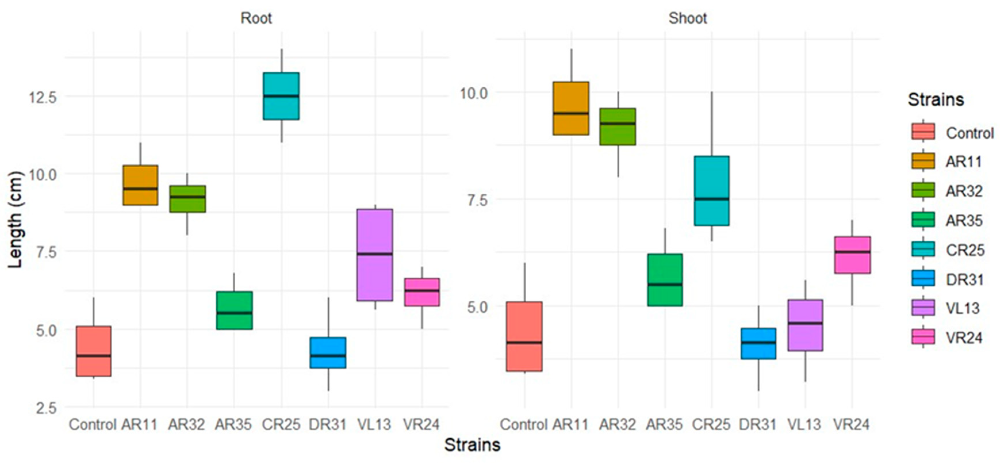
2.5. Biochemical Properties and Effect of the Endophytic Bacteria on Pea Plant Growth

In this study, 20% of the isolated bacteria exhibited positive results in the indole production test, while 44% demonstrated nitrogen fixation potential. Based on these and other test results, eight isolated strains were selected to study their effects on pea seed germination and growth promotion. The selected isolates are listed in Table 1.
The analysis of seed germination across various endophytic bacteria revealed that germination started within 48 h. The inoculation with VS32 suppressed pea seed germination. However, the seeds inoculated with AR11 and AR32 exhibited 100% seed germination rates. VL13 showed the least effect on seed germination, with only a 78% rate observed. There were no significant differences between isolates for seed germination.
Although some strains, like CR25, AR11, AR32, and VR24 did not exhibit a significant increase in root length, there was significant lateral root branching. DR31 not only did not make any significant effects on increasing shoot and root length but also demonstrated no positive impact on root branching (Figure 8).
AR11 and CR25 exhibited the most significant (p < 0.05) impact on root and shoot length compared to the control. While AR32 had a smaller effect on root length than CR25 (no significant difference), it displayed a greater effect on shoot length compared to CR25. DR31 did not exhibit any significant effect on pea root and shoot length compared to the control (Figure 9).

3. Discussion

Endophytic bacteria play a crucial role in influencing the essential functions of host plants. They can enhance plant growth and induce resistance against pathogens. Utilizing endophytes in crop production has the potential to decrease the need for fertilizers and pesticides, thereby enhancing the sustainability of crop cultivation [20]. Non-native endophytes have been studied for their potential use as biofertilizers. For example, researchers have evaluated endophytic rhizobia from clover roots for their effectiveness in enhancing rice growth as biofertilizers [21]. In this research, we studied how non-native endophytic bacteria isolated from Artemisia sp. suppress Fusarium c.f. oxysporum extracted from pea root and promote pea plant growth.

3.1. Identification of Isolated Endophytic Bacteria

Plants greatly benefit from endophytic bacteria, which stay in host plant tissues and have no negative effects [22]. Medicinal plants like Artemisia annua can host a variety of functionally significant endophytic bacteria in their leaves, stems, and roots [23]. Ashitha et al. (2019) reported a diverse community of endophytic bacteria, including Arthrobacter sp., Pseudomonas sp., Microbacterium sp., Psychrobacter sp., Enterobacter sp., Bacillus sp., Kosakonia cowanii, and Bacillus sp. associated with the medicinal plant A. nilagirica [24]. These findings demonstrate the remarkable diversity of endophytic bacteria found in various Artemisia species. In our investigation, we observed that the majority of effective endophytic bacteria belonged to the Bacillus and Pseudomonas genera. Among the endophytic bacteria identified, Bacillus cereus, B. subtilis, and B. thuringiensis were commonly found across all four Artemisia species, indicating their widespread presence in these plant ecosystems. B. altitudinis and B. mycoides were mostly associated with A. absinthium.

3.2. Antifungal Activity of Endophytic Bacteria

In our study, isolates that exhibited significant growth inhibition of Fusarium c.f. oxysporum were identified as Bacillus thuringiensis, B. cereus, B. velezensis, B. amyloliquefaciens, and Pseudomonas fluorescens. The antagonistic activity of these endophytic bacteria against F. oxysporum has been well-documented. For instance, B. thuringiensis has been reported to inhibit the growth of F. oxysporum in peanut and onion plants [25,26], B. cereus strains in tomato [27], B. velezensis in cucumber [28], B. amyloliquefaciens in banana [29], and Pseudomonas fluorescens in watermelon [30]. These findings align with our results, highlighting the potential of these endophytic bacteria as effective biocontrol agents against root rot pathogens.

3.3. Plant Growth-Promoting Characteristics

The biochemical properties of the isolated endophytic bacteria provided essential insights into their potential applications as plant growth-promoting agents. The capacity for nitrogen fixation and phosphate solubilization, along with indole production, were critical factors evaluated in this study. Strain AR11 exhibited strong phosphate solubilization, nitrogen fixation abilities, and increased plant growth. These multifunctional traits suggest that AR11 could be a plant growth promoter, aligning with previous studies that have highlighted B. thuringiensis as a powerful phosphate-solubilizing bacterium [31], nitrogen fixer [32] and effective plant growth-promoter [33]. Seed germination started within 48 h post-inoculation with selected endophytic bacteria, indicating their potential as biofertilizers. This is consistent with findings from previous studies where endophytic bacteria like Bacillus spp. and Pseudomonas spp. significantly enhanced seed germination and early seedling growth of tomatoes [34]. Conversely, strain VS32 suppressed pea seed germination, which suggests that not all endophytic bacteria have beneficial effects on germination and could have strain-specific or plant-specific interactions [35,36]. In terms of root development, strains CR25, AR11, AR32, and VR24 notably increased lateral root branching, although they did not significantly enhance root length. Lateral root branching is essential for establishing a resistant root system capable of better nutrient absorption [37]. The significant increase in root branching observed with CR25 (Pseudomonas syringae), indicates its substantial impact on root system architecture. This aligns with reported studies showing that Pseudomonas spp. modify root architecture and promote growth in Arabidopsis thaliana [38].

4. Materials and Methods

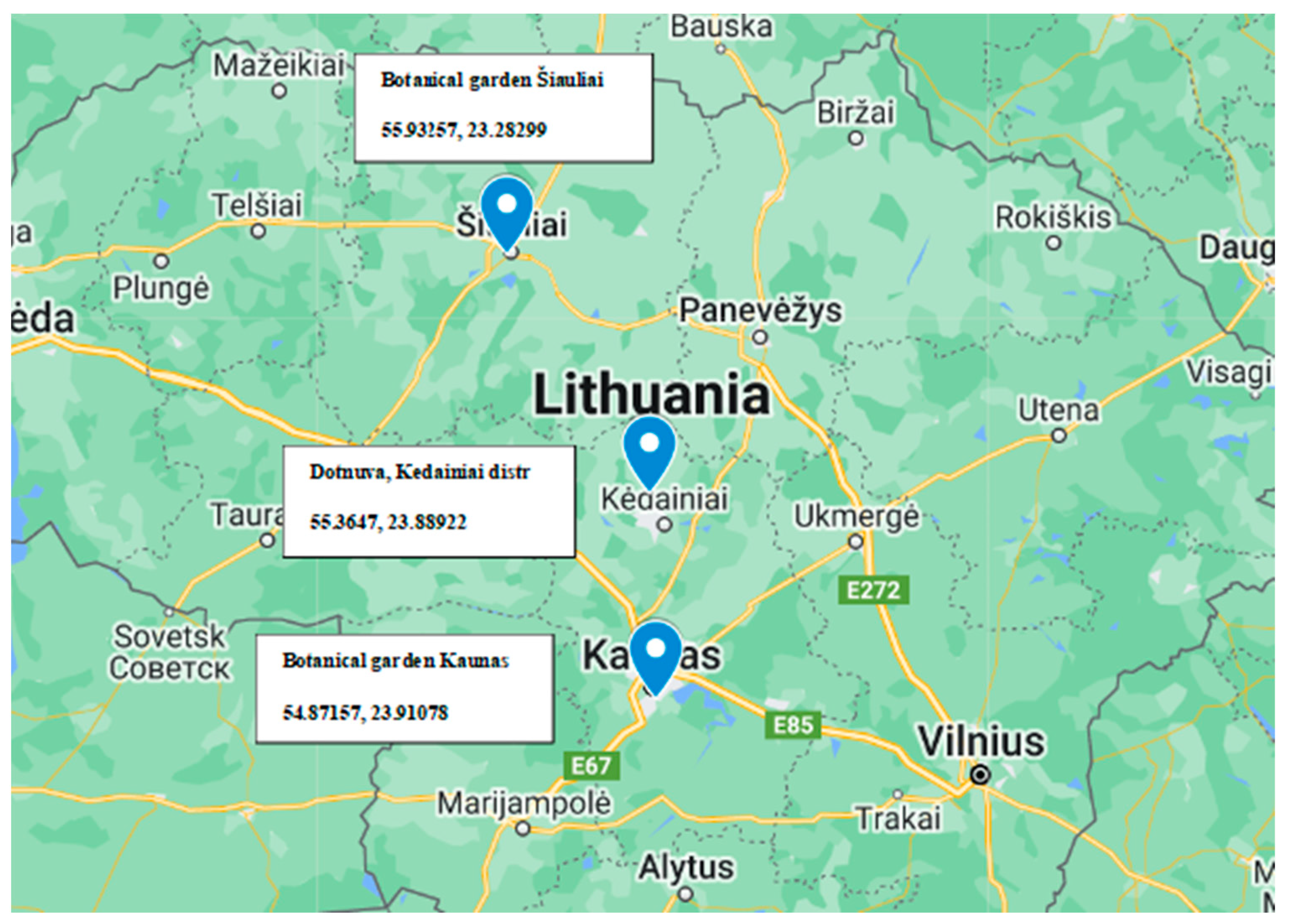
4.1. Plant Sample Collection

This study examined four species within the Artemisia genus: A. absinthium L., A. campestris L., and A. dubia Wall. ex-Besser, and A. vulgaris L. Among these, A. absinthium, A. campestris, and A. vulgaris are indigenous to the Lithuanian flora while A. dubia is introduced and cultivated. Plants from all tested species were collected in dry perennial anthropogenic herbaceous vegetation habitats at three distinct locations in Lithuania (Kaunas, Kėdainiai, Šiauliai) Figure 10, during the month of June 2021. Collected plants were at the growth stage of leaf development (main shoot). Subsequently, in the laboratory, the plant samples underwent thorough washing with water to eliminate all soil debris before being dried at room temperature. Surface disinfection was accomplished using aseptic immersion in 70% ethanol for 2 min after 5% sodium chloride. Subsequently, the samples were immersed in hypochlorite for 4 min and thoroughly rinsed using sterile distilled water, followed by a 2 min contact with 70% ethanol. Finally, after rinsing with sterile distilled water again, allowed to dry under sterile conditions. To ensure that all epiphytes were eliminated throughout the surface sterilization procedure, 100 µL of water from the final wash step was added onto the nutrient agar and incubated at 28 °C for 7 days and monitored for bacteria growth [17].

4.2. Isolation Endophytic Bacteria

Plant parts (stem, root, leaf) of each species that had been surface-sterilized were cut into small pieces. Endophytic microorganisms were isolated through a direct planting technique. Each plant part was cut into 1 × 1 cm sections and placed in Petri dishes containing TSA (tryptophan soy agar) medium. The plates were incubated at 28 °C for up to 15 days. On days 2, 5, 10, and 15, colonies were chosen and purified in TSA. Each plate was incubated for 14 days at a temperature of 27–29 °C. The colonies were chosen for each Petri dish based on their growth stage and morphology (color, size, and shape).

4.3. Molecular Identification of Isolated Endophytic Bacteria by 16srDNA

4.3.1. DNA Extraction from Bacteria Strains

The DNA extraction from isolated bacteria followed the CTAB DNA extraction protocol provided by Prof. Thorsten Mascher’s lab [39]. Bacteria were cultured in a 10 mL LB medium and incubated at 37 °C with shaker speed at 200 rpm until reaching an optical density (OD600) of 0.8–1.0. The cells were then harvested by centrifugation at 5000 rpm for 10 min at room temperature (RT). The cell pellet was resuspended in 400 µL of TEN buffers (10 mM Tris/HCl pH 8.0, 10 mM EDTA, 150 mM NaCl) in a 2 mL Eppendorf tube, to which 20 µL of lysozyme was added and incubated at 37 °C for 20 min. Subsequently, 2 µL of RNase was added and incubated at 65 °C for 3 min. Afterward, 40 µL of SDS, 1 µL of proteinase K, and 550 µL of TEN* buffer (10 mM Tris/HCl pH 8.0, 1 mM EDTA, 50 mM NaCl) were added, mixed thoroughly, and incubated at 60 °C for 2 h. Next, 900 µL of phenol (equilibrated with TE buffer, pH 7.5–8.0) was added, and the mixture was inverted to ensure thorough mixing before centrifugation at 13,000 rpm for 5 min. The upper phase was carefully transferred into a new 1.5 mL Eppendorf tube. This extraction process was repeated once with phenol and twice with chloroform: isoamyl alcohol (24:1). The final aqueous phase was transferred to a test tube containing 10 mL of −20 °C cold ethanol, where the DNA precipitated. After air drying, the DNA was dissolved in 100 µL of TEN* buffer and incubated overnight at 4 °C. The DNA quality and quantity were assessed with Nanodrop and Qubit analyses.

4.3.2. Taxonomic Identification

The bacteria 16S rDNA was amplified by PCR by universal primer pair 27F 5′-(AGAGTTTGATCMTGGCTCAG)-3′, and 1387R 5′-(GGGCGGWGTGTACAAG GC)-3′ [40]. The PCR amplification was conducted in a PCR thermal cycler (Bio-Rad My cycler, Berkeley, CA, USA), and comprised an initial denaturation at 95 °C for 5 min, followed by 25 cycles of 95 °C for 30 s, 56 °C for 30 s, 72 °C for 1.5 min, on the last cycle. The PCR amplicons were purified with the Gene JET PCR Purification Kit (Thermo Scientific, Dreieich, Germany) and the amplicons sequencing was conducted with Applied Biosystems 3730XL DNA Analyzer by BMR GENOMIC company (Padova, Italy). Retrieved DNA sequences were manually edited using Bio edit Sequence Alignment Editor Software (Version 7.2.5). The similarities of 16S rDNA genes between isolated endophytes and their phylogenetic neighbors were determined by using Basic Local Alignment Search Tool (BLAST) [41]. Multiple alignments were conducted using the CLUSTAL W Program in Bio edit Sequence Alignment Editor Software (Version 7.2.5) [42]. A phylogenetic tree based on 16S rRNA gene sequences was constructed by the neighbor-joining method, using a distance algorithm with a bootstrap of 1000, with Molecular Evolutionary Genetics Analysis (MEGA X) software version 11 [43]. The evolutionary distances were computed using the maximum composite likelihood method and were in the units of the number of base substitutions per site.

4.4. Pathogenic Fungi Isolation

Pathogenic fungi were obtained from diseased pea roots. Following surface sterilization, the samples were air-dried on sterile filter paper. Sections of roots, approximately 4 × 1 cm in size, were then placed onto Petri dishes containing potato dextrose agar (PDA) isolation medium. After obtaining pure cultures, they were placed at 25 °C for 5 days to grow before morphological evaluation. Microscopic observations of the two isolated fungal cultures were conducted using a light microscope and consisted of an evaluation of the septum and conidia size, shape, and color. Colony morphology was evaluated for colony color and growth pattern. After identification based on morphological characteristics, the ITS3f (5′-GCATCGATGAAGAACGCAGC-3′) and ITS4r (5′-TCCTCCGCTTATTGATATGC-3′) [44] primers were employed for molecular identification of the ITS region of pathogenic fungi. To assess the pathogenicity of each isolate, we utilized the pathogenicity test on pea plants [45].

4.5. Growth Inhibition Effect of Endophytic Bacteria on Pea Root Rot Pathogen

The growth inhibition activity of the isolated bacteria was evaluated by using the dual plate culture technique. Bacteria grown on the TSA for 24 h were used. The fungi were initially inoculated in the center of the plate with PDA medium and allowed to grow for one day. The bacteria strains were co-inoculated on the same agar plate at a distance of 2.5 cm from the fungal strains. The plates were then incubated at 28 °C for 7 days. The percentage growth inhibition of fungal mycelium compared to the control. This calculation was conducted after 7 days using the formula: (R1 − R2)/R1 × 100, where R1 and R2 represented the radial growth of the pathogen in the absence and presence of the antagonist, respectively.

4.6. Evaluation of Isolates for Their Plant Growth Promotion Potential

4.6.1. Indole Production Test

The strains were inoculated to evaluate tubes with one 1% tryptone broth and were incubated at 28 °C for 48 h. Further, 1 mL of Kovac’s reagent (hydrochloric acid and p-dimethylaminobenzaldehyde in amyl alcohol) was added to each tube, the tubes were shacked continuously for 10 to 15 min, then, they were left to settle so the reagent could rise to the surface. Tubes were checked for the reddening of the alcohol layer within a few minutes, which indicated indole production [46].

4.6.2. Phosphate Solubilization Activity of Endophytic Bacteria

Based on Pikovskaya [47] methods further created by Gupta et al. [48] which are widely applied as an evaluation tool [49], we assessed the potential of the isolates to solubilize phosphate on Pikovskaya’s agar medium (PVK). The PVK solid medium contains blue bromophenol, which produces halos around the colonies due to organic acid production. After inoculating fresh bacteria suspension (5 × 10⁸ CFU ml−1) on PVK agar media, the solubilization halo diameter around the colonies was estimated to determine bacteria solubilization ability. The solubilization index (SI) was calculated after 2, 7, and 10 days of incubation at 28 ± 2 °C using the following formula:
S I = C D + H D C D
where CD is the colony diameter, and HD is the halo zone diameter. All experiments were conducted with three repetitions [50].

4.6.3. Nitrogen Fixation Activity of Endophytic Bacteria

The nitrogen fixation test was conducted by utilizing Ashby’s N-free medium (NFM), composed of 10 g of mannitol, 0.2 g of KH2PO4, 0.2 g of MgSO4·7H2O, 0.2 g of NaCl, 0.1 g of CaSO4·2H2O, 5 g of CaCO3, and adjusted to pH 7.0–7.5 with 1.8 g of agar in 1 L of distilled water [51]. Bacteria strains displaying normal growth on NFM plates were identified as potential nitrogen fixers [52]. Escherichia coli and Rhizobium bacteria are considered as the negative and positive control, respectively.

4.6.4. Impact of Endophytic Bacteria on Pea Seed Germination

Seed Inoculation with Endophytic Bacteria

The seeds were surface sterilized in 70% (v/v) ethanol for 1 min followed by 10 min putting in 1% (v/v) sodium hypochlorite solution (Naclo-74.44 g/mol) and then washed six–seven times with sterile distilled water.
The suspensions of bacteria were prepared in LB medium and incubated overnight at 28 °C. After centrifugation at 5000× g for 5 min, the pellets were rinsed 2–3 times with sterile distilled water to remove any residual culture medium. Diluted with sterile 0.03 M MgSO4 and adjusted the concentration to 108 cfu mL–1 by spectrophotometer at 600 nm. Pea seeds were soaked in a suspension of bacteria containing sterile 0.03 M MgSO4 for 4 h. The seeds were air-dried under laminar airflow conditions for 2 h.

Determination of Seed Germination and Plant Growth

The seed germination test was conducted by placing four inoculated seeds in transparent test containers. The transparent test containers were filled with wet sand and covered with black filter paper according to the Phytotoxkit protocol [53]. The seeds were put 0.5–1.0 cm from the top of the transparent containers. Four containers were used for each treatment, and they were placed in a growth chamber (16 h light/ 8 h darkness) at 23 °C. The percentage of germination was recorded after 48 h. Root and shoot length were measured after the ten days.

4.7. Statistical Analysis

In this study, statistical analyses were conducted through analysis of variance (ANOVA) using R studio statistical software (v 4.2.3). A post hoc test (Tukey’s HSD) was performed to test the significance of the differences among the treatments (p < 0.05).

5. Conclusions

This study underscored the significant pathogen inhibition and plant growth enhancement potential of endophytic bacteria isolated from different Artemisia species. The diversity of endophytic bacteria, including strains of Bacillus and Pseudomonas, highlights their functional importance in promoting plant health. The growth inhibition activity against Fusarium c.f. oxysporum by strains like B. thuringiensis, B. cereus, B. velezensis, B. amyloliquefaciens, and Pseudomonas fluorescens underscores their role as effective biocontrol agents. Furthermore, the biochemical properties, including phosphate solubilization, nitrogen fixation, and indole production, particularly by strain AR11, demonstrate their potential as plant growth-promoting agents. AR11 emerged as a promising candidate for further investigation as a microbial biofertilizer, potentially offering an environmentally friendly alternative to chemical pesticides for crop protection.

Author Contributions

S.H., S.S., S.K., D.L.S. and E.N.; methodology, S.H.; software, S.H. and S.K.; validation, S.H. and S.S. formal analysis, S.H. and S.S.; investigation, S.H., S.S. and E.N.; resources, S.H.; data curation, S.H.; writing—original draft preparation, S.H., S.S., S.K., D.L.S. and E.N.; writing—review and editing, S.H.; visualization, S.S. and D.L.S.; supervision, D.L.S. and S.S.; funding acquisition. All authors have read and agreed to the published version of the manuscript.

Funding

This research received no external funding.

Data Availability Statement

Data are contained within the article.

Conflicts of Interest

The authors declare no conflict of interest.

References

  1. Bódalo, A.; Borrego, R.; Garrido, C.; Bolivar-Anillo, H.J.; Cantoral, J.M.; Vela-Delgado, M.D.; González-Rodríguez, V.E.; Carbú, M. In Vitro Studies of Endophytic Bacteria Isolated from Ginger (Zingiber officinale) as Potential Plant-Growth-Promoting and Biocontrol Agents against Botrytis cinerea and Colletotrichum acutatum. Plants 2023, 12, 4032. [Google Scholar] [CrossRef] [PubMed]
  2. Morales-Cedeño, L.R.; Orozco-Mosqueda, M.d.C.; Loeza-Lara, P.D.; Parra-Cota, F.I.; de los Santos-Villalobos, S.; Santoyo, G. Plant Growth-Promoting Bacterial Endophytes as Biocontrol Agents of Pre- and Post-Harvest Diseases: Fundamentals, Methods of Application and Future Perspectives. Microbiol. Res. 2021, 242, 126612. [Google Scholar] [CrossRef] [PubMed]
  3. Yang, L.-L.; Jiang, Z.; Li, Y.; Wang, E.-T.; Zhi, X.-Y. Plasmids Related to the Symbiotic Nitrogen Fixation Are Not Only Cooperated Functionally but also May Have Evolved over a Time Span in Family Rhizobiaceae. Genome Biol. Evol. 2020, 12, 2002–2014. [Google Scholar] [CrossRef]
  4. Fan, D.; Smith, D.L. Characterization of Selected Plant Growth-Promoting Rhizobacteria and Their Non-Host Growth Promotion Effects. Microbiol. Spectr. 2021, 9, e0027921. [Google Scholar] [CrossRef] [PubMed]
  5. Duca, D.; Lorv, J.; Patten, C.L.; Rose, D.; Glick, B.R. Indole-3-Acetic Acid in Plant–Microbe Interactions. Antonie Van. Leeuwenhoek 2014, 106, 85–125. [Google Scholar] [CrossRef] [PubMed]
  6. Prudent, M.; Salon, C.; Souleimanov, A.; Emery, R.J.N.; Smith, D.L. Soybean Is Less Impacted by Water Stress Using Bradyrhizobium japonicum and Thuricin-17 from Bacillus thuringiensis. Agron. Sustain. Dev. 2015, 35, 749–757. [Google Scholar] [CrossRef]
  7. Pham, V.H.T.; Kim, J.; Chang, S.; Chung, W. Bacterial Biosorbents, an Efficient Heavy Metals Green Clean-Up Strategy: Prospects, Challenges, and Opportunities. Microorganisms 2022, 10, 610. [Google Scholar] [CrossRef] [PubMed]
  8. Ho, Y.-N.; Mathew, D.C.; Hsiao, S.-C.; Shih, C.-H.; Chien, M.-F.; Chiang, H.-M.; Huang, C.-C. Selection and Application of Endophytic Bacterium Achromobacter xylosoxidans Strain F3B for Improving Phytoremediation of Phenolic Pollutants. J. Hazard. Mater. 2012, 219–220, 43–49. [Google Scholar] [CrossRef] [PubMed]
  9. Sakai, M.; Hosoda, A.; Ogura, K.; Ikenaga, M. The Growth of Steroidobacter agariperforans sp. Nov., a Novel Agar-Degrading Bacterium Isolated from Soil, Is Enhanced by the Diffusible Metabolites Produced by Bacteria Belonging to Rhizobiales. Microbes Environ. 2014, 29, 89–95. [Google Scholar] [CrossRef]
  10. Zhang, X.; Chen, L.; Liu, X.; Wang, C.; Chen, X.; Xu, G.; Deng, K. Synergic Degradation of Diesel by Scirpus triqueter and Its Endophytic Bacteria. Environ. Sci. Pollut. Res. 2014, 21, 8198–8205. [Google Scholar] [CrossRef]
  11. Hadian, S.; Supronienė, S. The Potential of Artemisia spp. Plant Extracts and Endophytic Bacteria to Increase Plant Productivity: A Review. Zemdirb. Agric. 2023, 110, 87–94. [Google Scholar] [CrossRef]
  12. Awasthi, A.; Bharti, N.; Nair, P.; Singh, R.; Shukla, A.K.; Gupta, M.M.; Darokar, M.P.; Kalra, A. Synergistic Effect of Glomus mosseae and Nitrogen Fixing Bacillus subtilis Strain Daz26 on Artemisinin Content in Artemisia annua L. Appl. Soil Ecol. 2011, 49, 125–130. [Google Scholar] [CrossRef]
  13. Ebu, S.M.; Adem, M.A.; Dekebo, A.; Olani, A. Isolation and Identification of Endophytic Bacterial Isolates from the Leaves, Roots, and Stems Parts of Artemisia annua, Moringa oleifera, and Ocimum lamiifolium Plants. Curr. Microbiol. 2023, 80, 405. [Google Scholar] [CrossRef] [PubMed]
  14. Gordon, T.R. Fusarium oxysporum and the Fusarium Wilt Syndrome. Annu. Rev. Phytopathol. 2017, 55, 23–39. [Google Scholar] [CrossRef] [PubMed]
  15. Edel-Hermann, V.; Lecomte, C. Current Status of Fusarium oxysporum Formae Speciales and Races. Phytopathology 2019, 109, 512–530. [Google Scholar] [CrossRef] [PubMed]
  16. Nazir, N.; Badri, Z.A.; Bhat, N.A.; Bhat, F.A.; Sultan, P.; Bhat, T.A.; Rather, M.A.; Sakina, A. Effect of the Combination of Biological, Chemical Control and Agronomic Technique in Integrated Management Pea Root Rot and Its Productivity. Sci. Rep. 2022, 12, 11348. [Google Scholar] [CrossRef] [PubMed]
  17. El-Saadony, M.T.; Saad, A.M.; Najjar, A.A.; Alzahrani, S.O.; Alkhatib, F.M.; Shafi, M.E.; Selem, E.; Desoky, E.-S.M.; Fouda, S.E.E.; El-Tahan, A.M.; et al. The Use of Biological Selenium Nanoparticles to Suppress Triticum aestivum L. Crown and Root Rot Diseases Induced by Fusarium Species and Improve Yield under Drought and Heat Stress. Saudi J. Biol. Sci. 2021, 28, 4461–4471. [Google Scholar] [CrossRef]
  18. Sriwati, R.; Maulidia, V.; Intan, N.; Oktarina, H.; Syamsuddin; Khairan, K.; Skala, L.; Mahmud, T. Endophytic Bacteria as Biological Agents to Control Fusarium Wilt Disease and Promote Tomato Plant Growth. Physiol. Mol. Plant Pathol. 2023, 125, 101994. [Google Scholar] [CrossRef]
  19. Maulidia, V.; Sriwati, R.; Soesanto, L.; Syamsuddin; Hamaguchi, T.; Hasegawa, K. Endophytic Bacteria Isolated from Higher Plant in Aceh, Indonesia, and Their Chemical Compounds Activity against Fusarium oxysporum f. sp. lycopersici. Egypt. J. Biol. Pest. Control 2021, 31, 31. [Google Scholar] [CrossRef]
  20. Ghimire, K.; Peta, V.; Bücking, H.; Caffe, M. Effect of Non-Native Endophytic Bacteria on Oat (Avena sativa L.) Growth. Int. J. Plant Biol. 2023, 14, 827–844. [Google Scholar] [CrossRef]
  21. Yanni, Y.G.; Dazzo, F.B.; Zidan, M.I. Beneficial Endophytic Rhizobia as Biofertilizer Inoculants for Rice and the Spatial Ecology of This Bacteria–Plant Association. In Bacteria in Agrobiology: Crop Ecosystems; Springer: Berlin/Heidelberg, Germany, 2011; pp. 265–294. [Google Scholar] [CrossRef]
  22. Wu, W.; Chen, W.; Liu, S.; Wu, J.; Zhu, Y.; Qin, L.; Zhu, B. Beneficial Relationships Between Endophytic Bacteria and Medicinal Plants. Front. Plant Sci. 2021, 12, 646146. [Google Scholar] [CrossRef] [PubMed]
  23. Husseiny, S.; Dishisha, T.; Soliman, H.A.; Adeleke, R.; Raslan, M. Characterization of Growth Promoting Bacterial Endophytes Isolated from Artemisia annua L. S. Afr. J. Bot. 2021, 143, 238–247. [Google Scholar] [CrossRef]
  24. Ashitha, A.; Midhun, S.J.; Sunil, M.A.; Nithin, T.U.; Radhakrishnan, E.K.; Mathew, J. Bacterial Endophytes from Artemisia nilagirica (Clarke) Pamp., with Antibacterial Efficacy against Human Pathogens. Microb. Pathog. 2019, 135, 103624. [Google Scholar] [CrossRef] [PubMed]
  25. Du, H.; Li, C. Study on the Mechanism of Peanut Resistance to Fusarium oxysporum Infection Induced by Bacillus thuringiensis TG5. Front. Microbiol. 2024, 14, 1251660. [Google Scholar] [CrossRef] [PubMed]
  26. Shin, J.-H.; Lee, H.-K.; Lee, S.-C.; Han, Y.-K. Biological Control of Fusarium Oxysporum, the Causal Agent of Fusarium Basal Rot in Onion by Bacillus spp. Plant Pathol. J. 2023, 39, 600–613. [Google Scholar] [CrossRef] [PubMed]
  27. Pazarlar, S.; Madriz-Ordeñana, K.; Thordal-Christensen, H. Bacillus cereus EC9 Protects Tomato against Fusarium Wilt through JA/ET-Activated Immunity. Front. Plant Sci. 2022, 13, 1090947. [Google Scholar] [CrossRef] [PubMed]
  28. Abdelkhalek, A.; Behiry, S.I.; Al-Askar, A.A. Bacillus velezensis PEA1 Inhibits Fusarium oxysporum Growth and Induces Systemic Resistance to Cucumber Mosaic Virus. Agronomy 2020, 10, 1312. [Google Scholar] [CrossRef]
  29. Cruz Martín, M.; Leyva, L.; Acosta Suárez, M.; Pichardo, T.; Bermúdez Caraballoso, I.; Alvarado Capó, Y. Antifungal Activity of Bacillus amyloliquefaciens against Fusarium oxysporum f. sp. Cubense Race 1. Agron. Mesoam. 2021, 32, 466–478. [Google Scholar] [CrossRef]
  30. Meyer, S.L.F.; Everts, K.L.; Gardener, B.M.; Masler, E.P.; Abdelnabby, H.M.E.; Skantar, A.M. Assessment of DAPG-Producing Pseudomonas fluorescens for Management of Meloidogyne incognita and Fusarium oxysporum on Watermelon. J. Nematol. 2016, 48, 43–53. [Google Scholar] [CrossRef]
  31. Delfim, J.; Dijoo, Z.K. Bacillus thuringiensis as a Biofertilizer and Plant Growth Promoter. In Microbiota and Biofertilizers; Springer International Publishing: Cham, Switzerland, 2021; Volume 2, pp. 251–265. [Google Scholar] [CrossRef]
  32. Berza, B.; Sekar, J.; Vaiyapuri, P.; Pagano, M.C.; Assefa, F. Evaluation of Inorganic Phosphate Solubilizing Efficiency and Multiple Plant Growth Promoting Properties of Endophytic Bacteria Isolated from Root Nodules Erythrina brucei. BMC Microbiol. 2022, 22, 276. [Google Scholar] [CrossRef]
  33. de Almeida, J.R.; Bonatelli, M.L.; Batista, B.D.; Teixeira-Silva, N.S.; Mondin, M.; dos Santos, R.C.; Bento, J.M.S.; de Almeida Hayashibara, C.A.; Azevedo, J.L.; Quecine, M.C. Bacillus thuringiensis RZ2MS9, a Tropical Plant Growth-promoting Rhizobacterium, Colonizes Maize Endophytically and Alters the Plant’s Production of Volatile Organic Compounds during Co-inoculation with Azospirillum brasilense Ab-V5. Environ. Microbiol. Rep. 2021, 13, 812–821. [Google Scholar] [CrossRef] [PubMed]
  34. Widnyana, I.K.; Javandira, C. Activities Pseudomonas spp. and Bacillus sp. to Stimulate Germination and Seedling Growth of Tomato Plants. Agric. Agric. Sci. Procedia 2016, 9, 419–423. [Google Scholar] [CrossRef]
  35. Ma, Y.; Rajkumar, M.; Luo, Y.; Freitas, H. Inoculation of Endophytic Bacteria on Host and Non-Host Plants—Effects on Plant Growth and Ni Uptake. J. Hazard. Mater. 2011, 195, 230–237. [Google Scholar] [CrossRef] [PubMed]
  36. Zhao, C.; Onyino, J.; Gao, X. Current Advances in the Functional Diversity and Mechanisms Underlying Endophyte–Plant Interactions. Microorganisms 2024, 12, 779. [Google Scholar] [CrossRef] [PubMed]
  37. Duque, L.O.; Villordon, A. Root Branching and Nutrient Efficiency: Status and Way Forward in Root and Tuber Crops. Front. Plant Sci. 2019, 10, 237. [Google Scholar] [CrossRef] [PubMed]
  38. Kuhl-Nagel, T.; Rodriguez, P.A.; Gantner, I.; Chowdhury, S.P.; Schwehn, P.; Rosenkranz, M.; Weber, B.; Schnitzler, J.-P.; Kublik, S.; Schloter, M.; et al. Novel Pseudomonas sp. SCA7 Promotes Plant Growth in Two Plant Families and Induces Systemic Resistance in Arabidopsis thaliana. Front. Microbiol. 2022, 13, 923515. [Google Scholar] [CrossRef] [PubMed]
  39. LMU-Munich. Available online: https://2012.igem.org/Team:LMU-Munich (accessed on 10 July 2023).
  40. Marchesi, J.R.; Sato, T.; Weightman, A.J.; Martin, T.A.; Fry, J.C.; Hiom, S.J.; Wade, W.G. Design and Evaluation of Useful Bacterium-Specific PCR Primers That Amplify Genes Coding for Bacterial 16S rRNA. Appl. Environ. Microbiol. 1998, 64. [Google Scholar] [CrossRef]
  41. NCBI. Available online: https://blast.ncbi.nlm.nih.gov/Blast.cgi (accessed on 5 November 2023).
  42. Thompson, J.D.; Higgins, D.G.; Gibson, T.J. CLUSTAL W: Improving the Sensitivity of Progressive Multiple Sequence Alignment through Sequence Weighting, Position-Specific Gap Penalties and Weight Matrix Choice. Nucleic Acids Res. 1994, 22, 4673–4680. [Google Scholar] [CrossRef]
  43. Kumar, S.; Stecher, G.; Li, M.; Knyaz, C.; Tamura, K. MEGA X: Molecular Evolutionary Genetics Analysis across Computing Platforms. Mol. Biol. Evol. 2018, 35, 1547–1549. [Google Scholar] [CrossRef]
  44. Mitchell, J.I.; Zuccaro, A. Sequences, the Environment and Fungi. Mycologist 2006, 20, 62–74. [Google Scholar] [CrossRef]
  45. Sakoda, T.; Komuta, K.; Fujiwara, Y. Tokyo Sub-Station, Yokohama Plant Protection Station A Rapid and Simple Method for Inoculation with Fusarium oxysporum f. sp. Pisi by Using Hydroponic Culture. Res. Bull. Plant Prot. Jpn. 2018, 54, 77–82. Available online: https://www.maff.go.jp/pps/j/guidance/r_bulletin/pdf/rb054_006.pdf (accessed on 5 November 2023).
  46. Bhagya, M. Isolation and Characterization of Endophytic Bacteria from Nodule, Root and Seeds of Greengram (Vigna radiata L.). Int. J. Pure Appl. Biosci. 2019, 7, 319–328. [Google Scholar] [CrossRef]
  47. Pikovskaya, R.I. Mobilization of Phosphorus in Soil in Connection with Vital Activity of Some Microbial Species. Mikrobiologiya 1948, 17, 362–370. [Google Scholar]
  48. Gupta, R.; Singal, R.; Shankar, A.; Kuhad, R.C.; Saxena, R.K. A Modified Plate Assay for Screening Phosphate Solubilizing Microorganisms. J. Gen. Appl. Microbiol. 1994, 40, 255–260. [Google Scholar] [CrossRef]
  49. Wendimu, A.; Yoseph, T.; Ayalew, T. Ditching Phosphatic Fertilizers for Phosphate-Solubilizing Biofertilizers: A Step towards Sustainable Agriculture and Environmental Health. Sustainability 2023, 15, 1713. [Google Scholar] [CrossRef]
  50. Rfaki, A.; Nassiri, L.; Ibijbijen, J. Isolation and Characterization of Phosphate solubilizing Bacteria from the Rhizosphere of Faba Bean (Vicia faba L.) in Meknes Region, Morocco. Br. Microbiol. Res. J. 2015, 6, 247–254. [Google Scholar] [CrossRef]
  51. Ashby, S.F. Some Observations on the Assimilation of Atmospheric Nitrogen by a Free Living Soil Organism.—Azotobacter Chroococcum of Beijerinck. J. Agric. Sci. 1907, 2, 35–51. [Google Scholar] [CrossRef]
  52. Li, J.H.; Wang, E.T.; Chen, W.F.; Chen, W.X. Genetic Diversity and Potential for Promotion of Plant Growth Detected in Nodule Endophytic Bacteria of Soybean Grown in Heilongjiang Province of China. Soil. Biol. Biochem. 2008, 40, 238–246. [Google Scholar] [CrossRef]
  53. Baran, A.; Tarnawski, M. Phytotoxkit/Phytotestkit and Microtox® as Tools for Toxicity Assessment of Sediments. Ecotoxicol. Environ. Saf. 2013, 98, 19–27. [Google Scholar] [CrossRef]
Figure 1. Four distinct species of Artemisia plants and some endophytic bacteria were isolated with different colony shapes, colors, and margins.
Plants 13 01684 g001
Figure 2. Phylogenetic tree reconstruction based on 16S rDNA sequences of endophytic bacteria isolated from A. absinthium, A. campestris, A. dubia, and A. vulgar. Neighbor joining was used to infer evolutionary history. The optimum tree is presented. The percentage of replicate trees with the relevant taxa clustered together in the bootstrap test (1000 repetitions) is displayed next to the branches. The sum of branch length for A. absinthium was 0.46909484, A. campestris 2.14063322, A. dubia 039531614, and A. vulgaris 2.39212283. Distinct strains, isolated from various plant parts such as roots, leaves, and stems, were differentiated by color, with strains from roots represented in red, leaves in green, and stems in blue.
Plants 13 01684 g002aPlants 13 01684 g002b
Figure 3. A dual culture method to determine the inhibitory effect of endophytic bacteria isolates on Fusarium spp. Inhibitory activity of AR11 against F. c.f. oxysporum—right, control plate—left.
Plants 13 01684 g003
Figure 4. Percentage growth inhibition of endophytic bacteria on F. c.f. oxysporum. Significant differences were observed in the ability of endophytic bacteria against F. c.f. oxysporum (p < 0.001) on PDA media at 28 °C. Each data point represents the mean value of three replicates. The endophytic bacteria are grouped according to the plant parts they were isolated from as follows: AL (A. absinthium, leaf), AR (A. absinthium, root), AS (A. absinthium, stem), CL (A. campestris, leaf), CR (A. campestris, root), CS (A. campestris, stem), DL (A. dubia, leaf) DR (A. dubia, root) DS (A. dubia, stem), VL (A. vulgaris, leaf), VR (A. vulgaris, root), VS (A. vulgaris, stem).
Plants 13 01684 g004
Figure 5. A comparison of the isolated endophytic bacteria to inhibit Fusarium c.f. oxysporum per plant. Data are based on mean percent inhibition across all isolates, the width of the violin plot illustrates the range of growth inhibition potential values within each Artemisia species.
Plants 13 01684 g005
Figure 6. Formation halo zone by phosphate-solubilizing bacteria on Pikovskaya agar plate.
Plants 13 01684 g006
Figure 7. Phosphate solubilization index of isolates over the specified time points (2, 7, and 10 days). The solubilization index (SI) was computed by evaluating the ratio of the sum of colony diameter (CD) and halo zone diameter (HD) to the colony diameter (SI = (CD + HD)/CD).
Plants 13 01684 g007
Figure 8. Effect of different endophytic bacteria strains on pea plant growth and root branching after 10 days.
Plants 13 01684 g008
Figure 9. Effect of different bacteria strains on shoot and root length compared to control after 10 days.
Plants 13 01684 g009
Figure 10. Geographical coordinates of Artemisia spp. used for strain isolation.
Plants 13 01684 g010
Table 1. Characteristics of selected endophytic bacteria.
IsolatePlant Species, PartsRelated SpeciesSimilarityGrowth
Inhibition
PSIIndole
Production
Test
Nitrogen
Fixation
Test
AR11A. absinthium, rootBacillus thuringiensis
IAM12077
100%90.83%2.93++
AR32A. absinthium, rootBacillus cereus
ATCC14579
99.57%83.8%2.7+
AR35A. absinthium, rootBacillus velezensis
FZB42
100%80.8%2.33+
VR24A. vulgaris, rootBacillus thuringiensis
IAM12077
99.91%88%-+
VL13A. vulgaris, leafBacillus cereus
ATCC14579
99.98%56.36%2.5+
VS32A. vulgaris, stemBacillus cereus
ATCC14579
99.9154.43%2.3+
CR25A. campestris, rootPseudomonas fluorescens
IAM 12022
100%75.86%2.1++
DR31A. dubia, rootBacillus amyloliquefaciens
NBRC15535
100%60%2.6+
(+/−) means positive and negative reaction to test.
Disclaimer/Publisher’s Note: The statements, opinions and data contained in all publications are solely those of the individual author(s) and contributor(s) and not of MDPI and/or the editor(s). MDPI and/or the editor(s) disclaim responsibility for any injury to people or property resulting from any ideas, methods, instructions or products referred to in the content.

