Article

It Is Not All about Alkaloids—Overlooked Secondary Constituents in Roots and Rhizomes of Gelsemium sempervirens (L.) J.St.-Hil

by
Lilo K. Mailänder
1,2,*,
Khadijeh Nosrati Gazafroudi
1,2,
Peter Lorenz
1,
Rolf Daniels
2,†,
Florian C. Stintzing
1,† and
Dietmar R. Kammerer
1,*,†
1
Department of Analytical Development and Research, Section Phytochemical Research, WALA Heilmittel GmbH, Dorfstraße 1, DE-73087 Bad Boll/Eckwälden, Germany
2
Department of Pharmaceutical Technology, Tübingen University, Auf der Morgenstelle 8, DE-72076 Tübingen, Germany
*
Authors to whom correspondence should be addressed.
The authors are members of the MOCS (‘more than one constituent substances’) initiative (www.stintmed.de/initiative-vielstoffgemische).
Plants 2024, 13(16), 2208; https://doi.org/10.3390/plants13162208
Submission received: 5 June 2024 / Revised: 1 August 2024 / Accepted: 5 August 2024 / Published: 9 August 2024
(This article belongs to the Section Phytochemistry)

Abstract

:
Gelsemium sempervirens (L.) J.St.-Hil. is an evergreen shrub occurring naturally in North and Middle America. So far, more than 120 alkaloids have been identified in this plant in addition to steroids, coumarins and iridoids, and its use in traditional medicine has been traced back to these compound classes. However, a comprehensive phytochemical investigation of the plant with a special focus on further compound classes has not yet been performed. Therefore, the present study aimed at an extensive HPLC-MSn characterization of secondary metabolites and, for the first time, reports the occurrence of various depsides and phenolic glycerides in G. sempervirens roots and rhizomes, consisting of benzoic and cinnamic acid derivatives as well as dicarboxylic acids. Furthermore, mono- and disaccharides were assigned by GC-MS. Applying the Folin–Ciocalteu assay, the phenolic content of extracts obtained with different solvents was estimated to range from 30 to 50% calculated as chlorogenic acid equivalents per g dry weight and was related to the DPPH radical scavenging activity of the respective extracts. Upon lactic acid fermentation of aqueous G. sempervirens extracts, degradation of phenolic esters was observed going along with the formation of low-molecular volatile metabolites.

1. Introduction

G. sempervirens (L.) J.St.-Hil. (GS), also known as yellow jessamine, is an evergreen vine with a cylindrical rhizome and wiry roots [1]. With its fragrant yellow flowers, it is often used as an ornamental plant [1]. GS belongs to the genus Gelsemium, the only genus within the Gelsemiaceae plant family, which comprises only three highly toxic species [2]. While G. elegans (Gardner and Champ.) Benth. is distributed in Southern China and Southeast Asia, GS (Figure 1) and G. rankinii Small originate from North and Middle America [1,2]. Among these species, G. elegans has been phytochemically best studied, mainly by Asian research groups [3,4]. In contrast, fewer data are available on GS, while G. rankinii has been very scarcely investigated [2]. Previous publications have mainly focused on indole and oxindole alkaloids, of which more than 120 different constituents have been isolated from GS and G. elegans [2]. Based on their complex core structures, these have been classified into six different types: gelsemine, koumine, gelsedine, humantenine, sarpagine, and yohimbane. In addition, approximately ten steroids, twenty-five iridoids, and five coumarins have also been characterized in both species [5,6,7]. Further constituents such as phenolic acids, lignans, and saccharides have only been assigned in G. elegans using HPLC-MS [8,9].
Various pharmacological effects have been described for the three Gelsemium species. Different solvent extracts as well as isolated alkaloids have been found to exhibit anti-inflammatory, cytotoxic, and immunostimulatory activities and modulate noradrenaline and serotonin uptake, among many others [1,2]. The antinociceptive effects of the plant are often, but not exclusively, attributed to the alkaloids gelsemine and koumine and rely on the activation of spinal glycine receptors [10,11,12], which may also explain its neurotoxicity. Despite its toxicity, GS is traditionally applied as a medicinal plant for the treatment of neuralgia and fever [2] and was shown to have anxiolytic effects in mice [13].
Many of the phenolic compounds assigned in this study belong to the depsides, a substance class not previously described in the Gelsemiaceae. Depsides are defined as condensation products of two or more aromatic hydroxycarboxylic acids connected by an ester bond [14]. Depsides occur in many lichen species, where they are mainly composed of methyl- or alkyl-substituted dihydroxybenzoic acids but have also been found in fungi and higher plants [14]. Various interesting bioactivities, such as analgesic, antimalarial, neuroprotective, and wound healing activities, have been demonstrated for lichen depsides [15]. Rosmarinic acid, an ester of caffeic acid and 3,4-dihydroxyphenyllactic acid, is one of the best-studied non-lichen depsides, which was first isolated from rosemary (Rosmarinus officinalis) in 1958 [16]. Its antioxidant and anti-inflammatory activities have been exploited for the treatment of inflammatory diseases such as colitis or arthritis [17]. It is, however, sensitive to oxidation and easily degraded by fermentation or digestion processes [18].
To obtain information on the enzyme-catalyzed conversion of GS depsides and other constituents, aqueous GS extracts were subjected to a model fermentation using the ubiquitous lactic acid bacterium (LAB) Lactiplantibacillus plantarum. Contrary to most other LABs, L. plantarum is able to metabolize phenolic compounds such as ferulic, coumaric, caffeic, and gallic acids [19,20]. This metabolism normally comprises an esterase activity followed by decarboxylation reactions [21] resulting in the production of, e.g., ethyl or vinyl phenols or pyrogallol [19]. These volatile metabolites are also known from red wine [22], for example.
To the best of our knowledge, no comprehensive phytochemical analysis of GS roots and rhizomes with a focus on non-alkaloid metabolites has been reported so far. Consequently, this study aimed to expand the phytochemical knowledge of the Gelsemiaceae plant family and provide new perspectives on composition, antioxidant activity, and the fermentative metabolism of GS root and rhizome constituents. Therefore, an exhaustive extraction of the phytoconstituents with solvents of different polarity, i.e., dichloromethane, ethyl acetate, and n-butanol, was performed followed by thorough HPLC-DAD-MSn and GC-MS analyses. In this way, both primary and secondary metabolites were assigned, with the focus of this study being clearly on the latter. The presented results may form a basis for further pharmacological studies supporting and potentially expanding the aforementioned medicinal applications, although the toxicity of the plant limits its use.

2. Results

2.1. Estimation of Total Phenolic Contents by Folin–Ciocalteu Assay

The Folin–Ciocalteu colorimetric assay was applied to estimate the phenolic contents of the GS extracts. Absorbance values and calibration data are displayed in the Supporting Information (Part I). Gallic and chlorogenic acids were used for calibration purposes, and total phenolic contents were calculated as gallic acid equivalents (GAE) or chlorogenic acid equivalents (CAE). As displayed in Figure 2, GAE and CAE differed by almost 50%. Ethyl acetate (EtOAc) extracts showed the highest phenolic contents of 536.6 µg CAE/mg dry weight and 274.0 µg GAE/mg dry weight, respectively. The lowest phenolic contents were determined in n-butanol (n-BuOH) extracts, while dichloromethane (DCM) extracts ranged in between.

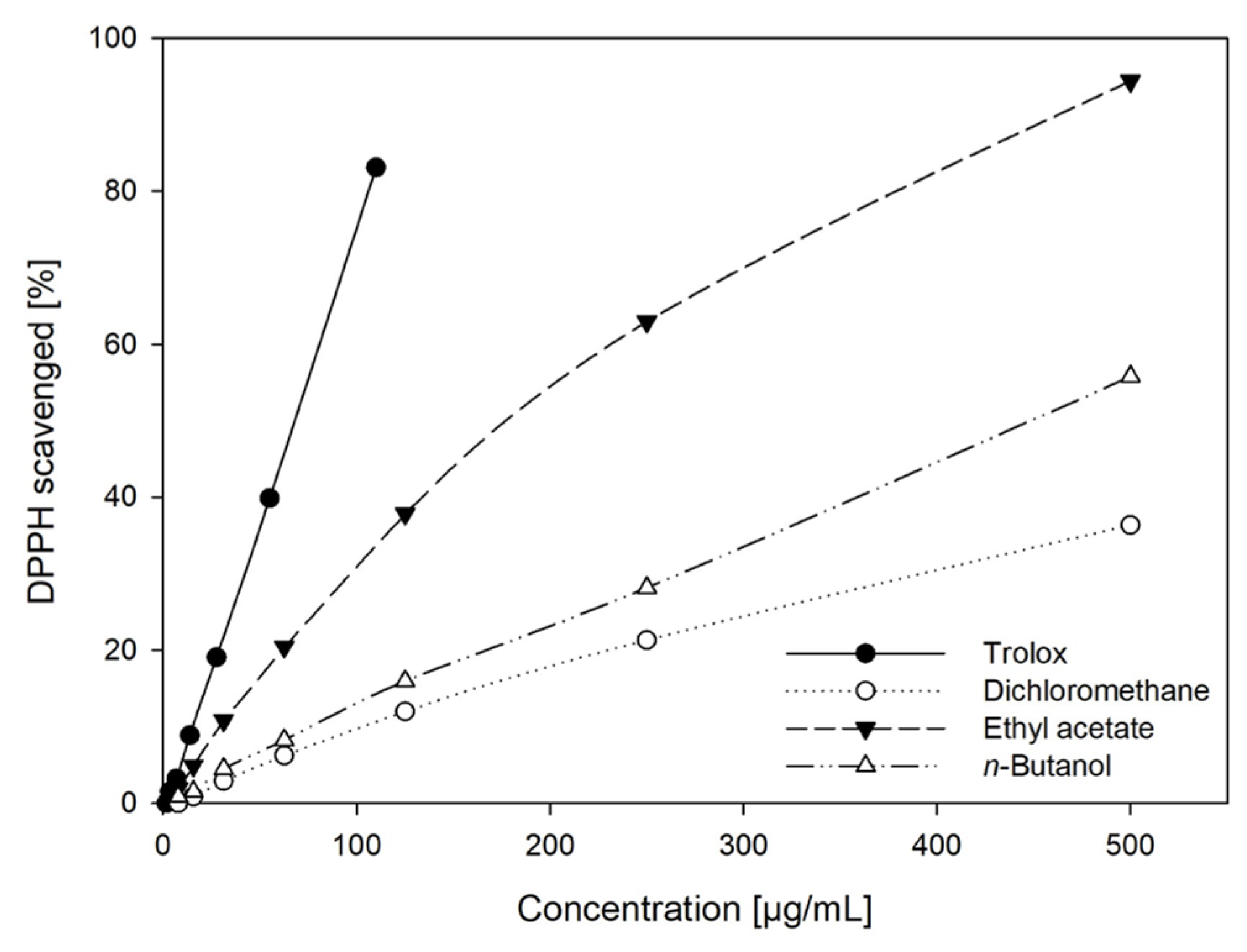
2.2. DPPH Radical Scavenging Activity of GS Extracts

In addition to their phenolic contents, the 2,2-diphenyl-1-picrylhydrazyl (DPPH) scavenging activities of the aforementioned extracts and trolox as reference compounds were determined spectrophotometrically at 516 nm (Supporting Information Part II). The percentage of scavenged DPPH was calculated relative to the maximum amount scavenged and is displayed in Figure 3. The correlation between extract concentration and DPPH scavenging activity was linear for trolox (r2 = 1) and the BuOH extract (r2 = 0.99) in the entire concentration range examined. For the EtOAc and DCM extracts, linearity was only found at concentrations < 100 µg/mL. Generally, EtOAc extracts exhibited the highest radical scavenging activities. While the BuOH extract had the least total phenolic content, its antioxidant activity was stronger than that of the DCM extract. This is presumably due to differences in the phenolic composition of both extracts as our analyses have shown (Section 2.3 and Section 2.4).

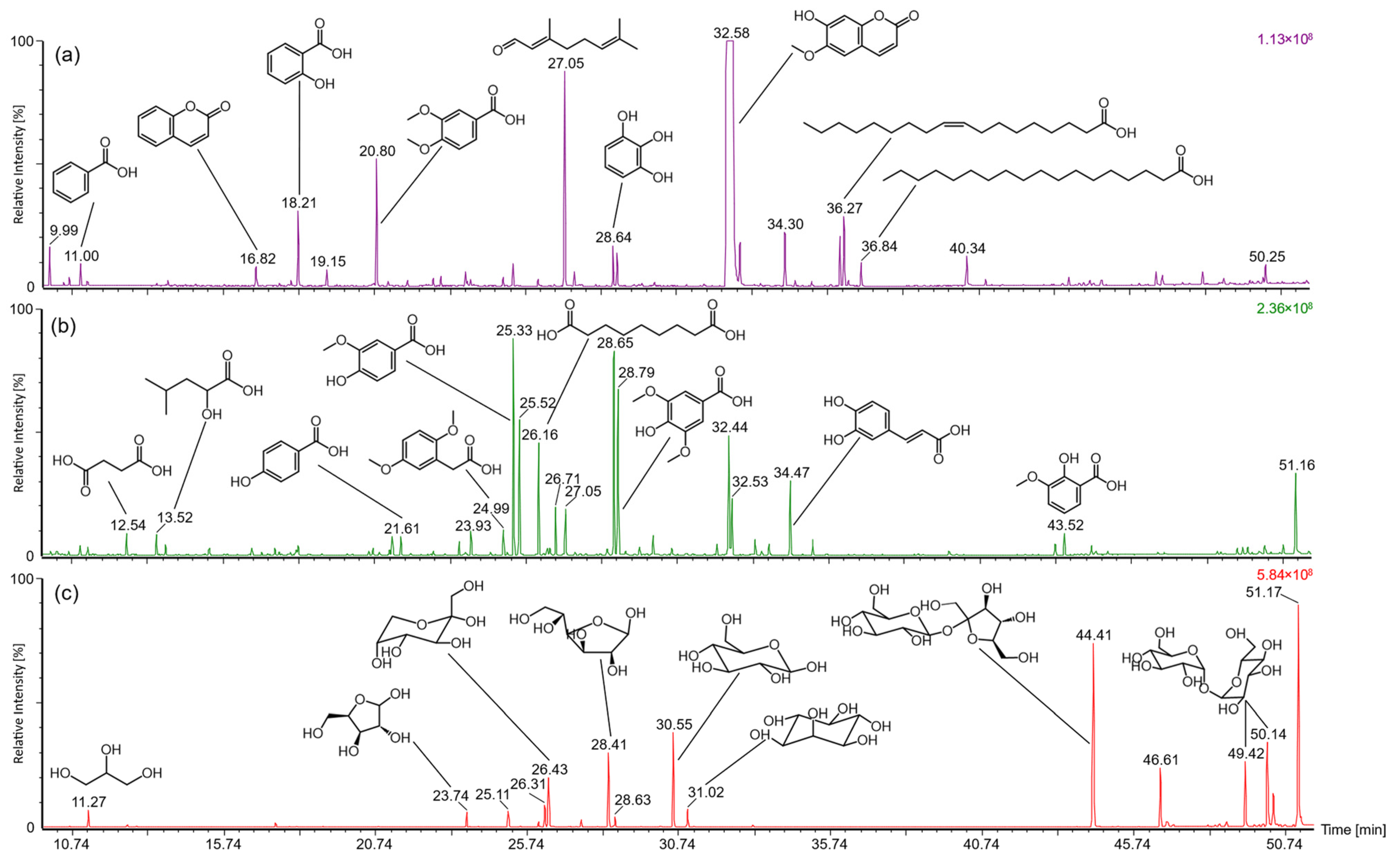
2.3. Analysis of Low Molecular Constituents by GC-MS

DCM, EtOAc, and BuOH extracts were analyzed by GC-MS after the silylation of individual constituents with BSTFA (Figure 4). For characterizing individual compounds, the obtained mass spectra (Table 1) were compared with the NIST database (National Institute of Standards and Technology, match factor > 800) and with MassBank Europe (massbank.eu, version 2.2.3). In DCM extracts, scopoletin (tR 32.6 min) was by far the most abundant component, followed by citral (tR 27.1 min). A variety of low molecular phenolic substances were eluted in a retention time range of 13–30 min. Among these, the three most abundant signals were attributed to coumarin (tR 16.6 min), salicylic acid (tR 18.2 min), and veratric acid (tR 20.8 min). Between 34 and 37 min, adenine (tR 34.3 min) and various fatty acids were eluted. Among the latter, linoleic, oleic, and stearic acids were the predominant compounds assigned based on their specific mass spectra. At higher retention times, mass spectrometric investigations indicated the presence of sterols. However, with match factors < 600, their exact identity could not be clarified. Various dicarboxylic and phenolic acids as well as scopoletin were characterized as main constituents in the chromatograms of EtOAc extracts. Finally, the polar BuOH extracts mainly contained a number of saccharides. Pentoses such as xylose and arabinose and hexoses such as fructose and glucose were eluted between 23 and 31 min. Disaccharides such as sucrose or trehalose were detected between 44 and 52 min. Furthermore, for aglycone analysis, the components of EtOAc and BuOH extracts were hydrolyzed with hydrochloric acid. The corresponding results can be found in the Supporting Information (Part III).

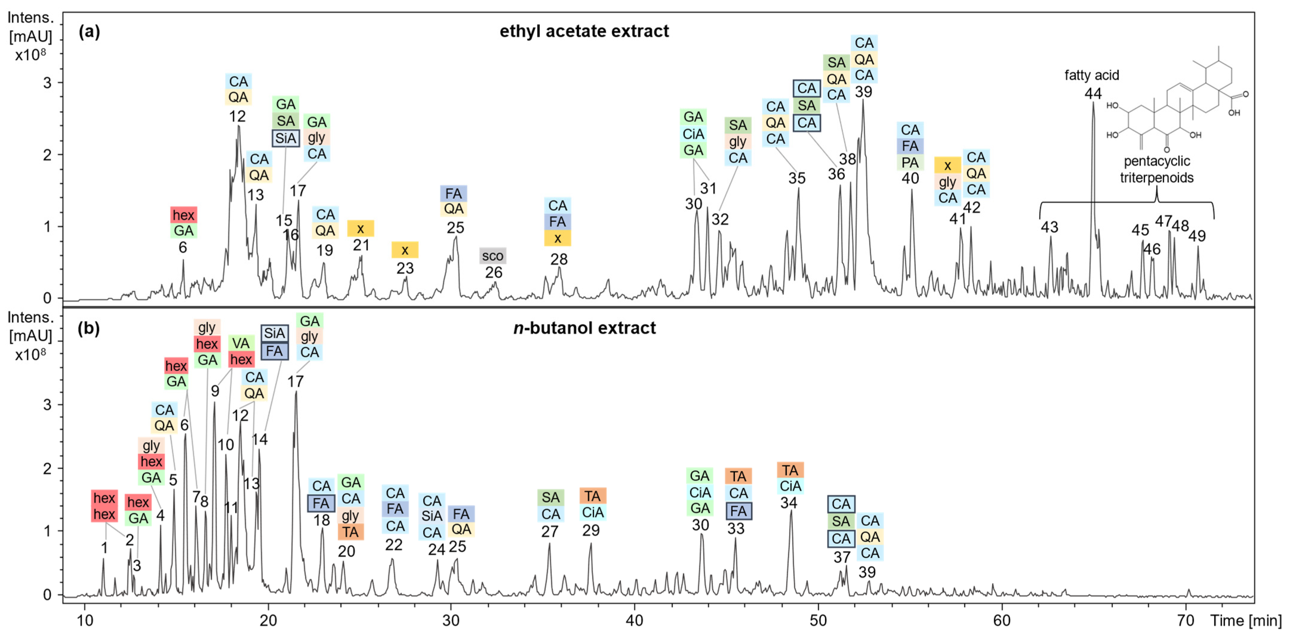
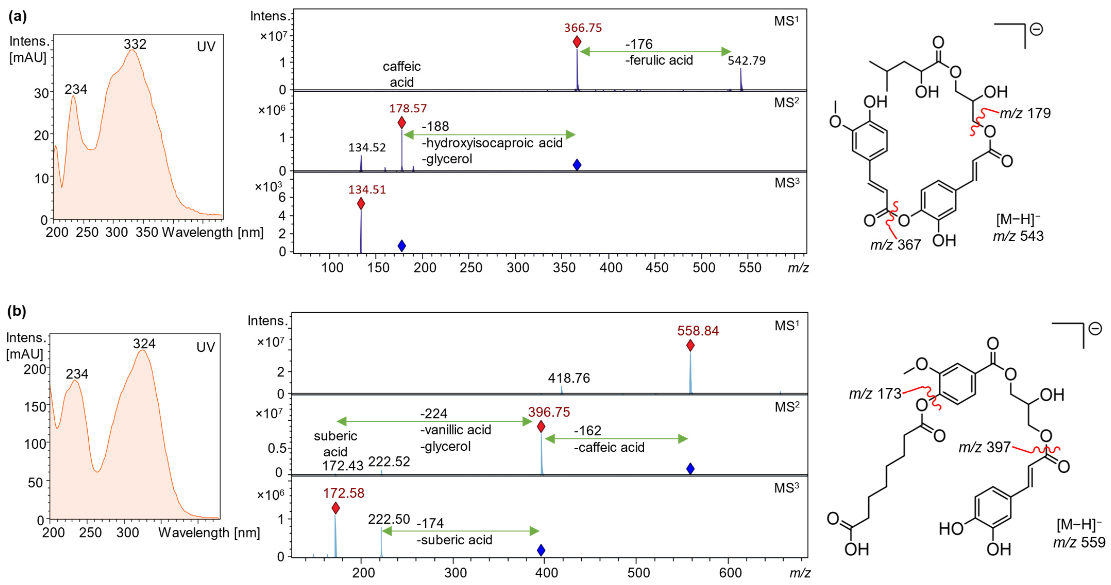
2.4. Analysis of Polar Secondary Constituents by HPLC-DAD-MSn

Applying HPLC-DAD-ESI-MSn in negative ionization mode, a variety of mostly phenolic substances were characterized based on the mass-to-charge ratios (m/z) of their precursor and fragment ions in comparison to the constituents and aglyca assigned in EtOAc and BuOH extracts by GC-MS. Moreover, for tentative structure elucidation, the fragmentation behavior of individual components in combination with UV spectral characteristics was compared to data published in the literature. A plethora of substances was assigned to depsidic structures, i.e., esters of two or three phenolcarboxylic acids, as displayed in Figure 5. Comprehensive HPLC-DAD-MSn characteristics are displayed in Table 2.
Hydroxybenzoic acids such as gallic acid and its derivatives are characterized by two marked UV maxima at around 215 and 270 nm. Hydroxycinnamic acids such as ferulic and caffeic acids typically exhibit absorption maxima at approx. 220 and 320 nm [23]. The coumarin scopoletin has a characteristic UV spectrum with maxima at 207, 228, 296, and 342 nm [24]. However, at low analyte concentrations, UV spectra become less conclusive due to poor signal intensities.
The characterization of phenolic esters and depsides was based on their molecular masses and neutral losses upon fragmentation and is exemplified in Figure 6 for compounds 28 and 41. The most frequent molecular ions [M−H] were detected at m/z 149 (tartaric acid), 169 (gallic acid), 179 (caffeic acid or hexose, distinguishable by the UV spectrum), 191 (quinic acid), 193 and 195 (ferulic and dihydroferulic acid) and 197 (syringic acid), the corresponding neutral losses were 17 Da less [25,26,27,28,29,30]. Interestingly, various neutral losses (compounds 4, 8, 16, 17, 20, 28, 32, 41) pointed to constituents being composed of a phenolic acid moiety and glycerol [26,31], which was also detected by GC-MS. In accordance with the literature, we assumed that the phenolic acids are esterified with the primary hydroxy groups of glycerol as a consequence of their higher reactivity [31,32].
In the following section, compound assignment is exemplified for some representative substances. Compound 4 (tR 14.2 min) exhibited a neutral loss of 242 Da in the MS2 experiment, which may be due to glycerol (92−17−17) and methyl gallic acid (184) moiety, producing a hexoside fragment ion (m/z 179). An isomer of this compound was eluted after 16.6 min (8). The compounds eluting next, 9 and 10 (tR 17.1 and 17.7 min), revealed similar fragmentation patterns. A loss of 64 Da in the first fragmentation step pointed to sulfite or furan derivatives, the subsequent loss of 164 Da may be due to veratric, i.e., dimethoxybenzoic, acid, which has also been detected by GC-MS. As above, the MS3 base peak at m/z 179 indicated hexosides. Then, compound 16 (tR 21.4 min), exhibiting an m/z at 505, showed a base peak at m/z 145 in the MS2 experiment which corresponds to [M−H−360], possibly [M−H−gallic acid−dihydrosinapinic acid]. This is equivalent to the molar weight of ethylsuccinic or methylglutaric acid, both being aliphatic dicarboxylic acids. As a neutral loss of succinic acid (100 Da) may be assumed from the MS1 spectrum and succinic acid was also found by GC-MS, the substance was characterized as ethylsuccinyl-galloyl-dihydrosinapic acid. A neutral loss of 226 Da (gallic acid + glycerol) indicated compound 17 (tR 21.7 min) to be a gallic and caffeic acid glyceride and compound 20 (tR 24.1 min) to be a tartaric acid ester thereof. In contrast, compound 27 (tR 35.4 min) had an [M−H] ion at m/z 359 and showed a loss of caffeic acid in the first fragmentation step. This may point to either caffeoyl-dihydroxyphenyllactic, i.e., rosmarinic, or caffeoyl-syringic acid. The latter assignment appears more plausible due to accordance with the GC-MS analyses. Furthermore, compounds 29 and 34 (tR 37.6 and 48.6 min) were detected as formic acid adducts [M−H+46] of the respective molecular ions. Based on the results of GC-MS analyses and the findings for compound 16, the neutral loss of 128 Da (compound 29) was assigned to ethylsuccinic acid, and 142 Da (compound 34) to propylsuccinic acid. The neutral loss of 144 Da in the MS3 experiments may be due to a hydroxycoumarin or a methylcinnamic acid moiety (compounds 2931, 34). However, due to the lack of distinct UV maxima >300 nm, which would be expected for coumarins, the latter was assumed. Compounds 30 and 31 (tR 43.5 and 44.0 min) could not be unambiguously characterized. Losses of 168 and 182 Da, possibly methyl- and dimethylgallic (syringic) acid, yielded MS2 base peaks at m/z 327. This ion was further fragmented yielding an MS3 base peak at m/z 183 (neutral loss of 144 Da, see above), which may either be ascribed to trihydroxyphenylacetic or methylgallic acid. Finally, the molecular ions of compounds 43 (tR 62.7 min) and 4549 (tR 67.7–70.9 min) indicated the presence of pentacyclic triterpenoids such as gelse-norursanes, which have been described earlier [9,33].
Alkaloids form the most intensely investigated group of secondary metabolites in Gelsemium species. Expectedly, these were only detected by LC-MS in positive ionization mode [4,8,9]. However, since these have been well investigated and described in the literature, they were outside the focus of the present study. For chromatograms and a peak list please refer to the Supporting Information (Part IV). As deduced from the intensities of the base peak chromatograms, the alkaloid concentrations were higher in BuOH than in EtOAc extracts (Supporting Information Figure S4.1). In contrast, the latter were characterized by a more complex profile of individual compounds, particularly in a later retention time range. Gelsemine, N-methylgelsedilam, gelsemicine, and sempervirine were the main alkaloids detected in the BuOH extracts.

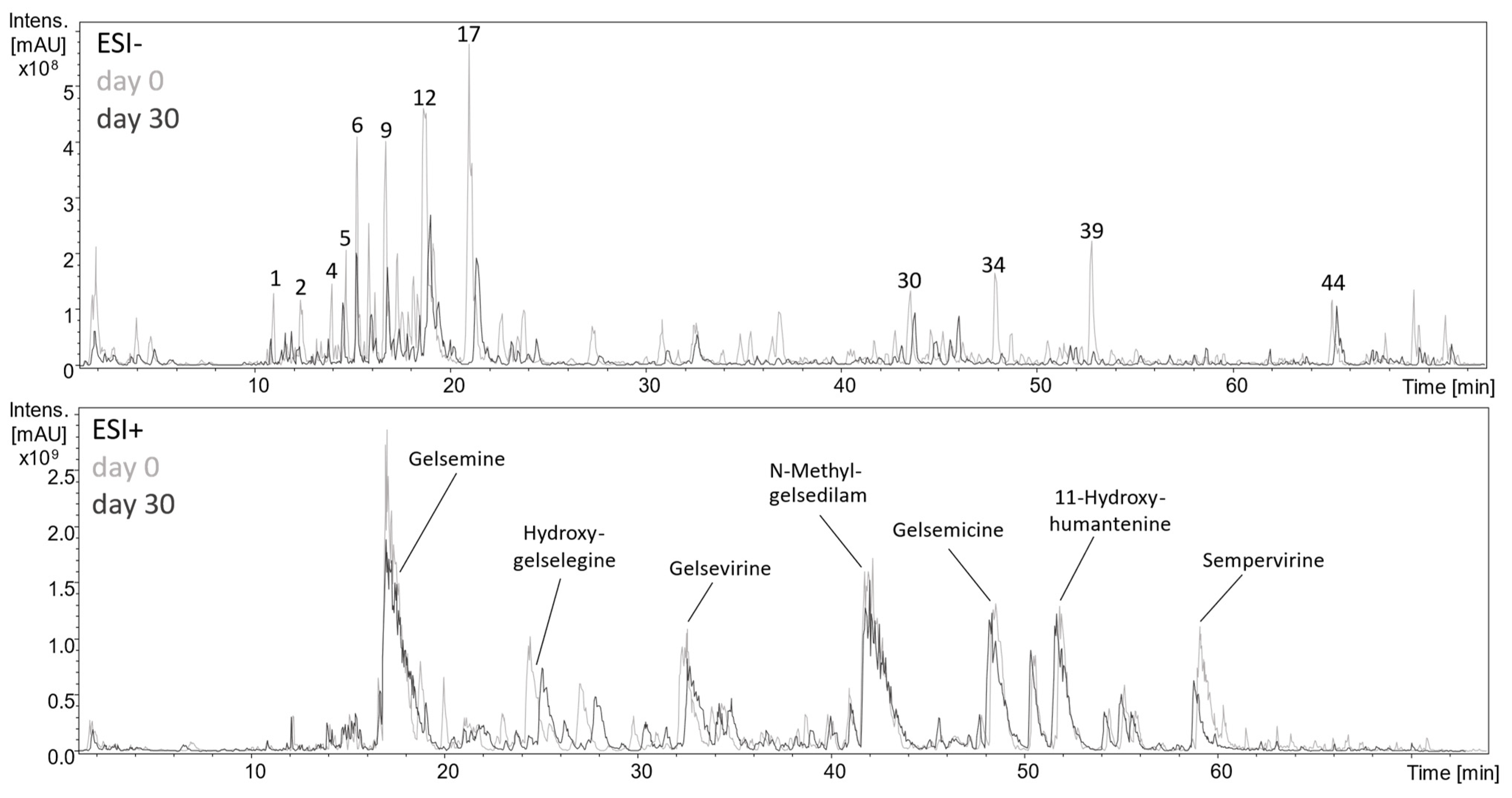
2.5. Metabolism of Phenolic Constituents upon Lactic Acid Fermentation

During the experiments, the progressive formation of a pleasant and spicy flavor of aqueous GS extracts was noticed. This observation is presumably due to the metabolic conversion of phenolic compounds caused by the microbial flora or endogenous plant enzymes after cell decompartmentation. Interestingly, the volatile compound formation was accelerated by inoculating the suspended plant material with L. plantarum, going along with a pH drop from 4.9 to 3.4 within three days as a result of lactic acid formation.
It is well known, that depsides and other esters are unstable upon enzymatic digestion and are rapidly hydrolyzed [18,34]. Accordingly, the HPLC-MSn base peak chromatogram (Figure 7) showed a marked degradation of phenolic compounds, especially depsides and glycerides, within 30 days of lactofermentation. As can be deduced from the UV traces (Supporting Information Figure S4.2), the chlorogenic acid content decreased by almost 50% within seven days but then slowed down with declining microbial viability. While the caffeic acid content increased correspondingly, the coumarin scopoletin was obviously not metabolized. Except for a slight decrease in gelsemine and sempervirine contents, only minor changes were monitored in the alkaloid spectrum.
For analyzing the volatile constituents contributing to the intense smell of fermented aqueous GS extracts, these were extracted with diethyl ether and injected into the GC-MS system without prior derivatization. As described for DCM and EtOAc extracts, benzoic, salicylic, isovanillic, azelaic, protocatechuic, syringic, ferulic, and caffeic acids were assigned (Figure 8, for mass spectral data, see Supporting Information Table S3.2). In addition, aliphatic compounds such as propylcyclohexene, methylethylidene-cyclohexane, and dimethyloctene were assigned based on a comparison of mass spectral data with the NIST database. Among oxygen-containing metabolites, ethylcatechol, hydroxy-methylbenzaldehyde, and the sesquiterpenoid oxo-α-ionol were detected. Two isomeric compounds at retention times of 36.2 and 36.3 min, exhibiting a molecular mass of 222 Da, could not be further characterized. However, the molecular weight could indicate a hydroxy-dimethoxy-coumarin [35].

3. Discussion

3.1. Phenolic Content and Antioxidant Activity

The Folin–Ciocalteu (FC) and the DPPH assay are both used for assessing the antioxidant activity of plant extracts [36]. The FC assay corroborated the high content of phenolic constituents in GS extracts. More precisely, when calculated as chlorogenic acid equivalents, the dry matter of EtOAc extracts was composed of about 50% phenolic substances, the corresponding values of DCM and BuOH extracts amounted to 40% and 30%, respectively. However, it should be kept in mind that due to the unspecific redox reaction of the Folin assay, it may only be regarded as a semiquantitative method and rather as an indication of the reductive potential of the sample [37,38]. Although phenolic substances generally have strong antioxidant effects, the extent markedly depends on the molecular structure, e.g., the number of phenolic hydroxyl groups [37]. Consequently, the results calculated as gallic and chlorogenic acid equivalents differed by almost 50%. This phenomenon is well known and is due to the higher reducing capacity of a galloyl group compared to catechol or hydroxycinnamic acid groups and has been documented in the literature [37,39]. These effects were also reflected by the fact that despite higher phenolic contents of DCM extracts as deduced from the FC assay, their DPPH radical scavenging activity was weaker than that of BuOH extracts. Accordingly, differences in the phenolic profiles of the extracts could be shown in our analyses. Furthermore, while trolox and BuOH extracts showed a linear correlation between the percentage of scavenged DPPH and concentration in the entire concentration range tested, the curves of EtOAc and DCM flattened at concentrations > 100 µg/mL. This indicates that not only concentration but also the exact chemical composition affects the characteristics of such extracts and that concentration-dependent interactions between individual components may occur.

3.2. Phytochemical and Bioactivity Profiling of G. sempervirens Roots and Rhizomes

Saccharides were detected in the GC-MS chromatograms of crude BuOH extracts as well as in the aqueous residues after solvent partitioning. Rhizomes often contain high amounts of starch since they commonly serve as storage organs [40]. However, the extraction procedure and derivatization prior to analysis discriminate oligomeric and polymeric saccharides. This is the reason why only low molecular saccharides were covered in the present analysis. Among these, various ubiquitous pentoses and hexoses as well as disaccharides could be assigned. The occurrence of significant amounts of sugars in rhizomes is also known from other plants such as Polygonatum species [29]. Interestingly, while only saccharides were characterized in crude BuOH extracts, various phenolic constituents were detected after hydrolysis with hydrochloric acid and extraction with ethyl acetate. This indicates that part of the phenolics naturally occur in bound forms but can be released by enzymatic or acidic cleavage. In contrast, chromatograms of EtOAc extracts were comparable prior to and after hydrolysis in terms of peak profile and compound assignment.
Among phenolics, a variety of benzoic and cinnamic acid derivatives were assigned via GC-MS analysis, with vanillic, gentisic, syringic, and caffeic acids being the main phenolic acids in both, EtOAc and BuOH extracts. Furthermore, coumarins, fatty acids, dicarboxylic acids, saccharides, and glycerol were assigned based on comparison with the NIST database. A number of these substances such as scopoletin, vanillic acid, and chlorogenic acid have previously been assigned in G. elegans [8,9,41], which in combination with the alkaloids emphasizes the phytochemical similarity of the two Gelsemium species despite their origin from different continents [1]. The monoterpene citral, a mixture of the isomers geranial and neral, presumably contributes to the smell of GS roots but has not been detected in its flowers so far [42].
The LC-MS investigations performed in the present study expanded the knowledge of the complex composition of the phenolic profile, as not only monomers but also higher molecular weight compounds can be detected using this technique. The assignment of most substances was based on their mass spectrometric behavior and UV spectra, which was aligned with the findings for the monomers obtained by GC-MS in crude and hydrolyzed EtOAc and BuOH extracts. Many of the compounds characterized according to this procedure belong to the depsides, i.e., esters of two or more phenolic acids [43]. Depsides have particularly been found in many lichen [15,44] and fungal [14] species, where they mostly consist of orsellinic, i.e., 2,4-dihydroxy-6-methylbenzoic, acid [14]. However, depsides of phenolic acids without methyl substitution of the aromatic ring have also been found in various higher plants such as aronia [45], rosemary [18], sage [46], or pineapple [47], thus across a wide range of plant families. The manifold bioactivities described for this compound class include cytotoxic, antimicrobial, analgesic, hepato-, nephro-, and neuroprotective as well as anti-inflammatory effects [15,17,46], which renders GS extracts potentially interesting from a pharmacological viewpoint.
Phenolic glycerides are another substance class characterized for the first time in the Gelsemiaceae in the present study. They have, however, also been detected in other plant families such as the Liliaceae [48], Bromeliaceae [49], and Asparagaceae [26], and are characteristic of propolis [31,50]. Like other phenolic substances, they exhibit marked antioxidant and anti-inflammatory activities [51,52]. Thus, while the toxicity of GS is attributed to alkaloids, the described inflammation, and eczema-reducing activity [2] may also be due to the phenolic constituents herein or due to an interplay of the different compound classes in the complex mixture.
Nor-ursane type triterpenoids such as the gelse-norursanes assigned in this study have been isolated from representatives of approx. 15 different plant families, mostly but not exclusively occurring in tropical and subtropical regions [33]. The natural habitat of GS from Florida to Virginia also fits in this climate zone [2]. Interesting bioactivities have been described for similar pentacyclic triterpenoids, for example, antidiabetic effects due to inhibition of the insulin-resistance-promoting enzyme tyrosine proteinase [53]. In addition, hepatoprotective effects [54] as well as cytotoxicity against leukemia, liver, breast, and colon cancer cells [55,56] were demonstrated.

3.3. Fermentation of Aqueous GS Root Extracts

The rapid formation of an aromatic odor within one week of fermentation in combination with a marked pH decrease indicated that the growth and viability of L. plantarum were not affected by the high alkaloid contents of GS. GC-MS analyses of the volatile compounds in fermented GS extracts revealed the presence of various low-molecular phenolic acids and metabolites thereof. As an example, 4-ethylcatechol may be produced from hydroxycinnamic acids by L. plantarum and is a well-known off-flavor component in various fermented foods [57]. γ-Amylbutyrolactone (γ-nonalactone) is an odor-active compound also found in whiskey [58], the sesquiterpenoid oxo-α-ionol is a metabolite produced by yeasts during winemaking [59]. However, it was not investigated if only a few character impact compounds account for the spicy smell of the fermented extracts, or if it is caused by a more complex variety of compounds detected in larger concentrations.
Using HPLC-MSn analysis in negative ionization mode, a degradation of saccharides, glycerides as well as depsides was observed as can be deduced from the base peak chromatograms. This is not surprising, since several esterases have been described in L. plantarum [21,60], which, among others, leads to the release of further compounds that may serve as substrates for bacterial metabolization. In contrast, only minor changes were detected in the alkaloid spectrum analyzed in positive ionization mode. The pH decline from 4.9 to 3.4 upon fermentation may have a stabilizing effect on the alkaloids, as they are protonated at lower pH values, increasing their solubility in water. Still, the decrease in gelsemine and sempervirine is most likely due to their poor solubility in aqueous systems, which leads to precipitation. Accordingly, both components were also found in the sediment formed during storage, after extraction of the latter with methanol. To conclude, fermentation appears to be a suitable method for the extraction of secondary metabolites from the plant matrix of GS as well as for preserving the obtained aqueous extracts, despite the metabolic degradation, particularly of phenolic constituents. The obtained extracts may, among others, be used for pharmaceutical applications, although their toxicity due to the alkaloid fraction must be considered.

4. Materials and Methods

4.1. Chemicals and Reagents

Acetone, acetonitrile, n-butanol (BuOH), chloroform, dichloromethane (DCM), ethyl acetate (EtOAc), hydrochloric acid, methanol (MeOH), methyl-tert-butyl ether (MTBE), sodium carbonate and sodium sulfate were purchased from Chemsolute (Th. Geyer GmbH & Co. KG, Renningen, Germany). Diethyl ether, gallic acid monohydrate (GA), and lactose were obtained from Carl Roth GmbH & Co. KG (Karlsruhe, Germany). N,O-Bis(trimethylsilyl)-trifluoroacetamide (BSTFA), dimethylformamide (DMF), and 2,2-diphenyl-1-picrylhydrazyl (DPPH) were from Sigma-Aldrich (St. Louis, Missouri, USA), formic acid from Fluka (Sigma Aldrich, St. Louis, MO, USA). Trolox was purchased from Cayman Chemical Company (Ann Arbor, MI, USA), and chlorogenic acid hemihydrate (CA) from Alfa Aesar (Karlsruhe, Germany). Folin–Ciocalteu’s reagent was from Merck KGaA (Darmstadt, Germany). Ultrapure water was produced with an ELGA Purelab Classic system (High Wycombe, UK) and used for all experiments.

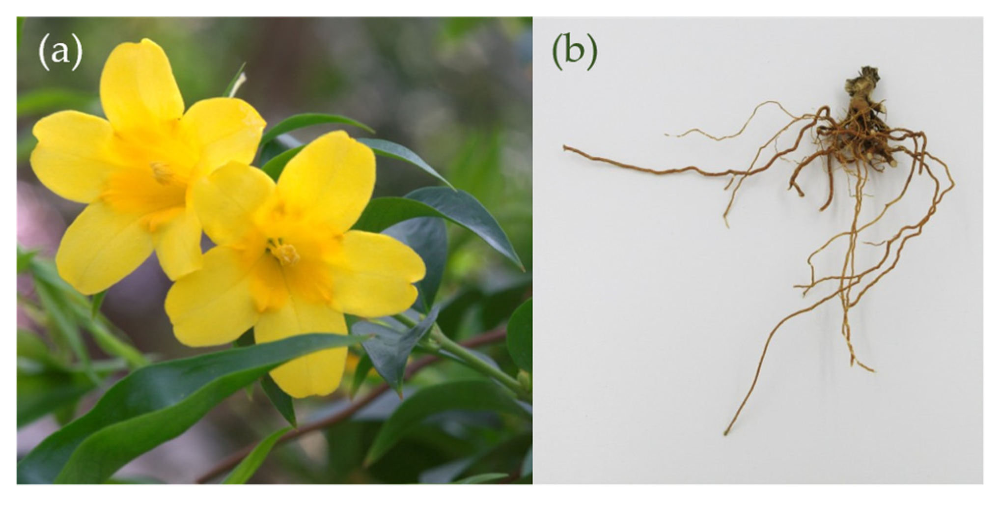
4.2. Plant Material and Extraction

Dried and cut G. sempervirens roots and rhizomes (Figure 9) were obtained from a commercial supplier (Albert Stephan export–import, Zweibrücken, Germany). A sample was deposited at the herbarium of the Institute of Botany, University of Hohenheim, Stuttgart (voucher number: HOH-022975).
For the extraction, 35 g plant material was mixed with 500 mL 70% acetone (v/v), bubbled with nitrogen for 5 min, and minced by Ultra-Turrax treatment (IKA Werke GmbH and Co. KG, Staufen, Germany; 3 min, 17,000 rpm). After another 10 min bubbling with nitrogen, the slurry was stored at 4 °C overnight. The mixture was then filtered over Celite® (Carl Roth GmbH & Co. KG, Karlsruhe, Germany) and the filter cake was re-extracted analogously. Then, the two filtrates were combined, the acetone was removed by rotary evaporation, and the residual aqueous phase was extracted successively with 3 × 100 mL of DCM, EtOAc, and n-BuOH. DCM and EtOAc extracts were dried over anhydrous sodium sulfate and filtered through filter paper (Whatman™ qualitative filter paper 2, GE Healthcare, Buckinghamshire, UK). Solvents were removed in vacuo and the dry residues were used for further experiments. Extraction was performed in duplicate.
For aglycone analyses, 50 mg of EtOAc or BuOH extracts were dissolved in 1 N hydrochloric acid (20 mL) and kept at 105 °C for 1 h. Aglycones were then extracted with 2 × 30 mL EtOAc, the organic phase was dried over sodium sulfate, filtered, and the solvent removed by rotary evaporation.

4.3. Fermentation Experiments

For the fermentation experiments, 20 g of dried roots were mixed with 500 mL of water containing 0.75% (w/v) lactose as substrate for microbial fermentation. The material was minced using an UltraTurrax (2 min, 17,000 rpm). This suspension was then inoculated with 1 mL of Lactiplantibacillus plantarum (previously Lactobacillus plantarum, GenBank accession number: MK841313.1; sequence length: 1083 base pairs; closest relative in National Center for Biotechnology Information: Lactobacillus plantarum strain 2.7.17, MK611349.1; similarity 100%) in MRS broth (5 × 10⁸ CFU/mL). After three days at 33 °C, the slurry was filtered through a cotton cloth yielding a turbid solution and the filtrate was kept in glass bottles at room temperature in the dark. After one week the turbid solution was filtered through filter paper yielding a clear solution, which was stored for a minimum of six months. Three temporally independent fermentations were conducted, each in duplicate or triplicate.

4.4. Estimation of the Total Phenolic Content by Folin–Ciocalteu Assay

The dried DCM, EtOAc, and BuOH extracts were dissolved in MeOH (250 µg/mL). Gallic acid (GA) and chlorogenic acid (CA) were used as reference substances in concentrations ranging from 3.5 to 55 µg/mL (GA) and 10–160 µg/mL (CA), respectively. A 20 µL amount of sample or reference solution and 40 µL of Folin–Ciocalteu’s reagent were mixed on a 96-well plate. The plate was shaken in the reader for one minute, and subsequently, 160 µL of sodium carbonate solution (700 mM) was added. After incubation (37 °C, 30 min), the absorbance at 765 nm was measured using a multiplate reader (Epoch2, Agilent Technologies Inc., Santa Clara, CA, USA). Calibration equations of GA and CA were calculated by plotting the absorbance values against the concentrations. The total phenolic content of the samples was then calculated as GA or CA equivalents [mg/g dry weight] by inserting the sample absorbance values into the regression equations. Analyses were performed in triplicate. Absorbance values and calibration data are provided in the Supporting Information (Part III).

4.5. Determination of the DPPH Radical Scavenging Activity

The DCM, EtOAc, and BuOH extracts were dissolved in MeOH and diluted to seven concentrations between 7.8 and 500 µg/mL. Trolox (1.7 to 110 µg/mL) was used as reference compound. 180 µL of DPPH solution (100 µM in MeOH) was then added to 20 µL of the test or reference solution or methanol as a blank sample in a 96-well plate. The plate was incubated at 37 °C for 45 min and then analyzed colorimetrically at 516 nm using a multiplate reader Epoch2. The percentage of scavenged DPPH was calculated from the maximum and sample absorbances (A) using the formula
DPPH scavenged [%] = (Amax − Asample)/Amax × 100%.
Analyses were performed in triplicate. Absorbance values are provided in the Supporting Information (Part IV).

4.6. GC-MS Analyses

For GC-MS analyses, crude DCM, EtOAc, and BuOH extracts and their aglycones (5–10 mg) were dissolved in DMF (500 µL) and mixed with BSTFA (200 µL). The solution was heated to 105 °C for 15 min in order to obtain trimethylsilyl derivatives of individual compounds.
For the extraction of volatile aroma composites from the fermented solutions, 40 mL of each sample was extracted with 2 × 10 mL diethyl ether. The solvent was removed under reduced pressure, and the residue was dissolved in 1 mL methyl-tert-butyl ether (MTBE) and directly used for analysis.
GC-MS analyses were conducted with a PerkinElmer Clarus 500 gas chromatograph (PerkinElmer, Inc., Shelton, CT, USA) coupled to a single quadrupole mass spectrometer operating in electron ionization (EI) mode at 70 eV according to [61]. Split injection (split ratio 30:1, injection volume 1.0 µL) was applied and a Zebron ZB-5MS capillary column (60 m × 0.25 mm i.d., 0.25 µm film thickness, 5% phenylpolysiloxane and 95% dimethylpolysiloxane coating; Phenomenex, Torrance, CA, USA) was used as a stationary phase. Helium served as carrier gas at a flow rate of 1 mL/min. The injector temperature was 250 °C, and the temperature of the column oven was 100–320 °C with a linear gradient of 4 °C/min. Data were acquired and processed using the software TurboMass (v.5.4.2, PerkinElmer, Inc., Waltham, MA, USA).

4.7. HPLC-DAD-ESI-MSn Analyses

For HPLC analyses, EtOAc and BuOH extracts were dissolved in MeOH or water (5 mg/mL), respectively, and aqueous samples were directly injected after filtration through a syringe filter (perfect-flow RC, 0.45 µm, WICOM, Heppenheim, Germany).
Reversed phase high performance liquid chromatography was carried out as described previously [61]. In brief, an Agilent 1200 HPLC system (Agilent Technologies, Inc., Palo Alto, CA, USA) equipped with binary pump, micro vacuum degasser, autosampler, thermostatic column compartment and UV/VIS diode array detector (DAD); a Kinetex® C18 reversed-phase column (2.6 µm particle size, 150 mm × 2.1 mm i.d., Phenomenex Ltd., Aschaffenburg, Germany); and a pre-column of the same material were used for chromatographic separation at 25 °C and a flow rate of 0.21 mL/min. 0.1% formic acid in water and acetonitrile were used as mobile phase.
For mass spectrometric detection, an HCTultra ion trap mass spectrometer (Bruker Daltonik GmbH, Bremen, Germany) with an ESI source was used. All extracts were analyzed in positive and negative ionization mode using a capillary voltage of + or −4000 V, respectively. The dry gas (N2) flow was 9.00 L/min, capillary temperature 365 °C, and nebulizer pressure 35 psi. MSn data were generated by performing collision-induced dissociation (CID) experiments. The instruments were controlled by Agilent LC 3D systems (Rev. B01.03SR1 (204)) and Bruker Daltonics EsquireControl software (V7.1).

5. Conclusions

In the investigation presented here, secondary metabolites in GS roots and rhizomes were comprehensively characterized with a special focus on phenolic constituents. Applying the Folin–Ciocalteu assay, total phenolic contents of 411, 537, and 291 µg chlorogenic acid equivalents per mg dry weight were determined in DCM, EtOAc, and BuOH extracts, respectively. Accordingly, pronounced antioxidant activity was determined using the DPPH antioxidant assay. Interestingly, the correlation between concentration and DPPH scavenging activity was not strictly linear for EtOAc and DCM extracts, indicating concentration-dependent interactions between individual components.
The identity of the phenolic compounds was studied by GC-MS and HPLC-DAD-MSn analyses and could mainly be assigned to depsides and phenolic glycerides consisting of various hydroxybenzoic, hydroxycinnamic, and dicarboxylic acids. A plethora of bioactivities have been reported for the aforementioned constituents, such as anti-inflammatory, analgesic, and neuroprotective action. They may therefore also contribute to the pharmacological effects described for GS, which have previously been attributed mainly to alkaloids.
Upon lactic acid fermentation with L. plantarum, depsides, glycerides, and other esters were rapidly degraded. Subsequently, the formation of low-molecular phenolic metabolites could be shown in GC-MS analyses. The obtained extracts remained microbially stable during the six-month period of investigation. The presented results expand the knowledge on the traditional medicinal plant GS and may open new perspectives of use, despite its toxicity limiting pharmaceutical applications.

Supplementary Materials

The following supporting information can be downloaded at: https://www.mdpi.com/article/10.3390/plants13162208/s1, Table S1.1. Calibration data for the Folin–Ciocalteu assay obtained with gallic acid. Table S1.2. Calibration data for the Folin–Ciocalteu assay obtained with chlorogenic acid. Table S1.3. Total phenolic content of G. sempervirens DCM, EtOAc, and BuOH extracts (250 µg/mL) calculated as µg gallic or chlorogenic acid equivalents per mg dry weight. Table S2.1. Calibration data for the DPPH radical scavenging assay obtained with trolox. Table S2.2. DPPH radical scavenging capacity of the DCM extract at different concentrations. Table S2.3. DPPH radical scavenging capacity of the ethyl acetate extract at different concentrations. Table S2.4. DPPH radical scavenging capacity of the n-butanol extract at different concentrations. Figure S3.1. GC-MS total ion current chromatograms of hydrolyzed (a) n-butanol and (b) ethyl acetate extracts after silylation. Table S3.1. Aglycones detected in ethyl acetate and n-butanol extracts after hydrolysis and silylation using GC-MS. Compound assignment was achieved by comparison with the NIST database (match factor >800). Table S3.2. GC-MS data of volatile compounds extracted with diethyl ether from aqueous fermented samples. Compound assignment was achieved by comparison with the NIST database (match factor >800). Figure S4.1. HPLC-DAD-ESI-MSn base peak chromatograms of (A) ethyl acetate and (B) n-butanol extracts of G. sempervirens roots and rhizomes recorded in positive ionization mode. Table S4.1. HPLC-DAD-MSn characteristics of individual secondary metabolites in ethyl acetate and n-butanol extracts from G. sempervirens roots and rhizomes obtained in positive ionization mode. Figure S4.2. Stacked display of UV chromatograms (328–332 nm) of aqueous fermented G. sempervirens extracts within 30 days of fermentation. References [62,63] are cited in the supplementary materials.

Author Contributions

Conceptualization, L.K.M.; Methodology, L.K.M. and P.L.; Investigation, L.K.M.; Data Curation, L.K.M. and K.N.G.; Writing—Original Draft Preparation, L.K.M.; Writing—Review and Editing, K.N.G., P.L., R.D., F.C.S. and D.R.K.; Visualization, L.K.M.; Supervision, D.R.K. All authors have read and agreed to the published version of the manuscript.

Funding

This research received no external funding.

Data Availability Statement

Data is contained within the article and supplementary material.

Acknowledgments

The authors wish to thank Rhinaixa Duque-Thüs (Institute of Botany, Hohenheim University) for the identification and storage of plant specimens in the herbarium of Hohenheim University and are grateful to Hannes Bitterling (WALA Heilmittel GmbH) for his assistance in GC-MS maintenance.

Conflicts of Interest

L.K.M., K.N.G., P.L., F.C.S. and D.R.K. are employed at WALA Heilmittel GmbH, they declare that the research was conducted in the absence of any commercial or financial relationships that could be construed as a potential conflict of interest. R.D. declares no conflict of interest.

References

  1. Dutt, V.; Thakur, S.; Dhar, V.J.; Sharma, A. The genus Gelsemium: An update. Pharmacogn. Rev. 2010, 4, 185–194. [Google Scholar] [CrossRef] [PubMed]
  2. Jin, G.-L.; Su, Y.-P.; Liu, M.; Xu, Y.; Yang, J.; Liao, K.-J.; Yu, C.-X. Medicinal plants of the genus Gelsemium (Gelsemiaceae, Gentianales)—A review of their phytochemistry, pharmacology, toxicology and traditional use. J. Ethnopharmacol. 2014, 152, 33–52. [Google Scholar] [CrossRef] [PubMed]
  3. Lin, H.; Qiu, H.; Cheng, Y.; Liu, M.; Chen, M.; Que, Y.; Que, W. Gelsemium elegans Benth: Chemical components, pharmacological effects, and toxicity mechanisms. Molecules 2021, 26, 7145. [Google Scholar] [CrossRef] [PubMed]
  4. Wang, J.; Zhang, J.; Zhang, C.; Sun, X.; Liao, X.; Zheng, W.; Yin, Q.; Yang, J.; Mao, D.; Wang, B.; et al. The qualitative and quantitative analyses of Gelsemium elegans. J. Pharm. Biomed. Anal. 2019, 172, 329–338. [Google Scholar] [CrossRef] [PubMed]
  5. Zhang, Z.; Wang, P.; Yuan, W.; Li, S. Steroids, alkaloids, and coumarins from Gelsemium sempervirens. Planta Med. 2008, 74, 1818–1822. [Google Scholar] [CrossRef] [PubMed]
  6. Jensen, S.R.; Kirk, O.; Nielsen, B.J.; Norrestam, R. 9-hydroxy substituted iridoids from Gelsemium sempervirens. Phytochemistry 1987, 26, 1725–1731. [Google Scholar] [CrossRef]
  7. Schun, Y.; Cordell, G.A. Cytotoxic steroids of Gelsemium sempervirens. J. Nat. Prod. 1987, 50, 195–198. [Google Scholar] [CrossRef] [PubMed]
  8. Liu, Y.-C.; Lin, L.; Cheng, P.; Sun, Z.-L.; Wu, Y.; Liu, Z.-Y. Fingerprint analysis of Gelsemium elegans by HPLC followed by the targeted identification of chemical constituents using HPLC coupled with quadrupole-time-of-flight mass spectrometry. Fitoterapia 2017, 121, 94–105. [Google Scholar] [CrossRef] [PubMed]
  9. Liu, Y.-C.; Xiao, S.; Yang, K.; Ling, L.; Sun, Z.-L.; Liu, Z.-Y. Comprehensive identification and structural characterization of target components from Gelsemium elegans by high-performance liquid chromatography coupled with quadrupole time-of-flight mass spectrometry based on accurate mass databases combined with MS/MS spectra. J. Mass Spectrom. 2017, 52, 378–396. [Google Scholar] [CrossRef] [PubMed]
  10. Zhang, J.-Y.; Wang, Y.-X. Gelsemium analgesia and the spinal glycine receptor/allopregnanolone pathway. Fitoterapia 2015, 100, 35–43. [Google Scholar] [CrossRef] [PubMed]
  11. Xu, W.-B.; Tang, M.-H.; Long, J.-Y.; Wang, W.-W.; Qin, J.-Y.; Qi, X.-J.; Liu, Z.-Y. Antinociceptive effect of gelsenicine, principal toxic alkaloids of Gelsemium, on prostaglandin E2-induced hyperalgesia in mice: Comparison with gelsemine and koumine. Biochem. Biophys. Res. Commun. 2023, 681, 55–61. [Google Scholar] [CrossRef] [PubMed]
  12. Shoaib, R.M.; Zhang, J.-Y.; Mao, X.-F.; Wang, Y.-X. Gelsemine and koumine, principal active ingredients of Gelsemium, exhibit mechanical antiallodynia via spinal glycine receptor activation-induced allopregnanolone biosynthesis. Biochem. Pharmacol. 2019, 161, 136–148. [Google Scholar] [CrossRef] [PubMed]
  13. Dutt, V.; Dhar, V.J.; Sharma, A. Antianxiety activity of Gelsemium sempervirens. Pharm. Biol. 2010, 48, 1091–1096. [Google Scholar] [CrossRef] [PubMed]
  14. Ibrahim, S.R.M.; Sirwi, A.; Eid, B.G.; Mohamed, S.G.A.; Mohamed, G.A. Fungal depsides-naturally inspiring molecules: Biosynthesis, structural characterization, and biological activities. Metabolites 2021, 11, 683. [Google Scholar] [CrossRef] [PubMed]
  15. Ureña-Vacas, I.; González-Burgos, E.; Divakar, P.K.; Gómez-Serranillos, M.P. Lichen depsides and tridepsides: Progress in pharmacological approaches. J. Fungi 2023, 9, 116. [Google Scholar] [CrossRef] [PubMed]
  16. Nadeem, M.; Imran, M.; Aslam Gondal, T.; Imran, A.; Shahbaz, M.; Muhammad Amir, R.; Wasim Sajid, M.; Batool Qaisrani, T.; Atif, M.; Hussain, G.; et al. Therapeutic potential of rosmarinic acid: A comprehensive review. Appl. Sci. 2019, 9, 3139. [Google Scholar] [CrossRef]
  17. Luo, C.; Zou, L.; Sun, H.; Peng, J.; Gao, C.; Bao, L.; Ji, R.; Jin, Y.; Sun, S. A review of the anti-inflammatory effects of rosmarinic acid on inflammatory diseases. Front. Pharmacol. 2020, 11, 153. [Google Scholar] [CrossRef] [PubMed]
  18. Gonçalves, G.A.; Corrêa, R.C.G.; Barros, L.; Dias, M.I.; Calhelha, R.C.; Correa, V.G.; Bracht, A.; Peralta, R.M.; Ferreira, I.C.F.R. Effects of in vitro gastrointestinal digestion and colonic fermentation on a rosemary (Rosmarinus officinalis L.) extract rich in rosmarinic acid. Food Chem. 2019, 271, 393–400. [Google Scholar] [CrossRef] [PubMed]
  19. Rodríguez, H.; Landete, J.M.; Rivas, B.d.l.; Muñoz, R. Metabolism of food phenolic acids by Lactobacillus plantarum CECT 748T. Food Chem. 2008, 107, 1393–1398. [Google Scholar] [CrossRef]
  20. Landete, J.M.; Curiel, J.A.; Rodríguez, H.; de Las Rivas, B.; Muñoz, R. Study of the inhibitory activity of phenolic compounds found in olive products and their degradation by Lactobacillus plantarum strains. Food Chem. 2008, 107, 320–326. [Google Scholar] [CrossRef]
  21. Landete, J.M.; Plaza-Vinuesa, L.; Montenegro, C.; Santamaría, L.; Reverón, I.; de Las Rivas, B.; Muñoz, R. The use of Lactobacillus plantarum esterase genes: A biotechnological strategy to increase the bioavailability of dietary phenolic compounds in lactic acid bacteria. Int. J. Food Sci. Nutr. 2021, 72, 1035–1045. [Google Scholar] [CrossRef] [PubMed]
  22. Leonard, W.; Zhang, P.; Ying, D.; Adhikari, B.; Fang, Z. Fermentation transforms the phenolic profiles and bioactivities of plant-based foods. Biotechnol. Adv. 2021, 49, 107763. [Google Scholar] [CrossRef]
  23. Singh, D.P.; Govindarajan, R.; Khare, A.; Rawat, A.K.S. Optimization of a high-performance liquid chromatography method for the separation and identification of six different classes of phenolics. J. Chromatogr. Sci. 2007, 45, 701–705. [Google Scholar] [CrossRef] [PubMed]
  24. Tasfiyati, A.N.; Antika, L.D.; Septama, A.W.; Hikmat, H.; Kurniawan, H.H.; Ariani, N. A validated HPLC-DAD method and comparison of different extraction techniques for analysis of scopoletin in noni-based products. Kuwait J. Sci. 2023, 50, 276–281. [Google Scholar] [CrossRef]
  25. Abu-Reidah, I.M.; Arráez-Román, D.; Segura-Carretero, A.; Fernández-Gutiérrez, A. Extensive characterisation of bioactive phenolic constituents from globe artichoke (Cynara scolymus L.) by HPLC-DAD-ESI-QTOF-MS. Food Chem. 2013, 141, 2269–2277. [Google Scholar] [CrossRef]
  26. Jiménez-Sánchez, C.; Lozano-Sánchez, J.; Rodríguez-Pérez, C.; Segura-Carretero, A.; Fernández-Gutiérrez, A. Comprehensive, untargeted, and qualitative RP-HPLC-ESI-QTOF/MS2 metabolite profiling of green asparagus (Asparagus officinalis). J. Food Comp. Anal. 2016, 46, 78–87. [Google Scholar] [CrossRef]
  27. Bunse, M.; Mailänder, L.K.; Lorenz, P.; Stintzing, F.C.; Kammerer, D.R. Evaluation of Geum urbanum L. extracts with respect to their antimicrobial potential. Chem. Biodivers. 2022, 19, e202100850. [Google Scholar] [CrossRef] [PubMed]
  28. Clifford, M.N.; Johnston, K.L.; Knight, S.; Kuhnert, N. Hierarchical scheme for LC-MSn identification of chlorogenic acids. J. Agric. Food Chem. 2003, 51, 2900–2911. [Google Scholar] [CrossRef]
  29. Jin, J.; Lao, J.; Zhou, R.; He, W.; Qin, Y.; Zhong, C.; Xie, J.; Liu, H.; Wan, D.; Zhang, S.; et al. Simultaneous identification and dynamic analysis of saccharides during steam processing of rhizomes of Polygonatum cyrtonema by HPLC-QTOF-MS/MS. Molecules 2018, 23, 2855. [Google Scholar] [CrossRef] [PubMed]
  30. Schütz, K.; Kammerer, D.R.; Carle, R.; Schieber, A. Characterization of phenolic acids and flavonoids in dandelion (Taraxacum officinale WEB. ex WIGG.) root and herb by high-performance liquid chromatography/electrospray ionization mass spectrometry. Rapid Commun. Mass Spectrom. 2005, 19, 179–186. [Google Scholar] [CrossRef] [PubMed]
  31. Bertrams, J.; Müller, M.B.; Kunz, N.; Kammerer, D.R.; Stintzing, F.C. Phenolic compounds as marker compounds for botanical origin determination of German propolis samples based on TLC and TLC-MS. J. Appl. Bot. Food Qual. 2013, 86, 143–153. [Google Scholar] [CrossRef]
  32. Lorenz, P.; Knittel, D.N.; Conrad, J.; Lotter, E.M.; Heilmann, J.; Stintzing, F.C.; Kammerer, D.R. 1-Acetyl-3-(3R)-hydroxyfatty acylglycerols: Lipid Compounds from Euphrasia rostkoviana Hayne and E. tetraquetra (Bréb.) Arrond. Chem. Biodivers. 2016, 13, 602–612. [Google Scholar] [CrossRef] [PubMed]
  33. Wu, H.-R.; He, X.-F.; Jin, X.-J.; Pan, H.; Shi, Z.-N.; Xu, D.-D.; Yao, X.-J.; Zhu, Y. New nor-ursane type triterpenoids from Gelsemium elegans. Fitoterapia 2015, 106, 175–183. [Google Scholar] [CrossRef] [PubMed]
  34. Lorenz, P.; Conrad, J.; Stintzing, F.C. Metabolic fate of depsides and alkaloid constituents in aqueous extracts from Mercurialis perennis L. during fermentation. Chem. Biodivers. 2013, 10, 1706–1723. [Google Scholar] [CrossRef] [PubMed]
  35. Schnitzler, P.; Schneider, S.; Stintzing, F.C.; Carle, R.; Reichling, J. Efficacy of an aqueous Pelargonium sidoides extract against herpesvirus. Phytomedicine 2008, 15, 1108–1116. [Google Scholar] [CrossRef] [PubMed]
  36. Platzer, M.; Kiese, S.; Herfellner, T.; Schweiggert-Weisz, U.; Eisner, P. How does the phenol structure influence the results of the Folin-Ciocalteu assay? Antioxidants 2021, 10, 811. [Google Scholar] [CrossRef] [PubMed]
  37. Pérez, M.; Dominguez-López, I.; Lamuela-Raventós, R.M. The chemistry behind the Folin-Ciocalteu method for the estimation of (poly)phenol content in food: Total phenolic intake in a mediterranean dietary pattern. J. Agric. Food Chem. 2023, 71, 17543–17553. [Google Scholar] [CrossRef] [PubMed]
  38. Ainsworth, E.A.; Gillespie, K.M. Estimation of total phenolic content and other oxidation substrates in plant tissues using Folin-Ciocalteu reagent. Nat. Protoc. 2007, 2, 875–877. [Google Scholar] [CrossRef] [PubMed]
  39. Kyoung Chun, O.; Kim, D.-O. Consideration on equivalent chemicals in total phenolic assay of chlorogenic acid-rich plums. Food Res. Int. 2004, 37, 337–342. [Google Scholar] [CrossRef]
  40. White, L.M. Carbohydrate reserves of grasses: A review. J. Range Manag. 1973, 26, 13–18. [Google Scholar] [CrossRef]
  41. Peng, Y.-L.; Liang, J.-J.; Xue, Y.; Khan, A.; Zhang, P.-P.; Feng, T.-T.; Song, D.; Zhou, Y.; Wei, X. Genus Gelsemium and its endophytic fungi—Comprehensive review of their traditional uses, phytochemistry, pharmacology, and toxicology. Curr. Top. Med. Chem. 2023, 23, 2452–2487. [Google Scholar] [CrossRef] [PubMed]
  42. Obi Johnson, B.; Golonka, A.M.; Blackwell, A.; Vazquez, I.; Wolfram, N. Floral scent variation in the heterostylous species Gelsemium sempervirens. Molecules 2019, 24, 2818. [Google Scholar] [CrossRef]
  43. Chen, L.; Wei, X.; Matsuda, Y. Depside bond formation by the starter-unit acyltransferase domain of a fungal polyketide synthase. J. Am. Chem. Soc. 2022, 144, 19225–19230. [Google Scholar] [CrossRef] [PubMed]
  44. Norouzi, H.; Sohrabi, M.; Yousefi, M.; Boustie, J. Tridepsides as potential bioactives: A review on their chemistry and the global distribution of their lichenic and non-lichenic natural sources. Front. Fungal Biol. 2023, 4, 1088966. [Google Scholar] [CrossRef] [PubMed]
  45. Ekiert, H.M.; Szopa, A.; Kubica, P. High production of depsides and other phenolic acids in different types of shoot cultures of three Aronias: Aronia melanocarpa, Aronia arbutifolia, Aronia × prunifolia. In Plant Cell and Tissue Differentiation and Secondary Metabolites: Fundamentals and Applications, 1st ed.; Ramawat, K.G., Ekiert, H.M., Goyal, S., Eds.; Springer International Publishing; Imprint Springer: Cham, Switzerland, 2021; pp. 337–364. ISBN 978-3-030-30184-2. [Google Scholar]
  46. Jin, Q.; Hu, X.; Deng, Y.; Hou, J.; Lei, M.; Ji, H.; Zhou, J.; Qu, H.; Wu, W.; Guo, D. Four new depsides isolated from Salvia miltiorrhiza and their significant nerve-protective activities. Molecules 2018, 23, 3274. [Google Scholar] [CrossRef] [PubMed]
  47. Steingass, C.B.; Glock, M.P.; Schweiggert, R.M.; Carle, R. Studies into the phenolic patterns of different tissues of pineapple (Ananas comosus L. Merr.) infructescence by HPLC-DAD-ESI-MS(n) and GC-MS analysis. Anal. Bioanal. Chem. 2015, 407, 6463–6479. [Google Scholar] [CrossRef] [PubMed]
  48. Shimomura, H.; Sashida, Y.; Mimaki, Y. Phenolic glycerides from Lilium auratum. Phytochemistry 1987, 26, 844–845. [Google Scholar] [CrossRef]
  49. Delaporte, R.H.; Guzen, K.P.; Laverde, A.; dos Santos, A.R.; Sarragiotto, M.H. Phenylpropanoid glycerols from Tillandsia streptocarpa Baker (Bromeliaceae). Biochem. Syst. Ecol. 2006, 34, 599–602. [Google Scholar] [CrossRef]
  50. Ristivojević, P.; Trifković, J.; Gašić, U.; Andrić, F.; Nedić, N.; Tešić, Ž.; Milojković-Opsenica, D. Ultrahigh-performance liquid chromatography and mass spectrometry (UHPLC-LTQ/Orbitrap/MS/MS) study of phenolic profile of Serbian poplar type propolis. Phytochem. Anal. 2015, 26, 127–136. [Google Scholar] [CrossRef] [PubMed]
  51. Guzelmeric, E.; Yuksel, P.I.; Yaman, B.K.; Sipahi, H.; Celik, C.; Kırmızıbekmez, H.; Aydın, A.; Yesilada, E. Comparison of antioxidant and anti-inflammatory activity profiles of various chemically characterized Turkish propolis sub-types: Which propolis type is a promising source for pharmaceutical product development? J. Pharm. Biomed. Anal. 2021, 203, 114196. [Google Scholar] [CrossRef] [PubMed]
  52. Okińczyc, P.; Widelski, J.; Szperlik, J.; Żuk, M.; Mroczek, T.; Skalicka-Woźniak, K.; Sakipova, Z.; Widelska, G.; Kuś, P.M. Impact of plant origin on Eurasian propolis on phenolic profile and classical antioxidant activity. Biomolecules 2021, 11, 68. [Google Scholar] [CrossRef] [PubMed]
  53. Na, M.; Thuong, P.T.; Hwang, I.H.; Bae, K.; Kim, B.Y.; Osada, H.; Ahn, J.S. Protein tyrosine phosphatase 1B inhibitory activity of 24-norursane triterpenes isolated from Weigela subsessilis. Phytother. Res. 2010, 24, 1716–1719. [Google Scholar] [CrossRef] [PubMed]
  54. Wang, H.-Q.; Ma, S.-G.; Zhang, D.; Li, Y.-H.; Qu, J.; Li, Y.; Liu, Y.-B.; Yu, S.-S. Oxygenated pentacyclic triterpenoids from the stems and branches of Enkianthus chinensis. Bioorg. Chem. 2021, 111, 104866. [Google Scholar] [CrossRef] [PubMed]
  55. Wu, X.-D.; He, J.; Li, X.-Y.; Dong, L.-B.; Gong, X.; Gao, X.; Song, L.-D.; Li, Y.; Peng, L.-Y.; Zhao, Q.-S. Triterpenoids and steroids with cytotoxic activity from Emmenopterys henryi. Planta Med. 2013, 79, 1356–1361. [Google Scholar] [CrossRef] [PubMed]
  56. Zheng, C.-J.; Pu, J.; Zhang, H.; Han, T.; Rahman, K.; Qin, L.-P. Sesquiterpenoids and norterpenoids from Vitex negundo. Fitoterapia 2012, 83, 49–54. [Google Scholar] [CrossRef] [PubMed]
  57. Santamaría, L.; Reverón, I.; de Felipe, F.L.; de Las Rivas, B.; Muñoz, R. Ethylphenol formation by Lactobacillus plantarum: Identification of the enzyme involved in the reduction of vinylphenols. Appl. Environ. Microbiol. 2018, 84, e01064-18. [Google Scholar] [CrossRef] [PubMed]
  58. Poisson, L.; Schieberle, P. Characterization of the most odor-active compounds in an American Bourbon whisky by application of the aroma extract dilution analysis. J. Agric. Food Chem. 2008, 56, 5813–5819. [Google Scholar] [CrossRef] [PubMed]
  59. de Ovalle, S.; Brena, B.; González-Pombo, P. Influence of beta glucosidases from native yeast on the aroma of Muscat and Tannat wines. Food Chem. 2021, 346, 128899. [Google Scholar] [CrossRef] [PubMed]
  60. Esteban-Torres, M.; Mancheño, J.M.; de Las Rivas, B.; Muñoz, R. Characterization of a cold-active esterase from Lactobacillus plantarum suitable for food fermentations. J. Agric. Food Chem. 2014, 62, 5126–5132. [Google Scholar] [CrossRef] [PubMed]
  61. Mailänder, L.K.; Lorenz, P.; Bitterling, H.; Stintzing, F.C.; Daniels, R.; Kammerer, D.R. Phytochemical characterization of chamomile (Matricaria recutita L.) roots and evaluation of their antioxidant and antibacterial potential. Molecules 2022, 27, 8508. [Google Scholar] [CrossRef] [PubMed]
  62. Que, W.; Chen, M.; Yang, L.; Zhang, B.; Zhao, Z.; Liu, M.; Cheng, Y.; Qiu, H. A network pharmacology-based investigation on the bioactive ingredients and molecular mechanisms of Gelsemium elegans Benth against colorectal cancer. BMC Complement. Med. Ther. 2021, 21, 99. [Google Scholar] [CrossRef] [PubMed]
  63. Kogure, N.; Someya, A.; Urano, A.; Kitajima, M.; Takayama, H. Total synthesis and full NMR assignment of ourouparine, a yohimbane-type indole alkaloid isolated from Gelsemium sempervirens. J. Nat. Med. 2007, 61, 208–212. [Google Scholar] [CrossRef]
Figure 1. (a) Flowers of G. sempervirens. © Horst Arne Schneider. (b) Roots and rhizome of G. sempervirens. Photo: L. Mailänder.
Plants 13 02208 g001
Figure 2. Total phenolic contents of dichloromethane (DCM), ethyl acetate (EtOAc), and n-butanol (n-BuOH) extracts of G. sempervirens roots and rhizomes. Results are expressed as µg gallic acid equivalents (GAE)/mg dry weight (dw) and µg chlorogenic acid equivalents (CAE)/mg dw, respectively; mean ± SD; n = 3.
Plants 13 02208 g002
Figure 3. Percentage of DPPH scavenged by different solvent extracts from G. sempervirens roots and rhizomes in comparison to trolox as reference compound.
Plants 13 02208 g003
Figure 4. GC-MS profiles of secondary constituents in (a) dichloromethane, (b) ethyl acetate, and (c) n-butanol extracts after silylation. Compound assignment is presented in Table 1.
Plants 13 02208 g004
Figure 5. HPLC-MSn base peak chromatograms of an (a) ethyl acetate and (b) n-butanol extract of dried G. sempervirens roots and rhizomes showing the occurrence of di- and tridepsides. The most abundant monomeric constituents are designated as follows: CA caffeic acid; CiA cinnamic acid; FA ferulic acid; GA gallic acid; gly glycerol; hex hexose; PA protocatechuic acid; QA quinic acid; SA syringic acid; sco scopoletin; SiA sinapinic acid; TA tartaric acid; VA veratric acid; x minor constituent (see Table 2); dihydro derivatives are displayed in bordered boxes.
Plants 13 02208 g005
Figure 6. UV spectral and mass spectrometric structure assignment exemplified for compounds 28 (a) and 41 (b).
Plants 13 02208 g006
Figure 7. HPLC-MSn base peak chromatograms of fermentation samples on day 0 (light grey) and 30 (dark grey) were recorded in negative (ESI−; top) and positive (ESI+; bottom) ionization mode. Peak numbers correspond to Table 2, LC-MS data of the alkaloids can be found in the Supporting Information (Table S4.1).
Plants 13 02208 g007
Figure 8. GC-MS total ion current chromatogram of volatile compounds extracted with diethyl ether from six months old aqueous fermented G. sempervirens extracts.
Plants 13 02208 g008
Figure 9. Dried G. sempervirens plant material used in this study.
Plants 13 02208 g009
Table 1. Compound assignment of substances detected using GC-MS in (a) dichloromethane, (b) ethyl acetate, and (c) n-butanol extracts after silylation. Corresponding chromatograms are illustrated in Figure 4.
tR
[min]
Constituent
(TMS Derivative)
MW
[Da]
Fragment m/z (Intensity %)
(a) Dichloromethane
10.02-Octenoic acid 214.1214 (38), 199 (82), 124 (100), 109 (62), 73 (88), 55 (28)
11.0Benzoic acid194.3194 (7), 179 (100), 135 (66), 105 (65), 77 (49)
16.8Coumarin146.1146 (96), 118 (100), 89 (45), 75 (12), 77 (49)
18.2Salicylic acid282.5267 (100), 209 (10), 73 (82)
19.2Vanillin224.3224 (26), 209 (46), 194 (100), 73 (21)
20.8Veratric acid254.4254 (44), 239 (100), 136 (93), 73 (90)
27.1Citral152.2152 (53), 107 (26), 84 (73), 69 (100)
28.6Pyrogallol342.7329 (36), 239 (34), 209 (21), 147 (49), 93 (32), 73 (100)
28.8Syringic acid342.5342 (67), 327 (100), 312 (79), 297 (63), 253 (39), 73 (71)
32.6Scopoletin264.3264 (48), 234 (100), 206 (37), 191 (8), 176 (10), 73 (36)
34.3Adenine279.5294 (37), 279 (25), 264 (100), 73 (30)
36.2Linoleic acid352.3337 (31), 129 (37), 117 (32), 95 (44), 81 (58), 73 (100), 55 (54)
36.3Oleic acid354.3354 (2), 339 (51), 129 (68), 117 (91), 73 (100), 55 (63)
36.8Stearic acid356.3356 (6), 341 (78), 132 (63), 117 (100), 73 (80), 55
40.3Undefined sterol 440 (4), 369 (8), 225 (75), 130 (23), 93 (17), 73 (100)
50.2Undefined sterol 386 (7), 371 (37), 281 (36), 269 (46), 207 (42), 73 (100)
(b) Ethyl acetate
12.5Succinic acid262.4247 (11), 147 (100), 73 (35)
13.52-Hydroxy-isocaproic acid276.5247 (66), 159 (82), 147 (47), 115 (19), 73 (100)
21.6Salicylic acid282.5282 (22), 267 (38), 193 (18), 73 (100)
23.9Suberic acid318.6303 (58), 213 (30), 147 (71), 73 (100), 69 (44), 55 (66)
25.02,5-Dimethoxy-phenylacetic acid268.4268 (16), 253 (32), 209 (38), 134 (25), 105 (36), 91 (29), 73 (100)
25.3Vanillic acid312.1 312 (57), 297 (100), 282 (34), 267 (67), 253 (51), 223 (51), 126 (31), 73 (49)
25.5Gentisic acid370.6370 (4), 355 (100), 73 (60)
26.2Azelaic acid332.6317 (51), 201 (52), 147 (26), 129 (31), 117 (29), 73 (100), 55 (48)
26.7Protocatechuic acid370.6370 (49), 355 (26), 311 (21), 193 (100), 73 (53)
27.1Citral152.2152 (42), 107 (23), 93 (23), 84 (67), 81 (29), 75 (60), 69 (100)
28.7Pyrogallol342.7329 (27), 239 (29), 209 (18), 147 (46), 143 (34), 119 (28), 103 (28), 73 (100)
28.8Syringic acid342.5342 (66), 327 (100), 312 (76), 297 (66), 253 (43), 149 (32), 141 (33), 73 (77)
32.4Scopoletin264.3264 (50), 234 (100), 206 (40), 191 (8), 176 (11), 73 (31)
32.5Vanillylmandelic acid414.7428 (64), 297 (100), 73 (83)
34.5Caffeic acid396.7396 (71), 381 (19), 219 (100), 191 (16), 73 (79)
43.5Methoxysalicylic acid312.5297 (100), 73 (48)
51.2Saccharide derivative 331 (30), 253 (85), 217 (100), 204 (21), 147 (31), 103 (25), 93 (27), 73 (89)
(c) n-Butanol
11.3Glycerol308.6218 (19), 205 (58), 147 (90), 133 (20), 117 (33), 103 (31), 73 (100)
23.7Xylose438.8217 (39), 204 (100), 191 (41), 147 (33), 73 (66)
25.1Arabinose438.8217 (45), 204 (100), 191 (45), 147 (34), 73 (83)
26.3Fructofuranose541.1437 (13), 217 (78), 147 (28), 73 (100)
26.4Fructopyranose541.1437 (24), 217 (27), 204 (78), 147 (37), 73 (100)
28.4Glucose541.1217 (18), 204 (100), 191 (50), 147 (24), 73 (62)
28.6Galactose541.1329 (22), 239 (23), 217 (18), 204 (63), 191 (35), 147 (56), 143 (27), 73 (100),
30.6Glucopyranose541.1217 (19), 204 (100), 191 (54), 147 (24), 73 (62)
31.0Myo-Inositol613.2318 (23), 305 (32), 217 (70), 191 (27), 147 (48), 133 (33), 73 (100)
44.4Sucrose919.7437 (18), 361 (100), 217 (43), 147 (25), 103 (19), 73 (81)
46.6Unknown394.5394 (22), 351 (21), 323 (16), 134 (16), 108 (100), 73 (43)
49.4Disaccharide919.7361 (94), 340 (38), 251 (38), 217 (33), 204 (20), 191 (28), 147 (31), 73 (100)
50.1Disaccharide919.7373 (19), 217 (18), 204 (100), 147 (16), 73 (47)
51.2Saccharide derivative 331 (29), 253 (85), 217 (100), 204 (22), 147 (26), 103 (23), 93 (24), 73 (83)
Table 2. HPLC-DAD-MSn characteristics of individual secondary metabolites in ethyl acetate (EtOAc) and n-butanol (BuOH) extracts from G. sempervirens roots and rhizomes obtained in negative ionization mode. Corresponding chromatograms are illustrated in Figure 5.
Peak NumbertR [min]λmax
[nm]
Negative Ionization m/zCompound Assignment
EtOAc aBuOH bMS1MS2MS3
111.1-421 c, 411225, 179 c161, 143 c, 119Saccharide
212.4204393 d347 c185, 161 c, 143Saccharide
312.5ND e375 c213 c169 cGallic acid hexoside
414.2206, 230, 308421 c179 c143 cMethylgallic acid hexoside glycerol ester
514.9204, 324353 c191 c, 179173 c4-O-Caffeoylquinic acid
6615.5238375 c213, 169 c151, 125 c, 109Gallic acid hexoside
716.1252375 c213 c, 169125, 107 cGallic acid hexoside
816.6208, 254421 c179 c143 c, 119, 89Methylgallic acid hexoside glycerol ester
917.1209, 254407 c, 397343 c179 c, 161, 143, 119Veratric acid hexoside derivative
1017.7204, 288407 c343, 179 c143, 119, 89, 83Veratric acid hexoside derivative
1118.0204, 225, 290sh, 328377 d331 c161 cHydroxycoumarin and azelaic acid ester
121218.5218, 236, 312, 328707, 353 c191173, 127, 111, 935-O-Caffeoylquinic acid
131319.4220, 240sh, 290sh, 326353 c191, 173 c127, 934-O-Caffeoylquinic acid
1419.6224, 324403 c, 353237, 195 c165, 151, 97Dihydroferuloyl-dihydrosinapinic acid
15 21.1216, 274557 c197 c181, 153, 137 cSyringyl-galloyl-dihydrosinapinic acid
16 21.4222, 296505 c, 405145 c Succinyl-galloyl-dihydrosinapinic acid derivative
171721.7226, 302405 c225, 179 c89 cGallic and caffeic acid glyceride
1823.0230, 326403 d357, 195 c, 179151, 125 cCaffeoyl-dihydroferulic acid
19 23.1230, 316353 c191 c171, 1273-O-Caffeoylquinic acid
2024.1308537 c311 c293, 233, 149 c, 101Caffeoyl-galloyl-tartaric acid glyceride
21 25.1218, 316391 f, 195 c151 c Dihydroferulic or dimethylphenyl-acetic acid
2226.8238, 324517 c193 c176, 149, 134 cDicaffeoyl-ferulic acid
23 27.6226, 264363 c315 c, 272300, 272, 256 cMethylellagic acid derivative
2429.3238, 324547 c367, 325, 295, 265, 223 c205, 163 cDicaffeoyl-sinapinic acid
252530.3212, 324367 c191 c, 173173 c, 93Feruloylquinic acid
26 32.4207, 228, 296, 342191 c176 c Scopoletin
2735.4308359 c197 c153 c, 135, 109Caffeoylsyringic acid
28 35.9204, 234, 332543, 367 c179 c, 161135 cFeruloyl-caffeoyl- hydroxyisocaproic acid glyceride
2937.6224, 282467 d421 c, 293293 c, 191, 149Tartaric and methylcinnamic acid ester
303043.5224, 282495 c463, 327 c311, 183Digalloylcinnamic acid derivative
31 44.0222, 260sh, 286509 c327 c183 cDigalloylcinnamic acid derivative
32 44.6222, 264, 300465 c433 c289, 271 c, 179Caffeoylsyringic acid glyceride
3345.5228549 d503 c371 c, 161Dihydroferuloyl-caffeoyltartaric acid
3448.6232481 d435 c, 293293 c, 149Tartaric and methylcinnamic acid ester
35 49.0222, 235, 300sh, 326515 c353 c191 c, 179, 1353,5-Di-O-caffeoylquinic acid
36 51.2222, 282539 c, 515523 c, 341197 cDi-dihydrocaffeoyl-syringic acid methyl ester
3751.5224, 288541 c523 c197 cDi-dihydrocaffeoyl-syringic acid methyl ester derivative
38 51.8232, 292533 c371 c197, 173 cSyringyl- caffeoyl-quinic acid
393952.5220, 240, 300sh, 326515 c353 c173 c4,5-Di-O-caffeoylquinic acid
40 55.1232, 324491 c315 c153 cFeruloyl-caffeoyl-protocatechuic acid
41 57.8234, 324559 c397 c223, 173 cCaffeoyl-vanillyl-suberic acid glyceride
42 58.4232, 290, 340515 c353 c191 c, 179, 1733,4-Di-O-caffeoylquinic acid
43 62.7232, 326501 c483 c, 465, 439419 c, 403Gelse-norursane A
44 65.0-329 c211, 293, 229 c, 171, 158 Trihydroxy-octadecenoic acid isomer
45 67.7-483 c419, 391 c, 379, 203321 cGelse-norursane derivative
46 68.2-515 c471 c453 c, 427Gelse-norursane C derivative
47 69.1-485 c467 c437 c, 355Gelse-norursane B
48 69.4-485 c467, 405 c363 cGelse-norursane B
49 70.9-469 c405 c375 cGelse-norursane E
a Signals in ethyl acetate extract, see Figure 2; b signals in n-butanol extract; c ion isolated for subsequent fragmentation; d formic acid adduct [M−H+HCOOH]; e not detected; f dimeric ion [2M−H].
Disclaimer/Publisher’s Note: The statements, opinions and data contained in all publications are solely those of the individual author(s) and contributor(s) and not of MDPI and/or the editor(s). MDPI and/or the editor(s) disclaim responsibility for any injury to people or property resulting from any ideas, methods, instructions or products referred to in the content.

