Systematic Review

Natural Products for Melanoma Therapy: From Traditional Medicine to Modern Drug Discovery

1
College of Korean Medicine, Kyung Hee University, Seoul 02447, Republic of Korea
2
Department of Pathology, College of Korean Medicine, Kyung Hee University, Hoegidong Dongdaemun-gu, Seoul 02447, Republic of Korea
3
KM Convergence Research Division, Korea Institute of Oriental Medicine, Daejeon 34054, Republic of Korea
4
Department of Biological Sciences, State Islamic University of Sunan Kalijaga (UIN Sunan Kalijaga), Yogyakarta 55281, Indonesia
5
Laboratory of Experimental Pathology, Federal University of São João del Rei-CCO/UFSJ, Divinópolis 35501-296, Brazil
*
Author to whom correspondence should be addressed.
These authors contributed equally to this work.
Plants 2025, 14(6), 951; https://doi.org/10.3390/plants14060951
Submission received: 14 January 2025 / Revised: 9 March 2025 / Accepted: 11 March 2025 / Published: 18 March 2025

Abstract

:
Melanoma, a type of skin cancer originating from melanocytes, represents a significant public health concern according to the World Health Organization. It is one of the most commonly diagnosed cancers worldwide, particularly affecting populations in Europe and North America, with an increasing incidence in Asia. The rise emphasizes the need for diversified treatment approaches. Conventional treatments for melanoma, including immunotherapy, chemotherapy, and targeted therapies like the FDA-approved Opdivo and Relatlimab, often come with severe side effects and high relapse rates. Consequently, natural products have gained considerable attention for their potential to enhance therapeutic outcomes and reduce adverse effects. This systematic review evaluates the anti-cancer properties of natural products against melanoma, examining 52 studies from PubMed and Google Scholar. Our analysis focuses on the antioxidant, anti-angiogenesis, anti-metastatic, and apoptosis-inducing activities of these compounds, also discussing the regulatory factors involved. The findings advocate for intensified research into natural products as complementary agents in melanoma treatment, aiming to improve efficacy and patient quality of life. Further in vitro, in vivo, and clinical trials are essential to validate their effectiveness and integrate them into standard care protocols.

1. Introduction

The skin, the largest organ of the human body, is susceptible to various stressors such as sunlight, injury, and infection [1]. As of 2024, melanoma ranked 17th in incidence and 22nd in mortality rates among cancers [2]. According to the Global Cancer Observatory, melanoma caused 9.7 million deaths among 20 million new cases of cancer in people around the globe in 2022 [3]. In addition, it is expected to affect 21.3 million in 2025, along with demographic changes [4]. This points to one of the crucial takeaways and motivations of this research, which is that melanoma has been slowly showing its prevalence in Asia in recent years [5].
Asian patients with melanoma have a 27% higher mortality rate compared to other Caucasian patients [5]. The National Institutes of Health (NIH) National Cancer Institute claims that superficially spreading melanoma makes up 70% of melanoma cases, and it is the most common type of melanoma that many are familiar with. According to the National Institutes of Health (NIH) National Cancer Institute, melanoma is a disease that arises from the malignant transformation of melanocytes. Melanocytes are responsible for producing melanin, the pigment that gives skin its color. It commonly develops on sun-exposed skin, and it can also occur in less visible areas, such as the eyes, nose, and throat. Although the exact cause of melanoma is unclear, a major contributing factor is believed to be exposure to ultraviolet (UV) radiation from sunlight and artificial sources like tanning beds. People subjected to prolonged UV exposure have a higher likelihood of developing melanoma. Excessive sun exposure can also contribute to skin pigmentation changes. Some plant-derived extracts have been studied for their potential effects on melanin production and UV-induced skin damage [6]. Melanoma is most common in individuals in their mid-40s and above, and is known for its high metastatic potential, often growing silently and undetected, making early intervention crucial. Fortunately, when detected early, melanoma can be effectively treated [7].
The World Health Organization (WHO) identifies melanoma as one of the most common endocrine-rooted cancers [8]. Until recently, melanoma was predominantly diagnosed in populations in the United States and Europe. However, according to the Global Cancer Observatory, in 2020, over 10,000 new cases were reported in Asia, following trends seen in Western countries. Extrapolating from these data, it is possible that melanoma will become one of the more frequently diagnosed cancers in Asian countries. As global temperatures continue to rise due to climate change, people will be increasingly exposed to higher levels of UV radiation, which could lead to an increase in melanoma incidence.
Melanoma has historically been more common in men than in women [9]. By 2020, more than 52,000 melanoma-related deaths occurred globally, regardless of gender. Additionally, medical data published by the Mayo Clinic in April 2023 emphasized that men, particularly those over 50, are at a higher risk of melanoma, with their risk increasing as they age. As melanoma continues to emerge as a serious cancer, preventive measures and treatment strategies must be thoroughly researched.
There are several methods to treat melanoma, including immunotherapy, targeted therapies, chemotherapy, radiation therapy, and surgery [10]. Novel FDA-approved drugs, such as Opdivo (nivolumab) and Relatlimab, are available, and early detection could drastically increase the 5% 5-year-overall survival rate to 50% [11,12]. However, melanoma is notorious for its tendency to relapse, even after treatment. As noted by Berthenet and Weber, melanoma cells often exhibit limited apoptosis during chemotherapy [13]. However, when pitavastatin, an FDA-approved drug, has been used alongside chemotherapy, active apoptotic activity has been observed compared to chemotherapy alone [14].
Although there are other monoclonal antibodies, besides those mentioned above, targeting melanoma, this review emphasizes the role and potential of traditional medicine in the field of cancer. Adjuvant treatment with the natural products often used in Traditional Chinese Medicine (TCM) and Traditional Korean Medicine (TKM) may reduce side effects, overcome drug resistance and increase survival rates [15], resulting in improved treatment outcomes in melanoma patients. Chemotherapy and immunotherapy cause deleterious effects on the human body. Various medicinal plants have been traditionally used in treating skin diseases and inflammation, and studies have reported that certain plant-derived compounds exhibit anticancer and anti-inflammatory properties [16]. Such therapies not only target cancer cells, but also affect healthy cells, contributing to side effects like weight loss and fatigue. If the synergistic effects of natural products with skin cancer therapies could reduce the number of chemotherapy sessions required, they could improve patients’ quality of life, the key goal in modern medicine [17]. Single TCM and TKM elements and pure natural compounds, along with their efficacy against melanoma cells, should be investigated to prompt future studies on adjuvant therapies. The authors strongly believe that natural products have great potential use in treating melanoma, a cancer prone to relapse, and should be further investigated in the medical field.

2. Methods

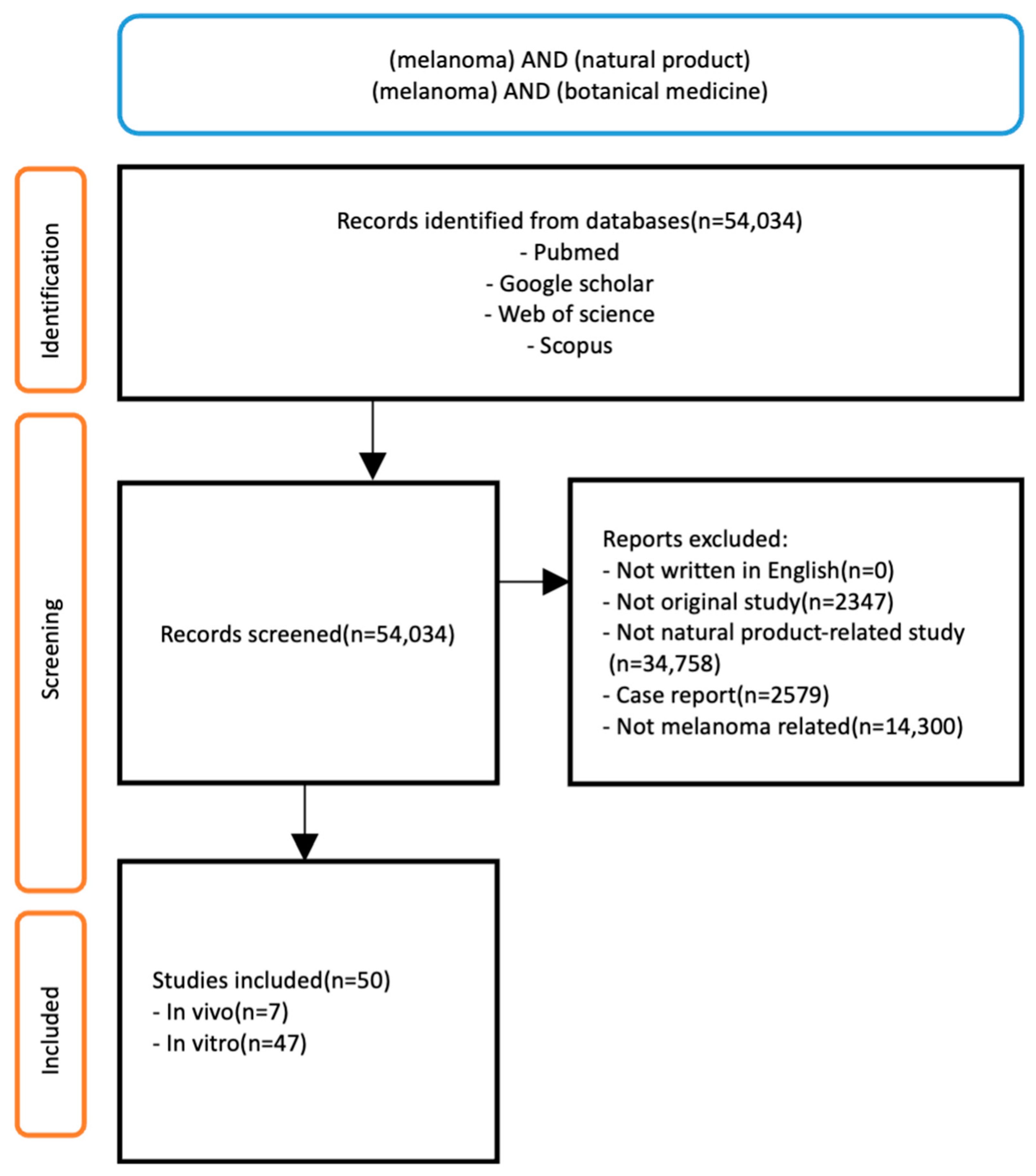
2.1. Search Strategy

Research papers addressing natural products that are effective in targeting melanoma were collected from February 2023 to March 2024 using databases PubMed, Google Scholar, Web of Science, and Scopus, using different combinations of keywords, such as the following: skin cancer, melanoma, natural products, herbal medicine, botanical medicine, plant extracts, antioxidant, anti-cancer, apoptosis, reactive oxygen species (ROS), angiogenesis and metastasis.

2.2. Study Selection Criteria

The selection of studies was based on the following inclusion criteria: (1) studies regarding the efficacy of natural products in any dosage form on melanoma cell or patients; (2) preclinical (in vivo, in vitro) studies; (3) isolated natural product; (4) original research articles; (5) articles written in English; (6) articles published within the last 10 years. The exclusion criteria were as follows: (1) articles regarding synthetic compounds; (2) systematic reviews, meta-analyses, review articles, conference papers and case reports; (3) articles in which the full text is unavailable. The resulting articles were revised to manually identify and exclude the articles that did not meet the criteria above. To identify articles of interest, all the articles retrieved from two databases were manually reviewed and checked for duplication.

2.3. Data Extraction

The obtained information included the author’s name, the publication year, the disease targeted, the extracted natural product, the cell line/animal model type, the dose and duration of extract administered, and the mechanisms of action (if reported) (Figure 1).

3. Anti-Melanoma Effect of Natural Products

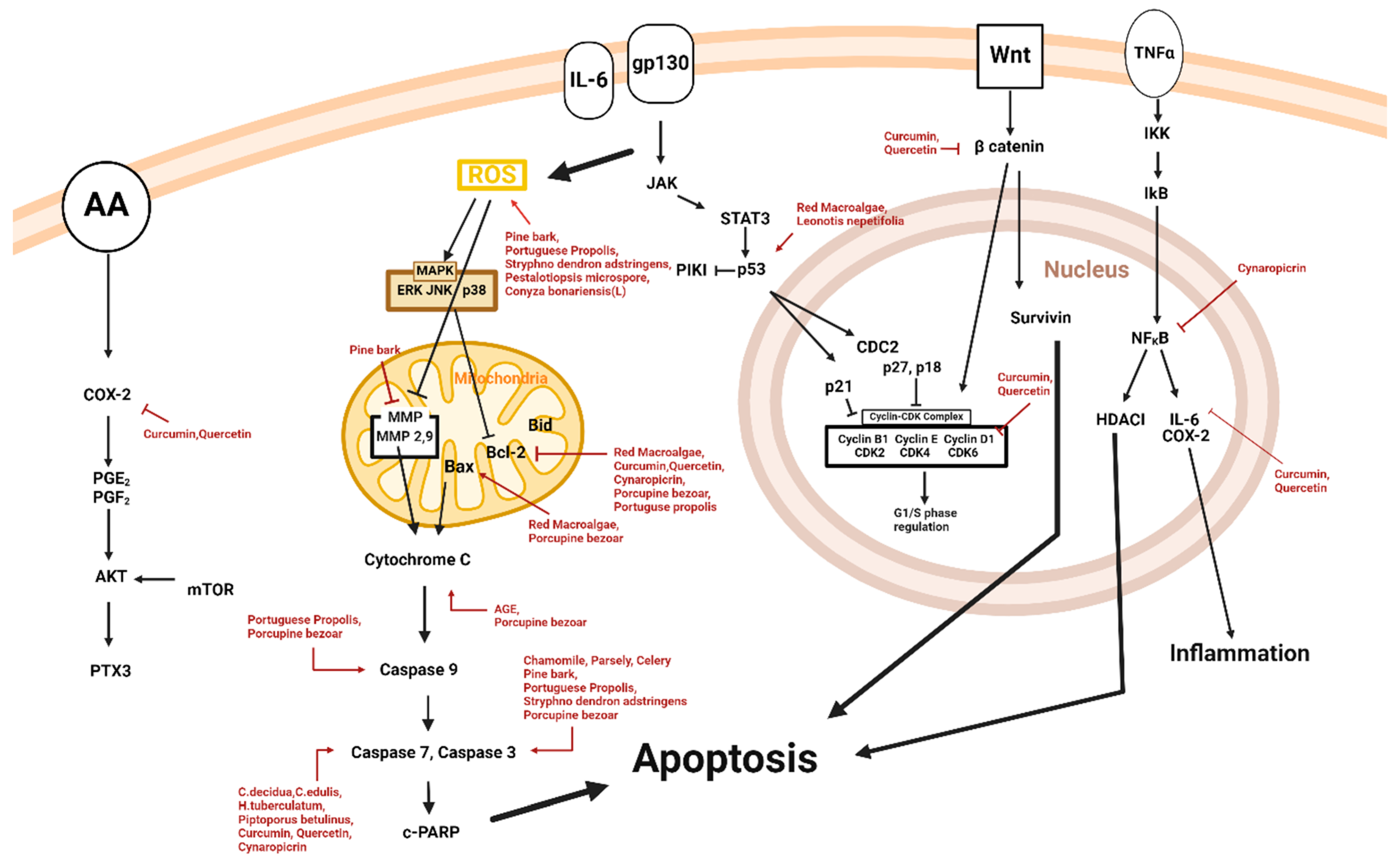
3.1. Apoptosis and Natural Products

Natural products ranging from extracts to single compounds, such as flavonoids, exhibit apoptotic effects on malignant melanoma cells (Table 1 and Figure 2).
Khan et al. reported the apoptotic effect of porcupine bezoar on A375 melanoma cells [18]. Porcupine bezoar at a dose of 3 µg/mL for 72 h had the highest apoptotic effect on A375 cells. The natural product exhibited apoptosis by the mitochondria apoptosis pathway, down-regulating Bcl-2, and up-regulating Bax, cyt-c, c-caspase-3, and c-capase-9. Srivastava et al. investigated the apoptotic effects of curcumin and quercetin on the A375 malignant melanoma cell line [19]. Curcumin and quercetin, at a dose of 1.5, 3.1, 6.3, 12.5, 25, or 50 μM for 24 h, resulted in the activation of apoptotic enzymes in a concentration-dependent manner. Apoptosis was induced through the down-regulation of DVL2, β-catenin, cyclin D1, Cox2, Axin2, Bcl-2, c-caspase-3, and c-caspase-7. Netala et al. assessed the apoptotic effects of the fungus Pestalotiopsis microspore on the B16F10 melanoma cell line [20]. Pestalotiopsis microspore at doses of 10, 20, 30, 40, 50, 100, 150, and 200 µg/mL induced apoptosis through the up-regulation of reactive oxygen species (ROS). And Ohkubo et al. demonstrated the apoptotic effects of aged garlic extract (AGE) on the M14 WT melanoma cell line [21]. Doses of AGE ranging from 0 to 10 mg/mL, exposed to cells for 24, 48, and 72 h, led to the up-regulation of proteins Cytc, Smac, and Diablo in a dose-dependent manner. Barros et al. highlighted the apoptotic effects of cashew gum on B16F10 cells and in C57BL/6 mice [22]. Doses of 3.125 to 100 μg/mL for 72 h activated apoptotic proteins in a dose-dependent manner. In vivo, C57BL/6 mice were intraperitoneally injected with 50 and 100 mg/kg of cashew gum for 14 consecutive days, which down-regulated γH2AX, ultimately inducing apoptosis in melanoma cells. Al Qathama et al. reported the apoptotic effects of Saudi medicinal plants on A375 and B16F10 melanoma cell lines [23]. C. decidua, and C. edulis, and H. tuberculatum, at a dose of 0.45 to 15 µg/mL for 48 h, induced apoptosis through the down-regulation of cleaved caspase-3 and cleaved caspase-7. Danciu et al. illustrated the apoptotic effects of chamomile, parsley and celery on the A375 melanoma cell line [24]. Dosages of chamomile, parsley and celery ranging from 10 to 60 μg/mL exposed to the melanoma cell line for 72 h resulted in the activation of various proteins in a dose-dependent manner. Apoptosis was induced by up-regulating c-casapse-3 and down-regulating IL-10. Ferreira et al. reported the apoptotic effect of Conyza bonariensis (L.) on SK-MEL-28, a melanoma cell line [25]. Exposing SK-MEL-28 to 20 μg/mL of Conyza bonariensis (L.) plant extract for 48 h induced apoptosis by activating ROS. Huang et al. investigated the apoptotic effects of Curcuma longa Linn extract on A2048 melanoma cells [26]. Doses of 5 to 25 μΜ of the extract, consisting of curcumin, demthoxycurcumin and bismethoxycurcumin, for 24 and 48 h resulted in apoptosis, coherent with the results of Srivastava et al. Also, Figueiredo et al. studied the apoptotic effects of curzerene extracted from Eugenia uniflora. SKMEL-19 cells were treated with 5 and 10 μM for 72 h [27]. This limited cell migration and enhanced chromatin condensation, promoting apoptosis. Kaushik et al. demonstrated the apoptotic effects of Hibiscus and Cinnamomum on the G361 melanoma cell line [28]. Hibiscus and Cinnamomum, at a dose of 62.5 to 1000 μg/mL for 48 h, caused apoptosis in a dose-dependent manner. Apoptosis was stimulated through the major metabolites found in Hibiscus and Cinnamomum. Merecz-Sadowska et al. demonstrated the apoptotic effect of Leonotis nepetifolia root extract on the A375 melanoma cell line [29]. Leonotis nepetifolia, at a dose of 1500 mg/mL for 24 h, had the highest apoptotic effect on A375 cells. Applying Leonotis nepetifolia root extract for 24 h resulted in the up-regulation of TP53 and p-53 in a concentration-dependent manner. Thaichinda et al. highlighted the apoptotic effects of pine bark on the human malignant A375 melanoma cell line [30]. Pinus maritima, at a dose of 5 to 100 µg/mL for 48 h, caused apoptotic activity by stimulating various proteins in a dose-dependent manner. Apoptosis was induced through the up-regulation of ROS and c-caspase 3 and the down-regulation of MMP-9. Bożek et al. demonstrated the apoptotic effects of Piptoporus betulinus ethanolic extracts on A375 cells [31]. Doses of 2.5 to 10 μL/mL for 24 h resulted in the up-regulation of caspase-3 activity. Oliveira et al. illustrated the apoptotic effects of Portuguese propolis, Apis mellifera L., on BRAF, A375, and WM9 melanoma cell lines [32]. Dosages of Apis mellifera L. ranging from 5 to 60 μg/mL for 72 h resulted in the activation of various proteins in a dose-dependent manner. Apoptosis was induced through the up-regulation of ROS, Bax, c-caspase-3, c-caspase-9 and p53, and the down-regulation of Bcl-XL and Bcl-2. Prasedya et al. demonstrated the apoptotic effects of red macroalgae, Gelidium latifolium, on the B16F10 malignant melanoma cell line [33]. Red macroalgae at a dose of 10 to 200 μg/mL for 72 h resulted in the activation of apoptotic enzymes in a concentration-dependent manner. Apoptosis was induced through the up-regulation of p53, Bax and Bak and the down-regulation of Bcl-2. Chatti et al. reported the apoptotic effect of Rhamnus alaternus on the B16F10 melanoma cell line [34]. Rhamnus alaternus at a dose of 20 μg/mL for 48 h had the highest apoptotic effect on B16F10 cells. Rhamnus alaternus aided in inhibiting cell proliferation and cell cycle arrest by damaging DNA. In addition to the proliferating effects of Rhamnus alaternus, apoptosis was also induced in a dose-dependent fashion. Baldivia et al. demonstrated the apoptotic effect of Stryphnodendron adstringens aqueous extract (SAAE) on B16F10Nex-2 melanoma cell line [35]. Likewise, SAAE at a dose of 500 µg/mL for 48 h had the highest apoptotic effect. Applying SAAE for 48 h resulted in the up-regulation of intracellular ROS level and c-caspase-3 activation in a concentration-dependent manner. Aimvijarn et al. claimed the induction of apoptosis by Thai water lily in B16 malignant melanoma cell line [36]. Thai water lily, applied at a dose of 200 to 1000 μg/mL for 24 h, resulted in apoptosis in a concentration-dependent manner. Apoptosis was induced through the stimulation of the transcription factor APF/Refl, which acted as an inhibitor of cellular invasion by acting as a cellular oxidant. Cicco et al. claimed the induction of apoptosis by cynaropicrin in A375 and SK-MEL-28 melanoma cell lines [30]. Cynaropicrin, applied at a dose of 30 μM for 24 and 48 h, induced apoptosis through the up-regulation of cleaved caspase-3 and cleaved caspase-7, and the down-regulation of Bcl-2 and NF-KB.
Table 1. Apoptosis-inducing compounds.
ClassificationDrugSourceCell Line/Animal ModelDose/DurationEfficacyMechanismReference
AnimalPorcupine bezoarHystrix BrachyuraA3751, 2, 3, 4 µg/mL; 24, 48, 72, 96 hInduction of apoptotic activity↑Bax, cyt-c, c-caspase-3, c-caspase-9
↓Bcl-2
[18]
Diarylheptanoid, FlavonoidCurcumin, quercetinCurcuma longa L.A3751.5, 3.1, 6.3, 12.5, 25, 50 μM; 24 hInduction of apoptosis↓DVL2, β-catenin, cyclin D1, Cox2, Axin2, Bcl-2
↑c-caspase-3, c-caspase-7
[19]
FungiPestalotiopsis microsporaGymnema sylvestre leavesB16F1010, 20, 30, 40, 50, 100, 150, 200 μg/mLInduction of apoptosis↑ROS[20]
Plant ExtractAged garlicAllium sativum L.M14 WT1, 5, 7, 10 mg/mL; 24, 48, 72 hInitiation of intrinsic apoptosis↑Cyt c, Smac, Diablo[21]
Plant ExtractCashew gumAnacardium occidentale Linn B16F103.125, 6.25, 12.5, 25, 50, 100 μg/mL; 72 hInduction of cell death and apoptosis↓γH2AX[22]
C57BL/6 mice50, 100 mg/kg; 14 d
Plant ExtractC. decidua,
C. edulis
H. tuberculatum,
C. decidua,
C. edulis
H. tuberculatum
A375, B16F100.45, 10, 15 µg/mL; 48 hInduction of apoptosis↑c-caspase-3, c-caspase-7[23]
Plant ExtractChamomile, parsley, celeryMatricaria chamomilla L., Petroselinum crispum (Mill.) Nym. ex A. W. Hill, Apium graveolens L.A37510, 30, 60 μg/mL; 72 hInduction of apoptotic activity↑c-caspase-3
↓ IL-10
[24]
Plant ExtractConyza bonariensis (L.)Conyza bonariensis (L.)SK-MEL-2820 μg/mL; 48 hInduction of apoptosis↑ROS[25]
Plant ExtractCurcumin, demethoxycurcumin, bisdemethoxycurcuminCurcuma longa LinnA20585, 10, 15, 20, 25 μΜ; 24, 48 hInitiation of apoptosis [26]
Plant ExtractCurzereneEugenia unifloraHCT-116, AGP-01, SKMEL-19, MRC-55, 10 μM; 72 hInduction of apoptosis [27]
Plant ExtractHibiscus, cinnamomumHibiscus syriacus, Cinnamomum loureirii NeesG36162.5, 125, 250, 500, 1000 μg/mL; 48 hInduction of apoptosis [28]
Plant ExtractLeonotis nepetifoliaLeonotis nepetifoliaA375500 µg/mL; 24 hInduction of apoptosis↑p53[29]
Plant ExtractPine barkPinus maritimaA3755, 25, 50, 100 µg/mL; 12, 24, 48 hInduction of apoptosis↑ROS, c-caspase-3
↓MMP-9
[37]
Plant ExtractPiptoporus betulinusPiptoporus betulinusHs27, A375, WM1152.5, 5, 10 μL/mL; 24 hInduction of apoptosis↑c-caspase-3, c-caspase-7[31]
Plant ExtractPortuguese propolisApis mellifera L.A375, WM95, 10, 15, 20, 25, 30, 35, 40, 45, 50, 55, 60 µg/mL; 72 hActivation of apoptosis↑ROS, Bax, c-caspase-3, c-caspase-9, p53
↓Bcl-XL, Bcl-2
[32]
Plant ExtractRed macroalgaeGelidium latifoliumB16F1010, 50, 100, 200 μg/mL; 72 hInduction of apoptosis↑p53, Bax, Bak
↓Bcl-2
[33]
Plant ExtractRhamnus alaternusRhamnus alaternusB16F1010, 20, 40 µg/mL; 48 hInduction of apoptosis [34]
Plant ExtractStryphnodendron adstringensStryphnodendron adstringensB16F1010, 25, 50, 125, 250, 500 µg/mL; 24, 48 hPromotion of apoptosis↑ROS, c-caspase-3[35]
Plant ExtractThai water lilyNymphaea stellateB16F10200, 400, 600 800, 1000 μg/mL; 24 hInduction of apoptosis [36]
TerpenoidCynaropicrinCentaurea drabifolia subsp.detonsaA37530 μM; 24, 48 hInduction of apoptosis↑c-caspase-3, c-caspase-7
↓Bcl-2, NF-KB
[30]
Abbreviations: Bax, Bcl-2-associated X protein; cyt-c, cytochrome c; c-caspase, cleaved caspase; Bcl-2-antagonist/killer; DVL2, dishevelled segment polarity protein 2; β-catenin, beta-catenin; Cox2, prostaglandin–endoperoxide synthase 2; Axin2, axis inhibition protein 2; ROS, reactive oxygen species; Smac, second mitochondria-derived activator of caspase; Diablo, direct inhibitor of apoptosis-binding protein with low pI; γH2AX, phosphorylated histone H2AX; IL-10, interleukin 10; p53, cellular tumor antigen p53; MMP-9, matrix metalloproteinases 9; Bcl-XL, B-cell lymphoma—extra large.

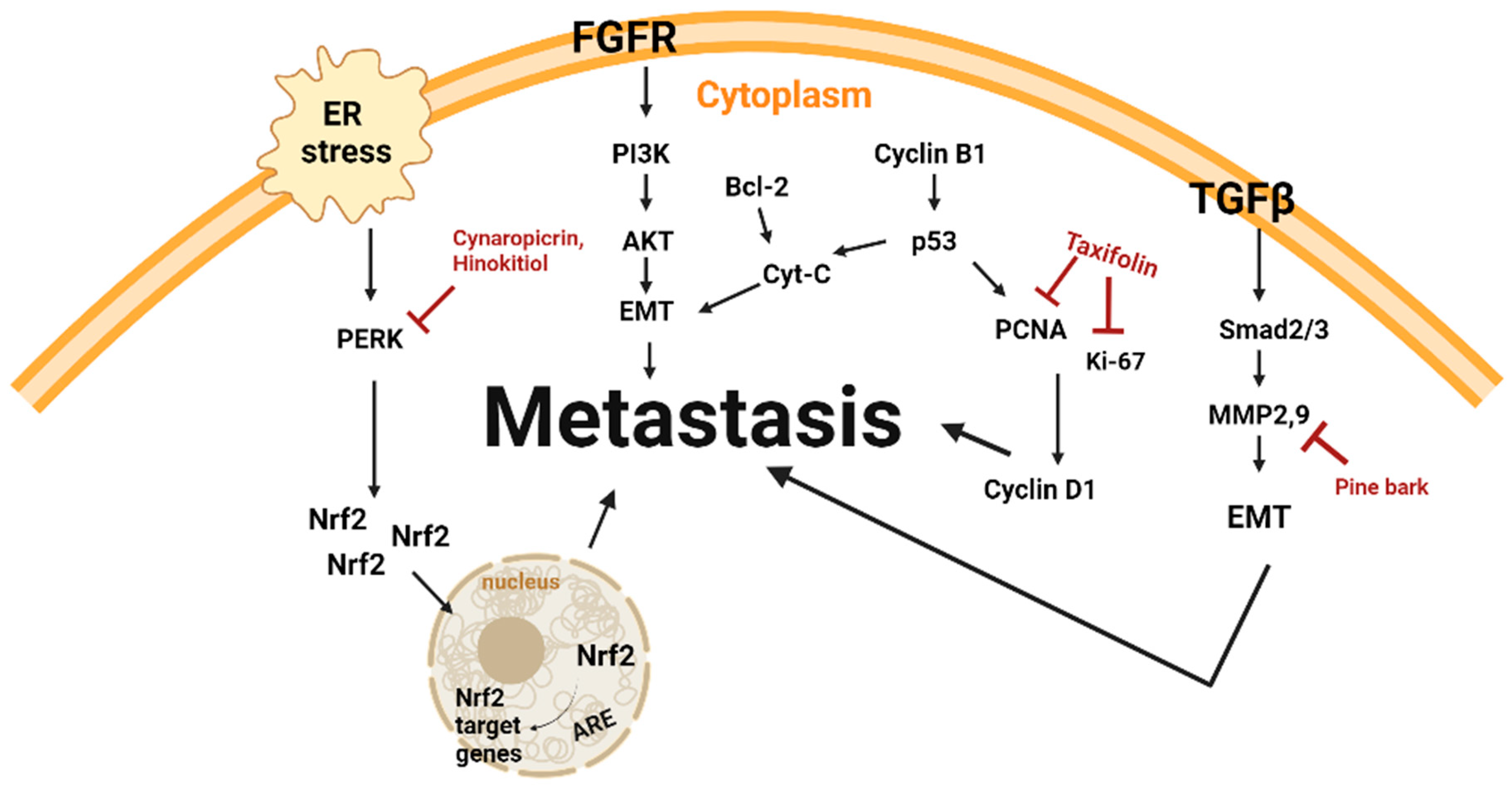
3.2. Anti-Metastasis and Natural Products

Polyphenol-rich extract and terpenoid-type single compounds have exhibited anti-metastatic properties when exposed to malignant melanoma cells (Table 2 and Figure 3).
Xu et al. described anti-proliferation and anti-migration in melanoma-induced C57/BL6 mice [38]. Here, 30 mg/kg and 60 mg/kg of taxifolin, extracted from the roots of Larix olgensis, were intraperitoneally injected for 11 days. Proteins Ki67 and PCNA were down-regulated, resulting in the inhibition of metastasis. Silva et al. claimed the anti-metastatic property of Eleutherine bulbosa bulbs in melanoma-induced C57BL6 mice [39]. Mice treated with 100, 500, and 1000 μg/mL of plant extract for 11 days exhibited anti-metastatic effects. Thaichinda et al. reported the anti-metastatic properties of pine bark extract (PBE) in melanoma skin cancer cell migration and invasion [37]. A dose of 50 μg/mL of PBE for 45 h reduced the migration and invasion of A375 cells. Additionally, PBE downregulated MMP-9, a protein expressed by cell lines derived from advanced primary melanomas, thereby reducing cancer cell invasion. De Cicco et al. demonstrated the anti-metastatic effect of cynaropicrin [30]. The natural product inhibited cell proliferation, apoptosis, migration, and invasion, which decreased metastatic activity in A375 cells. The up-regulation of Nrf2 and down-regulation of phosho-ERK and p65 reduced the activation of ERK as well as the translocation of NF-KB into the nucleus, reducing the proliferation of human metastatic melanoma cells. Wu et al. reported hinokitiol’s anti-metastatic properties [40]. Hinokitiol showed an inhibitory effect on cell migration, leading to a decrease in metastatic activity in B16F10 cells. In C57BL/6 mice and BALB/c mice, injecting hinokitiol via the tail vein impeded tumor metastasis, which was shown through a decrease in the weight of the tumors. Proteins phospho-AKT and phospho-ERK were significantly suppressed, acting against cancer-promoting pathways.

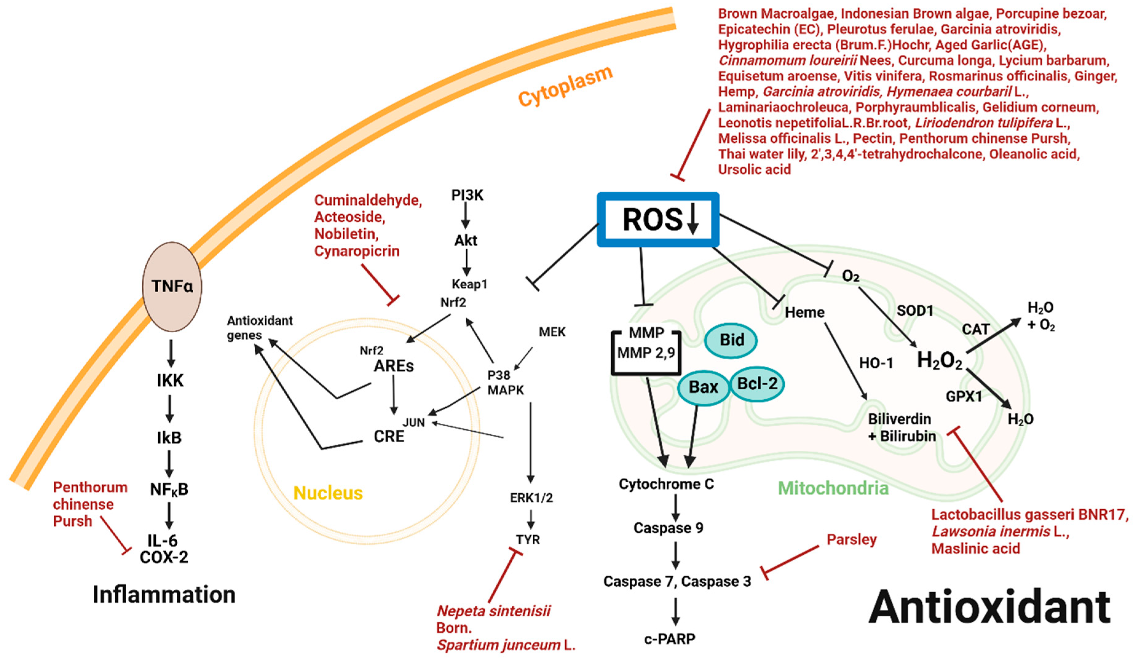
3.3. Antioxidant and Natural Products

Single compounds ranging from flavonoids to glycosides, and extracts ranging from calcified material to herbaceous plants, possess antioxidant properties against malignant melanoma cells (Table 3 and Figure 4).
Prasedya et al. reported the antioxidative effect of Sargassum cristaefolium (SCE) on B16F10 melanoma cell [41]. At doses of 1, 10, 50, and 100 μg/mL for an hour, the cellular ROS level in cells exposed to SCE was significantly downregulated. Sunarwidhi et al. reported the antioxidant effects of Indonesian brown algae on the B16F10 malignant melanoma cell line [42]. Indonesian brown algae at doses of 50, 100, 500, 1000, and 4000 μg/mL for 16 h, evaluated by ABTS scavenging, resulted in augmented antioxidant activity in a dose-dependent manner. Antioxidant activity was here induced through the down-regulation of ROS. Khan et al. reported the antioxidant effects of Hystrix Brachyura Bezoar on the B375 malignant melanoma cell line [18]. Hystrix Brachyura Bezoar at doses of 1, 2, 3, and 4 μg/mL for 72 h, evaluated by dot–blot assays, resulted in antioxidant activity in a concentration-dependent manner. Antioxidant activity was induced through the down-regulation of ROS. Lee et al. claimed the antioxidant effects of Lactobacillus gasseri BNR17 on the B16-F10 melanoma cell line and HaCaT epidermal keratinocyte cell line [43]. Lactobacillus gasseri BNR17, at doses of 0.16, 0.312, 0.625, 1.25, 2.5, 5, and 10% v/v for 24 h, resulted in antioxidant activity in a concentration-dependent manner. Antioxidant activity was induced through the up-regulation of HO-1, CAT, GPX1 and SOD1 and the down-regulation of ROS. Kim et al. confirmed the antioxidative effects that the essential oils of Chrysanthemum boreale MAKINO(CBM) demonstrated in B16BL6 melanoma cells [44]. The antioxidant and anti-melanogenesis properties were evident when exposed to CBM at doses of 1, 10, and 30 μg/mL for 48 h. The effects were exhibited through the down-regulation of TYR and the up-regulation of p38, MAPK, and Erk1/2. Mustapha et al. documented the antioxidative trait of epicatechin extracted from Hawthorn, Crataegus azarolus L., on the melanoma cell line [45]. B16F10 cells were exposed to doses of 10, 20, 30, 40, and 50 μmol/L of epicatechin for 1 h. The ROS level diminished, exhibiting evidence of an antioxidative effect. Arung et al. illustrated the antioxidant effects of Glyasperin A on the B16 malignant melanoma cell line [46]. Glyasperin A at doses of 2.4, 4.8, 6.0, and 7.2 μM resulted in antioxidant activity in a concentration-dependent manner, determined by diphenyl-2-picrylhydrazyl (DPPH), 2,2′-azi- no-bis-3-ethylbenzthiazoline-6-sulphonic acid (ABTS), superoxide dismutase-like activity (SOSA), and oxygen radical absorbance capacity (ORAC) assays. Yang et al. claimed the cellular effects of Pleurotus ferulae in B16 melanoma cells [47]. Doses of 0.5, 1, 2, 5, and 10 μg/mL had antioxidant and antitumor activities by limiting cellular ROS activity. Cheimonidi et al. illustrated the induction of antioxidant activity in the B16F1 and B16F10 cell lines and C57BL/6 mice through exposure to acteoside [48]. Acteoside, at doses of 0.2, 0.4, 0.6, 0.8, and 1 mM for 24 h, resulted in the activation of related proteins in a dose-dependent manner. C57BL/6 mice were intraperitonially injected with acteoside at doses of 1 and 2.5 mg/mouse for 13 consecutive days. Antioxidant activity was induced through the up-regulation of Akt, CREB1, MEK1 and WNK1 and the down-regulation of STAT1 and STAT3. Lee et al. demonstrated the antioxidant effect of Hygrophila erecta [49]. The natural product induced the antioxidant defense system, apoptotic effects, inflammatory responses, and a melanin synthesizing effect in both NHDF and B16F1 cell lines. Hygrophila erecta at doses at doses of 1, 2, 4, 8, 16, 31, 62, 125, 250, 500, and 1000 μg/mL for 24 h resulted in the suppression of ROS levels and NO concentration associated with the activation of SOD and Nrf2. Ohkubo et al. reported the antioxidant effects of aged garlic extract (AGE) on the human malignant melanoma M14 wild type cell line [21]. AGE extract at doses of 1, 5, and 10 mg/mL for 40 min induced oxidative stress by decreasing mitochondrial sulfhydryl groups in a dose-dependent manner. Antioxidant activity was induced through the down-regulation of ROS. Kaushik et al. demonstrated the antioxidant effects of Cinnamomum loureirii Nees on the G361 malignant melanoma cell line [28]. Cinnamomum loureirii Nees at a dose of 1 mg/mL for 30 min, evaluated by DPPH free radical inhibition, resulted in augmented antioxidant activity in a dose-dependent manner. Antioxidant activity was induced through the down-regulation of ROS. Žitek et al. demonstrated the antioxidant effects of Curcuma longa, Lycium barbarum, Equisetum arvense, Vitis vinifera, and Rosmarinus officinalis extracts on the WM-266-4 metastatic melanoma cell line [50]. The extracts from the raw materials, applied at doses of 0.001, 0.01, 0.1, 1, 5, 10, 20, and 100 μg/mL for 24 h, resulted in antioxidant activity in a concentration-dependent manner, mediated through the down-regulation of ROS. Also, Žitek et al. claimed the antioxidant effects of ginger and hemp on the B16F10 malignant melanoma cell line [51]. A combination of hemp extract and ginger, at doses of 0.01, 0.1, 1, 5, 10, 20, and 50 mg/mL for 24 h, exhibited antioxidant activity in a concentration-dependent manner by down-regulating ROS levels. Chatatkun et al. claimed the antioxidant effects of Garcinia atroviridis on the B16F10 malignant melanoma cell line [52] at doses of 62.5, 125, 250, 500, and 1000 μg/mL, in a dose-dependent manner, through free radical scavenging activity, as determined by DPPH and ABTS assays. Antioxidant activity was induced through the down-regulation of ROS. Spera et al. demonstrated the antioxidant effects of Hymenaea courbaril L. on the B16F10 malignant melanoma cell line [53]. Hymenaea courbaril L., at a dose of 250 μg/mL for 30 min, evaluated by DPPH radical scavenging activity, resulted in antioxidant activity in a dose-dependent manner. Antioxidant activity was induced through the down-regulation of ROS. Díaz et al. claimed the antioxidant effects of Laminaria ochroleuca, Porphyra umbilicalis and Gelidium corneum on G-361 malignant melanoma cell line [54]. At doses of 25, 50, 75, 100, 150, 200, 300, 400, and 500 μg/mL for 16 h, these extracts demonstrated antioxidant activity in a dose-dependent manner through the down-regulation of ROS. Raja et al. demonstrated the antioxidant effects of Lawsonia inermis L. on B16F10 malignant melanoma cell line-bearing C57BL/6 mice [55]. Lawsonia inermis L. at doses of 500 mg/kg and 1000 mg/kg for 40 days resulted in antioxidant activity in a concentration-dependent manner. Antioxidant activity was induced through the down-regulation of GSH-Px, SOD, and CAT. Merecz-Sadowska et al. demonstrated the antioxidant effects of Leonotis nepetifolia (L.) R. Br. extract on the A375 malignant melanoma cell line [29]. Leonotis nepetifolia (L.) R. Br., applied at a dose of 10 μg/mL for 16 h, evaluated by ABTS scavenging, resulted in increased antioxidant activity in a dose-dependent manner. Antioxidant activity was induced through the down-regulation of ROS and Mdm-2. Furthermore, Quassinti et al. claimed the antioxidant effects of Liriodendron tulipifera L. on the A375 malignant melanoma cell line [56]. Liriodendron tulipifera L., at a dose of 5 mg/mL for 72 h, evaluated by DPPH, ABTS, and ferric reducing antioxidant power (FRAP) assays, resulted in antioxidant activity in a concentration-dependent manner. Antioxidant activity was induced through the down-regulation of ROS. Sipos et al. reported the antioxidant effects of Melissa officinalis L. on HaCaT human keratinocyte and A375 malignant melanoma cell line [57]. Melissa officinalis L., at doses of 0.1, 0.5, 1, 3, and 5 mg/mL for 20 min, evaluated by DPPH free radical consumption, resulted in enhanced antioxidant activity in a concentration-dependent manner through the down-regulation of ROS. Akaberi et al. showed the antioxidative property of Nepeta sintenisii Bornm [58]. B16F10 melanoma cells treated with 50 μg/mL of Nepeta sintenisii Bornm showed the suppression of oxidative stress and TYR in the cell line. Danciu et al. illustrated the antioxidant effect of parsley on the A375 melanoma cell line [24]. Dosages of parsley ranging from 10 to 60 μg/mL exposed to a melanoma cell line for 72 h resulted in the activation of various proteins in a dose-dependent manner. Antioxidant activity was induced by up-regulating c-casapse-3 and down-regulating IL-10. Wikiera et al. reported the effects of pectin extracted from dried apple pomace [59]. An increased extracellular ROS level was observed through 24-h exposure to 0.2, 0.4, 0.6, 0.8, and 1 mg/mL, encouraging antioxidant activities in B16F10 cells. Jeong et al. reported the antioxidant effects of Penthorum chinense Pursh on the B16-F10 melanoma cell line and HaCaT epidermal keratinocyte cell line [60]. Penthorum chinense Pursh at doses of 50 and 100 μg/mL for 24 h resulted in antioxidant activity in a concentration-dependent manner. Antioxidant activity was induced through the down-regulation of ROS, IL-6, COX-2, p-ERK, p-p38, p-JNK, and MMP. Nanni et al. claimed the antioxidant effect of Spartium junceum L. on the B16F10 malignant melanoma cell line [61]. Spartium junceum L., at doses of 4, 8 and 10 mg/mL for 4, 24, and 48 h, inhibited the growth of the melanoma cell line, which resulted in the activation of various proteins in a dose- and time-dependent manner. Antioxidant activity was induced through the down-regulation of MITF. Baldivia et al., demonstrating the antioxidant effect of Stryphnodendron adstringens aqueous extract (SAAE) [35]. SAAE at a dose of 50 µg/mL had the highest antioxidant effect on B16F10Nex-2 melanoma cell line. SAAE showed antioxidant activity by scavenging ABTS and DPPH free-radicals, and proved its antioxidant capacity through reduced malondialdehyde (MDA) levels. Aimvijarn et al. claimed the antioxidant effects of Nymphaea stellate extracted from Thai water lily on the B16F10 malignant melanoma cell line [36]. Nymphaea stellate, applied at doses of 200, 400, 600 800, and 1000 μg/mL for 24 h, evaluated by fluorescent 2,7-dichlorodyhydrofluorescein diacetate (DCFH-DA), resulted in antioxidant activity in a concentration-dependent manner. Antioxidant activity was induced through the down-regulation of ROS. Ciorîţă et al. reported the induction of antioxidant activity by Vinca minor, V. herbacea, V. major and V. major var. variegata against HaCaT and the A375 malignant melanoma cell line [62]. Vinca minor, V. herbacea, V. major and V. major var. variegata extracts, at concentrations of 0.09, 0.5, 1, 2, and 3% for 24 h and 72 h, resulted in antioxidant activity in a dose-dependent manner, along with antibacterial and antitumor effects. Zhong et al. claimed the induction of antioxidant activity against the B16F10 cell line and C57BL/6 mice through exposure to 2′,3,4,4′-tetrahydrochalcone [63]. 2′,3,4,4′-tetrahydrochalcone, at doses of 1, 10, 20, and 40 μM/L for 30 min, resulted in the activation of related proteins in a dose-dependent manner. C57BL/6 mice were injected with 2′,3,4,4′-tetrahydrochalcone in doses of 8.5 and 10 μg for 40 days. Antioxidant activity was induced through the down-regulation of ROS. Feng et al. reported the antioxidant effects of nobiletin on the SK-MEL-28 malignant melanoma cell line [64]. Nobiletin, applied at doses of 5, 15, and 45 μM for 30 min and evaluated by DCFH-DA, where the collected cells’ fluorescence intensity was detected using a flow cytometer, resulted in increased antioxidant activity in a dose-dependent manner. Antioxidant activity was induced through the up-regulation of Keap1 and down-regulation of Nrf2 and ROS. Cicco et al. claimed the induction of antioxidant activity of cynaropicrin on A375 melanoma cell lines [30]. Cynaropicrin, applied at doses of 3, 10, and 30 μM for 24 h and 48 h, resulted in the activation of antioxidant activity in a concentration-dependent manner. Antioxidant activity was induced through the up-regulation Nrf2 and down-regulation of pERK and p65. Mokhtari et al. illustrated the antioxidant effects of maslinic acid on A10 and B16F10 malignant melanoma cell lines [65]. Maslinic acid, applied at doses of 10.6, 21.6, 42.3, and 84.6 μM for 24 h, resulted in the activation of antioxidant activity in a dose-dependent manner. Antioxidant activity was induced through the down-regulation of ROS, CAT, G6PDH, SOD, GST, GPX, and GR. Alvarado et al. demonstrated the antioxidant effects of oleanolic acid and ursolic acid extract from Plumeria obtusa on B16 malignant melanoma cell line [66]. Oleanolic acid and ursolic acid at a dose of 2 mg/mL for 90 min, evaluated by DPPH free radical activity, resulted in antioxidant activity in a dose-dependent manner. Antioxidant activity was induced through the down-regulation of ROS.

3.4. Anti-Angiogenesis and Natural Product

Extracts from natural compounds that have already been addressed possess properties of anti-angiogenesis on malignant melanoma cells (Table 4).
Sipos et al. claimed the induction of angiogenesis in HaCaT human keratinocyte, A375 malignant melanoma cell line and SKH-1 mice through exposure to Melissa officinalis L. extract [57]. Melissa officinalis L., at doses of 20, 100, 250, 500, and 1000 μg/mL for 24 h, resulted in the activation of proteins in a concentration-dependent manner. SKH-1 mice were topically applied with Melissa officinalis L. at a dose of 5 mg/mL every 2 days for 2 weeks. This resulted in the angiogenetic behavior of the specified melanoma cell.

4. Discussion

Cancer is related to an imbalance between reactive oxygen species (ROS) and antioxidants, as well as increased oxidative stress. Metabolic disturbances and signaling abnormalities cause the ROS level to increase, and eventually result in carcinogenesis [67]. Apoptosis is a form of programmed cell death. Cells that malfunction due to various changes in the body activate self-cell death [68]. However, most of the cells that do not notice their defect fail to turn on self-cell death systems, and form cancer. All cancers have the inclination to metastasize, which is when cancer cells break away from the primary location and move to secondary and tertiary locations [69]. The cancer cells go through this process by traveling through lymph nodes or blood vessels, which is angiogenesis [70]. Oxidative stress also plays a critical role in melanoma progression, as excessive reactive oxygen species (ROS) can promote DNA damage and alter key signaling pathways [71]. Curcumin has shown potential use as an adjuvant therapy in cancer treatment. A clinical trial combining curcumin with gemcitabine in pancreatic cancer reported a partial response and stable disease in some patients, highlighting its possible role in melanoma [72]. Figure 5 illustrates the chemical structures of key natural compounds discussed in this review, including curcumin (PubChem CID: 969516), quercetin (PubChem CID: 5280343), acteoside (PubChem CID: 5281800), cynaropicrin (PubChem CID: 119093), and hinokitiol (PubChem CID: 3611). These compounds have been reported to exhibit significant biological activities in melanoma treatment, such as apoptosis induction, oxidative stress regulation, and anti-metastatic effects. According to the Skin Cancer Foundation, a form of cancer that begins in a type of cell known as a melanocyte is melanoma. Among various skin cancers, like basal cell carcinoma and squamous cell carcinoma, malignant melanoma is the most dangerous type of skin cancer due to its ability to metastasize to other organs more rapidly if it is not treated at an early stage [73]. Various types of treatment methods that are available for curing melanoma have been approved by the FDA. Yet, due to its extreme tendency to relapse, melanomas do not always respond to these therapeutics [74]. Also, the death of melanoma cells is insignificant even with chemotherapy, and the treatment itself reduces the patients’ quality of life. Chemotherapy causes vomiting, nausea, fever, perforations, severe infection from within, diarrhea, constipation, and allergic reactions [75]. Thus, this calls for the adjuvant treatment of existing cancer therapeutics with natural products in curing melanoma. Natural products will provide patients with an easier approach to cure melanoma, one of the most commonly occurring cancers that roots from the endocrine systems in the body.
In this systematic review, the antioxidant activity and anti-cancer activity of natural products against melanoma were scrutinized. A375 and SK-MEL-28 are human melanoma cell lines that are widely used to assess proliferation and apoptotic responses to natural compounds [76]. B16F10, a murine melanoma cell line, is commonly employed in metastatic models due to its high invasiveness [77]. WM115, derived from primary melanoma, is utilized in studies investigating early-stage melanoma progression and genetic variations [78]. Understanding these distinctions is essential for accurately interpreting in vitro findings and evaluating the therapeutic potential of natural compounds in melanoma. Furthermore, the classification, origin, experimental model, dose, duration, efficacy, and mechanisms of action cited in studies covering single compounds up to complex decoctions were systematically categorized. In total, four tables were assembled based on the anti-cancer mechanisms of the natural products. The systematic reviews published within the last 10 years regarding the effects of natural products on melanoma were mainly included. This comprehensive approach allows for a clear understanding of the potential of various natural products. In addition, the detailed organization of the data in each of the tables facilitates the understanding and potential replication of our experimental results. The existing reviews have focused more on the effects of natural products on the adverse symptoms caused by adjuvant treatments. The antioxidant activity and anti-cancer activity of all the natural products included in this review were selective to melanoma cancer cell lines. Among several systematic reviews regarding melanoma, Garbe et al. only included therapeutics in clinical practice, and Al Qathama et al. focused only on natural active principles. There were no systematic reviews that solely examined the association between melanoma and natural products in general. However, in this paper, all studies that discussed the intrinsic efficacies of natural products were analyzed by forming four different categories of cancer mechanisms. In particular, in some studies, Cinnamomum loureirii Nees, which down-regulates ROS, acetoside, which up-regulates Akt, CREB1, MEK1, and WNK1 and downer-gulates STAT1 and STAT3, and pectin, which down-regulates ROS, were administrated at a low concentration of 1 mg/mL or less, illustrating their potential to be utilized as main compounds for treating melanoma.
However, there are several drawbacks to this systematic review. First, it only evaluated studies published in English, which may have resulted in a lack of comprehensive data. Furthermore, the lack of clinical trials and the shortage of research on anti-angiogenesis and anti-metastasis effects related to natural products and melanoma are problems. Future studies should include additional search engines beyond databases such as Google Scholar and PubMed, so as to gather more extensive data, including studies published in other languages. While twenty studies on apoptosis and thirty-four studies on antioxidant effects were identified, there was a limited number of studies related to anti-angiogenesis and anti-metastasis, requiring the further exploration of these categories. Moreover, some of the studies lacked either in vitro or in vivo investigations, highlighting the need for further examinations to exemplify the effects of natural products on melanoma. Studies focusing on Stryphnodendron adstringens, Glyasperin A, Eleutherine bulbosa bulbs, Rhamnus alaternus, Thai water lily, Hibiscus and Cinnamomum, Curzerene, and Curcuma longa Linn also require additional in-depth mechanistic research in order to better understand the impacts of natural products on melanoma. It is essential to elucidate how these substances work at the cellular level, as optimizing the efficacy of natural product-based therapies depends on understanding their molecular mechanisms. Considering these factors, broader studies on the effects of natural substances on melanoma are strongly advised. Despite its limitations, this systematic review offers an overview of the potential therapeutic benefits of natural products in treating melanoma, and highlights their significant role in cancer therapy.

5. Conclusions

This study has reviewed the anti-cancer effects of natural products on melanoma by classifying forty-three studies in terms of antioxidant and anti-cancer mechanisms. The regulatory factors that activate anti-cancer activity were presented for an overview of the effects of natural products. Natural products are promising, as they have the capacity to treat melanoma. Therefore, additional in vitro, in vivo and clinical studies should be conducted in order to fully utilize and develop such prospective resources.

Author Contributions

Conceptualization and writing—original draft preparation S.A., J.A. and B.K.; data curation, S.A., J.A., D.L. and B.K.; writing—review and editing, S.A., J.A., H.N.K., S.K., C.-H.A., R.A.S., R.I.M.A.R. and B.K.; visualization, S.A., J.A., D.L., S.K. and B.K.; supervision, B.K.; project administration, B.K.; funding acquisition, B.K. All authors have read and agreed to the published version of the manuscript.

Funding

This research was supported by the Undergraduate Research Program (URP) of the College of Korean Medicine, Kyung Hee University, Republic of Korea, in 2024, the Basic Science Research Program through the National Research Foundation of Korea (NRF) funded by the Ministry of Education (NRF-2020R1I1A2066868), the National Research Foundation of Korea (NRF) grant funded by the Korea government (MSIT) (No. 2020R1A5A2019413), a grant of the Korea Health Technology R&D Project through the Korea Health Industry Development Institute (KHIDI), funded by the Ministry of Health & Welfare, Republic of Korea (grant number: RS-2020-KH087790), the Starting growth Technological R&D Program (TIPS Program, (No. RS-2024-00507224)) funded by the Ministry of SMEs and Startups (MSS, Korea) in 2024 and the National Research Foundation of Korea (NRF) grant funded by the Korea government (MSIT) (RS-2024-00350362).

Conflicts of Interest

The authors declare no conflict of interest.

References

  1. PDQ Adult Treatment Editorial Board. Melanoma Treatment (PDQ®)—Patient Version; National Cancer Institute: Bethesda, MD, USA, 2023. [Google Scholar]
  2. Ferlay, J.; Ervik, M.; Lam, F.; Laversanne, M.; Colombet, M.; Mery, L.; Piñeros, M.; Znaor, A.; Soerjomataram, I.; Bray, F. Melanoma of Skin. In Cancer Today; Global Cancer Observatory: Lyon, France, 2024. [Google Scholar]
  3. Bray, F.; Laversanne, M.; Sung, H.; Ferlay, J.; Siegel, R.L.; Soerjomataram, I.; Jemal, A. Global cancer statistics 2022: GLOBOCAN estimates of incidence and mortality worldwide for 36 cancers in 185 countries. CA Cancer J. Clin. 2024, 74, 229–263. [Google Scholar] [CrossRef] [PubMed]
  4. Estimated Number of New Cases from 2022 to 2025, Both Sexes, Age [0–85+]. Globocan 2022. Available online: https://gco.iarc.fr/tomorrow/en/dataviz/isotype?years=2025&single_unit=10000&cancers=16 (accessed on 3 January 2025).
  5. Osman, A.; Nigro, A.R.; Brauer, S.F.; Borda, L.J.; Roberts, A.A. Epidemiology and primary location of melanoma in Asian patients: A surveillance, epidemiology, and end result-based study. JAAD Int. 2024, 16, 77–78. [Google Scholar] [CrossRef] [PubMed]
  6. Maretha, D.E.; Afriansyah, D.; Wati, D.S.; Masri, M.; Dwiyanti, A.R.; Hanif, M.I.; Wardoyo, S. Test comparison of seeds and skins extract of duku’s fruit (Lansium domesticum Corr.) against the amount of melanin pigment of skin mencit (Mus musculus) to prevent premature aging of the skin. Adv. Tradit. Med. 2022, 22, 875–883. [Google Scholar] [CrossRef]
  7. Petrie, T.; Samatham, R.; Witkowski, A.M.; Esteva, A.; Leachman, S.A. Melanoma Early Detection: Big Data, Bigger Picture. J. Investig. Dermatol. 2019, 139, 25–30. [Google Scholar] [CrossRef]
  8. Jeong, M.; Cho, J.; Lim, D.; Choi, M.; Park, Y.; Cheong, Y.; Kang, Y.; Kang, I.; Kim, S.; Kim, D. The biological effects of Rosa rugosa extract on keratinocyte differentiation and enhancement of skin barrier function. Adv. Tradit. Med. 2024. [Google Scholar] [CrossRef]
  9. Morgese, F.; Sampaolesi, C.; Torniai, M.; Conti, A.; Ranallo, N.; Giacchetti, A.; Serresi, S.; Onofri, A.; Burattini, M.; Ricotti, G.; et al. Gender Differences and Outcomes in Melanoma Patients. Oncol. Ther. 2020, 8, 103–114. [Google Scholar] [CrossRef]
  10. Ragab, M.; Choudhry, H.; Al-Rabia, M.W.; Binyamin, S.S.; Aldarmahi, A.A.; Mansour, R.F. Early and accurate detection of melanoma skin cancer using hybrid level set approach. Front. Physiol. 2022, 13, 965630. [Google Scholar] [CrossRef]
  11. Treating Melanoma Skin Cancer. Available online: https://www.cancer.org/cancer/types/melanoma-skin-cancer/treating.html (accessed on 3 January 2025).
  12. Kahlon, N.; Doddi, S.; Yousif, R.; Najib, S.; Sheikh, T.; Abuhelwa, Z.; Burmeister, C.; Hamouda, D.M. Melanoma Treatments and Mortality Rate Trends in the US, 1975 to 2019. JAMA Netw Open 2022, 5, e2245269. [Google Scholar] [CrossRef]
  13. Berthenet, K.; Weber, K.; Ichim, G. Sometimes even apoptosis fails: Implications for cancer. Mol. Cell. Oncol. 2020, 7, 1797430. [Google Scholar] [CrossRef]
  14. Al-Qatati, A.; Aliwaini, S. Combined pitavastatin and dacarbazine treatment activates apoptosis and autophagy resulting in synergistic cytotoxicity in melanoma cells. Oncol. Lett. 2017, 14, 7993–7999. [Google Scholar] [CrossRef]
  15. Lee, D.-h.; Seong, S.; Won, Y. Traditional Korean medicine as second-line treatment of metastatic colorectal cancer: A case report. Med. Case Rep. Study Protoc. 2021, 2, e0042. [Google Scholar] [CrossRef]
  16. Farida, S.; Jenie, R.I.; Fakhrudin, N. Calophyllum inophyllum: A Comprehensive Analysis of its Ethnobotanical, Phytochemical, and Pharmacological Properties. Maj. Obat Tradis. 2024, 29, 121–142. [Google Scholar] [CrossRef]
  17. Schröder, S.; Lee, S.; Efferth, T.; Motoo, Y. Acupuncture and herbal medicine for cancer patients. Evid. Based Complement. Altern. Med. 2013, 2013, 313751. [Google Scholar] [CrossRef] [PubMed]
  18. Firus Khan, A.Y.; Abdullah Asuhaimi, F.; Jalal, T.K.; Roheem, F.O.; Natto, H.A.; Johan, M.F.; Ahmed, Q.U.; Abdul Wahab, R. Hystrix brachyura Bezoar Characterization, Antioxidant Activity Screening, and Anticancer Activity on Melanoma Cells (A375): A Preliminary Study. Antioxidants 2019, 8, 39. [Google Scholar] [CrossRef]
  19. Srivastava, N.S.; Srivastava, R.A.K. Curcumin and quercetin synergistically inhibit cancer cell proliferation in multiple cancer cells and modulate Wnt/β-catenin signaling and apoptotic pathways in A375 cells. Phytomedicine 2019, 52, 117–128. [Google Scholar] [CrossRef]
  20. Netala, V.R.; Bethu, M.S.; Pushpalatha, B.; Baki, V.B.; Aishwarya, S.; Rao, J.V.; Tartte, V. Biogenesis of silver nanoparticles using endophytic fungus Pestalotiopsis microspora and evaluation of their antioxidant and anticancer activities. Int. J. Nanomed. 2016, 11, 5683–5696. [Google Scholar] [CrossRef]
  21. Ohkubo, S.; Dalla Via, L.; Grancara, S.; Kanamori, Y.; García-Argáez, A.N.; Canettieri, G.; Arcari, P.; Toninello, A.; Agostinelli, E. The antioxidant, aged garlic extract, exerts cytotoxic effects on wild-type and multidrug-resistant human cancer cells by altering mitochondrial permeability. Int. J. Oncol. 2018, 53, 1257–1268. [Google Scholar] [CrossRef]
  22. Barros, A.B.; Moura, A.F.; Silva, D.A.; Oliveira, T.M.; Barreto, F.S.; Ribeiro, W.L.C.; Alves, A.; Araújo, A.J.; Moraes Filho, M.O.; Iles, B.; et al. Evaluation of antitumor potential of cashew gum extracted from Anacardium occidentale Linn. Int. J. Biol. Macromol. 2020, 154, 319–328. [Google Scholar] [CrossRef]
  23. AlQathama, A.; Bader, A.; Al-Rehaily, A.; Gibbons, S.; Prieto, J.M. In vitro cytotoxic activities of selected Saudi medicinal plants against human malignant melanoma cells (A375) and the isolation of their active principles. Eur. J. Integr. Med. 2022, 49, 102083. [Google Scholar] [CrossRef]
  24. Danciu, C.; Zupko, I.; Bor, A.; Schwiebs, A.; Radeke, H.; Hancianu, M.; Cioanca, O.; Alexa, E.; Oprean, C.; Bojin, F.; et al. Botanical Therapeutics: Phytochemical Screening and Biological Assessment of Chamomile, Parsley and Celery Extracts against A375 Human Melanoma and Dendritic Cells. Int. J. Mol. Sci. 2018, 19, 3624. [Google Scholar] [CrossRef]
  25. Ferreira, R.C.; Duarte, S.S.; de Sousa, V.M.; de Souza, R.R.M.; Marques, K.K.G.; de Abrantes, R.A.; do Nascimento, Y.M.; de Sousa, N.F.; Scotti, M.T.; Scotti, L.; et al. The Essential Oil from Conyza bonariensis (L.) Cronquist (Asteraceae) Exerts an In Vitro Antimelanoma Effect by Inducing Apoptosis and Modulating the MAPKs, NF-κB, and PKB/AKT Signaling Pathways. Pharmaceuticals 2023, 16, 1553. [Google Scholar] [CrossRef] [PubMed]
  26. Huang, C.; Lu, H.F.; Chen, Y.H.; Chen, J.C.; Chou, W.H.; Huang, H.C. Curcumin, demethoxycurcumin, and bisdemethoxycurcumin induced caspase-dependent and -independent apoptosis via Smad or Akt signaling pathways in HOS cells. BMC Complement. Med. Ther. 2020, 20, 68. [Google Scholar] [CrossRef] [PubMed]
  27. Figueiredo, P.L.B.; Pinto, L.C.; da Costa, J.S.; da Silva, A.R.C.; Mourão, R.H.V.; Montenegro, R.C.; da Silva, J.K.R.; Maia, J.G.S. Composition, antioxidant capacity and cytotoxic activity of Eugenia uniflora L. chemotype-oils from the Amazon. J. Ethnopharmacol. 2019, 232, 30–38. [Google Scholar] [CrossRef] [PubMed]
  28. Kaushik, N.; Oh, H.; Lim, Y.; Kumar Kaushik, N.; Nguyen, L.N.; Choi, E.H.; Kim, J.H. Screening of Hibiscus and Cinnamomum Plants and Identification of Major Phytometabolites in Potential Plant Extracts Responsible for Apoptosis Induction in Skin Melanoma and Lung Adenocarcinoma Cells. Front. Bioeng. Biotechnol. 2021, 9, 779393. [Google Scholar] [CrossRef]
  29. Merecz-Sadowska, A.; Sitarek, P.; Śliwiński, T.; Zajdel, K.; Malinowska, K.; Zielińska-Bliźniewska, H.; Kucharska, E.; Zajdel, R. In vitro and In silico Studies on Leonotis nepetifolia (L.) R. Br. Root Extract against Cancer Cells. Curr. Pharm. Biotechnol. 2022, 23, 1383–1395. [Google Scholar] [CrossRef]
  30. De Cicco, P.; Busà, R.; Ercolano, G.; Formisano, C.; Allegra, M.; Taglialatela-Scafati, O.; Ianaro, A. Inhibitory effects of cynaropicrin on human melanoma progression by targeting MAPK, NF-κB, and Nrf-2 signaling pathways in vitro. Phytother. Res. 2021, 35, 1432–1442. [Google Scholar] [CrossRef]
  31. Bożek, J.; Tomala, J.; Wójcik, S.; Kamińska, B.; Brand, I.; Pocheć, E.; Szostak, E. Effects of Piptoporus betulinus Ethanolic Extract on the Proliferation and Viability of Melanoma Cells and Models of Their Cell Membranes. Int. J. Mol. Sci. 2022, 23, 13907. [Google Scholar] [CrossRef]
  32. Oliveira, R.D.; Celeiro, S.P.; Barbosa-Matos, C.; Freitas, A.S.; Cardoso, S.M.; Viana-Pereira, M.; Almeida-Aguiar, C.; Baltazar, F. Portuguese Propolis Antitumoral Activity in Melanoma Involves ROS Production and Induction of Apoptosis. Molecules 2022, 27, 3533. [Google Scholar] [CrossRef]
  33. Prasedya, E.S.; Ardiana, N.; Padmi, H.; Ilhami, B.T.K.; Martyasari, N.W.R.; Sunarwidhi, A.L.; Nikmatullah, A.; Widyastuti, S.; Sunarpi, H.; Frediansyah, A. The Antiproliferative and Apoptosis-Inducing Effects of the Red Macroalgae Gelidium latifolium Extract against Melanoma Cells. Molecules 2021, 26, 6568. [Google Scholar] [CrossRef]
  34. Bouhlel Chatti, I.; Ben Toumia, I.; Krichen, Y.; Maatouk, M.; Chekir Ghedira, L.; Krifa, M. Assessment of Rhamnus alaternus Leaves Extract: Phytochemical Characterization and Antimelanoma Activity. J. Med. Food 2022, 25, 910–917. [Google Scholar] [CrossRef]
  35. Baldivia, D.D.S.; Leite, D.F.; Castro, D.T.H.; Campos, J.F.; Santos, U.P.D.; Paredes-Gamero, E.J.; Carollo, C.A.; Silva, D.B.; de Picoli Souza, K.; Dos Santos, E.L. Evaluation of In Vitro Antioxidant and Anticancer Properties of the Aqueous Extract from the Stem Bark of Stryphnodendron adstringens. Int. J. Mol. Sci. 2018, 19, 2432. [Google Scholar] [CrossRef]
  36. Aimvijarn, P.; Palipoch, S.; Okada, S.; Suwannalert, P. Thai Water Lily Extract Induces B16 Melanoma Cell Apoptosis and Inhibits Cellular Invasion Through the Role of Cellular Oxidants. Asian Pac. J. Cancer Prev. 2018, 19, 149–153. [Google Scholar] [PubMed]
  37. Thaichinda, S.; Tancharoen, S.; Kanekura, T.; Higashi, Y.; Dararat, P.; Kikuchi, K.; Nararatwanchai, T. Pinus maritima Extract Induces Apoptosis in Human Malignant Melanoma Cells via ROS/Caspase-3 Signaling. Nat. Product. Commun. 2020, 15, 1934578X20926889. [Google Scholar] [CrossRef]
  38. Xu, L.; Zhang, L.; Zhang, S.; Yang, J.; Zhu, A.; Sun, J.; Kalvakolanu, D.V.; Cong, X.; Zhang, J.; Tang, J.; et al. Taxifolin inhibits melanoma proliferation/migration impeding USP18/Rac1/JNK/β-catenin oncogenic signaling. Phytomedicine 2024, 123, 155199. [Google Scholar] [CrossRef]
  39. Silva, R.; Alves, C.P.; Barbosa, F.C.; Santos, H.H.; Adão, K.M.; Granero, F.O.; Figueiredo, C.C.M.; Figueiredo, C.R.; Nicolau-Junior, N.; Silva, L.P. Antioxidant, antitumoral, antimetastatic effect and inhibition of collagenase enzyme activity of Eleutherine bulbosa (Dayak onion) extract: In vitro, in vivo and in silico approaches. J. Ethnopharmacol. 2024, 318 Pt B, 117005. [Google Scholar] [CrossRef]
  40. Wu, Y.J.; Hsu, W.J.; Wu, L.H.; Liou, H.P.; Pangilinan, C.R.; Tyan, Y.C.; Lee, C.H. Hinokitiol reduces tumor metastasis by inhibiting heparanase via extracellular signal-regulated kinase and protein kinase B pathway. Int. J. Med. Sci. 2020, 17, 403–413. [Google Scholar] [CrossRef]
  41. Prasedya, E.S.; Padmi, H.; Ilhami, B.T.K.; Martyasari, N.W.R.; Sunarwidhi, A.L.; Widyastuti, S.; Khairinisa, M.A.; Cokrowati, N.; Simangunsong, E.E.; Frediansyah, A. Brown Macroalgae Sargassum cristaefolium Extract Inhibits Melanin Production and Cellular Oxygen Stress in B16F10 Melanoma Cells. Molecules 2022, 27, 8585. [Google Scholar] [CrossRef]
  42. Sunarwidhi, A.L.; Hernawan, A.; Frediansyah, A.; Widyastuti, S.; Martyasari, N.W.R.; Abidin, A.S.; Padmi, H.; Handayani, E.; Utami, N.W.P.; Maulana, F.A.; et al. Multivariate Analysis Revealed Ultrasonic-Assisted Extraction Improves Anti-Melanoma Activity of Non-Flavonoid Compounds in Indonesian Brown Algae Ethanol Extract. Molecules 2022, 27, 7509. [Google Scholar] [CrossRef]
  43. Meng, Z.; Oh, S. Antioxidant and Antimelanogenic Activities of Kimchi-Derived Limosilactobacillus fermentum JNU532 in B16F10 Melanoma Cells. J. Microbiol. Biotechnol. 2021, 31, 990–998. [Google Scholar] [CrossRef]
  44. Kim, D.Y.; Won, K.J.; Hwang, D.I.; Park, S.M.; Kim, B.; Lee, H.M. Chemical Composition, Antioxidant and Anti-melanogenic Activities of Essential Oils from Chrysanthemum boreale Makino at Different Harvesting Stages. Chem. Biodivers. 2018, 15, e1700506. [Google Scholar] [CrossRef]
  45. Mustapha, N.; Mokdad-Bzéouich, I.; Maatouk, M.; Ghedira, K.; Hennebelle, T.; Chekir-Ghedira, L. Antitumoral, antioxidant, and antimelanogenesis potencies of Hawthorn, a potential natural agent in the treatment of melanoma. Melanoma Res. 2016, 26, 211–222. [Google Scholar] [CrossRef] [PubMed]
  46. Arung, E.T.; Sinamabela, J.R.; Rosamah, E.; Kusuma, I.W.; Kuspradini, H.; Alam, A.E.; Amen, Y.; Tanaka, H.; Satria, D.; Shimizu, K.; et al. Antioxidant and Antimelanogenesis Activities of Glyasperin A From Macaranga pruinosa Leaves. Nat. Product. Commun. 2019, 14, 1934578X19867192. [Google Scholar] [CrossRef]
  47. Yang, Y.; Fu, C.; Zhou, F.; Luo, X.; Li, J.; Zhao, J.; He, J.; Li, X.; Li, J. Chemical composition, antioxidant and antitumor activities of sub-fractions of wild and cultivated Pleurotus ferulae ethanol extracts. PeerJ 2018, 6, e6097. [Google Scholar] [CrossRef]
  48. Cheimonidi, C.; Samara, P.; Polychronopoulos, P.; Tsakiri, E.N.; Nikou, T.; Myrianthopoulos, V.; Sakellaropoulos, T.; Zoumpourlis, V.; Mikros, E.; Papassideri, I.; et al. Selective cytotoxicity of the herbal substance acteoside against tumor cells and its mechanistic insights. Redox Biol. 2018, 16, 169–178. [Google Scholar] [CrossRef]
  49. Lee, S.J.; Kim, J.E.; Choi, Y.J.; Jin, Y.J.; Roh, Y.J.; Seol, A.Y.; Song, H.J.; Park, S.H.; Uddin, M.S.; Lee, S.W.; et al. Antioxidative Role of Hygrophila erecta (Brum. F.) Hochr. on UV-Induced Photoaging of Dermal Fibroblasts and Melanoma Cells. Antioxidants 2022, 11, 1317. [Google Scholar] [CrossRef]
  50. Žitek, T.; Dariš, B.; Finšgar, M.; Knez, Ž.; Bjelić, D.; Knez Hrnčič, M. The Effect of Polyphenolics in Extracts from Natural Materials on Metabolic Activity of Metastatic Melanoma WM-266-4 Cells. Appl. Sci. 2020, 10, 3499. [Google Scholar] [CrossRef]
  51. Žitek, T.; Leitgeb, M.; Golle, A.; Dariš, B.; Knez, Ž.; Knez Hrnčič, M. The Influence of Hemp Extract in Combination with Ginger on the Metabolic Activity of Metastatic Cells and Microorganisms. Molecules 2020, 25, 4992. [Google Scholar] [CrossRef]
  52. Chatatikun, M.; Supjaroen, P.; Promlat, P.; Chantarangkul, C.; Waranuntakul, S.; Nawarat, J.; Tangpong, J.; Chiabchalard, A. Antioxidant and Tyrosinase Inhibitory Properties of an Aqueous Extract of Garcinia atroviridis Griff. ex. T. Anderson Fruit Pericarps. Polym. J. 2020, 12, 71–78. [Google Scholar] [CrossRef]
  53. Spera, K.D.; Figueiredo, P.A.; Santos, P.C.E.; Barbosa, F.C.; Alves, C.P.; Dokkedal, A.L.; Saldanha, L.L.; Silva, L.P.; Figueiredo, C.R.; Ferreira, P.C.; et al. Genotoxicity, anti-melanoma and antioxidant activities of Hymenaea courbaril L. seed extract. An. Acad. Bras. Cienc. 2019, 91, e20180446. [Google Scholar] [CrossRef]
  54. Abdala Díaz, R.T.; Casas Arrojo, V.; Arrojo Agudo, M.A.; Cárdenas, C.; Dobretsov, S.; Figueroa, F.L. Immunomodulatory and Antioxidant Activities of Sulfated Polysaccharides from Laminaria ochroleuca, Porphyra umbilicalis, and Gelidium corneum. Mar. Biotechnol. 2019, 21, 577–587. [Google Scholar] [CrossRef]
  55. Raja, W.; Pandey, S. Antitumoral effect of lawsonia inermis extract on melanoma tumor-bearing C57BL/6 mice. Pharmacogn. Mag. 2020, 16, 435. [Google Scholar]
  56. Quassinti, L.; Maggi, F.; Ortolani, F.; Lupidi, G.; Petrelli, D.; Vitali, L.A.; Miano, A.; Bramucci, M. Exploring new applications of tulip tree (Liriodendron tulipifera L.): Leaf essential oil as apoptotic agent for human glioblastoma. Environ. Sci. Pollut. Res. Int. 2019, 26, 30485–30497. [Google Scholar] [CrossRef] [PubMed]
  57. Sipos, S.; Moacă, E.A.; Pavel, I.Z.; Avram, Ş.; Crețu, O.M.; Coricovac, D.; Racoviceanu, R.M.; Ghiulai, R.; Pană, R.D.; Şoica, C.M.; et al. Melissa officinalis L. Aqueous Extract Exerts Antioxidant and Antiangiogenic Effects and Improves Physiological Skin Parameters. Molecules 2021, 26, 2369. [Google Scholar] [CrossRef] [PubMed]
  58. Akaberi, M.; Emami, S.A.; Vatani, M.; Tayarani-Najaran, Z. Evaluation of Antioxidant and Anti-Melanogenic Activity of Different Extracts of Aerial Parts of N. Sintenisii in Murine Melanoma B16F10 Cells. Iran. J. Pharm. Res. 2018, 17, 225–235. [Google Scholar]
  59. Wikiera, A.; Grabacka, M.; Byczyński, Ł.; Stodolak, B.; Mika, M. Enzymatically Extracted Apple Pectin Possesses Antioxidant and Antitumor Activity. Molecules 2021, 26, 1434. [Google Scholar] [CrossRef]
  60. Jeong, D.; Lee, J.; Park, S.H.; Kim, Y.A.; Park, B.J.; Oh, J.; Sung, G.H.; Aravinthan, A.; Kim, J.H.; Kang, H.; et al. Antiphotoaging and Antimelanogenic Effects of Penthorum chinense Pursh Ethanol Extract due to Antioxidant- and Autophagy-Inducing Properties. Oxid. Med. Cell Longev. 2019, 2019, 9679731. [Google Scholar] [CrossRef]
  61. Nanni, V.; Canuti, L.; Gismondi, A.; Canini, A. Hydroalcoholic extract of Spartium junceum L. flowers inhibits growth and melanogenesis in B16-F10 cells by inducing senescence. Phytomedicine 2018, 46, 1–10. [Google Scholar] [CrossRef]
  62. Ciorîță, A.; Zăgrean-Tuza, C.; Moț, A.C.; Carpa, R.; Pârvu, M. The Phytochemical Analysis of Vinca L. Species Leaf Extracts Is Correlated with the Antioxidant, Antibacterial, and Antitumor Effects. Molecules 2021, 26, 3040. [Google Scholar] [CrossRef]
  63. Zhong, H.; Zhou, J.; An, X.H.; Hua, Y.R.; Lai, Y.F.; Zhang, R.; Ahmad, O.; Zhang, Y.; Shang, J. Natural product-based design, synthesis and biological evaluation of 2′,3,4,4′-tetrahydrochalcone analogues as antivitiligo agents. Bioorg Chem. 2019, 87, 523–533. [Google Scholar] [CrossRef]
  64. Feng, S.; Zhou, Y.; Huang, H.; Lin, Y.; Zeng, Y.; Han, S.; Huang, K.; Liu, Q.; Zhu, W.; Yuan, Z.; et al. Nobiletin Induces Ferroptosis in Human Skin Melanoma Cells Through the GSK3β-Mediated Keap1/Nrf2/HO-1 Signalling Pathway. Front. Genet. 2022, 13, 865073. [Google Scholar] [CrossRef]
  65. Mokhtari, K.; Pérez-Jiménez, A.; García-Salguero, L.; Lupiáñez, J.A.; Rufino-Palomares, E.E. Unveiling the Differential Antioxidant Activity of Maslinic Acid in Murine Melanoma Cells and in Rat Embryonic Healthy Cells Following Treatment with Hydrogen Peroxide. Molecules 2020, 25, 4020. [Google Scholar] [CrossRef] [PubMed]
  66. Alvarado, H.L.; Calpena, A.C.; Garduño-Ramírez, M.L.; Ortiz, R.; Melguizo, C.; Prados, J.C.; Clares, B. Nanoemulsion Strategy for Ursolic and Oleanic Acids Isolates from Plumeria Obtusa Improves Antioxidant and Cytotoxic Activity in Melanoma Cells. Anticancer Agents Med. Chem. 2018, 18, 847–853. [Google Scholar] [CrossRef] [PubMed]
  67. Luo, M.; Zhou, L.; Huang, Z.; Li, B.; Nice, E.C.; Xu, J.; Huang, C. Antioxidant Therapy in Cancer: Rationale and Progress. Antioxidants 2022, 11, 1128. [Google Scholar] [CrossRef] [PubMed]
  68. Kari, S.; Subramanian, K.; Altomonte, I.A.; Murugesan, A.; Yli-Harja, O.; Kandhavelu, M. Programmed cell death detection methods: A systematic review and a categorical comparison. Apoptosis 2022, 27, 482–508. [Google Scholar] [CrossRef]
  69. Gerstberger, S.; Jiang, Q.; Ganesh, K. Metastasis. Cell 2023, 186, 1564–1579. [Google Scholar] [CrossRef]
  70. Bielenberg, D.R.; Zetter, B.R. The Contribution of Angiogenesis to the Process of Metastasis. Cancer J. 2015, 21, 267–273. [Google Scholar] [CrossRef]
  71. Bagati, A.; Moparthy, S.; Fink, E.E.; Bianchi-Smiraglia, A.; Yun, D.H.; Kolesnikova, M.; Udartseva, O.O.; Wolff, D.W.; Roll, M.V.; Lipchick, B.C.; et al. KLF9-dependent ROS regulate melanoma progression in stage-specific manner. Oncogene 2019, 38, 3585–3597. [Google Scholar] [CrossRef]
  72. Epelbaum, R.; Schaffer, M.; Vizel, B.; Badmaev, V.; Bar-Sela, G. Curcumin and gemcitabine in patients with advanced pancreatic cancer. Nutr. Cancer 2010, 62, 1137–1141. [Google Scholar] [CrossRef]
  73. Khayyati Kohnehshahri, M.; Sarkesh, A.; Mohamed Khosroshahi, L.; HajiEsmailPoor, Z.; Aghebati-Maleki, A.; Yousefi, M.; Aghebati-Maleki, L. Current status of skin cancers with a focus on immunology and immunotherapy. Cancer Cell Int. 2023, 23, 174. [Google Scholar] [CrossRef]
  74. Davis, L.E.; Shalin, S.C.; Tackett, A.J. Current state of melanoma diagnosis and treatment. Cancer Biol. Ther. 2019, 20, 1366–1379. [Google Scholar] [CrossRef]
  75. Natarelli, N.; Aleman, S.J.; Mark, I.M.; Tran, J.T.; Kwak, S.; Botto, E.; Aflatooni, S.; Diaz, M.J.; Lipner, S.R. A Review of Current and Pipeline Drugs for Treatment of Melanoma. Pharmaceuticals 2024, 17, 214. [Google Scholar] [CrossRef] [PubMed]
  76. Scatozza, F.; Moschella, F.; D’Arcangelo, D.; Rossi, S.; Tabolacci, C.; Giampietri, C.; Proietti, E.; Facchiano, F.; Facchiano, A. Nicotinamide inhibits melanoma in vitro and in vivo. J. Exp. Clin. Cancer Res. 2020, 39, 211. [Google Scholar] [CrossRef] [PubMed]
  77. Li, Z.; Zhang, Q.; Zhang, X.; Jin, Q.; Yue, Q.; Li, N.; Liu, H.; Fujimoto, M.; Jin, G. Dihydroartemisinin inhibits melanoma migration and metastasis by affecting angiogenesis. Phytother. Res. 2023. [Google Scholar] [CrossRef] [PubMed]
  78. Chandrasekaran, S.; DeLouise, L.A. Enriching and characterizing cancer stem cell sub-populations in the WM115 melanoma cell line. Biomaterials 2011, 32, 9316–9327. [Google Scholar] [CrossRef]
Figure 1. Flowchart of included studies.
Plants 14 00951 g001
Figure 2. Schematic diagram of apoptotic mechanisms of natural products.
Plants 14 00951 g002
Figure 3. Schematic diagram of metastasis mechanisms and natural products.
Plants 14 00951 g003
Figure 4. Schematic diagram of antioxidant mechanisms and natural products.
Plants 14 00951 g004
Figure 5. Chemical structures of selected natural compounds with promising anti-melanoma activity: acteoside (PubChem CID: 5281800), curcumin (PubChem CID: 969516), cynaropicrin (PubChem CID: 119093), hinokitiol (PubChem CID: 3611), and quercetin (PubChem CID: 5280343).
Plants 14 00951 g005
Table 2. Anti-metastasis-inducing compounds.
ClassificationCompoundSourceCell Line/Animal ModelDose/DurationEfficacyMechanismReference
FlavonoidTaxifolinLarix olgensis rootsC57/BL6 mice30, 60 mg/kg; 11 dInhibition of metastasis and proliferation↓PCNA[38]
Plant ExtractEleutherine bulbosa bulbsEleutherine bulbosaC57BL6 mice100, 500, 1000 μg/mL; 14 dAnti-metastatic property [39]
Plant ExtractPine barkPinus maritimaA3751, 5, 25, 50 μg/mL;
45 h
Inhibition of metastasis↓MMP-9[37]
TerpenoidCynaropicrinCentaurea drabifolia subsp. detonsaA3753, 10 μM;
0, 24, 48 h
Inhibition of metastasis↑Nrf2
↓pERK, p65
[30]
TerpenoidHinokitiolChamaecyparis taiwanensisB16F101250 nM;
0, 6, 12 h
Inhibition of metastasis↓pAKT, pERK[40]
C57BL/6 mice, BALB/c mice1250 nM;
15 d
Abbreviations: PCNA, proliferating cell nuclear antigen; MMP-9, matrix metallopeptidase; Nrf2, nuclear factor erythroid-2-related factor 2; pERK, phospho-extracellular-regulated kinase; p65, nuclear factor NF-kappa-B p65 subunit; pAKT, phospho-protein kinase B.
Table 3. Antioxidant property-inducing single compounds.
ClassificationCompoundSourceCell Line/Animal ModelDose/DurationEfficacyMechanismReference
AlgaeProtistaBrown macroalgaeSargassum cristaefolium (SCE)B16F101, 10, 50, 100 μg/mL; 1 hInitiation of antioxidant properties↓ROS[41]
AlgaeProtistaIndonesian Brown algaeSargassum polycystum, Sargassum cristaefolium, Sargassum aquifolium Turbinaria ornataB16F1050, 100, 500, 1000, 4000 μg/mL; 16 hInduction of antioxidant activity↓ROS[42]
AnimalPorcupine bezoarHystrix Brachyura BezoarA3751, 2, 3, 4 μg/mL; 72 hInduction of antioxidant capacity↓ROS[18]
BacteriaLactobacillus gasseri BNR17 B16-F10, HaCaT0.16, 0.312, 0.625, 1.25, 2.5, 5, 10% v/vInduction of antioxidant activity↑HO-1, CAT, GPX1, SOD1
↓ROS
[43]
BenzaldehydeCuminaldehydeChrysanthemum boreale MAKINOB16BL61, 10, 30 μg/mL; 48 hInduction of antioxidant response and antimelanogenesis properties↑Akt, CREB1, MEK1, WNK1, TAT1, STAT3, p38 MAPK, Erk1/2
↓TYR
[44]
FlavonoidEpicatechin (EC)Crataegus azarolus L.B16F1010, 20, 30, 40, 50 μmol/l; 1 hAntioxidant property↓ROS[45]
FlavonoidGlyasperin AMacaranga pruinosaB162.4, 4.8, 6.0, 7.2 μMInduction of antioxidant activity [46]
FungiPleurotus ferulaePleurotus ferulaeB16, Eca-109, BGC823, HeLa0.5, 1, 2, 5, 10 mg/mLInduction of antioxidant, antitumor activities↓ROS[47]
PhenylethanoidActeosideLippia citriodoraB16F1, B16F100.2, 0.4, 0.6, 0.8, 1 mM; 24 hInduction of antioxidant activity↑Akt, CREB1, MEK1, WNK1
↓ ROS, STAT1, STAT3
[48]
Plant Extract4-methoxycinnamic acid, 4-methoxybenzoic acid, methyl linoleate, asterriquinone C-1Hygrophila erecta (Brum. F.) Hochr.B16F101, 2, 4, 8, 16, 31, 62, 125, 250, 500, 1000 μg/mL; 24 hInduction of antioxidative role↑SOD, Nrf2
↓ROS, NO
[49]
Plant ExtractAged garlic extract
(AGE)
Allium sativum L.M14 WT1, 5, 10 mg/mL; 40 minInduction of antioxidant activity↓ROS[21]
Plant
Extract
Cinnamomum loureirii NeesCinnamomum loureirii NeesG3611 mg/mL; 30 minInduction of antioxidant activity↓ROS[28]
Plant ExtractCurcuma longa, Lycium barbarum, Equisetum arvense, Vitis vinifera, Rosmarinus officinalisCurcuma longa, Lycium barbarum, Equisetum arvense, Vitis vinifera, Rosmarinus officinalisWM-266-40.001, 0.01, 0.1, 1, 5, 10, 20, 100 μg/mL; 24 hInduction of antioxidant activity↓ROS[50]
Plant ExtractGinger, hempZingiber officinale, Cannabis sativa L.B16F100.01, 0.1, 1, 5, 10, 20, 50 mg/mL; 24 hInduction of antioxidant activity↓ROS[51]
Plant ExtractGarcinia atroviridisGarcinia atroviridis Griff. ex. T. AndersonB16F1062.5, 125, 250, 500, 1000 μg/mLInduction of pro-antioxidant activity↓ROS[52]
Plant ExtractHymenaea courbaril L.Hymenaea courbaril L.B16F10250 μg/mLInduction of antioxidant activity↓ROS[53]
Plant ExtractLaminaria ochroleuca, Porphyra umbilicalis, Gelidium corneumLaminaria ochroleuca, Porphyra umbilicalis, Gelidium corneumG-36125, 50, 75, 100, 150, 200, 300, 400, and 500 μg/mL; 16 hInduction of antioxidant property↓ROS[54]
Plant ExtractLawsonia inermis L.Lawsonia inermis L.C57BL/6 mice500, 1000 mg/kg; 40 dExhibition of optimum antioxidant activity and protection against oxidative stress↓GSH-Px, SOD, CAT[55]
Plant ExtractLeonotis nepetifolia (L.) R. Br. rootLeonotis nepetifolia (L.) R. Br.A37510 μg/mL; 16 hPossession of antioxidant activity↓ROS, Mdm-2[29]
Plant ExtractLiriodendron tulipifera L.Liriodendron tulipifera L.A3755 mg/mL; 72 hInduction of antioxidant activity↓ROS[56]
Plant ExtractMelissa officinalis L.Melissa officinalis L.HaCaT, A3750.1, 0.5, 1, 3, 5 mg/mL; 20 minInduction of antioxidant activity↓ROS[57]
Plant ExtractNepeta sintenisii Born.Nepeta sintenisii Born.B16F1050 μg/mL; 24 hInduction of antioxidant activity↓ROS, TYR[58]
Plant ExtractParsleyParsleyA37510, 30, 60 μg/mL; 72 hInduction of antioxidant and anti-inflammatory activity↑c-caspase-3
↓ IL-10
[24]
Plant ExtractPectinDried apple pomaceHT-29, B16F100.2, 0.4, 0.6, 0.8, 1 mg/mL; 24 hInduction of antioxidant activity↓ROS[59]
Plant ExtractPenthorum chinense PurshPenthorum chinense PurshB16-F10, HaCaT50, 100 μg/mL; 24 hInduction of antioxidative effect↓ROS, IL-6, COX-2, p-ERK, p-p38, p-JNK, MMP[60]
Plant ExtractSpartium junceum L.Spartium junceum L.B16F104, 8, 10 mg/mL; 4, 24, 48 hInduction of antioxidant activity↓ MIFT[61]
Plant ExtractStryphnodendron adstringens (Mart.) Coville (Fabaceae)Stryphnodendron adstringens (Mart.) Coville (Fabaceae)B16F10Nex-210, 50 μg/mL;Induction of antioxidant activity [35]
Plant ExtractThai water lilyNymphaea stellateB16F10200, 400, 600, 800, 1000 μg/mL; 24 hInduction of antioxidant activities↓ROS[36]
Plant ExtractVinca minor,
V. herbacea,
V. major,
V. major var. variegata
Vinca minor,
V. herbacea,
V. major,
V. major var. variegata
HaCaT, A3750.09, 0.5, 1, 2, 3%; 24, 72 hInduction of antioxidant, antibacterial and antitumor activity [62]
Polyphenol2′,3,4,4′-tetrahydrochalconeVernohia anthelmintica (L.) willdB16F101, 10, 20, 40 μM/L; 30 minInduction of antioxidant activity↓ROS[63]
C57BL/6 mice8.5, 10 μg; 40 d
PolyphenolNobiletinCitrus unshiu MarkovichSK-MEL-285, 15, 45 μM; 30 minInduction of antioxidant activity↑Keap1
↓Nrf2, ROS
[64]
TerpenoidCynaropicrinCentaurea drabifolia subsp. detonsaA3753, 10 μM;
0, 24, 48 h
Inhibition of metastasis↑Nrf2
↓pERK, p65
[30]
TriterpenoidMaslinic acidOlea europaea L.A10, B16F1010.6, 21.6, 42.3,
84.6 μM; 24 h
Induction of antioxidant activity↓ROS, CAT, G6PDH, SOD, GST, GPX, GR[65]
TriterpenoidOleanolic acid, Ursolic acidPlumeria obtusaB162 mg/mL; 90 minInduction of antioxidant activity↓ROS[66]
Abbreviations: ROS, reactive oxygen species; HO-1, heme oxygenase 1; CAT, catalase; GPX, glutathione peroxidase; SOD, superoxide dismutase; Akt, protein kinase B; CREB, cAMP responsive element binding protein; MEK, mitogen-activated protein kinase; WNK, lysine deficient protein kinase; TAT, trans-activator of transcription; STAT, signal transducer and activator of transcription; p38 MAPK, p38 mitogen-activated protein kinase; Erk1/2, extracellular-regulated kinase 1/2; TYR, tyrosinase; Nrf2, nuclear factor erythroid-2-related factor 2; NO, nitric oxide; GSH-Px, glutathione peroxidase 1; Mdm-2, mouse double minute 2 homolog; c-caspase, cleaved caspase; IL, interleukin; COX-2, prostaglandin-endoperoxide synthase 2; p-ERK, phospho-extracellular-regulated kinase; p-p38, phosphorylated p38 mitogen-activated protein kinases; p-JNK, phosphorylated stress-activated protein kinase; MMP, matrix metallopeptidase; MITF, microphthalmia-associated transcription factor; Keap1, kelch-like ECH-associated protein 1; p65, nuclear factor NF-kappa-B p65 subunit; G6PDH, glucose-6-phosphate 1-dehydrogenase; GST, glutathione S-transferase; GPX, glutathione peroxidase; GR, glutathione reductase.
Table 4. Anti-angiogenesis-inducing single compounds.
ClassificationCompoundSourceCell Line/Animal ModelDose/DurationEfficacyMechanismReference
Plant ExtractMelissa officinalis L.Melissa officinalis L.HaCaT, A37520, 100, 250, 500, 1000 μg/mL; 24 hInduction of antiangiogenic and antioxidant activity [57]
SKH-1 mice5 mg/mL; 14 d
Disclaimer/Publisher’s Note: The statements, opinions and data contained in all publications are solely those of the individual author(s) and contributor(s) and not of MDPI and/or the editor(s). MDPI and/or the editor(s) disclaim responsibility for any injury to people or property resulting from any ideas, methods, instructions or products referred to in the content.

