applsci-logo

Journal Browser

Journal Browser

Medical Imaging: Advanced Techniques and Applications

A special issue of Applied Sciences (ISSN 2076-3417). This special issue belongs to the section "Biomedical Engineering".

Deadline for manuscript submissions: closed (30 December 2022) | Viewed by 16422
Special Issue Editor


E-Mail Website
Guest Editor
Department of Cariology, Endodontology and Periodontology, University of Leipzig, Liebigstraße 12, 04103 Leipzig, Germany
Interests: adhesive mechanics in dentistry; imaging procedures for early detection of caries; micro-computed and optical coherence tomography

Special Issue Information

Dear Colleagues,

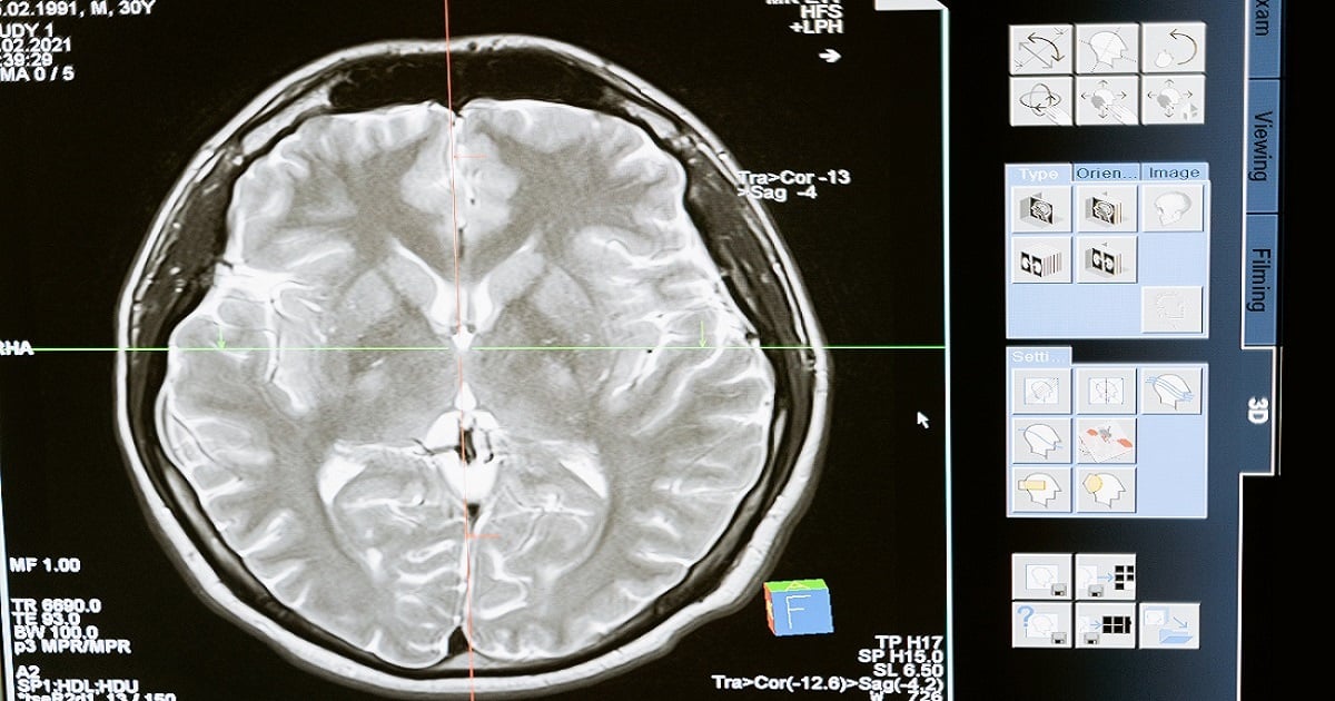
Medical imaging encompasses different imaging modalities and processes to image the human body for diagnostic and treatment purposes and therefore plays an important role in initiatives to improve public health for all population groups. 

There are many different types of medical imaging techniques, which use different technologies to produce images for different purposes, for example,  radiography, magnetic resonance imaging, ultrasound and tomography, depending on the physical nature of the waves employed and the method of image capture. There is no single imaging technology which is superior to the rest as each has its own advantages and disadvantages. 

Research into the application of medical images is usually the preserve of radiology and the medical sub-discipline relevant to medical condition or area of medical science (neuroscience, cardiology, psychiatry, psychology, dentistry, etc.) under investigation. Many of the techniques developed for medical imaging also have scientific and industrial applications. One of the most exciting areas currently under research is the application of artificial intelligence (AI) to medical imaging. 

This Special Issue of the journal Applied Sciences, “Medical Imaging: Advanced Techniques and Applications” is dedicated to covering some of the recent advances in this novel technology and applications.

Dr. Hartmut Schneider
Guest Editor

Manuscript Submission Information

Manuscripts should be submitted online at www.mdpi.com by registering and logging in to this website. Once you are registered, click here to go to the submission form. Manuscripts can be submitted until the deadline. All submissions that pass pre-check are peer-reviewed. Accepted papers will be published continuously in the journal (as soon as accepted) and will be listed together on the special issue website. Research articles, review articles as well as short communications are invited. For planned papers, a title and short abstract (about 100 words) can be sent to the Editorial Office for announcement on this website.

Submitted manuscripts should not have been published previously, nor be under consideration for publication elsewhere (except conference proceedings papers). All manuscripts are thoroughly refereed through a single-blind peer-review process. A guide for authors and other relevant information for submission of manuscripts is available on the Instructions for Authors page. Applied Sciences is an international peer-reviewed open access semimonthly journal published by MDPI.

Please visit the Instructions for Authors page before submitting a manuscript. The Article Processing Charge (APC) for publication in this open access journal is 2400 CHF (Swiss Francs). Submitted papers should be well formatted and use good English. Authors may use MDPI's English editing service prior to publication or during author revisions.

Keywords

  • medical imaging
  • optical imaging
  • biomedical image analysis
  • computer-assisted diagnosis
  • artificial intelligence in biomedicine
  • 2D and 3D modeling
  • Radiography
  • Tomography
  • Magnetic resonance imaging
  • Ultrasound
  • microscopy

Published Papers (7 papers)

Order results
Result details
Select all
Export citation of selected articles as:

Research

24 pages, 8197 KiB  
Article
Development and Validation of Two Intact Lumbar Spine Finite Element Models for In Silico Investigations: Comparison of the Bone Modelling Approaches
by Mate Turbucz, Agoston Jakab Pokorni, György Szőke, Zoltan Hoffer, Rita Maria Kiss, Aron Lazary and Peter Endre Eltes
Appl. Sci. 2022, 12(20), 10256; https://doi.org/10.3390/app122010256 - 12 Oct 2022
Cited by 9 | Viewed by 3855
Abstract
Finite element (FE) analyses contribute to a better understanding of the human lumbar spine’s biomechanics and serve as an effective predictive tool. This study aims to present the development of two L1–L5 FE models using literature-based (LBM) and patient-specific (PSM) bone material assignment [...] Read more.
Finite element (FE) analyses contribute to a better understanding of the human lumbar spine’s biomechanics and serve as an effective predictive tool. This study aims to present the development of two L1–L5 FE models using literature-based (LBM) and patient-specific (PSM) bone material assignment approaches. The geometry of the lumbar spine was developed based on quantitative computed tomography scans. The LBM and the PSM were compared under pure and combined loads. Various biomechanical parameters were investigated to validate the models. The total range of motion of the LBM in pure flexion-extension, lateral bending, and axial rotation were 30.9°, 29°, and 13.7°, respectively, while for the PSM, it was 31.6°, 28.6°, and 14.1°. The required computational time of the PSM to complete against pure and combined loads were 12.1 and 16.6 times higher on average compared to the LBM. This study demonstrated that both models agree with experimental and in silico results, although the cumulative distribution of the stress and characterization of strain values showed a noteworthy difference between the two models. Based on these findings, the clinically-focused biomechanical FE studies must perceive the differences in internal mechanical parameters and computational demand between the different bone modelling approaches. Full article
(This article belongs to the Special Issue Medical Imaging: Advanced Techniques and Applications)
Show Figures

Figure 1

10 pages, 2816 KiB  
Article
Rapid Quantification of Tissue Perfusion Properties with a Two-Stage Look-Up Table
by Bin Yang
Appl. Sci. 2022, 12(8), 3745; https://doi.org/10.3390/app12083745 - 8 Apr 2022
Viewed by 1696
Abstract
Tissue perfusion properties reveal crucial information pertinent to clinical diagnosis and treatment. Multispectral spatial frequency domain imaging (SFDI) is an emerging imaging technique that has been widely used to quantify tissue perfusion properties. However, slow processing speed limits its usefulness in real-time imaging [...] Read more.
Tissue perfusion properties reveal crucial information pertinent to clinical diagnosis and treatment. Multispectral spatial frequency domain imaging (SFDI) is an emerging imaging technique that has been widely used to quantify tissue perfusion properties. However, slow processing speed limits its usefulness in real-time imaging applications. In this study, we present a two-stage look-up table (LUT) approach that accurately and rapidly quantifies optical (absorption and reduced scattering maps) and perfusion (total hemoglobin and oxygen saturation maps) properties using stage-1 and stage-2 LUTs, respectively, based on reflectance images at 660 and 850 nm. The two-stage LUT can be implemented on both CPU and GPU computing platforms. Quantifying tissue perfusion properties using the simulated diffuse reflectance images, we achieved a quantification speed of 266, 174, and 74 frames per second for three image sizes 512 × 512, 1024 × 1024, and 2048 × 2048 pixels, respectively. Quantification of tissue perfusion properties was highly accurate with only 3.5% and 2.5% error for total hemoglobin and oxygen saturation quantification, respectively. The two-stage LUT has the potential to be integrated with dual-sensor cameras to enable real-time quantification of tissue hemodynamics. Full article
(This article belongs to the Special Issue Medical Imaging: Advanced Techniques and Applications)
Show Figures

Figure 1

12 pages, 1114 KiB  
Communication
AS-OCT and Ocular Hygrometer as Innovative Tools in Dry Eye Disease Diagnosis
by Daniele Gaudenzi, Tommaso Mori, Salvatore Crugliano, Antonella Grasso, Carlo Frontini, Antonella Carducci, Siddharth Yadav, Roberto Sgrulletta, Emiliano Schena, Marco Coassin and Antonio Di Zazzo
Appl. Sci. 2022, 12(3), 1647; https://doi.org/10.3390/app12031647 - 4 Feb 2022
Cited by 5 | Viewed by 1929
Abstract
Dry eye disease (DED) is one of the conditions that most commonly leads patients to visit an ophthalmologist. Fast and accurate diagnosis relieves patient discomfort and spares them from long-term effects on the ocular surface. Many tests used in the diagnosis of DED [...] Read more.
Dry eye disease (DED) is one of the conditions that most commonly leads patients to visit an ophthalmologist. Fast and accurate diagnosis relieves patient discomfort and spares them from long-term effects on the ocular surface. Many tests used in the diagnosis of DED may be considered subjective as they rely on an experienced observer for image interpretation, resulting in variations in diagnosis. On one hand, the non-contact nature of the anterior segment optical coherence tomography (AS-OCT) device and its rapid image acquisition enable the measurement of the tear meniscus parameter without reflex tearing. On the other hand, an ocular hygrometer allows a rapid, safe, but also efficient, analysis and is associated with low costs and the repeatability of the procedure. Full article
(This article belongs to the Special Issue Medical Imaging: Advanced Techniques and Applications)
Show Figures

Figure 1

9 pages, 5076 KiB  
Article
Analysis of Weighted Fraction of Length for Interfacial Gap in Cervical Composite Restorations as a Function of the Number of B-Scans of OCT Volume Scans
by Hartmut Schneider, Tobias Meißner, Claudia Rüger and Rainer Haak
Appl. Sci. 2021, 11(21), 10285; https://doi.org/10.3390/app112110285 - 2 Nov 2021
Cited by 2 | Viewed by 1158
Abstract
In dental research, the morphometric assessment of restorations is a challenge. This also applies to the assessment of the length of interfacial adhesive defects in composite restorations as a measure of tooth-restoration bond failure. The determined mean fractions of interfacial gap length on [...] Read more.
In dental research, the morphometric assessment of restorations is a challenge. This also applies to the assessment of the length of interfacial adhesive defects in composite restorations as a measure of tooth-restoration bond failure. The determined mean fractions of interfacial gap length on enamel and dentin interfaces deviate from the true means (N ∞), depending on the number (Ni) of object layers assessed. Cervical composite restorations were imaged with spectral domain optical coherence tomography (SD-OCT). The mean fractions of interfacial gap length on enamel and dentin were determined for an increasing number of OCT cross-sectional images (B-scans) per restoration and were graphically displayed as a function of the number of B-scans. As the number of B-scans increased, the calculated object means approached a range of ±2.5%. This analysis is appropriate for displaying the relationship between the determined mean fraction of interfacial gap length at the enamel/dentin-restoration interface and the number of B-scans. Full article
(This article belongs to the Special Issue Medical Imaging: Advanced Techniques and Applications)
Show Figures

Figure 1

23 pages, 49144 KiB  
Article
Light-Convolution Dense Selection U-Net (LDS U-Net) for Ultrasound Lateral Bony Feature Segmentation
by Sunetra Banerjee, Juan Lyu, Zixun Huang, Hung Fat Frank Leung, Timothy Tin-Yan Lee, De Yang, Steven Su, Yongping Zheng and Sai-Ho Ling
Appl. Sci. 2021, 11(21), 10180; https://doi.org/10.3390/app112110180 - 30 Oct 2021
Cited by 11 | Viewed by 2573
Abstract
Scoliosis is a widespread medical condition where the spine becomes severely deformed and bends over time. It mostly affects young adults and may have a permanent impact on them. A periodic assessment, using a suitable modality, is necessary for its early detection. Conventionally, [...] Read more.
Scoliosis is a widespread medical condition where the spine becomes severely deformed and bends over time. It mostly affects young adults and may have a permanent impact on them. A periodic assessment, using a suitable modality, is necessary for its early detection. Conventionally, the usually employed modalities include X-ray and MRI, which employ ionising radiation and are expensive. Hence, a non-radiating 3D ultrasound imaging technique has been developed as a safe and economic alternative. However, ultrasound produces low-contrast images that are full of speckle noise, and skilled intervention is necessary for their processing. Given the prevalent occurrence of scoliosis and the limitations of scalability of human expert interventions, an automatic, fast, and low-computation assessment technique is being developed for mass scoliosis diagnosis. In this paper, a novel hybridized light-weight convolutional neural network architecture is presented for automatic lateral bony feature identification, which can help to develop a fully-fledged automatic scoliosis detection system. The proposed architecture, Light-convolution Dense Selection U-Net (LDS U-Net), can accurately segment ultrasound spine lateral bony features, from noisy images, thanks to its capabilities of smartly selecting only the useful information and extracting rich deep layer features from the input image. The proposed model is tested using a dataset of 109 spine ultrasound images. The segmentation result of the proposed network is compared with basic U-Net, Attention U-Net, and MultiResUNet using various popular segmentation indices. The results show that LDS U-Net provides a better segmentation performance compared to the other models. Additionally, LDS U-Net requires a smaller number of parameters and less memory, making it suitable for a large-batch screening process of scoliosis without a high computational requirement. Full article
(This article belongs to the Special Issue Medical Imaging: Advanced Techniques and Applications)
Show Figures

Figure 1

10 pages, 2930 KiB  
Article
Artifact Reduction in Compressed Sensing Averaging Techniques for High-Resolution Magnetic Resonance Images
by Jeong-Min Shim, Young-Bo Kim and Chang-Ki Kang
Appl. Sci. 2021, 11(21), 9802; https://doi.org/10.3390/app11219802 - 20 Oct 2021
Cited by 1 | Viewed by 1816
Abstract
This study aims to introduce a new compressed sensing averaging (CSA) technique for the reduction of blurring and/or ringing artifacts, depending on the k-space sampling ratio. A full k-space dataset and three randomly undersampled datasets were obtained for CSA images in a brain [...] Read more.
This study aims to introduce a new compressed sensing averaging (CSA) technique for the reduction of blurring and/or ringing artifacts, depending on the k-space sampling ratio. A full k-space dataset and three randomly undersampled datasets were obtained for CSA images in a brain phantom and a healthy subject. An additional simulation was performed to assess the effect of the undersampling ratio on the images and the signal-to-noise ratios (SNRs). The image sharpness, spatial resolution, and contrast between tissues were analyzed and compared with other CSA techniques. Compared to CSA with multiple acquisition (CSAM) at 25%, 35%, and 45% undersampling, the reduction rates of the k-space lines of CSA with keyhole (CSAK) were 10%, 15%, and 22%, respectively, and the acquisition time was reduced by 16%, 23%, and 32%, respectively. In the simulation performed with a full sampling k-space dataset, the SNR decreased to 10.41, 9.80, and 8.86 in the white matter and 9.69, 9.35, and 8.46 in the gray matter, respectively. In addition, the ringing artifacts became substantially more predominant as the number of sampling lines decreased. The 50% modulation transfer functions were 0.38, 0.43, and 0.54 line pairs per millimeter for CSAM, CSAK with high-frequency sharing (CSAKS), and CSAK with high-frequency copying (CSAKC), respectively. In this study, we demonstrated that the smaller the sampling line, the more severe the ringing artifact, and that the CSAKC technique proposed to overcome the artifacts that occur when using CSA techniques did not generate artifacts, while it increased spatiotemporal resolution. Full article
(This article belongs to the Special Issue Medical Imaging: Advanced Techniques and Applications)
Show Figures

Figure 1

13 pages, 1809 KiB  
Article
Perioperative Chemotherapy with FLOT Scheme in Resectable Gastric Adenocarcinoma: A Preliminary Correlation between TRG and Radiomics
by Giovanni Maria Garbarino, Marta Zerunian, Eva Berardi, Federico Mainardi, Emanuela Pilozzi, Michela Polici, Gisella Guido, Carlotta Rucci, Tiziano Polidori, Mariarita Tarallo, Giovanni Guglielmo Laracca, Elsa Iannicelli, Paolo Mercantini, Bruno Annibale, Andrea Laghi and Damiano Caruso
Appl. Sci. 2021, 11(19), 9211; https://doi.org/10.3390/app11199211 - 3 Oct 2021
Cited by 1 | Viewed by 1984
Abstract
Perioperative chemotherapy (p-ChT) with a fluorouracil plus leucovorin, oxaliplatin, and docetaxel (FLOT) scheme is the gold standard of care for locally advanced gastric cancer. We aimed to test CT radiomics performance in early response prediction for p-ChT. Patients with advanced gastric cancer who [...] Read more.
Perioperative chemotherapy (p-ChT) with a fluorouracil plus leucovorin, oxaliplatin, and docetaxel (FLOT) scheme is the gold standard of care for locally advanced gastric cancer. We aimed to test CT radiomics performance in early response prediction for p-ChT. Patients with advanced gastric cancer who underwent contrast enhanced CT prior to and post p-ChT were retrospectively enrolled. Histologic evaluation of resected specimens was used as the reference standard, and patients were divided into responders (TRG 1a-1b) and non-responders (TRG 2-3) according to their Becker tumor regression grade (TRG). A volumetric region of interest including the whole tumor tissue was drawn from a CT portal-venous phase before and after p-ChT; 120 radiomic features, both first and second order, were extracted. CT radiomics performances were derived from baseline CT radiomics alone and ΔRadiomics to predict response to p-ChT according to the TRG and tested using a receiver operating characteristic (ROC) curve. The final population comprised 15 patients, 6 (40%) responders and 9 (60%) non-responders. Among pre-treatment CT radiomics parameters, Shape, GLCM, First order, and NGTDM features showed a significant ability to discriminate between responders and non-responders (p < 0.011), with Cluster Shade and Autocorrelation (GLCM features) having AUC = 0.907. ΔRadiomics showed significant differences for Shape, GLRLM, GLSZM, and NGTDM features (p < 0.007). MeshVolume (Shape feature) and LongRunEmphasis (GLRLM feature) had AUC = 0.889. In conclusion, CT radiomics may represent an important supportive approach for the radiologic evaluation of advanced gastric cancer patients. Full article
(This article belongs to the Special Issue Medical Imaging: Advanced Techniques and Applications)
Show Figures

Figure 1

Back to TopTop