Next Issue
Volume 10, July
Previous Issue
Volume 10, May


Viruses, Volume 10, Issue 6 (June 2018) – 61 articles

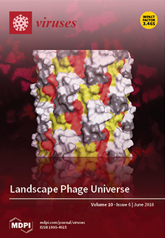
Cover Story (view full-size image): Filamentous phages have attracted the attention of bioengineers as structurally flexible bio-nanomaterials with a wide spectrum of potential applications. Phage landscape, which is formed by a genetically predetermined combination of thousands of functional organic groups, can be redesigned using genetic engineering methods. Furthermore, phage landscape can be randomly diversified to form multibillion clone landscape phage libraries, which can be screened for desired properties. The paper describes the history of the landscape phage invention, and gives some examples of their application. View Paper here.
  • Issues are regarded as officially published after their release is announced to the table of contents alert mailing list.
  • You may sign up for e-mail alerts to receive table of contents of newly released issues.
  • PDF is the official format for papers published in both, html and pdf forms. To view the papers in pdf format, click on the "PDF Full-text" link, and use the free Adobe Reader to open them.
Order results
Result details
Section
Select all
Export citation of selected articles as:
19 pages, 7882 KiB  
Article
Identification and Characterization of Type IV Pili as the Cellular Receptor of Broad Host Range Stenotrophomonas maltophilia Bacteriophages DLP1 and DLP2
by Jaclyn G. McCutcheon, Danielle L. Peters and Jonathan J. Dennis
Viruses 2018, 10(6), 338; https://doi.org/10.3390/v10060338 - 20 Jun 2018
Cited by 39 | Viewed by 5621
Abstract
Bacteriophages DLP1 and DLP2 are capable of infecting both Stenotrophomonas maltophilia and Pseudomonas aeruginosa strains, two highly antibiotic resistant bacterial pathogens, which is unusual for phages that typically exhibit extremely limited host range. To explain their unusual cross-order infectivity and differences in host [...] Read more.
Bacteriophages DLP1 and DLP2 are capable of infecting both Stenotrophomonas maltophilia and Pseudomonas aeruginosa strains, two highly antibiotic resistant bacterial pathogens, which is unusual for phages that typically exhibit extremely limited host range. To explain their unusual cross-order infectivity and differences in host range, we have identified the type IV pilus as the primary receptor for attachment. Screening of a P. aeruginosa PA01 mutant library, a host that is susceptible to DLP1 but not DLP2, identified DLP1-resistant mutants with disruptions in pilus structural and regulatory components. Subsequent complementation of the disrupted pilin subunit genes in PA01 restored DLP1 infection. Clean deletion of the major pilin subunit, pilA, in S. maltophilia strains D1585 and 280 prevented phage binding and lysis by both DLP1 and DLP2, and complementation restored infection by both. Transmission electron microscopy shows a clear interaction between DLP1 and pili of both D1585 and PA01. These results support the identity of the type IV pilus as the receptor for DLP1 and DLP2 infection across their broad host ranges. This research further characterizes DLP1 and DLP2 as potential “anti-virulence” phage therapy candidates for the treatment of multidrug resistant bacteria from multiple genera. Full article
(This article belongs to the Special Issue Phage-Host Interactions)
Show Figures

Figure 1

22 pages, 856 KiB  
Review
Visualizing Viral Infection In Vivo by Multi-Photon Intravital Microscopy
by Xaver Sewald
Viruses 2018, 10(6), 337; https://doi.org/10.3390/v10060337 - 20 Jun 2018
Cited by 13 | Viewed by 6399
Abstract
Viral pathogens have adapted to the host organism to exploit the cellular machinery for virus replication and to modulate the host cells for efficient systemic dissemination and immune evasion. Much of our knowledge of the effects that virus infections have on cells originates [...] Read more.
Viral pathogens have adapted to the host organism to exploit the cellular machinery for virus replication and to modulate the host cells for efficient systemic dissemination and immune evasion. Much of our knowledge of the effects that virus infections have on cells originates from in vitro imaging studies using experimental culture systems consisting of cell lines and primary cells. Recently, intravital microscopy using multi-photon excitation of fluorophores has been applied to observe virus dissemination and pathogenesis in real-time under physiological conditions in living organisms. Critical steps during viral infection and pathogenesis could be studied by direct visualization of fluorescent virus particles, virus-infected cells, and the immune response to viral infection. In this review, I summarize the latest research on in vivo studies of viral infections using multi-photon intravital microscopy (MP-IVM). Initially, the underlying principle of multi-photon microscopy is introduced and experimental challenges during microsurgical animal preparation and fluorescent labeling strategies for intravital imaging are discussed. I will further highlight recent studies that combine MP-IVM with optogenetic tools and transcriptional analysis as a powerful approach to extend the significance of in vivo imaging studies of viral pathogens. Full article
(This article belongs to the Special Issue Application of Advanced Imaging to the Study of Virus Replication)
Show Figures

Graphical abstract

19 pages, 693 KiB  
Review
Harnessing T Follicular Helper Cell Responses for HIV Vaccine Development
by Julia Niessl and Daniel E. Kaufmann
Viruses 2018, 10(6), 336; https://doi.org/10.3390/v10060336 - 19 Jun 2018
Cited by 5 | Viewed by 8011
Abstract
Passive administration of broadly neutralizing antibodies (bNAbs) capable of recognizing a broad range of viral strains to non-human primates has led to protection from infection with chimeric SIV/HIV virus (SHIV). This data suggests that generating protective antibody responses could be an effective strategy [...] Read more.
Passive administration of broadly neutralizing antibodies (bNAbs) capable of recognizing a broad range of viral strains to non-human primates has led to protection from infection with chimeric SIV/HIV virus (SHIV). This data suggests that generating protective antibody responses could be an effective strategy for an HIV vaccine. However, classic vaccine approaches have failed so far to induce such protective antibodies in HIV vaccine trials. HIV-specific bNAbs identified in natural infection show high levels of somatic hypermutations, demonstrating that they underwent extensive affinity maturation. It is likely that to gain ability to recognize diverse viral strains, vaccine-induced humoral responses will also require complex, iterative maturation. T follicular helper cells (Tfh) are a specialized CD4+ T cell subset that provides help to B cells in the germinal center for the generation of high-affinity and long-lasting humoral responses. It is therefore probable that the quality and quantity of Tfh responses upon vaccination will impact development of bNAbs. Here, we review studies that advanced our understanding of Tfh differentiation, function and regulation. We discuss correlates of Tfh responses and bNAb development in natural HIV infection. Finally, we highlight recent strategies to optimize Tfh responses upon vaccination and their impact on prophylactic HIV vaccine research. Full article
(This article belongs to the Special Issue HIV Vaccines)
Show Figures

Figure 1

11 pages, 790 KiB  
Review
Phage Genetic Engineering Using CRISPR–Cas Systems
by Asma Hatoum-Aslan
Viruses 2018, 10(6), 335; https://doi.org/10.3390/v10060335 - 19 Jun 2018
Cited by 53 | Viewed by 10697
Abstract
Since their discovery over a decade ago, the class of prokaryotic immune systems known as CRISPR–Cas have afforded a suite of genetic tools that have revolutionized research in model organisms spanning all domains of life. CRISPR-mediated tools have also emerged for the natural [...] Read more.
Since their discovery over a decade ago, the class of prokaryotic immune systems known as CRISPR–Cas have afforded a suite of genetic tools that have revolutionized research in model organisms spanning all domains of life. CRISPR-mediated tools have also emerged for the natural targets of CRISPR–Cas immunity, the viruses that specifically infect bacteria, or phages. Despite their status as the most abundant biological entities on the planet, the majority of phage genes have unassigned functions. This reality underscores the need for robust genetic tools to study them. Recent reports have demonstrated that CRISPR–Cas systems, specifically the three major types (I, II, and III), can be harnessed to genetically engineer phages that infect diverse hosts. Here, the mechanisms of each of these systems, specific strategies used, and phage editing efficacies will be reviewed. Due to the relatively wide distribution of CRISPR–Cas systems across bacteria and archaea, it is anticipated that these immune systems will provide generally applicable tools that will advance the mechanistic understanding of prokaryotic viruses and accelerate the development of novel technologies based on these ubiquitous organisms. Full article
(This article belongs to the Special Issue Applications of CRISPR Technology in Virology 2018)
Show Figures

Figure 1

20 pages, 3604 KiB  
Article
Biochemical and Functional Characterization of Mouse Mammary Tumor Virus Full-Length Pr77Gag Expressed in Prokaryotic and Eukaryotic Cells
by Akhil Chameettachal, Vineeta Narayana Pillai, Lizna Mohamed Ali, Fathima Nuzra Nagoor Pitchai, Mustafa Taleb Ardah, Farah Mustafa, Roland Marquet and Tahir Aziz Rizvi
Viruses 2018, 10(6), 334; https://doi.org/10.3390/v10060334 - 18 Jun 2018
Cited by 9 | Viewed by 5085
Abstract
The mouse mammary tumor virus (MMTV) Pr77Gag polypeptide is an essential retroviral structural protein without which infectious viral particles cannot be formed. This process requires specific recognition and packaging of dimerized genomic RNA (gRNA) by Gag during virus assembly. Most of the [...] Read more.
The mouse mammary tumor virus (MMTV) Pr77Gag polypeptide is an essential retroviral structural protein without which infectious viral particles cannot be formed. This process requires specific recognition and packaging of dimerized genomic RNA (gRNA) by Gag during virus assembly. Most of the previous work on retroviral assembly has used either the nucleocapsid portion of Gag, or other truncated Gag derivatives—not the natural substrate for virus assembly. In order to understand the molecular mechanism of MMTV gRNA packaging process, we expressed and purified full-length recombinant Pr77Gag-His6-tag fusion protein from soluble fractions of bacterial cultures. We show that the purified Pr77Gag-His6-tag protein retained the ability to assemble virus-like particles (VLPs) in vitro with morphologically similar immature intracellular particles. The recombinant proteins (with and without His6-tag) could both be expressed in prokaryotic and eukaryotic cells and had the ability to form VLPs in vivo. Most importantly, the recombinant Pr77Gag-His6-tag fusion proteins capable of making VLPs in eukaryotic cells were competent for packaging sub-genomic MMTV RNAs. The successful expression and purification of a biologically active, full-length MMTV Pr77Gag should lay down the foundation towards performing RNA–protein interaction(s), especially for structure-function studies and towards understanding molecular intricacies during MMTV gRNA packaging and assembly processes. Full article
(This article belongs to the Section Animal Viruses)
Show Figures

Figure 1

13 pages, 922 KiB  
Review
Neutralizing Antibody-Based Prevention of Cell-Associated HIV-1 Infection
by Matthew S Parsons, Roger Le Grand and Stephen J Kent
Viruses 2018, 10(6), 333; https://doi.org/10.3390/v10060333 - 18 Jun 2018
Cited by 8 | Viewed by 4706
Abstract
Improved vaccine-mediated protection against HIV-1 requires a thorough understanding of the mode of HIV-1 transmission and how various immune responses control transmission. Cell-associated HIV-1 is infectious and contributes to HIV-1 transmission in humans. Non-human primate models of cell-associated SIV infection demonstrate that cell-associated [...] Read more.
Improved vaccine-mediated protection against HIV-1 requires a thorough understanding of the mode of HIV-1 transmission and how various immune responses control transmission. Cell-associated HIV-1 is infectious and contributes to HIV-1 transmission in humans. Non-human primate models of cell-associated SIV infection demonstrate that cell-associated SIV is more infectious than cell-free SIV. In a recently described chimeric simian–human immunodeficiency virus (SHIV) macaque model, it was demonstrated that an occult infection with cell-associated SHIV can be established that evades passive protection with a broadly neutralizing antibody (bnAb). Indeed, considerable in vitro data shows that bnAbs have less efficacy against cell-associated HIV-1 than cell-free HIV-1. Optimizing the protective capacity of immune responses such as bnAbs against cell-associated infections may be needed to maximize their protective efficacy. Full article
(This article belongs to the Special Issue HIV Vaccines)
Show Figures

Figure 1

19 pages, 2632 KiB  
Article
Transcriptomic Analysis of the Campylobacter jejuni Response to T4-Like Phage NCTC 12673 Infection
by Jessica C. Sacher, Annika Flint, James Butcher, Bob Blasdel, Hayley M. Reynolds, Rob Lavigne, Alain Stintzi and Christine M. Szymanski
Viruses 2018, 10(6), 332; https://doi.org/10.3390/v10060332 - 16 Jun 2018
Cited by 38 | Viewed by 6132
Abstract
Campylobacter jejuni is a frequent foodborne pathogen of humans. As C. jejuni infections commonly arise from contaminated poultry, phage treatments have been proposed to reduce the C. jejuni load on farms to prevent human infections. While a prior report documented the transcriptome of [...] Read more.
Campylobacter jejuni is a frequent foodborne pathogen of humans. As C. jejuni infections commonly arise from contaminated poultry, phage treatments have been proposed to reduce the C. jejuni load on farms to prevent human infections. While a prior report documented the transcriptome of C. jejuni phages during the carrier state life cycle, transcriptomic analysis of a lytic C. jejuni phage infection has not been reported. We used RNA-sequencing to profile the infection of C. jejuni NCTC 11168 by the lytic T4-like myovirus NCTC 12673. Interestingly, we found that the most highly upregulated host genes upon infection make up an uncharacterized operon (cj0423–cj0425), which includes genes with similarity to T4 superinfection exclusion and antitoxin genes. Other significantly upregulated genes include those involved in oxidative stress defense and the Campylobactermultidrug efflux pump (CmeABC). We found that phage infectivity is altered by mutagenesis of the oxidative stress defense genes catalase (katA), alkyl-hydroxyperoxidase (ahpC), and superoxide dismutase (sodB), and by mutagenesis of the efflux pump genes cmeA and cmeB. This suggests a role for these gene products in phage infection. Together, our results shed light on the phage-host dynamics of an important foodborne pathogen during lytic infection by a T4-like phage. Full article
(This article belongs to the Special Issue Phage-Host Interactions)
Show Figures

Figure 1

14 pages, 1976 KiB  
Article
Pseudomonas PB1-Like Phages: Whole Genomes from Metagenomes Offer Insight into an Abundant Group of Bacteriophages
by Siobhan C. Watkins, Emily Sible and Catherine Putonti
Viruses 2018, 10(6), 331; https://doi.org/10.3390/v10060331 - 16 Jun 2018
Cited by 9 | Viewed by 5528
Abstract
Despite the abundance, ubiquity and impact of environmental viruses, their inherent genomic plasticity and extreme diversity pose significant challenges for the examination of bacteriophages on Earth. Viral metagenomic studies have offered insight into broader aspects of phage ecology and repeatedly uncover genes to [...] Read more.
Despite the abundance, ubiquity and impact of environmental viruses, their inherent genomic plasticity and extreme diversity pose significant challenges for the examination of bacteriophages on Earth. Viral metagenomic studies have offered insight into broader aspects of phage ecology and repeatedly uncover genes to which we are currently unable to assign function. A combined effort of phage isolation and metagenomic survey of Chicago’s nearshore waters of Lake Michigan revealed the presence of Pbunaviruses, relatives of the Pseudomonas phage PB1. This prompted our expansive investigation of PB1-like phages. Genomic signatures of PB1-like phages and Pbunaviruses were identified, permitting the unambiguous distinction between the presence/absence of these phages in soils, freshwater and wastewater samples, as well as publicly available viral metagenomic datasets. This bioinformatic analysis led to the de novo assembly of nine novel PB1-like phage genomes from a metagenomic survey of samples collected from Lake Michigan. While this study finds that Pbunaviruses are abundant in various environments of Northern Illinois, genomic variation also exists to a considerable extent within individual communities. Full article
(This article belongs to the Special Issue Bacteriophage Genomes and Genomics: News from the Wild)
Show Figures

Figure 1

16 pages, 3294 KiB  
Article
Host Long Noncoding RNA lncRNA-PAAN Regulates the Replication of Influenza A Virus
by Jing Wang, Yujia Wang, Rui Zhou, Jianyuan Zhao, Yongxin Zhang, Dongrong Yi, Quanjie Li, Jinming Zhou, Fei Guo, Chen Liang, Xiaoyu Li and Shan Cen
Viruses 2018, 10(6), 330; https://doi.org/10.3390/v10060330 - 16 Jun 2018
Cited by 46 | Viewed by 5834
Abstract
The productive infection of influenza A virus (IAV) depends on host factors. However, the involvement of long non-coding RNAs (lncRNAs) in IAV infection remains largely uninvestigated. In this work, we have discovered a human lncRNA, named lncRNA-PAAN (PA-associated noncoding RNA) that enhances IAV [...] Read more.
The productive infection of influenza A virus (IAV) depends on host factors. However, the involvement of long non-coding RNAs (lncRNAs) in IAV infection remains largely uninvestigated. In this work, we have discovered a human lncRNA, named lncRNA-PAAN (PA-associated noncoding RNA) that enhances IAV replication. The level of lncRNA-PAAN increases upon infection of IAV, but not other viruses, nor interferon treatment, suggesting specific up-regulation of lncRNA-PAAN expression by IAV. Silencing lncRNA-PAAN significantly decreases IAV replication through impairing the activity of viral RNA-dependent RNA polymerase (RdRp). This function of lncRNA-PAAN is a result of its association with viral PA protein, a key component of IAV RNA polymerase complex. Consequently, depletion of lncRNA-PAAN prevents the formation of functional RdRp. Together, these results suggest that lncRNA-PAAN promotes the assembly of viral RNA polymerase, thus warranting efficient viral RNA synthesis. Elucidating the functions of lncRNAs in IAV infection is expected to advance our understanding of IAV pathogenesis and open new avenues to the development of novel anti-IAV therapeutics. Full article
(This article belongs to the Special Issue What’s New with Flu?)
Show Figures

Figure 1

18 pages, 1070 KiB  
Review
Viral Determinants of Virulence in Tick-Borne Flaviviruses
by Eliza M. Kellman, Danielle K. Offerdahl, Wessam Melik and Marshall E. Bloom
Viruses 2018, 10(6), 329; https://doi.org/10.3390/v10060329 - 16 Jun 2018
Cited by 33 | Viewed by 5526
Abstract
Tick-borne flaviviruses have a global distribution and cause significant human disease, including encephalitis and hemorrhagic fever, and often result in neurologic sequelae. There are two distinct properties that determine the neuropathogenesis of a virus. The ability to invade the central nervous system (CNS) [...] Read more.
Tick-borne flaviviruses have a global distribution and cause significant human disease, including encephalitis and hemorrhagic fever, and often result in neurologic sequelae. There are two distinct properties that determine the neuropathogenesis of a virus. The ability to invade the central nervous system (CNS) is referred to as the neuroinvasiveness of the agent, while the ability to infect and damage cells within the CNS is referred to as its neurovirulence. Examination of laboratory variants, cDNA clones, natural isolates with varying pathogenicity, and virally encoded immune evasion strategies have contributed extensively to our understanding of these properties. Here we will review the major viral determinants of virulence that contribute to pathogenesis and influence both neuroinvasiveness and neurovirulence properties of tick-borne flaviviruses, focusing particularly on the envelope protein (E), nonstructural protein 5 (NS5), and the 3′ untranslated region (UTR). Full article
(This article belongs to the Special Issue Biology and Treatment of Tick-Borne Viral Pathogens)
Show Figures

Figure 1

16 pages, 41413 KiB  
Article
Whole Genome Analysis of Two Novel Type 2 Porcine Reproductive and Respiratory Syndrome Viruses with Complex Genome Recombination between Lineage 8, 3, and 1 Strains Identified in Southwestern China
by Long Zhou, Runmin Kang, Yi Zhang, Mengdie Ding, Bo Xie, Yiming Tian, Xuan Wu, Lei Zuo, Xin Yang and Hongning Wang
Viruses 2018, 10(6), 328; https://doi.org/10.3390/v10060328 - 15 Jun 2018
Cited by 36 | Viewed by 4512
Abstract
Recombination among porcine reproductive and respiratory syndrome viruses (PRRSVs) is thought to contribute to the emergence of new PRRSV variants. In this study, two newly emerged PRRSV strains, designated SCcd16 and SCya17, are isolated from lung tissues of piglets in Southwestern China. Genome [...] Read more.
Recombination among porcine reproductive and respiratory syndrome viruses (PRRSVs) is thought to contribute to the emergence of new PRRSV variants. In this study, two newly emerged PRRSV strains, designated SCcd16 and SCya17, are isolated from lung tissues of piglets in Southwestern China. Genome comparative analysis reveals that SCcd16/SCya17 exhibit 93.1%/93.2%, 86.9%/87.0%, 85.3%/85.7%, and 83.6%/82.0% nucleotide similarity to PRRSVs JXA1, VR-2332, QYYZ and NADC30, respectively. They only exhibit 44.8%/45.1% sequence identity with LV (PRRSV-1), indicating that both emergent strains belong to the PRRSV-2 genotype. Genomic sequence alignment shows that SCcd16 and SCya17 have the same discontinuous 30-amino acid (aa) deletion in Nsp2 of the highly pathogenic Chinese PRRSV strain JXA1, when compared to strain VR-2332. Notably, SCya17 shows a unique 5-nt deletion in its 3’-UTR. Phylogenetic analysis shows that both of the isolates are classified in the QYYZ-like lineage based on ORF5 genotyping, whereas they appear to constitute an inter-lineage between JXA1-like and QYYZ-like lineages based on their genomic sequences. Furthermore, recombination analyses reveal that the two newly emerged PRRSV isolates share the same novel recombination pattern. They have both likely originated from multiple recombination events between lineage 8 (JXA1-like), lineage 1 (NADC30-like), and lineage 3 (QYYZ-like) strains that have circulated in China recently. The genomic data from SCcd16 and SCya17 indicate that there is on going evolution of PRRSV field strains through genetic recombination, leading to outbreaks in the pig populations in Southwestern China. Full article
(This article belongs to the Special Issue Emerging Viruses)
Show Figures

Figure 1

11 pages, 488 KiB  
Review
The Diversity of Bacterial Lifestyles Hampers Bacteriophage Tenacity
by Marta Lourenço, Luisa De Sordi and Laurent Debarbieux
Viruses 2018, 10(6), 327; https://doi.org/10.3390/v10060327 - 15 Jun 2018
Cited by 28 | Viewed by 6091
Abstract
Phage therapy is based on a simple concept: the use of a virus (bacteriophage) that is capable of killing specific pathogenic bacteria to treat bacterial infections. Since the pioneering work of Félix d’Herelle, bacteriophages (phages) isolated in vitro have been shown to be [...] Read more.
Phage therapy is based on a simple concept: the use of a virus (bacteriophage) that is capable of killing specific pathogenic bacteria to treat bacterial infections. Since the pioneering work of Félix d’Herelle, bacteriophages (phages) isolated in vitro have been shown to be of therapeutic value. Over decades of study, a large number of rather complex mechanisms that are used by phages to hijack bacterial resources and to produce their progeny have been deciphered. While these mechanisms have been identified and have been studied under optimal conditions in vitro, much less is known about the requirements for successful viral infections in relevant natural conditions. This is particularly true in the context of phage therapy. Here, we highlight the parameters affecting phage replication in both in vitro and in vivo environments, focusing, in particular, on the mammalian digestive tract. We propose avenues for increasing the knowledge-guided implementation of phages as therapeutic tools. Full article
(This article belongs to the Special Issue Hurdles for Phage Therapy (PT) to Become a Reality)
Show Figures

Figure 1

11 pages, 2337 KiB  
Article
NS1 Antigenemia and Viraemia Load: Potential Markers of Progression to Dengue Fatal Outcome?
by Priscila Conrado Guerra Nunes, Rita Maria Ribeiro Nogueira, Manoela Heringer, Thaís Chouin-Carneiro, Cintia Damasceno dos Santos Rodrigues, Ana Maria Bispo De Filippis, Monique Da Rocha Queiroz Lima and Flávia Barreto Dos Santos
Viruses 2018, 10(6), 326; https://doi.org/10.3390/v10060326 - 14 Jun 2018
Cited by 24 | Viewed by 4775
Abstract
Dengue is a worldwide problem characterized by a multifactorial pathogenesis. Considering the viral components, it is known that high viremia or high levels of the secreted nonstructural protein 1 (NS1) may be associated with a more severe disease. We aimed to characterize the [...] Read more.
Dengue is a worldwide problem characterized by a multifactorial pathogenesis. Considering the viral components, it is known that high viremia or high levels of the secreted nonstructural protein 1 (NS1) may be associated with a more severe disease. We aimed to characterize the NS1 antigenemia and viremia in dengue fatal and non-fatal cases, as potential markers of progression to a fatal outcome. NS1 antigenemia and viremia were determined in Brazilian dengue fatal cases (n = 40) and non-fatal cases (n = 40), representative of the four dengue virus (DENV) serotypes. Overall, the fatal cases presented higher NS1 levels and viremia. Moreover, the fatal cases from secondary infections showed significantly higher NS1 levels than the non-fatal ones. Here, irrespective of the disease outcome, DENV-1 cases presented higher NS1 levels than the other serotypes. However, DENV-2 and DENV-4 fatal cases had higher NS1 antigenemia than the non-fatal cases with the same serotype. The viremia in the fatal cases was higher than in the non-fatal ones, with DENV-3 and DENV-4 presenting higher viral loads. Viral components, such as NS1 and viral RNA, may be factors influencing the disease outcome. However, the host immune status, comorbidities, and access to adequate medical support cannot be ruled out as interfering in the disease outcome. Full article
(This article belongs to the Special Issue 6th Pan-American Dengue Research Network Meeting)
Show Figures

Figure 1

11 pages, 1374 KiB  
Article
A Simple and Robust Approach for Evaluation of Antivirals Using a Recombinant Influenza Virus Expressing Gaussia Luciferase
by Ping Li, Qinghua Cui, Lin Wang, Xiujuan Zhao, Yingying Zhang, Balaji Manicassamy, Yong Yang, Lijun Rong and Ruikun Du
Viruses 2018, 10(6), 325; https://doi.org/10.3390/v10060325 - 13 Jun 2018
Cited by 17 | Viewed by 4393
Abstract
Influenza A virus (IAV) causes seasonal epidemics and occasional but devastating pandemics, which are major public health concerns. Because the effectiveness of seasonal vaccines is highly variable and the currently available drugs are limited in their efficacy because of the emergence of drug [...] Read more.
Influenza A virus (IAV) causes seasonal epidemics and occasional but devastating pandemics, which are major public health concerns. Because the effectiveness of seasonal vaccines is highly variable and the currently available drugs are limited in their efficacy because of the emergence of drug resistance, there is an urgent need to develop novel antivirals. In this study, we characterized a recombinant IAV-carrying Gaussia luciferase (Gluc) gene and determined its potential as a tool for evaluating therapeutics. We demonstrated that this recombinant IAV is replication-competent in tissue culture and pathogenic in mice, although it is slightly attenuated compared to the parental virus. Luciferase expression correlated well with virus propagation both in vitro and in vivo, providing a simple measure for viral replication in tissue culture and in mouse lungs. To demonstrate the utility of this virus, ribavirin and oseltamivir phosphate were used to treat the IAV-infected cells and mice, and we observed the dose-dependent inhibition of viral replication by a luciferase assay. Moreover, the decreased luciferase expression in the infected lungs could predict the protective efficacy of antiviral interventions as early as day 2 post virus challenge. In summary, this study provides a new and quantitative approach to evaluate antivirals against IAV. Full article
(This article belongs to the Section Animal Viruses)
Show Figures

Figure 1

10 pages, 1557 KiB  
Brief Report
Production of Bacteriophages by Listeria Cells Entrapped in Organic Polymers
by Brigitte Roy, Cécile Philippe, Martin J. Loessner, Jacques Goulet and Sylvain Moineau
Viruses 2018, 10(6), 324; https://doi.org/10.3390/v10060324 - 13 Jun 2018
Cited by 7 | Viewed by 5120
Abstract
Applications for bacteriophages as antimicrobial agents are increasing. The industrial use of these bacterial viruses requires the production of large amounts of suitable strictly lytic phages, particularly for food and agricultural applications. This work describes a new approach for phage production. Phages H387 [...] Read more.
Applications for bacteriophages as antimicrobial agents are increasing. The industrial use of these bacterial viruses requires the production of large amounts of suitable strictly lytic phages, particularly for food and agricultural applications. This work describes a new approach for phage production. Phages H387 (Siphoviridae) and A511 (Myoviridae) were propagated separately using Listeria ivanovii host cells immobilised in alginate beads. The same batch of alginate beads could be used for four successive and efficient phage productions. This technique enables the production of large volumes of high-titer phage lysates in continuous or semi-continuous (fed-batch) cultures. Full article
(This article belongs to the Special Issue Hurdles for Phage Therapy (PT) to Become a Reality)
Show Figures

Figure 1

8 pages, 449 KiB  
Comment
Phage Therapy Faces Evolutionary Challenges
by Clara Torres-Barceló
Viruses 2018, 10(6), 323; https://doi.org/10.3390/v10060323 - 12 Jun 2018
Cited by 72 | Viewed by 8452
Abstract
Antibiotic resistance evolution in bacteria indicates that one of the challenges faced by phage therapy is that, sooner or later, bacteria will evolve resistance to phages. Evidently, this is the case of every known antimicrobial therapy, but here this is also part of [...] Read more.
Antibiotic resistance evolution in bacteria indicates that one of the challenges faced by phage therapy is that, sooner or later, bacteria will evolve resistance to phages. Evidently, this is the case of every known antimicrobial therapy, but here this is also part of a ubiquitous natural process of co-evolution between phages and bacteria. Fundamental evolutionary studies hold some clues that are crucial to limit the problematic process of bacterial resistance during phage applications. First, I discuss here the importance of defining evolutionary and ecological factors influencing bacterial resistance and phage counter-defense mechanisms. Then, I comment on the interest of determining the co-evolutionary dynamics between phages and bacteria that may allow for selecting the conditions that will increase the probability of therapeutic success. I go on to suggest the varied strategies that may ensure the long-term success of phage therapy, including analysis of internal phage parameters and personalized treatments. In practical terms, these types of approaches will define evolutionary criteria regarding how to develop, and when to apply, therapeutic phage cocktails. Integrating this perspective in antimicrobial treatments, such as phage therapy, is among the necessary steps to expand its use in the near future, and to ensure its durability and success. Full article
(This article belongs to the Special Issue Hurdles for Phage Therapy (PT) to Become a Reality)
Show Figures

Figure 1

11 pages, 1408 KiB  
Article
Self-Assembled Nanoporous Biofilms from Functionalized Nanofibrous M13 Bacteriophage
by Vasanthan Devaraj, Jiye Han, Chuntae Kim, Yong-Cheol Kang and Jin-Woo Oh
Viruses 2018, 10(6), 322; https://doi.org/10.3390/v10060322 - 12 Jun 2018
Cited by 15 | Viewed by 5261
Abstract
Highly periodic and uniform nanostructures, based on a genetically engineered M13 bacteriophage, displayed unique properties at the nanoscale that have the potential for a variety of applications. In this work, we report a multilayer biofilm with self-assembled nanoporous surfaces involving a nanofiber-like genetically [...] Read more.
Highly periodic and uniform nanostructures, based on a genetically engineered M13 bacteriophage, displayed unique properties at the nanoscale that have the potential for a variety of applications. In this work, we report a multilayer biofilm with self-assembled nanoporous surfaces involving a nanofiber-like genetically engineered 4E-type M13 bacteriophage, which was fabricated using a simple pulling method. The nanoporous surfaces were effectively formed by using the networking-like structural layers of the M13 bacteriophage during self-assembly. Therefore, an external template was not required. The actual M13 bacteriophage-based fabricated multilayered biofilm with porous nanostructures agreed well with experimental and simulation results. Pores formed in the final layer had a diameter of about 150–500 nm and a depth of about 15–30 nm. We outline a filter application for this multilayered biofilm that enables selected ions to be extracted from a sodium chloride solution. Here, we describe a simple, environmentally friendly, and inexpensive fabrication approach with large-scale production potential. The technique and the multi-layered biofilms produced may be applied to sensor, filter, plasmonics, and bio-mimetic fields. Full article
(This article belongs to the Special Issue Biotechnological Applications of Phage and Phage-Derived Proteins)
Show Figures

Figure 1

14 pages, 2522 KiB  
Article
The HPV E2 Transcriptional Transactivation Protein Stimulates Cellular DNA Polymerase Epsilon
by Michaelle Chojnacki and Thomas Melendy
Viruses 2018, 10(6), 321; https://doi.org/10.3390/v10060321 - 12 Jun 2018
Cited by 7 | Viewed by 3429
Abstract
The papillomavirus (PV) protein E2 is one of only two proteins required for viral DNA replication. E2 is the viral transcriptional regulator/activation protein as well as the initiator of viral DNA replication. E2 is known to interact with various cellular DNA replication proteins, [...] Read more.
The papillomavirus (PV) protein E2 is one of only two proteins required for viral DNA replication. E2 is the viral transcriptional regulator/activation protein as well as the initiator of viral DNA replication. E2 is known to interact with various cellular DNA replication proteins, including the PV E1 protein, the cellular ssDNA binding complex (RPA), and topoisomerase I. Recently, we observed that cellular DNA polymerase ε (pol ε) interacts with the PV helicase protein, E1. E1 stimulates its activity with a very high degree of specificity, implicating pol ε in PV DNA replication. In this paper, we evaluated whether E2 also shows a functional interaction with pol ε. We found that E2 stimulates the DNA synthesis activity of pol ε, independently of pol ε’ s processivity factors, RFC, PCNA, and RPA, or E1. This appears to be specific for pol ε, as cellular DNA polymerase δ is unaffected by E1. However, unlike other known stimulatory factors of pol ε, E2 does not affect the processivity of pol ε. The domains of E2 were analyzed individually and in combination for their ability to stimulate pol ε. Both the transactivation and hinge domains were found to be important for this stimulation, while the E2 DNA-binding domain was dispensable. These findings support a role for E2 beyond E1 recruitment in viral DNA replication, demonstrate a novel functional interaction in PV DNA replication, and further implicate cellular pol ε in PV DNA replication. Full article
(This article belongs to the Section Animal Viruses)
Show Figures

Figure 1

21 pages, 11936 KiB  
Review
Changes in the EV-A71 Genome through Recombination and Spontaneous Mutations: Impact on Virulence
by Madiiha Bibi Mandary and Chit Laa Poh
Viruses 2018, 10(6), 320; https://doi.org/10.3390/v10060320 - 12 Jun 2018
Cited by 25 | Viewed by 6321
Abstract
Enterovirus 71 (EV-A71) is a major etiological agent of hand, foot and mouth disease (HFMD) that mainly affects young children less than five years old. The onset of severe HFMD is due to neurological complications bringing about acute flaccid paralysis and pulmonary oedema. [...] Read more.
Enterovirus 71 (EV-A71) is a major etiological agent of hand, foot and mouth disease (HFMD) that mainly affects young children less than five years old. The onset of severe HFMD is due to neurological complications bringing about acute flaccid paralysis and pulmonary oedema. In this review, we address how genetic events such as recombination and spontaneous mutations could change the genomic organization of EV-A71, leading to an impact on viral virulence. An understanding of the recombination mechanism of the poliovirus and non-polio enteroviruses will provide further evidence of the emergence of novel strains responsible for fatal HFMD outbreaks. We aim to see if the virulence of EV-A71 is contributed solely by the presence of fatal strains or is due to the co-operation of quasispecies within a viral population. The phenomenon of quasispecies within the poliovirus is discussed to reflect viral fitness, virulence and its implications for EV-A71. Ultimately, this review gives an insight into the evolution patterns of EV-A71 by looking into its recombination history and how spontaneous mutations would affect its virulence. Full article
(This article belongs to the Special Issue Viral Recombination: Ecology, Evolution and Pathogenesis)
Show Figures

Figure 1

9 pages, 514 KiB  
Article
The First Isolation and Whole Genome Sequencing of Murray Valley Encephalitis Virus from Cerebrospinal Fluid of a Patient with Encephalitis
by Jessica S. Russell, Leon Caly, Renata Kostecki, Sarah L. McGuinness, Glen Carter, Dieter Bulach, Torsten Seemann, Tim P. Stinear, Rob Baird, Mike Catton and Julian Druce
Viruses 2018, 10(6), 319; https://doi.org/10.3390/v10060319 - 11 Jun 2018
Cited by 12 | Viewed by 4721
Abstract
Murray Valley Encephalitis virus (MVEV) is a mosquito-borne Flavivirus. Clinical presentation is rare but severe, with a case fatality rate of 15–30%. Here we report a case of MVEV from the cerebrospinal fluid (CSF) of a patient in the Northern Territory in Australia. [...] Read more.
Murray Valley Encephalitis virus (MVEV) is a mosquito-borne Flavivirus. Clinical presentation is rare but severe, with a case fatality rate of 15–30%. Here we report a case of MVEV from the cerebrospinal fluid (CSF) of a patient in the Northern Territory in Australia. Initial diagnosis was performed using both MVEV-specific real-time, and Pan-Flavivirus conventional, Polymerase Chain Reaction (PCR), with confirmation by Sanger sequencing. Subsequent isolation, the first from CSF, was conducted in Vero cells and the observed cytopathic effect was confirmed by increasing viral titre in the real-time PCR. Isolation allowed for full genome sequencing using the Scriptseq V2 RNASeq library preparation kit. A consensus genome for VIDRL-MVE was generated and phylogenetic analysis identified it as Genotype 2. This is the first reported isolation, and full genome sequencing of MVEV from CSF. It is also the first time Genotype 2 has been identified in humans. As such, this case has significant implications for public health surveillance, epidemiology, and the understanding of MVEV evolution. Full article
(This article belongs to the Special Issue Emerging Viruses)
Show Figures

Figure 1

12 pages, 704 KiB  
Communication
Small RNA NGS Revealed the Presence of Cherry Virus A and Little Cherry Virus 1 on Apricots in Hungary
by Dániel Baráth, Nikoletta Jaksa-Czotter, János Molnár, Tünde Varga, Júlia Balássy, Luca Krisztina Szabó, Zoltán Kirilla, Gábor E. Tusnády, Éva Preininger and Éva Várallyay
Viruses 2018, 10(6), 318; https://doi.org/10.3390/v10060318 - 11 Jun 2018
Cited by 13 | Viewed by 4943
Abstract
Fruit trees, such as apricot trees, are constantly exposed to the attack of viruses. As they are propagated in a vegetative way, this risk is present not only in the field, where they remain for decades, but also during their propagation. Metagenomic diagnostic [...] Read more.
Fruit trees, such as apricot trees, are constantly exposed to the attack of viruses. As they are propagated in a vegetative way, this risk is present not only in the field, where they remain for decades, but also during their propagation. Metagenomic diagnostic methods, based on next generation sequencing (NGS), offer unique possibilities to reveal all the present pathogens in the investigated sample. Using NGS of small RNAs, a special field of these techniques, we tested leaf samples of different varieties of apricot originating from an isolator house or open field stock nursery. As a result, we identified Cherry virus A (CVA) and little cherry virus 1 (LChV-1) for the first time in Hungary. The NGS results were validated by RT-PCR and also by Northern blot in the case of CVA. Cloned and Sanger sequenced viral-specific PCR products enabled us to investigate their phylogenetic relationships. However, since these pathogens have not been described in our country before, their role in symptom development and modification during co-infection with other viruses requires further investigation. Full article
(This article belongs to the Special Issue Fruit Tree Viruses and Viroids)
Show Figures

Figure 1

12 pages, 1546 KiB  
Article
Antiviral Effects of Clinically-Relevant Interferon-α and Ribavirin Regimens against Dengue Virus in the Hollow Fiber Infection Model (HFIM)
by Camilly P. Pires de Mello, George L. Drusano, Jaime L. Rodriquez, Ajeet Kaushik and Ashley N. Brown
Viruses 2018, 10(6), 317; https://doi.org/10.3390/v10060317 - 9 Jun 2018
Cited by 21 | Viewed by 4316
Abstract
Dengue virus (DENV) is the most prevalent mosquito-borne viral illness in humans. Currently, there are no therapeutic agents available to prevent or treat DENV infections. Our objective was to fill this unmet medical need by evaluating the antiviral activity of interferon-α (IFN) and [...] Read more.
Dengue virus (DENV) is the most prevalent mosquito-borne viral illness in humans. Currently, there are no therapeutic agents available to prevent or treat DENV infections. Our objective was to fill this unmet medical need by evaluating the antiviral activity of interferon-α (IFN) and ribavirin (RBV) as a combination therapy against DENV. DENV-infected Vero and Huh-7 cells were exposed to RBV and/or IFN, and the viral burden was quantified over time by plaque assay. Drug-drug interactions for antiviral effect were determined by fitting a mathematical model to the data. We then assessed clinically-relevant exposures of IFN plus RBV using the hollow fiber infection model (HFIM) system. RBV monotherapy was only effective against DENV at toxic concentrations in Vero and Huh-7 cells. IFN, as a single agent, did inhibit DENV replication at physiological concentrations and viral suppression was substantial in Huh-7 cells (Half maximal effective concentration (EC50) = 58.34 IU/mL). As a combination therapy, RBV plus IFN was additive for viral suppression in both cell lines; however, enhancement of antiviral activity at clinically-achievable concentrations was observed only in Huh-7 cells. Finally, clinical exposures of RBV plus IFN suppressed DENV replication by 99% even when treatment was initiated 24 h post-infection in the HFIM. Further evaluation revealed that the antiviral effectiveness of the combination regimen against DENV is mostly attributed to activity associated with IFN. These findings suggest that IFN is a potential therapeutic strategy for the treatment of DENV. Full article
(This article belongs to the Section Viral Immunology, Vaccines, and Antivirals)
Show Figures

Figure 1

17 pages, 283 KiB  
Review
Non-Primate Lentiviral Vectors and Their Applications in Gene Therapy for Ocular Disorders
by Vincenzo Cavalieri, Elena Baiamonte and Melania Lo Iacono
Viruses 2018, 10(6), 316; https://doi.org/10.3390/v10060316 - 9 Jun 2018
Cited by 24 | Viewed by 4609
Abstract
Lentiviruses have a number of molecular features in common, starting with the ability to integrate their genetic material into the genome of non-dividing infected cells. A peculiar property of non-primate lentiviruses consists in their incapability to infect and induce diseases in humans, thus [...] Read more.
Lentiviruses have a number of molecular features in common, starting with the ability to integrate their genetic material into the genome of non-dividing infected cells. A peculiar property of non-primate lentiviruses consists in their incapability to infect and induce diseases in humans, thus providing the main rationale for deriving biologically safe lentiviral vectors for gene therapy applications. In this review, we first give an overview of non-primate lentiviruses, highlighting their common and distinctive molecular characteristics together with key concepts in the molecular biology of lentiviruses. We next examine the bioengineering strategies leading to the conversion of lentiviruses into recombinant lentiviral vectors, discussing their potential clinical applications in ophthalmological research. Finally, we highlight the invaluable role of animal organisms, including the emerging zebrafish model, in ocular gene therapy based on non-primate lentiviral vectors and in ophthalmology research and vision science in general. Full article
(This article belongs to the Special Issue Nonprimate Lentivirus)
15 pages, 2473 KiB  
Article
Genetic Characterization and Phylogenetic Analysis of Small Ruminant Lentiviruses Detected in Spanish Assaf Sheep with Different Mammary Lesions
by Elena Gayo, Vincenzo Cuteri, Laura Polledo, Giacomo Rossi, Juan F. García Marín and Silvia Preziuso
Viruses 2018, 10(6), 315; https://doi.org/10.3390/v10060315 - 9 Jun 2018
Cited by 9 | Viewed by 3526
Abstract
Small Ruminant Lentiviruses (SRLVs) are widespread in many countries and cause economically relevant, slow, and persistent diseases in sheep and goats. Monitoring the genetic diversity of SRLVs is useful to improve the diagnostic tools used in the eradication programs. In this study, SRLVs [...] Read more.
Small Ruminant Lentiviruses (SRLVs) are widespread in many countries and cause economically relevant, slow, and persistent diseases in sheep and goats. Monitoring the genetic diversity of SRLVs is useful to improve the diagnostic tools used in the eradication programs. In this study, SRLVs detected in Spanish Assaf sheep with different grades of lymphoproliferative mastitis were sequenced. Genetic characterization showed that most samples belonged to type A and were closer to Spanish SRLV isolates previously classified as A2/A3. Four samples belonged to subtype B2 and showed higher homology with Italian B2 strains than with Spanish B2 isolates. Amino acid sequences of immuno-dominant epitopes in the gag region were very conserved while more alterations were found in the LTR sequences. No significant correlations were found between grades of mastitis and alterations in the sequences although samples with similar histological features were phylogenetically closer to each other. Broader genetic characterization surveys in samples with different grades of SRLV-lesions are required for evaluating potential correlations between SRLV sequences and the severity of diseases. Full article
(This article belongs to the Special Issue Nonprimate Lentivirus)
Show Figures

Figure 1

17 pages, 3658 KiB  
Article
Understanding Oxidative Stress in Aedes during Chikungunya and Dengue Virus Infections Using Integromics Analysis
by Jatin Shrinet, Neel Sarovar Bhavesh and Sujatha Sunil
Viruses 2018, 10(6), 314; https://doi.org/10.3390/v10060314 - 9 Jun 2018
Cited by 16 | Viewed by 4572
Abstract
Arboviral infection causes dysregulation of cascade of events involving numerous biomolecules affecting fitness of mosquito to combat virus. In response of the viral infection mosquito’s defense mechanism get initiated. Oxidative stress is among the first host responses triggered by the vector. Significant number [...] Read more.
Arboviral infection causes dysregulation of cascade of events involving numerous biomolecules affecting fitness of mosquito to combat virus. In response of the viral infection mosquito’s defense mechanism get initiated. Oxidative stress is among the first host responses triggered by the vector. Significant number of information is available showing changes in the transcripts and/or proteins upon Chikungunya virus and Dengue virus mono-infections and as co-infections. In the present study, we collected different -omics data available in the public database along with the data generated in our laboratory related to mono-infections or co-infections of these viruses. We analyzed the data and classified them into their respective pathways to study the role of oxidative stress in combating arboviral infection in Aedes mosquito. The analysis revealed that the oxidative stress related pathways functions in harmonized manner. Full article
Show Figures

Figure 1

18 pages, 2089 KiB  
Article
The Odd “RB” Phage—Identification of Arabinosylation as a New Epigenetic Modification of DNA in T4-Like Phage RB69
by Julie A. Thomas, Jared Orwenyo, Lai-Xi Wang and Lindsay W. Black
Viruses 2018, 10(6), 313; https://doi.org/10.3390/v10060313 - 8 Jun 2018
Cited by 16 | Viewed by 5387
Abstract
In bacteriophages related to T4, hydroxymethylcytosine (hmC) is incorporated into the genomic DNA during DNA replication and is then further modified to glucosyl-hmC by phage-encoded glucosyltransferases. Previous studies have shown that RB69 shares a core set of genes with T4 and relatives. However, [...] Read more.
In bacteriophages related to T4, hydroxymethylcytosine (hmC) is incorporated into the genomic DNA during DNA replication and is then further modified to glucosyl-hmC by phage-encoded glucosyltransferases. Previous studies have shown that RB69 shares a core set of genes with T4 and relatives. However, unlike the other “RB” phages, RB69 is unable to recombine its DNA with T4 or with the other “RB” isolates. In addition, despite having homologs to the T4 enzymes used to synthesize hmC, RB69 has no identified homolog to known glucosyltransferase genes. In this study we sought to understand the basis for RB69’s behavior using high-pH anion exchange chromatography (HPAEC) and mass spectrometry. Our analyses identified a novel phage epigenetic DNA sugar modification in RB69 DNA, which we have designated arabinosyl-hmC (ara-hmC). We sought a putative glucosyltranserase responsible for this novel modification and determined that RB69 also has a novel transferase gene, ORF003c, that is likely responsible for the arabinosyl-specific modification. We propose that ara-hmC was responsible for RB69 being unable to participate in genetic exchange with other hmC-containing T-even phages, and for its described incipient speciation. The RB69 ara-hmC also likely protects its DNA from some anti-phage type-IV restriction endonucleases. Several T4-related phages, such as E. coli phage JS09 and Shigella phage Shf125875 have homologs to RB69 ORF003c, suggesting the ara-hmC modification may be relatively common in T4-related phages, highlighting the importance of further work to understand the role of this modification and the biochemical pathway responsible for its production. Full article
(This article belongs to the Special Issue Phage-Host Interactions)
Show Figures

Figure 1

19 pages, 2919 KiB  
Article
Comparison of Porcine Airway and Intestinal Epithelial Cell Lines for the Susceptibility and Expression of Pattern Recognition Receptors upon Influenza Virus Infection
by Milton Thomas, Max Pierson, Tirth Uprety, Laihua Zhu, Zhiguang Ran, Chithra C. Sreenivasan, Dan Wang, Ben Hause, David H. Francis, Feng Li and Radhey S. Kaushik
Viruses 2018, 10(6), 312; https://doi.org/10.3390/v10060312 - 7 Jun 2018
Cited by 13 | Viewed by 5017
Abstract
Influenza viruses infect the epithelial cells of the swine respiratory tract. Cell lines derived from the respiratory tract of pigs could serve as an excellent in vitro model for studying the pathogenesis of influenza viruses. In this study, we examined the replication of [...] Read more.
Influenza viruses infect the epithelial cells of the swine respiratory tract. Cell lines derived from the respiratory tract of pigs could serve as an excellent in vitro model for studying the pathogenesis of influenza viruses. In this study, we examined the replication of influenza viruses in the MK1-OSU cell line, which was clonally derived from pig airway epithelium. MK1-OSU cells expressed both cytokeratin and vimentin proteins and displayed several sugar moieties on the cell membrane. These cells also expressed both Sial2-3Gal and Sial2-6Gal receptors and were susceptible to swine influenza A, but not to human B and C viruses. Interestingly, these cells were also permissive to infection by influenza D virus that utilized 9-O-acetylated glycans. To study the differences in the expression of pattern recognition receptors (PRRs) upon influenza virus infection in the respiratory and digestive tract, we compared the protein expression of various PRRs in MK1-OSU cells with that in the SD-PJEC cell line, a clonally derived cell line from the porcine jejunal epithelium. Toll-like receptor 7 (TLR-7) and melanoma differentiation-associated protein 5 (MDA5) receptors showed decreased expression in influenza A infected MK1-OSU cells, while only TLR-7 expression decreased in SD-PJEC cells. Further research is warranted to study the mechanism behind the virus-mediated suppression of these proteins. Overall, this study shows that the porcine respiratory epithelial cell line, MK1-OSU, could serve as an in-vitro model for studying the pathogenesis and innate immune responses to porcine influenza viruses. Full article
(This article belongs to the Section Animal Viruses)
Show Figures

Figure 1

17 pages, 15198 KiB  
Review
Landscape Phage: Evolution from Phage Display to Nanobiotechnology
by Valery A. Petrenko
Viruses 2018, 10(6), 311; https://doi.org/10.3390/v10060311 - 7 Jun 2018
Cited by 44 | Viewed by 8173
Abstract
The development of phage engineering technology has led to the construction of a novel type of phage display library—a collection of nanofiber materials with diverse molecular landscapes accommodated on the surface of phage particles. These new nanomaterials, called the “landscape phage”, serve as [...] Read more.
The development of phage engineering technology has led to the construction of a novel type of phage display library—a collection of nanofiber materials with diverse molecular landscapes accommodated on the surface of phage particles. These new nanomaterials, called the “landscape phage”, serve as a huge resource of diagnostic/detection probes and versatile construction materials for the preparation of phage-functionalized biosensors and phage-targeted nanomedicines. Landscape-phage-derived probes interact with biological threat agents and generate detectable signals as a part of robust and inexpensive molecular recognition interfaces introduced in mobile detection devices. The use of landscape-phage-based interfaces may greatly improve the sensitivity, selectivity, robustness, and longevity of these devices. In another area of bioengineering, landscape-phage technology has facilitated the development and testing of targeted nanomedicines. The development of high-throughput phage selection methods resulted in the discovery of a variety of cancer cell-associated phages and phage proteins demonstrating natural proficiency to self-assemble into various drug- and gene-targeting nanovehicles. The application of this new “phage-programmed-nanomedicines” concept led to the development of a number of cancer cell-targeting nanomedicine platforms, which demonstrated anticancer efficacy in both in vitro and in vivo experiments. This review was prepared to attract the attention of chemical scientists and bioengineers seeking to develop functionalized nanomaterials and use them in different areas of bioscience, medicine, and engineering. Full article
(This article belongs to the Special Issue Biotechnological Applications of Phage and Phage-Derived Proteins)
Show Figures

Graphical abstract

10 pages, 3985 KiB  
Essay
Development of Phage Lysins as Novel Therapeutics: A Historical Perspective
by Vincent A. Fischetti
Viruses 2018, 10(6), 310; https://doi.org/10.3390/v10060310 - 7 Jun 2018
Cited by 121 | Viewed by 9729
Abstract
Bacteriophage lysins and related bacteriolytic enzymes are now considered among the top antibiotic alternatives for solving the mounting resistance problem. Over the past 17 years, lysins have been widely developed against Gram-positive and recently Gram-negative pathogens, and successfully tested in a variety of [...] Read more.
Bacteriophage lysins and related bacteriolytic enzymes are now considered among the top antibiotic alternatives for solving the mounting resistance problem. Over the past 17 years, lysins have been widely developed against Gram-positive and recently Gram-negative pathogens, and successfully tested in a variety of animal models to demonstrate their efficacy. A lysin (CF-301) directed to methicillin resistant Staphylococcus aureus (MRSA) has effectively completed phase 1 human clinical trials, showing safety in this novel therapeutic class. To validate efficacy, CF-301 is currently the first lysin to enter phase 2 human trials to treat hospitalized patients with MRSA bacteremia or endocarditis. If successful, it could be the defining moment leading to the acceptance of lysins as an alternative to small molecule antibiotics. This article is a detailed account of events leading to the first therapeutic use and ultimate development of phage-encoded lysins as novel anti-infectives. Full article
(This article belongs to the Special Issue Phage Lytic Enzymes and Their Applications)
Show Figures

Figure 1

13 pages, 2065 KiB  
Article
Structure of an Acinetobacter Broad-Range Prophage Endolysin Reveals a C-Terminal α-Helix with the Proposed Role in Activity against Live Bacterial Cells
by Nina N. Sykilinda, Alena Y. Nikolaeva, Mikhail M. Shneider, Dmitry V. Mishkin, Artem A. Patutin, Vladimir O. Popov, Konstantin M. Boyko, Natalia L. Klyachko and Konstantin A. Miroshnikov
Viruses 2018, 10(6), 309; https://doi.org/10.3390/v10060309 - 6 Jun 2018
Cited by 25 | Viewed by 4752
Abstract
Proteins that include enzymatic domain degrading the bacterial cell wall and a domain providing transport through the bacterial outer membrane are considered as prospective compounds to combat pathogenic Gram-negative bacteria. This paper presents an isolation and study of an enzyme of this class [...] Read more.
Proteins that include enzymatic domain degrading the bacterial cell wall and a domain providing transport through the bacterial outer membrane are considered as prospective compounds to combat pathogenic Gram-negative bacteria. This paper presents an isolation and study of an enzyme of this class naturally encoded in the prophage region of Acinetobacter baumannii AB 5075 genome. Recombinant protein expressed in E. coli exhibits an antimicrobial activity with respect to live cultures of Gram-negative bacteria reducing the population of viable bacteria by 1.5–2 log colony forming units (CFU)/mL. However the protein becomes rapidly inactivated and enables the bacteria to restore the population. AcLys structure determined by X-ray crystallography reveals a predominantly α—helical fold similar to bacteriophage P22 lysozyme. The С-terminal part of AcLys polypeptide chains forms an α—helix enriched by Lys and Arg residues exposed outside of the protein globule. Presumably this type of structure of the C-terminal α—helix has evolved evolutionally enabling the endolysin to pass the inner membrane during the host lysis or, potentially, to penetrate the outer membrane of the Gram-negative bacteria. Full article
(This article belongs to the Special Issue Phage Lytic Enzymes and Their Applications)
Show Figures

Figure 1

Previous Issue
Next Issue
Back to TopTop