Next Issue
Volume 10, February
Previous Issue
Volume 9, December


Toxins, Volume 10, Issue 1 (January 2018) – 48 articles

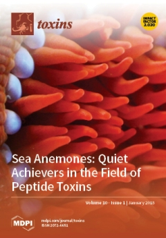
Cover Story (view full-size image): The peptide toxin ShK highlights the potential of sea anemones to produce valuable therapeutic leads, and the widespread abundance of closely-related peptides across many phyla suggests that the ShK scaffold can support a range of functions. Current genomic, transcriptomic and proteomic studies of sea anemones promise to greatly expand the number of ShK analogues and to identify a range of novel peptide families. The cover photo shows the tentacles (red) and acrorhagi (blue) of one of the sea anemones from south-eastern Queensland that we are investigating for new peptide toxins. View this paper
  • Issues are regarded as officially published after their release is announced to the table of contents alert mailing list.
  • You may sign up for e-mail alerts to receive table of contents of newly released issues.
  • PDF is the official format for papers published in both, html and pdf forms. To view the papers in pdf format, click on the "PDF Full-text" link, and use the free Adobe Reader to open them.
Order results
Result details
Section
Select all
Export citation of selected articles as:
20 pages, 2164 KiB  
Review
Allelopathic and Bloom-Forming Picocyanobacteria in a Changing World
by Sylwia Śliwińska-Wilczewska, Jakub Maculewicz, Aldo Barreiro Felpeto and Adam Latała
Toxins 2018, 10(1), 48; https://doi.org/10.3390/toxins10010048 - 20 Jan 2018
Cited by 45 | Viewed by 6694
Abstract
Picocyanobacteria are extremely important organisms in the world’s oceans and freshwater ecosystems. They play an essential role in primary production and their domination in phytoplankton biomass is common in both oligotrophic and eutrophic waters. Their role is expected to become even more relevant [...] Read more.
Picocyanobacteria are extremely important organisms in the world’s oceans and freshwater ecosystems. They play an essential role in primary production and their domination in phytoplankton biomass is common in both oligotrophic and eutrophic waters. Their role is expected to become even more relevant with the effect of climate change. However, this group of photoautotrophic organisms still remains insufficiently recognized. Only a few works have focused in detail on the occurrence of massive blooms of picocyanobacteria, their toxicity and allelopathic activity. Filling the gap in our knowledge about the mechanisms involved in the proliferation of these organisms could provide a better understanding of aquatic environments. In this review, we gathered and described recent information about allelopathic activity of picocyanobacteria and occurrence of their massive blooms in many aquatic ecosystems. We also examined the relationships between climate change and representative picocyanobacterial genera from freshwater, brackish and marine ecosystems. This work emphasizes the importance of studying the smallest picoplanktonic fractions of cyanobacteria. Full article
Show Figures

Figure 1

18 pages, 1597 KiB  
Article
Responses of Oat Grains to Fusarium poae and F. langsethiae Infections and Mycotoxin Contaminations
by Charlotte Martin, Torsten Schöneberg, Susanne Vogelgsang, Carla Susana Mendes Ferreira, Romina Morisoli, Mario Bertossa, Thomas D. Bucheli, Brigitte Mauch-Mani and Fabio Mascher
Toxins 2018, 10(1), 47; https://doi.org/10.3390/toxins10010047 - 20 Jan 2018
Cited by 23 | Viewed by 5150
Abstract
Recent increases of Fusarium head blight (FHB) disease caused by infections with F. poae (FP) and F. langsethiae (FL) have been observed in oats. These pathogens are producers of nivalenol (NIV) and T-2/HT-2 toxin (T-2/HT-2), respectively, which are now considered major issues for [...] Read more.
Recent increases of Fusarium head blight (FHB) disease caused by infections with F. poae (FP) and F. langsethiae (FL) have been observed in oats. These pathogens are producers of nivalenol (NIV) and T-2/HT-2 toxin (T-2/HT-2), respectively, which are now considered major issues for cereal food and feed safety. To date, the impact of FP and FL on oat grains has not yet been identified, and little is known about oat resistance elements against these pathogens. In the present study, the impact of FL and FP on oat grains was assessed under different environmental conditions in field experiments with artificial inoculations. The severity of FP and FL infection on grains were compared across three field sites, and the resistance against NIV and T-2/HT2 accumulation was assessed for seven oat genotypes. Grain weight, β-glucan content, and protein content were compared between infected and non-infected grains. Analyses of grain infection showed that FL was able to cause infection on the grain only in the field site with the highest relative humidity, whereas FP infected grains in all field sites. The FP infection of grains resulted in NIV contamination (between 30–500 μg/kg). The concentration of NIV in grains was not conditioned by environmental conditions. FL provoked an average contamination of grains with T-2/HT-2 (between 15–132 μg/kg). None of the genotypes was able to fully avoid toxin accumulation. The general resistance of oat grains against toxin accumulation was weak, and resistance against NIV accumulation was strongly impacted by the interaction between the genotype and the environment. Only the genotype with hull-less grains showed partial resistance to both NIV and T-2/HT-2 contamination. FP and FL infections could change the β-glucan content in grains, depending on the genotypes and environmental conditions. FP and FL did not have a significant impact on the thousand kernel weight (TKW) and protein content. Hence, resistance against toxin accumulation remains the only indicator of FHB resistance in oat. Our results highlight the need for new oat genotypes with enhanced resistance against both NIV and T-2/HT-2 to ensure food and feed safety. Full article
(This article belongs to the Collection Understanding Mycotoxin Occurrence in Food and Feed Chains)
Show Figures

Figure 1

2 pages, 505 KiB  
Editorial
Announcing the 2018 Toxins Travel Awards for Post-Doctoral Fellows
by Jay W. Fox
Toxins 2018, 10(1), 46; https://doi.org/10.3390/toxins10010046 - 19 Jan 2018
Viewed by 2728
Abstract
This year we enjoyed a large number of very highy meritorious applications for our annual Toxins Travel Awards.[...] Full article
Show Figures

Figure 1

11 pages, 727 KiB  
Article
Chronic Dietary Intake of Enniatin B in Broiler Chickens Has Low Impact on Intestinal Morphometry and Hepatic Histology, and Shows Limited Transfer to Liver Tissue
by Sophie Fraeyman, Siska Croubels, Mathias Devreese, Richard Ducatelle, Michael Rychlik and Gunther Antonissen
Toxins 2018, 10(1), 45; https://doi.org/10.3390/toxins10010045 - 18 Jan 2018
Cited by 11 | Viewed by 4049
Abstract
The Fusarium mycotoxin enniatin B (ENN B) is a so-called emerging mycotoxin frequently contaminating poultry feed. To investigate the impact of chronic ENN B exposure on animal health, broiler chickens were fed either a diet naturally contaminated with ENN B (2352 µg/kg) or [...] Read more.
The Fusarium mycotoxin enniatin B (ENN B) is a so-called emerging mycotoxin frequently contaminating poultry feed. To investigate the impact of chronic ENN B exposure on animal health, broiler chickens were fed either a diet naturally contaminated with ENN B (2352 µg/kg) or a control diet (135 µg/kg) for 2, 7, 14, or 21 days. ENN B concentrations were determined in plasma and liver using a validated ultra-high performance liquid chromatography—tandem mass spectrometry UHPLC-MS/MS method. Liver was evaluated histologically, and the villus length and crypt depth of the duodenum, jejunum, and ileum were measured. Histopathology of the livers did not reveal major abnormalities. Feeding an ENN B-contaminated diet could possibly inhibit the proliferation of enterocytes in the duodenal crypts, but did not affect villus length, crypt depth, or villus length-crypt depth ratio of the jejunum and ileum. ENN B levels in plasma and liver were significantly higher in the ENN B-fed group and ranged between <25–264 pg/mL and <0.05–0.85 ng/g, respectively. ENN B carry-over rates from feed to liver tissue were 0.005–0.014% and 0.034–0.109% in the ENN B and control group, respectively. Carry-over rates were low and indicated a limited contribution of poultry tissue-derived products to the total dietary ENN B intake for humans. The above results support the opinion of the European Food Safety Authority stating that adverse health effects from ENN B in broiler chickens are unlikely. Full article
Show Figures

Graphical abstract

3 pages, 191 KiB  
Editorial
“Bacterial Toxins” Section in the Journal Toxins: A Fantastic Multidisciplinary Interplay between Bacterial Pathogenicity Mechanisms, Physiological Processes, Genomic Evolution, and Subsequent Development of Identification Methods, Efficient Treatment, and Prevention of Toxigenic Bacteria
by Michel R. Popoff
Toxins 2018, 10(1), 44; https://doi.org/10.3390/toxins10010044 - 18 Jan 2018
Cited by 4 | Viewed by 3739
Abstract
Toxins are powerful pathogenicity factors produced by certain bacteria, fungi, animals, and plants which mediate drastic interactions of these pathogens on the organism host[...] Full article
(This article belongs to the Section Bacterial Toxins)
12 pages, 1902 KiB  
Article
The Peptide PnPP-19, a Spider Toxin Derivative, Activates μ-Opioid Receptors and Modulates Calcium Channels
by Ana C. N. Freitas, Steve Peigneur, Flávio H. P. Macedo, José E. Menezes-Filho, Paul Millns, Liciane F. Medeiros, Maria A. Arruda, Jader Cruz, Nicholas D. Holliday, Jan Tytgat, Gareth Hathway and Maria E. De Lima
Toxins 2018, 10(1), 43; https://doi.org/10.3390/toxins10010043 - 15 Jan 2018
Cited by 14 | Viewed by 6739
Abstract
The synthetic peptide PnPP-19 comprehends 19 amino acid residues and it represents part of the primary structure of the toxin δ-CNTX-Pn1c (PnTx2-6), isolated from the venom of the spider Phoneutria nigriventer. Behavioural tests suggest that PnPP-19 induces antinociception by activation of CB1, [...] Read more.
The synthetic peptide PnPP-19 comprehends 19 amino acid residues and it represents part of the primary structure of the toxin δ-CNTX-Pn1c (PnTx2-6), isolated from the venom of the spider Phoneutria nigriventer. Behavioural tests suggest that PnPP-19 induces antinociception by activation of CB1, μ and δ opioid receptors. Since the peripheral and central antinociception induced by PnPP-19 involves opioid activation, the aim of this work was to identify whether this synthetic peptide could directly activate opioid receptors and investigate the subtype selectivity for μ-, δ- and/or κ-opioid receptors. Furthermore, we also studied the modulation of calcium influx driven by PnPP-19 in dorsal root ganglion neurons, and analyzed whether this modulation was opioid-mediated. PnPP-19 selectively activates μ-opioid receptors inducing indirectly inhibition of calcium channels and hereby impairing calcium influx in dorsal root ganglion (DRG) neurons. Interestingly, notwithstanding the activation of opioid receptors, PnPP-19 does not induce β-arrestin2 recruitment. PnPP-19 is the first spider toxin derivative that, among opioid receptors, selectively activates μ-opioid receptors. The lack of β-arrestin2 recruitment highlights its potential for the design of new improved opioid agonists. Full article
(This article belongs to the Special Issue Toxins in Drug Discovery and Pharmacology)
Show Figures

Figure 1

24 pages, 1405 KiB  
Article
Comparative Response of the Hepatic Transcriptomes of Domesticated and Wild Turkey to Aflatoxin B1
by Kent M. Reed, Kristelle M. Mendoza, Juan E. Abrahante and Roger A. Coulombe
Toxins 2018, 10(1), 42; https://doi.org/10.3390/toxins10010042 - 13 Jan 2018
Cited by 14 | Viewed by 4584
Abstract
The food-borne mycotoxin aflatoxin B1 (AFB1) poses a significant risk to poultry, which are highly susceptible to its hepatotoxic effects. Domesticated turkeys (Meleagris gallopavo) are especially sensitive, whereas wild turkeys (M. g. silvestris) are more resistant. [...] Read more.
The food-borne mycotoxin aflatoxin B1 (AFB1) poses a significant risk to poultry, which are highly susceptible to its hepatotoxic effects. Domesticated turkeys (Meleagris gallopavo) are especially sensitive, whereas wild turkeys (M. g. silvestris) are more resistant. AFB1 toxicity entails bioactivation by hepatic cytochrome P450s to the electrophilic exo-AFB1-8,9-epoxide (AFBO). Domesticated turkeys lack functional hepatic GST-mediated detoxification of AFBO, and this is largely responsible for the differences in resistance between turkey types. This study was designed to characterize transcriptional changes induced in turkey livers by AFB1, and to contrast the response of domesticated (susceptible) and wild (more resistant) birds. Gene expression responses to AFB1 were examined using RNA-sequencing. Statistically significant differences in gene expression were observed among treatment groups and between turkey types. Expression analysis identified 4621 genes with significant differential expression (DE) in AFB1-treated birds compared to controls. Characterization of DE transcripts revealed genes dis-regulated in response to toxic insult with significant association of Phase I and Phase II genes and others important in cellular regulation, modulation of apoptosis, and inflammatory responses. Constitutive expression of GSTA3 was significantly higher in wild birds and was significantly higher in AFB1-treated birds when compared to controls for both genetic groups. This pattern was also observed by qRT-PCR in other wild and domesticated turkey strains. Results of this study emphasize the differential response of these genetically distinct birds, and identify genes and pathways that are differentially altered in aflatoxicosis. Full article
(This article belongs to the Collection Aflatoxins)
Show Figures

Figure 1

14 pages, 4007 KiB  
Article
Transketolase Is Identified as a Target of Herbicidal Substance α-Terthienyl by Proteomics
by Bin Zhao, Jingqian Huo, Ning Liu, Jinlin Zhang and Jingao Dong
Toxins 2018, 10(1), 41; https://doi.org/10.3390/toxins10010041 - 12 Jan 2018
Cited by 17 | Viewed by 4622
Abstract
α-terthienyl is a natural phytotoxin isolated originally from Flaveria bidentis (L.) Kuntze. The bioassay presented here shows the strong herbicidal activity of α-terthienyl on Digitaria sanguinalis, Arabidopsis thaliana and Chlamydomonas reinhardtii. The α-terthienyl-induced response of A. thaliana at [...] Read more.
α-terthienyl is a natural phytotoxin isolated originally from Flaveria bidentis (L.) Kuntze. The bioassay presented here shows the strong herbicidal activity of α-terthienyl on Digitaria sanguinalis, Arabidopsis thaliana and Chlamydomonas reinhardtii. The α-terthienyl-induced response of A. thaliana at the protein level was analyzed at different times. Changes in the protein expression profiles were analyzed by two-dimensional gel electrophoresis and liquid chromatography tandem mass spectrometry (LC-MS/MS) mass spectrometry. Sixteen protein spots were identified that showed reproducible changes in the expression of at least 2-fold when compared to the control. Among these 16 spots, three were up-regulated and 13 were down-regulated. The decreased expression of several proteins associated with energy production and carbon metabolism suggested that these processes were affected by α-terthienyl. To search for the candidate proteins in this screen, A. thaliana T-DNA mutants of the candidate proteins were used to test their susceptibility to α-terthienyl. Amongst the others, attkl1, a mutant of transketolase, exhibited a significantly lower sensitivity to α-terthienyl when hit compared with Col-0. Based on the identification of the proteins associated with the response to α-terthienyl by proteomics, a candidate target protein transketolase was identified. Full article
(This article belongs to the Section Plant Toxins)
Show Figures

Figure 1

17 pages, 5724 KiB  
Article
Molecular and Physiological Effects on the Small Intestine of Weaner Pigs Following Feeding with Deoxynivalenol-Contaminated Feed
by J. Alex Pasternak, Vaishnavi Iyer Aka Aiyer, Glenn Hamonic, A. Denise Beaulieu, Daniel A. Columbus and Heather L. Wilson
Toxins 2018, 10(1), 40; https://doi.org/10.3390/toxins10010040 - 12 Jan 2018
Cited by 22 | Viewed by 5363
Abstract
We intended to assess how exposure of piglets to deoxynivalenol (DON)-contaminated feed impacted their growth, immune response and gut development. Piglets were fed traditional Phase I, Phase II and Phase III diets with the control group receiving 0.20–0.40 ppm DON (referred to as [...] Read more.
We intended to assess how exposure of piglets to deoxynivalenol (DON)-contaminated feed impacted their growth, immune response and gut development. Piglets were fed traditional Phase I, Phase II and Phase III diets with the control group receiving 0.20–0.40 ppm DON (referred to as the Control group) and treatment group receiving much higher level of DON-contaminated wheat (3.30–3.80 ppm; referred to as DON-contaminated group). Feeding a DON-contaminated diet had no impact on average daily feed intake (ADFI) (p < 0.08) or average daily gain (ADG) (p > 0.10) but it did significantly reduce body weight over time relative to the control piglets (p < 0.05). Cytokine analysis after initial exposure to the DON-contaminated feed did not result in significant differences in serum interleukin (IL) IL1β, IL-8, IL-13, tumor necrosis factor (TNF)-α or interferon (IFN)-γ. After day 24, no obvious changes in jejunum or ileum gut morphology, histology or changes in gene expression for IL-1β, IL-6, IL-10, TNFα, or Toll-like receptor (TLR)-4 genes. IL-8 showed a trend towards increased expression in the ileum in DON-fed piglets. A significant increase in gene expression for claudin (CLDN) 7 gene expression and a trend towards increased CLDN 2-expression was observed in the ileum in piglets fed the highly DON-contaminated wheat. Because CLDN localization was not negatively affected, we believe that it is unlikely that gut permeability was affected. Exposure to DON-contaminated feed did not significantly impact weaner piglet performance or gut physiology. Full article
(This article belongs to the Special Issue Effects of Mycotoxins on the Intestine)
Show Figures

Figure 1

29 pages, 7713 KiB  
Article
Analysis of Microcystins in Cyanobacterial Blooms from Freshwater Bodies in England
by Andrew D. Turner, Monika Dhanji-Rapkova, Alison O’Neill, Lewis Coates, Adam Lewis and Katy Lewis
Toxins 2018, 10(1), 39; https://doi.org/10.3390/toxins10010039 - 11 Jan 2018
Cited by 68 | Viewed by 6908
Abstract
Cyanobacterial blooms in freshwater bodies in England are currently monitored reactively, with samples containing more than 20,000 cells/mL of potentially toxin-producing species by light microscopy resulting in action by the water body owner. Whilst significantly reducing the risk of microcystin exposure, there is [...] Read more.
Cyanobacterial blooms in freshwater bodies in England are currently monitored reactively, with samples containing more than 20,000 cells/mL of potentially toxin-producing species by light microscopy resulting in action by the water body owner. Whilst significantly reducing the risk of microcystin exposure, there is little data describing the levels of these toxins present in cyanobacterial blooms. This study focused on the quantitative LC-MS/MS analysis of microcystins in freshwater samples, collected across England during 2016 and found to contain potentially toxin-producing cyanobacteria. More than 50% of samples contained quantifiable concentrations of microcystins, with approximately 13% exceeding the WHO medium health threshold of 20 μg/L. Toxic samples were confirmed over a nine-month period, with a clear increase in toxins during late summer, but with no apparent geographical patterns. No statistical relationships were found between total toxin concentrations and environmental parameters. Complex toxin profiles were determined and profile clusters were unrelated to cyanobacterial species, although a dominance of MC-RR was determined in water samples from sites associated with lower rainfall. 100% of samples with toxins above the 20 μg/L limit contained cell densities above 20,000 cells/mL or cyanobacterial scum, showing the current regime is suitable for public health. Conversely, with only 18% of cell density threshold samples having total microcystins above 20 μg/L, there is the potential for reactive water closures to unnecessarily impact upon the socio-economics of the local population. In the future, routine analysis of bloom samples by LC-MS/MS would provide a beneficial confirmatory approach to the current microscopic assessment, aiding both public health and the needs of water users and industry. Full article
Show Figures

Figure 1

13 pages, 313 KiB  
Review
Updated Overview of Infrared Spectroscopy Methods for Detecting Mycotoxins on Cereals (Corn, Wheat, and Barley)
by Cecile Levasseur-Garcia
Toxins 2018, 10(1), 38; https://doi.org/10.3390/toxins10010038 - 10 Jan 2018
Cited by 55 | Viewed by 6432
Abstract
Each year, mycotoxins cause economic losses of several billion US dollars worldwide. Consequently, methods must be developed, for producers and cereal manufacturers, to detect these toxins and to comply with regulations. Chromatographic reference methods are time consuming and costly. Thus, alternative methods such [...] Read more.
Each year, mycotoxins cause economic losses of several billion US dollars worldwide. Consequently, methods must be developed, for producers and cereal manufacturers, to detect these toxins and to comply with regulations. Chromatographic reference methods are time consuming and costly. Thus, alternative methods such as infrared spectroscopy are being increasingly developed to provide simple, rapid, and nondestructive methods to detect mycotoxins. This article reviews research conducted over the last eight years into the use of near-infrared and mid-infrared spectroscopy to monitor mycotoxins in corn, wheat, and barley. More specifically, we focus on the Fusarium species and on the main fusariotoxins of deoxynivalenol, zearalenone, and fumonisin B1 and B2. Quantification models are insufficiently precise to satisfy the legal requirements. Sorting models with cutoff levels are the most promising applications. Full article
(This article belongs to the Collection Understanding Mycotoxin Occurrence in Food and Feed Chains)
6 pages, 399 KiB  
Editorial
Acknowledgement to Reviewers of Toxins in 2017
by Toxins Editorial Office
Toxins 2018, 10(1), 37; https://doi.org/10.3390/toxins10010037 - 09 Jan 2018
Viewed by 3148
Abstract
Peer review is an essential part in the publication process, ensuring that Toxins maintains high quality standards for its published papers.[...] Full article
15 pages, 2935 KiB  
Review
Sea Anemones: Quiet Achievers in the Field of Peptide Toxins
by Peter J. Prentis, Ana Pavasovic and Raymond S. Norton
Toxins 2018, 10(1), 36; https://doi.org/10.3390/toxins10010036 - 08 Jan 2018
Cited by 85 | Viewed by 12873
Abstract
Sea anemones have been understudied as a source of peptide and protein toxins, with relatively few examined as a source of new pharmacological tools or therapeutic leads. This is surprising given the success of some anemone peptides that have been tested, such as [...] Read more.
Sea anemones have been understudied as a source of peptide and protein toxins, with relatively few examined as a source of new pharmacological tools or therapeutic leads. This is surprising given the success of some anemone peptides that have been tested, such as the potassium channel blocker from Stichodactyla helianthus known as ShK. An analogue of this peptide, ShK-186, which is now known as dalazatide, has successfully completed Phase 1 clinical trials and is about to enter Phase 2 trials for the treatment of autoimmune diseases. One of the impediments to the exploitation of sea anemone toxins in the pharmaceutical industry has been the difficulty associated with their high-throughput discovery and isolation. Recent developments in multiple ‘omic’ technologies, including genomics, transcriptomics and proteomics, coupled with advanced bioinformatics, have opened the way for large-scale discovery of novel sea anemone toxins from a range of species. Many of these toxins will be useful pharmacological tools and some will hopefully prove to be valuable therapeutic leads. Full article
(This article belongs to the Special Issue Selected Papers from the 2017 Venoms to Drugs Conference)
Show Figures

Figure 1

19 pages, 2117 KiB  
Article
Biological and Proteolytic Variation in the Venom of Crotalus scutulatus scutulatus from Mexico
by Miguel Borja, Edgar Neri-Castro, Gamaliel Castañeda-Gaytán, Jason L. Strickland, Christopher L. Parkinson, Juan Castañeda-Gaytán, Roberto Ponce-López, Bruno Lomonte, Alejandro Olvera-Rodríguez, Alejandro Alagón and Rebeca Pérez-Morales
Toxins 2018, 10(1), 35; https://doi.org/10.3390/toxins10010035 - 08 Jan 2018
Cited by 34 | Viewed by 8820
Abstract
Rattlesnake venoms may be classified according to the presence/absence and relative abundance of the neurotoxic phospholipases A 2 s (PLA 2 s), such as Mojave toxin, and snake venom metalloproteinases (SVMPs). In Mexico, studies to determine venom variation in Mojave Rattlesnakes (Crotalus [...] Read more.
Rattlesnake venoms may be classified according to the presence/absence and relative abundance of the neurotoxic phospholipases A 2 s (PLA 2 s), such as Mojave toxin, and snake venom metalloproteinases (SVMPs). In Mexico, studies to determine venom variation in Mojave Rattlesnakes (Crotalus scutulatus scutulatus) are limited and little is known about the biological and proteolytic activities in this species. Tissue (34) and venom (29) samples were obtained from C. s. scutulatus from different locations within their distribution in Mexico. Mojave toxin detection was carried out at the genomic (by PCR) and protein (by ELISA) levels for all tissue and venom samples. Biological activity was tested on representative venoms by measuring LD 50 and hemorrhagic activity. To determine the approximate amount of SVMPs, 15 venoms were separated by RP-HPLC and variation in protein profile and proteolytic activity was evaluated by SDS-PAGE (n = 28) and Hide Powder Azure proteolytic analysis (n = 27). Three types of venom were identified in Mexico which is comparable to the intraspecific venom diversity observed in the Sonoran Desert of Arizona, USA: Venom Type A (∼Type II), with Mojave toxin, highly toxic, lacking hemorrhagic activity, and with scarce proteolytic activity; Type B (∼Type I), without Mojave toxin, less toxic than Type A, highly hemorrhagic and proteolytic; and Type A + B, containing Mojave toxin, as toxic as venom Type A, variable in hemorrhagic activity and with intermediate proteolytic activity. We also detected a positive correlation between SVMP abundance and hemorrhagic and proteolytic activities. Although more sampling is necessary, our results suggest that venoms containing Mojave toxin and venom lacking this toxin are distributed in the northwest and southeast portions of the distribution in Mexico, respectively, while an intergradation in the middle of both zones is present. Full article
(This article belongs to the Section Animal Venoms)
Show Figures

Figure 1

57 pages, 1440 KiB  
Review
Biochemical and Clinical Impact of Organic Uremic Retention Solutes: A Comprehensive Update
by Raymond Vanholder, Anneleen Pletinck, Eva Schepers and Griet Glorieux
Toxins 2018, 10(1), 33; https://doi.org/10.3390/toxins10010033 - 08 Jan 2018
Cited by 218 | Viewed by 12304
Abstract
In this narrative review, the biological/biochemical impact (toxicity) of a large array of known individual uremic retention solutes and groups of solutes is summarized. We classified these compounds along their physico-chemical characteristics as small water-soluble compounds or groups, protein bound compounds and middle [...] Read more.
In this narrative review, the biological/biochemical impact (toxicity) of a large array of known individual uremic retention solutes and groups of solutes is summarized. We classified these compounds along their physico-chemical characteristics as small water-soluble compounds or groups, protein bound compounds and middle molecules. All but one solute (glomerulopressin) affected at least one mechanism with the potential to contribute to the uremic syndrome. In general, several mechanisms were influenced for each individual solute or group of solutes, with some impacting up to 7 different biological systems of the 11 considered. The inflammatory, cardio-vascular and fibrogenic systems were those most frequently affected and they are one by one major actors in the high morbidity and mortality of CKD but also the mechanisms that have most frequently been studied. A scoring system was built with the intention to classify the reviewed compounds according to the experimental evidence of their toxicity (number of systems affected) and overall experimental and clinical evidence. Among the highest globally scoring solutes were 3 small water-soluble compounds [asymmetric dimethylarginine (ADMA); trimethylamine-N-oxide (TMAO); uric acid], 6 protein bound compounds or groups of protein bound compounds [advanced glycation end products (AGEs); p-cresyl sulfate; indoxyl sulfate; indole acetic acid; the kynurenines; phenyl acetic acid;] and 3 middle molecules [β2-microglobulin; ghrelin; parathyroid hormone). In general, more experimental data were provided for the protein bound molecules but for almost half of them clinical evidence was missing in spite of robust experimental data. The picture emanating is one of a complex disorder, where multiple factors contribute to a multisystem complication profile, so that it seems of not much use to pursue a decrease of concentration of a single compound. Full article
(This article belongs to the Special Issue Novel Issues in Uremic Toxicity)
Show Figures

Graphical abstract

25 pages, 2965 KiB  
Article
Azemiopsin, a Selective Peptide Antagonist of Muscle Nicotinic Acetylcholine Receptor: Preclinical Evaluation as a Local Muscle Relaxant
by Irina V. Shelukhina, Maxim N. Zhmak, Alexander V. Lobanov, Igor A. Ivanov, Alexandra I. Garifulina, Irina N. Kravchenko, Ekaterina A. Rasskazova, Margarita A. Salmova, Elena A. Tukhovskaya, Vladimir A. Rykov, Gulsara A. Slashcheva, Natalya S. Egorova, Inessa S. Muzyka, Victor I. Tsetlin and Yuri N. Utkin
Toxins 2018, 10(1), 34; https://doi.org/10.3390/toxins10010034 - 07 Jan 2018
Cited by 26 | Viewed by 5566
Abstract
Azemiopsin (Az), a linear peptide from the Azemiops feae viper venom, contains no disulfide bonds, is a high-affinity and selective inhibitor of nicotinic acetylcholine receptor (nAChR) of muscle type and may be considered as potentially applicable nondepolarizing muscle relaxant. In this study, we [...] Read more.
Azemiopsin (Az), a linear peptide from the Azemiops feae viper venom, contains no disulfide bonds, is a high-affinity and selective inhibitor of nicotinic acetylcholine receptor (nAChR) of muscle type and may be considered as potentially applicable nondepolarizing muscle relaxant. In this study, we investigated its preclinical profile in regard to in vitro and in vivo efficacy, acute and chronic toxicity, pharmacokinetics, allergenic capacity, immunotoxicity and mutagenic potency. The peptide effectively inhibited (IC50 ~ 19 nM) calcium response of muscle nAChR evoked by 30 μM (EC100) acetylcholine but was less potent (IC50 ~ 3 μM) at α7 nAChR activated by 10 μM (EC50) acetylcholine and had a low affinity to α4β2 and α3-containing nAChR, as well as to GABAA or 5HT3 receptors. Its muscle relaxant effect was demonstrated at intramuscular injection to mice at doses of 30–300 µg/kg, 30 µg/kg being the initial effective dose and 90 µg/kg—the average effective dose. The maximal muscle relaxant effect of Az was achieved in 10 min after the administration and elimination half-life of Az in mice was calculated as 20–40 min. The longest period of Az action observed at a dose of 300 µg/kg was 55 min. The highest acute toxicity (LD50 510 μg/kg) was observed at intravenous injection of Az, at intramuscular or intraperitoneal administration it was less toxic. The peptide showed practically no immunotoxic, allergenic or mutagenic capacity. Overall, the results demonstrate that Az has good drug-like properties for the application as local muscle relaxant and in its parameters, is not inferior to the relaxants currently used. However, some Az modification might be effective to extend its narrow therapeutic window, a typical characteristic and a weak point of all nondepolarizing myorelaxants. Full article
(This article belongs to the Special Issue Toxins in Drug Discovery and Pharmacology)
Show Figures

Graphical abstract

13 pages, 2536 KiB  
Article
Development of a Deimmunized Bispecific Immunotoxin dDT2219 against B-Cell Malignancies
by Joerg U. Schmohl, Deborah Todhunter, Elizabeth Taras, Veronika Bachanova and Daniel A. Vallera
Toxins 2018, 10(1), 32; https://doi.org/10.3390/toxins10010032 - 06 Jan 2018
Cited by 20 | Viewed by 4330
Abstract
Diphtheria toxin (DT) related targeted toxins are effective in cancer treatment, but efficacy diminishes in time because of their immunogenic potential and/or former vaccinations. In order to overcome this limitation for DT2219, a promising bispecific targeted toxin which targets CD19 and CD22, we [...] Read more.
Diphtheria toxin (DT) related targeted toxins are effective in cancer treatment, but efficacy diminishes in time because of their immunogenic potential and/or former vaccinations. In order to overcome this limitation for DT2219, a promising bispecific targeted toxin which targets CD19 and CD22, we deimmunized the DT moiety, and thereby developed an exciting improved drug (dDT2219) which still has the potential to sufficiently target B-cell malignancies but also limits clearance because of its reduced immunogenicity. The DT moiety was modified by inducing point mutations in prominent positions on the molecular surface. The new engineered dDT2219 was tested for activity, efficacy, and specificity using functional assays, proliferation assays, and flow cytometry. Furthermore, 12 samples of Chronic Lymphatic Leukemia (CLL) patients were used to assess binding. Immunogenicity was determined using a BALB/c mouse model. dDT2219 was efficient and specific against B-cell malignancies such as Bukitt-Lymphoma cell lines Daudi and Raji. dDT2219 showed specific binding on targets and on CLL samples. Intraperitoneal vaccination of immune competent mice showed that even after multiple administrations with increasing doses, induction of neutralizing antibodies was significantly lower in the dDT2219 treated animal group. The new dDT2219 combines potent anti-tumor cell activity with a reduced immunogenicity. With regard to the frequent development of neutralizing antibodies after multiple administrations with immunotoxins, dDT2219 shows promise to overcome this limitation and thus might maintain effectiveness even after multiple treatment cycles. Full article
Show Figures

Figure 1

12 pages, 5076 KiB  
Article
Light-Irradiation Wavelength and Intensity Changes Influence Aflatoxin Synthesis in Fungi
by Tadahiro Suzuki
Toxins 2018, 10(1), 31; https://doi.org/10.3390/toxins10010031 - 05 Jan 2018
Cited by 14 | Viewed by 4814
Abstract
Fungi respond to light irradiation by forming conidia and occasionally synthesizing mycotoxins. Several light wavelengths, such as blue and red, affect the latter. However, the relationship between light irradiation and mycotoxin synthesis varies depending on the fungal species or strain. This study focused [...] Read more.
Fungi respond to light irradiation by forming conidia and occasionally synthesizing mycotoxins. Several light wavelengths, such as blue and red, affect the latter. However, the relationship between light irradiation and mycotoxin synthesis varies depending on the fungal species or strain. This study focused on aflatoxin (AF), which is a mycotoxin, and the types of light irradiation that increase AF synthesis. Light-irradiation tests using the visible region indicated that blue wavelengths in the lower 500 nm region promoted AF synthesis. In contrast, red wavelengths of 660 nm resulted in limited significant changes compared with dark conditions. Irradiation tests with different intensity levels indicated that a low light intensity increased AF synthesis. For one fungal strain, light irradiation decreased the AF synthesis under all wavelength conditions. However, the decrease was mitigated by 525 nm low intensity irradiation. Thus, blue-green low intensity irradiation may increase AF synthesis in fungi. Full article
(This article belongs to the Collection Aflatoxins)
Show Figures

Figure 1

14 pages, 591 KiB  
Article
Biological Control Products for Aflatoxin Prevention in Italy: Commercial Field Evaluation of Atoxigenic Aspergillus flavus Active Ingredients
by Antonio Mauro, Esther Garcia-Cela, Amedeo Pietri, Peter J. Cotty and Paola Battilani
Toxins 2018, 10(1), 30; https://doi.org/10.3390/toxins10010030 - 05 Jan 2018
Cited by 69 | Viewed by 7924 | Correction
Abstract
Since 2003, non-compliant aflatoxin concentrations have been detected in maize produced in Italy. The most successful worldwide experiments in aflatoxin prevention resulted from distribution of atoxigenic strains of Aspergillus flavus to displace aflatoxin-producers during crop development. The displacement results in lower aflatoxin concentrations [...] Read more.
Since 2003, non-compliant aflatoxin concentrations have been detected in maize produced in Italy. The most successful worldwide experiments in aflatoxin prevention resulted from distribution of atoxigenic strains of Aspergillus flavus to displace aflatoxin-producers during crop development. The displacement results in lower aflatoxin concentrations in harvested grain. The current study evaluated in field performances of two atoxigenic strains of A. flavus endemic to Italy in artificially inoculated maize ears and in naturally contaminated maize. Co-inoculation of atoxigenic strains with aflatoxin producers resulted in highly significant reductions in aflatoxin concentrations (>90%) in both years only with atoxigenic strain A2085. The average percent reduction in aflatoxin B1 concentration in naturally contaminated maize fields was 92.3%, without significant differences in fumonisins between treated and control maize. The vegetative compatibility group of A2085 was the most frequently recovered A. flavus in both treated and control plots (average 61.9% and 53.5% of the A. flavus, respectively). A2085 was therefore selected as an active ingredient for biocontrol products and deposited under provisions of the Budapest Treaty in the Belgian Co-Ordinated Collections of Micro-Organisms (BCCM/MUCL) collection (accession MUCL54911). Further work on development of A2085 as a tool for preventing aflatoxin contamination in maize produced in Italy is ongoing with the commercial product named AF-X1™. Full article
(This article belongs to the Collection Understanding Mycotoxin Occurrence in Food and Feed Chains)
Show Figures

Figure 1

23 pages, 9091 KiB  
Article
A Dipteran’s Novel Sucker Punch: Evolution of Arthropod Atypical Venom with a Neurotoxic Component in Robber Flies (Asilidae, Diptera)
by Stephan Holger Drukewitz, Nico Fuhrmann, Eivind A. B. Undheim, Alexander Blanke, Julien Giribaldi, Rosanna Mary, Guillaume Laconde, Sébastien Dutertre and Björn Marcus Von Reumont
Toxins 2018, 10(1), 29; https://doi.org/10.3390/toxins10010029 - 05 Jan 2018
Cited by 29 | Viewed by 15792
Abstract
Predatory robber flies (Diptera, Asilidae) have been suspected to be venomous due to their ability to overpower well-defended prey. However, details of their venom composition and toxin arsenal remained unknown. Here, we provide a detailed characterization of the venom system of robber flies [...] Read more.
Predatory robber flies (Diptera, Asilidae) have been suspected to be venomous due to their ability to overpower well-defended prey. However, details of their venom composition and toxin arsenal remained unknown. Here, we provide a detailed characterization of the venom system of robber flies through the application of comparative transcriptomics, proteomics and functional morphology. Our results reveal asilid venoms to be dominated by peptides and non-enzymatic proteins, and that the majority of components in the crude venom is represented by just ten toxin families, which we have named Asilidin1–10. Contrary to what might be expected for a liquid-feeding predator, the venoms of robber flies appear to be rich in novel peptides, rather than enzymes with a putative pre-digestive role. The novelty of these peptides suggests that the robber fly venom system evolved independently from hematophagous dipterans and other pancrustaceans. Indeed, six Asilidins match no other venom proteins, while three represent known examples of peptide scaffolds convergently recruited to a toxic function. Of these, members of Asilidin1 closely resemble cysteine inhibitor knot peptides (ICK), of which neurotoxic variants occur in cone snails, assassin bugs, scorpions and spiders. Synthesis of one of these putative ICKs, U-Asilidin1-Mar1a, followed by toxicity assays against an ecologically relevant prey model revealed that one of these likely plays a role as a neurotoxin involved in the immobilization of prey. Our results are fundamental to address these insights further and to understand processes that drive venom evolution in dipterans as well as other arthropods. Full article
(This article belongs to the Section Animal Venoms)
Show Figures

Figure 1

11 pages, 1669 KiB  
Article
Determination of Multi-Class Mycotoxins in Tartary Buckwheat by Ultra-Fast Liquid Chromatography Coupled with Triple Quadrupole Mass Spectrometry
by Guixing Ren, Yichen Hu, Jinming Zhang, Liang Zou and Gang Zhao
Toxins 2018, 10(1), 28; https://doi.org/10.3390/toxins10010028 - 04 Jan 2018
Cited by 14 | Viewed by 5305
Abstract
Considering crops are susceptible to toxicogenic fungi during plantation, pre-processing and storage, an ultra-fast liquid chromatography coupled with triple quadrupole mass spectrometry (UFLC-QTrap-MS/MS) method was developed and validated for simultaneous determination of the 12 most frequent mycotoxins, including aflatoxin B1, B [...] Read more.
Considering crops are susceptible to toxicogenic fungi during plantation, pre-processing and storage, an ultra-fast liquid chromatography coupled with triple quadrupole mass spectrometry (UFLC-QTrap-MS/MS) method was developed and validated for simultaneous determination of the 12 most frequent mycotoxins, including aflatoxin B1, B2, G1, G2, HT-2, T-2 toxin, ochratoxin A, fumonisin B1, B2, zearalanone, zearalenone, and deoxynivalenol, in 14 batches of Tartary buckwheat cultivar, collected from different origins in Sichuan Province, China. Differing from those complicated approaches, a simple and cost-efficient pretreatment method based on dilute-and-shoot was employed. Based on optimized chromatographic and mass spectrometry conditions, these 12 mycotoxins could be analyzed with high correlation coefficients (all over 0.995), high precision (RSD 0.47–9.26%), stability (RSD 0.72–11.36%), and recovery (79.52% to 108.92%, RSD 4.35–14.27%). Furthermore, this analysis method exhibited good determination performance with little disturbance of the matrix effect. Finally, this proposed method was applied for 14 batches of Tartary buckwheat seeds, in which aflatoxin B1 (AFB1) was detected in one moldy cultivar, Meigu No. 2, with its concentration exceeding the maximum residue limits set by EU regulations. The method thus established, which has significant advantages, could provide a preferred determination approach candidate for measurement of multiple mycotoxins measurement in Tartary buckwheat, even other kinds of foodstuffs. Full article
(This article belongs to the Collection Understanding Mycotoxin Occurrence in Food and Feed Chains)
Show Figures

Figure 1

14 pages, 5464 KiB  
Review
Correction of Malocclusion by Botulinum Neurotoxin Injection into Masticatory Muscles
by Hyun Seok and Seong-Gon Kim
Toxins 2018, 10(1), 27; https://doi.org/10.3390/toxins10010027 - 02 Jan 2018
Cited by 16 | Viewed by 9407
Abstract
Botulinum toxin (BTX) is a neurotoxin, and its injection in masticatory muscles induces muscle weakness and paralysis. This paralytic effect of BTX induces growth retardation of the maxillofacial bones, changes in dental eruption and occlusion state, and facial asymmetry. Using masticatory muscle paralysis [...] Read more.
Botulinum toxin (BTX) is a neurotoxin, and its injection in masticatory muscles induces muscle weakness and paralysis. This paralytic effect of BTX induces growth retardation of the maxillofacial bones, changes in dental eruption and occlusion state, and facial asymmetry. Using masticatory muscle paralysis and its effect via BTX, BTX can be used for the correction of malocclusion after orthognathic surgery and mandible fracture. The paralysis of specific masticatory muscles by BTX injection reduces the tensional force to the mandible and prevents relapse and changes in dental occlusion. BTX injection in the anterior belly of digastric and mylohyoid muscle prevents the open-bite and deep bite of dental occlusion and contributes to mandible stability after orthognathic surgery. The effect of BTX injection in masticatory muscles for maxillofacial bone growth and dental occlusion is reviewed in this article. The clinical application of BTX is also discussed for the correction of dental malocclusion and suppression of post-operative relapse after mandibular surgery. Full article
Show Figures

Figure 1

17 pages, 1621 KiB  
Article
Co-Occurrence of Microcystins and Taste-and-Odor Compounds in Drinking Water Source and Their Removal in a Full-Scale Drinking Water Treatment Plant
by Lixia Shang, Muhua Feng, Xiangen Xu, Feifei Liu, Fan Ke and Wenchao Li
Toxins 2018, 10(1), 26; https://doi.org/10.3390/toxins10010026 - 02 Jan 2018
Cited by 52 | Viewed by 6022
Abstract
The co-occurrence of cyanotoxins and taste-and-odor compounds are a growing concern for drinking water treatment plants (DWTPs) suffering cyanobacteria in water resources. The dissolved and cell-bound forms of three microcystin (MC) congeners (MC-LR, MC-RR and MC-YR) and four taste-and-odor compounds (geosmin, 2-methyl isoborneol, [...] Read more.
The co-occurrence of cyanotoxins and taste-and-odor compounds are a growing concern for drinking water treatment plants (DWTPs) suffering cyanobacteria in water resources. The dissolved and cell-bound forms of three microcystin (MC) congeners (MC-LR, MC-RR and MC-YR) and four taste-and-odor compounds (geosmin, 2-methyl isoborneol, β-cyclocitral and β-ionone) were investigated monthly from August 2011 to July 2012 in the eastern drinking water source of Lake Chaohu. The total concentrations of microcystins and taste-and-odor compounds reached 8.86 μg/L and 250.7 ng/L, respectively. The seasonal trends of microcystins were not consistent with those of the taste-and-odor compounds, which were accompanied by dominant species Microcystis and Dolichospermum. The fate of the cyanobacteria and metabolites were determined simultaneously after the processes of coagulation/flocculation, sedimentation, filtration and chlorination in the associated full-scale DWTP. The dissolved fractions with elevated concentrations were detected after some steps and the breakthrough of cyanobacteria and metabolites were even observed in finished water. Chlorophyll-a limits at intake were established for the drinking water source based on our investigation of multiple metabolites, seasonal variations and their elimination rates in the DWTP. Not only microcystins but also taste-and-odor compounds should be taken into account to guide the management in source water and in DWTPs. Full article
(This article belongs to the Special Issue Cyanobacteria and Cyanotoxins: New Advances and Future Challenges)
Show Figures

Graphical abstract

18 pages, 1522 KiB  
Review
Protective and Detoxifying Effects Conferred by Dietary Selenium and Curcumin against AFB1-Mediated Toxicity in Livestock: A Review
by Aniket Limaye, Roch-Chui Yu, Cheng-Chun Chou, Je-Ruei Liu and Kuan-Chen Cheng
Toxins 2018, 10(1), 25; https://doi.org/10.3390/toxins10010025 - 02 Jan 2018
Cited by 81 | Viewed by 6671
Abstract
Aflatoxin B1 (AFB1), among other aflatoxins of the aflatoxin family, is the most carcinogenic and hazardous mycotoxin to animals and human beings with very high potency leading to aflatoxicosis. Selenium is an essential trace mineral possessing powerful antioxidant functions. Selenium is widely reported [...] Read more.
Aflatoxin B1 (AFB1), among other aflatoxins of the aflatoxin family, is the most carcinogenic and hazardous mycotoxin to animals and human beings with very high potency leading to aflatoxicosis. Selenium is an essential trace mineral possessing powerful antioxidant functions. Selenium is widely reported as an effective antioxidant against aflatoxicosis. By preventing oxidative liver damage, suppressing pro-apoptotic proteins and improving immune status in AFB1 affected animals; selenium confers specific protection against AFB1 toxicity. Meticulous supplementation of animal feed by elemental selenium in the organic and inorganic forms has proven to be effective to ameliorate AFB1 toxicity. Curcumin is another dietary agent of importance in tackling aflatoxicosis. Curcumin is one of the major active ingredients in the tubers of a spice Curcuma longa L., a widely reported antioxidant, anticarcinogenic agent with reported protective potential against aflatoxin-mediated liver damage. Curcumin restricts the aflatoxigenic potential of Aspergillus flavus. Curcumin inhibits cytochrome P450 isoenzymes, particularly CYP2A6 isoform; thereby reducing the formation of AFB1-8, 9-epoxide and other toxic metabolites causing aflatoxicosis. In this review, we have briefly reviewed important aflatoxicosis symptoms among animals. With the main focus on curcumin and selenium, we have reviewed their underlying protective mechanisms in different animals along with their extraction and production methods for feed applications. Full article
(This article belongs to the Section Mycotoxins)
Show Figures

Figure 1

16 pages, 6550 KiB  
Article
ROS-Mediated Cell Cycle Arrest and Apoptosis Induced by Zearalenone in Mouse Sertoli Cells via ER Stress and the ATP/AMPK Pathway
by Wang-Long Zheng, Bing-Jie Wang, Ling Wang, Yu-Ping Shan, Hui Zou, Rui-Long Song, Tao Wang, Jian-Hong Gu, Yan Yuan, Xue-Zhong Liu, Guo-Qiang Zhu, Jian-Fa Bai, Zong-Ping Liu and Jian-Chun Bian
Toxins 2018, 10(1), 24; https://doi.org/10.3390/toxins10010024 - 01 Jan 2018
Cited by 90 | Viewed by 6969
Abstract
Zearalenone (ZEA) can perturb the differentiation of cells, reduce the generation of reproductive cells and induce a death of germ cells, but the molecular mechanism remains unclear. In order to investigate the potential mechanism of ZEA-induced cell cycle arrest and apoptosis, we studied [...] Read more.
Zearalenone (ZEA) can perturb the differentiation of cells, reduce the generation of reproductive cells and induce a death of germ cells, but the molecular mechanism remains unclear. In order to investigate the potential mechanism of ZEA-induced cell cycle arrest and apoptosis, we studied the effects of ZEA on cell proliferation, cell-cycle distribution, cell-cycle-related proteins, cell death, cell apoptosis, ROS generation and the ATP/AMPK pathway in Sertoli cells. The role of ROS, ER stress and the ATP/AMPK pathway in ZEA-induced cell-cycle arrest and cell apoptosis was explored by using the antioxidant NAC, ER stress inhibitor 4-PBA and the AMPK inhibitor dorsomorphin, respectively. The results revealed that ZEA inhibited the cell proliferation, influenced the distribution of the cell cycle and induced cell apoptosis through the ATP/AMPK pathway. The ATP/AMPK pathway was regulated by ER stress that was induced by ROS generation after exposure to ZEA. Taking these together, this study provided evidence that ROS regulated the process of ZEA-induced cell cycle arrest and cell apoptosis through ER stress and the ATP/AMPK signal ways. Full article
(This article belongs to the Collection Fusarium Toxins – Relevance for Human and Animal Health)
Show Figures

Figure 1

24 pages, 4807 KiB  
Article
Comparative Venomics of the Vipera ammodytes transcaucasiana and Vipera ammodytes montandoni from Turkey Provides Insights into Kinship
by Benjamin-Florian Hempel, Maik Damm, Bayram Göçmen, Mert Karis, Mehmet Anıl Oguz, Ayse Nalbantsoy and Roderich D. Süssmuth
Toxins 2018, 10(1), 23; https://doi.org/10.3390/toxins10010023 - 01 Jan 2018
Cited by 18 | Viewed by 10140
Abstract
The Nose-horned Viper (Vipera ammodytes) is one of the most widespread and venomous snakes in Europe, which causes high frequent snakebite accidents. The first comprehensive venom characterization of the regional endemic Transcaucasian Nose-horned Viper (Vipera ammodytes transcaucasiana) and the [...] Read more.
The Nose-horned Viper (Vipera ammodytes) is one of the most widespread and venomous snakes in Europe, which causes high frequent snakebite accidents. The first comprehensive venom characterization of the regional endemic Transcaucasian Nose-horned Viper (Vipera ammodytes transcaucasiana) and the Transdanubian Sand Viper (Vipera ammodytes montandoni) is reported employing a combination of intact mass profiling and bottom-up proteomics. The bottom-up analysis of both subspecies identified the major snake protein families of viper venoms. Furthermore, intact mass profiling revealed the presence of two tripeptidic metalloprotease inhibitors and their precursors. While previous reports applied multivariate analysis techniques to clarify the taxonomic status of the subspecies, an accurate classification of Vipera ammodytes transcaucasiana is still part of the ongoing research. The comparative analysis of the viper venoms on the proteome level reveals a close relationship between the Vipera ammodytes subspecies, which could be considered to clarify the classification of the Transcaucasian Nose-horned Viper. However, the slightly different ratio of some venom components could be indicating interspecific variations of the two studied subspecies or intraspecies alternations based on small sample size. Additionally, we performed a bioactivity screening with the crude venoms against several human cancerous and non-cancerous cell lines, which showed interesting results against a human breast adenocarcinoma epithelial cell line. Several fractions of Vipera a. transcaucasiana demonstrated a strong cytotoxic effect on triple negative MDA MB 231 breast cancer cells. Full article
(This article belongs to the Section Animal Venoms)
Show Figures

Graphical abstract

7232 KiB  
Article
A Single Neonatal Exposure to BMAA in a Rat Model Produces Neuropathology Consistent with Neurodegenerative Diseases
by Laura Louise Scott and Timothy Grant Downing
Toxins 2018, 10(1), 22; https://doi.org/10.3390/toxins10010022 - 29 Dec 2017
Cited by 44 | Viewed by 6880
Abstract
Although cyanobacterial β-N-methylamino-l-alanine (BMAA) has been implicated in the development of Alzheimer’s Disease (AD), Parkinson’s Disease (PD) and Amyotrophic Lateral Sclerosis (ALS), no BMAA animal model has reproduced all the neuropathology typically associated with these neurodegenerative diseases. We present [...] Read more.
Although cyanobacterial β-N-methylamino-l-alanine (BMAA) has been implicated in the development of Alzheimer’s Disease (AD), Parkinson’s Disease (PD) and Amyotrophic Lateral Sclerosis (ALS), no BMAA animal model has reproduced all the neuropathology typically associated with these neurodegenerative diseases. We present here a neonatal BMAA model that causes β-amyloid deposition, neurofibrillary tangles of hyper-phosphorylated tau, TDP-43 inclusions, Lewy bodies, microbleeds and microgliosis as well as severe neuronal loss in the hippocampus, striatum, substantia nigra pars compacta, and ventral horn of the spinal cord in rats following a single BMAA exposure. We also report here that BMAA exposure on particularly PND3, but also PND4 and 5, the critical period of neurogenesis in the rodent brain, is substantially more toxic than exposure to BMAA on G14, PND6, 7 and 10 which suggests that BMAA could potentially interfere with neonatal neurogenesis in rats. The observed selective toxicity of BMAA during neurogenesis and, in particular, the observed pattern of neuronal loss observed in BMAA-exposed rats suggest that BMAA elicits its effect by altering dopamine and/or serotonin signaling in rats. Full article
(This article belongs to the Special Issue The Cyanobacterial Neurotoxin BMAA)
Show Figures

Figure 1

4064 KiB  
Article
Anti-Helicobacter pylori Properties of the Ant-Venom Peptide Bicarinalin
by Jesus Guzman, Nathan Téné, Axel Touchard, Denis Castillo, Haouaria Belkhelfa, Laila Haddioui-Hbabi, Michel Treilhou and Michel Sauvain
Toxins 2018, 10(1), 21; https://doi.org/10.3390/toxins10010021 - 29 Dec 2017
Cited by 23 | Viewed by 7222
Abstract
The venom peptide bicarinalin, previously isolated from the ant Tetramorium bicarinatum, is an antimicrobial agent with a broad spectrum of activity. In this study, we investigate the potential of bicarinalin as a novel agent against Helicobacter pylori, which causes several gastric [...] Read more.
The venom peptide bicarinalin, previously isolated from the ant Tetramorium bicarinatum, is an antimicrobial agent with a broad spectrum of activity. In this study, we investigate the potential of bicarinalin as a novel agent against Helicobacter pylori, which causes several gastric diseases. First, the effects of synthetic bicarinalin have been tested against Helicobacter pylori: one ATCC strain, and forty-four isolated from stomach ulcer biopsies of Peruvian patients. Then the cytoxicity of bicarinalin on human gastric cells and murine peritoneal macrophages was measured using XTT and MTT assays, respectively. Finally, the preventive effect of bicarinalin was evaluated by scanning electron microscopy using an adherence assay of H. pylori on human gastric cells treated with bicarinalin. This peptide has a potent antibacterial activity at the same magnitude as four antibiotics currently used in therapies against H. pylori. Bicarinalin also inhibited adherence of H. pylori to gastric cells with an IC50 of 0.12 μg·mL−1 and had low toxicity for human cells. Scanning electron microscopy confirmed that bicarinalin can significantly decrease the density of H. pylori on gastric cells. We conclude that Bicarinalin is a promising compound for the development of a novel and effective anti-H. pylori agent for both curative and preventive use. Full article
(This article belongs to the Special Issue Venom and Toxin as Targeted Therapy)
Show Figures

Figure 1

3419 KiB  
Review
Muscle Selection for Focal Limb Dystonia
by Barbara Illowsky Karp and Katharine Alter
Toxins 2018, 10(1), 20; https://doi.org/10.3390/toxins10010020 - 29 Dec 2017
Cited by 6 | Viewed by 6830
Abstract
Selection of muscles for botulinum toxin injection for limb dystonia is particularly challenging. Limb dystonias vary more widely in the pattern of dystonic movement and involved muscles than cervical dystonia or blepharospasm. The large variation in how healthy individuals perform skilled hand movements, [...] Read more.
Selection of muscles for botulinum toxin injection for limb dystonia is particularly challenging. Limb dystonias vary more widely in the pattern of dystonic movement and involved muscles than cervical dystonia or blepharospasm. The large variation in how healthy individuals perform skilled hand movements, the large number of muscles in the hand and forearm, and the presence of compensatory actions in patients with dystonia add to the complexity of choosing muscles for injection. In this article, we discuss approaches to selecting upper and lower extremity muscles for chemodenervation treatment of limb dystonia. Full article
(This article belongs to the Special Issue Muscle Selection for BoNT)
Show Figures

Figure 1

3054 KiB  
Article
Impact of the Oral Adsorbent AST-120 on Organ-Specific Accumulation of Uremic Toxins: LC-MS/MS and MS Imaging Techniques
by Emiko Sato, Daisuke Saigusa, Eikan Mishima, Taeko Uchida, Daisuke Miura, Tomomi Morikawa-Ichinose, Kiyomi Kisu, Akiyo Sekimoto, Ritsumi Saito, Yuji Oe, Yotaro Matsumoto, Yoshihisa Tomioka, Takefumi Mori, Nobuyuki Takahashi, Hiroshi Sato, Takaaki Abe, Toshimitsu Niwa and Sadayoshi Ito
Toxins 2018, 10(1), 19; https://doi.org/10.3390/toxins10010019 - 28 Dec 2017
Cited by 63 | Viewed by 7985
Abstract
Elevated circulating uremic toxins are associated with a variety of symptoms and organ dysfunction observed in patients with chronic kidney disease (CKD). Indoxyl sulfate (IS) and p-cresyl sulfate (PCS) are representative uremic toxins that exert various harmful effects. We recently showed that [...] Read more.
Elevated circulating uremic toxins are associated with a variety of symptoms and organ dysfunction observed in patients with chronic kidney disease (CKD). Indoxyl sulfate (IS) and p-cresyl sulfate (PCS) are representative uremic toxins that exert various harmful effects. We recently showed that IS induces metabolic alteration in skeletal muscle and causes sarcopenia in mice. However, whether organ-specific accumulation of IS and PCS is associated with tissue dysfunction is still unclear. We investigated the accumulation of IS and PCS using liquid chromatography/tandem mass spectrometry in various tissues from mice with adenine-induced CKD. IS and PCS accumulated in all 15 organs analyzed, including kidney, skeletal muscle, and brain. We also visualized the tissue accumulation of IS and PCS with immunohistochemistry and mass spectrometry imaging techniques. The oral adsorbent AST-120 prevented some tissue accumulation of IS and PCS. In skeletal muscle, reduced accumulation following AST-120 treatment resulted in the amelioration of renal failure-associated muscle atrophy. We conclude that uremic toxins can accumulate in various organs and that AST-120 may be useful in treating or preventing organ dysfunction in CKD, possibly by reducing tissue accumulation of uremic toxins. Full article
(This article belongs to the Special Issue Uremia and Cardiovascular Disease)
Show Figures

Figure 1

Previous Issue
Next Issue
Back to TopTop