Next Issue
Volume 7, February
Previous Issue
Volume 6, December


Antioxidants, Volume 7, Issue 1 (January 2018) – 21 articles

Cover Story (view full-size image): 
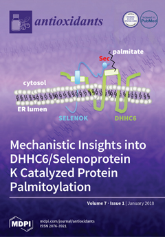
We have previously shown that the DHHC6 enzyme binds to selenoprotein K (SELENOK) in the endoplasmic reticulum membrane to catalyze the post-translational palmitoylation of several proteins. Herein, we reveal that the selenocysteine residue in SELENOK (Sec92) functions to stabilize the palmitoylated DHHC6 intermediate by protecting the thioester bond between the palmitate and cysteine residue in DHHC6 from hydrolysis. This allows transfer of the palmitate from DHHC6 to cysteine residues on target proteins, thereby increasing the catalytic efficiency of this reaction. View the paper here.
  • Issues are regarded as officially published after their release is announced to the table of contents alert mailing list.
  • You may sign up for e-mail alerts to receive table of contents of newly released issues.
  • PDF is the official format for papers published in both, html and pdf forms. To view the papers in pdf format, click on the "PDF Full-text" link, and use the free Adobe Reader to open them.
Order results
Result details
Section
Select all
Export citation of selected articles as:
21 pages, 5903 KiB  
Article
The Addition of Manganese Porphyrins during Radiation Inhibits Prostate Cancer Growth and Simultaneously Protects Normal Prostate Tissue from Radiation Damage
by Arpita Chatterjee, Yuxiang Zhu, Qiang Tong, Elizabeth A. Kosmacek, Eliezer Z. Lichter and Rebecca E. Oberley-Deegan
Antioxidants 2018, 7(1), 21; https://doi.org/10.3390/antiox7010021 - 25 Jan 2018
Cited by 33 | Viewed by 6621
Abstract
Radiation therapy is commonly used for prostate cancer treatment; however, normal tissues can be damaged from the reactive oxygen species (ROS) produced by radiation. In separate reports, we and others have shown that manganese porphyrins (MnPs), ROS scavengers, protect normal cells from radiation-induced [...] Read more.
Radiation therapy is commonly used for prostate cancer treatment; however, normal tissues can be damaged from the reactive oxygen species (ROS) produced by radiation. In separate reports, we and others have shown that manganese porphyrins (MnPs), ROS scavengers, protect normal cells from radiation-induced damage but inhibit prostate cancer cell growth. However, there have been no studies demonstrating that MnPs protect normal tissues, while inhibiting tumor growth in the same model. LNCaP or PC3 cells were orthotopically implanted into athymic mice and treated with radiation (2 Gy, for 5 consecutive days) in the presence or absence of MnPs. With radiation, MnPs enhanced overall life expectancy and significantly decreased the average tumor volume, as compared to the radiated alone group. MnPs enhanced lipid oxidation in tumor cells but reduced oxidative damage to normal prostate tissue adjacent to the prostate tumor in combination with radiation. Mechanistically, MnPs behave as pro-oxidants or antioxidants depending on the level of oxidative stress inside the treated cell. We found that MnPs act as pro-oxidants in prostate cancer cells, while in normal cells and tissues the MnPs act as antioxidants. For the first time, in the same in vivo model, this study reveals that MnPs enhance the tumoricidal effect of radiation and reduce oxidative damage to normal prostate tissue adjacent to the prostate tumor in the presence of radiation. This study suggests that MnPs are effective radio-protectors for radiation-mediated prostate cancer treatment. Full article
(This article belongs to the Special Issue Superoxide Dismutase (SOD) Enzymes, Mimetics and Oxygen Radicals)
Show Figures

Figure 1

14 pages, 414 KiB  
Article
Polyphenolic Compounds Analysis of Old and New Apple Cultivars and Contribution of Polyphenolic Profile to the In Vitro Antioxidant Capacity
by Josephine Kschonsek, Theresa Wolfram, Annette Stöckl and Volker Böhm
Antioxidants 2018, 7(1), 20; https://doi.org/10.3390/antiox7010020 - 24 Jan 2018
Cited by 135 | Viewed by 9182
Abstract
Polyphenols are antioxidant ingredients in apples and are related to human health because of their free radical scavenging activities. The polyphenolic profiles of old and new apple cultivars (n = 15) were analysed using high-performance liquid chromatography (HPLC) with diode array detection [...] Read more.
Polyphenols are antioxidant ingredients in apples and are related to human health because of their free radical scavenging activities. The polyphenolic profiles of old and new apple cultivars (n = 15) were analysed using high-performance liquid chromatography (HPLC) with diode array detection (DAD). The in vitro antioxidant capacity was determined by total phenolic content (TPC) assay, hydrophilic trolox equivalent antioxidant capacity (H-TEAC) assay and hydrophilic oxygen radical absorbance (H-ORAC) assay. Twenty polyphenolic compounds were identified in all investigated apples by HPLC analysis. Quercetin glycosides (203 ± 108 mg/100 g) were the main polyphenols in the peel and phenolic acids (10 ± 5 mg/100 g) in the flesh. The calculated relative contribution of single compounds indicated flavonols (peel) and vitamin C (flesh) as the major contributors to the antioxidant capacity, in all cultivars investigated. The polyphenolic content (HPLC data) of the flesh differed significantly between old (29 ± 7 mg/100 g) and new (13 ± 4 mg/100 g) cultivars, and the antioxidant capacity of old apple cultivars was up to 30% stronger compared to new ones. Full article
(This article belongs to the Special Issue Feature Papers in Antioxidants in 2018)
Show Figures

Figure 1

21 pages, 3511 KiB  
Article
Interplay between Selenium Levels and Replicative Senescence in WI-38 Human Fibroblasts: A Proteomic Approach
by Ghania Hammad, Yona Legrain, Zahia Touat-Hamici, Stéphane Duhieu, David Cornu, Anne-Laure Bulteau and Laurent Chavatte
Antioxidants 2018, 7(1), 19; https://doi.org/10.3390/antiox7010019 - 20 Jan 2018
Cited by 21 | Viewed by 4956
Abstract
Selenoproteins are essential components of antioxidant defense, redox homeostasis, and cell signaling in mammals, where selenium is found in the form of a rare amino acid, selenocysteine. Selenium, which is often limited both in food intake and cell culture media, is a strong [...] Read more.
Selenoproteins are essential components of antioxidant defense, redox homeostasis, and cell signaling in mammals, where selenium is found in the form of a rare amino acid, selenocysteine. Selenium, which is often limited both in food intake and cell culture media, is a strong regulator of selenoprotein expression and selenoenzyme activity. Aging is a slow, complex, and multifactorial process, resulting in a gradual and irreversible decline of various functions of the body. Several cellular aspects of organismal aging are recapitulated in the replicative senescence of cultured human diploid fibroblasts, such as embryonic lung fibroblast WI-38 cells. We previously reported that the long-term growth of young WI-38 cells with high (supplemented), moderate (control), or low (depleted) concentrations of selenium in the culture medium impacts their replicative lifespan, due to rapid changes in replicative senescence-associated markers and signaling pathways. In order to gain insight into the molecular link between selenium levels and replicative senescence, in the present work, we have applied a quantitative proteomic approach based on 2-Dimensional Differential in-Gel Electrophoresis (2D-DIGE) to the study of young and presenescent cells grown in selenium-supplemented, control, or depleted media. Applying a restrictive cut-off (spot intensity ±50% and a p value < 0.05) to the 2D-DIGE analyses revealed 81 differentially expressed protein spots, from which 123 proteins of interest were identified by mass spectrometry. We compared the changes in protein abundance for three different conditions: (i) spots varying between young and presenescent cells, (ii) spots varying in response to selenium concentration in young cells, and (iii) spots varying in response to selenium concentration in presenescent cells. Interestingly, a 72% overlap between the impact of senescence and selenium was observed in our proteomic results, demonstrating a strong interplay between selenium, selenoproteins, and replicative senescence. Full article
(This article belongs to the Special Issue Selenium and Selenoproteins for Optimal Health)
Show Figures

Graphical abstract

13 pages, 1491 KiB  
Article
Superoxide Dismutase Mimetic GC4419 Enhances the Oxidation of Pharmacological Ascorbate and Its Anticancer Effects in an H2O2-Dependent Manner
by Collin D. Heer, Andrew B. Davis, David B. Riffe, Brett A. Wagner, Kelly C. Falls, Bryan G. Allen, Garry R. Buettner, Robert A. Beardsley, Dennis P. Riley and Douglas R. Spitz
Antioxidants 2018, 7(1), 18; https://doi.org/10.3390/antiox7010018 - 19 Jan 2018
Cited by 31 | Viewed by 7196
Abstract
Lung cancer, together with head and neck cancer, accounts for more than one-fourth of cancer deaths worldwide. New, non-toxic therapeutic approaches are needed. High-dose IV vitamin C (aka, pharmacological ascorbate; P-AscH) represents a promising adjuvant to radiochemotherapy that exerts its anti-cancer [...] Read more.
Lung cancer, together with head and neck cancer, accounts for more than one-fourth of cancer deaths worldwide. New, non-toxic therapeutic approaches are needed. High-dose IV vitamin C (aka, pharmacological ascorbate; P-AscH) represents a promising adjuvant to radiochemotherapy that exerts its anti-cancer effects via metal-catalyzed oxidation to form H2O2. Mn(III)-porphyrins possessing superoxide dismutase (SOD) mimetic activity have been shown to increase the rate of oxidation of AscH, enhancing the anti-tumor effects of AscH in several cancer types. The current study demonstrates that the Mn(II)-containing pentaazamacrocyclic selective SOD mimetic GC4419 may serve as an AscH/O2•− oxidoreductase as evidenced by the increased rate of oxygen consumption, steady-state concentrations of ascorbate radical, and H2O2 production in complete cell culture media. GC4419, but not CuZnSOD, was shown to significantly enhance the toxicity of AscH in H1299, SCC25, SQ20B, and Cal27 cancer cell lines. This enhanced cancer cell killing was dependent upon the catalytic activity of the SOD mimetic and the generation of H2O2, as determined using conditional overexpression of catalase in H1299T cells. GC4419 combined with AscH was also capable of enhancing radiation-induced cancer cell killing. Currently, AscH and GC4419 are each being tested separately in clinical trials in combination with radiation therapy. Data presented here support the hypothesis that the combination of GC4419 and AscH may provide an effective means by which to further enhance radiation therapy responses. Full article
(This article belongs to the Special Issue Superoxide Dismutase (SOD) Enzymes, Mimetics and Oxygen Radicals)
Show Figures

Figure 1

7 pages, 1046 KiB  
Perspective
Cytokine Response to Exercise and Its Modulation
by Katsuhiko Suzuki
Antioxidants 2018, 7(1), 17; https://doi.org/10.3390/antiox7010017 - 17 Jan 2018
Cited by 96 | Viewed by 11979
Abstract
Strenuous exercise induces such inflammatory responses as leukocytosis (neutrophilia) and symptoms as delayed-onset muscle soreness and swelling. However, the association between inflammatory mediator cytokines and oxidative stress is not fully delineated. Herein, in addition to basic background information on cytokines, research findings on [...] Read more.
Strenuous exercise induces such inflammatory responses as leukocytosis (neutrophilia) and symptoms as delayed-onset muscle soreness and swelling. However, the association between inflammatory mediator cytokines and oxidative stress is not fully delineated. Herein, in addition to basic background information on cytokines, research findings on exertional effects on cytokine release and the underlying mechanisms and triggers are introduced. Then, the associations among cytokine responses, oxidative stress, and tissue damage are described not only in overloaded skeletal muscle, but also in other internal organs. Furthermore, we introduce preventive countermeasures against the exhaustive exercise-induced pathogenesis together with the possibility of antioxidant interventions. Full article
(This article belongs to the Special Issue Exercise and Inflammation)
Show Figures

Figure 1

18 pages, 816 KiB  
Review
Metabolic Alterations in Cancer Cells and the Emerging Role of Oncometabolites as Drivers of Neoplastic Change
by Zhengqiu Zhou, Elochukwu Ibekwe and Yevgen Chornenkyy
Antioxidants 2018, 7(1), 16; https://doi.org/10.3390/antiox7010016 - 17 Jan 2018
Cited by 29 | Viewed by 9190
Abstract
The mitochondrion is an important organelle and provides energy for a plethora of intracellular reactions. Metabolic dysregulation has dire consequences for the cell, and alteration in metabolism has been identified in multiple disease states—cancer being one. Otto Warburg demonstrated that cancer cells, in [...] Read more.
The mitochondrion is an important organelle and provides energy for a plethora of intracellular reactions. Metabolic dysregulation has dire consequences for the cell, and alteration in metabolism has been identified in multiple disease states—cancer being one. Otto Warburg demonstrated that cancer cells, in the presence of oxygen, undergo glycolysis by reprogramming their metabolism—termed “aerobic glycolysis”. Alterations in metabolism enable cancer cells to gain a growth advantage by obtaining precursors for macromolecule biosynthesis, such as nucleic acids and lipids. To date, several molecules, termed “oncometabolites”, have been identified to be elevated in cancer cells and arise from mutations in nuclear encoded mitochondrial enzymes. Furthermore, there is evidence that oncometabolites can affect mitochondrial dynamics. It is believed that oncometabolites can assist in reprogramming enzymatic pathways and providing cancer cells with selective advantages. In this review, we will touch upon the effects of normal and aberrant mitochondrial metabolism in normal and cancer cells, the advantages of metabolic reprogramming, effects of oncometabolites on metabolism and mitochondrial dynamics and therapies aimed at targeting oncometabolites and metabolic aberrations. Full article
(This article belongs to the Special Issue Mitochondrial Shape Change in Physio-Pathology)
Show Figures

Figure 1

15 pages, 994 KiB  
Review
Regulation of Mitochondrial Dynamics by Proteolytic Processing and Protein Turnover
by Sumaira Ali and Gavin P. McStay
Antioxidants 2018, 7(1), 15; https://doi.org/10.3390/antiox7010015 - 17 Jan 2018
Cited by 19 | Viewed by 5691
Abstract
The mitochondrial network is a dynamic organization within eukaryotic cells that participates in a variety of essential cellular processes, such as adenosine triphosphate (ATP) synthesis, central metabolism, apoptosis and inflammation. The mitochondrial network is balanced between rates of fusion and fission that respond [...] Read more.
The mitochondrial network is a dynamic organization within eukaryotic cells that participates in a variety of essential cellular processes, such as adenosine triphosphate (ATP) synthesis, central metabolism, apoptosis and inflammation. The mitochondrial network is balanced between rates of fusion and fission that respond to pathophysiologic signals to coordinate appropriate mitochondrial processes. Mitochondrial fusion and fission are regulated by proteins that either reside in or translocate to the inner or outer mitochondrial membranes or are soluble in the inter-membrane space. Mitochondrial fission and fusion are performed by guanosine triphosphatases (GTPases) on the outer and inner mitochondrial membranes with the assistance of other mitochondrial proteins. Due to the essential nature of mitochondrial function for cellular homeostasis, regulation of mitochondrial dynamics is under strict control. Some of the mechanisms used to regulate the function of these proteins are post-translational proteolysis and/or turnover, and this review will discuss these mechanisms required for correct mitochondrial network organization. Full article
(This article belongs to the Special Issue Mitochondrial Shape Change in Physio-Pathology)
Show Figures

Figure 1

16 pages, 3604 KiB  
Review
Subcellular Reactive Oxygen Species (ROS) in Cardiovascular Pathophysiology
by Sarah Aldosari, Maan Awad, Elizabeth O. Harrington, Frank W. Sellke and M. Ruhul Abid
Antioxidants 2018, 7(1), 14; https://doi.org/10.3390/antiox7010014 - 16 Jan 2018
Cited by 75 | Viewed by 12558
Abstract
There exist two opposing perspectives regarding reactive oxygen species (ROS) and their roles in angiogenesis and cardiovascular system, one that favors harmful and causal effects of ROS, while the other supports beneficial effects. Recent studies have shown that interaction between ROS in different [...] Read more.
There exist two opposing perspectives regarding reactive oxygen species (ROS) and their roles in angiogenesis and cardiovascular system, one that favors harmful and causal effects of ROS, while the other supports beneficial effects. Recent studies have shown that interaction between ROS in different sub-cellular compartments plays a crucial role in determining the outcomes (beneficial vs. deleterious) of ROS exposures on the vascular system. Oxidant radicals in one cellular organelle can affect the ROS content and function in other sub-cellular compartments in endothelial cells (ECs). In this review, we will focus on a critical fact that the effects or the final phenotypic outcome of ROS exposure to EC are tissue- or organ-specific, and depend on the spatial (subcellular localization) and temporal (duration of ROS exposure) modulation of ROS levels. Full article
(This article belongs to the Special Issue ROS Derived from NADPH Oxidase (NOX) in Angiogenesis)
Show Figures

Figure 1

24 pages, 1168 KiB  
Review
Reactive Oxygen Species and Mitochondrial Dynamics: The Yin and Yang of Mitochondrial Dysfunction and Cancer Progression
by Jan Ježek, Katrina F. Cooper and Randy Strich
Antioxidants 2018, 7(1), 13; https://doi.org/10.3390/antiox7010013 - 16 Jan 2018
Cited by 329 | Viewed by 16529
Abstract
Mitochondria are organelles with a highly dynamic ultrastructure maintained by a delicate equilibrium between its fission and fusion rates. Understanding the factors influencing this balance is important as perturbations to mitochondrial dynamics can result in pathological states. As a terminal site of nutrient [...] Read more.
Mitochondria are organelles with a highly dynamic ultrastructure maintained by a delicate equilibrium between its fission and fusion rates. Understanding the factors influencing this balance is important as perturbations to mitochondrial dynamics can result in pathological states. As a terminal site of nutrient oxidation for the cell, mitochondrial powerhouses harness energy in the form of ATP in a process driven by the electron transport chain. Contemporaneously, electrons translocated within the electron transport chain undergo spontaneous side reactions with oxygen, giving rise to superoxide and a variety of other downstream reactive oxygen species (ROS). Mitochondrially-derived ROS can mediate redox signaling or, in excess, cause cell injury and even cell death. Recent evidence suggests that mitochondrial ultrastructure is tightly coupled to ROS generation depending on the physiological status of the cell. Yet, the mechanism by which changes in mitochondrial shape modulate mitochondrial function and redox homeostasis is less clear. Aberrant mitochondrial morphology may lead to enhanced ROS formation, which, in turn, may deteriorate mitochondrial health and further exacerbate oxidative stress in a self-perpetuating vicious cycle. Here, we review the latest findings on the intricate relationship between mitochondrial dynamics and ROS production, focusing mainly on its role in malignant disease. Full article
(This article belongs to the Special Issue Mitochondrial Shape Change in Physio-Pathology)
Show Figures

Figure 1

13 pages, 1164 KiB  
Review
Vitamin E as a Treatment for Nonalcoholic Fatty Liver Disease: Reality or Myth?
by Hamza El Hadi, Roberto Vettor and Marco Rossato
Antioxidants 2018, 7(1), 12; https://doi.org/10.3390/antiox7010012 - 16 Jan 2018
Cited by 94 | Viewed by 16351
Abstract
Obesity is one of the major epidemics of this millennium, and its incidence is growing worldwide. Following the epidemics of obesity, nonalcoholic fatty liver disease (NAFLD) has become a disease of increasing prevalence and a leading cause of morbidity and mortality closely related [...] Read more.
Obesity is one of the major epidemics of this millennium, and its incidence is growing worldwide. Following the epidemics of obesity, nonalcoholic fatty liver disease (NAFLD) has become a disease of increasing prevalence and a leading cause of morbidity and mortality closely related to cardiovascular disease, malignancies, and cirrhosis. It is believed that oxidative stress is a main player in the development and progression of NAFLD. Currently, a pharmacological approach has become necessary in NAFLD because of a failure to modify lifestyle and dietary habits in most patients. Vitamin E is a potent antioxidant that has been shown to reduce oxidative stress in NAFLD. This review summarizes the biological activities of vitamin E, with a primary focus on its therapeutic efficacy in NAFLD. Full article
(This article belongs to the Special Issue Vitamin E)
Show Figures

Figure 1

10 pages, 1068 KiB  
Article
Effect of the Extraction Process on the Biological Activity of Lyophilized Apricot Extracts Recovered from Apricot Pomace
by Dina Cheaib, Nada El Darra, Hiba N. Rajha, Iman El-Ghazzawi, Richard G. Maroun and Nicolas Louka
Antioxidants 2018, 7(1), 11; https://doi.org/10.3390/antiox7010011 - 14 Jan 2018
Cited by 18 | Viewed by 7273
Abstract
The preservation of polyphenols in fruits by lyophilization has gained great interest in the recent decades. The present study aims to assess the impact of the pre-treatment extraction methods heat-assisted extraction (HAE) and infrared (IR) on lyophilized apricot pomace extracts. Then to test [...] Read more.
The preservation of polyphenols in fruits by lyophilization has gained great interest in the recent decades. The present study aims to assess the impact of the pre-treatment extraction methods heat-assisted extraction (HAE) and infrared (IR) on lyophilized apricot pomace extracts. Then to test the conservation of polyphenols quantities as well as their bioactivities (antiradical and antibacterial) in lyophilized extract. An aqueous extract was obtained through either heat-assisted extraction or infrared pre-treatments then lyophilized to obtain a dried form. Results showed that the content of polyphenols, the antiradical and antibacterial activities in lyophilized extracts exhibited a slighter decrease in infrared sample compared to the heat-assisted extraction ones. The High-performance liquid chromatography (HPLC) analysis showed that lyophilized extracts IR and HAE preserved the same phenolic molecules (rutin, catechin and epicatechin) detected in liquid extracts (IR and HAE) with a smaller yield. Lyophilization can be used as a widely process in the food industry to conserve many bioactive molecules. Full article
Show Figures

Figure 1

3 pages, 157 KiB  
Editorial
Dietary Antioxidants and Health Promotion
by Dejian Huang
Antioxidants 2018, 7(1), 9; https://doi.org/10.3390/antiox7010009 - 12 Jan 2018
Cited by 55 | Viewed by 8494
Abstract
Accumulating scientific evidence suggests that over-production of reactive oxygen species (ROS) may be the root cause of chronic diseases such as cancer, cardiovascular diseases, neurodegeneration, and ageing per se [1,2].[...] Full article
(This article belongs to the Special Issue Dietary Antioxidants and Health Promotion)
20 pages, 1894 KiB  
Review
Long-Chain Metabolites of Vitamin E: Metabolic Activation as a General Concept for Lipid-Soluble Vitamins?
by Martin Schubert, Stefan Kluge, Lisa Schmölz, Maria Wallert, Francesco Galli, Marc Birringer and Stefan Lorkowski
Antioxidants 2018, 7(1), 10; https://doi.org/10.3390/antiox7010010 - 12 Jan 2018
Cited by 46 | Viewed by 7984
Abstract
Vitamins E, A, D and K comprise the class of lipid-soluble vitamins. For vitamins A and D, a metabolic conversion of precursors to active metabolites has already been described. During the metabolism of vitamin E, the long-chain metabolites (LCMs) 13′-hydroxychromanol (13′-OH) and 13′-carboxychromanol [...] Read more.
Vitamins E, A, D and K comprise the class of lipid-soluble vitamins. For vitamins A and D, a metabolic conversion of precursors to active metabolites has already been described. During the metabolism of vitamin E, the long-chain metabolites (LCMs) 13′-hydroxychromanol (13′-OH) and 13′-carboxychromanol (13′-COOH) are formed by oxidative modification of the side-chain. The occurrence of these metabolites in human serum indicates a physiological relevance. Indeed, effects of the LCMs on lipid metabolism, apoptosis, proliferation and inflammatory actions as well as tocopherol and xenobiotic metabolism have been shown. Interestingly, there are several parallels between the actions of the LCMs of vitamin E and the active metabolites of vitamin A and D. The recent findings that the LCMs exert effects different from that of their precursors support their putative role as regulatory metabolites. Hence, it could be proposed that the mode of action of the LCMs might be mediated by a mechanism similar to vitamin A and D metabolites. If the physiological relevance and this concept of action of the LCMs can be confirmed, a general concept of activation of lipid-soluble vitamins via their metabolites might be deduced. Full article
(This article belongs to the Special Issue Vitamin E)
Show Figures

Figure 1

3 pages, 170 KiB  
Editorial
Acknowledgement to Reviewers of Antioxidants in 2017
by Antioxidants Editorial Office
Antioxidants 2018, 7(1), 8; https://doi.org/10.3390/antiox7010008 - 10 Jan 2018
Viewed by 2523
Abstract
Peer review is an essential part in the publication process, ensuring that Antioxidants maintains high quality standards for its published papers.[...] Full article
21 pages, 1542 KiB  
Review
Exercise and Mitochondrial Dynamics: Keeping in Shape with ROS and AMPK
by Adam J. Trewin, Brandon J. Berry and Andrew P. Wojtovich
Antioxidants 2018, 7(1), 7; https://doi.org/10.3390/antiox7010007 - 06 Jan 2018
Cited by 90 | Viewed by 14037
Abstract
Exercise is a robust stimulus for mitochondrial adaptations in skeletal muscle which consequently plays a central role in enhancing metabolic health. Despite this, the precise molecular events that underpin these beneficial effects remain elusive. In this review, we discuss molecular signals generated during [...] Read more.
Exercise is a robust stimulus for mitochondrial adaptations in skeletal muscle which consequently plays a central role in enhancing metabolic health. Despite this, the precise molecular events that underpin these beneficial effects remain elusive. In this review, we discuss molecular signals generated during exercise leading to altered mitochondrial morphology and dynamics. In particular, we focus on the interdependence between reactive oxygen species (ROS) and redox homeostasis, the sensing of cellular bioenergetic status via 5’ adenosine monophosphate (AMP)-activated protein kinase (AMPK), and the regulation of mitochondrial fission and fusion. Precisely how exercise regulates the network of these responses and their effects on mitochondrial dynamics is not fully understood at present. We highlight the limitations that exist with the techniques currently available, and discuss novel molecular tools to potentially advance the fields of redox biology and mitochondrial bioenergetics. Ultimately, a greater understanding of these processes may lead to novel mitochondria-targeted therapeutic strategies to augment or mimic exercise in order to attenuate or reverse pathophysiology. Full article
(This article belongs to the Special Issue Mitochondrial Shape Change in Physio-Pathology)
Show Figures

Figure 1

9 pages, 236 KiB  
Review
Genotypic and Environmental Effects on Tocopherol Content in Almond
by Ossama Kodad, Rafel Socias i Company and José M. Alonso
Antioxidants 2018, 7(1), 6; https://doi.org/10.3390/antiox7010006 - 05 Jan 2018
Cited by 38 | Viewed by 6510
Abstract
Almond is the most important nut species worldwide and almond kernels show the highest levels of tocopherols among all nuts. In almond, tocopherols not only play a substantial role as a healthy food for human consumption, but also in protecting lipids against oxidation [...] Read more.
Almond is the most important nut species worldwide and almond kernels show the highest levels of tocopherols among all nuts. In almond, tocopherols not only play a substantial role as a healthy food for human consumption, but also in protecting lipids against oxidation and, thus, lengthening the storage time of almond kernels. The main tocopherol homologues detected in almond in decreasing content and biological importance are α-, γ-, δ-, and β-tocopherol. Tocopherol concentration in almond depends on the genotype and the environment, such as the climatic conditions of the year and the growing management of the orchard. The range of variability for the different tocopherol homologues is of 335–657 mg/kg of almond oil for α-, 2–50 for γ-, and 0.1–22 for β-tocopherol. Drought and heat have been the most important stresses affecting tocopherol content in almond, with increased levels at higher temperatures and in water deficit conditions. The right cultivar and the most appropriate growing conditions may be selected to obtain crops with effective kernel storage and for the most beneficial effects of almond consumption for human nutrition and health. Full article
(This article belongs to the Special Issue Vitamin E)
16 pages, 747 KiB  
Review
Singlet Oxygen and Free Radical Reactions of Retinoids and Carotenoids—A Review
by Ruth Edge and T. George Truscott
Antioxidants 2018, 7(1), 5; https://doi.org/10.3390/antiox7010005 - 01 Jan 2018
Cited by 107 | Viewed by 10538
Abstract
We report on studies of reactions of singlet oxygen with carotenoids and retinoids and a range of free radical studies on carotenoids and retinoids with emphasis on recent work, dietary carotenoids and the role of oxygen in biological processes. Many previous reviews are [...] Read more.
We report on studies of reactions of singlet oxygen with carotenoids and retinoids and a range of free radical studies on carotenoids and retinoids with emphasis on recent work, dietary carotenoids and the role of oxygen in biological processes. Many previous reviews are cited and updated together with new data not previously reviewed. The review does not deal with computational studies but the emphasis is on laboratory-based results. We contrast the ease of study of both singlet oxygen and polyene radical cations compared to neutral radicals. Of particular interest is the switch from anti- to pro-oxidant behavior of a carotenoid with change of oxygen concentration: results for lycopene in a cellular model system show total protection of the human cells studied at zero oxygen concentration, but zero protection at 100% oxygen concentration. Full article
(This article belongs to the Special Issue Carotenoids—Antioxidant Properties)
Show Figures

Figure 1

3198 KiB  
Article
Selenoprotein K Increases Efficiency of DHHC6 Catalyzed Protein Palmitoylation by Stabilizing the Acyl-DHHC6 Intermediate
by Gregory J. Fredericks, FuKun W. Hoffmann, Robert J. Hondal, Sharon Rozovsky, Johann Urschitz and Peter R. Hoffmann
Antioxidants 2018, 7(1), 4; https://doi.org/10.3390/antiox7010004 - 29 Dec 2017
Cited by 23 | Viewed by 5201
Abstract
Selenoprotein K (SELENOK) is a selenocysteine (Sec)-containing protein localized in the endoplasmic reticulum (ER) membrane where it interacts with the DHHC6 (where single letter symbols represent Asp-His-His-Cys amino acids) enzyme to promote protein acyl transferase (PAT) reactions. PAT reactions involve the DHHC enzymatic [...] Read more.
Selenoprotein K (SELENOK) is a selenocysteine (Sec)-containing protein localized in the endoplasmic reticulum (ER) membrane where it interacts with the DHHC6 (where single letter symbols represent Asp-His-His-Cys amino acids) enzyme to promote protein acyl transferase (PAT) reactions. PAT reactions involve the DHHC enzymatic capture of palmitate via a thioester bond to cysteine (Cys) residues that form an unstable palmitoyl-DHHC intermediate, followed by transfer of palmitate to Cys residues of target proteins. How SELENOK facilitates this reaction has not been determined. Splenocyte microsomal preparations from wild-type mice versus SELENOK knockout mice were used to establish PAT assays and showed decreased PAT activity (~50%) under conditions of SELENOK deficiency. Using recombinant, soluble versions of DHHC6 along with SELENOK containing Sec92, Cys92, or alanine (Ala92), we evaluated the stability of the acyl-DHHC6 intermediate and its capacity to transfer the palmitate residue to Cys residues on target peptides. Versions of SELENOK containing either Ala or Cys residues in place of Sec were equivalently less effective than Sec at stabilizing the acyl-DHHC6 intermediate or promoting PAT activity. These data suggest that Sec92 in SELENOK serves to stabilize the palmitoyl-DHHC6 intermediate by reducing hydrolyzation of the thioester bond until transfer of the palmitoyl group to the Cys residue on the target protein can occur. Full article
(This article belongs to the Special Issue Selenium and Selenoproteins for Optimal Health)
Show Figures

Graphical abstract

7311 KiB  
Review
Natural Nanoparticles: A Particular Matter Inspired by Nature
by Sharoon Griffin, Muhammad Irfan Masood, Muhammad Jawad Nasim, Muhammad Sarfraz, Azubuike Peter Ebokaiwe, Karl-Herbert Schäfer, Cornelia M. Keck and Claus Jacob
Antioxidants 2018, 7(1), 3; https://doi.org/10.3390/antiox7010003 - 29 Dec 2017
Cited by 146 | Viewed by 23024
Abstract
During the last couple of decades, the rapidly advancing field of nanotechnology has produced a wide palette of nanomaterials, most of which are considered as “synthetic” and, among the wider public, are often met with a certain suspicion. Despite the technological sophistication behind [...] Read more.
During the last couple of decades, the rapidly advancing field of nanotechnology has produced a wide palette of nanomaterials, most of which are considered as “synthetic” and, among the wider public, are often met with a certain suspicion. Despite the technological sophistication behind many of these materials, “nano” does not always equate with “artificial”. Indeed, nature itself is an excellent nanotechnologist. It provides us with a range of fine particles, from inorganic ash, soot, sulfur and mineral particles found in the air or in wells, to sulfur and selenium nanoparticles produced by many bacteria and yeasts. These nanomaterials are entirely natural, and, not surprisingly, there is a growing interest in the development of natural nanoproducts, for instance in the emerging fields of phyto- and phyco-nanotechnology. This review will highlight some of the most recent—and sometimes unexpected—advances in this exciting and diverse field of research and development. Naturally occurring nanomaterials, artificially produced nanomaterials of natural products as well as naturally occurring or produced nanomaterials of natural products all show their own, particular chemical and physical properties, biological activities and promise for applications, especially in the fields of medicine, nutrition, cosmetics and agriculture. In the future, such natural nanoparticles will not only stimulate research and add a greener outlook to a traditionally high-tech field, they will also provide solutions—pardon—suspensions for a range of problems. Here, we may anticipate specific biogenic factories, valuable new materials based on waste, the effective removal of contaminants as part of nano-bioremediation, and the conversion of poorly soluble substances and materials to biologically available forms for practical uses. Full article
(This article belongs to the Special Issue Inspired by Nature: Antioxidants and Nanotechnology)
Show Figures

Graphical abstract

3720 KiB  
Review
Vitamin E Biosynthesis and Its Regulation in Plants
by Laurent Mène-Saffrané
Antioxidants 2018, 7(1), 2; https://doi.org/10.3390/antiox7010002 - 25 Dec 2017
Cited by 93 | Viewed by 14140
Abstract
Vitamin E is one of the 13 vitamins that are essential to animals that do not produce them. To date, six natural organic compounds belonging to the chemical family of tocochromanols—four tocopherols and two tocotrienols—have been demonstrated as exhibiting vitamin E activity in [...] Read more.
Vitamin E is one of the 13 vitamins that are essential to animals that do not produce them. To date, six natural organic compounds belonging to the chemical family of tocochromanols—four tocopherols and two tocotrienols—have been demonstrated as exhibiting vitamin E activity in animals. Edible plant-derived products, notably seed oils, are the main sources of vitamin E in the human diet. Although this vitamin is readily available, independent nutritional surveys have shown that human populations do not consume enough vitamin E, and suffer from mild to severe deficiency. Tocochromanols are mostly produced by plants, algae, and some cyanobacteria. Tocochromanol metabolism has been mainly studied in higher plants that produce tocopherols, tocotrienols, plastochromanol-8, and tocomonoenols. In contrast to the tocochromanol biosynthetic pathways that are well characterized, our understanding of the physiological and molecular mechanisms regulating tocochromanol biosynthesis is in its infancy. Although it is known that tocochromanol biosynthesis is strongly conditioned by the availability in homogentisate and polyprenyl pyrophosphate, its polar and lipophilic biosynthetic precursors, respectively, the mechanisms regulating their biosyntheses are barely known. This review summarizes our current knowledge of tocochromanol biosynthesis in plants, and highlights future challenges regarding the understanding of its regulation. Full article
(This article belongs to the Special Issue Vitamin E)
Show Figures

Figure 1

1825 KiB  
Article
Biological Implications of Differential Expression of Mitochondrial-Shaping Proteins in Parkinson’s Disease
by Sara Rocha, Ana Freitas, Sofia C. Guimaraes, Rui Vitorino, Miguel Aroso and Maria Gomez-Lazaro
Antioxidants 2018, 7(1), 1; https://doi.org/10.3390/antiox7010001 - 21 Dec 2017
Cited by 14 | Viewed by 6655
Abstract
It has long been accepted that mitochondrial function and morphology is affected in Parkinson’s disease, and that mitochondrial function can be directly related to its morphology. So far, mitochondrial morphological alterations studies, in the context of this neurodegenerative disease, have been performed through [...] Read more.
It has long been accepted that mitochondrial function and morphology is affected in Parkinson’s disease, and that mitochondrial function can be directly related to its morphology. So far, mitochondrial morphological alterations studies, in the context of this neurodegenerative disease, have been performed through microscopic methodologies. The goal of the present work is to address if the modifications in the mitochondrial-shaping proteins occurring in this disorder have implications in other cellular pathways, which might constitute important pathways for the disease progression. To do so, we conducted a novel approach through a thorough exploration of the available proteomics-based studies in the context of Parkinson’s disease. The analysis provided insight into the altered biological pathways affected by changes in the expression of mitochondrial-shaping proteins via different bioinformatic tools. Unexpectedly, we observed that the mitochondrial-shaping proteins altered in the context of Parkinson’s disease are, in the vast majority, related to the organization of the mitochondrial cristae. Conversely, in the studies that have resorted to microscopy-based techniques, the most widely reported alteration in the context of this disorder is mitochondria fragmentation. Cristae membrane organization is pivotal for mitochondrial ATP production, and changes in their morphology have a direct impact on the organization and function of the oxidative phosphorylation (OXPHOS) complexes. To understand which biological processes are affected by the alteration of these proteins we analyzed the binding partners of the mitochondrial-shaping proteins that were found altered in Parkinson’s disease. We showed that the binding partners fall into seven different cellular components, which include mitochondria, proteasome, and endoplasmic reticulum (ER), amongst others. It is noteworthy that, by evaluating the biological process in which these modified proteins are involved, we showed that they are related to the production and metabolism of ATP, immune response, cytoskeleton alteration, and oxidative stress, amongst others. In summary, with our bioinformatics approach using the data on the modified proteins in Parkinson’s disease patients, we were able to relate the alteration of mitochondrial-shaping proteins to modifications of crucial cellular pathways affected in this disease. Full article
(This article belongs to the Special Issue Mitochondrial Shape Change in Physio-Pathology)
Show Figures

Figure 1

Previous Issue
Next Issue
Back to TopTop