


Brain Sci. 2024, 14(5), 434; https://doi.org/10.3390/brainsci14050434 (registering DOI) - 26 Apr 2024
Abstract
Research indicates that brain-region-specific synapse loss and dysfunction are early hallmarks and stronger neurobiological correlates of cognitive decline in Alzheimer’s disease (AD) than amyloid plaque and neurofibrillary tangle counts or neuronal loss. Even though the precise mechanisms underlying increased synaptic pruning in AD
[...] Read more.
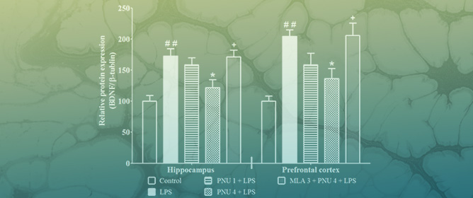
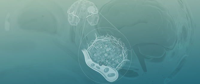
Research indicates that brain-region-specific synapse loss and dysfunction are early hallmarks and stronger neurobiological correlates of cognitive decline in Alzheimer’s disease (AD) than amyloid plaque and neurofibrillary tangle counts or neuronal loss. Even though the precise mechanisms underlying increased synaptic pruning in AD are still unknown, it has been confirmed that dysregulation of the balance between complement activation and inhibition is a crucial driver of its pathology. The complement includes three distinct activation mechanisms, with the activation products C3a and C5a, potent inflammatory effectors, and a membrane attack complex (MAC) leading to cell lysis. Besides pro-inflammatory cytokines, the dysregulated complement proteins released by activated microglia bind to amyloid β at the synaptic regions and cause the microglia to engulf the synapses. Additionally, research indicating that microglia-removed synapses are not always degenerating and that suppression of synaptic engulfment can repair cognitive deficits points to an essential opportunity for intervention that can prevent the loss of intact synapses. In this study, we focus on the latest research on the role and mechanisms of complement-mediated microglial synaptic pruning at different stages of AD to find the right targets that could interfere with complement dysregulation and be relevant for therapeutic intervention at the early stages of the disease.
Full article
(This article belongs to the Section Neurodegenerative Diseases)


