


Cancers, Volume 16, Issue 13 (July-1 2024) – 204 articles
Cover Story (view full-size image):
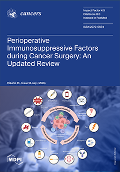
Surgical excision of primary tumors is the gold standard and best curative treatment for solid tumors. However, the manipulation of tumor releases circulating tumor cells and the inflammatory pain generated during procedures promotes an immunosuppressive stress environment. Moreover, several perioperative factors impair the physiological anticancer immune response, such as chemotherapy, undernutrition, anemia or addictions, thus compromising the benefits of oncological surgery. This review offers a comprehensive and explained list of immunosuppressive factors occurring in the perioperative period of cancer interventions and discusses pharmacological and non-pharmacological strategies to optimally stimulate the immune system with the aim of helping clinicians to improve cancer outcomes. View this paper
- Issues are regarded as officially published after their release is announced to the table of contents alert mailing list.
- You may sign up for e-mail alerts to receive table of contents of newly released issues.
- PDF is the official format for papers published in both, html and pdf forms. To view the papers in pdf format, click on the "PDF Full-text" link, and use the free Adobe Reader to open them.
Previous Issue
Next Issue