Next Issue
Volume 14, February-1
Previous Issue
Volume 14, January-1


Diagnostics, Volume 14, Issue 2 (January-2 2024) – 113 articles

Cover Story (view full-size image): 
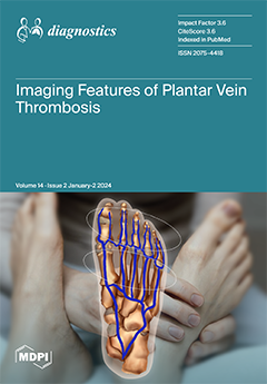
Plantar vein thrombosis is a venous disorder affecting deep plantar veins that can manifest with non-specific localized pain, plantar foot pain, swelling, and sensation of fullness. Plantar veins are not routinely assessed during sonographic scans for deep venous thrombosis, which makes plantar venous thrombosis a commonly missed diagnosis. This paper provides a comprehensive review of the venous anatomy of the foot and imaging findings of plantar venous thrombosis as well as discusses the current literature on the topic and its differential diagnoses. View this paper
  • Issues are regarded as officially published after their release is announced to the table of contents alert mailing list.
  • You may sign up for e-mail alerts to receive table of contents of newly released issues.
  • PDF is the official format for papers published in both, html and pdf forms. To view the papers in pdf format, click on the "PDF Full-text" link, and use the free Adobe Reader to open them.
Order results
Result details
Section
Select all
Export citation of selected articles as:
11 pages, 988 KiB  
Article
Use of [18F]Fluorodeoxyglucose Positron Emission Tomography/Computed Tomography after Curative Treatment of Non-Small-Cell Lung Cancer Patients: A Nationwide Cohort Study
by Kasper Foged Guldbrandsen, Liza Sopina, Torben Riis Rasmussen and Barbara Malene Fischer
Diagnostics 2024, 14(2), 233; https://doi.org/10.3390/diagnostics14020233 - 22 Jan 2024
Viewed by 833
Abstract
[¹⁸F]Fluorodeoxyglucose positron emission tomography/computed tomography ([¹⁸F]FDG PET/CT) is a valuable imaging tool in the post-treatment management of non-small-cell lung cancer (NSCLC). This study aimed to investigate the trends in utilization and factors associated with the use of [¹⁸F]FDG PET/CT after curative-intent treatment. Data [...] Read more.
[¹⁸F]Fluorodeoxyglucose positron emission tomography/computed tomography ([¹⁸F]FDG PET/CT) is a valuable imaging tool in the post-treatment management of non-small-cell lung cancer (NSCLC). This study aimed to investigate the trends in utilization and factors associated with the use of [¹⁸F]FDG PET/CT after curative-intent treatment. Data from 13,758 NSCLC patients diagnosed between 2007 and 2020 identified in the Danish Lung Cancer Registry, who underwent curative-intent treatment, were analyzed using multivariable regression. The results showed a significant increase in the use of [¹⁸F]FDG PET/CT scans, from 10.4 per 100 patients per year in 2007 to 39.6 in 2013, followed by a period of stability. Higher utilization rates were observed in patients who received radiotherapy (22% increase compared to surgical resection) and in patients with stage II–III disease (14% and 20% increase compared to stage I, respectively). Additionally, utilization was increased when other diagnostic procedures were performed, such as MRI, ultrasound, endoscopy, and biopsy. These findings highlight an increasing reliance on [¹⁸F]FDG PET/CT in post-treatment NSCLC, especially after radiotherapy and in patients with locally advanced disease, where treatment-induced radiographic changes and an increased risk of recurrence present a significant diagnostic challenge. Full article
(This article belongs to the Special Issue 18F-FDG PET/CT: Current and Future Clinical Applications)
Show Figures

Figure 1

10 pages, 1942 KiB  
Article
Home Haemoglobin Monitoring for the Titration of Erythropoietin-Stimulating Agents in Chronic Kidney Disease: A Pragmatic Pilot Trial
by Richard Bodington, Madeline Lok and Sunil Bhandari
Diagnostics 2024, 14(2), 232; https://doi.org/10.3390/diagnostics14020232 - 22 Jan 2024
Viewed by 716
Abstract
Background. No trials of POCT device pathways have been published in the field of anaemia of CKD. We describe the results of a year of use of a novel POCT device with its associated eHealth pathway in the home monitoring of ESA therapy, [...] Read more.
Background. No trials of POCT device pathways have been published in the field of anaemia of CKD. We describe the results of a year of use of a novel POCT device with its associated eHealth pathway in the home monitoring of ESA therapy, with the aim of evaluating device performance and pathway feasibility. Methods: We used a POCT device designed for home self-testing, able to measure Hb from a drop of capillary blood (Luma, Entia, UK). Results were shared with HCPs via an associated mobile application. The pilot ran from August 2020 to March 2022 in a single UK renal centre. All adult non-dialysis-dependent-CKD patients on ESAs were eligible for inclusion. Participants were mailed the device and trained remotely. Participants were encouraged to self-test twice weekly for up to 1 year, with data collected on a pragmatic basis. Lab and Luma’s results were compared. Results: Seventeen participants returned comparable datasets (underwent ≥ 4 lab Hb measurements and self-tested for >5 months) with a mean testing frequency of 1.6 tests/wk. 1062 Luma and 113 lab Hb results were analysed. The coefficient of variation (CV) for raw results was 8.3% with a bias of −2.0% and SD 8.5. The percentage of Luma results differing by >10% lab results was 30.9%, dropping to 17.7% using an 8-point-moving-average. Participants stated they preferred Luma to traditional ESA monitoring and recommended the pathway to others. Conclusion: One year of home self-testing with Luma yielded comparable device utility to other POCT haematology analysers derived via HCP testing. Innovative pilot trials such as this form the basis for new empowering and home-based models of care required and desired by patients and HCPs. Full article
(This article belongs to the Special Issue 21st Century Point-of-Care, Near-Patient and Critical Care Testing)
Show Figures

Figure 1

11 pages, 1431 KiB  
Article
Performance Evaluation of the STANDARD i-Q COVID-19 Ag Test with Nasal and Oral Swab Specimens from Symptomatic Patients
by Jong Do Seo, Hee-Won Moon, Eunju Shin, Ji Young Kim, Sang-Gyu Choi, Ju Ae Lee, Jeong Hwa Choi and Yeo-Min Yun
Diagnostics 2024, 14(2), 231; https://doi.org/10.3390/diagnostics14020231 - 22 Jan 2024
Viewed by 688
Abstract
We evaluated the diagnostic performance of the STANDARD i-Q COVID-19 Ag Test, which was developed to detect viral antigens, using nasal and oral swabs. Sixty positive and 100 negative samples were analyzed. We determined the distribution of the Ct values according to the [...] Read more.
We evaluated the diagnostic performance of the STANDARD i-Q COVID-19 Ag Test, which was developed to detect viral antigens, using nasal and oral swabs. Sixty positive and 100 negative samples were analyzed. We determined the distribution of the Ct values according to the day of sample collection after symptom onset, the diagnostic performance of the total samples and subgroups separated by Ct value or time of sample collection, and the Ct value at which maximal accuracy was expected. No differences were observed in Ct values, except for the samples obtained on the day of symptom onset. The diagnostic sensitivity and specificity of the oral swabs were 75.0 and 100.0%, respectively, whereas those of the nasal swabs were 85.0 and 98.0%, respectively. The sensitivity was higher in samples with a high viral load collected earlier than those collected later, although the difference was not significant. False-negative results were confirmed in all samples with a Ct value ≥ 30.0. These results indicate that tests using oral and nasal swabs are helpful for diagnosing acute symptomatic cases with suspected high viral loads. Our tests exhibited relatively low sensitivity but high specificity rates, indicating the need to assess negative antigen test results. Full article
(This article belongs to the Special Issue Point-of-Care Testing for Infectious Disease)
Show Figures

Figure 1

17 pages, 3455 KiB  
Article
Color Analysis of Merkel Cell Carcinoma: A Comparative Study with Cherry Angiomas, Hemangiomas, Basal Cell Carcinomas, and Squamous Cell Carcinomas
by Dimitra Koumaki, Georgios Manios, Marios Papadakis, Aikaterini Doxastaki, Georgios Vasileiou Zacharopoulos, Alexander Katoulis and Andreas Manios
Diagnostics 2024, 14(2), 230; https://doi.org/10.3390/diagnostics14020230 - 22 Jan 2024
Viewed by 1206
Abstract
Merkel cell carcinoma (MCC) is recognized as one of the most malignant skin tumors. Its rarity might explain the limited exploration of digital color studies in this area. The objective of this study was to delineate color alterations in MCCs compared to benign [...] Read more.
Merkel cell carcinoma (MCC) is recognized as one of the most malignant skin tumors. Its rarity might explain the limited exploration of digital color studies in this area. The objective of this study was to delineate color alterations in MCCs compared to benign lesions resembling MCC, such as cherry angiomas and hemangiomas, along with other non-melanoma skin cancer lesions like basal cell carcinoma (BCC) and squamous cell carcinoma (SCC), utilizing computer-aided digital color analysis. This was a retrospective study where clinical images of the color of the lesion and adjacent normal skin from 11 patients with primary MCC, 11 patients with cherry angiomas, 12 patients with hemangiomas, and 12 patients with BCC/SCC (totaling 46 patients) were analyzed using the RGB (red, green, and blue) and the CIE Lab color system. The Lab color system aided in estimating the Individual Typology Angle (ITA) change in the skin, and these results are documented in this study. It was demonstrated that the estimation of color components can assist in the differential diagnosis of these types of lesions because there were significant differences in color parameters between MCC and other categories of skin lesions such as hemangiomas, common skin carcinomas, and cherry hemangiomas. Significant differences in values were observed in the blue color of RGB (p = 0.003) and the b* parameter of Lab color (p < 0.0001) of MCC versus cherry angiomas. Similarly, the mean a* value of Merkel cell carcinoma (MCC) compared to basal cell carcinoma and squamous cell carcinoma showed a statistically significant difference (p < 0.0001). Larger prospective studies are warranted to further validate the clinical application of these findings. Full article
(This article belongs to the Section Pathology and Molecular Diagnostics)
Show Figures

Figure 1

16 pages, 902 KiB  
Review
New Therapeutic Approaches for the Treatment of Patients with Metabolic Dysfunction-Associated Steatotic Liver Disease (MASLD) and Increased Cardiovascular Risk
by Marija Branković, Marija Dukić, Tijana Gmizić, Višeslav Popadić, Novica Nikolić, Ana Sekulić, Milica Brajković, Jelena Đokić, Edvin Mahmutović, Ratko Lasica, Marko Vojnović and Tamara Milovanović
Diagnostics 2024, 14(2), 229; https://doi.org/10.3390/diagnostics14020229 - 22 Jan 2024
Cited by 2 | Viewed by 1380
Abstract
Metabolic dysfunction-associated steatotic liver disease (MASLD) was previously known as nonalcoholic fatty liver disease (NAFLD). The main characteristic of the disease is the process of long-term liver inflammation, which leads to hepatocyte damage followed by liver fibrosis and eventually cirrhosis. Additionally, these patients [...] Read more.
Metabolic dysfunction-associated steatotic liver disease (MASLD) was previously known as nonalcoholic fatty liver disease (NAFLD). The main characteristic of the disease is the process of long-term liver inflammation, which leads to hepatocyte damage followed by liver fibrosis and eventually cirrhosis. Additionally, these patients are at a greater risk for developing cardiovascular diseases (CVD). They have several pathophysiological mechanisms in common, primarily lipid metabolism disorders and lipotoxicity. Lipotoxicity is a factor that leads to the occurrence of heart disease and the occurrence and progression of atherosclerosis. Atherosclerosis, as a multifactorial disease, is one of the predominant risk factors for the development of ischemic heart disease. Therefore, CVD are one of the most significant carriers of mortality in patients with metabolic syndrome. So far, no pharmacotherapy has been established for the treatment of MASLD, but patients are advised to reduce their body weight and change their lifestyle. In recent years, several trials of different drugs, whose basic therapeutic indications include other diseases, have been conducted. Because it has been concluded that they can have beneficial effects in the treatment of these conditions as well, in this paper, the most significant results of these studies will be presented. Full article
(This article belongs to the Special Issue Diagnosis, Prognosis, and Management of Cardiovascular Disease)
Show Figures

Figure 1

24 pages, 1018 KiB  
Review
Diagnosis and Management of Gastrointestinal Manifestations in Children with Cystic Fibrosis
by Dana-Teodora Anton-Păduraru, Alina Mariela Murgu, Laura Iulia Bozomitu, Dana Elena Mîndru, Codruța Olimpiada Iliescu Halițchi, Felicia Trofin, Carmen Iulia Ciongradi, Ioan Sârbu, Irina Mihaela Eṣanu and Alice Nicoleta Azoicăi
Diagnostics 2024, 14(2), 228; https://doi.org/10.3390/diagnostics14020228 - 22 Jan 2024
Viewed by 972
Abstract
Cystic fibrosis (CF) is primarily known for its pulmonary consequences, which are extensively explored in the existing literature. However, it is noteworthy that individuals with CF commonly display gastrointestinal (G-I) manifestations due to the substantial presence of the cystic fibrosis transmembrane conductance regulator [...] Read more.
Cystic fibrosis (CF) is primarily known for its pulmonary consequences, which are extensively explored in the existing literature. However, it is noteworthy that individuals with CF commonly display gastrointestinal (G-I) manifestations due to the substantial presence of the cystic fibrosis transmembrane conductance regulator (CFTR) protein in the intestinal tract. Recognized as pivotal nonpulmonary aspects of CF, G-I manifestations exhibit a diverse spectrum. Identifying and effectively managing these manifestations are crucial for sustaining health and influencing the overall quality of life for CF patients. This review aims to synthesize existing knowledge, providing a comprehensive overview of the G-I manifestations associated with CF. Each specific G-I manifestation, along with the diagnostic methodologies and therapeutic approaches, is delineated, encompassing the impact of innovative treatments targeting the fundamental effects of CF on the G-I tract. The findings underscore the imperative for prompt diagnosis and meticulous management of G-I manifestations, necessitating a multidisciplinary team approach for optimal care and enhancement of the quality of life for affected individuals. In conclusion, the authors emphasize the urgency for further clinical studies to establish a more robust evidence base for managing G-I symptoms within the context of this chronic disease. Such endeavors are deemed essential for advancing understanding and refining the clinical care of CF patients with G-I manifestations. Full article
(This article belongs to the Special Issue Pediatric Gastrointestinal Diseases: Diagnosis and Management)
Show Figures

Figure 1

17 pages, 2591 KiB  
Article
An Enhanced LightGBM-Based Breast Cancer Detection Technique Using Mammography Images
by Abdul Rahaman Wahab Sait and Ramprasad Nagaraj
Diagnostics 2024, 14(2), 227; https://doi.org/10.3390/diagnostics14020227 - 22 Jan 2024
Viewed by 1064
Abstract
Breast cancer (BC) is the leading cause of mortality among women across the world. Earlier screening of BC can significantly reduce the mortality rate and assist the diagnostic process to increase the survival rate. Researchers employ deep learning (DL) techniques to detect BC [...] Read more.
Breast cancer (BC) is the leading cause of mortality among women across the world. Earlier screening of BC can significantly reduce the mortality rate and assist the diagnostic process to increase the survival rate. Researchers employ deep learning (DL) techniques to detect BC using mammogram images. However, these techniques are resource-intensive, leading to implementation complexities in real-life environments. The performance of convolutional neural network (CNN) models depends on the quality of mammogram images. Thus, this study aimed to build a model to detect BC using a DL technique. Image preprocessing techniques were used to enhance image quality. The authors developed a CNN model using the EfficientNet B7 model’s weights to extract the image features. Multi-class classification of BC images was performed using the LightGBM model. The Optuna algorithm was used to fine-tune LightGBM for image classification. In addition, a quantization-aware training (QAT) strategy was followed to implement the proposed model in a resource-constrained environment. The authors generalized the proposed model using the CBIS-DDSM and CMMD datasets. Additionally, they combined these two datasets to ensure the model’s generalizability to diverse images. The experimental findings revealed that the suggested BC detection model produced a promising result. The proposed BC detection model obtained an accuracy of 99.4%, 99.9%, and 97.0%, and Kappa (K) values of 96.9%, 96.9%, and 94.1% in the CBIS-DDSM, CMMD, and combined datasets. The recommended model streamlined the BC detection process in order to achieve an exceptional outcome. It can be deployed in a real-life environment to support physicians in making effective decisions. Graph convolutional networks can be used to improve the performance of the proposed model. Full article
(This article belongs to the Special Issue Artificial Intelligence in Cancers)
Show Figures

Figure 1

0 pages, 282 KiB  
Review
Exceptional Liver Transplant Indications: Unveiling the Uncommon Landscape
by Teodor Cabel, Cristina Madalina Pascu, Catalin Stefan Ghenea, Bogdan Florin Dumbrava, Deniz Gunsahin, Andreea Andrunache, Livia-Marieta Negoita, Afrodita Panaitescu, Ecaterina Mihaela Rinja, Christopher Pavel, Oana-Mihaela Plotogea, Madalina Stan-Ilie, Vasile Sandru and Mariana Mihaila
Diagnostics 2024, 14(2), 226; https://doi.org/10.3390/diagnostics14020226 - 21 Jan 2024
Viewed by 1100
Abstract
Liver transplantation represents the definitive intervention for various etiologies of liver failure and encompasses a spectrum of rare indications crucial to understanding the diverse landscape of end-stage liver disease, with significantly improved survival rates over the past three decades. Apart from commonly encountered [...] Read more.
Liver transplantation represents the definitive intervention for various etiologies of liver failure and encompasses a spectrum of rare indications crucial to understanding the diverse landscape of end-stage liver disease, with significantly improved survival rates over the past three decades. Apart from commonly encountered liver transplant indications such as decompensated cirrhosis and liver cancer, several rare diseases can lead to transplantation. Recognition of these rare indications is essential, providing a lifeline to individuals facing complex liver disorders where conventional treatments fail. Collaborative efforts among healthcare experts lead not only to timely interventions but also to the continuous refinement of transplant protocols. This continued evolution in transplant medicine promises hope for those facing diverse and rare liver diseases, marking a paradigm shift in the landscape of liver disease management. Full article
11 pages, 2512 KiB  
Article
Real-Time Tracheal Ultrasound vs. Capnography for Intubation Confirmation during CPR Wearing a Powered Air-Purifying Respirator in COVID-19 Era
by Seungwan Eun, Hee Yoon, Soo Yeon Kang, Ik Joon Jo, Sejin Heo, Hansol Chang, Guntak Lee, Jong Eun Park, Taerim Kim, Se Uk Lee, Sung Yeon Hwang and Sun-Young Baek
Diagnostics 2024, 14(2), 225; https://doi.org/10.3390/diagnostics14020225 - 21 Jan 2024
Viewed by 920
Abstract
This study aimed to compare the accuracy of real-time trans-tracheal ultrasound (TTUS) with capnography to confirm intubation in cardiopulmonary resuscitation (CPR) while wearing a powered air-purifying respirator (PAPR). This setting reflects increased caution due to contagious diseases. This single-center, prospective, comparative study enrolled [...] Read more.
This study aimed to compare the accuracy of real-time trans-tracheal ultrasound (TTUS) with capnography to confirm intubation in cardiopulmonary resuscitation (CPR) while wearing a powered air-purifying respirator (PAPR). This setting reflects increased caution due to contagious diseases. This single-center, prospective, comparative study enrolled patients requiring CPR while wearing a PAPR who visited the emergency department of a tertiary medical center from December 2020 to August 2022. A physician performed the TTUS in real time and recorded the tube placement assessment. Another healthcare provider attached waveform capnography to the tube and recorded end-tidal carbon dioxide (EtCO2) after five ventilations. The accuracy and agreement of both methods compared with direct laryngoscopic visualization of tube placement, and the time taken by both methods was evaluated. Thirty-three patients with cardiac arrest were analyzed. TTUS confirmed tube placement with 100% accuracy, sensitivity, and specificity, whereas capnography demonstrated 97% accuracy, 96.8% sensitivity, and 100% specificity. The Kappa values for TTUS and capnography compared to direct visualization were 1.0 and 0.7843, respectively. EtCO2 was measured in 45 (37–59) seconds (median (interquartile range)), whereas TTUS required only 12 (8–23) seconds, indicating that TTUS was significantly faster (p < 0.001). No significant correlation was found between the physician’s TTUS proficiency and image acquisition time. This study demonstrated that TTUS is more accurate and faster than EtCO2 measurement for confirming endotracheal tube placement during CPR, particularly in the context of PAPR usage in pandemic conditions. Full article
(This article belongs to the Special Issue The Utility of Ultrasound in Emergency Medicine)
Show Figures

Figure 1

13 pages, 507 KiB  
Article
Analysis of Out-of-Hospital First Aid for Recovery of Spontaneous Circulation after Cardiac Arrest in Korea
by Hyeon-Ji Lee, Mi-Young Choi and Young-Soon Choi
Diagnostics 2024, 14(2), 224; https://doi.org/10.3390/diagnostics14020224 - 20 Jan 2024
Viewed by 808
Abstract
The characteristics of an individual patient experiencing out-of-hospital cardiac arrest who recovered spontaneous circulation with the assistance of witnesses and paramedics were examined. The analysis of bystander cardiopulmonary resuscitation (CPR) and the professional first aid efforts of paramedics in the pre-hospital environment is [...] Read more.
The characteristics of an individual patient experiencing out-of-hospital cardiac arrest who recovered spontaneous circulation with the assistance of witnesses and paramedics were examined. The analysis of bystander cardiopulmonary resuscitation (CPR) and the professional first aid efforts of paramedics in the pre-hospital environment is pivotal to enhancing the survival rate of out-of-hospital cardiac arrest patients. The data used in this study were extracted from the Korea Centers for Disease Control and Prevention (KCDC) nationally recognized statistics, Acute Heart Failure big data survey. Out-of-hospital cardiac arrest (OHCA) customer data were collected from the Gangwon Fire Headquarters public information database as social management data. The data were analyzed using SPSS 24. The study’s results emphasized the significance of offering basic CPR training to the public. This is evident from the fact that 90.5% of the first witnesses in the study performed CPR on OHCA patients, resulting in the recovery of spontaneous circulation (ROSC). The majority of patients with ROSC were male, with the highest age group being 41–50 years. Heart disease, hypertension, and diabetes were common medical conditions. The rate of witnessing cardiac arrest was high. Among the first witnesses, about 78.4% were of cardiac arrest incidents involving family members, co-workers, or acquaintances; 12.2% were on-duty medical healthcare personnel; and 9.5% were off-duty healthcare personnel. Cardiac arrest was treated in 83.8% of cases, with 90% of witnesses performing CPR. The percentage of witnesses that used an automated external defibrillator (AED) was 13.5%. In this study, the rates of ECG monitoring, CPR performance, and defibrillation performed by paramedics were high, but intravascular access and drug administration had a lower rate of performance. The time elapsed depended on the patient’s physical fitness. The study found that paramedics had the highest CPC restoration rate in patients with cardiac arrest, followed by EMTs and nurses. Significant differences were observed in cerebral performance scores after care by these paramedics and nurses. To increase the performance of AEDs, more AEDs should be installed in public spaces so that the public can access them conveniently in cases of emergency. In addition, it is necessary to improve the quality of professional first aid physical activity services performed by first-class paramedics. Full article
(This article belongs to the Special Issue Advances in the Diagnosis and Management of Congenital Heart Disease)
Show Figures

Figure 1

2 pages, 950 KiB  
Interesting Images
Over-Inflating a Tracheostomy Tube Cuff for Tracheo-Innominate Artery Fistula
by Yi-Chia Hsieh, Wen-Ruei Tang and Ying-Yuan Chen
Diagnostics 2024, 14(2), 223; https://doi.org/10.3390/diagnostics14020223 - 20 Jan 2024
Viewed by 731
Abstract
We report an angiographic image of a 58-year-old woman with profuse bleeding from a tracheo-innominate artery fistula. It may not have been possible to obtain this valuable image if adequate initial resuscitation and an over-inflated tracheostomy tube cuff had not been administered to [...] Read more.
We report an angiographic image of a 58-year-old woman with profuse bleeding from a tracheo-innominate artery fistula. It may not have been possible to obtain this valuable image if adequate initial resuscitation and an over-inflated tracheostomy tube cuff had not been administered to stop bleeding during an emergency. Full article
(This article belongs to the Collection Interesting Images)
Show Figures

Figure 1

6 pages, 4584 KiB  
Interesting Images
Fatal Subarachnoid Hemorrhage in a Deep Brain Stimulation Patient: Displacement of Stimulation Leads for Deep Brain Stimulation Indicate Subarachnoid Hemorrhage on X-ray
by Gregor Bara, Valeri Borger and Jaroslaw Maciaczyk
Diagnostics 2024, 14(2), 222; https://doi.org/10.3390/diagnostics14020222 - 19 Jan 2024
Viewed by 682
Abstract
We depict the rare case of a patient with aneurysmatic subarachnoid hemorrhage previously treated with deep brain stimulation for Parkinson’s disease. Initial CT scans showed a Fisher grade 4 subarachnoid hemorrhage with lead displacement due to midline-shift. CT angiogram revealed a supra-ophthalmic aneurysm [...] Read more.
We depict the rare case of a patient with aneurysmatic subarachnoid hemorrhage previously treated with deep brain stimulation for Parkinson’s disease. Initial CT scans showed a Fisher grade 4 subarachnoid hemorrhage with lead displacement due to midline-shift. CT angiogram revealed a supra-ophthalmic aneurysm of the internal carotid artery. The patient subsequently underwent clipping of the aneurysm and decompressive hemicraniecomy. Full article
(This article belongs to the Collection Interesting Images)
Show Figures

Figure 1

19 pages, 6077 KiB  
Article
Self-Diagnosis of SARS-CoV-2 from Saliva Samples at Home: Isothermal Amplification Enabled by Do-It-Yourself Portable Incubators and Laminated Poly-ethyl Sulfonate Membranes
by Sergio Bravo-González, Everardo González-González, Valeria Perales-Salinas, Iram Pablo Rodríguez-Sánchez, Jose E. Ortiz-Castillo, Adriana Vargas-Martínez, Victor H. Perez-Gonzalez, Claudia Maribel Luna-Aguirre, Grissel Trujillo-de Santiago and Mario Moisés Alvarez
Diagnostics 2024, 14(2), 221; https://doi.org/10.3390/diagnostics14020221 - 19 Jan 2024
Viewed by 1088
Abstract
COVID-19 made explicit the need for rethinking the way in which we conduct testing for epidemic emergencies. During the COVID-19 pandemic, the dependence on centralized lab facilities and resource-intensive methodologies (e.g., RT-qPCR methods) greatly limited the deployment of widespread testing efforts in many [...] Read more.
COVID-19 made explicit the need for rethinking the way in which we conduct testing for epidemic emergencies. During the COVID-19 pandemic, the dependence on centralized lab facilities and resource-intensive methodologies (e.g., RT-qPCR methods) greatly limited the deployment of widespread testing efforts in many developed and underdeveloped countries. Here, we illustrate the development of a simple and portable diagnostic kit that enables self-diagnosis of COVID-19 at home from saliva samples. We describe the development of a do-it-yourself (DIY) incubator for Eppendorf tubes that can be used to conduct SARS-CoV-2 detection with competitive sensitivity and selectivity from saliva at home. In a proof-of-concept experiment, we assembled Eppendorf-tube incubators at our home shop, prepared a single-tube mix of reagents and LAMP primers in our lab, and deployed these COVID-19 detection kits using urban delivery systems (i.e., Rappifavor or Uber) to more than 15 different locations in Monterrey, México. This straightforward strategy enabled rapid and cost-effective at-home molecular diagnostics of SARS-CoV-2 from real saliva samples with a high sensitivity (100%) and high selectivity (87%). Full article
(This article belongs to the Special Issue 21st Century Point-of-Care, Near-Patient and Critical Care Testing)
Show Figures

Figure 1

11 pages, 2568 KiB  
Article
Colorectal Cancer Risk Prediction Using the rs4939827 Polymorphism of the SMAD7 Gene in the Romanian Population
by Lucian-Flavius Herlo, Raluca Dumache, Ciprian Duta, Octavia Vita, Adriana Marina Mercioni, Lavinia Stelea, Roxana Sirli and Stela Iurciuc
Diagnostics 2024, 14(2), 220; https://doi.org/10.3390/diagnostics14020220 - 19 Jan 2024
Viewed by 814
Abstract
Colorectal cancer (CRC) is globally recognized as a prevalent malignancy known for its significant mortality rate. Recent years have witnessed a rising incidence trend in colorectal cancer, emphasizing the necessity for early diagnosis. Our study focused on examining the impact of the SMAD7 [...] Read more.
Colorectal cancer (CRC) is globally recognized as a prevalent malignancy known for its significant mortality rate. Recent years have witnessed a rising incidence trend in colorectal cancer, emphasizing the necessity for early diagnosis. Our study focused on examining the impact of the SMAD7 gene variant rs4939827 on the risk of colorectal cancer occurrence. The composition of our study group included 340 individuals, comprising 170 CRC diagnosed patients and 170 healthy controls. We performed genotyping of all biological samples using the TaqMan assay on the ABI 7500 Real-Time PCR System (Applied Biosystems, Waltham, MA, USA). This investigation focused on the rs4939827 gene variant, assessing its association with CRC risk and clinicopathological characteristics. Genotyping results for the SMAD7 gene variant rs4939827 revealed a 42.6% prevalence of the C allele in CRC patients (p = 0.245) and a 22.8% prevalence of the T allele in control subjects (p = 0.109). This study concluded that there was an elevated risk of CRC in the dominant model for CC/CT+TT, with a p-value of 0.113 and an odds ratio (OR) of 2.781, within a 95% confidence interval (CI) of 0.998 to 3.456. The findings of our research indicate a correlation between variants of the SMAD7 gene and the likelihood of developing colorectal cancer in our study population. Consequently, these results could be instrumental in facilitating early diagnosis of colorectal cancer utilizing information on single-nucleotide polymorphism (SNP) and genetic susceptibility to the disease. Full article
(This article belongs to the Special Issue Molecular Markers in Metabolic Diseases and Cancer)
Show Figures

Figure 1

11 pages, 4204 KiB  
Brief Report
Post-COVID-19 Vaccination Myocarditis: A Histopathologic Study on a Monocentric Series of Six Cases
by Hyo-Suk Ahn, Yuran Ahn, Jaehyuk Jang, Seonghyun Bu, Sungmin Lim, Chanjoon Kim, Jong-Min Lee, Kyungji Lee and Kyung-Jin Seo
Diagnostics 2024, 14(2), 219; https://doi.org/10.3390/diagnostics14020219 - 19 Jan 2024
Viewed by 918
Abstract
Many reports on the development of myocarditis following coronavirus disease 2019 (COVID-19) vaccination (PCVM) have emerged. However, only a few case studies have investigated endomyocardial biopsy (EMB) results. This study describes the clinicopathologic features of PCVM. We surveyed all hospitalized patients in a [...] Read more.
Many reports on the development of myocarditis following coronavirus disease 2019 (COVID-19) vaccination (PCVM) have emerged. However, only a few case studies have investigated endomyocardial biopsy (EMB) results. This study describes the clinicopathologic features of PCVM. We surveyed all hospitalized patients in a single university hospital in Korea and identified six cases of PCVM. All six patients underwent EMB, five of whom were men aged 15–85 years. All patients developed cardiac dysfunction. Among these patients, two had mild disease without sequelae, whereas the other four had dilated cardiomyopathy with depressed cardiac function. All six cases demonstrated lymphohistiocytic myocarditis. Two of our cases fulfilled the criterion of CD3+ T lymphocytes > 7 cells/mm2 (Case nos. 3 and 6), while the remaining four cases did not fulfill the Dallas criteria. In conclusion, most PCVM cases showed mild degree inflammation histopathologically, and some cases could not fulfill the Dallas criteria and were classified as borderline myocarditis. Full article
(This article belongs to the Special Issue Diagnosis, Prognosis, and Management of Cardiovascular Disease)
Show Figures

Figure 1

15 pages, 4128 KiB  
Article
A New Method to Evaluate Lower Esophageal Distension Capacity in Eosinophilic Esophagitis by Using Functional Lumen Imaging Probe (EndoFLIP™)
by Sergio Casabona-Francés, Ancor Sanz-García, Guillermo J. Ortega, Cecilio Santander, Teresa Perez-Fernandez, Pedro Majano, José Maria Olalla, Rocio Juárez-Tosina, Ralph Mueller, Stephen Attwood and Alfredo Lucendo
Diagnostics 2024, 14(2), 218; https://doi.org/10.3390/diagnostics14020218 - 19 Jan 2024
Viewed by 938
Abstract
Endoluminal functional lumen impedance planimetry (EndoFLIPTM) has become the gold standard to evaluate esophageal distensibility, although the study itself and its analysis present challenges. We propose here a new method to assess lower esophageal distension capacity that overcomes several limitations of [...] Read more.
Endoluminal functional lumen impedance planimetry (EndoFLIPTM) has become the gold standard to evaluate esophageal distensibility, although the study itself and its analysis present challenges. We propose here a new method to assess lower esophageal distension capacity that overcomes several limitations of prior approaches, including incomplete and corrupted EndoFLIPTM recordings. Esophageal distension capacity was evaluated with a 16-channel EndoFLIPTM in 10 controls and 14 patients with eosinophilic esophagitis (EoE). Controls were evaluated once. EoE patients were evaluated at baseline and after at least six weeks of treatment with orodispersible budesonide tablets, 1 mg bd. Balloon volumes were increased by 5 mL stepwise, either reaching a maximum volume of 60 mL or a maximum balloon pressure of 60 mmHg. Recordings were analyzed with a homemade R script. The mean esophageal diameter at 60 mL, D (60 mL), was calculated or extrapolated depending on whether the 60 mL volume was reached. By fitting a Michaelis–Menten curve across all measured diameters throughout all constant volume steps, the mean D (60 mL) was estimated. For control subjects, the mean ± SD value of D (60 mL) was 17.08 ± 1.69 mm, and for EoE patients at baseline, D (60 mL) was 14.51 ± 2.68 mm. After six weeks of treatment of EoE patients, D (60 mL) significantly increased to 16.22 ± 1.86 mm (paired Wilcoxon signed test: p = 0.0052), although the values for control subjects were not reached. The estimated mean esophageal diameter at 60 mL is a good proxy for esophageal distension capacity, which correlates with clinical outcomes in EoE. The method presented in this study overcomes difficulties encountered during the standard measurement protocol, allowing the analysis of recordings from incomplete and corrupted registries. Full article
(This article belongs to the Section Medical Imaging and Theranostics)
Show Figures

Figure 1

15 pages, 5754 KiB  
Article
Differences in Anatomic Adaptation and Injury Patterns Related to Valgus Extension Overload in Overhead Throwing Athletes
by Kathryn J. Stevens, Akshay S. Chaudhari and Karin J. Kuhn
Diagnostics 2024, 14(2), 217; https://doi.org/10.3390/diagnostics14020217 - 19 Jan 2024
Viewed by 641
Abstract
The purpose of our study was to determine differences in adaptative and injury patterns in the elbow related to valgus extension overload (VEO) in overhead throwing athletes by age. A total of 86 overhead throwing athletes and 23 controls underwent MRI or MR [...] Read more.
The purpose of our study was to determine differences in adaptative and injury patterns in the elbow related to valgus extension overload (VEO) in overhead throwing athletes by age. A total of 86 overhead throwing athletes and 23 controls underwent MRI or MR arthrography (MRA) of the elbow. Throwing athletes were divided by age into three groups: ≤16 years (26 subjects), 17–19 years (25 subjects), and ≥20 years (35 subjects). Consensus interpretation of each MRI was performed, with measurements of ulnar collateral ligament (UCL) thickness and subchondral sclerosis at the radial head, humeral trochlea, and olecranon process. A higher frequency of apophyseal and stress injuries was seen in adolescent athletes and increased incidence of soft tissue injuries was observed in older athletes. Early adaptive and degenerative changes were observed with high frequency independent of age. Significant differences were observed between athletes and controls for UCL thickness (p < 0.001) and subchondral sclerosis at the radial head (p < 0.001), humeral trochlea (p < 0.001), and olecranon process (p < 0.001). Significant differences based on athlete age were observed for UCL thickness (p < 0.001) and subchondral sclerosis at the olecranon process (p = 0.002). Our study highlights differences in anatomic adaptations related to VEO at the elbow between overhead throwing athletes and control subjects, as well as across age in throwing athletes. Full article
(This article belongs to the Special Issue Musculoskeletal Imaging 2023)
Show Figures

Figure 1

11 pages, 1687 KiB  
Article
Intra-Observer and Inter-Observer Variability of Intraocular Lens Measurements Using an Interferometry Metrology Device
by Benjamin Stern, Alain Saad, Roxane Flamant, Luc Joannes and Damien Gatinel
Diagnostics 2024, 14(2), 216; https://doi.org/10.3390/diagnostics14020216 - 19 Jan 2024
Viewed by 785
Abstract
The NIMO TEMPO (Lambda-X, Nivelles, Belgium) is a novel, user-friendly and compact device designed for in vitro optical analysis of refractive and diffractive intraocular lenses (IOLs). This device analyzes the IOL wavefront and generates a synthetic eye model for numerical computation. The objective [...] Read more.
The NIMO TEMPO (Lambda-X, Nivelles, Belgium) is a novel, user-friendly and compact device designed for in vitro optical analysis of refractive and diffractive intraocular lenses (IOLs). This device analyzes the IOL wavefront and generates a synthetic eye model for numerical computation. The objective of this study was to evaluate the precision of this innovative device. Intra- and inter-observer variability were calculated using a two-way analysis of variance (ANOVA) after conducting ten measurements of eight different IOL models, with each measurement being repeated by three distinct operators (resulting in a total of 30 measurements for each IOL). The device demonstrated satisfactory intra- and inter-observer variability in evaluating IOL power and modulation transfer function (MTF) profiles, with values of 0.066 and 0.078 diopters for IOL power and 0.018 and 0.019 for MTF measurements, respectively. Furthermore, this hybrid optical and numerical in vitro IOL wavefront analyzer appears to have several advantages over conventional optical bench devices. It reduces the need for operator manipulation, and allows for numerical modeling of various optical environments, including cornea models and apertures. In conclusion, this novel metrology device designed for refractive and diffractive IOLs appears to provide a satisfactory precision, making it a promising tool in the field of IOL metrology. Full article
(This article belongs to the Section Optical Diagnostics)
Show Figures

Figure 1

16 pages, 5188 KiB  
Article
An Automatic Needle Puncture Path-Planning Method for Thermal Ablation of Lung Tumors
by Zhengshuai Wang, Weiwei Wu, Shuicai Wu, Zhuhuang Zhou and Honghai Zhang
Diagnostics 2024, 14(2), 215; https://doi.org/10.3390/diagnostics14020215 - 19 Jan 2024
Cited by 2 | Viewed by 741
Abstract
Computed tomography (CT)-guided thermal ablation is an emerging treatment method for lung tumors. Ablation needle path planning in preoperative diagnosis is of critical importance. In this work, we proposed an automatic needle path-planning method for thermal lung tumor ablation. First, based on the [...] Read more.
Computed tomography (CT)-guided thermal ablation is an emerging treatment method for lung tumors. Ablation needle path planning in preoperative diagnosis is of critical importance. In this work, we proposed an automatic needle path-planning method for thermal lung tumor ablation. First, based on the improved cube mapping algorithm, binary classification was performed on the surface of the bounding box of the patient’s CT image to obtain a feasible puncture area that satisfied all hard constraints. Then, for different clinical soft constraint conditions, corresponding grayscale constraint maps were generated, respectively, and the multi-objective optimization problem was solved by combining Pareto optimization and weighted product algorithms. Finally, several optimal puncture paths were planned within the feasible puncture area obtained for the clinicians to choose. The proposed method was evaluated with 18 tumors of varying sizes (482.79 mm3 to 9313.81 mm3) and the automatically planned paths were compared and evaluated with manually planned puncture paths by two clinicians. The results showed that over 82% of the paths (74 of 90) were considered reasonable, with clinician A finding the automated planning path superior in 7 of 18 cases, and clinician B in 9 cases. Additionally, the time efficiency of the algorithm (35 s) was much higher than that of manual planning. The proposed method is expected to aid clinicians in preoperative path planning for thermal ablation of lung tumors. By providing a valuable reference for the puncture path during preoperative diagnosis, it may reduce the clinicians’ workload and enhance the objectivity and rationality of the planning process, which in turn improves the effectiveness of treatment. Full article
(This article belongs to the Special Issue Artificial Intelligence and Robotics in Interventional Radiology)
Show Figures

Figure 1

18 pages, 3110 KiB  
Article
Advanced Diagnostics of Respiratory Distress Syndrome in Premature Infants Treated with Surfactant and Budesonide through Computer-Assisted Chest X-ray Analysis
by Tijana Prodanovic, Suzana Petrovic Savic, Nikola Prodanovic, Aleksandra Simovic, Suzana Zivojinovic, Jelena Cekovic Djordjevic and Dragana Savic
Diagnostics 2024, 14(2), 214; https://doi.org/10.3390/diagnostics14020214 - 19 Jan 2024
Viewed by 850
Abstract
This research addresses the respiratory distress syndrome (RDS) in preterm newborns caused by insufficient surfactant synthesis, which can lead to serious complications, including pneumothorax, pulmonary hypertension, and pulmonary hemorrhage, increasing the risk of a fatal outcome. By analyzing chest radiographs and blood gases, [...] Read more.
This research addresses the respiratory distress syndrome (RDS) in preterm newborns caused by insufficient surfactant synthesis, which can lead to serious complications, including pneumothorax, pulmonary hypertension, and pulmonary hemorrhage, increasing the risk of a fatal outcome. By analyzing chest radiographs and blood gases, we specifically focus on the significant contributions of these parameters to the diagnosis and analysis of the recovery of patients with RDS. The study involved 32 preterm newborns, and the analysis of gas parameters before and after the administration of surfactants and inhalation corticosteroid therapy revealed statistically significant changes in values of parameters such as FiO2, pH, pCO2, HCO3, and BE (Sig. < 0.05), while the pO2 parameter showed a potential change (Sig. = 0.061). Parallel to this, the research emphasizes the development of a lung segmentation algorithm implemented in the MATLAB programming environment. The key steps of the algorithm include preprocessing, segmentation, and visualization for a more detailed understanding of the recovery dynamics after RDS. These algorithms have achieved promising results, with a global accuracy of 0.93 ± 0.06, precision of 0.81 ± 0.16, and an F-score of 0.82 ± 0.14. These results highlight the potential application of algorithms in the analysis and monitoring of recovery in newborns with RDS, also underscoring the need for further development of software solutions in medicine, particularly in neonatology, to enhance the diagnosis and treatment of preterm newborns with respiratory distress syndrome. Full article
(This article belongs to the Special Issue Chest X-ray Detection and Classification of Chest Abnormalities)
Show Figures

Figure 1

13 pages, 3426 KiB  
Article
Weight Gain and Increased Body Mass Index in Patients with Hepatitis C after Eradication Using Direct-Acting Antiviral Therapy in Taiwan
by Chun-Hsien Chen, Yung-Yu Hsieh, Wei-Ming Chen, Chien-Heng Shen, Kuo-Liang Wei, Kao-Chi Chang, Yuan-Jie Ding, Sheng-Nan Lu, Chao-Hung Hung and Te-Sheng Chang
Diagnostics 2024, 14(2), 213; https://doi.org/10.3390/diagnostics14020213 - 19 Jan 2024
Viewed by 777
Abstract
Few studies have reported weight gain in patients with hepatitis C virus (HCV) infection treated with direct-acting antiviral agents (DAAs). This retrospective cohort study identified factors associated with substantial weight gain after DAA treatment in Taiwan. This study involved patients treated using DAAs [...] Read more.
Few studies have reported weight gain in patients with hepatitis C virus (HCV) infection treated with direct-acting antiviral agents (DAAs). This retrospective cohort study identified factors associated with substantial weight gain after DAA treatment in Taiwan. This study involved patients treated using DAAs at the Chiayi and Yunlin branches of Chang Gung Memorial Hospital from 1 January 2017 to 31 October 2020. Body weight data were collected at the start of DAA therapy and 2 years after the confirmation of a sustained virologic response. We performed multiple logistic regression to evaluate the clinical and laboratory parameters associated with a large body mass index (BMI) increase (≥5%). The mean BMI was 25.56 ± 4.07 kg/m2 at baseline and 25.77 ± 4.29 kg/m2 at the endpoint (p = 0.005). A considerable reduction in fibrosis-4 (FIB-4) score was a significant predictor of a large BMI increase (OR: 1.168; 95% CI: 1.047–1.304, p = 0.006). By contrast, older age (OR: 0.979; 95% CI: 0.963–0.996, p = 0.013) and a higher baseline BMI (OR: 0.907; 95% CI: 0.863–0.954, p < 0.001) were associated with a reduced risk of a large increase in BMI at the endpoint. In summary, a larger BMI increase was closely associated with a younger age, lower baseline BMI, and higher FIB-4 score reduction. Notably, differences in DAA regimens did not affect outcomes. Future studies are needed to elucidate the long-term effects and metabolic outcomes associated with this body weight change and investigate the exact underlying mechanisms. Full article
(This article belongs to the Section Clinical Laboratory Medicine)
Show Figures

Figure 1

12 pages, 1009 KiB  
Review
Best Practices in Nuclear Imaging for the Diagnosis of Transthyretin Amyloid Cardiomyopathy (ATTR-CM) in KSA: The Eagle Eyes of Local Experts
by Abdullah Alqarni, Ahmed Aljizeeri, Aquib Mohammadidrees Bakhsh, Hossam Ahmed Maher El-Zeftawy, Hussein R. Farghaly, Mukhtar Ahmed M. Alqadhi, Mushref Algarni, Zain Mohammed Asiri, Ahmed Osman, Haya Haddadin, Islam Alayary and Mouaz H. Al-Mallah
Diagnostics 2024, 14(2), 212; https://doi.org/10.3390/diagnostics14020212 - 18 Jan 2024
Viewed by 1391
Abstract
Transthyretin amyloid cardiomyopathy (ATTR-CM) is a complex and serious form of heart failure caused by the accumulation of transthyretin amyloid protein in the heart muscle. Variable symptoms of ATTR-CM can lead to a delayed diagnosis. Recognizing the diagnostic indicators is crucial to promptly [...] Read more.
Transthyretin amyloid cardiomyopathy (ATTR-CM) is a complex and serious form of heart failure caused by the accumulation of transthyretin amyloid protein in the heart muscle. Variable symptoms of ATTR-CM can lead to a delayed diagnosis. Recognizing the diagnostic indicators is crucial to promptly detect this condition. A targeted literature review was conducted to examine the latest international consensus recommendations on a comprehensive diagnosis of ATTR-CM. Additionally, a panel consisting of nuclear medicine expert consultants (n = 10) and nuclear imaging technicians (n = 2) convened virtually from the Kingdom of Saudi Arabia (KSA) to formulate best practices for ATTR-CM diagnosis. The panel reached a consensus on a standard diagnostic pathway for ATTR-CM, which commences by evaluating the presence of clinical red flags and initiating a cardiac workup to assess the patient’s echocardiogram. Cardiac magnetic resonance imaging may be needed, in uncertain cases. When there is a high suspicion of ATTR-CM, patients undergo nuclear scintigraphy and hematologic tests to rule out primary or light-chain amyloidosis. The expert panel emphasized that implementing best practices will support healthcare professionals in KSA to improve their ability to detect and diagnose ATTR-CM more accurately and promptly. Diagnosing ATTR-CM accurately and early can reduce morbidity and mortality rates through appropriate treatment. Full article
(This article belongs to the Special Issue 18F-FDG PET/CT: Current and Future Clinical Applications)
Show Figures

Figure 1

12 pages, 1091 KiB  
Article
Quantitative Analysis of Retinal Perfusion in Patients with Frontotemporal Dementia Using Optical Coherence Tomography Angiography
by Eliane Luisa Esser, Larissa Lahme, Sebastian Dierse, Raphael Diener, Nicole Eter, Heinz Wiendl, Thomas Duning, Matthias Pawlowski, Julia Krämer and Maged Alnawaiseh
Diagnostics 2024, 14(2), 211; https://doi.org/10.3390/diagnostics14020211 - 18 Jan 2024
Viewed by 776
Abstract
Background: Optical coherence tomography angiography (OCT-A) provides detailed visualization of the perfusion of the vascular network of the eye. While in other forms of dementia, such as Alzheimer’s disease and mild cognitive impairment, reduced retinal perfusion was frequently reported, data of patients with [...] Read more.
Background: Optical coherence tomography angiography (OCT-A) provides detailed visualization of the perfusion of the vascular network of the eye. While in other forms of dementia, such as Alzheimer’s disease and mild cognitive impairment, reduced retinal perfusion was frequently reported, data of patients with frontotemporal dementia (FTD) are lacking. Objective: Retinal and optic nerve head perfusion was evaluated in patients with FTD with OCT-A. Quantitative OCT-A metrics were analyzed and correlated with clinical markers and vascular cerebral lesions in FTD patients. Methods: OCT-A was performed in 18 eyes of 18 patients with FTD and 18 eyes of 18 healthy participants using RTVue XR Avanti with AngioVue. In addition, patients underwent a detailed ophthalmological, neurological, and neuropsychological examination, cerebral magnetic resonance imaging (MRI), and lumbar puncture. Results: The flow density in the optic nerve head (ONH) and in the superficial capillary plexus (SCP) of the macula of patients was significantly lower compared to that of healthy controls (p < 0.001). Similarly, the VD in the deep capillary plexus (DCP) of the macula of patients was significantly lower compared to that of healthy controls (p < 0.001). There was no significant correlation between the flow density data, white matter lesions in brain MRI, cognitive deficits, and cerebrospinal fluid markers of dementia. Conclusions: Patients with FTD showed a reduced flow density in the ONH, and in the superficial and deep retinal capillary plexus of the macula, when compared with that of healthy controls. Quantitative analyses of retinal perfusion using OCT-A may therefore help in the diagnosis and monitoring of FTD. Larger and longitudinal studies are necessary to evaluate if OCT-A is a suitable biomarker for patients with FTD. Full article
Show Figures

Figure 1

16 pages, 40726 KiB  
Review
Autoimmune Hepatitis: From Evolution to Current Status—A Pathologist’s Perspective
by Puja Sakhuja and Surbhi Goyal
Diagnostics 2024, 14(2), 210; https://doi.org/10.3390/diagnostics14020210 - 18 Jan 2024
Viewed by 1002
Abstract
Autoimmune hepatitis (AIH) is a chronic, relapsing and remitting, immune-mediated liver disease that progresses to cirrhosis if left untreated. A significant number of patients may present with acute hepatitis or acute liver failure, which are often misdiagnosed as toxic liver injury. AIH shows [...] Read more.
Autoimmune hepatitis (AIH) is a chronic, relapsing and remitting, immune-mediated liver disease that progresses to cirrhosis if left untreated. A significant number of patients may present with acute hepatitis or acute liver failure, which are often misdiagnosed as toxic liver injury. AIH shows a preponderance in young women but may be seen in children and the elderly. Diagnosis requires the integration of clinical, biochemical, and serologic parameters, along with supportive liver histology and exclusion of other causes of liver disease. Liver biopsy is a prerequisite for diagnosis of AIH, to assess severity and stage of disease, exclude other entities, and recognize any concurrent morbidities. No single biomarker or histologic feature is pathognomonic for AIH. The diagnostic and histologic criteria have undergone several modifications since the original scoring system was proposed by the International Autoimmune Hepatitis Group (IAIHG) in 1993. Recently, the IAIHG has proposed consensus recommendations for histologic criteria, relevant for both acute and chronic AIH. This review article will describe the evolving diagnostic criteria for AIH, with their limitations and utility, and with an emphasis on the role of liver histology in the diagnosis and management of AIH. Full article
(This article belongs to the Special Issue Pathology of Hepatobiliary Diseases)
Show Figures

Figure 1

16 pages, 977 KiB  
Article
Anemia and Iron Deficiency Predict All-Cause Mortality in Patients with Heart Failure and Preserved Ejection Fraction: 6-Year Follow-Up Study
by Fatoş Dilan Köseoğlu and Bülent Özlek
Diagnostics 2024, 14(2), 209; https://doi.org/10.3390/diagnostics14020209 - 18 Jan 2024
Viewed by 1105
Abstract
Aims: The aim of this study was to assess the prevalence of anemia and iron deficiency in patients with heart failure with preserved ejection fraction (HFpEF) and its impact on clinical outcomes. Methods: We retrospectively analyzed 212 patients with HFpEF and identified anemia [...] Read more.
Aims: The aim of this study was to assess the prevalence of anemia and iron deficiency in patients with heart failure with preserved ejection fraction (HFpEF) and its impact on clinical outcomes. Methods: We retrospectively analyzed 212 patients with HFpEF and identified anemia as a serum hemoglobin level of less than 13 g/dL in men and less than 12 g/dL in women. Additionally, ID was defined as a serum ferritin concentration < 100 ng/mL or 100–299 ng/mL with transferrin saturation < 20%. Patients were followed up for an average of 66.2 ± 12.1 months, with the endpoint being all-cause mortality among patients with HFpEF, both with and without anemia and iron deficiency. Furthermore, we explored other predictors of all-cause mortality. Results: The average age of the entire group was 70.6 ± 10.5 years, with females comprising 55% of the patients. Anemia was present in 81 (38.2%) patients, while 108 (50.9%) had iron deficiency. At the end of the follow-up period, 60 (28.3%) of the patients had passed away. Patients with anemia displayed more heart failure (HF) symptoms, diastolic dysfunction, higher NT-pro-BNP levels, and worse baseline functional capacity than those without. Similarly, patients with iron deficiency showed more pronounced HF symptoms and worse functional capacity than those without. The results from the multivariable analyses revealed that anemia (hazard ratio [HR]: 5.401, 95% confidence interval [CI]: 4.303–6.209, log-rank p = 0.001), advanced age, iron deficiency (HR: 3.502, 95% CI: 2.204–6.701, log-rank p = 0.015), decreased left ventricular ejection fraction, chronic kidney disease, and paroxysmal nocturnal dyspnea were all independently associated with all-cause mortality. Conclusions: It is essential to consider anemia and iron deficiency as common comorbidities in managing and prognosis HFpEF, as they significantly increase mortality risk. Full article
(This article belongs to the Section Clinical Laboratory Medicine)
Show Figures

Figure 1

18 pages, 1657 KiB  
Review
Anderson–Fabry Disease: Red Flags for Early Diagnosis of Cardiac Involvement
by Annamaria Iorio, Fabiana Lucà, Andrea Pozzi, Carmelo Massimiliano Rao, Cristina Chimenti, Stefania Angela Di Fusco, Roberta Rossini, Giorgio Caretta, Stefano Cornara, Simona Giubilato, Irene Di Matteo, Concetta Di Nora, Anna Pilleri, Sandro Gelsomino, Roberto Ceravolo, Carmine Riccio, Massimo Grimaldi, Furio Colivicchi, Fabrizio Oliva, Michele Massimo Gulizia and the Cardiac Rare Diseases Working Group Associazione Nazionale Medici Cardiologi Ospedalieri (ANMCO)add Show full author list remove Hide full author list
Diagnostics 2024, 14(2), 208; https://doi.org/10.3390/diagnostics14020208 - 18 Jan 2024
Cited by 1 | Viewed by 1377
Abstract
Anderson–Fabry disease (AFD) is a lysosome storage disorder resulting from an X-linked inheritance of a mutation in the galactosidase A (GLA) gene encoding for the enzyme alpha-galactosidase A (α-GAL A). This mutation results in a deficiency or absence of α-GAL A activity, with [...] Read more.
Anderson–Fabry disease (AFD) is a lysosome storage disorder resulting from an X-linked inheritance of a mutation in the galactosidase A (GLA) gene encoding for the enzyme alpha-galactosidase A (α-GAL A). This mutation results in a deficiency or absence of α-GAL A activity, with a progressive intracellular deposition of glycosphingolipids leading to organ dysfunction and failure. Cardiac damage starts early in life, often occurring sub-clinically before overt cardiac symptoms. Left ventricular hypertrophy represents a common cardiac manifestation, albeit conduction system impairment, arrhythmias, and valvular abnormalities may also characterize AFD. Even in consideration of pleiotropic manifestation, diagnosis is often challenging. Thus, knowledge of cardiac and extracardiac diagnostic “red flags” is needed to guide a timely diagnosis. Indeed, considering its systemic involvement, a multidisciplinary approach may be helpful in discerning AFD-related cardiac disease. Beyond clinical pearls, a practical approach to assist clinicians in diagnosing AFD includes optimal management of biochemical tests, genetic tests, and cardiac biopsy. We extensively reviewed the current literature on AFD cardiomyopathy, focusing on cardiac “red flags” that may represent key diagnostic tools to establish a timely diagnosis. Furthermore, clinical findings to identify patients at higher risk of sudden death are also highlighted. Full article
(This article belongs to the Special Issue Diagnostic and Clinical Application of Magnetic Resonance Imaging)
Show Figures

Figure 1

13 pages, 1927 KiB  
Systematic Review
Diagnostic Accuracy of Deep Learning for the Prediction of Osteoporosis Using Plain X-rays: A Systematic Review and Meta-Analysis
by Tzu-Yun Yen, Chan-Shien Ho, Yueh-Peng Chen and Yu-Cheng Pei
Diagnostics 2024, 14(2), 207; https://doi.org/10.3390/diagnostics14020207 - 18 Jan 2024
Cited by 1 | Viewed by 1269
Abstract
(1) Background: This meta-analysis assessed the diagnostic accuracy of deep learning model-based osteoporosis prediction using plain X-ray images. (2) Methods: We searched PubMed, Web of Science, SCOPUS, and Google Scholar from no set beginning date to 28 February 2023, for eligible studies that [...] Read more.
(1) Background: This meta-analysis assessed the diagnostic accuracy of deep learning model-based osteoporosis prediction using plain X-ray images. (2) Methods: We searched PubMed, Web of Science, SCOPUS, and Google Scholar from no set beginning date to 28 February 2023, for eligible studies that applied deep learning methods for diagnosing osteoporosis using X-ray images. The quality of studies was assessed using the Quality Assessment of Diagnostic Accuracy Studies-2 criteria. The area under the receiver operating characteristic curve (AUROC) was used to quantify the predictive performance. Subgroup, meta-regression, and sensitivity analyses were performed to identify the potential sources of study heterogeneity. (3) Results: Six studies were included; the pooled AUROC, sensitivity, and specificity were 0.88 (95% confidence interval [CI] 0.85–0.91), 0.81 (95% CI 0.78–0.84), and 0.87 (95% CI 0.81–0.92), respectively, indicating good performance. Moderate heterogeneity was observed. Mega-regression and subgroup analyses were not performed due to the limited number of studies included. (4) Conclusion: Deep learning methods effectively extract bone density information from plain radiographs, highlighting their potential for opportunistic screening. Nevertheless, additional prospective multicenter studies involving diverse patient populations are required to confirm the applicability of this novel technique. Full article
(This article belongs to the Section Machine Learning and Artificial Intelligence in Diagnostics)
Show Figures

Figure 1

5 pages, 1257 KiB  
Interesting Images
Detection of Pneumothorax in Severe Acute Respiratory Distress Syndrome—Lung Ultrasound Pitfalls
by Konrad Mendrala, Sylweriusz Kosiński, Tomasz Czober, Paweł Podsiadło, Szymon Skoczyński and Tomasz Darocha
Diagnostics 2024, 14(2), 206; https://doi.org/10.3390/diagnostics14020206 - 18 Jan 2024
Viewed by 751
Abstract
Lung ultrasound is gaining popularity as a quick, easy, and accurate method for the detection of pneumothorax. The typical sonographic features of pneumothorax are the absence of lung sliding, the presence of a lung point, the absence of a lung pulse, and the [...] Read more.
Lung ultrasound is gaining popularity as a quick, easy, and accurate method for the detection of pneumothorax. The typical sonographic features of pneumothorax are the absence of lung sliding, the presence of a lung point, the absence of a lung pulse, and the absence of B-lines. However, we found that in some cases, each of these elements might be misleading. Full article
(This article belongs to the Special Issue New Advances in Lung Ultrasound)
Show Figures

Figure 1

4 pages, 1630 KiB  
Interesting Images
High-Grade Infection after Branched Endovascular Aortic Repair in Patient with Recent COVID-19 Hospitalization
by Alireza Mohseni, Alessia Di Girolamo, Rocco Cangiano, Marta Ascione, Luca di Marzo and Wassim Mansour
Diagnostics 2024, 14(2), 205; https://doi.org/10.3390/diagnostics14020205 - 18 Jan 2024
Viewed by 887
Abstract
In the context of the COVID-19 pandemic, the global healthcare landscape has undergone significant transformations, particularly impacting the management of complex medical conditions such as aortic aneurysms. This study focuses on a 76-year-old female patient with a history of extensive cardiovascular surgeries, including [...] Read more.
In the context of the COVID-19 pandemic, the global healthcare landscape has undergone significant transformations, particularly impacting the management of complex medical conditions such as aortic aneurysms. This study focuses on a 76-year-old female patient with a history of extensive cardiovascular surgeries, including aortic valve replacement, Bentall operation, and Frozen Elephant Trunk procedure, who presented with a type II thoracoabdominal aortic aneurysm post-COVID-19 recovery. A comprehensive frailty assessment using the Modified Frailty Index and a two-phase endovascular approach for aneurysm treatment, considering the patient’s frailty and complex medical history was performed. Upon successful aneurysm management, the patient’s postoperative course was complicated by COVID-19 reinfection and Enterococcus faecalis superinfection, highlighting the increased risk of bacterial superinfections and the challenges posed by antimicrobial resistance in COVID-19 patients. The study underscores the necessity of vigilant postoperative surveillance and a multidisciplinary approach in managing such complex cases, highlighting the importance of personalized care strategies, integrating cardiovascular and infectious disease management, and adapting healthcare practices to the unique challenges of the pandemic. This case contributes to the evolution of knowledge on managing aortic aneurysms in the COVID-19 era, advocating for patient-centric treatment approaches and continuous research into long-term patient outcomes. Full article
(This article belongs to the Section Medical Imaging and Theranostics)
Show Figures

Figure 1

15 pages, 1496 KiB  
Article
Ultrasound in the Diagnosis of Non-Expandable Lung: A Prospective Observational Study of M-Mode, B-Mode, and 2D-Shear Wave Elastography
by Jesper Koefod Petersen, Katrine Fjaellegaard, Daniel B. Rasmussen, Gitte Alstrup, Asbjørn Høegholm, Jatinder Singh Sidhu, Pradeesh Sivapalan, Oke Gerke, Rahul Bhatnagar, Paul Frost Clementsen, Christian B. Laursen and Uffe Bodtger
Diagnostics 2024, 14(2), 204; https://doi.org/10.3390/diagnostics14020204 - 18 Jan 2024
Viewed by 882
Abstract
Background: Non-expandable lung (NEL) has severe implications for patient symptoms and impaired lung function, as well as crucial implications for the management of malignant pleural effusion (MPE). Indwelling pleural catheters have shown good symptom relief for patients with NEL; hence, identifying patients early [...] Read more.
Background: Non-expandable lung (NEL) has severe implications for patient symptoms and impaired lung function, as well as crucial implications for the management of malignant pleural effusion (MPE). Indwelling pleural catheters have shown good symptom relief for patients with NEL; hence, identifying patients early in their disease is vital. With the inability of the lung to achieve pleural apposition following thoracentesis and the formation of a hydropneumothorax, traditionally, chest X-ray and clinical symptoms have been used to make the diagnosis following thoracentesis. It is our aim to investigate whether ultrasound measurement of lung movement during respiration can predict NEL before thoracentesis, thereby aiding clinicians in their planning for the optimal treatment of affected patients. Methods: A total of 49 patients were consecutively included in a single-centre trial performed at a pleural clinic. Patients underwent protocolled ultrasound assessment pre-thoracentesis with measurements of lung and diaphragm movement and shear wave elastography measurements of the pleura and pleural effusion at the planned site of thoracentesis. Results: M-mode measurements of lung movement provided the best diagnostic ROC-curve results, with an AUC of 0.81. Internal validity showed good results utilising the calibration belt test and Brier test. Conclusion: M-mode measurement of lung movement shows promise in diagnosing NEL before thoracentesis in patients with known or suspected MPE. A validation cohort is needed to confirm the results. Full article
(This article belongs to the Special Issue Advances in Cardiovascular and Pulmonary Imaging)
Show Figures

Figure 1

Previous Issue
Back to TopTop