ijms-logo

Journal Browser

Journal Browser

Feature Papers in Molecular Neurobiology

A topical collection in International Journal of Molecular Sciences (ISSN 1422-0067). This collection belongs to the section "Molecular Neurobiology".

Viewed by 305371

Editor


E-Mail Website
Collection Editor
1. Department of Brain and Behavioral Sciences, University of Pavia, 27100 Pavia, Italy
2. IRCCS Mondino Foundation, 27100 Pavia, Italy
Interests: basal ganglia; striatum; dopamine; acetylcholine; Parkinson’s disease; dystonia; synaptic plasticity; neurotransmitters

Topical Collection Information

Dear Colleagues,

The Topical Collection “Feature Papers in Molecular Neurobiology” aims to collect both high-quality original research articles and reviews in the field of molecular neurobiology. This Topical Collection is also open to articles describing novel technologies aimed at identifying disease biomarkers. Editorial Board Members of this IJMS Section are highly encouraged to contribute by submitting their reports on relevant advances in their field. Board members are also encouraged to invite colleagues and experts to consider submitting their work to this Topical Collection.

Dr. Antonio Pisani
Collection Editor

Manuscript Submission Information

Manuscripts should be submitted online at www.mdpi.com by registering and logging in to this website. Once you are registered, click here to go to the submission form. Manuscripts can be submitted until the deadline. All submissions that pass pre-check are peer-reviewed. Accepted papers will be published continuously in the journal (as soon as accepted) and will be listed together on the collection website. Research articles, review articles as well as short communications are invited. For planned papers, a title and short abstract (about 100 words) can be sent to the Editorial Office for announcement on this website.

Submitted manuscripts should not have been published previously, nor be under consideration for publication elsewhere (except conference proceedings papers). All manuscripts are thoroughly refereed through a single-blind peer-review process. A guide for authors and other relevant information for submission of manuscripts is available on the Instructions for Authors page. International Journal of Molecular Sciences is an international peer-reviewed open access semimonthly journal published by MDPI.

Please visit the Instructions for Authors page before submitting a manuscript. There is an Article Processing Charge (APC) for publication in this open access journal. For details about the APC please see here. Submitted papers should be well formatted and use good English. Authors may use MDPI's English editing service prior to publication or during author revisions.

Published Papers (92 papers)

2024

Jump to: 2023, 2022, 2021, 2020

17 pages, 812 KiB  
Review
Metabolic Dysfunctions, Dysregulation of the Autonomic Nervous System, and Echocardiographic Parameters in Borderline Personality Disorder: A Narrative Review
by Paola Bozzatello, Giacomo Marin, Giulio Gabriele, Claudio Brasso, Paola Rocca and Silvio Bellino
Int. J. Mol. Sci. 2024, 25(22), 12286; https://doi.org/10.3390/ijms252212286 - 15 Nov 2024
Viewed by 730
Abstract
Borderline personality disorder (BPD) is a complex psychiatric disorder characterized by an unstable sense of self and identity, emotional dysregulation, impulsivity, and disturbed interpersonal relationships. This narrative review examines the interplay between dysregulation of the autonomic nervous system, metabolic changes, and cardiovascular risk [...] Read more.
Borderline personality disorder (BPD) is a complex psychiatric disorder characterized by an unstable sense of self and identity, emotional dysregulation, impulsivity, and disturbed interpersonal relationships. This narrative review examines the interplay between dysregulation of the autonomic nervous system, metabolic changes, and cardiovascular risk in BPD. Altered heart rate variability (HRV), reflecting the dysregulation of the autonomic nervous system, is associated with some BPD core symptoms, such as emotional instability and impulsivity. Dysregulation of the hypothalamic–pituitary–adrenal (HPA) axis, often stemming from early trauma, contributes to chronic inflammation and elevated allostatic load, which further increases cardiovascular risk. Metabolic dysfunctions in BPD, such as elevated body mass index (BMI), high blood pressure, and inflammatory markers like C-reactive protein (CRP), exacerbate these risks. Speckle-tracking echocardiography, particularly global longitudinal strain (GLS) and biomarkers such as homocysteine and epicardial fat, could be considered early predictors of cardiovascular events in individuals with BPD. Chronic stress, inflammation, and maladaptive stress responses further heighten cardiovascular vulnerability, potentially accelerating biological aging and cognitive decline. A literature search covering the period from 2014 to 2024 on PubMed identified 189 studies on this topic, of which 37 articles were deemed eligible for this review. These included cross-sectional, longitudinal, case–control, randomised controlled trials (RCTs), reviews, and meta-analysis designs, with sample sizes ranging from 14 to 5969 participants. The main limitations were that only one database was searched, the time of publications was limited, non-English manuscripts were excluded, and the quality of each paper was not commented on. This narrative review aims to provide an overview of recent evidence obtained on this topic, pointing out a direction for future research. Full article
Show Figures

Figure 1

18 pages, 3156 KiB  
Article
Neurosteroids Alter p-ERK Levels and Tau Distribution, Restraining the Effects of High Extracellular Calcium
by Vasiliki Konsta, Maria Paschou, Nikoleta Koti, Maria Evangelia Vlachou, Pantelis Livanos, Maria Xilouri and Panagiota Papazafiri
Int. J. Mol. Sci. 2024, 25(21), 11637; https://doi.org/10.3390/ijms252111637 - 30 Oct 2024
Viewed by 802
Abstract
Neurosteroids are undeniably regarded as neuroprotective mediators, regulating brain function by rapid non-genomic actions involving interference with microtubules. Conversely, hyperphosphorylated Tau is considered responsible for the onset of a plethora of neurodegenerative diseases, as it dissociates from microtubules, leading to their destabilization, thus [...] Read more.
Neurosteroids are undeniably regarded as neuroprotective mediators, regulating brain function by rapid non-genomic actions involving interference with microtubules. Conversely, hyperphosphorylated Tau is considered responsible for the onset of a plethora of neurodegenerative diseases, as it dissociates from microtubules, leading to their destabilization, thus impairing synaptic vesicle transport and neurotransmission. Consequently, we aimed to investigate the effects of neurosteroids, specifically allopregnanolone (Allo) and dehydroepiandrosterone (DHEA), on the levels of total and phosphorylated at Serine 404 Tau (p-Tau) in C57BL/6 mice brain slices. In total tissue extracts, we found that neurosteroids elevated both total and p-Tau levels without significantly altering the p-Tau/Tau ratio. In addition, the levels of several enzymes implicated in Tau phosphorylation did not display significant differences between conditions, suggesting that neurosteroids influence Tau distribution rather than its phosphorylation. Hence, we subsequently examined the mitochondria-enriched subcellular fraction where, again, both p-Tau and total Tau levels were increased in the presence of neurosteroids. These effects seem actin-dependent, as disrupting actin polymerization by cytochalasin B preserved Tau levels. Furthermore, co-incubation with high [Ca2+] and neurosteroids mitigated the effects of Ca2+ overload, pointing to cytoskeletal remodeling as a potential mechanism underlying neurosteroid-induced neuroprotection. Full article
Show Figures

Figure 1

16 pages, 1523 KiB  
Article
Enhanced ROS Production and Mitochondrial Metabolic Shifts in CD4+ T Cells of an Autoimmune Uveitis Model
by Ronja Söth, Anne L. C. Hoffmann and Cornelia A. Deeg
Int. J. Mol. Sci. 2024, 25(21), 11513; https://doi.org/10.3390/ijms252111513 - 26 Oct 2024
Viewed by 722
Abstract
Equine recurrent uveitis (ERU) is a spontaneously occurring autoimmune disease and one of the leading causes of blindness in horses worldwide. Its similarities to autoimmune-mediated uveitis in humans make it a unique spontaneous animal model for this disease. Although many aspects of ERU [...] Read more.
Equine recurrent uveitis (ERU) is a spontaneously occurring autoimmune disease and one of the leading causes of blindness in horses worldwide. Its similarities to autoimmune-mediated uveitis in humans make it a unique spontaneous animal model for this disease. Although many aspects of ERU pathogenesis have been elucidated, it remains not fully understood and requires further research. CD4+ T cells have been a particular focus of research. In a previous study, we showed metabolic alterations in CD4+ T cells from ERU cases, including an increased basal oxygen consumption rate (OCR) and elevated compensatory glycolysis. To further investigate the underlying reasons for and consequences of these metabolic changes, we quantified reactive oxygen species (ROS) production in CD4+ T cells from ERU cases and compared it to healthy controls, revealing significantly higher ROS production in ERU-affected horses. Additionally, we aimed to define mitochondrial fuel oxidation of glucose, glutamine, and long-chain fatty acids (LCFAs) and identified significant differences between CD4+ T cells from ERU cases and controls. CD4+ T cells from ERU cases showed a lower dependency on mitochondrial glucose oxidation and greater metabolic flexibility for the mitochondrial oxidation of glucose and LCFAs, indicating an enhanced ability to switch to alternative fuels when necessary. Full article
Show Figures

Figure 1

18 pages, 6789 KiB  
Article
Proof of Concept for Genome Profiling of the Neurofibroma/Sarcoma Sequence in Neurofibromatosis Type 1
by Ilenia Rita Cannizzaro, Mirko Treccani, Antonietta Taiani, Enrico Ambrosini, Sabrina Busciglio, Sofia Cesarini, Anita Luberto, Erika De Sensi, Barbara Moschella, Pierpacifico Gismondi, Cinzia Azzoni, Lorena Bottarelli, Giovanna Giordano, Domenico Corradi, Enrico Maria Silini, Valentina Zanatta, Federica Cennamo, Patrizia Bertolini, Patrizia Caggiati, Davide Martorana, Vera Uliana, Antonio Percesepe and Valeria Bariliadd Show full author list remove Hide full author list
Int. J. Mol. Sci. 2024, 25(19), 10822; https://doi.org/10.3390/ijms251910822 - 9 Oct 2024
Viewed by 1153
Abstract
Neurofibromatosis type 1 (NF1) is an autosomal dominant genetic disorder characterized by the predisposition to develop tumors such as malignant peripheral nerve sheath tumors (MPNSTs) which represents the primary cause of death for NF1-affected patients. Regardless of the high incidence and mortality, the [...] Read more.
Neurofibromatosis type 1 (NF1) is an autosomal dominant genetic disorder characterized by the predisposition to develop tumors such as malignant peripheral nerve sheath tumors (MPNSTs) which represents the primary cause of death for NF1-affected patients. Regardless of the high incidence and mortality, the molecular mechanisms underneath MPNST growth and metastatic progression remain poorly understood. In this proof-of-concept study, we performed somatic whole-exome sequencing (WES) to profile the genomic alterations in four samples from a patient with NF1-associated MPNST, consisting of a benign plexiform neurofibroma, a primary MPNST, and metastases from lung and skin tissues. By comparing genomic patterns, we identified a high level of variability across samples with distinctive genetic changes which allow for the definition of profiles of the early phase with respect to the late metastatic stages. Pathogenic and likely pathogenic variants were abundant in the primary tumor, whereas the metastatic samples exhibited a high level of copy-number variations (CNVs), highlighting a possible genomic instability in the late phases. The most known MPNST-related genes, such as TP53 and SUZ12, were identified in CNVs observed within the primary tumor. Pathway analysis of altered early genes in MPNST pointed to a potential role in cell motility, division and metabolism. Moreover, we employed survival analysis with the TCGA sarcoma genomic dataset on 262 affected patients, in order to corroborate the predictive significance of the identified early and metastatic MPNST driver genes. Specifically, the expression changes related to the mutated genes, such as in RBMX, PNPLA6 and AGAP2, were associated with reduced patient survival, distinguishing them as potential prognostic biomarkers. This study underlines the relevance of integrating genomic results with clinical information for early diagnosis and prognostic understanding of tumor aggressiveness. Full article
Show Figures

Figure 1

21 pages, 1564 KiB  
Review
NMDARs in Alzheimer’s Disease: Between Synaptic and Extrasynaptic Membranes
by Sergio Escamilla, Javier Sáez-Valero and Inmaculada Cuchillo-Ibáñez
Int. J. Mol. Sci. 2024, 25(18), 10220; https://doi.org/10.3390/ijms251810220 - 23 Sep 2024
Viewed by 1498
Abstract
N-methyl-D-aspartate receptors (NMDARs) are glutamate receptors with key roles in synaptic communication and plasticity. The activation of synaptic NMDARs initiates plasticity and stimulates cell survival. In contrast, the activation of extrasynaptic NMDARs can promote cell death underlying a potential mechanism of neurodegeneration occurring [...] Read more.
N-methyl-D-aspartate receptors (NMDARs) are glutamate receptors with key roles in synaptic communication and plasticity. The activation of synaptic NMDARs initiates plasticity and stimulates cell survival. In contrast, the activation of extrasynaptic NMDARs can promote cell death underlying a potential mechanism of neurodegeneration occurring in Alzheimer’s disease (AD). The distribution of synaptic versus extrasynaptic NMDARs has emerged as an important parameter contributing to neuronal dysfunction in neurodegenerative diseases including AD. Here, we review the concept of extrasynaptic NMDARs, as this population is present in numerous neuronal cell membranes but also in the membranes of various non-neuronal cells. Previous evidence regarding the membranal distribution of synaptic versus extrasynaptic NMDRs in relation to AD mice models and in the brains of AD patients will also be reviewed. Full article
Show Figures

Figure 1

15 pages, 12795 KiB  
Article
Cholesterol-Dependent Serotonin Insertion Controlled by Gangliosides in Model Lipid Membranes
by Jacques Fantini, Fodil Azzaz, Ryad Bennaï, Nouara Yahi and Henri Chahinian
Int. J. Mol. Sci. 2024, 25(18), 10194; https://doi.org/10.3390/ijms251810194 - 23 Sep 2024
Viewed by 842
Abstract
Serotonin is distinct among synaptic neurotransmitters because it is amphipathic and released from synaptic vesicles at concentrations superior to its water solubility limit (270 mM in synaptic vesicles for a solubility limit of 110 mM). Hence, serotonin is mostly aggregated in the synaptic [...] Read more.
Serotonin is distinct among synaptic neurotransmitters because it is amphipathic and released from synaptic vesicles at concentrations superior to its water solubility limit (270 mM in synaptic vesicles for a solubility limit of 110 mM). Hence, serotonin is mostly aggregated in the synaptic cleft, due to extensive aromatic stacking. This important characteristic has received scant attention, as most representations of the serotonergic synapse take as warranted that serotonin molecules are present as monomers after synaptic vesicle exocytosis. Using a combination of in silico and physicochemical approaches and a new experimental device mimicking synaptic conditions, we show that serotonin aggregates are efficiently dissolved by gangliosides (especially GM1) present in postsynaptic membranes. This initial interaction, driven by electrostatic forces, attracts serotonin from insoluble aggregates and resolves micelles into monomers. Serotonin also interacts with cholesterol via a set of CH-π and van der Waals interactions. Thus, gangliosides and cholesterol act together as a functional serotonin-collecting funnel on brain cell membranes. Based on this unique mode of interaction with postsynaptic membranes, we propose a new model of serotonergic transmission that takes into account the post-exocytosis solubilizing effect of gangliosides and cholesterol on serotonin aggregates. Full article
Show Figures

Figure 1

16 pages, 3114 KiB  
Article
Exploring the PDZ, DUF, and LIM Domains of Pdlim5 in Dendrite Branching
by Yogesh Srivastava, Maxsam Donta, Lydia L. Mireles, Adriana Paulucci-Holthauzen, Leilei Shi, Mark T. Bedford, M. Neal Waxham and Pierre D. McCrea
Int. J. Mol. Sci. 2024, 25(15), 8326; https://doi.org/10.3390/ijms25158326 - 30 Jul 2024
Viewed by 883
Abstract
The branched architecture of neuronal dendrites is a key factor in how neurons form ordered networks and discoveries continue to be made identifying proteins and protein–protein interactions that direct or execute the branching and extension of dendrites. Our prior work showed that the [...] Read more.
The branched architecture of neuronal dendrites is a key factor in how neurons form ordered networks and discoveries continue to be made identifying proteins and protein–protein interactions that direct or execute the branching and extension of dendrites. Our prior work showed that the molecular scaffold Pdlim5 and delta-catenin, in conjunction, are two proteins that help regulate the branching and elongation of dendrites in cultured hippocampal neurons and do so through a phosphorylation-dependent mechanism triggered by upstream glutamate signaling. In this report we have focused on Pdlim5’s multiple scaffolding domains and how each contributes to dendrite branching. The three identified regions within Pdlim5 are the PDZ, DUF, and a trio of LIM domains; however, unresolved is the intra-molecular conformation of Pdlim5 as well as which domains are essential to regulate dendritic branching. We address Pdlim5’s structure and function by examining the role of each of the domains individually and using deletion mutants in the context of the full-length protein. Results using primary hippocampal neurons reveal that the Pdlim5 DUF domain plays a dominant role in increasing dendritic branching. Neither the PDZ domain nor the LIM domains alone support increased branching. The central role of the DUF domain was confirmed using deletion mutants in the context of full-length Pdlim5. Guided by molecular modeling, additional domain mapping studies showed that the C-terminal LIM domain forms a stable interaction with the N-terminal PDZ domain, and we identified key amino acid residues at the interface of each domain that are needed for this interaction. We posit that the central DUF domain of Pdlim5 may be subject to modulation in the context of the full-length protein by the intra-molecular interaction between the N-terminal PDZ and C-terminal LIM domains. Overall, our studies reveal a novel mechanism for the regulation of Pdlim5’s function in the regulation of neuronal branching and highlight the critical role of the DUF domain in mediating these effects. Full article
Show Figures

Figure 1

19 pages, 3115 KiB  
Article
Analytical Post-Embedding Immunogold–Electron Microscopy with Direct Gold-Labelled Monoclonal Primary Antibodies against RIBEYE A- and B-Domain Suggests a Refined Model of Synaptic Ribbon Assembly
by Stella Papadopoulos, René Tinschert, Iason Papadopoulos, Xenia Gerloff and Frank Schmitz
Int. J. Mol. Sci. 2024, 25(13), 7443; https://doi.org/10.3390/ijms25137443 - 6 Jul 2024
Viewed by 1354
Abstract
Synaptic ribbons are the eponymous specializations of continuously active ribbon synapses. They are primarily composed of the RIBEYE protein that consists of a unique amino-terminal A-domain and carboxy-terminal B-domain that is largely identical to the ubiquitously expressed transcriptional regulator protein CtBP2. Both RIBEYE [...] Read more.
Synaptic ribbons are the eponymous specializations of continuously active ribbon synapses. They are primarily composed of the RIBEYE protein that consists of a unique amino-terminal A-domain and carboxy-terminal B-domain that is largely identical to the ubiquitously expressed transcriptional regulator protein CtBP2. Both RIBEYE A-domain and RIBEYE B-domain are essential for the assembly of the synaptic ribbon, as shown by previous analyses of RIBEYE knockout and knockin mice and related investigations. How exactly the synaptic ribbon is assembled from RIBEYE subunits is not yet clear. To achieve further insights into the architecture of the synaptic ribbon, we performed analytical post-embedding immunogold–electron microscopy with direct gold-labelled primary antibodies against RIBEYE A-domain and RIBEYE B-domain for improved ultrastructural resolution. With direct gold-labelled monoclonal antibodies against RIBEYE A-domain and RIBEYE B-domain, we found that both domains show a very similar localization within the synaptic ribbon of mouse photoreceptor synapses, with no obvious differential gradient between the centre and surface of the synaptic ribbon. These data favour a model of the architecture of the synaptic ribbon in which the RIBEYE A-domain and RIBEYE B-domain are located similar distances from the midline of the synaptic ribbon. Full article
Show Figures

Figure 1

12 pages, 1563 KiB  
Article
The Role of Piromelatine on Peripheral and Hippocampal Insulin Resistance in Rat Offspring Exposed to Chronic Maternal Stress
by Natasha Ivanova, Milena Atanasova, Dora Terzieva, Katerina Georgieva and Jana Tchekalarova
Int. J. Mol. Sci. 2024, 25(13), 7022; https://doi.org/10.3390/ijms25137022 - 27 Jun 2024
Viewed by 979
Abstract
Prenatal stress (PNS), which alters the hypothalamic-pituitary-adrenal axis function in the offspring, predisposes to insulin resistance (IR) in later life and is associated with numerous disorders, including cognitive and memory impairments. At present, our main goal is to assess the effects of chronic [...] Read more.
Prenatal stress (PNS), which alters the hypothalamic-pituitary-adrenal axis function in the offspring, predisposes to insulin resistance (IR) in later life and is associated with numerous disorders, including cognitive and memory impairments. At present, our main goal is to assess the effects of chronic piromelatine (Pir) administration, a melatonin analogue, on PNS-provoked IR in the periphery and the hippocampus in male and female offspring. Pregnant Sprague–Dawley rats were exposed to chronic stress (one short-term stressor on a daily basis and one long-term stressor on a nightly basis) from the first gestation week until birth. Vehicle or Pir 20 mg/kg were administered intraperitoneally for 21 days. Plasma glucose, serum insulin levels, and the homeostasis model assessment of insulin resistance (HOMA-IR) were determined as markers of peripheral IR. For the hippocampal IR assessment, insulin receptors (IRs) and glucose transporter 4 (GLUT4) were examined. Prenatally stressed offspring of both sexes indicated enhanced plasma glucose and serum insulin concentrations, increased HOMA-IR, and decreased hippocampal GLUT4 only in male rats. The PNS-induced changes were corrected by chronic treatment with Pir. The present results suggest that the melatoninergic compound Pir exerts beneficial effects on altered glucose/insulin homeostasis in PNS-exposed offspring. Full article
Show Figures

Figure 1

19 pages, 2111 KiB  
Article
Correlation between Neurotransmitters (Dopamine, Epinephrine, Norepinephrine, Serotonin), Prognostic Nutritional Index, Glasgow Prognostic Score, Systemic Inflammatory Response Markers, and TNM Staging in a Cohort of Colorectal Neuroendocrine Tumor Patients
by Radu Cristian Cîmpeanu, Mihail Virgil Boldeanu, Roxana-Viorela Ahrițculesei, Alina Elena Ciobanu, Anda-Mihaela Cristescu, Dragoș Forțofoiu, Isabela Siloși, Daniel-Nicolae Pirici, Sergiu-Marian Cazacu, Lidia Boldeanu and Cristin Constantin Vere
Int. J. Mol. Sci. 2024, 25(13), 6977; https://doi.org/10.3390/ijms25136977 - 26 Jun 2024
Cited by 1 | Viewed by 1592
Abstract
Neuroendocrine tumors are uncommon in the gastrointestinal system but can develop in the majority of the body’s epithelial organs. Our goal was to examine the presence and clinical application of serum dopamine (DA), serotonin (ST), norepinephrine (NE), and epinephrine (EPI), in addition to [...] Read more.
Neuroendocrine tumors are uncommon in the gastrointestinal system but can develop in the majority of the body’s epithelial organs. Our goal was to examine the presence and clinical application of serum dopamine (DA), serotonin (ST), norepinephrine (NE), and epinephrine (EPI), in addition to determining the significance of the Prognostic Nutritional Index (PNI), Glasgow Prognostic Score (GPS), and systemic inflammatory response (SIR) markers as a prognostic factor for patients with colorectal neuroendocrine tumors (CR-NETs), in various tumor–node–metastasis (TNM) stages. We also wanted to identify the possible connection between them. This study included 25 consecutive patients who were diagnosed with CR-NETs and a control group consisting of 60 patients with newly diagnosed colorectal cancer (CRC). We used the Enzyme-Linked Immunosorbent Assay (ELISA) technique. This study revealed that CR-NET patients showed significantly higher serum levels of DA compared to CRC patients. We showed that serum DA was present in the early stages of CR-NETs, with increasing levels as we advanced through the TNM stages. Moreover, we found a close relationship between the levels of DA and the inflammation and nutritional status of the CR-NET patients in this study. CR-NET patients from the PNI < 47.00 subgroup had a higher level of DA than those from the PNI ≥ 47.00 subgroup. Pearson’s correlation analysis revealed correlations between DA, PNI, and the neutrophil/lymphocyte ratio (NLR) and the platelet/lymphocyte ratio (PLR). Both hematological indices were negatively correlated with albumin (ALB). Our investigation’s findings relating to the PNI, GPS, SIR, and DA indicate that these tools can be markers of nutritional and systemic inflammatory status, are simple to use, and are repeatable. Further research on this topic could provide valuable insights into which biomarkers to incorporate into clinical practice for the management of CR-NET patients. Full article
Show Figures

Figure 1

23 pages, 4448 KiB  
Article
Prodromic Inflammatory–Oxidative Stress in Peritoneal Leukocytes of Triple-Transgenic Mice for Alzheimer’s Disease
by Noemí Ceprián, Irene Martínez de Toda, Ianire Maté, Antonio Garrido, Lydia Gimenez-Llort and Mónica De la Fuente
Int. J. Mol. Sci. 2024, 25(13), 6976; https://doi.org/10.3390/ijms25136976 - 26 Jun 2024
Viewed by 1361
Abstract
Inflammatory–oxidative stress is known to be pivotal in the pathobiology of Alzheimer’s disease (AD), but the involvement of this stress at the peripheral level in the disease’s onset has been scarcely studied. This study investigated the pro-inflammatory profile and oxidative stress parameters in [...] Read more.
Inflammatory–oxidative stress is known to be pivotal in the pathobiology of Alzheimer’s disease (AD), but the involvement of this stress at the peripheral level in the disease’s onset has been scarcely studied. This study investigated the pro-inflammatory profile and oxidative stress parameters in peritoneal leukocytes from female triple-transgenic mice for AD (3xTgAD) and non-transgenic mice (NTg). Peritoneal leukocytes were obtained at 2, 4, 6, 12, and 15 months of age. The concentrations of TNFα, INFγ, IL-1β, IL-2, IL-6, IL-17, and IL-10 released in cultures without stimuli and mitogen concanavalin A and lipopolysaccharide presence were measured. The concentrations of reduced glutathione (GSH), oxidized glutathione (GSSG), lipid peroxidation, and Hsp70 were also analyzed in the peritoneal cells. Our results showed that although there was a lower release of pro-inflammatory cytokines by 3xTgAD mice, this response was uncontrolled and overstimulated, especially at a prodromal stage at 2 months of age. In addition, there were lower concentrations of GSH in leukocytes from 3xTgAD and higher amounts of lipid peroxides at 2 and 4 months, as well as, at 6 months, a lower concentration of Hsp70. In conclusion, 3xTgAD mice show a worse pro-inflammatory response and higher oxidative stress than NTg mice during the prodromal stages, potentially supporting the idea that Alzheimer’s disease could be a consequence of peripheral alteration in the leukocyte inflammation–oxidation state. Full article
Show Figures

Figure 1

15 pages, 740 KiB  
Article
Chronic Treatment with Oxcarbazepine Attenuates Its Anticonvulsant Effect in the Maximal Electroshock Model in Mice
by Kinga Borowicz-Reutt and Monika Banach
Int. J. Mol. Sci. 2024, 25(12), 6751; https://doi.org/10.3390/ijms25126751 - 19 Jun 2024
Viewed by 1078
Abstract
The objective of this study was to assess the impact of acute and chronic treatment with oxcarbazepine on its anticonvulsant activity, neurological adverse effects, and protective index in mice. Oxcarbazepine was administered in four protocols: once or twice daily for one week (7 [...] Read more.
The objective of this study was to assess the impact of acute and chronic treatment with oxcarbazepine on its anticonvulsant activity, neurological adverse effects, and protective index in mice. Oxcarbazepine was administered in four protocols: once or twice daily for one week (7 × 1 or 7 × 2) and once or twice daily for two weeks (14 × 1 or 14 × 2). A single dose of the drug was employed as a control. The anticonvulsant effect was evaluated in the maximal electroshock test in mice. Motor and long-term memory impairment were assessed using the chimney test and the passive avoidance task, respectively. The concentrations of oxcarbazepine in the brain and plasma were determined via high-performance liquid chromatography. Two weeks of oxcarbazepine treatment resulted in a significant reduction in the anticonvulsant (in the 14 × 1; 14 × 2 protocols) and neurotoxic (in the 14 × 2 schedule) effects of this drug. In contrast, the protective index for oxcarbazepine in the 14 × 2 protocol was found to be lower than that calculated for the control. No significant deficits in memory or motor coordination were observed following repeated administration of oxcarbazepine. The plasma and brain concentrations of this anticonvulsant were found to be significantly higher in the one-week protocols. Chronic treatment with oxcarbazepine may result in the development of tolerance to its anticonvulsant and neurotoxic effects, which appears to be dependent on pharmacodynamic mechanisms. Full article
Show Figures

Figure 1

15 pages, 1187 KiB  
Article
Inhibition of Protein Synthesis Attenuates Formation of Traumatic Memory and Normalizes Fear-Induced c-Fos Expression in a Mouse Model of Posttraumatic Stress Disorder
by Tatyana A. Zamorina, Olga I. Ivashkina, Ksenia A. Toropova and Konstantin V. Anokhin
Int. J. Mol. Sci. 2024, 25(12), 6544; https://doi.org/10.3390/ijms25126544 - 13 Jun 2024
Viewed by 1199
Abstract
Posttraumatic stress disorder (PTSD) is a debilitating psychosomatic condition characterized by impairment of brain fear circuits and persistence of exceptionally strong associative memories resistant to extinction. In this study, we investigated the neural and behavioral consequences of inhibiting protein synthesis, a process known [...] Read more.
Posttraumatic stress disorder (PTSD) is a debilitating psychosomatic condition characterized by impairment of brain fear circuits and persistence of exceptionally strong associative memories resistant to extinction. In this study, we investigated the neural and behavioral consequences of inhibiting protein synthesis, a process known to suppress the formation of conventional aversive memories, in an established PTSD animal model based on contextual fear conditioning in mice. Control animals were subjected to the conventional fear conditioning task. Utilizing c-Fos neural activity mapping, we found that the retrieval of PTSD and normal aversive memories produced activation of an overlapping set of brain structures. However, several specific areas, such as the infralimbic cortex and the paraventricular thalamic nucleus, showed an increase in the PTSD group compared to the normal aversive memory group. Administration of protein synthesis inhibitor before PTSD induction disrupted the formation of traumatic memories, resulting in behavior that matched the behavior of mice with usual aversive memory. Concomitant with this behavioral shift was a normalization of brain c-Fos activation pattern matching the one observed in usual fear memory. Our findings demonstrate that inhibiting protein synthesis during traumatic experiences significantly impairs the development of PTSD in a mouse model. These data provide insights into the neural underpinnings of protein synthesis-dependent traumatic memory formation and open prospects for the development of new therapeutic strategies for PTSD prevention. Full article
Show Figures

Figure 1

12 pages, 1774 KiB  
Article
Unburned Tobacco Smoke Affects Neuroinflammation-Related Pathways in the Rat Mesolimbic System
by Camilla Morosini, Fabio Vivarelli, Laura Rullo, Emilia Volino, Loredana Maria Losapio, Moreno Paolini, Patrizia Romualdi, Donatella Canistro and Sanzio Candeletti
Int. J. Mol. Sci. 2024, 25(10), 5259; https://doi.org/10.3390/ijms25105259 - 11 May 2024
Cited by 1 | Viewed by 1527
Abstract
Tobacco use disorder represents a significant public health challenge due to its association with various diseases. Despite awareness efforts, smoking rates remain high, partly due to ineffective cessation methods and the spread of new electronic devices. This study investigated the impact of prolonged [...] Read more.
Tobacco use disorder represents a significant public health challenge due to its association with various diseases. Despite awareness efforts, smoking rates remain high, partly due to ineffective cessation methods and the spread of new electronic devices. This study investigated the impact of prolonged nicotine exposure via a heat-not-burn (HnB) device on selected genes and signaling proteins involved in inflammatory processes in the rat ventral tegmental area (VTA) and nucleus accumbens (NAc), two brain regions associated with addiction to different drugs, including nicotine. The results showed a reduction in mRNA levels for PPARα and PPARγ, two nuclear receptors and anti-inflammatory transcription factors, along with the dysregulation of gene expression of the epigenetic modulator KDM6s, in both investigated brain areas. Moreover, decreased PTEN mRNA levels and higher AKT phosphorylation were detected in the VTA of HnB-exposed rats with respect to their control counterparts. Finally, significant alterations in ERK 1/2 phosphorylation were observed in both mesolimbic areas, with VTA decrease and NAc increase, respectively. Overall, the results suggest that HnB aerosol exposure disrupts intracellular pathways potentially involved in the development and maintenance of the neuroinflammatory state. Moreover, these data highlight that, similar to conventional cigarettes, HnB devices use affects specific signaling pathways shaping neuroinflammatory process in the VTA and NAc, thus triggering mechanisms that are currently considered as potentially relevant for the development of addictive behavior. Full article
Show Figures

Figure 1

23 pages, 12836 KiB  
Article
Integrated Transcriptomic and Proteomic Study of the Mechanism of Action of the Novel Small-Molecule Positive Allosteric Modulator 1 in Targeting PAC1-R for the Treatment of D-Gal-Induced Aging Mice
by Lili Liang, Shang Chen, Wanlin Su, Huahua Zhang and Rongjie Yu
Int. J. Mol. Sci. 2024, 25(7), 3872; https://doi.org/10.3390/ijms25073872 - 30 Mar 2024
Cited by 2 | Viewed by 1409
Abstract
Small-molecule positive allosteric modulator 1 (SPAM1), which targets pituitary adenylate cyclase-activating polypeptide receptor 1 (PAC1-R), has been found to have a neuroprotective effect, and the underlying mechanism was explored in this study. First, using a D-galactose (D-gal)-induced aging mouse model, we confirmed that [...] Read more.
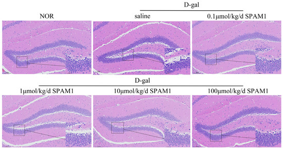
Small-molecule positive allosteric modulator 1 (SPAM1), which targets pituitary adenylate cyclase-activating polypeptide receptor 1 (PAC1-R), has been found to have a neuroprotective effect, and the underlying mechanism was explored in this study. First, using a D-galactose (D-gal)-induced aging mouse model, we confirmed that SPAM1 improves the structure of the hippocampal dentate gyrus and restores the number of neurons. Compared with D-gal model mice, SPAM1-treated mice showed up-regulated expression of Sirtuin 6 (SIRT6) and Lamin B1 and down-regulated expression of YinYang 1 (YY1) and p16. A similar tendency was observed in senescent RGC-5 cells induced by long-term culture, indicating that SPAM1 exhibits significant in vitro and in vivo anti-senescence activity in neurons. Then, using whole-transcriptome sequencing and proteomic analysis, we further explored the mechanism behind SPAM1’s neuroprotective effects and found that SPAM is involved in the longevity-regulating pathway. Finally, the up-regulation of neurofilament light and medium polypeptides indicated by the proteomics results was further confirmed by Western blotting. These results help to lay a pharmacological network foundation for the use of SPAM1 as a potent anti-aging therapeutic drug to combat neurodegeneration with anti-senescence, neuroprotective, and nerve regeneration activity. Full article
Show Figures

Figure 1

20 pages, 3077 KiB  
Article
Neuronal Protection by Ha-RAS-GTPase Signaling through Selective Downregulation of Plasmalemmal Voltage-Dependent Anion Channel-1
by Sebastian Neumann, Konstantin Kuteykin-Teplyakov and Rolf Heumann
Int. J. Mol. Sci. 2024, 25(5), 3030; https://doi.org/10.3390/ijms25053030 - 6 Mar 2024
Viewed by 2128
Abstract
The small GTPase RAS acts as a plasma membrane-anchored intracellular neurotrophin counteracting neuronal degeneration in the brain, but the underlying molecular mechanisms are largely unknown. In transgenic mice expressing constitutively activated V12-Ha-RAS selectively in neurons, proteome analysis uncovered a 70% decrease in voltage-dependent [...] Read more.
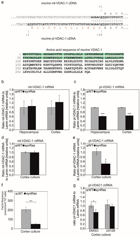
The small GTPase RAS acts as a plasma membrane-anchored intracellular neurotrophin counteracting neuronal degeneration in the brain, but the underlying molecular mechanisms are largely unknown. In transgenic mice expressing constitutively activated V12-Ha-RAS selectively in neurons, proteome analysis uncovered a 70% decrease in voltage-dependent anion channel-1 (VDAC-1) in the cortex and hippocampus. We observed a corresponding reduction in the levels of mRNA splicing variant coding for plasma membrane-targeted VDAC-1 (pl-VDAC-1) while mRNA levels for mitochondrial membrane VDAC-1 (mt-VDAC-1) remained constant. In primary cortical neurons derived from V12-Ha-RAS animals, a decrease in pl-VDAC-1 mRNA levels was observed, accompanied by a concomitant reduction in the ferricyanide reductase activity associated with VDAC-1 protein. Application of MEK inhibitor U0126 to transgenic cortical neurons reconstituted pl-VDAC-1 mRNA to reach wild-type levels. Excitotoxic glutamate-induced cell death was strongly attenuated in transgenic V12-Ha-RAS overexpressing cortical cultures. Consistently, a neuroprotective effect could also be achieved in wild-type cortical cultures by the extracellular application of channel-blocking antibody targeting the N-terminus of VDAC-1. These results may encourage novel therapeutic approaches toward blocking pl-VDAC-1 by monoclonal antibody targeting for complementary treatments in transplantation and neurodegenerative disease. Full article
Show Figures

Figure 1

20 pages, 7288 KiB  
Article
Neurotrophins and Trk Neurotrophin Receptors in the Retina of Adult Killifish (Nothobranchius guentheri)
by Caterina Porcino, Kamel Mhalhel, Marilena Briglia, Marzio Cometa, Maria Cristina Guerrera, Patrizia Germana Germanà, Giuseppe Montalbano, Maria Levanti, Rosaria Laurà, Francesco Abbate, Antonino Germanà and Marialuisa Aragona
Int. J. Mol. Sci. 2024, 25(5), 2732; https://doi.org/10.3390/ijms25052732 - 27 Feb 2024
Cited by 1 | Viewed by 1541
Abstract
Specific subpopulations of neurons in nerve and sensory systems must be developed and maintained, and this is accomplished in significant part by neurotrophins (NTs) and the signaling receptors on which they act, called tyrosine protein kinase receptors (Trks). The neurotrophins–tyrosine protein kinase receptors [...] Read more.
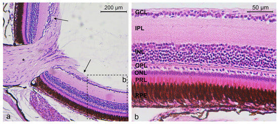
Specific subpopulations of neurons in nerve and sensory systems must be developed and maintained, and this is accomplished in significant part by neurotrophins (NTs) and the signaling receptors on which they act, called tyrosine protein kinase receptors (Trks). The neurotrophins–tyrosine protein kinase receptors (NTs/Trks) system is involved in sensory organ regulation, including the visual system. An NTs/Trks system alteration is associated with neurodegeneration related to aging and diseases, including retinal pathologies. An emergent model in the field of translational medicine, for instance, in aging study, is the annual killifish belonging to the Nothobranchius genus, thanks to its short lifespan. Members of this genus, such as Nothobranchius guentheri, and humans share a similar retinal stratigraphy. Nevertheless, according to the authors’ knowledge, the occurrence and distribution of the NTs/Trks system in the retina of N. guentheri has never been investigated before. Therefore, the present study aimed to localize neurotrophin BDNF, NGF, and NT-3 and TrkA, TrkB, and TrkC receptors in the N. guentheri retina using the immunofluorescence method. The present investigation demonstrates, for the first time, the occurrence of the NTs/Trks system in N. guentheri retina and, consequently, the potential key role of these proteins in the biology and survival of the retinal cells. Full article
Show Figures

Figure 1

9 pages, 1279 KiB  
Communication
ATP-Induced Contractile Response of Esophageal Smooth Muscle in Mice
by Yuji Suzuki, Yasutake Shimizu and Takahiko Shiina
Int. J. Mol. Sci. 2024, 25(4), 1985; https://doi.org/10.3390/ijms25041985 - 6 Feb 2024
Viewed by 1306
Abstract
The tunica muscularis of mammalian esophagi is composed of striated muscle and smooth muscle. Contraction of the esophageal striated muscle portion is mainly controlled by cholinergic neurons. On the other hand, smooth muscle contraction and relaxation are controlled not only by cholinergic components [...] Read more.
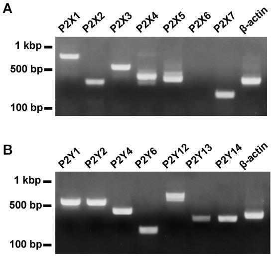
The tunica muscularis of mammalian esophagi is composed of striated muscle and smooth muscle. Contraction of the esophageal striated muscle portion is mainly controlled by cholinergic neurons. On the other hand, smooth muscle contraction and relaxation are controlled not only by cholinergic components but also by non-cholinergic components in the esophagus. Adenosine triphosphate (ATP) is known to regulate smooth muscle contraction and relaxation in the gastrointestinal tract via purinergic receptors. However, the precise mechanism of purinergic regulation in the esophagus is still unclear. Therefore, the aim of the present study was to clarify the effects of ATP on the mechanical responses of the esophageal muscle in mice. An isolated segment of the mouse esophagus was placed in a Magnus’s tube and longitudinal mechanical responses were recorded. Exogenous application of ATP induced contractile responses in the esophageal preparations. Tetrodotoxin, a blocker of voltage-dependent sodium channels in neurons and striated muscle, did not affect the ATP-induced contraction. The ATP-evoked contraction was blocked by pretreatment with suramin, a purinergic receptor antagonist. RT-PCR revealed the expression of mRNA of purinergic receptor genes in the mouse esophageal tissue. The findings suggest that purinergic signaling might regulate the motor activity of mouse esophageal smooth muscle. Full article
Show Figures

Figure 1

12 pages, 765 KiB  
Review
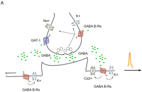
Disinhibition Is an Essential Network Motif Coordinated by GABA Levels and GABA B Receptors
by Nelson Villalobos
Int. J. Mol. Sci. 2024, 25(2), 1340; https://doi.org/10.3390/ijms25021340 - 22 Jan 2024
Cited by 2 | Viewed by 1875
Abstract
Network dynamics are crucial for action and sensation. Changes in synaptic physiology lead to the reorganization of local microcircuits. Consequently, the functional state of the network impacts the output signal depending on the firing patterns of its units. Networks exhibit steady states in [...] Read more.
Network dynamics are crucial for action and sensation. Changes in synaptic physiology lead to the reorganization of local microcircuits. Consequently, the functional state of the network impacts the output signal depending on the firing patterns of its units. Networks exhibit steady states in which neurons show various activities, producing many networks with diverse properties. Transitions between network states determine the output signal generated and its functional results. The temporal dynamics of excitation/inhibition allow a shift between states in an operational network. Therefore, a process capable of modulating the dynamics of excitation/inhibition may be functionally important. This process is known as disinhibition. In this review, we describe the effect of GABA levels and GABAB receptors on tonic inhibition, which causes changes (due to disinhibition) in network dynamics, leading to synchronous functional oscillations. Full article
Show Figures

Figure 1

16 pages, 664 KiB  
Review
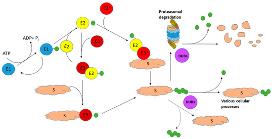
Ubiquitin Carboxyl-Terminal Hydrolase L1 and Its Role in Parkinson’s Disease
by Olga Buneeva and Alexei Medvedev
Int. J. Mol. Sci. 2024, 25(2), 1303; https://doi.org/10.3390/ijms25021303 - 21 Jan 2024
Cited by 4 | Viewed by 3159
Abstract
Ubiquitin carboxyl-terminal hydrolase L1 (UCHL1), also known as Parkinson’s disease protein 5, is a highly expressed protein in the brain. It plays an important role in the ubiquitin–proteasome system (UPS), where it acts as a deubiquitinase (DUB) enzyme. Being the smallest member of [...] Read more.
Ubiquitin carboxyl-terminal hydrolase L1 (UCHL1), also known as Parkinson’s disease protein 5, is a highly expressed protein in the brain. It plays an important role in the ubiquitin–proteasome system (UPS), where it acts as a deubiquitinase (DUB) enzyme. Being the smallest member of the UCH family of DUBs, it catalyzes the reaction of ubiquitin precursor processing and the cleavage of ubiquitinated protein remnants, thus maintaining the level of ubiquitin monomers in the brain cells. UCHL1 mutants, containing amino acid substitutions, influence catalytic activity and its aggregability. Some of them protect cells and transgenic mice in toxin-induced Parkinson’s disease (PD) models. Studies of putative protein partners of UCHL1 revealed about sixty individual proteins located in all major compartments of the cell: nucleus, cytoplasm, endoplasmic reticulum, plasma membrane, mitochondria, and peroxisomes. These include proteins related to the development of PD, such as alpha-synuclein, amyloid-beta precursor protein, ubiquitin-protein ligase parkin, and heat shock proteins. In the context of the catalytic paradigm, the importance of these interactions is not clear. However, there is increasing understanding that UCHL1 exhibits various effects in a catalytically independent manner through protein–protein interactions. Since this protein represents up to 5% of the soluble protein in the brain, PD-related changes in its structure will have profound effects on the proteomes/interactomes in which it is involved. Growing evidence is accumulating that the role of UCHL1 in PD is obviously determined by a balance of canonic catalytic activity and numerous activity-independent protein–protein interactions, which still need better characterization. Full article
Show Figures

Figure 1

2023

Jump to: 2024, 2022, 2021, 2020

14 pages, 919 KiB  
Review
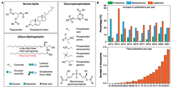
Lipids as Emerging Biomarkers in Neurodegenerative Diseases
by Justin Wei, Li Chin Wong and Sebastian Boland
Int. J. Mol. Sci. 2024, 25(1), 131; https://doi.org/10.3390/ijms25010131 - 21 Dec 2023
Cited by 5 | Viewed by 3979
Abstract
Biomarkers are molecules that can be used to observe changes in an individual’s biochemical or medical status and provide information to aid diagnosis or treatment decisions. Dysregulation in lipid metabolism in the brain is a major risk factor for many neurodegenerative disorders, including [...] Read more.
Biomarkers are molecules that can be used to observe changes in an individual’s biochemical or medical status and provide information to aid diagnosis or treatment decisions. Dysregulation in lipid metabolism in the brain is a major risk factor for many neurodegenerative disorders, including frontotemporal dementia, Alzheimer’s disease, Parkinson’s disease, and amyotrophic lateral sclerosis. Thus, there is a growing interest in using lipids as biomarkers in neurodegenerative diseases, with the anionic phospholipid bis(monoacylglycerol)phosphate and (glyco-)sphingolipids being the most promising lipid classes thus far. In this review, we provide a general overview of lipid biology, provide examples of abnormal lysosomal lipid metabolism in neurodegenerative diseases, and discuss how these insights might offer novel and promising opportunities in biomarker development and therapeutic discovery. Finally, we discuss the challenges and opportunities of lipid biomarkers and biomarker panels in diagnosis, prognosis, and/or treatment response in the clinic. Full article
Show Figures

Figure 1

30 pages, 2004 KiB  
Review
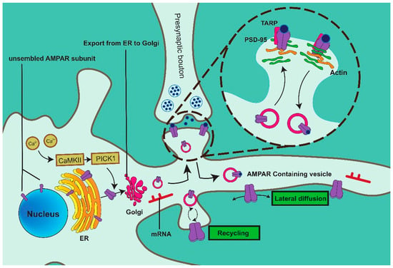
Molecular Mechanisms of AMPA Receptor Trafficking in the Nervous System
by Yi-Yang Cao, Ling-Ling Wu, Xiao-Nan Li, Yu-Lian Yuan, Wan-Wei Zhao, Jing-Xuan Qi, Xu-Yu Zhao, Natalie Ward and Jiao Wang
Int. J. Mol. Sci. 2024, 25(1), 111; https://doi.org/10.3390/ijms25010111 - 21 Dec 2023
Cited by 3 | Viewed by 2687
Abstract
Synaptic plasticity enhances or reduces connections between neurons, affecting learning and memory. Postsynaptic AMPARs mediate greater than 90% of the rapid excitatory synaptic transmission in glutamatergic neurons. The number and subunit composition of AMPARs are fundamental to synaptic plasticity and the formation of [...] Read more.
Synaptic plasticity enhances or reduces connections between neurons, affecting learning and memory. Postsynaptic AMPARs mediate greater than 90% of the rapid excitatory synaptic transmission in glutamatergic neurons. The number and subunit composition of AMPARs are fundamental to synaptic plasticity and the formation of entire neural networks. Accordingly, the insertion and functionalization of AMPARs at the postsynaptic membrane have become a core issue related to neural circuit formation and information processing in the central nervous system. In this review, we summarize current knowledge regarding the related mechanisms of AMPAR expression and trafficking. The proteins related to AMPAR trafficking are discussed in detail, including vesicle-related proteins, cytoskeletal proteins, synaptic proteins, and protein kinases. Furthermore, significant emphasis was placed on the pivotal role of the actin cytoskeleton, which spans throughout the entire transport process in AMPAR transport, indicating that the actin cytoskeleton may serve as a fundamental basis for AMPAR trafficking. Additionally, we summarize the proteases involved in AMPAR post-translational modifications. Moreover, we provide an overview of AMPAR transport and localization to the postsynaptic membrane. Understanding the assembly, trafficking, and dynamic synaptic expression mechanisms of AMPAR may provide valuable insights into the cognitive decline associated with neurodegenerative diseases. Full article
Show Figures

Figure 1

14 pages, 12490 KiB  
Review
Molecular Pathogenesis of Central and Peripheral Nervous System Complications in Anderson–Fabry Disease
by Antonino Tuttolomondo, Irene Baglio, Renata Riolo, Federica Todaro, Gaspare Parrinello, Salvatore Miceli and Irene Simonetta
Int. J. Mol. Sci. 2024, 25(1), 61; https://doi.org/10.3390/ijms25010061 - 20 Dec 2023
Cited by 3 | Viewed by 2797
Abstract
Fabry disease (FD) is a recessive monogenic disease linked to chromosome X due to more than two hundred mutations in the alfa-galactosidase A (GLA) gene. Modifications of the GLA gene may cause the progressive accumulation of globotriaosylceramide (Gb3) and its deacylated form, globotriasylsphingosine [...] Read more.
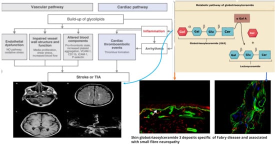
Fabry disease (FD) is a recessive monogenic disease linked to chromosome X due to more than two hundred mutations in the alfa-galactosidase A (GLA) gene. Modifications of the GLA gene may cause the progressive accumulation of globotriaosylceramide (Gb3) and its deacylated form, globotriasylsphingosine (lyso-Gb3), in lysosomes of several types of cells of the heart, kidneys, skin, eyes, peripheral and central nervous system (not clearly and fully demonstrated), and gut with different and pleiotropic clinical symptoms. Among the main symptoms are acroparesthesias and pain crisis (involving the peripheral nervous system), hypohidrosis, abdominal pain, gut motility abnormalities (involving the autonomic system), and finally, cerebrovascular ischemic events due to macrovascular involvement (TIA and stroke) and lacunar strokes and white matter abnormalities due to a small vessel disease (SVS). Gb3 lysosomal accumulation causes cytoplasmatic disruption and subsequent cell death. Additional consequences of Gb3 deposits are inflammatory processes, abnormalities of leukocyte function, and impaired trafficking of some types of immune cells, including lymphocytes, monocytes, CD8+ cells, B cells, and dendritic cells. The involvement of inflammation in AFD pathogenesis conflicts with the reported poor correlation between CRP levels as an inflammation marker and clinical scores such as the Mainz Severity Score Index (MSSI). Also, some authors have suggested an autoimmune reaction is involved in the disease’s pathogenetic mechanism after the α-galactosidase A deficiency. Some studies have reported a high degree of neuronal apoptosis inhibiting protein as a critical anti-apoptotic mediator in children with Fabry disease compared to healthy controls. Notably, this apoptotic upregulation did not change after treatment with enzymatic replacement therapy (ERT), with a further upregulation of the apoptosis-inducing factor after ERT started. Gb3-accumulation has been reported to increase the degree of oxidative stress indexes and the production of reactive oxygen species (ROS). Lipids and proteins have been reported as oxidized and not functioning. Thus, neurological complications are linked to different pathogenetic molecular mechanisms. Progressive accumulation of Gb3 represents a possible pathogenetic event of peripheral nerve involvement. In contrast, central nervous system participation in the clinical setting of cerebrovascular ischemic events seems to be due to the epitheliopathy of Anderson–Fabry disease with lacunar lesions and white matter hyperintensities (WMHs). In this review manuscript, we revised molecular mechanisms of peripheral and central neurological complications of Anderson–Fabry Disease. The management of Fabry disease may be improved by the identification of biomarkers that reflect the clinical course, severity, and progression of the disease. Intensive research on biomarkers has been conducted over the years to detect novel markers that may potentially be used in clinical practice as a screening tool, in the context of the diagnostic process and as an indicator of response to treatment. Recent proteomic or metabolomic studies are in progress, investigating plasma proteome profiles in Fabry patients: these assessments may be useful to characterize the molecular pathology of the disease, improve the diagnostic process, and monitor the response to treatment. Full article
Show Figures

Figure 1

17 pages, 1844 KiB  
Article
Direct Current Stimulation Modulates Synaptic Facilitation via Distinct Presynaptic Calcium Channels
by Sreerag Othayoth Vasu and Hanoch Kaphzan
Int. J. Mol. Sci. 2023, 24(23), 16866; https://doi.org/10.3390/ijms242316866 - 28 Nov 2023
Cited by 1 | Viewed by 1145
Abstract
Transcranial direct current stimulation (tDCS) is a subthreshold neurostimulation technique known for ameliorating neuropsychiatric conditions. The principal mechanism of tDCS is the differential polarization of subcellular neuronal compartments, particularly the axon terminals that are sensitive to external electrical fields. Yet, the underlying mechanism [...] Read more.
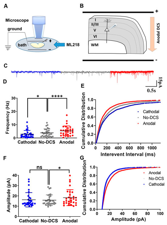
Transcranial direct current stimulation (tDCS) is a subthreshold neurostimulation technique known for ameliorating neuropsychiatric conditions. The principal mechanism of tDCS is the differential polarization of subcellular neuronal compartments, particularly the axon terminals that are sensitive to external electrical fields. Yet, the underlying mechanism of tDCS is not fully clear. Here, we hypothesized that direct current stimulation (DCS)-induced modulation of presynaptic calcium channel conductance alters axon terminal dynamics with regard to synaptic vesicle release. To examine the involvement of calcium-channel subtypes in tDCS, we recorded spontaneous excitatory postsynaptic currents (sEPSCs) from cortical layer-V pyramidal neurons under DCS while selectively inhibiting distinct subtypes of voltage-dependent calcium channels. Blocking P/Q or N-type calcium channels occluded the effects of DCS on sEPSCs, demonstrating their critical role in the process of DCS-induced modulation of spontaneous vesicle release. However, inhibiting T-type calcium channels did not occlude DCS-induced modulation of sEPSCs, suggesting that despite being active in the subthreshold range, T-type calcium channels are not involved in the axonal effects of DCS. DCS modulates synaptic facilitation by regulating calcium channels in axon terminals, primarily via controlling P/Q and N-type calcium channels, while T-type calcium channels are not involved in this mechanism. Full article
Show Figures

Figure 1

18 pages, 1659 KiB  
Article
Reduced Levels of Lacrimal Glial Cell Line-Derived Neurotrophic Factor (GDNF) in Patients with Focal Epilepsy and Focal Epilepsy with Comorbid Depression: A Biomarker Candidate
by Alexander A. Shpak, Flora K. Rider, Tatiana A. Druzhkova, Marina Y. Zhanina, Sofya B. Popova, Alla B. Guekht and Natalia V. Gulyaeva
Int. J. Mol. Sci. 2023, 24(23), 16818; https://doi.org/10.3390/ijms242316818 - 27 Nov 2023
Cited by 2 | Viewed by 1354
Abstract
Our previous studies showed that in patients with brain diseases, neurotrophic factors in lacrimal fluid (LF) may change more prominently than in blood serum (BS). Since glial cell line-derived neurotrophic factor (GDNF) is involved in the control of neuronal networks in an epileptic [...] Read more.
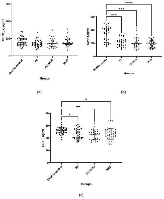
Our previous studies showed that in patients with brain diseases, neurotrophic factors in lacrimal fluid (LF) may change more prominently than in blood serum (BS). Since glial cell line-derived neurotrophic factor (GDNF) is involved in the control of neuronal networks in an epileptic brain, we aimed to assess the GDNF levels in LF and BS as well as the BDNF and the hypothalamic–pituitary–adrenocortical and inflammation indices in BS of patients with focal epilepsy (FE) and epilepsy and comorbid depression (FE + MDD) and to compare them with those of patients with major depressive disorder (MDD) and healthy controls (HC). GDNF levels in BS were similar in patients and HC and higher in FE taking valproates. GDNF levels in LF were significantly lower in all patient groups compared to controls, and independent of drugs used. GDNF concentrations in LF and BS positively correlated in HC, but not in patient groups. BDNF level was lower in BS of patients compared with HC and higher in FE + MDD taking valproates. A reduction in the GDNF level in LF might be an important biomarker of FE. Logistic regression models demonstrated that the probability of FE can be evaluated using GDNF in LF and BDNF in BS; that of MDD using GDNF in LF and cortisol and TNF-α in BS; and that of epilepsy with MDD using GDNF in LF and TNF-α and BDNF in BS. Full article
Show Figures

Figure 1

19 pages, 1962 KiB  
Review
Noncoupled Mitochondrial Respiration as Therapeutic Approach for the Treatment of Metabolic Diseases: Focus on Transgenic Animal Models
by Artem P. Gureev, Alina A. Alimova, Denis N. Silachev and Egor Y. Plotnikov
Int. J. Mol. Sci. 2023, 24(22), 16491; https://doi.org/10.3390/ijms242216491 - 18 Nov 2023
Cited by 1 | Viewed by 2096
Abstract
Mitochondrial dysfunction contributes to numerous chronic diseases, and mitochondria are targets for various toxins and xenobiotics. Therefore, the development of drugs or therapeutic strategies targeting mitochondria is an important task in modern medicine. It is well known that the primary, although not the [...] Read more.
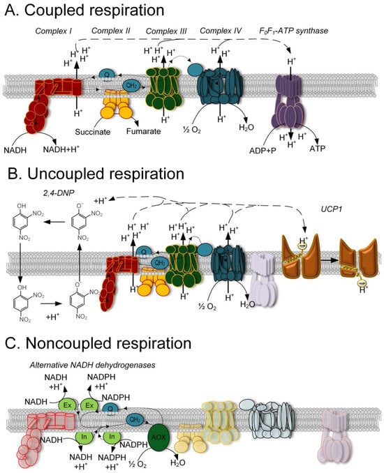
Mitochondrial dysfunction contributes to numerous chronic diseases, and mitochondria are targets for various toxins and xenobiotics. Therefore, the development of drugs or therapeutic strategies targeting mitochondria is an important task in modern medicine. It is well known that the primary, although not the sole, function of mitochondria is ATP generation, which is achieved by coupled respiration. However, a high membrane potential can lead to uncontrolled reactive oxygen species (ROS) production and associated dysfunction. For over 50 years, scientists have been studying various synthetic uncouplers, and for more than 30 years, uncoupling proteins that are responsible for uncoupled respiration in mitochondria. Additionally, the proteins of the mitochondrial alternative respiratory pathway exist in plant mitochondria, allowing noncoupled respiration, in which electron flow is not associated with membrane potential formation. Over the past two decades, advances in genetic engineering have facilitated the creation of various cellular and animal models that simulate the effects of uncoupled and noncoupled respiration in different tissues under various disease conditions. In this review, we summarize and discuss the findings obtained from these transgenic models. We focus on the advantages and limitations of transgenic organisms, the observed physiological and biochemical changes, and the therapeutic potential of uncoupled and noncoupled respiration. Full article
Show Figures

Figure 1

20 pages, 614 KiB  
Review
Astrocytes: Lessons Learned from the Cuprizone Model
by Markus Kipp
Int. J. Mol. Sci. 2023, 24(22), 16420; https://doi.org/10.3390/ijms242216420 - 16 Nov 2023
Cited by 2 | Viewed by 2609
Abstract
A diverse array of neurological and psychiatric disorders, including multiple sclerosis, Alzheimer’s disease, and schizophrenia, exhibit distinct myelin abnormalities at both the molecular and histological levels. These aberrations are closely linked to dysfunction of oligodendrocytes and alterations in myelin structure, which may be [...] Read more.
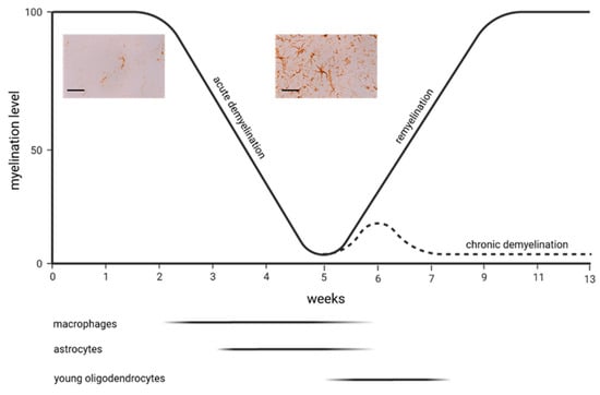
A diverse array of neurological and psychiatric disorders, including multiple sclerosis, Alzheimer’s disease, and schizophrenia, exhibit distinct myelin abnormalities at both the molecular and histological levels. These aberrations are closely linked to dysfunction of oligodendrocytes and alterations in myelin structure, which may be pivotal factors contributing to the disconnection of brain regions and the resulting characteristic clinical impairments observed in these conditions. Astrocytes, which significantly outnumber neurons in the central nervous system by a five-to-one ratio, play indispensable roles in the development, maintenance, and overall well-being of neurons and oligodendrocytes. Consequently, they emerge as potential key players in the onset and progression of a myriad of neurological and psychiatric disorders. Furthermore, targeting astrocytes represents a promising avenue for therapeutic intervention in such disorders. To gain deeper insights into the functions of astrocytes in the context of myelin-related disorders, it is imperative to employ appropriate in vivo models that faithfully recapitulate specific aspects of complex human diseases in a reliable and reproducible manner. One such model is the cuprizone model, wherein metabolic dysfunction in oligodendrocytes initiates an early response involving microglia and astrocyte activation, culminating in multifocal demyelination. Remarkably, following the cessation of cuprizone intoxication, a spontaneous process of endogenous remyelination occurs. In this review article, we provide a historical overview of studies investigating the responses and putative functions of astrocytes in the cuprizone model. Following that, we list previously published works that illuminate various aspects of the biology and function of astrocytes in this multiple sclerosis model. Some of the studies are discussed in more detail in the context of astrocyte biology and pathology. Our objective is twofold: to provide an invaluable overview of this burgeoning field, and, more importantly, to inspire fellow researchers to embark on experimental investigations to elucidate the multifaceted functions of this pivotal glial cell subpopulation. Full article
Show Figures

Figure 1

14 pages, 920 KiB  
Review
The Role of RFRP Neurons in the Allostatic Control of Reproductive Function
by Maggie C. Evans and Greg M. Anderson
Int. J. Mol. Sci. 2023, 24(21), 15851; https://doi.org/10.3390/ijms242115851 - 1 Nov 2023
Cited by 1 | Viewed by 1613
Abstract
Reproductive function is critical for species survival; however, it is energetically costly and physically demanding. Reproductive suppression is therefore a physiologically appropriate adaptation to certain ecological, environmental, and/or temporal conditions. This ‘allostatic’ suppression of fertility enables individuals to accommodate unfavorable reproductive circumstances and [...] Read more.
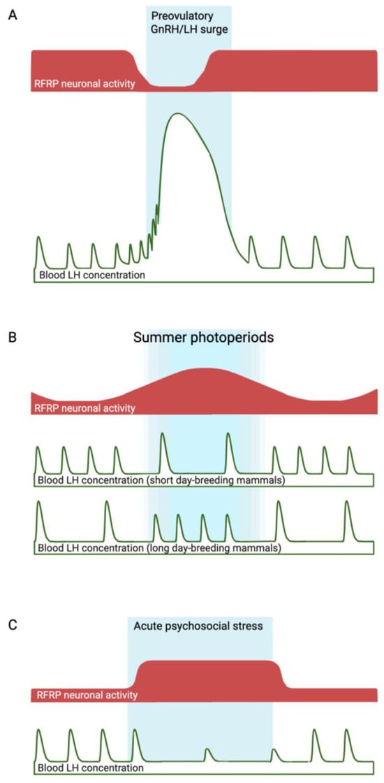
Reproductive function is critical for species survival; however, it is energetically costly and physically demanding. Reproductive suppression is therefore a physiologically appropriate adaptation to certain ecological, environmental, and/or temporal conditions. This ‘allostatic’ suppression of fertility enables individuals to accommodate unfavorable reproductive circumstances and safeguard survival. The mechanisms underpinning this reproductive suppression are complex, yet culminate with the reduced secretion of gonadotropin-releasing hormone (GnRH) from the hypothalamus, which in turn suppresses gonadotropin release from the pituitary, thereby impairing gonadal function. The focus of this review will be on the role of RFamide-related peptide (RFRP) neurons in different examples of allostatic reproductive suppression. RFRP neurons release the RFRP-3 peptide, which negatively regulates GnRH neurons and thus appears to act as a ‘brake’ on the neuroendocrine reproductive axis. In a multitude of predictable (e.g., pre-puberty, reproductive senescence, and seasonal or lactational reproductive quiescence) and unpredictable (e.g., metabolic, immune and/or psychosocial stress) situations in which GnRH secretion is suppressed, the RFRP neurons have been suggested to act as modulators. This review examines evidence for and against these roles. Full article
Show Figures

Figure 1

28 pages, 4557 KiB  
Review
The Role of Cysteine Protease Cathepsins B, H, C, and X/Z in Neurodegenerative Diseases and Cancer
by Veronika Stoka, Olga Vasiljeva, Hiroshi Nakanishi and Vito Turk
Int. J. Mol. Sci. 2023, 24(21), 15613; https://doi.org/10.3390/ijms242115613 - 26 Oct 2023
Cited by 8 | Viewed by 3286
Abstract
Papain-like cysteine proteases are composed of 11 human cysteine cathepsins, originally located in the lysosomes. They exhibit broad specificity and act as endopeptidases and/or exopeptidases. Among them, only cathepsins B, H, C, and X/Z exhibit exopeptidase activity. Recently, cysteine cathepsins have been found [...] Read more.
Papain-like cysteine proteases are composed of 11 human cysteine cathepsins, originally located in the lysosomes. They exhibit broad specificity and act as endopeptidases and/or exopeptidases. Among them, only cathepsins B, H, C, and X/Z exhibit exopeptidase activity. Recently, cysteine cathepsins have been found to be present outside the lysosomes and often participate in various pathological processes. Hence, they have been considered key signalling molecules. Their potentially hazardous proteolytic activities are tightly regulated. This review aims to discuss recent advances in understanding the structural aspects of these four cathepsins, mechanisms of their zymogen activation, regulation of their activities, and functional aspects of these enzymes in neurodegeneration and cancer. Neurodegenerative effects have been evaluated, particularly in Alzheimer’s disease, Parkinson’s disease, Huntington’s disease, amyotrophic lateral sclerosis, multiple sclerosis, and neuropsychiatric disorders. Cysteine cathepsins also participate in tumour progression and metastasis through the overexpression and secretion of proteases, which trigger extracellular matrix degradation. To our knowledge, this is the first review to provide an in-depth analysis regarding the roles of cysteine cathepsins B, H, C, and X in neurodegenerative diseases and cancer. Further advances in understanding the functions of cysteine cathepsins in these conditions will result in the development of novel, targeted therapeutic strategies. Full article
Show Figures

Figure 1

19 pages, 12475 KiB  
Article
Characterisation of GFAP-Expressing Glial Cells in the Dorsal Root Ganglion after Spared Nerve Injury
by Elena A. Konnova, Alexandru-Florian Deftu, Paul Chu Sin Chung, Marie Pertin, Guylène Kirschmann, Isabelle Decosterd and Marc R. Suter
Int. J. Mol. Sci. 2023, 24(21), 15559; https://doi.org/10.3390/ijms242115559 - 25 Oct 2023
Cited by 7 | Viewed by 2882
Abstract
Satellite glial cells (SGCs), enveloping primary sensory neurons’ somas in the dorsal root ganglion (DRG), contribute to neuropathic pain upon nerve injury. Glial fibrillary acidic protein (GFAP) serves as an SGC activation marker, though its DRG satellite cell specificity is debated. We employed [...] Read more.
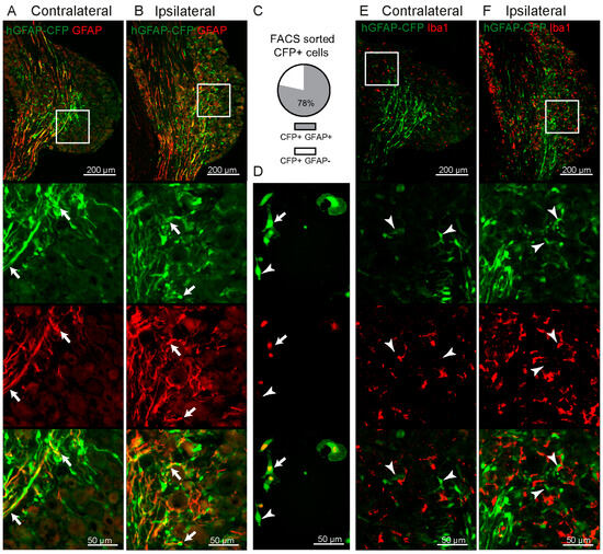
Satellite glial cells (SGCs), enveloping primary sensory neurons’ somas in the dorsal root ganglion (DRG), contribute to neuropathic pain upon nerve injury. Glial fibrillary acidic protein (GFAP) serves as an SGC activation marker, though its DRG satellite cell specificity is debated. We employed the hGFAP-CFP transgenic mouse line, designed for astrocyte studies, to explore its expression within the peripheral nervous system (PNS) after spared nerve injury (SNI). We used diverse immunostaining techniques, Western blot analysis, and electrophysiology to evaluate GFAP+ cell changes. Post-SNI, GFAP+ cell numbers increased without proliferation, and were found near injured ATF3+ neurons. GFAP+ FABP7+ SGCs increased, yet 75.5% of DRG GFAP+ cells lacked FABP7 expression. This suggests a significant subset of GFAP+ cells are non-myelinating Schwann cells (nmSC), indicated by their presence in the dorsal root but not in the ventral root which lacks unmyelinated fibres. Additionally, patch clamp recordings from GFAP+ FABP7−cells lacked SGC-specific Kir4.1 currents, instead displaying outward Kv currents expressing Kv1.1 and Kv1.6 channels specific to nmSCs. In conclusion, this study demonstrates increased GFAP expression in two DRG glial cell subpopulations post-SNI: GFAP+ FABP7+ SGCs and GFAP+ FABP7− nmSCs, shedding light on GFAP’s specificity as an SGC marker after SNI. Full article
Show Figures

Figure 1

18 pages, 986 KiB  
Article
Evaluation of an Image-Derived Input Function for Kinetic Modeling of Nicotinic Acetylcholine Receptor-Binding PET Ligands in Mice
by Matthew Zammit, Chien-Min Kao, Hannah J. Zhang, Hsiu-Ming Tsai, Nathanial Holderman, Samuel Mitchell, Eve Tanios, Mohammed Bhuiyan, Richard Freifelder, Anna Kucharski, William N. Green, Jogeshwar Mukherjee and Chin-Tu Chen
Int. J. Mol. Sci. 2023, 24(21), 15510; https://doi.org/10.3390/ijms242115510 - 24 Oct 2023
Viewed by 1337
Abstract
Positron emission tomography (PET) radioligands that bind with high-affinity to α4β2-type nicotinic receptors (α4β2Rs) allow for in vivo investigations of the mechanisms underlying nicotine addiction and smoking cessation. Here, we investigate the use of an image-derived arterial input function and the cerebellum for [...] Read more.
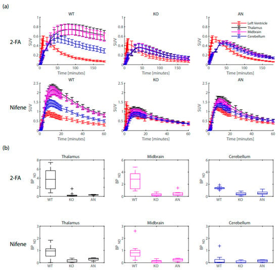
Positron emission tomography (PET) radioligands that bind with high-affinity to α4β2-type nicotinic receptors (α4β2Rs) allow for in vivo investigations of the mechanisms underlying nicotine addiction and smoking cessation. Here, we investigate the use of an image-derived arterial input function and the cerebellum for kinetic analysis of radioligand binding in mice. Two radioligands were explored: 2-[18F]FA85380 (2-FA), displaying similar pKa and binding affinity to the smoking cessation drug varenicline (Chantix), and [18F]Nifene, displaying similar pKa and binding affinity to nicotine. Time–activity curves of the left ventricle of the heart displayed similar distribution across wild type mice, mice lacking the β2-subunit for ligand binding, and acute nicotine-treated mice, whereas reference tissue binding displayed high variation between groups. Binding potential estimated from a two-tissue compartment model fit of the data with the image-derived input function were higher than estimates from reference tissue-based estimations. Rate constants of radioligand dissociation were very slow for 2-FA and very fast for Nifene. We conclude that using an image-derived input function for kinetic modeling of nicotinic PET ligands provides suitable results compared to reference tissue-based methods and that the chemical properties of 2-FA and Nifene are suitable to study receptor response to nicotine addiction and smoking cessation therapies. Full article
Show Figures

Figure 1

19 pages, 1211 KiB  
Review
The Impact of Neurotransmitters on the Neurobiology of Neurodegenerative Diseases
by Sarah E. Davis, Abagail B. Cirincione, Ana Catya Jimenez-Torres and Jun Zhu
Int. J. Mol. Sci. 2023, 24(20), 15340; https://doi.org/10.3390/ijms242015340 - 19 Oct 2023
Cited by 16 | Viewed by 3623
Abstract
Neurodegenerative diseases affect millions of people worldwide. Neurodegenerative diseases result from progressive damage to nerve cells in the brain or peripheral nervous system connections that are essential for cognition, coordination, strength, sensation, and mobility. Dysfunction of these brain and nerve functions is associated [...] Read more.
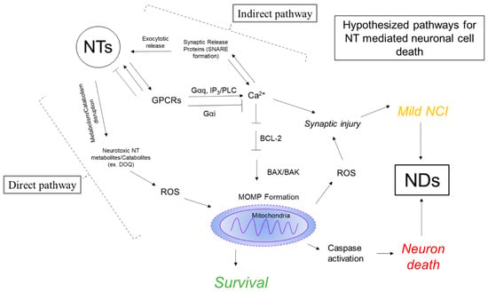
Neurodegenerative diseases affect millions of people worldwide. Neurodegenerative diseases result from progressive damage to nerve cells in the brain or peripheral nervous system connections that are essential for cognition, coordination, strength, sensation, and mobility. Dysfunction of these brain and nerve functions is associated with Alzheimer’s disease, Parkinson’s disease, Huntington’s disease, Amyotrophic lateral sclerosis, and motor neuron disease. In addition to these, 50% of people living with HIV develop a spectrum of cognitive, motor, and/or mood problems collectively referred to as HIV-Associated Neurocognitive Disorders (HAND) despite the widespread use of a combination of antiretroviral therapies. Neuroinflammation and neurotransmitter systems have a pathological correlation and play a critical role in developing neurodegenerative diseases. Each of these diseases has a unique pattern of dysregulation of the neurotransmitter system, which has been attributed to different forms of cell-specific neuronal loss. In this review, we will focus on a discussion of the regulation of dopaminergic and cholinergic systems, which are more commonly disturbed in neurodegenerative disorders. Additionally, we will provide evidence for the hypothesis that disturbances in neurotransmission contribute to the neuronal loss observed in neurodegenerative disorders. Further, we will highlight the critical role of dopamine as a mediator of neuronal injury and loss in the context of NeuroHIV. This review will highlight the need to further investigate neurotransmission systems for their role in the etiology of neurodegenerative disorders. Full article
Show Figures

Figure 1

21 pages, 1767 KiB  
Review
Advancing Spinal Cord Injury Treatment through Stem Cell Therapy: A Comprehensive Review of Cell Types, Challenges, and Emerging Technologies in Regenerative Medicine
by Chih-Wei Zeng
Int. J. Mol. Sci. 2023, 24(18), 14349; https://doi.org/10.3390/ijms241814349 - 20 Sep 2023
Cited by 13 | Viewed by 9397
Abstract
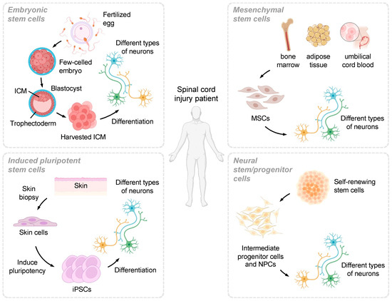
Spinal cord injuries (SCIs) can lead to significant neurological deficits and lifelong disability, with far-reaching physical, psychological, and economic consequences for affected individuals and their families. Current treatments for SCIs are limited in their ability to restore function, and there is a pressing [...] Read more.
Spinal cord injuries (SCIs) can lead to significant neurological deficits and lifelong disability, with far-reaching physical, psychological, and economic consequences for affected individuals and their families. Current treatments for SCIs are limited in their ability to restore function, and there is a pressing need for innovative therapeutic approaches. Stem cell therapy has emerged as a promising strategy to promote the regeneration and repair of damaged neural tissue following SCIs. This review article comprehensively discusses the potential of different stem cell types, such as embryonic stem cells (ESCs), induced pluripotent stem cells (iPSCs), mesenchymal stem cells (MSCs), and neural stem/progenitor cells (NSPCs), in SCI treatment. We provide an in-depth analysis of the unique advantages and challenges associated with each stem cell type, as well as the latest advancements in the field. Furthermore, we address the critical challenges faced in stem cell therapy for SCIs, including safety concerns, ethical considerations, standardization of protocols, optimization of transplantation parameters, and the development of effective outcome measures. We also discuss the integration of novel technologies such as gene editing, biomaterials, and tissue engineering to enhance the therapeutic potential of stem cells. The article concludes by emphasizing the importance of collaborative efforts among various stakeholders in the scientific community, including researchers, clinicians, bioengineers, industry partners, and patients, to overcome these challenges and realize the full potential of stem cell therapy for SCI patients. By fostering such collaborations and advancing our understanding of stem cell biology and regenerative medicine, we can pave the way for the development of groundbreaking therapies that improve the lives of those affected by SCIs. Full article
Show Figures

Figure 1

12 pages, 1811 KiB  
Communication
Astrocytic GPCR-Induced Ca2+ Signaling Is Not Causally Related to Local Cerebral Blood Flow Changes
by Katsuya Ozawa, Masaki Nagao, Ayumu Konno, Youichi Iwai, Marta Vittani, Peter Kusk, Tsuneko Mishima, Hirokazu Hirai, Maiken Nedergaard and Hajime Hirase
Int. J. Mol. Sci. 2023, 24(17), 13590; https://doi.org/10.3390/ijms241713590 - 2 Sep 2023
Cited by 6 | Viewed by 4131
Abstract
Activation of Gq-type G protein-coupled receptors (GPCRs) gives rise to large cytosolic Ca2+ elevations in astrocytes. Previous in vitro and in vivo studies have indicated that astrocytic Ca2+ elevations are closely associated with diameter changes in the nearby blood vessels, which [...] Read more.
Activation of Gq-type G protein-coupled receptors (GPCRs) gives rise to large cytosolic Ca2+ elevations in astrocytes. Previous in vitro and in vivo studies have indicated that astrocytic Ca2+ elevations are closely associated with diameter changes in the nearby blood vessels, which astrocytes enwrap with their endfeet. However, the causal relationship between astrocytic Ca2+ elevations and blood vessel diameter changes has been questioned, as mice with diminished astrocytic Ca2+ signaling show normal sensory hyperemia. We addressed this controversy by imaging cortical vasculature while optogenetically elevating astrocyte Ca2+ in a novel transgenic mouse line, expressing Opto-Gq-type GPCR Optoα1AR (Astro-Optoα1AR) in astrocytes. Blue light illumination on the surface of the somatosensory cortex induced Ca2+ elevations in cortical astrocytes and their endfeet in mice under anesthesia. Blood vessel diameter did not change significantly with Optoα1AR-induced Ca2+ elevations in astrocytes, while it was increased by forelimb stimulation. Next, we labeled blood plasma with red fluorescence using AAV8-P3-Alb-mScarlet in Astro-Optoα1AR mice. We were able to identify arterioles that display diameter changes in superficial areas of the somatosensory cortex through the thinned skull. Photo-stimulation of astrocytes in the cortical area did not result in noticeable changes in the arteriole diameters compared with their background strain C57BL/6. Together, compelling evidence for astrocytic Gq pathway-induced vasodiameter changes was not observed. Our results support the notion that short-term (<10 s) hyperemia is not mediated by GPCR-induced astrocytic Ca2+ signaling. Full article
Show Figures

Figure 1

16 pages, 2522 KiB  
Article
Depressive-like Behaviors Induced by mGluR5 Reduction in 6xTg in Mouse Model of Alzheimer’s Disease
by Youngkyo Kim, Jinho Kim, Shinwoo Kang and Keun-A Chang
Int. J. Mol. Sci. 2023, 24(16), 13010; https://doi.org/10.3390/ijms241613010 - 21 Aug 2023
Cited by 3 | Viewed by 2114
Abstract
Alzheimer’s disease (AD) is one representative dementia characterized by the accumulation of amyloid beta (Aβ) plaques and neurofibrillary tangles (NFTs) in the brain, resulting in cognitive decline and memory loss. AD is associated with neuropsychiatric symptoms, including major depressive disorder (MDD). Recent studies [...] Read more.
Alzheimer’s disease (AD) is one representative dementia characterized by the accumulation of amyloid beta (Aβ) plaques and neurofibrillary tangles (NFTs) in the brain, resulting in cognitive decline and memory loss. AD is associated with neuropsychiatric symptoms, including major depressive disorder (MDD). Recent studies showed a reduction in mGluR5 expression in the brains of stress-induced mice models and individuals with MDD compared to controls. In our study, we identified depressive-like behavior and memory impairment in a mouse model of AD, specifically in the 6xTg model with tau and Aβ pathologies. In addition, we investigated the expression of mGluR5 in the brains of 6xTg mice using micro-positron emission tomography (micro-PET) imaging, histological analysis, and Western blot analysis, and we observed a decrease in mGluR5 levels in the brains of 6xTg mice compared to wild-type (WT) mice. Additionally, we identified alterations in the ERK/AKT/GSK-3β signaling pathway in the brains of 6xTg mice. Notably, we identified a significant negative correlation between depressive-like behavior and the protein level of mGluR5 in 6xTg mice. Additionally, we also found a significant positive correlation between depressive-like behavior and AD pathologies, including phosphorylated tau and Aβ. These findings suggested that abnormal mGluR5 expression and AD-related pathologies were involved in depressive-like behavior in the 6xTg mouse model. Further research is warranted to elucidate the underlying mechanisms and explore potential therapeutic targets in the intersection of AD and depressive-like symptoms. Full article
Show Figures

Figure 1

16 pages, 3637 KiB  
Article
The Induction of Long-Term Potentiation by Medial Septum Activation under Urethane Anesthesia Can Alter Gene Expression in the Hippocampus
by Yulia V. Dobryakova, Konstantin Gerasimov, Yulia S. Spivak, Tinna Korotkova, Alena Koryagina, Angelina Deryabina, Vladimir A. Markevich and Alexey P. Bolshakov
Int. J. Mol. Sci. 2023, 24(16), 12970; https://doi.org/10.3390/ijms241612970 - 19 Aug 2023
Cited by 1 | Viewed by 1209
Abstract
We studied changes in the expression of early genes in hippocampal cells in response to stimulation of the dorsal medial septal area (dMSA), leading to long-term potentiation in the hippocampus. Rats under urethane anesthesia were implanted with stimulating electrodes in the ventral hippocampal [...] Read more.
We studied changes in the expression of early genes in hippocampal cells in response to stimulation of the dorsal medial septal area (dMSA), leading to long-term potentiation in the hippocampus. Rats under urethane anesthesia were implanted with stimulating electrodes in the ventral hippocampal commissure and dMSA and a recording electrode in the CA1 area of the hippocampus. We found that high-frequency stimulation (HFS) of the dMSA led to the induction of long-term potentiation in the synapses formed by the ventral hippocampal commissure on the hippocampal CA1 neurons. One hour after dMSA HFS, we collected the dorsal and ventral hippocampi on both the ipsilateral (damaged by the implanted electrode) and contralateral (intact) sides and analyzed the expression of genes by qPCR. The dMSA HFS led to an increase in the expression of bdnf and cyr61 in the ipsilateral hippocampi and egr1 in the ventral contralateral hippocampus. Thus, dMSA HFS under the conditions of degeneration of the cholinergic neurons in the medial septal area prevented the described increase in gene expression. The changes in cyr61 expression appeared to be dependent on the muscarinic M1 receptors. Our data suggest that the induction of long-term potentiation by dMSA activation enhances the expression of select early genes in the hippocampus. Full article
Show Figures

Figure 1

12 pages, 4997 KiB  
Article
Exposure of Cultured Hippocampal Neurons to the Mitochondrial Uncoupler Carbonyl Cyanide Chlorophenylhydrazone Induces a Rapid Growth of Dendritic Processes
by Liliia Kushnireva, Eduard Korkotian and Menahem Segal
Int. J. Mol. Sci. 2023, 24(16), 12940; https://doi.org/10.3390/ijms241612940 - 18 Aug 2023
Cited by 1 | Viewed by 1495
Abstract
A major route for the influx of calcium ions into neurons uses the STIM-Orai1 voltage-independent channel. Once cytosolic calcium ([Ca2+]i) elevates, it activates mitochondrial and endoplasmic calcium stores to affect downstream molecular pathways. In the present study, we employed a novel [...] Read more.
A major route for the influx of calcium ions into neurons uses the STIM-Orai1 voltage-independent channel. Once cytosolic calcium ([Ca2+]i) elevates, it activates mitochondrial and endoplasmic calcium stores to affect downstream molecular pathways. In the present study, we employed a novel drug, carbonyl cyanide chlorophenylhydrazone (CCCP), a mitochondrial uncoupler, to explore the role of mitochondria in cultured neuronal morphology. CCCP caused a sustained elevation of [Ca2+]i and, quite surprisingly, a massive increase in the density of dendritic filopodia and spines in the affected neurons. This morphological change can be prevented in cultures exposed to a calcium-free medium, Orai1 antagonist 2APB, or cells transfected with a mutant Orai1 plasmid. It is suggested that CCCP activates mitochondria through the influx of calcium to cause rapid growth of dendritic processes. Full article
Show Figures

Figure 1

16 pages, 3707 KiB  
Article
Acute Exposure to Microplastics Induced Changes in Behavior and Inflammation in Young and Old Mice
by Lauren Gaspar, Sydney Bartman, Giuseppe Coppotelli and Jaime M. Ross
Int. J. Mol. Sci. 2023, 24(15), 12308; https://doi.org/10.3390/ijms241512308 - 1 Aug 2023
Cited by 26 | Viewed by 40733
Abstract
Environmental pollutants have become quite ubiquitous over the past two centuries; of those, plastics, and in particular, microplastics (<5 mm), are among the most pervasive pollutants. Microplastics (MPs) have found their way into the air, water system, and food chain and are either [...] Read more.
Environmental pollutants have become quite ubiquitous over the past two centuries; of those, plastics, and in particular, microplastics (<5 mm), are among the most pervasive pollutants. Microplastics (MPs) have found their way into the air, water system, and food chain and are either purposely produced or are derived from the breakdown of larger plastic materials. Despite the societal advancements that plastics have allowed, the mismanagement of plastic waste has become a pressing global issue. Pioneering studies on MPs toxicity have shown that exposure to MPs induces oxidative stress, inflammation, and decreased cell viability in marine organisms. Current research suggests that these MPs are transported throughout the environment and can accumulate in human tissues; however, research on the health effects of MPs, especially in mammals, is still very limited. This has led our group to explore the biological and cognitive consequences of exposure to MPs in a rodent model. Following a three-week exposure to water treated with fluorescently-labeled pristine polystyrene MPs, young and old C57BL/6J mice were assessed using behavioral assays, such as open-field and light–dark preference, followed by tissue analyses using fluorescent immunohistochemistry, Western blot, and qPCR. Data from these assays suggest that short-term exposure to MPs induces both behavioral changes as well as alterations in immune markers in liver and brain tissues. Additionally, we noted that these changes differed depending on age, indicating a possible age-dependent effect. These findings suggest the need for further research to better understand the mechanisms by which microplastics may induce physiological and cognitive changes. Full article
Show Figures

Figure 1

20 pages, 4717 KiB  
Article
Recruitment of Corticotropin-Releasing Hormone (CRH) Neurons in Categorically Distinct Stress Reactions in the Mouse Brain
by Krisztina Horváth, Balázs Juhász, Dániel Kuti, Szilamér Ferenczi and Krisztina J. Kovács
Int. J. Mol. Sci. 2023, 24(14), 11736; https://doi.org/10.3390/ijms241411736 - 21 Jul 2023
Cited by 1 | Viewed by 2695
Abstract
Corticotropin-releasing hormone (CRH) neurons in the paraventricular hypothalamic nucleus (PVH) are in the position to integrate stress-related information and initiate adaptive neuroendocrine-, autonomic-, metabolic- and behavioral responses. In addition to hypophyseotropic cells, CRH is widely expressed in the CNS, however its involvement in [...] Read more.
Corticotropin-releasing hormone (CRH) neurons in the paraventricular hypothalamic nucleus (PVH) are in the position to integrate stress-related information and initiate adaptive neuroendocrine-, autonomic-, metabolic- and behavioral responses. In addition to hypophyseotropic cells, CRH is widely expressed in the CNS, however its involvement in the organization of the stress response is not fully understood. In these experiments, we took advantage of recently available Crh-IRES-Cre;Ai9 mouse line to study the recruitment of hypothalamic and extrahypothalamic CRH neurons in categorically distinct, acute stress reactions. A total of 95 brain regions in the adult male mouse brain have been identified as containing putative CRH neurons with significant expression of tdTomato marker gene. With comparison of CRH mRNA and tdTomato distribution, we found match and mismatch areas. Reporter mice were then exposed to restraint, ether, high salt, lipopolysaccharide and predator odor stress and neuronal activation was revealed by FOS immunocytochemistry. In addition to a core stress system, stressor-specific areas have been revealed to display activity marker FOS. Finally, activation of CRH neurons was detected by colocalization of FOS in tdTomato expressing cells. All stressors resulted in profound activation of CRH neurons in the hypothalamic paraventricular nucleus; however, a differential activation of pattern was observed in CRH neurons in extrahypothalamic regions. This comprehensive description of stress-related CRH neurons in the mouse brain provides a starting point for a systematic functional analysis of the brain stress system and its relation to stress-induced psychopathologies. Full article
Show Figures

Graphical abstract

14 pages, 11382 KiB  
Article
Spinal Cord Blood Vessels in Aged Mice Show Greater Levels of Hypoxia-Induced Vascular Disruption and Microglial Activation
by Sebok K. Halder and Richard Milner
Int. J. Mol. Sci. 2023, 24(14), 11235; https://doi.org/10.3390/ijms241411235 - 8 Jul 2023
Cited by 2 | Viewed by 1723
Abstract
In response to chronic mild hypoxia (CMH, 8% O2), spinal cord blood vessels launch a robust angiogenic response that is associated with transient disruption of the blood–spinal cord barrier (BSCB) which, in turn, triggers a microglial vasculo-protective response. Because hypoxia occurs [...] Read more.
In response to chronic mild hypoxia (CMH, 8% O2), spinal cord blood vessels launch a robust angiogenic response that is associated with transient disruption of the blood–spinal cord barrier (BSCB) which, in turn, triggers a microglial vasculo-protective response. Because hypoxia occurs in many age-related conditions, the goal of this study was to define how aging influences these responses by comparing events in young (8–10 weeks) and aged (20 months) mice. This revealed that aged mice had much greater (3–4-fold) levels of hypoxic-induced BSCB disruption than young mice and that, while the early stage of the angiogenic response in aged mice was no different to young mice, the maturation of newly formed vessels was significantly delayed. Interestingly, microglia in the spinal cords of aged mice were much more activated than young mice, even under normoxic conditions, and this was further enhanced by CMH, though, surprisingly, this resulted in reduced microglial clustering around leaky blood vessels and diminished vasculo-protection. Vascular disruption was associated with loss of myelin in spinal cord white matter (WM) in both young and aged mice. Furthermore, it was notable that the spinal cord of aged mice contained a lower density of Olig2+ oligodendroglial cells even under normoxic conditions and that CMH significantly reduced the density of Olig2+ cells in spinal cord WM of the aged, but not the young, mice. These results demonstrate that spinal cord blood vessels of aged mice are much more vulnerable to the damaging effects of hypoxia than young mice, in part due to the reduced vasculo-protection conferred by chronically activated microglial cells. These observations may have implications for the pathogenesis and/or treatment of spinal cord diseases such as amyotrophic lateral sclerosis (ALS) and suggest that an improvement in microglial function could offer therapeutic potential for treating these age-related conditions. Full article
Show Figures

Figure 1

15 pages, 4110 KiB  
Article
Altered Hippocampal and Striatal Expression of Endothelial Markers and VIP/PACAP Neuropeptides in a Mouse Model of Systemic Lupus Erythematosus
by Jayden Lee, Sarah Thomas Broome, Margo Iris Jansen, Mawj Mandwie, Grant J. Logan, Rubina Marzagalli, Giuseppe Musumeci and Alessandro Castorina
Int. J. Mol. Sci. 2023, 24(13), 11118; https://doi.org/10.3390/ijms241311118 - 5 Jul 2023
Cited by 2 | Viewed by 1839
Abstract
Neuropsychiatric systemic lupus erythematosus (NPSLE) is one of the most common and severe manifestations of lupus; however, its pathogenesis is still poorly understood. While there is sparse evidence suggesting that the ongoing autoimmunity may trigger pathogenic changes to the central nervous system (CNS) [...] Read more.
Neuropsychiatric systemic lupus erythematosus (NPSLE) is one of the most common and severe manifestations of lupus; however, its pathogenesis is still poorly understood. While there is sparse evidence suggesting that the ongoing autoimmunity may trigger pathogenic changes to the central nervous system (CNS) microvasculature, culminating in inflammatory/ischemic damage, further evidence is still needed. In this study, we used the spontaneous mouse model of SLE (NZBWF1 mice) to investigate the expression of genes and proteins associated with endothelial (dys)function: tissue and urokinase plasminogen activators (tPA and uPA), intercellular and vascular adhesion molecules 1 (ICAM-1 and VCAM-1), brain derived neurotrophic factor (BDNF), endothelial nitric oxide synthase (eNOS) and Krüppel-like factor 4 (KLF4) and neuroprotection/immune modulation: pituitary adenylate cyclase-activating peptide (PACAP), vasoactive intestinal peptide (VIP), PACAP receptor (PAC1), VIP receptors 1 and 2 (VPAC1 and VPAC2). Analyses were carried out both in the hippocampus and striatum of SLE mice of two different age groups (2 and 7 months old), since age correlates with disease severity. In the hippocampus, we identified a gene/protein expression profile indicative of mild endothelial dysfunction, which increased in severity in aged SLE mice. These alterations were paralleled by moderate alterations in the expression of VIP, PACAP and related receptors. In contrast, we report a robust upregulation of endothelial activation markers in the striatum of both young and aged mice, concurrent with significant induction of the VIP/PACAP system. These data identify molecular signatures of endothelial alterations in the hippocampus and striatum of NZBWF1 mice, which are accompanied by a heightened expression of endogenous protective/immune-modulatory neuropeptides. Collectively, our results support the idea that NPSLE may cause alterations of the CNS micro-vascular compartment that cannot be effectively counteracted by the endogenous activity of the neuropeptides PACAP and VIP. Full article
Show Figures

Figure 1

15 pages, 355 KiB  
Review
Neurotoxicity from Old and New Radiation Treatments for Brain Tumors
by Riccardo Soffietti, Alessia Pellerino, Francesco Bruno, Alessandro Mauro and Roberta Rudà
Int. J. Mol. Sci. 2023, 24(13), 10669; https://doi.org/10.3390/ijms241310669 - 26 Jun 2023
Cited by 6 | Viewed by 2093
Abstract
Research regarding the mechanisms of brain damage following radiation treatments for brain tumors has increased over the years, thus providing a deeper insight into the pathobiological mechanisms and suggesting new approaches to minimize this damage. This review has discussed the different factors that [...] Read more.
Research regarding the mechanisms of brain damage following radiation treatments for brain tumors has increased over the years, thus providing a deeper insight into the pathobiological mechanisms and suggesting new approaches to minimize this damage. This review has discussed the different factors that are known to influence the risk of damage to the brain (mainly cognitive disturbances) from radiation. These include patient and tumor characteristics, the use of whole-brain radiotherapy versus particle therapy (protons, carbon ions), and stereotactic radiotherapy in various modalities. Additionally, biological mechanisms behind neuroprotection have been elucidated. Full article
10 pages, 1432 KiB  
Case Report
Metabolic Biomarkers Differentiate a Surgical Intervertebral Disc from a Nonsurgical Intervertebral Disc
by Beata Toczylowska, Michal Woznica, Elzbieta Zieminska and Leszek Krolicki
Int. J. Mol. Sci. 2023, 24(13), 10572; https://doi.org/10.3390/ijms241310572 - 24 Jun 2023
Cited by 3 | Viewed by 1899
Abstract
Background: Degeneration of the intervertebral disc (IVD) is caused by disturbances in metabolic processes, which lead to structural disorders. The aim of this report is to analyze metabolic disorders in the degeneration process by comparing control discs with degenerated discs. In our research [...] Read more.
Background: Degeneration of the intervertebral disc (IVD) is caused by disturbances in metabolic processes, which lead to structural disorders. The aim of this report is to analyze metabolic disorders in the degeneration process by comparing control discs with degenerated discs. In our research on the nucleus pulposus (NP), we used NMR spectroscopy of extracts of hydrophilic and hydrophobic compounds of the tissue. Methods: Nuclear magnetic resonance (NMR) spectroscopy allows the study of biochemistry and cellular metabolism in vitro. Hydrophilic and hydrophobic compounds were extracted from the NP of the intervertebral disc. In the NMR spectra, metabolites were identified and quantitatively analyzed. The results of our research indicate disturbances in the biosynthesis and metabolism of cholesterol, the biosynthesis and degradation of various fatty acid groups, ketone bodies, or lysine, and the metabolism of glycerophospholipids, purines, glycine, inositol, galactose, alanine, glutamate, and pyruvate in the biosynthesis of valine and isoleucine, leucine. All these disorders indicate pathomechanisms related to oxidative stress, energy, neurotransmission disturbances, and disturbances in the structure and functioning of cell membranes, inflammation, or chronic pain generators. Conclusions: NMR spectroscopy allows the identification of metabolites differentiating surgical from nonsurgical discs. These data may provide guidance in in vivo MRS studies in assessing the severity of lesions of the disc. Full article
Show Figures

Figure 1

20 pages, 2864 KiB  
Review
Targeting the Limbic System: Insights into Its Involvement in Tinnitus
by Anurag Singh, Paul F. Smith and Yiwen Zheng
Int. J. Mol. Sci. 2023, 24(12), 9889; https://doi.org/10.3390/ijms24129889 - 8 Jun 2023
Cited by 3 | Viewed by 6419
Abstract
Tinnitus is originally derived from the Latin verb tinnire, which means “to ring”. Tinnitus, a complex disorder, is a result of sentient cognizance of a sound in the absence of an external auditory stimulus. It is reported in children, adults, and older [...] Read more.
Tinnitus is originally derived from the Latin verb tinnire, which means “to ring”. Tinnitus, a complex disorder, is a result of sentient cognizance of a sound in the absence of an external auditory stimulus. It is reported in children, adults, and older populations. Patients suffering from tinnitus often present with hearing loss, anxiety, depression, and sleep disruption in addition to a hissing and ringing in the ear. Surgical interventions and many other forms of treatment have been only partially effective due to heterogeneity in tinnitus patients and a lack of understanding of the mechanisms of tinnitus. Although researchers across the globe have made significant progress in understanding the underlying mechanisms of tinnitus over the past few decades, tinnitus is still deemed to be a scientific enigma. This review summarises the role of the limbic system in tinnitus development and provides insight into the development of potential target-specific tinnitus therapies. Full article
Show Figures

Figure 1

19 pages, 3183 KiB  
Article
High-Fat Diet Consumption Induces Neurobehavioral Abnormalities and Neuronal Morphological Alterations Accompanied by Excessive Microglial Activation in the Medial Prefrontal Cortex in Adolescent Mice
by Conghui Wang, Hong Li, Chen Chen, Xiuting Yao, Chenxi Yang, Zhehao Yu, Jiayi Ren, Yue Ming, Yi Huang, Yi Rong, Yu Ma and Lijie Liu
Int. J. Mol. Sci. 2023, 24(11), 9394; https://doi.org/10.3390/ijms24119394 - 28 May 2023
Cited by 4 | Viewed by 2540
Abstract
The association between a high-fat diet (HFD) consumption and emotional/cognitive disorders is widely documented. One distinctive feature of the prefrontal cortex (PFC), a kernel emotion- and cognition-related brain region, is its protracted adolescent maturation, which makes it highly vulnerable to the detrimental effects [...] Read more.
The association between a high-fat diet (HFD) consumption and emotional/cognitive disorders is widely documented. One distinctive feature of the prefrontal cortex (PFC), a kernel emotion- and cognition-related brain region, is its protracted adolescent maturation, which makes it highly vulnerable to the detrimental effects of environmental factors during adolescence. Disruption of the PFC structure and function is linked to emotional/cognitive disorders, especially those that emerge in late adolescence. A HFD consumption is common among adolescents, yet its potential effects on PFC-related neurobehavior in late adolescence and any related underlying mechanisms are yet to be established. In the present study, adolescent (postnatal days 28–56) male C57BL/6J mice were fed a control diet (CD) or a HFD and underwent behavioral tests in addition to Golgi staining and immunofluorescence targeting of the medial PFC (mPFC). The HFD-fed adolescent mice exhibited anxiety- and depression-like behavior and abnormal mPFC pyramidal neuronal morphology accompanied by alterations in microglial morphology indicative of a heightened state of activation and increased microglial PSD95+ inclusions signifying excessive phagocytosis of the synaptic material in the mPFC. These findings offer novel insights into the neurobehavioral effects due to adolescent HFD consumption and suggest a contributing role in microglial dysfunction and prefrontal neuroplasticity deficits for HFD-associated mood disorders in adolescents. Full article
Show Figures

Figure 1

19 pages, 719 KiB  
Review
Impact of Solute Carrier Transporters in Glioma Pathology: A Comprehensive Review
by Filippos Anagnostakis, Michail Kokkorakis, Mariam Markouli and Christina Piperi
Int. J. Mol. Sci. 2023, 24(11), 9393; https://doi.org/10.3390/ijms24119393 - 28 May 2023
Cited by 5 | Viewed by 3158
Abstract
Solute carriers (SLCs) are essential for brain physiology and homeostasis due to their role in transporting necessary substances across cell membranes. There is an increasing need to further unravel their pathophysiological implications since they have been proposed to play a pivotal role in [...] Read more.
Solute carriers (SLCs) are essential for brain physiology and homeostasis due to their role in transporting necessary substances across cell membranes. There is an increasing need to further unravel their pathophysiological implications since they have been proposed to play a pivotal role in brain tumor development, progression, and the formation of the tumor microenvironment (TME) through the upregulation and downregulation of various amino acid transporters. Due to their implication in malignancy and tumor progression, SLCs are currently positioned at the center of novel pharmacological targeting strategies and drug development. In this review, we discuss the key structural and functional characteristics of the main SLC family members involved in glioma pathogenesis, along with their potential targeting options to provide new opportunities for CNS drug design and more effective glioma management. Full article
Show Figures

Figure 1

14 pages, 2193 KiB  
Article
The Role of Dopamine D3 Receptors, Dysbindin, and Their Functional Interaction in the Expression of Key Genes for Neuroplasticity and Neuroinflammation in the Mouse Brain
by Veronica Rivi, Cristina Benatti, Joan M. C. Blom, Luca Pani, Nicoletta Brunello, Filippo Drago, Francesco Papaleo, Filippo Caraci, Federica Geraci, Sebastiano Alfio Torrisi, Gian Marco Leggio and Fabio Tascedda
Int. J. Mol. Sci. 2023, 24(10), 8699; https://doi.org/10.3390/ijms24108699 - 12 May 2023
Cited by 1 | Viewed by 1948
Abstract
Cognitive impairment in schizophrenia remains a clinically and pharmacologically unsolved challenge. Clinical and preclinical studies have revealed that the concomitant reduction in dysbindin (DYS) and dopamine receptor D3 functionality improves cognitive functions. However, the molecular machinery underlying this epistatic interaction has not yet [...] Read more.
Cognitive impairment in schizophrenia remains a clinically and pharmacologically unsolved challenge. Clinical and preclinical studies have revealed that the concomitant reduction in dysbindin (DYS) and dopamine receptor D3 functionality improves cognitive functions. However, the molecular machinery underlying this epistatic interaction has not yet been fully elucidated. The glutamate NMDA receptors and the neurotrophin BDNF, with their established role in promoting neuroplasticity, may be involved in the complex network regulated by the D3/DYS interaction. Furthermore, as inflammation is involved in the etiopathogenesis of several psychiatric diseases, including schizophrenia, the D3/DYS interaction may affect the expression levels of pro−inflammatory cytokines. Thus, by employing mutant mice bearing selective heterozygosis for D3 and/or DYS, we provide new insights into the functional interactions (single and synergic) between these schizophrenia susceptibility genes and the expression levels of key genes for neuroplasticity and neuroinflammation in three key brain areas for schizophrenia: the prefrontal cortex, striatum, and hippocampus. In the hippocampus, the epistatic interaction between D3 and DYS reversed to the wild-type level the downregulated mRNA levels of GRIN1 and GRIN2A were observed in DYS +/− and D3 +/− mice. In all the areas investigated, double mutant mice had higher BDNF levels compared to their single heterozygote counterparts, whereas D3 hypofunction resulted in higher pro−inflammatory cytokines. These results may help to clarify the genetic mechanisms and functional interactions involved in the etiology and development of schizophrenia. Full article
Show Figures

Figure 1

23 pages, 4482 KiB  
Article
Impact of Endurance Training on Regeneration of Axons, Glial Cells, and Inhibitory Neurons after Spinal Cord Injury: A Link between Functional Outcome and Regeneration Potential within the Lesion Site and in Adjacent Spinal Cord Tissue
by Katarina Kiss Bimbova, Maria Bacova, Alexandra Kisucka, Ján Gálik, Maria Ileninova, Tomas Kuruc, Martina Magurova and Nadezda Lukacova
Int. J. Mol. Sci. 2023, 24(10), 8616; https://doi.org/10.3390/ijms24108616 - 11 May 2023
Cited by 2 | Viewed by 2710
Abstract
Endurance training prior to spinal cord injury (SCI) has a beneficial effect on the activation of signaling pathways responsible for survival, neuroplasticity, and neuroregeneration. It is, however, unclear which training-induced cell populations are essential for the functional outcome after SCI. Adult Wistar rats [...] Read more.
Endurance training prior to spinal cord injury (SCI) has a beneficial effect on the activation of signaling pathways responsible for survival, neuroplasticity, and neuroregeneration. It is, however, unclear which training-induced cell populations are essential for the functional outcome after SCI. Adult Wistar rats were divided into four groups: control, six weeks of endurance training, Th9 compression (40 g/15 min), and pretraining + Th9 compression. The animals survived six weeks. Training alone increased the gene expression and protein level of immature CNP-ase oligodendrocytes (~16%) at Th10, and caused rearrangements in neurotrophic regulation of inhibitory GABA/glycinergic neurons at the Th10 and L2 levels, known to contain the interneurons with rhythmogenic potential. Training + SCI upregulated markers for immature and mature (CNP-ase, PLP1) oligodendrocytes by ~13% at the lesion site and caudally, and increased the number of GABA/glycinergic neurons in specific spinal cord regions. In the pretrained SCI group, the functional outcome of hindlimbs positively correlated with the protein levels of CNP-ase, PLP1, and neurofilaments (NF-l), but not with the outgrowing axons (Gap-43) at the lesion site and caudally. These results indicate that endurance training applied before SCI potentiates the repair in damaged spinal cord, and creates a suitable environment for neurological outcome. Full article
Show Figures

Figure 1

24 pages, 4263 KiB  
Article
Acute Cardiovascular and Cardiorespiratory Effects of JWH-018 in Awake and Freely Moving Mice: Mechanism of Action and Possible Antidotal Interventions?
by Beatrice Marchetti, Sabrine Bilel, Micaela Tirri, Giorgia Corli, Elisa Roda, Carlo Alessandro Locatelli, Elena Cavarretta, Fabio De-Giorgio and Matteo Marti
Int. J. Mol. Sci. 2023, 24(8), 7515; https://doi.org/10.3390/ijms24087515 - 19 Apr 2023
Cited by 4 | Viewed by 2527
Abstract
JWH-018 is the most known compound among synthetic cannabinoids (SCs) used for their psychoactive effects. SCs-based products are responsible for several intoxications in humans. Cardiac toxicity is among the main side effects observed in emergency departments: SCs intake induces harmful effects such as [...] Read more.
JWH-018 is the most known compound among synthetic cannabinoids (SCs) used for their psychoactive effects. SCs-based products are responsible for several intoxications in humans. Cardiac toxicity is among the main side effects observed in emergency departments: SCs intake induces harmful effects such as hypertension, tachycardia, chest pain, arrhythmias, myocardial infarction, breathing impairment, and dyspnea. This study aims to investigate how cardio-respiratory and vascular JWH-018 (6 mg/kg) responses can be modulated by antidotes already in clinical use. The tested antidotes are amiodarone (5 mg/kg), atropine (5 mg/kg), nifedipine (1 mg/kg), and propranolol (2 mg/kg). The detection of heart rate, breath rate, arterial oxygen saturation (SpO2), and pulse distention are provided by a non-invasive apparatus (Mouse Ox Plus) in awake and freely moving CD-1 male mice. Tachyarrhythmia events are also evaluated. Results show that while all tested antidotes reduce tachycardia and tachyarrhythmic events and improve breathing functions, only atropine completely reverts the heart rate and pulse distension. These data may suggest that cardiorespiratory mechanisms of JWH-018-induced tachyarrhythmia involve sympathetic, cholinergic, and ion channel modulation. Current findings also provide valuable impetus to identify potential antidotal intervention to support physicians in the treatment of intoxicated patients in emergency clinical settings. Full article
Show Figures

Figure 1

30 pages, 5778 KiB  
Article
An Efficient 2D Protocol for Differentiation of iPSCs into Mature Postmitotic Dopaminergic Neurons: Application for Modeling Parkinson’s Disease
by Olga S. Lebedeva, Elena I. Sharova, Dmitriy A. Grekhnev, Liubov O. Skorodumova, Irina V. Kopylova, Ekaterina M. Vassina, Arina Oshkolova, Iuliia V. Novikova, Alena V. Krisanova, Evgenii I. Olekhnovich, Vladimir A. Vigont, Elena V. Kaznacheyeva, Alexandra N. Bogomazova and Maria A. Lagarkova
Int. J. Mol. Sci. 2023, 24(8), 7297; https://doi.org/10.3390/ijms24087297 - 14 Apr 2023
Cited by 7 | Viewed by 4496
Abstract
About 15% of patients with parkinsonism have a hereditary form of Parkinson’s disease (PD). Studies on the early stages of PD pathogenesis are challenging due to the lack of relevant models. The most promising ones are models based on dopaminergic neurons (DAns) differentiated [...] Read more.
About 15% of patients with parkinsonism have a hereditary form of Parkinson’s disease (PD). Studies on the early stages of PD pathogenesis are challenging due to the lack of relevant models. The most promising ones are models based on dopaminergic neurons (DAns) differentiated from induced pluripotent stem cells (iPSCs) of patients with hereditary forms of PD. This work describes a highly efficient 2D protocol for obtaining DAns from iPSCs. The protocol is rather simple, comparable in efficiency with previously published protocols, and does not require viral vectors. The resulting neurons have a similar transcriptome profile to previously published data for neurons, and have a high level of maturity marker expression. The proportion of sensitive (SOX6+) DAns in the population calculated from the level of gene expression is higher than resistant (CALB+) DAns. Electrophysiological studies of the DAns confirmed their voltage sensitivity and showed that a mutation in the PARK8 gene is associated with enhanced store-operated calcium entry. The study of high-purity DAns differentiated from the iPSCs of patients with hereditary PD using this differentiation protocol will allow for investigators to combine various research methods, from patch clamp to omics technologies, and maximize information about cell function in normal and pathological conditions. Full article
Show Figures

Figure 1

21 pages, 2791 KiB  
Article
Co-Expression Network Analysis Identifies Molecular Determinants of Loneliness Associated with Neuropsychiatric and Neurodegenerative Diseases
by Jose A. Santiago, James P. Quinn and Judith A. Potashkin
Int. J. Mol. Sci. 2023, 24(6), 5909; https://doi.org/10.3390/ijms24065909 - 21 Mar 2023
Cited by 8 | Viewed by 4105
Abstract
Loneliness and social isolation are detrimental to mental health and may lead to cognitive impairment and neurodegeneration. Although several molecular signatures of loneliness have been identified, the molecular mechanisms by which loneliness impacts the brain remain elusive. Here, we performed a bioinformatics approach [...] Read more.
Loneliness and social isolation are detrimental to mental health and may lead to cognitive impairment and neurodegeneration. Although several molecular signatures of loneliness have been identified, the molecular mechanisms by which loneliness impacts the brain remain elusive. Here, we performed a bioinformatics approach to untangle the molecular underpinnings associated with loneliness. Co-expression network analysis identified molecular ‘switches’ responsible for dramatic transcriptional changes in the nucleus accumbens of individuals with known loneliness. Loneliness-related switch genes were enriched in cell cycle, cancer, TGF-β, FOXO, and PI3K-AKT signaling pathways. Analysis stratified by sex identified switch genes in males with chronic loneliness. Male-specific switch genes were enriched in infection, innate immunity, and cancer-related pathways. Correlation analysis revealed that loneliness-related switch genes significantly overlapped with 82% and 68% of human studies on Alzheimer’s (AD) and Parkinson’s diseases (PD), respectively, in gene expression databases. Loneliness-related switch genes, BCAM, NECTIN2, NPAS3, RBM38, PELI1, DPP10, and ASGR2, have been identified as genetic risk factors for AD. Likewise, switch genes HLA-DRB5, ALDOA, and GPNMB are known genetic loci in PD. Similarly, loneliness-related switch genes overlapped in 70% and 64% of human studies on major depressive disorder and schizophrenia, respectively. Nine switch genes, HLA-DRB5, ARHGAP15, COL4A1, RBM38, DMD, LGALS3BP, WSCD2, CYTH4, and CNTRL, overlapped with known genetic variants in depression. Seven switch genes, NPAS3, ARHGAP15, LGALS3BP, DPP10, SMYD3, CPXCR1, and HLA-DRB5 were associated with known risk factors for schizophrenia. Collectively, we identified molecular determinants of loneliness and dysregulated pathways in the brain of non-demented adults. The association of switch genes with known risk factors for neuropsychiatric and neurodegenerative diseases provides a molecular explanation for the observed prevalence of these diseases among lonely individuals. Full article
Show Figures

Figure 1

13 pages, 13856 KiB  
Article
Inhaled Nitric Oxide Promotes Angiogenesis in the Rodent Developing Brain
by Gauthier Loron, Julien Pansiot, Paul Olivier, Christiane Charriaut-Marlangue and Olivier Baud
Int. J. Mol. Sci. 2023, 24(6), 5871; https://doi.org/10.3390/ijms24065871 - 20 Mar 2023
Cited by 6 | Viewed by 1865
Abstract
Inhaled nitric oxide (iNO) is a therapy used in neonates with pulmonary hypertension. Some evidence of its neuroprotective properties has been reported in both mature and immature brains subjected to injury. NO is a key mediator of the VEGF pathway, and angiogenesis may [...] Read more.
Inhaled nitric oxide (iNO) is a therapy used in neonates with pulmonary hypertension. Some evidence of its neuroprotective properties has been reported in both mature and immature brains subjected to injury. NO is a key mediator of the VEGF pathway, and angiogenesis may be involved in the reduced vulnerability to injury of white matter and the cortex conferred by iNO. Here, we report the effect of iNO on angiogenesis in the developing brain and its potential effectors. We found that iNO promotes angiogenesis in the developing white matter and cortex during a critical window in P14 rat pups. This shift in the developmental program of brain angiogenesis was not related to a regulation of NO synthases by exogenous NO exposure, nor the VEGF pathway or other angiogenic factors. The effects of iNO on brain angiogenesis were found to be mimicked by circulating nitrate/nitrite, suggesting that these carriers may play a role in transporting NO to the brain. Finally, our data show that the soluble guanylate cyclase/cGMP signaling pathway is likely to be involved in the pro-angiogenetic effect of iNO through thrombospondin-1, a glycoprotein of the extracellular matrix, inhibiting soluble guanylate cyclase through CD42 and CD36. In conclusion, this study provides new insights into the biological basis of the effect of iNO in the developing brain. Full article
Show Figures

Figure 1

30 pages, 1062 KiB  
Review
Epilepsy and Attention Deficit Hyperactivity Disorder: Connection, Chance, and Challenges
by Hueng-Chuen Fan, Kuo-Liang Chiang, Kuang-Hsi Chang, Chuan-Mu Chen and Jeng-Dau Tsai
Int. J. Mol. Sci. 2023, 24(6), 5270; https://doi.org/10.3390/ijms24065270 - 9 Mar 2023
Cited by 16 | Viewed by 6988
Abstract
Comorbidities are common in children with epilepsy, with nearly half of the patients having at least one comorbidity. Attention deficit hyperactivity disorder (ADHD) is a psychiatric disorder characterized by hyperactivity and inattentiveness level disproportional to the child’s developmental stage. The burden of ADHD [...] Read more.
Comorbidities are common in children with epilepsy, with nearly half of the patients having at least one comorbidity. Attention deficit hyperactivity disorder (ADHD) is a psychiatric disorder characterized by hyperactivity and inattentiveness level disproportional to the child’s developmental stage. The burden of ADHD in children with epilepsy is high and can adversely affect the patients’ clinical outcomes, psychosocial aspects, and quality of life. Several hypotheses were proposed to explain the high burden of ADHD in childhood epilepsy; the well-established bidirectional connection and shared genetic/non-genetic factors between epilepsy and comorbid ADHD largely rule out the possibility of a chance in this association. Stimulants are effective in children with comorbid ADHD, and the current body of evidence supports their safety within the approved dose. Nonetheless, safety data should be further studied in randomized, double-blinded, placebo-controlled trials. Comorbid ADHD is still under-recognized in clinical practice. Early identification and management of comorbid ADHD are crucial to optimize the prognosis and reduce the risk of adverse long-term neurodevelopmental outcomes. The identification of the shared genetic background of epilepsy and ADHD can open the gate for tailoring treatment options for these patients through precision medicine. Full article
Show Figures

Figure 1

9 pages, 1897 KiB  
Article
Insights from Spatial Measures of Intolerance to Identifying Pathogenic Variants in Developmental and Epileptic Encephalopathies
by Michael Silk, Alex de Sá, Moshe Olshansky and David B. Ascher
Int. J. Mol. Sci. 2023, 24(6), 5114; https://doi.org/10.3390/ijms24065114 - 7 Mar 2023
Cited by 1 | Viewed by 1417
Abstract
Developmental and epileptic encephalopathies (DEEs) are a group of epilepsies with early onset and severe symptoms that sometimes lead to death. Although previous work successfully discovered several genes implicated in disease outcomes, it remains challenging to identify causative mutations within these genes from [...] Read more.
Developmental and epileptic encephalopathies (DEEs) are a group of epilepsies with early onset and severe symptoms that sometimes lead to death. Although previous work successfully discovered several genes implicated in disease outcomes, it remains challenging to identify causative mutations within these genes from the background variation present in all individuals due to disease heterogeneity. Nevertheless, our ability to detect possible pathogenic variants has continued to improve as in silico predictors of deleteriousness have advanced. We investigate their use in prioritising likely pathogenic variants in epileptic encephalopathy patients’ whole exome sequences. We showed that the inclusion of structure-based predictors of intolerance improved upon previous attempts to demonstrate enrichment within epilepsy genes. Full article
Show Figures

Figure 1

16 pages, 9169 KiB  
Article
Neurodegenerative Changes in the Brains of the 5xFAD Alzheimer’s Disease Model Mice Investigated by High-Field and High-Resolution Magnetic Resonance Imaging and Multi-Nuclei Magnetic Resonance Spectroscopy
by Chi-Hyeon Yoo, Jinho Kim, Hyeon-Man Baek, Keun-A Chang and Bo-Young Choe
Int. J. Mol. Sci. 2023, 24(6), 5073; https://doi.org/10.3390/ijms24065073 - 7 Mar 2023
Cited by 3 | Viewed by 2695
Abstract
This study aimed to investigate morphological and metabolic changes in the brains of 5xFAD mice. Structural magnetic resonance imaging (MRI) and 1H magnetic resonance spectroscopy (MRS) were obtained in 10- and 14-month-old 5xFAD and wild-type (WT) mice, while 31P MRS scans [...] Read more.
This study aimed to investigate morphological and metabolic changes in the brains of 5xFAD mice. Structural magnetic resonance imaging (MRI) and 1H magnetic resonance spectroscopy (MRS) were obtained in 10- and 14-month-old 5xFAD and wild-type (WT) mice, while 31P MRS scans were acquired in 11-month-old mice. Significantly reduced gray matter (GM) was identified by voxel-based morphometry (VBM) in the thalamus, hypothalamus, and periaqueductal gray areas of 5xFAD mice compared to WT mice. Significant reductions in N-acetyl aspartate and elevation of myo-Inositol were revealed by the quantification of MRS in the hippocampus of 5xFAD mice, compared to WT. A significant reduction in NeuN-positive cells and elevation of Iba1- and GFAP-positive cells supported this observation. The reduction in phosphomonoester and elevation of phosphodiester was observed in 11-month-old 5xFAD mice, which might imply a sign of disruption in the membrane synthesis. Commonly reported 1H MRS features were replicated in the hippocampus of 14-month-old 5xFAD mice, and a sign of disruption in the membrane synthesis and elevation of breakdown were revealed in the whole brain of 5xFAD mice by 31P MRS. GM volume reduction was identified in the thalamus, hypothalamus, and periaqueductal gray areas of 5xFAD mice. Full article
Show Figures

Figure 1

16 pages, 4578 KiB  
Article
Optic Nerve Injury Enhanced Mitochondrial Fission and Increased Mitochondrial Density without Altering the Uniform Mitochondrial Distribution in the Unmyelinated Axons of Retinal Ganglion Cells in a Mouse Model
by Takahiro Tsuji, Tomoya Murase, Yoshiyuki Konishi and Masaru Inatani
Int. J. Mol. Sci. 2023, 24(5), 4356; https://doi.org/10.3390/ijms24054356 - 22 Feb 2023
Viewed by 2201
Abstract
Glaucomatous optic neuropathy (GON), a major cause of blindness, is characterized by the loss of retinal ganglion cells (RGCs) and the degeneration of their axons. Mitochondria are deeply involved in maintaining the health of RGCs and their axons. Therefore, lots of attempts have [...] Read more.
Glaucomatous optic neuropathy (GON), a major cause of blindness, is characterized by the loss of retinal ganglion cells (RGCs) and the degeneration of their axons. Mitochondria are deeply involved in maintaining the health of RGCs and their axons. Therefore, lots of attempts have been made to develop diagnostic tools and therapies targeting mitochondria. Recently, we reported that mitochondria are uniformly distributed in the unmyelinated axons of RGCs, possibly owing to the ATP gradient. Thus, using transgenic mice expressing yellow fluorescent protein targeting mitochondria exclusively in RGCs within the retina, we assessed the alteration of mitochondrial distributions induced by optic nerve crush (ONC) via in vitro flat-mount retinal sections and in vivo fundus images captured with a confocal scanning ophthalmoscope. We observed that the mitochondrial distribution in the unmyelinated axons of survived RGCs after ONC remained uniform, although their density increased. Furthermore, via in vitro analysis, we discovered that the mitochondrial size is attenuated following ONC. These results suggest that ONC induces mitochondrial fission without disrupting the uniform mitochondrial distribution, possibly preventing axonal degeneration and apoptosis. The in vivo visualization system of axonal mitochondria in RGCs may be applicable in the detection of the progression of GON in animal studies and potentially in humans. Full article
Show Figures

Figure 1

14 pages, 1456 KiB  
Article
Whole Transcriptome Analysis of Hypothalamus in Mice during Short-Term Starvation
by Eun-Young Oh, Byong Seo Park, Hye Rim Yang, Ho Gyun Lee, Thai Hien Tu, Sunggu Yang, Mi-Ryung Han and Jae Geun Kim
Int. J. Mol. Sci. 2023, 24(4), 3204; https://doi.org/10.3390/ijms24043204 - 6 Feb 2023
Cited by 4 | Viewed by 3184
Abstract
Molecular profiling of the hypothalamus in response to metabolic shifts is a critical cue to better understand the principle of the central control of whole-body energy metabolism. The transcriptional responses of the rodent hypothalamus to short-term calorie restriction have been documented. However, studies [...] Read more.
Molecular profiling of the hypothalamus in response to metabolic shifts is a critical cue to better understand the principle of the central control of whole-body energy metabolism. The transcriptional responses of the rodent hypothalamus to short-term calorie restriction have been documented. However, studies on the identification of hypothalamic secretory factors that potentially contribute to the control of appetite are lacking. In this study, we analyzed the differential expression of hypothalamic genes and compared the selected secretory factors from the fasted mice with those of fed control mice using bulk RNA-sequencing. We verified seven secretory genes that were significantly altered in the hypothalamus of fasted mice. In addition, we determined the response of secretory genes in cultured hypothalamic cells to treatment with ghrelin and leptin. The current study provides further insights into the neuronal response to food restriction at the molecular level and may be useful for understanding the hypothalamic control of appetite. Full article
Show Figures

Figure 1

12 pages, 2266 KiB  
Article
Nitric Oxide Synthase Blockade Impairs Spontaneous Calcium Activity in Mouse Primary Hippocampal Culture Cells
by Rostislav A. Sokolov, David Jappy, Oleg V. Podgorny and Irina V. Mukhina
Int. J. Mol. Sci. 2023, 24(3), 2608; https://doi.org/10.3390/ijms24032608 - 30 Jan 2023
Cited by 1 | Viewed by 2068
Abstract
Oscillation of intracellular calcium concentration is a stable phenomenon that affects cellular function throughout the lifetime of both electrically excitable and non-excitable cells. Nitric oxide, a gaseous secondary messenger and the product of nitric oxide synthase (NOS), affects intracellular calcium dynamics. Using mouse [...] Read more.
Oscillation of intracellular calcium concentration is a stable phenomenon that affects cellular function throughout the lifetime of both electrically excitable and non-excitable cells. Nitric oxide, a gaseous secondary messenger and the product of nitric oxide synthase (NOS), affects intracellular calcium dynamics. Using mouse hippocampal primary cultures, we recorded the effect of NOS blockade on neuronal spontaneous calcium activity. There was a correlation between the amplitude of spontaneous calcium events and the number of action potentials (APs) (Spearman R = 0.94). There was a linear rise of DAF-FM fluorescent emission showing an increase in NO concentration with time in neurons (11.9 ± 1.0%). There is correlation between the integral of the signal from DAF-FM and the integral of the spontaneous calcium event signal from Oregon Green 488 (Spearman R = 0.58). Blockade of NOS affected the parameters of the spontaneous calcium events studied (amplitude, frequency, integral, rise slope and decay slope). NOS blockade by Nw-Nitro-L-arginine suppressed the amplitude and frequency of spontaneous calcium events. The NOS blocker 3-Bromo-7-Nitroindazole reduced the frequency but not the amplitude of spontaneous calcium activity. Blockade of the well-known regulator of NOS, calcineurin with cyclosporine A reduced the integral of calcium activity in neurons. The differences and similarities in the effects on the parameters of spontaneous calcium effects caused by different blockades of NO production help to improve understanding of how NO synthesis affects calcium dynamics in neurons. Full article
Show Figures

Figure 1

17 pages, 2374 KiB  
Article
Behavioral and Pharmacokinetics Studies of N-Methyl-2-Aminoindane (NM2AI) in Mice: An Aminoindane Briefly Used in the Illicit Drug Market
by Micaela Tirri, Giorgia Corli, Raffaella Arfè, Beatrice Marchetti, Sabrine Bilel, Tatiana Bernardi, Federica Boccuto, Sara Odoardi, Serena Mestria, Sabina Strano-Rossi and Matteo Marti
Int. J. Mol. Sci. 2023, 24(3), 1882; https://doi.org/10.3390/ijms24031882 - 18 Jan 2023
Viewed by 2237
Abstract
Drug forums are considered as the main platform sources that have contributed to the increase in NPS popularity, especially for those not yet known to law enforcement and therefore not yet illegal. An example is the new synthetic stimulant NM2AI, which has a [...] Read more.
Drug forums are considered as the main platform sources that have contributed to the increase in NPS popularity, especially for those not yet known to law enforcement and therefore not yet illegal. An example is the new synthetic stimulant NM2AI, which has a very short history of human use and abuse. Little is known regarding this compound, but some information from internet forums and the scientific literature indicates NM2AI as a structural derivate of MDAI, which is known for its entactogenic activity. Indeed, the purpose of this study is to evaluate, for the first time, the in vivo acute effect induced by the intraperitoneal injection of NM2AI (1–10–30–100 mg/kg) in mice. We demonstrate the sensory (by visual placing and object tests) and physiological (core temperature measurement) function variations, nociceptor (by tail pinch test) and strength (grip test) alterations, and sensorimotor (time on rod and mobility) decrease. Moreover, we verify the mild hallucinogenic effect of NM2AI (by startle/prepulse inhibition test). Lastly, we perform a pharmacokinetic study on mice blood samples, highlighting that the main active metabolite of NM2AI is 2-aminoindane (2AI). Taken together, our data confirm the suspected entactogenic activity of NM2AI; however, these in vivo effects appear atypical and less intense with respect to those induced by the classic stimulants, in surprising analogy with what is reported by networked users. Full article
Show Figures

Figure 1

46 pages, 1995 KiB  
Review
Molecular Mechanisms of Neuroinflammation in Aging and Alzheimer’s Disease Progression
by Felicia Liana Andronie-Cioara, Adriana Ioana Ardelean, Carmen Delia Nistor-Cseppento, Anamaria Jurcau, Maria Carolina Jurcau, Nicoleta Pascalau and Florin Marcu
Int. J. Mol. Sci. 2023, 24(3), 1869; https://doi.org/10.3390/ijms24031869 - 18 Jan 2023
Cited by 54 | Viewed by 8934
Abstract
Aging is the most prominent risk factor for late-onset Alzheimer’s disease. Aging associates with a chronic inflammatory state both in the periphery and in the central nervous system, the evidence thereof and the mechanisms leading to chronic neuroinflammation being discussed. Nonetheless, neuroinflammation is [...] Read more.
Aging is the most prominent risk factor for late-onset Alzheimer’s disease. Aging associates with a chronic inflammatory state both in the periphery and in the central nervous system, the evidence thereof and the mechanisms leading to chronic neuroinflammation being discussed. Nonetheless, neuroinflammation is significantly enhanced by the accumulation of amyloid beta and accelerates the progression of Alzheimer’s disease through various pathways discussed in the present review. Decades of clinical trials targeting the 2 abnormal proteins in Alzheimer’s disease, amyloid beta and tau, led to many failures. As such, targeting neuroinflammation via different strategies could prove a valuable therapeutic strategy, although much research is still needed to identify the appropriate time window. Active research focusing on identifying early biomarkers could help translating these novel strategies from bench to bedside. Full article
Show Figures

Figure 1

12 pages, 3108 KiB  
Article
Prevention of Chemotherapy-Induced Peripheral Neuropathy by Inhibiting C-X-C Motif Chemokine Receptor 2
by Hee Seong Cho, Young In Choi, Seon Uk Park, Yi Seul Han, Jean Kwon and Sung Jun Jung
Int. J. Mol. Sci. 2023, 24(3), 1855; https://doi.org/10.3390/ijms24031855 - 17 Jan 2023
Cited by 4 | Viewed by 2457
Abstract
Chemotherapy-induced peripheral neuropathy (CIPN) is a major drawback in the use of chemotherapeutic agents for patients with cancer. Although studies have investigated a broad number of molecules that might be related to CIPN, the differences in the chemokine pathways between various chemotherapeutic agents, [...] Read more.
Chemotherapy-induced peripheral neuropathy (CIPN) is a major drawback in the use of chemotherapeutic agents for patients with cancer. Although studies have investigated a broad number of molecules that might be related to CIPN, the differences in the chemokine pathways between various chemotherapeutic agents, such as vincristine and oxaliplatin, which are some of the most widely used treatments, have not been fully elucidated. We confirmed that the administration (intraperitoneal injections for seven days) of vincristine (0.1 mg/kg) and oxaliplatin (3 mg/kg) induced pain by using the von Frey behavioral test. Subsequent applications with vincristine and oxaliplatin led to mechanical allodynia that lasted more than one week from the fifth day. After the induction of mechanical allodynia, the mRNA expression of CXCR2, CXCL1, CXCL3, and CXCL5 was examined in the dorsal root ganglia (DRG) and spinal cord of the CIPN models. As a result, the mRNA expression of CXCR2 robustly increased in the lumbar spinal cord in the oxaliplatin-treated mice. Next, to evaluate the involvement of CXCR2 in CIPN, reparixin, a CXCR1/2 inhibitor, was administered intrathecally or intraperitoneally with vincristine or oxaliplatin and was further verified by treatment with ruxolitinib, which inhibits Janus kinase 2 downstream of the CXCR1/2 pathway. Reparixin and ruxolitinib blocked oxaliplatin-induced allodynia but not vincristine-induced allodynia, which suggests that CXCR2-related pathways are associated with the development of oxaliplatin-induced neuropathy. Together with the above results, this suggests that the prevention of oxaliplatin-induced neuropathy by CXCR2 inhibition can lead to successful chemotherapy, and it is important to provide appropriate countermeasures against CIPN development for each specific chemotherapeutic agent. Full article
Show Figures

Figure 1

14 pages, 2486 KiB  
Article
Ultrastructural Remodeling of the Blood–Brain Barrier and Neurovascular Unit by Lipopolysaccharide-Induced Neuroinflammation
by Michelle A. Erickson, Tatyana Shulyatnikova, William A. Banks and Melvin R. Hayden
Int. J. Mol. Sci. 2023, 24(2), 1640; https://doi.org/10.3390/ijms24021640 - 13 Jan 2023
Cited by 25 | Viewed by 3738
Abstract
The blood–brain barrier (BBB) is an interface primarily comprised of brain endothelial cells (BECs), separating the central nervous system (CNS) from the systemic circulation while carefully regulating the transport of molecules and inflammatory cells, and maintaining the required steady-state environment. Inflammation modulates many [...] Read more.
The blood–brain barrier (BBB) is an interface primarily comprised of brain endothelial cells (BECs), separating the central nervous system (CNS) from the systemic circulation while carefully regulating the transport of molecules and inflammatory cells, and maintaining the required steady-state environment. Inflammation modulates many BBB functions, but the ultrastructural cytoarchitectural changes of the BBB with inflammation are understudied. Inflammation was induced in male 8–10-week-old CD-1 mice with intraperitoneal lipopolysaccharide (LPS), using a regimen (3 mg/kg at 0, 6, and 24 h) that caused robust BBB disruption but had minimal lethality at the study timepoint of 28 h. Perfusion-fixed brains were collected and the frontal cortical layer III regions were analyzed using a transmission electron microscopy (TEM). The LPS-treated mice had pronounced ultrastructural remodeling changes in BECs that included plasma membrane ruffling, increased numbers of extracellular microvesicles, small exosome formation, aberrant BEC mitochondria, increased BEC transcytosis, while tight junctions appeared to be unaltered. Aberrant pericytes were contracted with rounded nuclei and a loss of their elongated cytoplasmic processes. Surveilling microglial cells were attracted to the neurovascular unit (NVU) of BECs, and astrocyte detachment and separation were associated with the formation of a perivascular space and pericapillary edema. The LPS treatment resulted in numerous ultrastructural aberrant remodeling changes to the neurovascular unit’s BECs, microglia, pericytes, and astrocytes. In summary, a disturbance of the NVU morphology is a consequence of LPS treatment. Full article
Show Figures

Graphical abstract

15 pages, 4305 KiB  
Article
ProNGF Expression and Targeting in Glioblastoma Multiforme
by Mark Marsland, Amiee Dowdell, Sam Faulkner, Phillip Jobling, Robert A. Rush, Craig Gedye, James Lynam, Cassandra P. Griffin, Mark Baker, Joanne Marsland, Chen Chen Jiang and Hubert Hondermarck
Int. J. Mol. Sci. 2023, 24(2), 1616; https://doi.org/10.3390/ijms24021616 - 13 Jan 2023
Cited by 5 | Viewed by 2728
Abstract
Glioblastoma multiforme (GBM) is the most lethal adult brain cancer. Temozolomide (TMZ), the standard chemotherapeutic drug used in GBM, has limited benefit and alternate therapies are needed to improve GBM treatment. Nerve growth factor (NGF) and its precursor proNGF are increasingly recognized as [...] Read more.
Glioblastoma multiforme (GBM) is the most lethal adult brain cancer. Temozolomide (TMZ), the standard chemotherapeutic drug used in GBM, has limited benefit and alternate therapies are needed to improve GBM treatment. Nerve growth factor (NGF) and its precursor proNGF are increasingly recognized as stimulators of human tumor progression. The expression and stimulatory effect of NGF on GBM cell growth has previously been reported, but the status of proNGF in GBM is unreported. In this study, we have investigated proNGF expression and biological activity in GBM. A clinical cohort of GBM (n = 72) and low-grade glioma (n = 20) was analyzed by immunohistochemistry for proNGF and digital quantification. ProNGF expression was significantly increased in GBM compared to low grade gliomas and proNGF was also detected in patient plasma samples. ProNGF was also detected in most GBM cell lines by Western blotting. Although anti-proNGF blocking antibodies inhibited cell growth in GBM cells with methylated MGMT gene promoter, targeting proNGF could not potentiate the efficacy of TMZ. In subcutaneous xenograft of human GBM cells, anti-proNGF antibodies slightly reduced tumor volume but had no impact on TMZ efficacy. In conclusion, this data reveals that proNGF is overexpressed in GBM and can stimulate cancer cell growth. The potential of proNGF as a clinical biomarker and therapeutic target warrants further investigations. Full article
Show Figures

Figure 1

15 pages, 1616 KiB  
Article
Aggression Results in the Phosphorylation of ERK1/2 in the Nucleus Accumbens and the Dephosphorylation of mTOR in the Medial Prefrontal Cortex in Female Syrian Hamsters
by Johnathan M. Borland, Desarae A. Dempsey, Anna C. Peyla, Megan A. L. Hall, Abigail L. Kohut-Jackson, Paul G. Mermelstein and Robert L. Meisel
Int. J. Mol. Sci. 2023, 24(2), 1379; https://doi.org/10.3390/ijms24021379 - 10 Jan 2023
Cited by 4 | Viewed by 2552
Abstract
Like many social behaviors, aggression can be rewarding, leading to behavioral plasticity. One outcome of reward-induced aggression is the long-term increase in the speed in which future aggression-based encounters is initiated. This form of aggression impacts dendritic structure and excitatory synaptic neurotransmission in [...] Read more.
Like many social behaviors, aggression can be rewarding, leading to behavioral plasticity. One outcome of reward-induced aggression is the long-term increase in the speed in which future aggression-based encounters is initiated. This form of aggression impacts dendritic structure and excitatory synaptic neurotransmission in the nucleus accumbens, a brain region well known to regulate motivated behaviors. Yet, little is known about the intracellular signaling mechanisms that drive these structural/functional changes and long-term changes in aggressive behavior. This study set out to further elucidate the intracellular signaling mechanisms regulating the plasticity in neurophysiology and behavior that underlie the rewarding consequences of aggressive interactions. Female Syrian hamsters experienced zero, two or five aggressive interactions and the phosphorylation of proteins in reward-associated regions was analyzed. We report that aggressive interactions result in a transient increase in the phosphorylation of extracellular-signal related kinase 1/2 (ERK1/2) in the nucleus accumbens. We also report that aggressive interactions result in a transient decrease in the phosphorylation of mammalian target of rapamycin (mTOR) in the medial prefrontal cortex, a major input structure to the nucleus accumbens. Thus, this study identifies ERK1/2 and mTOR as potential signaling pathways for regulating the long-term rewarding consequences of aggressive interactions. Furthermore, the recruitment profile of the ERK1/2 and the mTOR pathways are distinct in different brain regions. Full article
Show Figures

Figure 1

16 pages, 2292 KiB  
Article
Increase of Circulating Endothelial Progenitor Cells and Released Angiogenic Factors in Children with Moyamoya Arteriopathy
by Gemma Gorla, Tatiana Carrozzini, Giuliana Pollaci, Antonella Potenza, Sara Nava, Francesco Acerbi, Paolo Ferroli, Silvia Esposito, Veronica Saletti, Emilio Ciusani, Aida Zulueta, Eugenio A. Parati, Anna Bersano, Laura Gatti and Ignazio G. Vetrano
Int. J. Mol. Sci. 2023, 24(2), 1233; https://doi.org/10.3390/ijms24021233 - 8 Jan 2023
Cited by 1 | Viewed by 2149
Abstract
Moyamoya arteriopathy (MMA) is a rare cerebrovascular disorder that causes recurrent ischemic and hemorrhagic strokes, leading young patients to severe neurological deficits. The pathogenesis of MMA is still unknown. The disease onset in a wide number of pediatric cases raises the question of [...] Read more.
Moyamoya arteriopathy (MMA) is a rare cerebrovascular disorder that causes recurrent ischemic and hemorrhagic strokes, leading young patients to severe neurological deficits. The pathogenesis of MMA is still unknown. The disease onset in a wide number of pediatric cases raises the question of the role of genetic factors in the disease’s pathogenesis. In these patients, MMA’s clinical course, or progression, is largely unclear. By performing a comprehensive molecular and cellular profile in the plasma and CSF, respectively, of MMA pediatric patients, our study is aimed at assessing the levels of circulating endothelial progenitor cells (cEPC) and the release of selected proteins at an early disease stage to clarify MMA pathogenesis and progression. We employed cytofluorimetric methods and immunoassays in pediatric MMA patients and matched control subjects by age and sex. We detected increased levels of cEPC in peripheral blood and an upregulation of angiogenic markers in CSF (i.e., angiopoietin-2 and VEGF-A). This finding is probably associated with deregulated angiogenesis, as stated by the moderate severity of collateral vessel network development (Suzuki III-IV). The absence of significant modulation of neurofilament light in CSF led us to rule out the presence of substantial neuronal injury in MMA children. Despite the limited cohort of pediatric patients, we found some peculiar cellular and molecular characteristics in their blood and CSF samples. Our findings may be confirmed by wider and perspective studies to identify predictive or prognostic circulating biomarkers and potential therapeutic targets for personalized care of MMA pediatric patients. Full article
Show Figures

Figure 1

18 pages, 4472 KiB  
Article
A Bioengineering Strategy to Control ADAM10 Activity in Living Cells
by Francesco Pastore, Martina Battistoni, Raimondo Sollazzo, Pietro Renna, Fabiola Paciello, Domenica Donatella Li Puma, Eugenio Barone, Onur Dagliyan, Cristian Ripoli and Claudio Grassi
Int. J. Mol. Sci. 2023, 24(2), 917; https://doi.org/10.3390/ijms24020917 - 4 Jan 2023
Cited by 3 | Viewed by 2943
Abstract
A Disintegrin and Metalloprotease 10, also known as ADAM10, is a cell surface protease ubiquitously expressed in mammalian cells where it cuts several membrane proteins implicated in multiple physiological processes. The dysregulation of ADAM10 expression and function has been implicated in pathological conditions, [...] Read more.
A Disintegrin and Metalloprotease 10, also known as ADAM10, is a cell surface protease ubiquitously expressed in mammalian cells where it cuts several membrane proteins implicated in multiple physiological processes. The dysregulation of ADAM10 expression and function has been implicated in pathological conditions, including Alzheimer’s disease (AD). Although it has been suggested that ADAM10 is expressed as a zymogen and the removal of the prodomain results in its activation, other potential mechanisms for the ADAM10 proteolytic function and activation remain unclear. Another suggested mechanism is post-translational modification of the cytoplasmic domain, which regulates ADAM10-dependent protein ectodomain shedding. Therefore, the precise and temporal activation of ADAM10 is highly desirable to reveal the fine details of ADAM10-mediated cleavage mechanisms and protease-dependent therapeutic applications. Here, we present a strategy to control prodomain and cytosolic tail cleavage to regulate ADAM10 shedding activity without the intervention of small endogenous molecule signaling pathways. We generated a series of engineered ADAM10 analogs containing Tobacco Etch Virus protease (TEV) cleavage site (TEVcs), rendering ADAM10 cleavable by TEV. This strategy revealed that, in the absence of other stimuli, the TEV-mediated removal of the prodomain could not activate ADAM10. However, the TEV-mediated cleavage of the cytosolic domain significantly increased ADAM10 activity. Then, we generated ADAM10 with a minimal constitutively catalytic activity that increased significantly in the presence of TEV or after activating a chemically activatable TEV. Our results revealed a bioengineering strategy for controlling the ADAM10 activity in living cells, paving the way to obtain spatiotemporal control of ADAM10. Finally, we proved that our approach of controlling ADAM10 promoted α-secretase activity and the non-amyloidogenic cleavage of amyloid-β precursor protein (APP), thereby increasing the production of the neuroprotective soluble ectodomain (sAPPα). Our bioengineering strategy has the potential to be exploited as a next-generation gene therapy for AD. Full article
Show Figures

Figure 1

14 pages, 444 KiB  
Review
Microglial Activation and Priming in Alzheimer’s Disease: State of the Art and Future Perspectives
by Giulia Bivona, Matilda Iemmolo, Luisa Agnello, Bruna Lo Sasso, Caterina Maria Gambino, Rosaria Vincenza Giglio, Concetta Scazzone, Giulio Ghersi and Marcello Ciaccio
Int. J. Mol. Sci. 2023, 24(1), 884; https://doi.org/10.3390/ijms24010884 - 3 Jan 2023
Cited by 29 | Viewed by 7475
Abstract
Alzheimer’s Disease (AD) is the most common cause of dementia, having a remarkable social and healthcare burden worldwide. Amyloid β (Aβ) and protein Tau aggregates are disease hallmarks and key players in AD pathogenesis. However, it has been hypothesized that microglia can contribute [...] Read more.
Alzheimer’s Disease (AD) is the most common cause of dementia, having a remarkable social and healthcare burden worldwide. Amyloid β (Aβ) and protein Tau aggregates are disease hallmarks and key players in AD pathogenesis. However, it has been hypothesized that microglia can contribute to AD pathophysiology, as well. Microglia are CNS-resident immune cells belonging to the myeloid lineage of the innate arm of immunity. Under physiological conditions, microglia are in constant motion in order to carry on their housekeeping function, and they maintain an anti-inflammatory, quiescent state, with low expression of cytokines and no phagocytic activity. Upon various stimuli (debris, ATP, misfolded proteins, aggregates and pathogens), microglia acquire a phagocytic function and overexpress cytokine gene modules. This process is generally regarded as microglia activation and implies that the production of pro-inflammatory cytokines is counterbalanced by the synthesis and the release of anti-inflammatory molecules. This mechanism avoids excessive inflammatory response and inappropriate microglial activation, which causes tissue damage and brain homeostasis impairment. Once the pathogenic stimulus has been cleared, activated microglia return to the naïve, anti-inflammatory state. Upon repeated stimuli (as in the case of Aβ deposition in the early stage of AD), activated microglia shift toward a less protective, neurotoxic phenotype, known as “primed” microglia. The main characteristic of primed microglia is their lower capability to turn back toward the naïve, anti-inflammatory state, which makes these cells prone to chronic activation and favours chronic inflammation in the brain. Primed microglia have impaired defence capacity against injury and detrimental effects on the brain microenvironment. Additionally, priming has been associated with AD onset and progression and can represent a promising target for AD treatment strategies. Many factors (genetics, environmental factors, baseline inflammatory status of microglia, ageing) generate an aberrantly activated phenotype that undergoes priming easier and earlier than normally activated microglia do. Novel, promising targets for therapeutic strategies for AD have been sought in the field of microglia activation and, importantly, among those factors influencing the baseline status of these cells. The CX3CL1 pathway could be a valuable target treatment approach in AD, although preliminary findings from the studies in this field are controversial. The current review aims to summarize state of the art on the role of microglia dysfunction in AD pathogenesis and proposes biochemical pathways with possible targets for AD treatment. Full article
Show Figures

Figure 1

2022

Jump to: 2024, 2023, 2021, 2020

11 pages, 1038 KiB  
Article
Peripheral Inflammatory Markers TNF-α and CCL2 Revisited: Association with Parkinson’s Disease Severity
by Georgia Xiromerisiou, Chrysoula Marogianni, Ioannis C. Lampropoulos, Efthimios Dardiotis, Matthaios Speletas, Panagiotis Ntavaroukas, Anastasia Androutsopoulou, Fani Kalala, Nikolaos Grigoriadis and Stamatia Papoutsopoulou
Int. J. Mol. Sci. 2023, 24(1), 264; https://doi.org/10.3390/ijms24010264 - 23 Dec 2022
Cited by 24 | Viewed by 2601
Abstract
One of the major mediators of neuroinflammation in PD is tumour necrosis factor alpha (TNF-α), which, similar to other cytokines, is produced by activated microglia and astrocytes. Although TNF-α can be neuroprotective in the brain, long-term neuroinflammation and TNF release can be harmful, [...] Read more.
One of the major mediators of neuroinflammation in PD is tumour necrosis factor alpha (TNF-α), which, similar to other cytokines, is produced by activated microglia and astrocytes. Although TNF-α can be neuroprotective in the brain, long-term neuroinflammation and TNF release can be harmful, having a neurotoxic role that leads to death of oligodendrocytes, astrocytes, and neurons and, therefore, is associated with neurodegeneration. Apart from cytokines, a wide family of molecules with homologous structures, namely chemokines, play a key role in neuro-inflammation by drawing cytotoxic T-lymphocytes and activating microglia. The objective of the current study was to examine the levels of the serum TNF-α and CCL2 (Chemokine (C-C motif) ligand 2), also known as MCP-1 (Monocyte Chemoattractant Protein-1), in PD patients compared with healthy controls. We also investigated the associations between the serum levels of these two inflammatory mediators and a number of clinical symptoms, in particular, disease severity and cognition. Such an assessment may point to their prognostic value and provide some treatment hints. PD patients with advanced stage on the Hoehn–Yahr scale showed an increase in TNF-α levels compared with PD patients with stages 1 and 2 (p = 0.01). Additionally, the UPDRS score was significantly associated with TNF-α levels. CCL2 levels, however, showed no significant associations. Full article
Show Figures

Figure 1

16 pages, 2657 KiB  
Article
Transcriptome Profiling in the Hippocampi of Mice with Experimental Autoimmune Encephalomyelitis
by Poornima D. E. Weerasinghe-Mudiyanselage, Sohi Kang, Joong-Sun Kim, Jong-Choon Kim, Sung-Ho Kim, Hongbing Wang, Taekyun Shin and Changjong Moon
Int. J. Mol. Sci. 2022, 23(23), 14829; https://doi.org/10.3390/ijms232314829 - 27 Nov 2022
Cited by 6 | Viewed by 2193
Abstract
Experimental autoimmune encephalomyelitis (EAE), an animal model of multiple sclerosis (MS), approximates the key histopathological, clinical, and immunological features of MS. Hippocampal dysfunction in MS and EAE causes varying degrees of cognitive and emotional impairments and synaptic abnormalities. However, the molecular alterations underlying [...] Read more.
Experimental autoimmune encephalomyelitis (EAE), an animal model of multiple sclerosis (MS), approximates the key histopathological, clinical, and immunological features of MS. Hippocampal dysfunction in MS and EAE causes varying degrees of cognitive and emotional impairments and synaptic abnormalities. However, the molecular alterations underlying hippocampal dysfunctions in MS and EAE are still under investigation. The purpose of this study was to identify differentially expressed genes (DEGs) in the hippocampus of mice with EAE in order to ascertain potential genes associated with hippocampal dysfunction. Gene expression in the hippocampus was analyzed by RNA-sequencing and validated by reverse transcription-quantitative polymerase chain reaction (RT-qPCR). Gene expression analysis revealed 1202 DEGs; 1023 were upregulated and 179 were downregulated in the hippocampus of mice with EAE (p-value < 0.05 and fold change >1.5). Gene ontology (GO) analysis showed that the upregulated genes in the hippocampi of mice with EAE were associated with immune system processes, defense responses, immune responses, and regulation of immune responses, whereas the downregulated genes were related to learning or memory, behavior, and nervous system processes in the GO biological process. The expressions of hub genes from the search tool for the retrieval of interacting genes/proteins (STRING) analysis were validated by RT-qPCR. Additionally, gene set enrichment analysis showed that the upregulated genes in the hippocampus were associated with inflammatory responses: interferon-γ responses, allograft rejection, interferon-α responses, IL6_JAK_STAT3 signaling, inflammatory responses, complement, IL2_STAT5 signaling, TNF-α signaling via NF-κB, and apoptosis, whereas the downregulated genes were related to synaptic plasticity, dendritic development, and development of dendritic spine. This study characterized the transcriptome pattern in the hippocampi of mice with EAE and signaling pathways underpinning hippocampal dysfunction. However, further investigation is needed to determine the applicability of these findings from this rodent model to patients with MS. Collectively, these results indicate directions for further research to understand the mechanisms behind hippocampal dysfunction in EAE. Full article
Show Figures

Figure 1

25 pages, 3685 KiB  
Article
Reactive Microgliosis in Sepsis-Associated and Acute Hepatic Encephalopathies: An Ultrastructural Study
by Tatyana Shulyatnikova, Valerii Tumanskyi and Melvin R. Hayden
Int. J. Mol. Sci. 2022, 23(22), 14455; https://doi.org/10.3390/ijms232214455 - 21 Nov 2022
Cited by 2 | Viewed by 2669
Abstract
Sepsis and acute liver failure are associated with severe endogenous intoxication. Microglia, which are the resident immune brain cells, play diverse roles in central nervous system development, surveillance, and defense, as well as contributing to neuroinflammatory reactions. In particular, microglia are fundamental to [...] Read more.
Sepsis and acute liver failure are associated with severe endogenous intoxication. Microglia, which are the resident immune brain cells, play diverse roles in central nervous system development, surveillance, and defense, as well as contributing to neuroinflammatory reactions. In particular, microglia are fundamental to the pathophysiology of reactive toxic encephalopathies. We analyzed microglial ultrastructure, morphotypes, and phagocytosis in the sensorimotor cortex of cecal ligation and puncture (CLP) and acetaminophen-induced liver failure (AILF) Wistar rats. A CLP model induced a gradual shift of ~50% of surveillant microglia to amoeboid hypertrophic-like and gitter cell-like reactive phenotypes with active phagocytosis and frequent contacts with damaged neurons. In contrast, AILF microglia exhibited amoeboid, rod-like, and hypertrophic-like reactive morphotypes with minimal indications for efficient phagocytosis, and were mostly in contact with edematous astrocytes. Close interactions of reactive microglia with neurons, astrocytes, and blood–brain barrier components reflect an active contribution of these cells to the tissue adaptation and cellular remodeling to toxic brain damage. Partial disability of reactive microglia may affect the integrity and metabolism in all tissue compartments, leading to failure of the compensatory mechanisms in acute endogenous toxic encephalopathies. Full article
Show Figures

Graphical abstract

17 pages, 6134 KiB  
Article
4-Hydroxynonenal Modulates Blood–Brain Barrier Permeability In Vitro through Changes in Lipid Composition and Oxidative Status of Endothelial Cells and Astrocytes
by Marina Cindrić, Ana Čipak Gašparović, Lidija Milković, Ivana Tartaro Bujak, Branka Mihaljević, Neven Žarković and Kamelija Žarković
Int. J. Mol. Sci. 2022, 23(22), 14373; https://doi.org/10.3390/ijms232214373 - 19 Nov 2022
Cited by 3 | Viewed by 1743
Abstract
Blood brain barrier (BBB) is a dynamic interface responsible for proper functioning of brain, but also a major obstacle for effective treatment of neurological diseases. Increased levels of free radicals, in high ferrous and high lipid content surrounding, induce lipid peroxidation, leading to [...] Read more.
Blood brain barrier (BBB) is a dynamic interface responsible for proper functioning of brain, but also a major obstacle for effective treatment of neurological diseases. Increased levels of free radicals, in high ferrous and high lipid content surrounding, induce lipid peroxidation, leading to production of 4-hydroxynonenal (HNE). HNE modifies all key proteins responsible for proper brain functioning thus playing a major role in the onset of neurological diseases. To investigate HNE effects on BBB permeability, we developed two in vitro BBB models–‘physiological’ and ‘pathological’. The latter mimicked HNE modified extracellular matrix under oxidative stress conditions in brain pathologies. We showed that exogenous HNE induce activation of antioxidative defense systems by increasing catalase activity and glutathione content as well as reducing lipid peroxide levels in endothelial cells and astrocytes of ‘physiological’ model. While in ‘pathological’ model, exogenous HNE further increased lipid peroxidation levels of endothelial cells and astrocytes, followed by increase in Nrf2 and glutathione levels in endothelial cells. At lipid composition level, HNE caused increase in ω3 polyunsaturated fatty acid (PUFA) level in endothelial cells, followed by decrease in ω3 PUFA level and increase in monounsaturated fatty acid level in astrocytes. Using these models, we showed for the first time that HNE in ‘pathological’ model can reduce BBB permeability. Full article
Show Figures

Figure 1

14 pages, 3820 KiB  
Communication
Differential, Stage Dependent Detection of Peptidylarginine Deiminases and Protein Deimination in Lewy Body Diseases—Findings from a Pilot Study
by Audrey Mercer, Zane Jaunmuktane, Mariya Hristova and Sigrun Lange
Int. J. Mol. Sci. 2022, 23(21), 13117; https://doi.org/10.3390/ijms232113117 - 28 Oct 2022
Cited by 3 | Viewed by 2427
Abstract
Over 10 million people worldwide live with Parkinson’s disease (PD) and 4% of affected people are diagnosed before the age of 50. Research on early PD-related pathways is therefore of considerable importance. Peptidylarginine deiminases (PADs) are a family of calcium-activated enzymes that, through [...] Read more.
Over 10 million people worldwide live with Parkinson’s disease (PD) and 4% of affected people are diagnosed before the age of 50. Research on early PD-related pathways is therefore of considerable importance. Peptidylarginine deiminases (PADs) are a family of calcium-activated enzymes that, through post-translational deimination of arginine to citrulline, contribute to changes in protein function, including in pathological processes. Recent studies have highlighted roles for PADs in a range of neurological disorders including PD, but overall, investigations on PADs in Lewy body disease (LBD), including PD, are still scarce. Hence, the current pilot study aimed at performing an immunohistochemistry screen of post-mortem human brain sections from Braak stages 4-6 from PD patients, as well as patients with incidental LBD (ILBD). We assessed differences in PAD isozyme detection (assessing all five PADs), in total protein deimination/citrullination and histone H3 deimination—which is an indicator of epigenetic changes and extracellular trap formation (ETosis), which can elicit immune responses and has involvement in pathogenic conditions. The findings of our pilot study indicate that PADs and deimination are increased in cingulate cortex and hippocampus, particularly in earlier stages of the disease. PAD2 and PAD3 were the most strongly upregulated PAD isozymes, with some elevation also observed for PAD1, while PAD4 and PAD6 increase was less marked in PD brains. Total protein deimination and histone H3 deimination were furthermore increased in PD brains, with a considerable increase at earlier Braak stages, compared with controls. Our findings point to a significant contribution of PADs, which may further aid early disease biomarker discovery, in PD and other LBDs. Full article
Show Figures

Figure 1

21 pages, 12246 KiB  
Article
PRG3 and PRG5 C-Termini: Important Players in Early Neuronal Differentiation
by Nicola Brandt, Jan Philipp Willmer, Maurilyn S. Ayon-Olivas, Veronika Banicka, Martin Witt, Andreas Wree, Isabel Groß, Anne Gläser, Jens Hausmann and Anja U. Bräuer
Int. J. Mol. Sci. 2022, 23(21), 13007; https://doi.org/10.3390/ijms232113007 - 27 Oct 2022
Cited by 2 | Viewed by 2125
Abstract
The functional importance of neuronal differentiation of the transmembrane proteins’ plasticity-related genes 3 (PRG3) and 5 (PRG5) has been shown. Although their sequence is closely related, they promote different morphological changes in neurons. PRG3 was shown to promote neuritogenesis in primary neurons; PRG5 [...] Read more.
The functional importance of neuronal differentiation of the transmembrane proteins’ plasticity-related genes 3 (PRG3) and 5 (PRG5) has been shown. Although their sequence is closely related, they promote different morphological changes in neurons. PRG3 was shown to promote neuritogenesis in primary neurons; PRG5 contributes to spine induction in immature neurons and the regulation of spine density and morphology in mature neurons. Both exhibit intracellularly located C-termini of less than 50 amino acids. Varying C-termini suggested that these domains shape neuronal morphology differently. We generated mutant EGFP-fusion proteins in which the C-termini were either swapped between PRG3 and PRG5, deleted, or fused to another family member, plasticity-related gene 4 (PRG4), that was recently shown to be expressed in different brain regions. We subsequently analyzed the influence of overexpression in immature neurons. Our results point to a critical role of the PRG3 and PRG5 C-termini in shaping early neuronal morphology. However, the results suggest that the C-terminus alone might not be sufficient for promoting the morphological effects induced by PRG3 and PRG5. Full article
Show Figures

Figure 1

11 pages, 287 KiB  
Article
Association between Insertion-Deletion Polymorphism of the Angiotensin-Converting Enzyme Gene and Treatment Response to Antipsychotic Medications: A Study of Antipsychotic-Naïve First-Episode Psychosis Patients and Nonadherent Chronic Psychosis Patients
by Sergej Nadalin, Sanja Dević Pavlić, Vjekoslav Peitl, Dalibor Karlović, Lena Zatković, Smiljana Ristić, Alena Buretić-Tomljanović and Hrvoje Jakovac
Int. J. Mol. Sci. 2022, 23(20), 12180; https://doi.org/10.3390/ijms232012180 - 12 Oct 2022
Cited by 3 | Viewed by 2264
Abstract
We investigated whether a functional insertion/deletion (I/D) polymorphism of angiotensin-converting enzyme (ACE) influenced antipsychotic treatment. At baseline, and after 8 weeks of treatment with various antipsychotic medications, we assessed patients’ Positive and Negative Syndrome Scale (PANSS) scores, PANSS factors, and metabolic-syndrome-related parameters (fasting [...] Read more.
We investigated whether a functional insertion/deletion (I/D) polymorphism of angiotensin-converting enzyme (ACE) influenced antipsychotic treatment. At baseline, and after 8 weeks of treatment with various antipsychotic medications, we assessed patients’ Positive and Negative Syndrome Scale (PANSS) scores, PANSS factors, and metabolic-syndrome-related parameters (fasting plasma lipid and glucose levels, and body mass index). A total of 186 antipsychotic-naïve first-episode psychosis patients or nonadherent chronic psychosis individuals (99 males and 87 females) were genotyped by polymerase chain reaction analysis. The ACE-I/D polymorphism was significantly associated with changes in PANSS psychopathology only (p < 0.05). Compared to ACE-II homozygous males, ACE-DD homozygous and ACE-ID heterozygous males manifested significantly greater decreases in PANSS positive score, PANSS excitement factor, and PANSS cognitive factor. ACE-DD homozygous females manifested higher decreases in PANSS depression factor compared to ACE-II homozygous and ACE-ID heterozygous females. The polymorphism’s effect size was estimated as moderate to strong, while its contribution to the PANSS psychopathology ranged from ~5.4 to 8.7%, with the lowest contribution observed for PANSS positive score changes and the highest for PANSS depressive factor changes. Our results indicate that ACE-I/D polymorphism had a statistically significant but weak gender-specific impact on psychopathology data, and showed no association between ACE-I/D polymorphism and metabolic-syndrome-related parameters. Full article
30 pages, 5671 KiB  
Article
The Interplay between GSK3β and Tau Ser262 Phosphorylation during the Progression of Tau Pathology
by Liqing Song, Daniel E. Oseid, Evan A. Wells and Anne Skaja Robinson
Int. J. Mol. Sci. 2022, 23(19), 11610; https://doi.org/10.3390/ijms231911610 - 1 Oct 2022
Cited by 12 | Viewed by 3717
Abstract
Tau hyperphosphorylation has been linked directly to the formation of toxic neurofibrillary tangles (NFTs) in tauopathies, however, prior to NFT formation, the sequence of pathological events involving tau phosphorylation remains unclear. Here, the effect of glycogen synthase kinase 3β (GSK3β) on tau pathology [...] Read more.
Tau hyperphosphorylation has been linked directly to the formation of toxic neurofibrillary tangles (NFTs) in tauopathies, however, prior to NFT formation, the sequence of pathological events involving tau phosphorylation remains unclear. Here, the effect of glycogen synthase kinase 3β (GSK3β) on tau pathology was examined independently for each step of transcellular propagation; namely, tau intracellular aggregation, release, cellular uptake and seeding activity. We find that overexpression of GSK3β-induced phosphorylated 0N4R tau led to a higher level of tau oligomerization in SH-SY5Y neuroblastoma cells than wild type 0N4R, as determined by several orthogonal assays. Interestingly, the presence of GSK3β also enhanced tau release. Further, we demonstrated that cells endocytosed more monomeric tau protein when pre-phosphorylated by GSK3β. Using an extracellular vesicle (EVs)-assisted tau neuronal delivery system, we show that exosomal GSK3β-phosphorylated tau, when added to differentiated SH-SY5Y cells, induced more efficient tau transfer, showing much higher total tau levels and increased tau aggregate formation as compared to wild type exosomal tau. The role of a primary tau phosphorylation site targeted by microtubule-affinity regulating kinases (MARKs), Ser262, was tested by pseudo-phosphorylation using site-directed mutagenesis to aspartate (S262D). S262D tau overexpression significantly enhanced tau release and intracellular tau accumulation, which were concurrent with the increase of pathological states of tau, as determined by immunodetection. Importantly, phosphorylation-induced tau accumulation was augmented by co-transfecting S262D tau with GSK3β, suggesting a possible interplay between Ser262 phosphorylation and GSK3β activity in tau pathology. Lastly, we found that pre-treatment of cells with amyloid-β (Aβ) further tau phosphorylation and accumulation when Ser262 pre-phosphorylation was present, suggesting that S262 may be a primary mediator of Aβ-induced tau toxicity. These findings provide a potential therapeutic target for treating tau-related disorders by targeting specific phospho-tau isoforms and further elucidate the GSK3β-mediated pathological seeding mechanisms. Full article
Show Figures

Figure 1

27 pages, 1755 KiB  
Review
Symptomatic and Disease-Modifying Therapy Pipeline for Alzheimer’s Disease: Towards a Personalized Polypharmacology Patient-Centered Approach
by Xavier Morató, Vanesa Pytel, Sara Jofresa, Agustín Ruiz and Mercè Boada
Int. J. Mol. Sci. 2022, 23(16), 9305; https://doi.org/10.3390/ijms23169305 - 18 Aug 2022
Cited by 22 | Viewed by 10371
Abstract
Since 1906, when Dr. Alois Alzheimer first described in a patient “a peculiar severe disease process of the cerebral cortex”, people suffering from this pathology have been waiting for a breakthrough therapy. Alzheimer’s disease (AD) is an irreversible, progressive neurodegenerative brain disorder and [...] Read more.
Since 1906, when Dr. Alois Alzheimer first described in a patient “a peculiar severe disease process of the cerebral cortex”, people suffering from this pathology have been waiting for a breakthrough therapy. Alzheimer’s disease (AD) is an irreversible, progressive neurodegenerative brain disorder and the most common form of dementia in the elderly with a long presymptomatic phase. Worldwide, approximately 50 million people are living with dementia, with AD comprising 60–70% of cases. Pathologically, AD is characterized by the deposition of amyloid β-peptide (Aβ) in the neuropil (neuritic plaques) and blood vessels (amyloid angiopathy), and by the accumulation of hyperphosphorylated tau in neurons (neurofibrillary tangles) in the brain, with associated loss of synapses and neurons, together with glial activation, and neuroinflammation, resulting in cognitive deficits and eventually dementia. The current competitive landscape in AD consists of symptomatic treatments, of which there are currently six approved medications: three AChEIs (donepezil, rivastigmine, and galantamine), one NMDA-R antagonist (memantine), one combination therapy (memantine/donepezil), and GV-971 (sodium oligomannate, a mixture of oligosaccharides derived from algae) only approved in China. Improvements to the approved therapies, such as easier routes of administration and reduced dosing frequencies, along with the developments of new strategies and combined treatments are expected to occur within the next decade and will positively impact the way the disease is managed. Recently, Aducanumab, the first disease-modifying therapy (DMT) has been approved for AD, and several DMTs are in advanced stages of clinical development or regulatory review. Small molecules, mAbs, or multimodal strategies showing promise in animal studies have not confirmed that promise in the clinic (where small to moderate changes in clinical efficacy have been observed), and therefore, there is a significant unmet need for a better understanding of the AD pathogenesis and the exploration of alternative etiologies and therapeutic effective disease-modifying therapies strategies for AD. Therefore, a critical review of the disease-modifying therapy pipeline for Alzheimer’s disease is needed. Full article
Show Figures

Figure 1

16 pages, 1225 KiB  
Article
Consequences of Acute or Chronic Methylphenidate Exposure Using Ex Vivo Neurochemistry and In Vivo Electrophysiology in the Prefrontal Cortex and Striatum of Rats
by Mathieu Di Miceli, Asma Derf and Benjamin Gronier
Int. J. Mol. Sci. 2022, 23(15), 8588; https://doi.org/10.3390/ijms23158588 - 2 Aug 2022
Cited by 3 | Viewed by 5244
Abstract
Methylphenidate (MPH) is among the main drugs prescribed to treat patients with attention-deficit and hyperactivity disease (ADHD). MPH blocks both the norepinephrine and dopamine reuptake transporters (NET and DAT, respectively). Our study was aimed at further understanding the mechanisms by which MPH could [...] Read more.
Methylphenidate (MPH) is among the main drugs prescribed to treat patients with attention-deficit and hyperactivity disease (ADHD). MPH blocks both the norepinephrine and dopamine reuptake transporters (NET and DAT, respectively). Our study was aimed at further understanding the mechanisms by which MPH could modulate neurotransmitter efflux, using ex vivo radiolabelled neurotransmitter assays isolated from rats. Here, we observed significant dopamine and norepinephrine efflux from the prefrontal cortex (PFC) after MPH (100 µM) exposure. Efflux was mediated by both dopamine and norepinephrine terminals. In the striatum, MPH (100 µM) triggered dopamine efflux through both sodium- and vesicular-dependent mechanisms. Chronic MPH exposure (4 mg/kg/day/animal, voluntary oral intake) for 15 days, followed by a 28-day washout period, increased the firing rate of PFC pyramidal neurons, assessed by in vivo extracellular single-cell electrophysiological recordings, without altering the responses to locally applied NMDA, via micro-iontophoresis. Furthermore, chronic MPH treatment resulted in decreased efficiency of extracellular dopamine to modulate NMDA-induced firing activities of medium spiny neurons in the striatum, together with lower MPH-induced (100 µM) dopamine outflow, suggesting desensitization to both dopamine and MPH in striatal regions. These results indicate that MPH can modulate neurotransmitter efflux in brain regions enriched with dopamine and/or norepinephrine terminals. Further, long-lasting alterations of striatal and prefrontal neurotransmission were observed, even after extensive washout periods. Further studies will be needed to understand the clinical implications of these findings. Full article
Show Figures

Figure 1

25 pages, 4937 KiB  
Article
Chronic-Antibiotics Induced Gut Microbiota Dysbiosis Rescues Memory Impairment and Reduces β-Amyloid Aggregation in a Preclinical Alzheimer’s Disease Model
by Paola C. Bello-Medina, Karina Corona-Cervantes, Norma Gabriela Zavala Torres, Antonio González, Marcel Pérez-Morales, Diego A. González-Franco, Astrid Gómez, Jaime García-Mena, Sofía Díaz-Cintra and Gustavo Pacheco-López
Int. J. Mol. Sci. 2022, 23(15), 8209; https://doi.org/10.3390/ijms23158209 - 26 Jul 2022
Cited by 19 | Viewed by 3667
Abstract
Alzheimer’s disease (AD) is a multifactorial pathology characterized by β-amyloid (Aβ) deposits, Tau hyperphosphorylation, neuroinflammatory response, and cognitive deficit. Changes in the bacterial gut microbiota (BGM) have been reported as a possible etiological factor of AD. We assessed in offspring (F1) 3xTg, the [...] Read more.
Alzheimer’s disease (AD) is a multifactorial pathology characterized by β-amyloid (Aβ) deposits, Tau hyperphosphorylation, neuroinflammatory response, and cognitive deficit. Changes in the bacterial gut microbiota (BGM) have been reported as a possible etiological factor of AD. We assessed in offspring (F1) 3xTg, the effect of BGM dysbiosisdysbiosis in mothers (F0) at gestation and F1 from lactation up to the age of 5 months on Aβ and Tau levels in the hippocampus, as well as on spatial memory at the early symptomatic stage of AD. We found that BGM dysbiosisdysbiosis with antibiotics (Abx) treatment in F0 was vertically transferred to their F1 3xTg mice, as observed on postnatal day (PD) 30 and 150. On PD150, we observed a delay in spatial memory impairment and Aβ deposits, but not in Tau and pTau protein in the hippocampus at the early symptomatic stage of AD. These effects are correlated with relative abundance of bacteria and alpha diversity, and are specific to bacterial consortia. Our results suggest that this specific BGM could reduce neuroinflammatory responses related to cerebral amyloidosis and cognitive deficit and activate metabolic pathways associated with the biosynthesis of triggering or protective molecules for AD. Full article
Show Figures

Figure 1

12 pages, 3907 KiB  
Review
Fascial Innervation: A Systematic Review of the Literature
by Vidina Suarez-Rodriguez, Caterina Fede, Carmelo Pirri, Lucia Petrelli, Juan Francisco Loro-Ferrer, David Rodriguez-Ruiz, Raffaele De Caro and Carla Stecco
Int. J. Mol. Sci. 2022, 23(10), 5674; https://doi.org/10.3390/ijms23105674 - 18 May 2022
Cited by 41 | Viewed by 7813
Abstract
Currently, myofascial pain has become one of the main problems in healthcare systems. Research into its causes and the structures related to it may help to improve its management. Until some years ago, all the studies were focused on muscle alterations, as trigger [...] Read more.
Currently, myofascial pain has become one of the main problems in healthcare systems. Research into its causes and the structures related to it may help to improve its management. Until some years ago, all the studies were focused on muscle alterations, as trigger points, but recently, fasciae are starting to be considered a new, possible source of pain. This systematic review has been conducted for the purpose of analyze the current evidence of the muscular/deep fasciae innervation from a histological and/or immunohistochemical point of view. A literature search published between 2000 and 2021 was made in PubMed and Google Scholar. Search terms included a combination of fascia, innervation, immunohistochemical, and different immunohistochemical markers. Of the 23 total studies included in the review, five studies were performed in rats, four in mice, two in horses, ten in humans, and two in both humans and rats. There were a great variety of immunohistochemical markers used to detect the innervation of the fasciae; the most used were Protein Gene Marker 9.5 (used in twelve studies), Calcitonin Gene-Related Peptide (ten studies), S100 (ten studies), substance P (seven studies), and tyrosine hydroxylase (six studies). Various areas have been studied, with the thoracolumbar fascia being the most observed. Besides, the papers highlighted diversity in the density and type of innervation in the various fasciae, going from free nerve endings to Pacini and Ruffini corpuscles. Finally, it has been observed that the innervation is increased in the pathological fasciae. From this review, it is evident that fasciae are well innerved, their innervation have a particular distribution and precise localization and is composed especially by proprioceptors and nociceptors, the latter being more numerous in pathological situations. This could contribute to a better comprehension and management of pain. Full article
Show Figures

Figure 1

16 pages, 3840 KiB  
Article
Engineered Neutral Phosphorous Dendrimers Protect Mouse Cortical Neurons and Brain Organoids from Excitotoxic Death
by Inmaculada Posadas, Laura Romero-Castillo, Rosa-Anna Ronca, Andrii Karpus, Serge Mignani, Jean-Pierre Majoral, Mariángeles Muñoz-Fernández and Valentín Ceña
Int. J. Mol. Sci. 2022, 23(8), 4391; https://doi.org/10.3390/ijms23084391 - 15 Apr 2022
Cited by 7 | Viewed by 2680
Abstract
Nanoparticles are playing an increasing role in biomedical applications. Excitotoxicity plays a significant role in the pathophysiology of neurodegenerative diseases, such as Alzheimer’s or Parkinson’s disease. Glutamate ionotropic receptors, mainly those activated by N-methyl-D-aspartate (NMDA), play a key role in excitotoxic death by [...] Read more.
Nanoparticles are playing an increasing role in biomedical applications. Excitotoxicity plays a significant role in the pathophysiology of neurodegenerative diseases, such as Alzheimer’s or Parkinson’s disease. Glutamate ionotropic receptors, mainly those activated by N-methyl-D-aspartate (NMDA), play a key role in excitotoxic death by increasing intraneuronal calcium levels; triggering mitochondrial potential collapse; increasing free radicals; activating caspases 3, 9, and 12; and inducing endoplasmic reticulum stress. Neutral phosphorous dendrimers, acting intracellularly, have neuroprotective actions by interfering with NMDA-mediated excitotoxic mechanisms in rat cortical neurons. In addition, phosphorous dendrimers can access neurons inside human brain organoids, complex tridimensional structures that replicate a significant number of properties of the human brain, to interfere with NMDA-induced mechanisms of neuronal death. Phosphorous dendrimers are one of the few nanoparticles able to gain access to the inside of neurons, both in primary cultures and in brain organoids, and to exert pharmacological actions by themselves. Full article
Show Figures

Figure 1

17 pages, 2993 KiB  
Review
Dissecting the Molecular Mechanisms Surrounding Post-COVID-19 Syndrome and Neurological Features
by Mohamed S. Mohamed, Anton Johansson, Jörgen Jonsson and Helgi B. Schiöth
Int. J. Mol. Sci. 2022, 23(8), 4275; https://doi.org/10.3390/ijms23084275 - 12 Apr 2022
Cited by 22 | Viewed by 10310
Abstract
Many of the survivors of the novel coronavirus disease (COVID-19) are suffering from persistent symptoms, causing significant morbidity and decreasing their quality of life, termed “post-COVID-19 syndrome” or “long COVID”. Understanding the mechanisms surrounding PCS is vital to developing the diagnosis, biomarkers, and [...] Read more.
Many of the survivors of the novel coronavirus disease (COVID-19) are suffering from persistent symptoms, causing significant morbidity and decreasing their quality of life, termed “post-COVID-19 syndrome” or “long COVID”. Understanding the mechanisms surrounding PCS is vital to developing the diagnosis, biomarkers, and possible treatments. Here, we describe the prevalence and manifestations of PCS, and similarities with previous SARS epidemics. Furthermore, we look at the molecular mechanisms behind the neurological features of PCS, where we highlight important neural mechanisms that may potentially be involved and pharmacologically targeted, such as glutamate reuptake in astrocytes, the role of NMDA receptors and transporters (EAAT2), ROS signaling, astrogliosis triggered by NF-κB signaling, KNDy neurons, and hypothalamic networks involving Kiss1 (a ligand for the G-protein-coupled receptor 54 (GPR54)), among others. We highlight the possible role of reactive gliosis following SARS-CoV-2 CNS injury, as well as the potential role of the hypothalamus network in PCS manifestations. Full article
Show Figures

Figure 1

15 pages, 2595 KiB  
Article
Modulation of Metabolic Hormone Signaling via a Circadian Hormone and Biogenic Amine in Drosophila melanogaster
by Jason T. Braco, Jonathan M. Nelson, Cecil J. Saunders and Erik C. Johnson
Int. J. Mol. Sci. 2022, 23(8), 4266; https://doi.org/10.3390/ijms23084266 - 12 Apr 2022
Cited by 6 | Viewed by 2680
Abstract
In insects, adipokinetic hormone is the primary hormone responsible for the mobilization of stored energy. While a growing body of evidence has solidified the role of adipokinetic hormone (AKH) in modulating the physiological and behavioral responses to metabolic stress, little is known about [...] Read more.
In insects, adipokinetic hormone is the primary hormone responsible for the mobilization of stored energy. While a growing body of evidence has solidified the role of adipokinetic hormone (AKH) in modulating the physiological and behavioral responses to metabolic stress, little is known about the upstream endocrine circuit that directly regulates AKH release. We evaluated the AKH-producing cell (APC) transcriptome to identify potential regulatory elements controlling APC activity and found that a number of receptors showed consistent expression levels, including all known dopamine receptors and the pigment dispersing factor receptor (PDFR). We tested the consequences of targeted genetic knockdown and found that APC limited expression of RNAi elements corresponding to each dopamine receptor and caused a significant reduction in survival under starvation. In contrast, PDFR knockdown significantly extended lifespan under starvation, whereas expression of a tethered PDF in APCs resulted in significantly shorter lifespans. These manipulations caused various changes in locomotor activity under starvation. We used live-cell imaging to evaluate the acute effects of the ligands for these receptors on APC activation. Dopamine application led to a transient increase in intracellular calcium in a trehalose-dependent manner. Furthermore, coapplication of dopamine and ecdysone led to a complete loss of this response, suggesting that these two hormones act antagonistically. We also found that PDF application led to an increase in cAMP in APCs and that this response was dependent on expression of the PDFR in APCs. Together, these results suggest a complex circuit in which multiple hormones act on APCs to modulate metabolic state. Full article
Show Figures

Figure 1

2021

Jump to: 2024, 2023, 2022, 2020

17 pages, 3390 KiB  
Article
Synergistic Effect in Neurological Recovery via Anti-Apoptotic Akt Signaling in Umbilical Cord Blood and Erythropoietin Combination Therapy for Neonatal Hypoxic-Ischemic Brain Injury
by Jee In Choi, Joo-Wan Choi, Kyu-Ho Shim, Jin Seung Choung, Hyun-Jin Kim, Hye Ryeong Sim, Mi Ri Suh, Joo Eun Jung and MinYoung Kim
Int. J. Mol. Sci. 2021, 22(21), 11995; https://doi.org/10.3390/ijms222111995 - 5 Nov 2021
Cited by 9 | Viewed by 2572
Abstract
Our previous clinical studies demonstrated the synergistic therapeutic effect induced by co-administering recombinant human erythropoietin (rhEPO) in human umbilical cord blood (hUCB) therapy for children with cerebral palsy. However, the cellular mechanism beyond the beneficial effects in this combination therapy still needs to [...] Read more.
Our previous clinical studies demonstrated the synergistic therapeutic effect induced by co-administering recombinant human erythropoietin (rhEPO) in human umbilical cord blood (hUCB) therapy for children with cerebral palsy. However, the cellular mechanism beyond the beneficial effects in this combination therapy still needs to be elucidated. A hypoxic–ischemic encephalopathy (HIE) model of neonates, representing cerebral palsy, was prepared and randomly divided into five groups (hUCB+rhEPO combination, hUCB, and rhEPO treatments over HIE, HIE control, and sham). Seven days after, hUCB was administered intraperitoneally and the rhEPO injections were started. Neurobehavioral tests showed the best outcome in the combination therapy group, while the hUCB and rhEPO alone treatments also showed better outcomes compared with the control (p < 0.05). Inflammatory cytokines were downregulated by the treatments and attenuated most by the combination therapy (p < 0.05). The hUCB+rhEPO treatment also showed remarkable increase in phosphorylation of Akt and potentiation of anti-apoptotic responses with decreased Bax and increased Bcl-2 (p < 0.05). Pre-treatment of MK-2206, an Akt inhibitor, for the combination therapy depressed the anti-apoptotic effects. In conclusion, these findings suggest that the therapeutic effect of hUCB therapy might be potentiated by co-administration of rhEPO via augmentation of anti-inflammatory and anti-apoptotic responses related to the phosphorylation of Akt. Full article
Show Figures

Figure 1

24 pages, 4630 KiB  
Article
Optogenetically Controlled Activity Pattern Determines Survival Rate of Developing Neocortical Neurons
by I. Emeline Wong Fong Sang, Jonas Schroer, Lisa Halbhuber, Davide Warm, Jenq-Wei Yang, Heiko J. Luhmann, Werner Kilb and Anne Sinning
Int. J. Mol. Sci. 2021, 22(12), 6575; https://doi.org/10.3390/ijms22126575 - 19 Jun 2021
Cited by 16 | Viewed by 3363
Abstract
A substantial proportion of neurons undergoes programmed cell death (apoptosis) during early development. This process is attenuated by increased levels of neuronal activity and enhanced by suppression of activity. To uncover whether the mere level of activity or also the temporal structure of [...] Read more.
A substantial proportion of neurons undergoes programmed cell death (apoptosis) during early development. This process is attenuated by increased levels of neuronal activity and enhanced by suppression of activity. To uncover whether the mere level of activity or also the temporal structure of electrical activity affects neuronal death rates, we optogenetically controlled spontaneous activity of synaptically-isolated neurons in developing cortical cultures. Our results demonstrate that action potential firing of primary cortical neurons promotes neuronal survival throughout development. Chronic patterned optogenetic stimulation allowed to effectively modulate the firing pattern of single neurons in the absence of synaptic inputs while maintaining stable overall activity levels. Replacing the burst firing pattern with a non-physiological, single pulse pattern significantly increased cell death rates as compared to physiological burst stimulation. Furthermore, physiological burst stimulation led to an elevated peak in intracellular calcium and an increase in the expression level of classical activity-dependent targets but also decreased Bax/BCL-2 expression ratio and reduced caspase 3/7 activity. In summary, these results demonstrate at the single-cell level that the temporal pattern of action potentials is critical for neuronal survival versus cell death fate during cortical development, besides the pro-survival effect of action potential firing per se. Full article
Show Figures

Figure 1

18 pages, 8043 KiB  
Article
Involvement of Huntingtin in Development and Ciliary Beating Regulation of Larvae of the Sea Urchin, Hemicentrotus pulcherrimus
by Hideki Katow, Tomoko Katow, Hiromi Yoshida and Masato Kiyomoto
Int. J. Mol. Sci. 2021, 22(10), 5116; https://doi.org/10.3390/ijms22105116 - 12 May 2021
Cited by 4 | Viewed by 3149
Abstract
The multiple functions of the wild type Huntington’s disease protein of the sea urchin Hemicentrotus pulcherrimus (Hp-Htt) have been examined using the anti-Hp-Htt antibody (Ab) raised against synthetic oligopeptides. According to immunoblotting, Hp-Htt was detected as a single band at around the 350 [...] Read more.
The multiple functions of the wild type Huntington’s disease protein of the sea urchin Hemicentrotus pulcherrimus (Hp-Htt) have been examined using the anti-Hp-Htt antibody (Ab) raised against synthetic oligopeptides. According to immunoblotting, Hp-Htt was detected as a single band at around the 350 kDa region at the swimming blastula stage to the prism larva stage. From the 2-arm pluteus stage (2aPL), however, an additional smaller band at the 165 kDa region appeared. Immunohistochemically, Hp-Htt was detected in the nuclei and the nearby cytoplasm of the ectodermal cells from the swimming blastula stage, and the blastocoelar cells from the mid-gastrula stage. The Ab-positive signal was converged to the ciliary band-associated strand (CBAS). There, it was accompanied by several CBAS-marker proteins in the cytoplasm, such as glutamate decarboxylase. Application of Hp-Htt morpholino (Hp-Htt-MO) has resulted in shortened larval arms, accompanied by decreased 5-bromo-2-deoxyuridin (BrdU) incorporation by the ectodermal cells of the larval arms. Hp-Htt-MO also resulted in lowered ciliary beating activity, accompanied by a disordered swirling pattern formation around the body. These Hp-Htt-MO-induced deficiencies took place after the onset of CBAS system formation at the larval arms. Thus, Hp-Htt is involved in cell proliferation and the ciliary beating pattern regulation signaling system in pluteus larvae. Full article
Show Figures

Figure 1

18 pages, 3014 KiB  
Article
Antidepressants Differentially Regulate Intracellular Signaling from α1-Adrenergic Receptor Subtypes In Vitro
by Piotr Chmielarz, Justyna Kuśmierczyk, Katarzyna Rafa-Zabłocka, Katarzyna Chorązka, Marta Kowalska, Grzegorz Satała and Irena Nalepa
Int. J. Mol. Sci. 2021, 22(9), 4817; https://doi.org/10.3390/ijms22094817 - 1 May 2021
Viewed by 3060
Abstract
Currently utilized antidepressants have limited effectiveness and frequently incur undesired effects. Most antidepressants are thought to act via the inhibition of monoamine reuptake; however, direct binding to monoaminergic receptors has been proposed to contribute to both their clinical effectiveness and their side effects, [...] Read more.
Currently utilized antidepressants have limited effectiveness and frequently incur undesired effects. Most antidepressants are thought to act via the inhibition of monoamine reuptake; however, direct binding to monoaminergic receptors has been proposed to contribute to both their clinical effectiveness and their side effects, or lack thereof. Among the target receptors of antidepressants, α1‑adrenergic receptors (ARs) have been implicated in depression etiology, antidepressant action, and side effects. However, differences in the direct effects of antidepressants on signaling from the three subtypes of α1-ARs, namely, α1A-, α1B- and α1D‑ARs, have been little explored. We utilized cell lines overexpressing α1A-, α1B- or α1D-ARs to investigate the effects of the antidepressants imipramine (IMI), desipramine (DMI), mianserin (MIA), reboxetine (REB), citalopram (CIT) and fluoxetine (FLU) on noradrenaline-induced second messenger generation by those receptors. We found similar orders of inhibition at α1A-AR (IMI < DMI < CIT < MIA < REB) and α1D‑AR (IMI = DMI < CIT < MIA), while the α1B-AR subtype was the least engaged subtype and was inhibited with low potency by three drugs (MIA < IMI = DMI). In contrast to their direct antagonistic effects, prolonged incubation with IMI and DMI increased the maximal response of the α1B-AR subtype, and the CIT of both the α1A- and the α1B-ARs. Our data demonstrate a complex, subtype-specific modulation of α1-ARs by antidepressants of different groups. Full article
Show Figures

Figure 1

16 pages, 6827 KiB  
Article
Deficiency of Tristetraprolin Triggers Hyperthermia through Enhancing Hypothalamic Inflammation
by Da Yeon Jeong, Nuri Song, Hye Rim Yang, Thai Hien Tu, Byong Seo Park, Hara Kang, Jeong Woo Park, Byung Ju Lee, Sunggu Yang and Jae Geun Kim
Int. J. Mol. Sci. 2021, 22(7), 3328; https://doi.org/10.3390/ijms22073328 - 24 Mar 2021
Cited by 1 | Viewed by 2386
Abstract
Tristetraprolin (TTP), an RNA-binding protein, controls the stability of RNA by capturing AU-rich elements on their target genes. It has recently been identified that TTP serves as an anti-inflammatory protein by guiding the unstable mRNAs of pro-inflammatory proteins in multiple cells. However, it [...] Read more.
Tristetraprolin (TTP), an RNA-binding protein, controls the stability of RNA by capturing AU-rich elements on their target genes. It has recently been identified that TTP serves as an anti-inflammatory protein by guiding the unstable mRNAs of pro-inflammatory proteins in multiple cells. However, it has not yet been investigated whether TTP affects the inflammatory responses in the hypothalamus. Since hypothalamic inflammation is tightly coupled to the disturbance of energy homeostasis, we designed the current study to investigate whether TTP regulates hypothalamic inflammation and thereby affects energy metabolism by utilizing TTP-deficient mice. We observed that deficiency of TTP led to enhanced hypothalamic inflammation via stimulation of a variety of pro-inflammatory genes. In addition, microglial activation occurred in the hypothalamus, which was accompanied by an enhanced inflammatory response. In line with these molecular and cellular observations, we finally confirmed that deficiency of TTP results in elevated core body temperature and energy expenditure. Taken together, our findings unmask novel roles of hypothalamic TTP on energy metabolism, which is linked to inflammatory responses in hypothalamic microglial cells. Full article
Show Figures

Figure 1

18 pages, 4747 KiB  
Article
Generation of High-Yield, Functional Oligodendrocytes from a c-myc Immortalized Neural Cell Line, Endowed with Staminal Properties
by Mafalda Giovanna Reccia, Floriana Volpicelli, Eirkiur Benedikz, Åsa Fex Svenningsen and Luca Colucci-D’Amato
Int. J. Mol. Sci. 2021, 22(3), 1124; https://doi.org/10.3390/ijms22031124 - 23 Jan 2021
Cited by 1 | Viewed by 2936
Abstract
Neural stem cells represent a powerful tool to study molecules involved in pathophysiology of Nervous System and to discover new drugs. Although they can be cultured and expanded in vitro as a primary culture, their use is hampered by their heterogeneity and by [...] Read more.
Neural stem cells represent a powerful tool to study molecules involved in pathophysiology of Nervous System and to discover new drugs. Although they can be cultured and expanded in vitro as a primary culture, their use is hampered by their heterogeneity and by the cost and time needed for their preparation. Here we report that mes-c-myc A1 cells (A1), a neural cell line, is endowed with staminal properties. Undifferentiated/proliferating and differentiated/non-proliferating A1 cells are able to generate neurospheres (Ns) in which gene expression parallels the original differentiation status. In fact, Ns derived from undifferentiated A1 cells express higher levels of Nestin, Kruppel-like factor 4 (Klf4) and glial fibrillary protein (GFAP), markers of stemness, while those obtained from differentiated A1 cells show higher levels of the neuronal marker beta III tubulin. Interestingly, Ns differentiation, by Epidermal Growth Factors (EGF) and Fibroblast Growth Factor 2 (bFGF) withdrawal, generates oligodendrocytes at high-yield as shown by the expression of markers, Galactosylceramidase (Gal-C) Neuron-Glial antigen 2 (NG2), Receptor-Interacting Protein (RIP) and Myelin Basic Protein (MBP). Finally, upon co-culture, Ns-A1-derived oligodendrocytes cause a redistribution of contactin-associated protein (Caspr/paranodin) protein on neuronal cells, as primary oligodendrocytes cultures, suggesting that they are able to form compact myelin. Thus, Ns-A1-derived oligodendrocytes may represent a time-saving and low-cost tool to study the pathophysiology of oligodendrocytes and to test new drugs. Full article
Show Figures

Figure 1

2020

Jump to: 2024, 2023, 2022, 2021

22 pages, 4182 KiB  
Article
Effect of Mitochondrial and Cytosolic FXN Isoform Expression on Mitochondrial Dynamics and Metabolism
by Mauro Agrò and Javier Díaz-Nido
Int. J. Mol. Sci. 2020, 21(21), 8251; https://doi.org/10.3390/ijms21218251 - 4 Nov 2020
Cited by 9 | Viewed by 3388
Abstract
Friedreich’s ataxia (FRDA) is a neurodegenerative disease caused by recessive mutations in the frataxin gene that lead to a deficiency of the mitochondrial frataxin (FXN) protein. Alternative forms of frataxin have been described, with different cellular localization and tissue distribution, including a cerebellum-specific [...] Read more.
Friedreich’s ataxia (FRDA) is a neurodegenerative disease caused by recessive mutations in the frataxin gene that lead to a deficiency of the mitochondrial frataxin (FXN) protein. Alternative forms of frataxin have been described, with different cellular localization and tissue distribution, including a cerebellum-specific cytosolic isoform called FXN II. Here, we explored the functional roles of FXN II in comparison to the mitochondrial FXN I isoform, highlighting the existence of potential cross-talk between cellular compartments. To achieve this, we transduced two human cell lines of patient and healthy subjects with lentiviral vectors overexpressing the mitochondrial or the cytosolic FXN isoforms and studied their effect on the mitochondrial network and metabolism. We confirmed the cytosolic localization of FXN isoform II in our in vitro models. Interestingly, both cytosolic and mitochondrial isoforms have an effect on mitochondrial dynamics, affecting different parameters. Accordingly, increases of mitochondrial respiration were detected after transduction with FXN I or FXN II in both cellular models. Together, these results point to the existence of a potential cross-talk mechanism between the cytosol and mitochondria, mediated by FXN isoforms. A more thorough knowledge of the mechanisms of action behind the extra-mitochondrial FXN II isoform could prove useful in unraveling FRDA physiopathology. Full article
Show Figures

Figure 1

14 pages, 2365 KiB  
Article
The ALS-Related σ1R E102Q Mutant Eludes Ligand Control and Exhibits Anomalous Response to Calcium
by María Rodríguez-Muñoz, Elsa Cortés-Montero, Javier Garzón-Niño and Pilar Sánchez-Blázquez
Int. J. Mol. Sci. 2020, 21(19), 7339; https://doi.org/10.3390/ijms21197339 - 4 Oct 2020
Cited by 6 | Viewed by 2654
Abstract
Sigma receptor type 1 (σ1R) is a transmembrane protein expressed throughout the central nervous system and in certain peripheral tissues. The human σ1R E102Q mutation causes juvenile amyotrophic lateral sclerosis (ALS), likely by inducing a series of alterations in calcium efflux from the [...] Read more.
Sigma receptor type 1 (σ1R) is a transmembrane protein expressed throughout the central nervous system and in certain peripheral tissues. The human σ1R E102Q mutation causes juvenile amyotrophic lateral sclerosis (ALS), likely by inducing a series of alterations in calcium efflux from the endoplasmic reticulum (ER) to mitochondria that affects calcium homeostasis and cellular survival. Here, we report the influence of calcium on σ1R E102Q associations with glutamate N-methyl-D-aspartate receptors (NMDARs), binding immunoglobulin protein (BiP), and transient receptor potential calcium channels A1, V1, and M8. The mutant protein inhibited the binding of calmodulin to these calcium channels and interacted less with BiP than wild-type σ1R, thereby contributing to calcium homeostasis dysfunction. Mutant σ1R, but not wild-type σ1R, strongly bound to histidine triad nucleotide binding protein 1, which regulates neuromuscular synaptic organization and target selection through teneurin 1. While ligands regulated the association of σ1R wild-type with NMDARs and BiP, they failed to modulate the interaction between these proteins and the σ1R E102Q mutant. Thus, the σ1R E102Q mutant exhibited an anomalous response to cytosolic calcium levels, altered affinity for target proteins, and a loss of response to regulatory ligands. We believe that these modifications may contribute to the onset of juvenile ALS. Full article
Show Figures

Figure 1

22 pages, 3688 KiB  
Article
Altered Secretome and ROS Production in Olfactory Mucosa Stem Cells Derived from Friedreich’s Ataxia Patients
by Sara Pérez-Luz, Frida Loria, Yurika Katsu-Jiménez, Daniel Oberdoerfer, Oscar-Li Yang, Filip Lim, José Luis Muñoz-Blanco and Javier Díaz-Nido
Int. J. Mol. Sci. 2020, 21(18), 6662; https://doi.org/10.3390/ijms21186662 - 11 Sep 2020
Cited by 6 | Viewed by 2857
Abstract
Friedreich’s ataxia is the most common hereditary ataxia for which there is no cure or approved treatment at present. However, therapeutic developments based on the understanding of pathological mechanisms underlying the disease have advanced considerably, with the implementation of cellular models that mimic [...] Read more.
Friedreich’s ataxia is the most common hereditary ataxia for which there is no cure or approved treatment at present. However, therapeutic developments based on the understanding of pathological mechanisms underlying the disease have advanced considerably, with the implementation of cellular models that mimic the disease playing a crucial role. Human olfactory ecto-mesenchymal stem cells represent a novel model that could prove useful due to their accessibility and neurogenic capacity. Here, we isolated and cultured these stem cells from Friedreich´s ataxia patients and healthy donors, characterizing their phenotype and describing disease-specific features such as reduced cell viability, impaired aconitase activity, increased ROS production and the release of cytokines involved in neuroinflammation. Importantly, we observed a positive effect on patient-derived cells, when frataxin levels were restored, confirming the utility of this in vitro model to study the disease. This model will improve our understanding of Friedreich´s ataxia pathogenesis and will help in developing rationally designed therapeutic strategies. Full article
Show Figures

Figure 1

12 pages, 1055 KiB  
Article
MicroRNA-5572 Is a Novel MicroRNA-Regulating SLC30A3 in Sporadic Amyotrophic Lateral Sclerosis
by Hisaka Kurita, Saori Yabe, Tomoyuki Ueda, Masatoshi Inden, Akiyoshi Kakita and Isao Hozumi
Int. J. Mol. Sci. 2020, 21(12), 4482; https://doi.org/10.3390/ijms21124482 - 24 Jun 2020
Cited by 8 | Viewed by 2488
Abstract
Amyotrophic lateral sclerosis (ALS) is a progressive degenerative disease caused by the loss of motor neurons. Although the pathogenesis of sporadic ALS (sALS) remains unclear, it has recently been suggested that disorders of microRNA (miRNA) may be involved in neurodegenerative conditions. The purpose [...] Read more.
Amyotrophic lateral sclerosis (ALS) is a progressive degenerative disease caused by the loss of motor neurons. Although the pathogenesis of sporadic ALS (sALS) remains unclear, it has recently been suggested that disorders of microRNA (miRNA) may be involved in neurodegenerative conditions. The purpose of this study was to investigate miRNA levels in sALS and the target genes of miRNA. Microarray and real-time RT-PCR analyses revealed significantly-decreased levels of miR-139-5p and significantly increased levels of miR-5572 in the spinal cords of sALS patients compared with those in controls. We then focused on miR-5572, which has not been reported in ALS, and determined its target gene. By using TargetScan, we predicted SLC30A3 as the candidate target gene of miR-5572. In a previous study, we found decreased SLC30A3 levels in the spinal cords of sALS patients. We revealed that SLC30A3 was regulated by miR-5572. Taken together, these results demonstrate that the level of novel miRNA miR-5572 is increased in sALS and that SLC30A3 is one of the target genes regulated by miR-5572. Full article
Show Figures

Figure 1

Back to TopTop