-
 Mouse Models of HIV-Associated Atherosclerosis
 -
 Chronic Antibody-Mediated Rejection and Plasma Cell ER Stress: Opportunities and Challenges with Calcineurin Inhibitors
 -
 Microenvironmental Drivers of Glioma Progression
 -
 A Novel Insight into the Role of Obesity-Related Adipokines in Ovarian Cancer—State-of-the-Art Review and Future Perspectives
 -
 The Triad of Blood–Brain Barrier Integrity: Endothelial Cells, Astrocytes, and Pericytes in Perinatal Stroke Pathophysiology
Journal Description
International Journal of Molecular Sciences
International Journal of Molecular Sciences
is an international, peer-reviewed, open access journal providing an advanced forum for biochemistry, molecular and cell biology, molecular biophysics, molecular medicine, and all aspects of molecular research in chemistry, and is published semimonthly online by MDPI. The Australian Society of Plant Scientists (ASPS), Epigenetics Society, European Chitin Society (EUCHIS), Spanish Society for Cell Biology (SEBC) and others are affiliated with IJMS and their members receive a discount on the article processing charges.
- Open Access— free for readers, with article processing charges (APC) paid by authors or their institutions.
- High Visibility: indexed within Scopus, SCIE (Web of Science), PubMed, PMC, MEDLINE, Embase, CAPlus / SciFinder, and other databases.
- Journal Rank: JCR - Q1 (Biochemistry and Molecular Biology) / CiteScore - Q1 (Inorganic Chemistry)
- Rapid Publication: manuscripts are peer-reviewed and a first decision is provided to authors approximately 16.8 days after submission; acceptance to publication is undertaken in 2.6 days (median values for papers published in this journal in the second half of 2024).
- Recognition of Reviewers: reviewers who provide timely, thorough peer-review reports receive vouchers entitling them to a discount on the APC of their next publication in any MDPI journal, in appreciation of the work done.
- Testimonials: See what our editors and authors say about the IJMS.
- Companion journals for IJMS include: Biophysica, Stresses, Lymphatics and SynBio.
Impact Factor:
4.9 (2023);
5-Year Impact Factor:
5.6 (2023)
Latest Articles
LPAR6 Inhibits the Progression of Hepatocellular Carcinoma (HCC) by Suppressing the Nuclear Translocation of YAP/TAZ
Int. J. Mol. Sci. 2025, 26(9), 4205; https://doi.org/10.3390/ijms26094205 (registering DOI) - 29 Apr 2025
Abstract
Lysophosphatidic acid (LPA), a key bioactive lipid, modulates cellular functions through interactions with LPA receptors (LPAR1-6) of the G protein-coupled receptor (GPCR) family, participating in both physiological and pathological processes. While LPA/LPAR signaling typically promotes cancer progression by regulating angiogenesis and cancer cell
[...] Read more.
Lysophosphatidic acid (LPA), a key bioactive lipid, modulates cellular functions through interactions with LPA receptors (LPAR1-6) of the G protein-coupled receptor (GPCR) family, participating in both physiological and pathological processes. While LPA/LPAR signaling typically promotes cancer progression by regulating angiogenesis and cancer cell metastasis, our study unexpectedly reveals that LPA exhibits an inhibitory effect on cellular activity in hepatocellular carcinoma (HCC). We further investigate the specific receptor subtypes mediating these effects and elucidate the underlying mechanisms at the cellular, tissue, and organismal levels. Pharmacological studies demonstrated that LPA predominantly inhibits HCC progression through activation of LPAR6. Mechanistically, LPA/LPAR6 activation suppresses HCC proliferation, migration, and epithelial–mesenchymal transition (EMT). In vivo, LPAR6 overexpression in a nude mouse xenograft model significantly reduced tumor growth rate and volume, accompanied by decreased Ki-67 expression in tumor tissues, as shown by immunohistochemical analysis. Transcriptomic analysis combined with Western blot experiments demonstrated that LPA/LPAR6 inhibits YAP/TAZ nuclear translocation, thereby suppressing HCC cell proliferation and migration. In conclusion, these findings suggest that enhancing LPAR6 expression or developing LPAR6 agonists may offer a promising therapeutic strategy for adjuvant cancer treatment.
Full article
(This article belongs to the Section Molecular Pathology, Diagnostics, and Therapeutics)
►
Show Figures
Open AccessCase Report
Further Evidence of Early-Onset Osteoporosis and Bone Fractures as a New FGFR2-Related Phenotype
by
Alice Moroni, Elena Pedrini, Morena Tremosini, Alessia Di Cecco, Dario Cocciadiferro, Antonio Novelli, Lucia Santoro, Rosanna Cordiali, Luca Sangiorgi and Maria Gnoli
Int. J. Mol. Sci. 2025, 26(9), 4204; https://doi.org/10.3390/ijms26094204 (registering DOI) - 29 Apr 2025
Abstract
Primary osteoporosis in children and young adults often suggests a monogenic disease affecting bone microarchitecture and bone mineral density. While Osteogenesis Imperfecta (OI) is the most recognized genetic cause of recurrent fractures, many other genes involved in bone metabolism may contribute to osteoporosis.
[...] Read more.
Primary osteoporosis in children and young adults often suggests a monogenic disease affecting bone microarchitecture and bone mineral density. While Osteogenesis Imperfecta (OI) is the most recognized genetic cause of recurrent fractures, many other genes involved in bone metabolism may contribute to osteoporosis. Among them, FGFR2 plays a critical role in bone growth and development by regulating osteoblast differentiation and proliferation, as well as chondrogenesis. Germline pathogenic FGFR2 variants are typically associated with syndromic craniosynostosis, conditions not characterized by bone fragility or osteoporosis. A report recently identified FGFR2 as a potential cause of dominant early-onset osteoporosis and bone fractures in a family. We report the case of a child affected by severe osteoporosis with multiple fractures. We performed clinical exome sequencing in trio to investigate potential genetic causes of the observed phenotype and identified a likely mosaic pathogenic FGFR2 variant, absent in both parental samples. Our findings provide further evidence that FGFR2 pathogenic variants can lead to a novel non-syndromic bone mineralization disorder, reinforcing the role of FGFR2 in the pathogenesis of early-onset osteoporosis.
Full article
(This article belongs to the Special Issue Advances in Osteogenesis)
Open AccessArticle
Molecular Hydrogen Ameliorates Anti-Desmoglein 1 Antibody-Induced Pemphigus-Associated Interstitial Lung Disease by Inhibiting Oxidative Stress
by
Chang Tang, Lanting Wang, Zihua Chen, Xiangguang Shi, Yahui Chen, Jin Yang, Haiqing Gao, Chenggong Guan, Shan He, Luyao Zhang, Shenyuan Zheng, Fanping Yang, Sheng-An Chen, Li Ma, Zhen Zhang, Ying Zhao, Qingmei Liu, Jiucun Wang and Xiaoqun Luo
Int. J. Mol. Sci. 2025, 26(9), 4203; https://doi.org/10.3390/ijms26094203 (registering DOI) - 28 Apr 2025
Abstract
Pemphigus-associated interstitial lung disease (P-ILD) is a severe complication observed in pemphigus patients that is characterized by pulmonary interstitial inflammation and fibrosis. This study investigated the role of anti-desmoglein (Dsg) 1/3 antibodies in P-ILD pathogenesis and evaluated the therapeutic potential of molecular hydrogen
[...] Read more.
Pemphigus-associated interstitial lung disease (P-ILD) is a severe complication observed in pemphigus patients that is characterized by pulmonary interstitial inflammation and fibrosis. This study investigated the role of anti-desmoglein (Dsg) 1/3 antibodies in P-ILD pathogenesis and evaluated the therapeutic potential of molecular hydrogen (H2). Using a BALB/cJGpt mouse model, we demonstrated that anti-Dsg 1 antibodies, but not anti-Dsg 3 antibodies, induced interstitial inflammation and fibrosis. Immunofluorescence staining confirmed IgG deposition in the alveolar epithelium, suggesting immune complex formation and epithelial damage. Gene expression analysis revealed elevated pro-inflammatory cytokines (IL-1β, IL-13) and upregulated pro-fibrotic markers (α-SMA, S100A4, TGF-β, and collagen genes) in P-ILD progression. Elevated oxidative stress and impaired ROS metabolism further implied the role of oxidative damage in disease pathogenesis. To assess H2’s therapeutic potential, hydrogen-rich water was administered to P-ILD mice. H2 treatment significantly reduced oxidative stress, attenuated interstitial inflammation, and prevented pulmonary fibrosis. These protective effects were attributed to H2’s antioxidant properties, which restored the pro-oxidant–antioxidant balance. Our findings underscore the critical role of anti-Dsg 1 antibodies and oxidative stress in P-ILD and highlight H2 as a promising therapeutic agent for mitigating anti-Dsg 1 antibody-induced lung injury.
Full article
(This article belongs to the Section Molecular Pathology, Diagnostics, and Therapeutics)
►▼
Show Figures
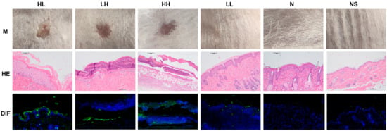
Figure 1
Open AccessReview
HIF-1α Pathway in COVID-19: A Scoping Review of Its Modulation and Related Treatments
by
Felipe Paes Gomes da Silva, Rafael Matte, David Batista Wiedmer, Arthur Paes Gomes da Silva, Rafaela Makiak Menin, Fernanda Bressianini Barbosa, Thainá Aymê Mocelin Meneguzzi, Sabrina Barancelli Pereira, Amanda Terres Fausto, Larissa Klug, Bruna Pinheiro Melim and Claudio Jose Beltrão
Int. J. Mol. Sci. 2025, 26(9), 4202; https://doi.org/10.3390/ijms26094202 (registering DOI) - 28 Apr 2025
Abstract
The COVID-19 pandemic, driven by SARS-CoV-2, has led to a global health crisis, highlighting the virus’s unique molecular mechanisms that distinguish it from other respiratory pathogens. It is known that the Hypoxia-Inducible Factor 1α (HIF-1α) activates a complex network of intracellular signaling pathways
[...] Read more.
The COVID-19 pandemic, driven by SARS-CoV-2, has led to a global health crisis, highlighting the virus’s unique molecular mechanisms that distinguish it from other respiratory pathogens. It is known that the Hypoxia-Inducible Factor 1α (HIF-1α) activates a complex network of intracellular signaling pathways regulating cellular energy metabolism, angiogenesis, and cell survival, contributing to the wide range of clinical manifestations of COVID-19, including Post-Acute COVID-19 Syndrome (PACS). Emerging evidence suggests that dysregulation of HIF-1α is a key driver of systemic inflammation, silent hypoxia, and pathological tissue remodeling in both the acute and post-acute phases of the disease. This scoping review was conducted following PRISMA-ScR guidelines and registered in INPLASY. It involved a literature search in Scopus and PubMed, supplemented by manual reference screening, with study selection facilitated by Rayyan software. Our analysis clarifies the dual role of HIF-1α, which may either worsen inflammatory responses and viral persistence or support adaptive mechanisms that reduce cellular damage. The potential for targeting HIF-1α therapeutically in COVID-19 is complex, requiring further investigation to clarify its precise role and translational applications. This review deepens the molecular understanding of SARS-CoV-2-induced cellular and tissue dysfunction in hypoxia, offering insights for improving clinical management strategies and addressing long-term sequelae.
Full article
(This article belongs to the Special Issue Novel Insights into Molecular Mechanisms of Pulmonary Pathology)
►▼
Show Figures
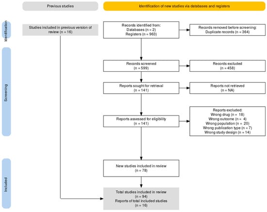
Figure 1
Open AccessArticle
Protective Effects of (-)-Butaclamol Against Gentamicin-Induced Ototoxicity: In Vivo and In Vitro Approaches
by
Sumin Hong, Eunjung Han, Saemi Park, Kyungtae Hyun, Yunkyoung Lee, Hyun woo Baek, Hwee-Jin Kim, Yoon Chan Rah and June Choi
Int. J. Mol. Sci. 2025, 26(9), 4201; https://doi.org/10.3390/ijms26094201 (registering DOI) - 28 Apr 2025
Abstract
Gentamicin-induced ototoxicity leads to irreversible sensorineural hearing loss due to structural and functional damage to inner ear hair cells. In this study, we identified (-)-butaclamol as a potent protective agent against gentamicin-induced cytotoxicity through high-content screening (HCS) of a natural compound library. (-)-Butaclamol
[...] Read more.
Gentamicin-induced ototoxicity leads to irreversible sensorineural hearing loss due to structural and functional damage to inner ear hair cells. In this study, we identified (-)-butaclamol as a potent protective agent against gentamicin-induced cytotoxicity through high-content screening (HCS) of a natural compound library. (-)-Butaclamol significantly enhanced cell viability in both HEI-OC1 cells and zebrafish neuromasts, demonstrating robust protection against gentamicin toxicity. Mechanistically, (-)-butaclamol inhibited intrinsic apoptosis, as evidenced by reduced TUNEL-positive cell counts and the downregulation of BAX and caspase-3, alongside the upregulation of BCL-2. Moreover, (-)-butaclamol activated key survival signaling pathways, including AKT/mTOR and ERK, while suppressing the inflammatory regulator NF-κB. Additional analyses revealed that (-)-butaclamol effectively mitigated oxidative stress and restored autophagic activity, as confirmed by CellROX and LysoTracker assays. Notably, TMRE staining showed that (-)-butaclamol preserved mitochondrial membrane potential in zebrafish hair cells, indicating mitochondrial protection. Collectively, these findings suggest that (-)-butaclamol exerts comprehensive cytoprotective effects against gentamicin-induced ototoxicity by modulating apoptosis, enhancing survival signaling, and restoring mitochondrial and cellular homeostasis. These results highlight the therapeutic potential of (-)-butaclamol and provide a foundation for future studies aimed at its clinical application.
Full article
(This article belongs to the Special Issue Programmed Cell Death and Oxidative Stress: 3rd Edition)
Open AccessCommunication
Genomic Alterations of the Infectious Bronchitis Virus (IBV) Strain of the GI-23 Lineage Induced by Passages in Chickens and Quails
by
Katarzyna Domanska-Blicharz, Joanna Sajewicz-Krukowska, Anna Lisowska, Justyna Opolska, Karolina Tarasiuk and Kamila Dziadek
Int. J. Mol. Sci. 2025, 26(9), 4200; https://doi.org/10.3390/ijms26094200 (registering DOI) - 28 Apr 2025
Abstract
Infectious bronchitis virus (IBV) of the GI-23 lineage, which first emerged in the Middle East in the late 1990s, has since spread worldwide. The factors driving its expansion, whether human involvement, wild bird migration, or the virus’s biological traits, are still unclear. This
[...] Read more.
Infectious bronchitis virus (IBV) of the GI-23 lineage, which first emerged in the Middle East in the late 1990s, has since spread worldwide. The factors driving its expansion, whether human involvement, wild bird migration, or the virus’s biological traits, are still unclear. This study aimed to trace the genome evolution of GI-23 IBV in chickens and its adaptability to quails, which are susceptible to both gamma- and deltacoronaviruses. Thirty specific-pathogen-free (SPF) birds, aged between two and three weeks, were used. Initially, three birds were inoculated with the G052/2016 IBV via the oculo-nasal route. On the third day post-infection (dpi), oropharyngeal swabs were collected from the whole group, pooled, and subsequently used to infect three next birds. This process was repeated nine more times during consecutive IBV passages (P-I–P-X), and eventually, virus sequencing was performed using Next-Generation Sequencing (NGS). The obtained results showed that quails were not susceptible to the IBV GI-23 lineage, as the virus RNA was detected in low amounts only during the first passage (QP-I) with no further detections in later rounds of IBV passaging. In chickens, only mild diarrhea symptoms appeared in a few individuals. The NGS analysis identified sixty-two single nucleotide variants (SNVs), thirty of which caused amino acid changes, twenty-eight were synonymous, and one SNV introduced a stop codon. Three SNVs were found in untranslated regions. However, none of these SNVs lasted beyond seven passages, with forty-four being unique SNVs. The Shannon entropy values measured during passages varied for pol1a, pol1b, S, 5a, 5b, and N genes, with overall genome complexity peaking at CP-VI and CP-X. The highest complexity was observed in the pol1a (CP-X) and S genes (CP-IV, CP-VI, CP-VIII, and CP-X). Along with the S gene that was under positive selection, eight codons in pol1a were also positively selected. These findings suggest that even in an adapted host, IBV variability does not stabilize without immune pressure, indicating continuous molecular changes within its genome.
Full article
(This article belongs to the Section Molecular Microbiology)
Open AccessReview
The Effect and Mechanism of Regular Exercise on Improving Insulin Impedance: Based on the Perspective of Cellular and Molecular Levels
by
Tingran Zhang, Yongsen Liu, Yi Yang, Jiong Luo and Chen Hao
Int. J. Mol. Sci. 2025, 26(9), 4199; https://doi.org/10.3390/ijms26094199 (registering DOI) - 28 Apr 2025
Abstract
Insulin resistance is more common in the elderly, and with the improvement in people’s living standards and changes in lifestyle habits, the incidence of insulin resistance in other age groups is also increasing year by year. Overweight and obesity caused by abnormal fat
[...] Read more.
Insulin resistance is more common in the elderly, and with the improvement in people’s living standards and changes in lifestyle habits, the incidence of insulin resistance in other age groups is also increasing year by year. Overweight and obesity caused by abnormal fat metabolism or accumulation can significantly reduce glucose intake, which is the direct cause of insulin resistance and the trigger for the occurrence and development of type II diabetes. This article reviews and analyzes relevant literature on empirical research on the effect of regular exercise on improving insulin resistance. It was found that the most important step in carbohydrate metabolism is the translocation of glucose transporter 4 (GLUT4) to the cell membrane, carrying water-soluble glucose through the lipid soluble cell membrane to complete carbohydrate transport. The process of glucose transporter protein translocation to the cell membrane can be driven by two different signaling pathways: one is the insulin information transfer pathway (ITP), the second is to induce the ITP of monophosphate-activated protein kinase (AMPK) through hypoxia or muscle contraction. For type II diabetes patients, the insulin signal transmission pathway through insulin receptors (IRS1, IRS2) and phosphatidylinositol 3-kinase (PI3K) (PI3K) is damaged, which results in the decrease in glucose absorption stimulated by insulin in skeletal muscle, while the noninsulin signal transmission pathway of AMPK in these patients is normal. It can be seen that regular exercise can regulate glucose intake and the metabolism of skeletal muscle, improve insulin resistance, reduce fasting blood glucose and glycosylated hemoglobin in diabetes patients, and thus, effectively regulate blood glucose. However, many steps in the molecular mechanism of how exercise training improves systemic insulin resistance are still not fully understood, and further discussion is needed in the future.
Full article
(This article belongs to the Special Issue Advances in Molecular Research of Diabetes Mellitus)
►▼
Show Figures

Figure 1
Open AccessReview
Evaluation of Illumina and Oxford Nanopore Sequencing for the Study of DNA Methylation in Alzheimer’s Disease and Frontotemporal Dementia
by
Lorenzo Pagano, Davide Lagrotteria, Alessandro Facconi, Claudia Saraceno, Antonio Longobardi, Sonia Bellini, Assunta Ingannato, Silvia Bagnoli, Tommaso Ducci, Alessandra Mingrino, Valentina Laganà, Ersilia Paparazzo, Barbara Borroni, Raffaele Maletta, Benedetta Nacmias, Alberto Montesanto and Roberta Ghidoni
Int. J. Mol. Sci. 2025, 26(9), 4198; https://doi.org/10.3390/ijms26094198 (registering DOI) - 28 Apr 2025
Abstract
DNA methylation is a critical epigenetic mechanism involved in numerous physiological processes. Alterations in DNA methylation patterns are associated with various brain disorders, including dementias such as Alzheimer’s disease (AD) and frontotemporal dementia (FTD). Investigating these alterations is essential for understanding the pathogenesis
[...] Read more.
DNA methylation is a critical epigenetic mechanism involved in numerous physiological processes. Alterations in DNA methylation patterns are associated with various brain disorders, including dementias such as Alzheimer’s disease (AD) and frontotemporal dementia (FTD). Investigating these alterations is essential for understanding the pathogenesis and progression of these disorders. Among the various methods for detecting DNA methylation, DNA sequencing is one of the most widely employed. Specifically, two main sequencing approaches are commonly used for DNA methylation analysis: bisulfite sequencing and single-molecule long-read sequencing. In this review, we compared the performances of CpG methylation detection obtained using two popular sequencing platforms, Illumina for bisulfite sequencing and Oxford Nanopore (ON) for long-read sequencing. Our comparison considers several factors, including accuracy, efficiency, genomic regions, costs, wet-lab protocols, and bioinformatics pipelines. We provide insights into the strengths and limitations of both methods with a particular focus on their application in research on AD and FTD.
Full article
(This article belongs to the Special Issue New Advances in Epigenetics and Epigenomics)
►▼
Show Figures

Figure 1
Open AccessArticle
rDNA Copy Number Variation and Methylation in Human and Mouse Sperm
by
Ramya Potabattula, Marcus Dittrich, Thomas Hahn, Martin Schorsch, Grazyna Ewa Ptak and Thomas Haaf
Int. J. Mol. Sci. 2025, 26(9), 4197; https://doi.org/10.3390/ijms26094197 (registering DOI) - 28 Apr 2025
Abstract
In this study, droplet digital PCR and deep bisulfite sequencing were used to study the absolute and active rDNA copy number (CN) and the effect of paternal age on human and mouse sperm. The absolute CN ranged from 98 to 404 (219 ±
[...] Read more.
In this study, droplet digital PCR and deep bisulfite sequencing were used to study the absolute and active rDNA copy number (CN) and the effect of paternal age on human and mouse sperm. The absolute CN ranged from 98 to 404 (219 ± 47) in human and from 98 to 177 (133 ± 14) in mouse sperm. Methylation of the human upstream control element/core promoter (UCE/CP) region and the 5′ external transcribed spacer, as well as that of the mouse CP, the spacer promoter, and 28S rDNA, significantly increased with donor age and absolute CN. Overall, rDNA hypomethylation was much more pronounced in mouse sperm, with 101.7 ± 11.4 copies showing a completely (0%) unmethylated and 11.3 ± 2.8 (8.5%) a slightly methylated (1–10%) CP region, compared to humans with 25.7 ± 9.5 (12%) completely unmethylated and 83.0 ± 19.8 slightly methylated UCE/CP regions. Although the absolute CN was much higher in human sperm, the number of copies with a hypomethylated (0–10%) promoter was comparable in humans (108.7 ± 28.3) and mice (113.0 ± 12.2). However, in mice, the majority (77%) of all copies were completely unmethylated, whereas in humans a high percentage (38%) showed one or two single CpG methylation errors. These different germline methylation dynamics may be due to species differences in reproductive strategies and lifespan. Complete demethylation of the sperm rDNA promoter in mice may be essential for embryonic genome activation, which already occurs at the 2-cell stage in mice and at the 4–8-cell stage in humans. The paternal age effect has been conserved between humans and mice with some notable differences. In humans, the number of hypomethylated (0–10%) copies decreased with age, whereas in mice only the completely unmethylated copies decreased with age. The number of methylated rDNA copies (>1% in mice and >10% in humans) significantly increased with age.
Full article
(This article belongs to the Special Issue New Advances in Germ Cell Research)
Open AccessArticle
Role of T3 in the Regulation of GRP78 on Granulosa Cells in Rat Ovaries
by
Yan Liu, Yilin Yao, Yakun Yu, Ying Sun, Mingqi Wu, Rui Chen, Haoyuan Feng, Shuaitian Guo, Yanzhou Yang and Cheng Zhang
Int. J. Mol. Sci. 2025, 26(9), 4196; https://doi.org/10.3390/ijms26094196 (registering DOI) - 28 Apr 2025
Abstract
Thyroid hormone (TH) plays a vital role in ovarian follicle development, and glucose-regulated protein 78 (GRP78) is involved in these processes, which is regulated by TH. However, the mechanisms are still unclear. To evaluate the possible mechanism of TH on the regulation of
[...] Read more.
Thyroid hormone (TH) plays a vital role in ovarian follicle development, and glucose-regulated protein 78 (GRP78) is involved in these processes, which is regulated by TH. However, the mechanisms are still unclear. To evaluate the possible mechanism of TH on the regulation of GRP78 expression, Cleavage Under Targets and Tagmentation (CUT & Tag) sequencing, luciferase assays, and Electrophoretic Mobility Shift Assays (EMSA) were employed to delineate the binding sites of thyroid hormone receptor β (TRβ) on the GRP78 promoter and to confirm the interactions. Additionally, Co-Immunoprecipitation (Co-IP) and Immunofluorescence (IF) assays were used to investigate the interactions between TRβ and the coactivator peroxisome proliferator-activated receptor γ coactivator 1α (PGC-1α) after triiodothyronine (T3) treatment with different concentrations. Our findings identified a thyroid hormone response element (TRE) on the GRP78 promoter and demonstrated that TRβ can activate GRP78 expression by interacting with PGC-1α. In order to simulate the condition of hyperthyroidism, granulosa cells (GCs) extracted from rats were treated by T3 with high concentrations, which decreased the expression of PGC-1α, resulting in decreased expressions of GRP78 and other ferroptosis-related markers such as glutathione peroxidase 4 (GPX4) and solute carrier family 7 member 11 (SLC7A11, xCT), thereby inducing ferroptosis in GCs. Taken together, the present study demonstrates that T3 induces cellular ferroptosis by binding TRE of the GRP78 promoter in ovarian GCs via TRβ. As a switcher, PGC-1α is also involved in these processes.
Full article
(This article belongs to the Section Molecular Endocrinology and Metabolism)
Open AccessReview
Analysis of Mechanisms for Electron Uptake by Methanothrix harundinacea 6Ac During Direct Interspecies Electron Transfer
by
Lei Wang, Xiaoman Shan, Yanhui Xu, Quan Xi, Haiming Jiang and Xia Li
Int. J. Mol. Sci. 2025, 26(9), 4195; https://doi.org/10.3390/ijms26094195 (registering DOI) - 28 Apr 2025
Abstract
Direct interspecies electron transfer (DIET) is a syntrophic metabolism wherein free electrons are directly transferred between microorganisms without the mediation of intermediates such as molecular hydrogen or formate. Previous research has demonstrated that Methanothrix harundinacea 6Ac is capable of reducing carbon dioxide through
[...] Read more.
Direct interspecies electron transfer (DIET) is a syntrophic metabolism wherein free electrons are directly transferred between microorganisms without the mediation of intermediates such as molecular hydrogen or formate. Previous research has demonstrated that Methanothrix harundinacea 6Ac is capable of reducing carbon dioxide through DIET. However, the mechanisms underlying electron uptake in M. harundinacea 6Ac during DIET remain poorly understood. This study aims to elucidate the electron and proton flux in M. harundinacea 6Ac during DIET and to propose a model for electron uptake in this organism, primarily based on the analysis of gene transcript levels, genomic characteristics of M. harundinacea 6Ac, and the pathways generating fully reduced ferridoxin (Fdred2−), reduced coenzyme F420 (F420H2), coenzyme M (CoM-SH), and coenzyme B (CoB-SH) during DIET. The findings suggest that membrane-bound heterodisulfide reductase (HdrED), F420H2-dehydrogenase lacking subunit F (Fpo−), and cytoplasmic heterodisulfide reductase (HdrABC)-subunit B of F420-reducing hydrogenase (FrhB) complex play critical roles in electron uptake in M. harundinacea 6Ac during DIET. Specifically, Fpo− is responsible for generating Fdred2− with reduced methanophenazine (MPH2), driven by a proton motive force, while HdrED facilitates the reduction of heterodisulfide of coenzyme M and coenzyme B (CoM-S-S-CoB) to CoM-SH and CoB-SH using MPH2. Additionally, cytoplasmic heterodisulfide reductase HdrABC and subunit B of coenzyme F420-hydrogenase complex (HdrABC-FrhB complex) catalyzes the reduction of oxidized coenzyme F420 (F420) to F420H2, utilizing CoM-SH, CoB-SH, and Fdred2−. This study represents the first genetics-based functional characterization of electron and proton flux in M. harundinacea 6Ac during DIET, providing a model for further investigation of electron uptake in Methanosaeta species. Furthermore, it deepens our understanding of the mechanisms underlying electron uptake in methanogens during DIET.
Full article
(This article belongs to the Section Physical Chemistry and Chemical Physics)
►▼
Show Figures
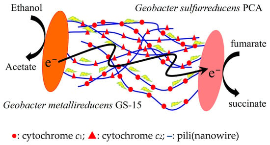
Figure 1
Open AccessArticle
Transcriptome-Wide Analysis and Experimental Validation from FFPE Tissue Identifies Stage-Specific Gene Expression Profiles Differentiating Adenoma, Carcinoma In-Situ and Adenocarcinoma in Colorectal Cancer Progression
by
Faisal Alhosani, Reem Sami Alhamidi, Burcu Yener Ilce, Alaa Muayad Altaie, Nival Ali, Alaa Mohamed Hamad, Axel Künstner, Cyrus Khandanpour, Hauke Busch, Basel Al-Ramadi, Rania Harati, Kadria Sayed, Ali AlFazari, Riyad Bendardaf and Rifat Hamoudi
Int. J. Mol. Sci. 2025, 26(9), 4194; https://doi.org/10.3390/ijms26094194 (registering DOI) - 28 Apr 2025
Abstract
Colorectal cancer (CRC) progression occurs through three stages: adenoma (pre-cancerous lesion), carcinoma in situ (CIS) and adenocarcinoma, with tumor stage playing a pivotal role in the prognosis and treatment outcomes. Despite therapeutic advancements, the lack of stage-specific biomarkers hinders the development of accurate
[...] Read more.
Colorectal cancer (CRC) progression occurs through three stages: adenoma (pre-cancerous lesion), carcinoma in situ (CIS) and adenocarcinoma, with tumor stage playing a pivotal role in the prognosis and treatment outcomes. Despite therapeutic advancements, the lack of stage-specific biomarkers hinders the development of accurate diagnostic tools and effective therapeutic strategies. This study aims to identify stage-specific gene expression profiles and key molecular mechanisms in CRC providing insights into molecular alterations across disease progression. Our methodological approach integrates the use of absolute gene set enrichment analysis (absGSEA) on formalin-fixed paraffin-embedded (FFPE)-derived transcriptomic data, combined with large-scale clinical validation and experimental confirmation. A comparative whole transcriptomic analysis (RNA-seq) was performed on FFPE samples including adenoma (n = 10), carcinoma in situ (CIS) (n = 8) and adenocarcinoma (n = 11) samples. Using absGSEA, we identified significant cellular pathways and putative molecular biomarkers associated with each stage of CRC progression. Key findings were then validated in a large independent CRC patient cohort (n = 1926), with survival analysis conducted from 1336 patients to assess the prognostic relevance of the candidate biomarkers. The key differentially expressed genes were experimentally validated using real-time PCR (RT-qPCR). Pathway analysis revealed that in CIS, apoptotic processes and Wnt signaling pathways were more prominent than in adenoma samples, while in adenocarcinoma, transcriptional co-regulatory mechanisms and protein kinase activity, which are critical for tumor growth and metastasis, were significantly enriched compared to adenoma. Additionally, extracellular matrix organization pathways were significantly enriched in adenocarcinoma compared to CIS. Distinct gene signatures were identified across CRC stages that differentiate between adenoma, CIS and adenocarcinoma. In adenoma, ARRB1, CTBP1 and CTBP2 were overexpressed, suggesting their involvement in early tumorigenesis, whereas in CIS, RPS3A and COL4A5 were overexpressed, suggesting their involvement in the transition from benign to malignant stage. In adenocarcinoma, COL1A2, CEBPZ, MED10 and PAWR were overexpressed, suggesting their involvement in advanced disease progression. Functional analysis confirmed that ARRB1 and CTBP1/2 were associated with early tumor development, while COL1A2 and CEBPZ were involved in extracellular matrix remodeling and transcriptional regulation, respectively. Experimental validation with RT-qPCR confirmed the differential expression of the candidate biomarkers (ARRB1, RPS3A, COL4A5, COL1A2 and MED10) across the three CRC stages reinforcing their potential as stage-specific biomarkers in CRC progression. These findings provide a foundation to distinguish between the CRC stages and for the development of accurate stage-specific diagnostic and prognostic biomarkers, which helps in the development of more effective therapeutic strategies for CRC.
Full article
(This article belongs to the Special Issue The Molecular Network, Key Biomarkers, and Therapeutic Targets in Colorectal Cancer)
►▼
Show Figures
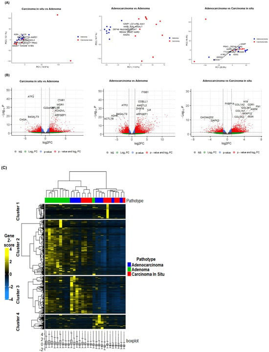
Figure 1
Open AccessArticle
Dysregulation of Labile Iron Predisposes Chemotherapy Resistant Cancer Cells to Ferroptosis
by
Luke V. Loftus, Louis T. A. Rolle, Bowen Wang, Kenneth J. Pienta and Sarah R. Amend
Int. J. Mol. Sci. 2025, 26(9), 4193; https://doi.org/10.3390/ijms26094193 (registering DOI) - 28 Apr 2025
Abstract
Despite centuries of research, metastatic cancer remains incurable due to resistance to all conventional cancer therapeutics. Alternative strategies leveraging non-proliferative vulnerabilities in cancer are required to overcome cancer recurrence. Ferroptosis is an iron dependent cell death pathway that has shown promising pre-clinical activity
[...] Read more.
Despite centuries of research, metastatic cancer remains incurable due to resistance to all conventional cancer therapeutics. Alternative strategies leveraging non-proliferative vulnerabilities in cancer are required to overcome cancer recurrence. Ferroptosis is an iron dependent cell death pathway that has shown promising pre-clinical activity in several contexts of therapeutic resistant cancer. However, ferroptosis sensitivity is highly variable across tissue types and cell states, posing a challenge for clinical translation. We describe a convergent phenotype induced by chemotherapy where cells surviving chemotherapy have dysregulated iron homeostasis, regardless of initial cell type or chemotherapy used. Elevated labile iron levels are counteracted by NRF2 signaling, yet the resulting antioxidant programs do not alleviate the labile iron burden. Selectively inhibiting GPX4 leads to uniform susceptibility to ferroptosis in surviving cells, highlighting the common reliance on lipid peroxidation defenses. Cellular iron dysregulation is a vulnerability of chemoresistant cancer cells that can be leveraged by triggering ferroptosis.
Full article
(This article belongs to the Section Molecular Biology)
►▼
Show Figures
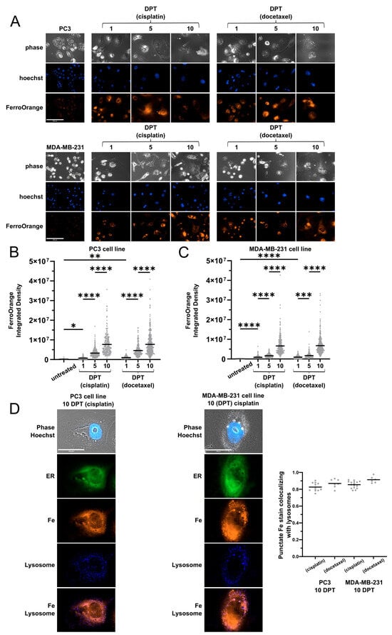
Figure 1
Open AccessReview
Innovations in Drug Discovery for Sickle Cell Disease Targeting Oxidative Stress and NRF2 Activation—A Short Review
by
Athena Starlard-Davenport, Chithra D. Palani, Xingguo Zhu and Betty S. Pace
Int. J. Mol. Sci. 2025, 26(9), 4192; https://doi.org/10.3390/ijms26094192 (registering DOI) - 28 Apr 2025
Abstract
Sickle cell disease (SCD) is a monogenic blood disorder characterized by abnormal hemoglobin S production, which polymerizes under hypoxia conditions to produce chronic red blood cell hemolysis, widespread organ damage, and vasculopathy. As a result of vaso-occlusion and ischemia-reperfusion injury, individuals with SCD
[...] Read more.
Sickle cell disease (SCD) is a monogenic blood disorder characterized by abnormal hemoglobin S production, which polymerizes under hypoxia conditions to produce chronic red blood cell hemolysis, widespread organ damage, and vasculopathy. As a result of vaso-occlusion and ischemia-reperfusion injury, individuals with SCD have recurrent pain episodes, infection, pulmonary disease, and fall victim to early death. Oxidative stress due to chronic hemolysis and the release of hemoglobin and free heme is a key driver of the clinical manifestations of SCD. The net result is the generation of reactive oxygen species that consume nitric oxide and overwhelm the antioxidant system due to a reduction in enzymes such as superoxide dismutase and glutathione peroxidase. The primary mechanism for handling cellular oxidative stress is the activation of antioxidant proteins by the transcription factor NRF2, a promising target for treatment development, given the significant role of oxidative stress in the clinical severity of SCD. In this review, we discuss the role of oxidative stress in health and the clinical complications of SCD, and the potential of NRF2 as a treatment target, offering hope for developing effective therapies for SCD. This task requires our collective dedication and focus.
Full article
(This article belongs to the Special Issue Oxidation in Human Health and Disease)
►▼
Show Figures
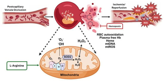
Figure 1
Open AccessArticle
Therapeutic Effects of Sigesbeckia pubescens Makino Against Atopic Dermatitis-Like Skin Inflammation Through the JAK2/STAT Signaling Pathway
by
Hyun-Kyung Song, Hye Jin Kim, Seong Cheol Kim and Taesoo Kim
Int. J. Mol. Sci. 2025, 26(9), 4191; https://doi.org/10.3390/ijms26094191 (registering DOI) - 28 Apr 2025
Abstract
Atopic dermatitis (AD), a chronic inflammatory skin condition, is a common allergic disorder. The human skin, the largest organ, serves as the first barrier in protecting the body against various external threats. Human epidermal keratinocytes (HEKs) in the epidermal layer and human dermal
[...] Read more.
Atopic dermatitis (AD), a chronic inflammatory skin condition, is a common allergic disorder. The human skin, the largest organ, serves as the first barrier in protecting the body against various external threats. Human epidermal keratinocytes (HEKs) in the epidermal layer and human dermal fibroblasts (HDFs) in the dermis of the skin are implicated in AD-associated skin inflammation through the secretion of diverse inflammatory mediators, including chemokines. Sigesbeckia pubescens Makino (SP), a traditional Korean and Chinese herbal remedy, is used for treating inflammatory conditions. While several pharmacological effects of SP extract (SPE) have been documented, its specific inhibitory effect on AD-related skin inflammation remains unexplored. Hence, oral administration of SPE to NC/Nga mice reduced the severity of house dust mite extract-induced dermatitis, accompanied by lowered levels of serum inflammatory mediators, decreased epidermal thickness, reduced mast cell infiltration, and restoration of skin barrier function within skin lesions. In conclusion, SPE has demonstrated the ability to alleviate skin inflammation and protect the skin barrier and shows potential as a therapeutic option for AD. SPE inhibited proinflammatory chemokine production by modulating the Janus kinase (JAK) 2/signal transducer and activator of transcription proteins (STAT) 1/STAT3 signaling pathway in IFN-γ- and TNF-α-stimulated skin cells.
Full article
(This article belongs to the Special Issue Molecular Mechanisms and Therapeutic Targets in Skin Diseases)
Open AccessArticle
Comparative Brain and Serum Exosome Expression of Biomarkers in an Experimental Model of Alzheimer-Type Neurodegeneration: Potential Relevance to Liquid Biopsy Diagnostics
by
Suzanne M. de la Monte, Yiwen Yang, Anjali Prabhu and Ming Tong
Int. J. Mol. Sci. 2025, 26(9), 4190; https://doi.org/10.3390/ijms26094190 (registering DOI) - 28 Apr 2025
Abstract
The development of more effective disease-modifying treatments for Alzheimer’s disease (AD) is compromised by the lack of streamlined measures to detect and monitor the full spectrum of neurodegeneration, including white matter pathology, which begins early. This study utilized an established intracerebral streptozotocin (STZ)
[...] Read more.
The development of more effective disease-modifying treatments for Alzheimer’s disease (AD) is compromised by the lack of streamlined measures to detect and monitor the full spectrum of neurodegeneration, including white matter pathology, which begins early. This study utilized an established intracerebral streptozotocin (STZ) model of AD to examine the potential utility of a non-invasive serum extracellular vesicle (SEV)-based liquid biopsy approach for detecting a broad range of molecular pathologies related to neurodegeneration. The design enabled comparative analysis of immunoreactivity in frontal lobe tissue (FLTX), frontal lobe-derived EVs (FLEVs), and SEVs. Long Evans rats were administered i.c. STZ or saline (control) on postnatal day 3 (P3). Morris Water Maze testing was performed from P24 to P27. On P31–32, the rats were sacrificed to harvest FLTX and serum for EV characterization. STZ caused brain atrophy, with deficits in spatial learning and memory. STZ significantly impacted FLEV and SEV nanoparticle abundance and size distributions and concordantly increased AD (Tau, pTau, and Aβ) and oxidative stress (ubiquitin, 4-HNE) biomarkers, as well as immunoreactivity to immature oligodendrocyte (PLP), non-myelinating glial (PDGFRA, GALC) proteins, MAG, nestin, and GFAP in FLTX and FLEV. The SEVs also exhibited concordant STZ-related effects, but they were limited to increased levels of 4-HNE, PLP, PDGFRA, GALC, MAG, and GFAP. The findings suggest that non-invasive EV-based liquid biopsy approaches could potentially be used to detect and monitor some aspects of AD-type neurodegeneration. Targeting brain-specific EVs in serum will likely increase the sensitivity of this promising non-invasive approach for diagnostic and clinical management.
Full article
(This article belongs to the Special Issue The Role of Extracellular Vesicles in Inflammatory Diseases)
►▼
Show Figures
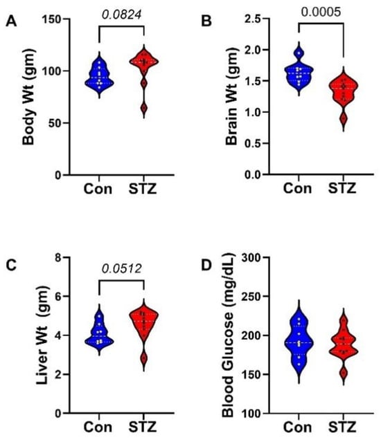
Figure 1
Open AccessArticle
Interaction Between Glycoside Hydrolase FsGH28c from Fusarium solani and PnPUB35 Confers Resistance in Piper nigrum
by
Shichao Liu, Tianci Xing, Ruibing Liu, Shengfeng Gao, Jianfeng Yang, Tian Tian, Chong Zhang, Shiwei Sun and Chenchen Zhao
Int. J. Mol. Sci. 2025, 26(9), 4189; https://doi.org/10.3390/ijms26094189 (registering DOI) - 28 Apr 2025
Abstract
Pathogens deploy various molecular mechanisms to overcome host defenses, among which glycoside hydrolases (GHs) play a critical role as virulence factors. Understanding the functional roles of these enzymes is essential for uncovering pathogen–host interactions and developing strategies for disease management. Fusarium wilt has
[...] Read more.
Pathogens deploy various molecular mechanisms to overcome host defenses, among which glycoside hydrolases (GHs) play a critical role as virulence factors. Understanding the functional roles of these enzymes is essential for uncovering pathogen–host interactions and developing strategies for disease management. Fusarium wilt has occurred in the main Piper nigrum cultivation regions, which seriously affects the yield and quality of P. nigrum. Here, we identified and characterized FsGH28c, a GH28 family member in Fusarium solani. Its expression was significantly upregulated during the infection of black pepper (Piper nigrum) roots by F. solani cv. WN-1, indicating its potential role in pathogenicity. FsGH28c elicited cell death in Nicotiana benthamiana and modulated the expression of genes related to pathogenesis. FsGH28c exerts a positive influence on the pathogenicity of F. solani. The knockout of FsGH28c mutant strains markedly attenuated F. solani ’s virulence in black pepper plants. The knockout mutant strains decrease the ability of F. solani to utilize carbon sources. The FsGH28c deletion did not affect mycelial growth on PDA but did impact spore development. We identified a U-box protein, PnPUB35, interacting with FsGH28c using yeast two-hybrid and bimolecular fluorescence complementation assays. PnPUB35 conferred enhanced resistance to F. solani in black pepper through positive regulation. These findings suggest that FsGH28c may function as a virulence factor by modulating host immune responses through its interaction with PnPUB35.
Full article
(This article belongs to the Special Issue Crop Stress Biology and Molecular Breeding: 5th Edition)
►▼
Show Figures
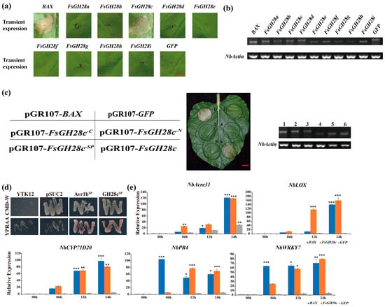
Figure 1
Open AccessReview
Unlocking the Pharmacological Potential of Myricetin Against Various Pathogenesis
by
Saleh A. Almatroodi and Arshad Husain Rahmani
Int. J. Mol. Sci. 2025, 26(9), 4188; https://doi.org/10.3390/ijms26094188 (registering DOI) - 28 Apr 2025
Abstract
Myricetin is a natural flavonoid with powerful antioxidant and anti-inflammatory potential commonly found in vegetables, fruits, nuts, and tea. The vital role of this flavonoid in the prevention and treatment of various diseases is evidenced by its ability to reduce inflammation and oxidative
[...] Read more.
Myricetin is a natural flavonoid with powerful antioxidant and anti-inflammatory potential commonly found in vegetables, fruits, nuts, and tea. The vital role of this flavonoid in the prevention and treatment of various diseases is evidenced by its ability to reduce inflammation and oxidative stress, maintain tissue architecture, and modulate cell signaling pathways. Thus, this review summarizes recent evidence on myricetin, focusing precisely on its mechanisms of action in various pathogenesis, including obesity, diabetes mellitus, arthritis, osteoporosis, liver, neuro, cardio, and reproductive system-associated pathogenesis. Moreover, it has been revealed that myricetin exhibits anti-microbial properties due to obstructive virulence factors, preventing biofilm formation and disrupting membrane integrity. Additionally, synergistic potential with other drugs and the role of myricetin-based nanoformulations in different diseases are properly discussed. This review seeks to increase the understanding of myricetin’s pharmacological potential in various diseases, principally highlighting its effective mechanisms of action. Further wide-ranging research, as well as more randomized and controlled clinical trial studies, should be executed to reconnoiter this compound’s therapeutic value, safety, and usefulness against various human pathogenesis.
Full article
(This article belongs to the Special Issue Effects of Bioactive Compounds in Oxidative Stress and Inflammation)
Open AccessArticle
Short- and Long-Term Evolution of Microbial Cultures: A Thermodynamic Perspective
by
Alberto Schiraldi
Int. J. Mol. Sci. 2025, 26(9), 4187; https://doi.org/10.3390/ijms26094187 (registering DOI) - 28 Apr 2025
Abstract
A thermodynamic description of cell duplication reflects Odum’s view of a feedback energy loop that sustains the transformation of the energy of substrates in the (higher quality) energy of new microbial cells with the dissipation of heat and the (lower quality) energy of
[...] Read more.
A thermodynamic description of cell duplication reflects Odum’s view of a feedback energy loop that sustains the transformation of the energy of substrates in the (higher quality) energy of new microbial cells with the dissipation of heat and the (lower quality) energy of catabolites. For a closed batch microbial culture, entropy increases during the whole growth and decay cycle, i.e., the production of entropy during the growth phase displays a rate proportional to the number of cell duplications per unit time, while during the decay phase, it depends on the death rate. Because of its high mobility, water is assumed to exhibit the same thermodynamic activity throughout the system. This assumption leads to the conclusion that an osmotic balance exists between cells and their surrounding medium, which, in a closed batch culture, can affect the rate and the extent of the microbial growth. Finally, the paper suggests a thermodynamic interpretation of the increase in fitness observed in a long-term evolution experiment (LTEE), based on the supposed exergy difference between the generating and generated cells in each duplication, which is also a measure of the “age” of the cells, i.e., aged cells die first. This produces microbial cultures richer in cells with enhanced duplication potential after the many thousand generations considered in an LTEE.
Full article
(This article belongs to the Special Issue Life’s Thermodynamics: Cells and Evolution)
►▼
Show Figures
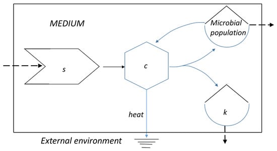
Figure 1
Open AccessArticle
Stevia Leaf Extract Fermented with Plant-Derived Lactobacillus plantarum SN13T Displays Anticancer Activity to Pancreatic Cancer PANC-1 Cell Line
by
Rentao Zhang, Narandalai Danshiitsoodol, Masafumi Noda, Sayaka Yonezawa, Keishi Kanno and Masanori Sugiyama
Int. J. Mol. Sci. 2025, 26(9), 4186; https://doi.org/10.3390/ijms26094186 (registering DOI) - 28 Apr 2025
Abstract
Pancreatic cancer is a highly malignant tumor that remains a significant global health burden. In this study, we demonstrated the anticancer potential of stevia leaf extract fermented with plant-derived Lactobacillus (L.) plantarum SN13T strain. Evaluation of antioxidant capacity (including DPPH and
[...] Read more.
Pancreatic cancer is a highly malignant tumor that remains a significant global health burden. In this study, we demonstrated the anticancer potential of stevia leaf extract fermented with plant-derived Lactobacillus (L.) plantarum SN13T strain. Evaluation of antioxidant capacity (including DPPH and ABTS radical scavenging activities and H2O2-induced oxidative damage repair in HEK-293 cells), as well as cytotoxicity against pancreatic cancer cells (PANC-1) and non-cancerous human embryonic kidney (HEK-293), revealed that the fermented extract exhibited significantly enhanced antioxidant activity and cytotoxicity against PANC-1 cells while showing minimal toxicity to HEK-293 cells compared to the unfermented extract. Further, validation through clonogenic, migration, and wound-healing assays demonstrated that the fermented extract effectively inhibited the proliferation and migration of PANC-1 cells. The active compound in the fermented extract has been identified as chlorogenic acid methyl ester (CAME), with a concentration of 374.4 μg/mL. Flow cytometry analysis indicated that CAME significantly arrested PANC-1 cells in the G0/G1 phase and induced apoptosis. Furthermore, CAME upregulated the expression of pro-apoptotic genes Bax, Bad, Caspase-3/9, Cytochrome c, and E-cadherin, while downregulating the anti-apoptotic gene Bcl-2. These findings suggest that CAME exerts potent cytotoxic effects on PANC-1 cells by inhibiting cell proliferation and migration, arresting the cell cycle, and regulating apoptosis-related gene expression. In conclusion, stevia leaf extract fermented with L. plantarum SN13T, which contains CAME, may serve as a promising candidate for pancreatic cancer treatment.
Full article
(This article belongs to the Special Issue Probiotics in Health and Disease)
►▼
Show Figures
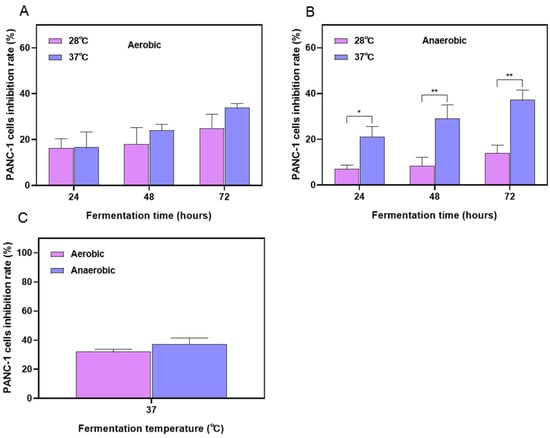
Figure 1

Journal Menu
► ▼ Journal Menu-
- IJMS Home
- Aims & Scope
- Editorial Board
- Reviewer Board
- Topical Advisory Panel
- Instructions for Authors
- Special Issues
- Topics
- Sections & Collections
- Article Processing Charge
- Indexing & Archiving
- Most Cited & Viewed
- Journal Statistics
- Journal History
- Journal Awards
- Society Collaborations
- Conferences
- Editorial Office
Journal Browser
► ▼ Journal Browser-
arrow_forward_ios
Forthcoming issue
arrow_forward_ios Current issue - Vol. 26 (2025)
- Vol. 25 (2024)
- Vol. 24 (2023)
- Vol. 23 (2022)
- Vol. 22 (2021)
- Vol. 21 (2020)
- Vol. 20 (2019)
- Vol. 19 (2018)
- Vol. 18 (2017)
- Vol. 17 (2016)
- Vol. 16 (2015)
- Vol. 15 (2014)
- Vol. 14 (2013)
- Vol. 13 (2012)
- Vol. 12 (2011)
- Vol. 11 (2010)
- Vol. 10 (2009)
- Vol. 9 (2008)
- Vol. 8 (2007)
- Vol. 7 (2006)
- Vol. 6 (2005)
- Vol. 5 (2004)
- Vol. 4 (2003)
- Vol. 3 (2002)
- Vol. 2 (2001)
- Vol. 1 (2000)
Highly Accessed Articles
Latest Books
E-Mail Alert
News
Topics
Topic in
BioChem, Biomedicines, Biomolecules, IJMS, Metabolites, Molecules
Natural Products in Prevention and Therapy of Metabolic Syndrome
Topic Editors: Jianbo Wan, Ligen LinDeadline: 30 April 2025
Topic in
Biomedicines, Brain Sciences, CIMB, Diagnostics, IJMS, IJTM
Autism: Molecular Bases, Diagnosis and Therapies, 2nd Volume
Topic Editors: Lello Zolla, Kunio YuiDeadline: 31 May 2025
Topic in
Biomolecules, IJMS, Molecules, Pharmaceutics
Advances in Diagnostics, Brain Delivery Systems and Therapeutics of Neurodegenerative Disease
Topic Editors: Ashok Iyaswamy, Chuanbin Yang, Abhimanyu ThakurDeadline: 11 June 2025
Topic in
Chemistry, Foods, IJMS, Molecules, Separations
Recent Trends and Advances in Food Authentication and Traceability
Topic Editors: Michael Kontominas, Anastasia BadekaDeadline: 30 June 2025

Conferences
Special Issues
Special Issue in
IJMS
Advances in Plant Cell Dynamics
Guest Editor: Andrei SmertenkoDeadline: 29 April 2025
Special Issue in
IJMS
Peptide Receptor Radionuclide Therapy (PRRT) and Targeted Alpha Therapy (TAT) in Cancer
Guest Editor: Behrooz H. YousefiDeadline: 29 April 2025
Special Issue in
IJMS
Advances in Molecular Insight into Neuroendocrine Cells
Guest Editor: Cristina VelazquezDeadline: 30 April 2025
Special Issue in
IJMS
Plant Responses to Heavy Metals: From Deficiency to Excess, 2nd Edition
Guest Editors: Anna D. Kozhevnikova, Ilya Vladimirovich SereginDeadline: 30 April 2025
Topical Collections
Topical Collection in
IJMS
Feature Papers in Molecular Toxicology
Collection Editor: Guido R.M.M. Haenen
Topical Collection in
IJMS
Feature Paper Collection in Molecular Endocrinology and Metabolism
Collection Editor: José L. Quiles
Topical Collection in
IJMS
Bioactive Natural Compounds for Therapeutics and Nutraceutical Applications
Collection Editors: Sonia A.O. Santos, Armando J. D. Silvestre, Raphael Grougnet, Vessela Balabanova
Topical Collection in
IJMS
30th Anniversary of IJMS: Updates and Advances in Biochemistry
Collection Editor: Claudiu T. Supuran


