Next Issue
Volume 9, November
Previous Issue
Volume 9, September


Viruses, Volume 9, Issue 10 (October 2017) – 47 articles

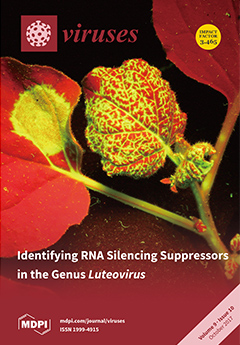
Cover Story (view full-size image): Viruses use silencing suppressor proteins to combat or avoid a plant’s intrinsic RNA silencing defense mechanism. In the family Luteoviridae, members of the genera Polerovirus and Enamovirus have a strong suppressor of local and systemic RNA silencing, named P0. Members of the genus Luteovirus, however, lack the corresponding ORF 0. We show that, in the absence of P0, luteoviruses have evolved a P4 movement protein that also displays strong systemic silencing suppression activity by inhibiting the accumulation of small RNAs. View this paper
  • Issues are regarded as officially published after their release is announced to the table of contents alert mailing list.
  • You may sign up for e-mail alerts to receive table of contents of newly released issues.
  • PDF is the official format for papers published in both, html and pdf forms. To view the papers in pdf format, click on the "PDF Full-text" link, and use the free Adobe Reader to open them.
Order results
Result details
Section
Select all
Export citation of selected articles as:
766 KiB  
Communication
Human Metapneumovirus Attachment Protein Contributes to Neutrophil Recruitment into the Airways of Infected Mice
by Nagarjuna R. Cheemarla and Antonieta Guerrero-Plata
Viruses 2017, 9(10), 310; https://doi.org/10.3390/v9100310 - 22 Oct 2017
Cited by 11 | Viewed by 4399
Abstract
Human Metapneumovirus (HMPV) is a leading respiratory pathogen that causes lower respiratory tract infections worldwide. Acute HMPV infection induces an exacerbated inflammatory neutrophilic response leading to bronchiolitis and pneumonia. However, the mechanism by which the virus regulates neutrophil infiltration into the airways still [...] Read more.
Human Metapneumovirus (HMPV) is a leading respiratory pathogen that causes lower respiratory tract infections worldwide. Acute HMPV infection induces an exacerbated inflammatory neutrophilic response leading to bronchiolitis and pneumonia. However, the mechanism by which the virus regulates neutrophil infiltration into the airways still remains unexplored. In this work, we used an experimental mouse model of HMPV infection to demonstrate that the attachment (G) protein of HMPV contributes to the recruitment of neutrophils into the airways and modulate the production of neutrophil chemoattractants and Type I IFN responses, specifically IFN-α. These findings provide the first evidence that the HMPV G protein contributes to the in vivo neutrophilic response to HMPV infection and furthers our understanding on virus induced inflammatory responses in the airways. Full article
(This article belongs to the Special Issue Viruses and Inflammation)
Show Figures

Figure 1

969 KiB  
Review
The Battle of RNA Synthesis: Virus versus Host
by Alex Harwig, Robert Landick and Ben Berkhout
Viruses 2017, 9(10), 309; https://doi.org/10.3390/v9100309 - 21 Oct 2017
Cited by 26 | Viewed by 8464
Abstract
Transcription control is the foundation of gene regulation. Whereas a cell is fully equipped for this task, viruses often depend on the host to supply tools for their transcription program. Over the course of evolution and adaptation, viruses have found diverse ways to [...] Read more.
Transcription control is the foundation of gene regulation. Whereas a cell is fully equipped for this task, viruses often depend on the host to supply tools for their transcription program. Over the course of evolution and adaptation, viruses have found diverse ways to optimally exploit cellular host processes such as transcription to their own benefit. Just as cells are increasingly understood to employ nascent RNAs in transcription regulation, recent discoveries are revealing how viruses use nascent RNAs to benefit their own gene expression. In this review, we first outline the two different transcription programs used by viruses, i.e., transcription (DNA-dependent) and RNA-dependent RNA synthesis. Subsequently, we use the distinct stages (initiation, elongation, termination) to describe the latest insights into nascent RNA-mediated regulation in the context of each relevant stage. Full article
(This article belongs to the Special Issue Structure-Function Relationships in Viral Polymerases)
Show Figures

Figure 1

934 KiB  
Review
Gammaherpesviral Tegument Proteins, PML-Nuclear Bodies and the Ubiquitin-Proteasome System
by Florian Full, Alexander S. Hahn, Anna K. Großkopf and Armin Ensser
Viruses 2017, 9(10), 308; https://doi.org/10.3390/v9100308 - 21 Oct 2017
Cited by 6 | Viewed by 7727
Abstract
Gammaherpesviruses like Epstein-Barr virus (EBV) and Kaposi’s sarcoma-associated herpesvirus (KSHV) subvert the ubiquitin proteasome system for their own benefit in order to facilitate viral gene expression and replication. In particular, viral tegument proteins that share sequence homology to the formylglycineamide ribonucleotide amidotransferase (FGARAT, [...] Read more.
Gammaherpesviruses like Epstein-Barr virus (EBV) and Kaposi’s sarcoma-associated herpesvirus (KSHV) subvert the ubiquitin proteasome system for their own benefit in order to facilitate viral gene expression and replication. In particular, viral tegument proteins that share sequence homology to the formylglycineamide ribonucleotide amidotransferase (FGARAT, or PFAS), an enzyme in the cellular purine biosynthesis, are important for disrupting the intrinsic antiviral response associated with Promyelocytic Leukemia (PML) protein-associated nuclear bodies (PML-NBs) by proteasome-dependent and independent mechanisms. In addition, all herpesviruses encode for a potent ubiquitin protease that can efficiently remove ubiquitin chains from proteins and thereby interfere with several different cellular pathways. In this review, we discuss mechanisms and functional consequences of virus-induced ubiquitination and deubiquitination for early events in gammaherpesviral infection. Full article
(This article belongs to the Special Issue Viruses, ERAD, and the Proteasome)
Show Figures

Figure 1

4573 KiB  
Article
The Operophtera brumata Nucleopolyhedrovirus (OpbuNPV) Represents an Early, Divergent Lineage within Genus Alphabaculovirus
by Robert L. Harrison, Daniel L. Rowley, Joseph D. Mowery, Gary R. Bauchan and John P. Burand
Viruses 2017, 9(10), 307; https://doi.org/10.3390/v9100307 - 21 Oct 2017
Cited by 22 | Viewed by 5843
Abstract
Operophtera brumata nucleopolyhedrovirus (OpbuNPV) infects the larvae of the winter moth, Operophtera brumata. As part of an effort to explore the pesticidal potential of OpbuNPV, an isolate of this virus from Massachusetts (USA)—OpbuNPV-MA—was characterized by electron microscopy of OpbuNPV occlusion bodies (OBs) [...] Read more.
Operophtera brumata nucleopolyhedrovirus (OpbuNPV) infects the larvae of the winter moth, Operophtera brumata. As part of an effort to explore the pesticidal potential of OpbuNPV, an isolate of this virus from Massachusetts (USA)—OpbuNPV-MA—was characterized by electron microscopy of OpbuNPV occlusion bodies (OBs) and by sequencing of the viral genome. The OBs of OpbuNPV-MA consisted of irregular polyhedra and contained virions consisting of a single rod-shaped nucleocapsid within each envelope. Presumptive cypovirus OBs were also detected in sections of the OB preparation. The OpbuNPV-MA genome assembly yielded a circular contig of 119,054 bp and was found to contain little genetic variation, with most polymorphisms occurring at a frequency of < 6%. A total of 130 open reading frames (ORFs) were annotated, including the 38 core genes of Baculoviridae, along with five homologous repeat (hr) regions. The results of BLASTp and phylogenetic analysis with selected ORFs indicated that OpbuNPV-MA is not closely related to other alphabaculoviruses. Phylogenies based on concatenated core gene amino acid sequence alignments placed OpbuNPV-MA on a basal branch lying outside other alphabaculovirus clades. These results indicate that OpbuNPV-MA represents a divergent baculovirus lineage that appeared early during the diversification of genus Alphabaculovirus. Full article
(This article belongs to the Section Invertebrate Viruses)
Show Figures

Figure 1

3472 KiB  
Communication
Mutation of a Conserved Nuclear Export Sequence in Chikungunya Virus Capsid Protein Disrupts Host Cell Nuclear Import
by Susan C. Jacobs, Adam Taylor, Lara J. Herrero, Suresh Mahalingam and John K. Fazakerley
Viruses 2017, 9(10), 306; https://doi.org/10.3390/v9100306 - 20 Oct 2017
Cited by 7 | Viewed by 4972
Abstract
Transmitted by mosquitoes; chikungunya virus (CHIKV) is responsible for frequent outbreaks of arthritic disease in humans. CHIKV is an arthritogenic alphavirus of the Togaviridae family. Capsid protein, a structural protein encoded by the CHIKV RNA genome, is able to translocate to the host [...] Read more.
Transmitted by mosquitoes; chikungunya virus (CHIKV) is responsible for frequent outbreaks of arthritic disease in humans. CHIKV is an arthritogenic alphavirus of the Togaviridae family. Capsid protein, a structural protein encoded by the CHIKV RNA genome, is able to translocate to the host cell nucleus. In encephalitic alphaviruses nuclear translocation induces host cell shut off; however, the role of capsid protein nuclear localisation in arthritogenic alphaviruses remains unclear. Using replicon systems, we investigated a nuclear export sequence (NES) in the N-terminal region of capsid protein; analogous to that found in encephalitic alphavirus capsid but uncharacterised in CHIKV. The chromosomal maintenance 1 (CRM1) export adaptor protein mediated CHIKV capsid protein export from the nucleus and a region within the N-terminal part of CHIKV capsid protein was required for active nuclear targeting. In contrast to encephalitic alphaviruses, CHIKV capsid protein did not inhibit host nuclear import; however, mutating the NES of capsid protein (∆NES) blocked host protein access to the nucleus. Interactions between capsid protein and the nucleus warrant further investigation. Full article
(This article belongs to the Special Issue Advances in Alphavirus Research)
Show Figures

Graphical abstract

6597 KiB  
Article
Structural and Functional Insight into Canarypox Virus CNP058 Mediated Regulation of Apoptosis
by Mohd Ishtiaq Anasir, Amy A. Baxter, Ivan K. H. Poon, Mark D. Hulett and Marc Kvansakul
Viruses 2017, 9(10), 305; https://doi.org/10.3390/v9100305 - 20 Oct 2017
Cited by 18 | Viewed by 5765
Abstract
Programmed cell death or apoptosis is an important component of host defense systems against viral infection. The B-cell lymphoma 2 (Bcl-2) proteins family is the main arbiter of mitochondrially mediated apoptosis, and viruses have evolved sequence and structural mimics of Bcl-2 to subvert [...] Read more.
Programmed cell death or apoptosis is an important component of host defense systems against viral infection. The B-cell lymphoma 2 (Bcl-2) proteins family is the main arbiter of mitochondrially mediated apoptosis, and viruses have evolved sequence and structural mimics of Bcl-2 to subvert premature host cell apoptosis in response to viral infection. The sequencing of the canarypox virus genome identified a putative pro-survival Bcl-2 protein, CNP058. However, a role in apoptosis inhibition for CNP058 has not been identified to date. Here, we report that CNP058 is able to bind several host cell pro-death Bcl-2 proteins, including Bak and Bax, as well as several BH3 only-proteins including Bim, Bid, Bmf, Noxa, Puma, and Hrk with high to moderate affinities. We then defined the structural basis for CNP058 binding to pro-death Bcl-2 proteins by determining the crystal structure of CNP058 bound to Bim BH3. CNP058 adopts the conserved Bcl-2 like fold observed in cellular pro-survival Bcl-2 proteins, and utilizes the canonical ligand binding groove to bind Bim BH3. We then demonstrate that CNP058 is a potent inhibitor of ultraviolet (UV) induced apoptosis in a cell culture model. Our findings suggest that CNP058 is a potent inhibitor of apoptosis that is able to bind to BH3 domain peptides from a broad range of pro-death Bcl-2 proteins, and may play a key role in countering premature host apoptosis. Full article
(This article belongs to the Special Issue Viral Infection and Apoptosis)
Show Figures

Figure 1

997 KiB  
Review
Hepatitis B Virus and DNA Damage Response: Interactions and Consequences for the Infection
by Andoni Gómez-Moreno and Urtzi Garaigorta
Viruses 2017, 9(10), 304; https://doi.org/10.3390/v9100304 - 19 Oct 2017
Cited by 25 | Viewed by 11386
Abstract
Hepatitis B virus (HBV) is a major etiologic agent of acute and chronic hepatitis, and end-stage liver disease. Establishment of HBV infection, progression to persistency and pathogenesis are determined by viral and cellular factors, some of which remain still undefined. Key steps of [...] Read more.
Hepatitis B virus (HBV) is a major etiologic agent of acute and chronic hepatitis, and end-stage liver disease. Establishment of HBV infection, progression to persistency and pathogenesis are determined by viral and cellular factors, some of which remain still undefined. Key steps of HBV life cycle e.g., transformation of genomic viral DNA into transcriptionally active episomal DNA (cccDNA) or transcription of viral mRNAs from cccDNA, take place in the nucleus of infected cells and strongly depend on enzymatic activities provided by cellular proteins. In this regard, DNA damage response (DDR) pathways and some DDR proteins are being recognized as important factors regulating the infection. On one hand, HBV highjacks specific DDR proteins to successfully complete some of the steps of its life cycle. On the other hand, HBV subverts DDR pathways to presumably create a cellular environment that favours its replication. Direct consequences of these interactions are: HBV DNA integration into host chromosomal DNA, and accumulation of mutations in host chromosomal DNA that could eventually trigger carcinogenic processes, which would explain in part the incidence of hepatocellular carcinoma in chronically infected patients. Unravelling the interactions that HBV establishes with DDR pathways might help identify new molecular targets for therapeutic intervention. Full article
(This article belongs to the Special Issue Viruses and the DNA Damage Response)
Show Figures

Figure 1

677 KiB  
Review
The Th17 Lineage: From Barrier Surfaces Homeostasis to Autoimmunity, Cancer, and HIV-1 Pathogenesis
by Vanessa Sue Wacleche, Alan Landay, Jean-Pierre Routy and Petronela Ancuta
Viruses 2017, 9(10), 303; https://doi.org/10.3390/v9100303 - 19 Oct 2017
Cited by 78 | Viewed by 10197
Abstract
The T helper 17 (Th17) cells represent a subset of CD4+ T-cells with unique effector functions, developmental plasticity, and stem-cell features. Th17 cells bridge innate and adaptive immunity against fungal and bacterial infections at skin and mucosal barrier surfaces. Although Th17 cells have [...] Read more.
The T helper 17 (Th17) cells represent a subset of CD4+ T-cells with unique effector functions, developmental plasticity, and stem-cell features. Th17 cells bridge innate and adaptive immunity against fungal and bacterial infections at skin and mucosal barrier surfaces. Although Th17 cells have been extensively studied in the context of autoimmunity, their role in various other pathologies is underexplored and remains an area of open investigation. This review summarizes the history of Th17 cell discovery and the current knowledge relative to the beneficial role of Th17 cells in maintaining mucosal immunity homeostasis. We further discuss the concept of Th17 pathogenicity in the context of autoimmunity, cancer, and HIV infection, and we review the most recent discoveries on molecular mechanisms regulating HIV replication/persistence in pathogenic Th17 cells. Finally, we stress the need for novel fundamental research discovery-based Th17-specific therapeutic interventions to treat pathogenic conditions associated with Th17 abnormalities, including HIV infection. Full article
(This article belongs to the Special Issue Homage to Mark Wainberg)
Show Figures

Figure 1

1075 KiB  
Editorial
Marine Viruses: Key Players in Marine Ecosystems
by Mathias Middelboe and Corina P. D. Brussaard
Viruses 2017, 9(10), 302; https://doi.org/10.3390/v9100302 - 18 Oct 2017
Cited by 72 | Viewed by 19390
Abstract
Viruses were recognized as the causative agents of fish diseases, such as infectious pancreatic necrosis and Oregon sockeye disease, in the early 1960s [1], and have since been shown to be responsible for diseases in all marine life from bacteria to protists, mollusks, [...] Read more.
Viruses were recognized as the causative agents of fish diseases, such as infectious pancreatic necrosis and Oregon sockeye disease, in the early 1960s [1], and have since been shown to be responsible for diseases in all marine life from bacteria to protists, mollusks, crustaceans, fish and mammals [2].[...] Full article
(This article belongs to the Special Issue Marine Viruses 2016)
Show Figures

Figure 1

3408 KiB  
Article
Preclinical Testing of an Oncolytic Parvovirus: Standard Protoparvovirus H-1PV Efficiently Induces Osteosarcoma Cell Lysis In Vitro
by Carsten Geiss, Zoltán Kis, Barbara Leuchs, Monika Frank-Stöhr, Jörg R. Schlehofer, Jean Rommelaere, Christiane Dinsart and Jeannine Lacroix
Viruses 2017, 9(10), 301; https://doi.org/10.3390/v9100301 - 17 Oct 2017
Cited by 17 | Viewed by 6094
Abstract
Osteosarcoma is the most frequent malignant disease of the bone. On the basis of early clinical experience in the 1960s with H-1 protoparvovirus (H-1PV) in osteosarcoma patients, this effective oncolytic virus was selected for systematic preclinical testing on various osteosarcoma cell cultures. A [...] Read more.
Osteosarcoma is the most frequent malignant disease of the bone. On the basis of early clinical experience in the 1960s with H-1 protoparvovirus (H-1PV) in osteosarcoma patients, this effective oncolytic virus was selected for systematic preclinical testing on various osteosarcoma cell cultures. A panel of five human osteosarcoma cell lines (CAL 72, H-OS, MG-63, SaOS-2, U-2OS) was tested. Virus oncoselectivity was confirmed by infecting non-malignant human neonatal fibroblasts and osteoblasts used as culture models of non-transformed mesenchymal cells. H-1PV was found to enter osteosarcoma cells and to induce viral DNA replication, transcription of viral genes, and translation to viral proteins. After H-1PV infection, release of infectious viral particles from osteosarcoma cells into the supernatant indicated successful viral assembly and egress. Crystal violet staining revealed progressive cytomorphological changes in all osteosarcoma cell lines. Infection of osteosarcoma cell lines with the standard H-1PV caused an arrest of the cell cycle in the G2 phase, and these lines had a limited capacity for standard H-1PV virus replication. The cytotoxicity of wild-type H-1PV virus towards osteosarcoma cells was compared in vitro with that of two variants, Del H-1PV and DM H-1PV, previously described as fitness variants displaying higher infectivity and spreading in human transformed cell lines of different origins. Surprisingly, wild-type H-1PV displayed the strongest cytostatic and cytotoxic effects in this analysis and thus seems the most promising for the next preclinical validation steps in vivo. Full article
(This article belongs to the Special Issue Protoparvoviruses: Friends or Foes?)
Show Figures

Figure 1

682 KiB  
Article
Random Network Models to Predict the Long-Term Impact of HPV Vaccination on Genital Warts
by Javier Díez-Domingo, Víctor Sánchez-Alonso, Rafael-J. Villanueva, Luis Acedo, José-Antonio Moraño and Javier Villanueva-Oller
Viruses 2017, 9(10), 300; https://doi.org/10.3390/v9100300 - 16 Oct 2017
Cited by 10 | Viewed by 5463
Abstract
The Human papillomaviruses (HPV) vaccine induces a herd immunity effect in genital warts when a large number of the population is vaccinated. This aspect should be taken into account when devising new vaccine strategies, like vaccination at older ages or male vaccination. Therefore, [...] Read more.
The Human papillomaviruses (HPV) vaccine induces a herd immunity effect in genital warts when a large number of the population is vaccinated. This aspect should be taken into account when devising new vaccine strategies, like vaccination at older ages or male vaccination. Therefore, it is important to develop mathematical models with good predictive capacities. We devised a sexual contact network that was calibrated to simulate the Spanish epidemiology of different HPV genotypes. Through this model, we simulated the scenario that occurred in Australia in 2007, where 12–13 year-old girls were vaccinated with a three-dose schedule of a vaccine containing genotypes 6 and 11, which protect against genital warts, and also a catch-up program in women up to 26 years of age. Vaccine coverage were 73 % in girls with three doses and with coverage rates decreasing with age until 52 % for 20–26 year-olds. A fast 59 % reduction in the genital warts diagnoses occurred in the model in the first years after the start of the program, similar to what was described in the literature. Full article
(This article belongs to the Section Viral Immunology, Vaccines, and Antivirals)
Show Figures

Figure 1

2066 KiB  
Article
Seed Transmission of Beet Curly Top Virus and Beet Curly Top Iran Virus in a Local Cultivar of Petunia in Iran
by Ameneh Anabestani, Seyed Ali Akbar Behjatnia, Keramat Izadpanah, Saeid Tabein and Gian Paolo Accotto
Viruses 2017, 9(10), 299; https://doi.org/10.3390/v9100299 - 16 Oct 2017
Cited by 22 | Viewed by 7923
Abstract
Beet curly top virus (BCTV) and beet curly top Iran virus (BCTIV) are known as the causal agents of curly top disease in beet and several other dicotyledonous plants in Iran. These viruses are transmitted by Circulifer species, and until now, there has [...] Read more.
Beet curly top virus (BCTV) and beet curly top Iran virus (BCTIV) are known as the causal agents of curly top disease in beet and several other dicotyledonous plants in Iran. These viruses are transmitted by Circulifer species, and until now, there has been no confirmed report of their seed transmission. A percentage (38.2–78.0%) of the seedlings developed from the seeds of a petunia local cultivar under insect-free conditions showed stunting, interveinal chlorosis, leaf curling, and vein swelling symptoms, and were infected by BCTV when tested by PCR. Presence of BCTV in seed extracts of petunia local cultivar was confirmed by PCR and IC-PCR, followed by sequencing. Agroinoculation of curly top free petunia plants with a BCTV infectious clone resulted in BCTV infection of plants and their developed seeds. These results show the seed infection and transmission of BCTV in a local cultivar of petunia. Similar experiments performed with BCTIV showed that this virus is also seed transmissible in the same cultivar of petunia, although with a lower rate (8.8–18.5%). Seed transmission of curly top viruses may have significant implications in the epidemiology of these viruses. Full article
(This article belongs to the Special Issue Geminiviruses)
Show Figures

Figure 1

2531 KiB  
Article
Secreted Expression of the Cap Gene of Porcine Circovirus Type 2 in Classical Swine Fever Virus C-Strain: Potential of C-Strain Used as a Vaccine Vector
by Lingkai Zhang, Yongfeng Li, Libao Xie, Xiao Wang, Xulei Gao, Yuan Sun and Hua-Ji Qiu
Viruses 2017, 9(10), 298; https://doi.org/10.3390/v9100298 - 16 Oct 2017
Cited by 10 | Viewed by 4867
Abstract
Bivalent vaccines based on live attenuated viruses expressing a heterologous protein are an attractive strategy to address co-infections with various pathogens in the field. Considering the excellent efficacy and safety of the lapinized live attenuated vaccine C-strain (HCLV strain) of classical swine fever [...] Read more.
Bivalent vaccines based on live attenuated viruses expressing a heterologous protein are an attractive strategy to address co-infections with various pathogens in the field. Considering the excellent efficacy and safety of the lapinized live attenuated vaccine C-strain (HCLV strain) of classical swine fever virus (CSFV), we proposed that C-strain has the potential as a viral vector for developing bivalent vaccines. To this end, we generated three recombinant viruses based on C-strain, one expressing the capsid (Cap) gene of porcine circovirus type 2 (PCV2) with the nuclear localization signal (NLS) (rHCLV-2ACap), and the other two expressing the PCV2 Cap gene without the NLS yet containing the signal peptide of the prolactin gene (rHCLV-pspCap) or that of the ubiquitin-specific peptidase gene (rHCLV-uspCap). All the recombinant viruses exhibited phenotypes similar to those of the parental virus and produced high-level anti-CSFV neutralizing antibodies (NAbs) in rabbits. Interestingly, rHCLV-uspCap and rHCLV-pspCap, but not rHCLV-2ACap, elicited detectable anti-Cap and -PCV2 NAbs in rabbits. Taken together, our data demonstrate that C-strain can be used as a viral vector to develop bivalent vaccines. Full article
(This article belongs to the Section Viral Immunology, Vaccines, and Antivirals)
Show Figures

Figure 1

2619 KiB  
Article
MicroRNA and mRNA Dysregulation in Astrocytes Infected with Zika Virus
by Robert A. Kozak, Anna Majer, Mia J. Biondi, Sarah J. Medina, Lee W. Goneau, Babu V. Sajesh, Jessy A. Slota, Vanessa Zubach, Alberto Severini, David Safronetz, Shannon L. Hiebert, Daniel R. Beniac, Timothy F. Booth, Stephanie A. Booth and Gary P. Kobinger
Viruses 2017, 9(10), 297; https://doi.org/10.3390/v9100297 - 14 Oct 2017
Cited by 58 | Viewed by 7110
Abstract
The Zika virus (ZIKV) epidemic is an ongoing public health concern. ZIKV is a flavivirus reported to be associated with microcephaly, and recent work in animal models demonstrates the ability of the virus to cross the placenta and affect fetal brain development. Recent [...] Read more.
The Zika virus (ZIKV) epidemic is an ongoing public health concern. ZIKV is a flavivirus reported to be associated with microcephaly, and recent work in animal models demonstrates the ability of the virus to cross the placenta and affect fetal brain development. Recent findings suggest that the virus preferentially infects neural stem cells and thereby deregulates gene expression, cell cycle progression, and increases cell death. However, neuronal stem cells are not the only brain cells that are susceptible to ZIKV and infection of other brain cells may contribute to disease progression. Herein, we characterized ZIKV replication in astrocytes, and profiled temporal changes in host microRNAs (miRNAs) and transcriptomes during infection. We observed the deregulation of numerous processes known to be involved in flavivirus infection, including genes involved in the unfolded protein response pathway. Moreover, a number of miRNAs were upregulated, including miR-30e-3p, miR-30e-5p, and, miR-17-5p, which have been associated with other flavivirus infections. This study highlights potential miRNAs that may be of importance in ZIKV pathogenesis. Full article
(This article belongs to the Section Animal Viruses)
Show Figures

Figure 1

978 KiB  
Article
A Next-Generation Sequencing Approach Uncovers Viral Transcripts Incorporated in Poxvirus Virions
by Marica Grossegesse, Joerg Doellinger, Berit Haldemann, Lars Schaade and Andreas Nitsche
Viruses 2017, 9(10), 296; https://doi.org/10.3390/v9100296 - 13 Oct 2017
Cited by 7 | Viewed by 5265
Abstract
Transcripts are known to be incorporated in particles of DNA viruses belonging to the families of Herpesviridae and Mimiviridae, but the presence of transcripts in other DNA viruses, such as poxviruses, has not been analyzed yet. Therefore, we first established a next-generation-sequencing [...] Read more.
Transcripts are known to be incorporated in particles of DNA viruses belonging to the families of Herpesviridae and Mimiviridae, but the presence of transcripts in other DNA viruses, such as poxviruses, has not been analyzed yet. Therefore, we first established a next-generation-sequencing (NGS)-based protocol, enabling the unbiased identification of transcripts in virus particles. Subsequently, we applied our protocol to analyze RNA in an emerging zoonotic member of the Poxviridae family, namely Cowpox virus. Our results revealed the incorporation of 19 viral transcripts, while host identifications were restricted to ribosomal and mitochondrial RNA. Most viral transcripts had an unknown and immunomodulatory function, suggesting that transcript incorporation may be beneficial for poxvirus immune evasion. Notably, the most abundant transcript originated from the D5L/I1R gene that encodes a viral inhibitor of the host cytoplasmic DNA sensing machinery. Full article
(This article belongs to the Special Issue Smallpox and Emerging Zoonotic Orthopoxviruses: What Is Coming Next?)
Show Figures

Graphical abstract

1901 KiB  
Article
Expression Profiles of Ligands for Activating Natural Killer Cell Receptors on HIV Infected and Uninfected CD4+ T Cells
by Alexandra Tremblay-McLean, Julie Bruneau, Bertrand Lebouché, Irene Lisovsky, Rujun Song and Nicole F. Bernard
Viruses 2017, 9(10), 295; https://doi.org/10.3390/v9100295 - 12 Oct 2017
Cited by 15 | Viewed by 6190
Abstract
Natural Killer (NK) cell responses to HIV-infected CD4 T cells (iCD4) depend on the integration of signals received through inhibitory (iNKR) and activating NK receptors (aNKR). iCD4 activate NK cells to inhibit HIV replication. HIV infection-dependent changes in the human leukocyte antigen (HLA) [...] Read more.
Natural Killer (NK) cell responses to HIV-infected CD4 T cells (iCD4) depend on the integration of signals received through inhibitory (iNKR) and activating NK receptors (aNKR). iCD4 activate NK cells to inhibit HIV replication. HIV infection-dependent changes in the human leukocyte antigen (HLA) ligands for iNKR on iCD4 are well documented. By contrast, less is known regarding the HIV infection related changes in ligands for aNKR on iCD4. We examined the aNKR ligand profiles HIV p24+ HIV iCD4s that maintained cell surface CD4 (iCD4+), did not maintain CD4 (iCD4) and uninfected CD4 (unCD4) T cells for expression of unique long (UL)-16 binding proteins-1 (ULBP-1), ULBP-2/5/6, ULBP-3, major histocompatibility complex (MHC) class 1-related (MIC)-A, MIC-B, CD48, CD80, CD86, CD112, CD155, Intercellular adhesion molecule (ICAM)-1, ICAM-2, HLA-E, HLA-F, HLA-A2, HLA-C, and the ligands to NKp30, NKp44, NKp46, and killer immunoglobulin-like receptor 3DS1 (KIR3DS1) by flow cytometry on CD4 T cells from 17 HIV-1 seronegative donors activated and infected with HIV. iCD4+ cells had higher expression of aNKR ligands than did unCD4. However, the expression of aNKR ligands on iCD4 where CD4 was downregulated (iCD4) was similar to (ULBP-1, ULBP-2/5/6, ULBP-3, MIC-A, CD48, CD80, CD86 and CD155) or significantly lower than (MIC-B, CD112 and ICAM-2) what was observed on unCD4. Thus, HIV infection can be associated with increased expression of aNKR ligands or either baseline or lower than baseline levels of aNKR ligands, concomitantly with the HIV-mediated downregulation of cell surface CD4 on infected cells. Full article
(This article belongs to the Special Issue Homage to Mark Wainberg)
Show Figures

Graphical abstract

1465 KiB  
Article
The Evolutionary History and Spatiotemporal Dynamics of the NC Lineage of Citrus Tristeza Virus
by María José Benítez-Galeano, Matías Castells and Rodney Colina
Viruses 2017, 9(10), 272; https://doi.org/10.3390/v9100272 - 12 Oct 2017
Cited by 8 | Viewed by 5273
Abstract
Citrus tristeza virus (CTV) is a major pathogen affecting citrus trees worldwide. However, few studies have focused on CTV’s evolutionary history and geographic behavior. CTV is locally dispersed by an aphid vector and long distance dispersion due to transportation of contaminated material. With [...] Read more.
Citrus tristeza virus (CTV) is a major pathogen affecting citrus trees worldwide. However, few studies have focused on CTV’s evolutionary history and geographic behavior. CTV is locally dispersed by an aphid vector and long distance dispersion due to transportation of contaminated material. With the aim to delve deeper into the CTV-NC (New Clade) genotype evolution, we estimated an evolution rate of 1.19 × 10⁻³ subs/site/year and the most common recent ancestor in 1977. Furthermore, the place of origin of the genotype was in the United States, and a great expansion of the population was observed in Uruguay. This expansion phase could be a consequence of the increment in the number of naïve citrus trees in Uruguayan orchards encompassing citrus industry growth in the past years. Full article
(This article belongs to the Section Viruses of Plants, Fungi and Protozoa)
Show Figures

Figure 1

10456 KiB  
Article
The Luteovirus P4 Movement Protein Is a Suppressor of Systemic RNA Silencing
by Adriana F. Fusaro, Deborah A. Barton, Kenlee Nakasugi, Craig Jackson, Melanie L. Kalischuk, Lawrence M. Kawchuk, Maite F. S. Vaslin, Regis L. Correa and Peter M. Waterhouse
Viruses 2017, 9(10), 294; https://doi.org/10.3390/v9100294 - 10 Oct 2017
Cited by 27 | Viewed by 7342
Abstract
The plant viral family Luteoviridae is divided into three genera: Luteovirus, Polerovirus and Enamovirus. Without assistance from another virus, members of the family are confined to the cells of the host plant’s vascular system. The first open reading frame (ORF) of [...] Read more.
The plant viral family Luteoviridae is divided into three genera: Luteovirus, Polerovirus and Enamovirus. Without assistance from another virus, members of the family are confined to the cells of the host plant’s vascular system. The first open reading frame (ORF) of poleroviruses and enamoviruses encodes P0 proteins which act as silencing suppressor proteins (VSRs) against the plant’s viral defense-mediating RNA silencing machinery. Luteoviruses, such as barley yellow dwarf virus-PAV (BYDV-PAV), however, have no P0 to carry out the VSR role, so we investigated whether other proteins or RNAs encoded by BYDV-PAV confer protection against the plant’s silencing machinery. Deep-sequencing of small RNAs from plants infected with BYDV-PAV revealed that the virus is subjected to RNA silencing in the phloem tissues and there was no evidence of protection afforded by a possible decoy effect of the highly abundant subgenomic RNA3. However, analysis of VSR activity among the BYDV-PAV ORFs revealed systemic silencing suppression by the P4 movement protein, and a similar, but weaker, activity by P6. The closely related BYDV-PAS P4, but not the polerovirus potato leafroll virus P4, also displayed systemic VSR activity. Both luteovirus and the polerovirus P4 proteins also showed transient, weak local silencing suppression. This suggests that systemic silencing suppression is the principal mechanism by which the luteoviruses BYDV-PAV and BYDV-PAS minimize the effects of the plant’s anti-viral defense. Full article
(This article belongs to the Section Viruses of Plants, Fungi and Protozoa)
Show Figures

Graphical abstract

414 KiB  
Review
Why Human Papillomavirus Acute Infections Matter
by Samuel Alizon, Carmen Lía Murall and Ignacio G. Bravo
Viruses 2017, 9(10), 293; https://doi.org/10.3390/v9100293 - 10 Oct 2017
Cited by 43 | Viewed by 11043
Abstract
Most infections by human papillomaviruses (HPVs) are `acute’, that is non-persistent. Yet, for HPVs, as for many other oncoviruses, there is a striking gap between our detailed understanding of chronic infections and our limited data on the early stages of infection. Here we [...] Read more.
Most infections by human papillomaviruses (HPVs) are `acute’, that is non-persistent. Yet, for HPVs, as for many other oncoviruses, there is a striking gap between our detailed understanding of chronic infections and our limited data on the early stages of infection. Here we argue that studying HPV acute infections is necessary and timely. Focusing on early interactions will help explain why certain infections are cleared while others become chronic or latent. From a molecular perspective, descriptions of immune effectors and pro-inflammatory pathways during the initial stages of infections have the potential to lead to novel treatments or to improved handling algorithms. From a dynamical perspective, adopting concepts from spatial ecology, such as meta-populations or meta-communities, can help explain why HPV acute infections sometimes last for years. Furthermore, cervical cancer screening and vaccines impose novel iatrogenic pressures on HPVs, implying that anticipating any viral evolutionary response remains essential. Finally, hints at the associations between HPV acute infections and fertility deserve further investigation given their high, worldwide prevalence. Overall, understanding asymptomatic and benign infections may be instrumental in reducing HPV virulence. Full article
(This article belongs to the Special Issue Expert Views on HPV Infection)
Show Figures

Figure 1

2693 KiB  
Article
Polyprotein Processing as a Determinant for in Vitro Activity of Semliki Forest Virus Replicase
by Maija K. Pietilä, Irina C. Albulescu, Martijn J. van Hemert and Tero Ahola
Viruses 2017, 9(10), 292; https://doi.org/10.3390/v9100292 - 7 Oct 2017
Cited by 9 | Viewed by 6296
Abstract
Semliki Forest virus (SFV) is an arthropod-borne alphavirus that induces membrane invaginations (spherules) in host cells. These harbor the viral replication complexes (RC) that synthesize viral RNA. Alphaviruses have four replicase or nonstructural proteins (nsPs), nsP1–4, expressed as polyprotein P1234. An early RC, [...] Read more.
Semliki Forest virus (SFV) is an arthropod-borne alphavirus that induces membrane invaginations (spherules) in host cells. These harbor the viral replication complexes (RC) that synthesize viral RNA. Alphaviruses have four replicase or nonstructural proteins (nsPs), nsP1–4, expressed as polyprotein P1234. An early RC, which synthesizes minus-strand RNA, is formed by the polyprotein P123 and the polymerase nsP4. Further proteolytic cleavage results in a late RC consisting of nsP1–4 and synthesizing plus strands. Here, we show that only the late RCs are highly active in RNA synthesis in vitro. Furthermore, we demonstrate that active RCs can be isolated from both virus-infected cells and cells transfected with the wild-type replicase in combination with a plasmid expressing a template RNA. When an uncleavable polyprotein P123 and polymerase nsP4 were expressed together with a template, high levels of minus-strand RNA were produced in cells, but RCs isolated from these cells were hardly active in vitro. Furthermore, we observed that the uncleavable polyprotein P123 and polymerase nsP4, which have previously been shown to form spherules even in the absence of the template, did not replicate an exogenous template. Consequently, we hypothesize that the replicase proteins were sequestered in spherules and were no longer able to recruit a template. Full article
(This article belongs to the Special Issue Advances in Alphavirus Research)
Show Figures

Figure 1

1247 KiB  
Review
Innate Immune Evasion Mediated by Flaviviridae Non-Structural Proteins
by Shun Chen, Zhen Wu, Mingshu Wang and Anchun Cheng
Viruses 2017, 9(10), 291; https://doi.org/10.3390/v9100291 - 7 Oct 2017
Cited by 79 | Viewed by 9891
Abstract
Flaviviridae-caused diseases are a critical, emerging public health problem worldwide. Flaviviridae infections usually cause severe, acute or chronic diseases, such as liver damage and liver cancer resulting from a hepatitis C virus (HCV) infection and high fever and shock caused by yellow fever. [...] Read more.
Flaviviridae-caused diseases are a critical, emerging public health problem worldwide. Flaviviridae infections usually cause severe, acute or chronic diseases, such as liver damage and liver cancer resulting from a hepatitis C virus (HCV) infection and high fever and shock caused by yellow fever. Many researchers worldwide are investigating the mechanisms by which Flaviviridae cause severe diseases. Flaviviridae can interfere with the host’s innate immunity to achieve their purpose of proliferation. For instance, dengue virus (DENV) NS2A, NS2B3, NS4A, NS4B and NS5; HCV NS2, NS3, NS3/4A, NS4B and NS5A; and West Nile virus (WNV) NS1 and NS4B proteins are involved in immune evasion. This review discusses the interplay between viral non-structural Flaviviridae proteins and relevant host proteins, which leads to the suppression of the host’s innate antiviral immunity. Full article
(This article belongs to the Collection Advances in Ebolavirus, Marburgvirus, and Cuevavirus Research)
Show Figures

Figure 1

1749 KiB  
Review
The Bcl-2 Family in Host-Virus Interactions
by Marc Kvansakul, Sofia Caria and Mark G. Hinds
Viruses 2017, 9(10), 290; https://doi.org/10.3390/v9100290 - 6 Oct 2017
Cited by 91 | Viewed by 10146
Abstract
Members of the B cell lymphoma-2 (Bcl-2) family are pivotal arbiters of mitochondrially mediated apoptosis, a process of fundamental importance during tissue development, homeostasis, and disease. At the structural and mechanistic level, the mammalian members of the Bcl-2 family are increasingly well understood, [...] Read more.
Members of the B cell lymphoma-2 (Bcl-2) family are pivotal arbiters of mitochondrially mediated apoptosis, a process of fundamental importance during tissue development, homeostasis, and disease. At the structural and mechanistic level, the mammalian members of the Bcl-2 family are increasingly well understood, with their interplay ultimately deciding the fate of a cell. Dysregulation of Bcl-2-mediated apoptosis underlies a plethora of diseases, and numerous viruses have acquired homologs of Bcl-2 to subvert host cell apoptosis and autophagy to prevent premature death of an infected cell. Here we review the structural biology, interactions, and mechanisms of action of virus-encoded Bcl-2 proteins, and how they impact on host-virus interactions to ultimately enable successful establishment and propagation of viral infections. Full article
(This article belongs to the Special Issue Viral Infection and Apoptosis)
Show Figures

Graphical abstract

2808 KiB  
Review
Telomere Dynamics in Immune Senescence and Exhaustion Triggered by Chronic Viral Infection
by Marcia Bellon and Christophe Nicot
Viruses 2017, 9(10), 289; https://doi.org/10.3390/v9100289 - 5 Oct 2017
Cited by 101 | Viewed by 13368
Abstract
The progressive loss of immunological memory during aging correlates with a reduced proliferative capacity and shortened telomeres of T cells. Growing evidence suggests that this phenotype is recapitulated during chronic viral infection. The antigenic volume imposed by persistent and latent viruses exposes the [...] Read more.
The progressive loss of immunological memory during aging correlates with a reduced proliferative capacity and shortened telomeres of T cells. Growing evidence suggests that this phenotype is recapitulated during chronic viral infection. The antigenic volume imposed by persistent and latent viruses exposes the immune system to unique challenges that lead to host T-cell exhaustion, characterized by impaired T-cell functions. These dysfunctional memory T cells lack telomerase, the protein capable of extending and stabilizing chromosome ends, imposing constraints on telomere dynamics. A deleterious consequence of this excessive telomere shortening is the premature induction of replicative senescence of viral-specific CD8+ memory T cells. While senescent cells are unable to expand, they can survive for extended periods of time and are more resistant to apoptotic signals. This review takes a closer look at T-cell exhaustion in chronic viruses known to cause human disease: Epstein–Barr virus (EBV), Hepatitis B/C/D virus (HBV/HCV/HDV), human herpesvirus 8 (HHV-8), human immunodeficiency virus (HIV), human T-cell leukemia virus type I (HTLV-I), human papillomavirus (HPV), herpes simplex virus-1/2(HSV-1/2), and Varicella–Zoster virus (VZV). Current literature linking T-cell exhaustion with critical telomere lengths and immune senescence are discussed. The concept that enduring antigen stimulation leads to T-cell exhaustion that favors telomere attrition and a cell fate marked by enhanced T-cell senescence appears to be a common endpoint to chronic viral infections. Full article
(This article belongs to the Special Issue Viruses and Telomeres)
Show Figures

Figure 1

648 KiB  
Review
Autophagy Proteins in Viral Exocytosis and Anti-Viral Immune Responses
by Christian Münz
Viruses 2017, 9(10), 288; https://doi.org/10.3390/v9100288 - 4 Oct 2017
Cited by 20 | Viewed by 8097
Abstract
Abstract: Autophagy-related (Atg) gene-encoded proteins were originally described for their crucial role in macroautophagy, a catabolic pathway for cytoplasmic constituent degradation in lysosomes. Recently it has become clear that modules of this machinery can also be used to influence endo- and exocytosis. [...] Read more.
Abstract: Autophagy-related (Atg) gene-encoded proteins were originally described for their crucial role in macroautophagy, a catabolic pathway for cytoplasmic constituent degradation in lysosomes. Recently it has become clear that modules of this machinery can also be used to influence endo- and exocytosis. This mini review discusses how these alternative Atg functions support virus replication and viral antigen presentation on major histocompatibility (MHC) class I and II molecules. A better understanding of the modular use of the macroautophagy machinery might enable us to manipulate these alternative functions of Atg proteins during anti-viral therapies and to attenuate virus-induced immune pathologies. Full article
(This article belongs to the Special Issue Viruses and Autophagy)
Show Figures

Graphical abstract

1047 KiB  
Brief Report
Sophoraflavenone G Restricts Dengue and Zika Virus Infection via RNA Polymerase Interference
by Alexandre Sze, David Olagnier, Samar Bel Hadj, Xiaoying Han, Xiao Hong Tian, Hong-Tao Xu, Long Yang, Qingwen Shi, Penghua Wang, Mark A. Wainberg, Jian Hui Wu and Rongtuan Lin
Viruses 2017, 9(10), 287; https://doi.org/10.3390/v9100287 - 3 Oct 2017
Cited by 12 | Viewed by 5078
Abstract
Flaviviruses including Zika, Dengue and Hepatitis C virus cause debilitating diseases in humans, and the former are emerging as global health concerns with no antiviral treatments. We investigated Sophora Flavecens, used in Chinese medicine, as a source for antiviral compounds. We isolated [...] Read more.
Flaviviruses including Zika, Dengue and Hepatitis C virus cause debilitating diseases in humans, and the former are emerging as global health concerns with no antiviral treatments. We investigated Sophora Flavecens, used in Chinese medicine, as a source for antiviral compounds. We isolated Sophoraflavenone G and found that it inhibited Hepatitis C replication, but not Sendai or Vesicular Stomatitis Virus. Pre- and post-infection treatments demonstrated anti-flaviviral activity against Dengue and Zika virus, via viral RNA polymerase inhibition. These data suggest that Sophoraflavenone G represents a promising candidate regarding anti-Flaviviridae research. Full article
(This article belongs to the Special Issue Homage to Mark Wainberg)
Show Figures

Figure 1

4455 KiB  
Article
Assessing Monkeypox Virus Prevalence in Small Mammals at the Human–Animal Interface in the Democratic Republic of the Congo
by Jeffrey B. Doty, Jean M. Malekani, Lem’s N. Kalemba, William T. Stanley, Benjamin P. Monroe, Yoshinori U. Nakazawa, Matthew R. Mauldin, Trésor L. Bakambana, Tobit Liyandja Dja Liyandja, Zachary H. Braden, Ryan M. Wallace, Divin V. Malekani, Andrea M. McCollum, Nadia Gallardo-Romero, Ashley Kondas, A. Townsend Peterson, Jorge E. Osorio, Tonie E. Rocke, Kevin L. Karem, Ginny L. Emerson and Darin S. Carrolladd Show full author list remove Hide full author list
Viruses 2017, 9(10), 283; https://doi.org/10.3390/v9100283 - 3 Oct 2017
Cited by 133 | Viewed by 12103
Abstract
During 2012, 2013 and 2015, we collected small mammals within 25 km of the town of Boende in Tshuapa Province, the Democratic Republic of the Congo. The prevalence of monkeypox virus (MPXV) in this area is unknown; however, cases of human infection were [...] Read more.
During 2012, 2013 and 2015, we collected small mammals within 25 km of the town of Boende in Tshuapa Province, the Democratic Republic of the Congo. The prevalence of monkeypox virus (MPXV) in this area is unknown; however, cases of human infection were previously confirmed near these collection sites. Samples were collected from 353 mammals (rodents, shrews, pangolins, elephant shrews, a potamogale, and a hyrax). Some rodents and shrews were captured from houses where human monkeypox cases have recently been identified, but most were trapped in forests and agricultural areas near villages. Real-time PCR and ELISA were used to assess evidence of MPXV infection and other Orthopoxvirus (OPXV) infections in these small mammals. Seven (2.0%) of these animal samples were found to be anti-orthopoxvirus immunoglobulin G (IgG) antibody positive (six rodents: two Funisciurus spp.; one Graphiurus lorraineus; one Cricetomys emini; one Heliosciurus sp.; one Oenomys hypoxanthus, and one elephant shrew Petrodromus tetradactylus); no individuals were found positive in PCR-based assays. These results suggest that a variety of animals can be infected with OPXVs, and that epidemiology studies and educational campaigns should focus on animals that people are regularly contacting, including larger rodents used as protein sources. Full article
(This article belongs to the Special Issue Smallpox and Emerging Zoonotic Orthopoxviruses: What Is Coming Next?)
Show Figures

Figure 1

463 KiB  
Review
Protoparvovirus Knocking at the Nuclear Door
by Elina Mäntylä, Michael Kann and Maija Vihinen-Ranta
Viruses 2017, 9(10), 286; https://doi.org/10.3390/v9100286 - 2 Oct 2017
Cited by 8 | Viewed by 6726
Abstract
Protoparvoviruses target the nucleus due to their dependence on the cellular reproduction machinery during the replication and expression of their single-stranded DNA genome. In recent years, our understanding of the multistep process of the capsid nuclear import has improved, and led to the [...] Read more.
Protoparvoviruses target the nucleus due to their dependence on the cellular reproduction machinery during the replication and expression of their single-stranded DNA genome. In recent years, our understanding of the multistep process of the capsid nuclear import has improved, and led to the discovery of unique viral nuclear entry strategies. Preceded by endosomal transport, endosomal escape and microtubule-mediated movement to the vicinity of the nuclear envelope, the protoparvoviruses interact with the nuclear pore complexes. The capsids are transported actively across the nuclear pore complexes using nuclear import receptors. The nuclear import is sometimes accompanied by structural changes in the nuclear envelope, and is completed by intranuclear disassembly of capsids and chromatinization of the viral genome. This review discusses the nuclear import strategies of protoparvoviruses and describes its dynamics comprising active and passive movement, and directed and diffusive motion of capsids in the molecularly crowded environment of the cell. Full article
(This article belongs to the Special Issue Protoparvoviruses: Friends or Foes?)
Show Figures

Figure 1

3457 KiB  
Article
Protein Inhibitor of Activated STAT2 Restricts HCV Replication by Modulating Viral Proteins Degradation
by Jing Guo, Dan Chen, Xiaoxiao Gao, Xue Hu, Yuan Zhou, Chunchen Wu, Yun Wang, Jizheng Chen, Rongjuan Pei and Xinwen Chen
Viruses 2017, 9(10), 285; https://doi.org/10.3390/v9100285 - 30 Sep 2017
Cited by 14 | Viewed by 5452
Abstract
Hepatitis C virus (HCV) replication in cells is controlled by many host factors. In this report, we found that protein inhibitor of activated STAT2 (PIAS2), which is a small ubiquitin-like modifier (SUMO) E3 ligase, restricted HCV replication. During infection, HCV core, NS3 and [...] Read more.
Hepatitis C virus (HCV) replication in cells is controlled by many host factors. In this report, we found that protein inhibitor of activated STAT2 (PIAS2), which is a small ubiquitin-like modifier (SUMO) E3 ligase, restricted HCV replication. During infection, HCV core, NS3 and NS5A protein expression, as well as the viral assembly and budding efficiency were enhanced when endogenous PIAS2 was knocked down, whereas exogenous PIAS2 expression decreased HCV core, NS3, and NS5A protein expression and the viral assembly and budding efficiency. PIAS2 did not influence the viral entry, RNA replication, and protein translation steps of the viral life cycle. When expressed together with SUMO1, PIAS2 reduced the HCV core, NS3 and NS5A protein levels expressed from individual plasmids through the proteasome pathway in a ubiquitin-independent manner; the stability of these proteins in the HCV infectious system was enhanced when PIAS2 was knocked down. Furthermore, we found that the core was SUMOylated at amino acid K78, and PIAS2 enhanced the SUMOylation level of the core. Full article
Show Figures

Figure 1

1500 KiB  
Review
Why do Individuals Differ in Viral Susceptibility? A Story Told by Model Organisms
by Lisa Van Sluijs, Gorben P. Pijlman and Jan E. Kammenga
Viruses 2017, 9(10), 284; https://doi.org/10.3390/v9100284 - 30 Sep 2017
Cited by 19 | Viewed by 7144
Abstract
Viral susceptibility and disease progression is determined by host genetic variation that underlies individual differences. Genetic polymorphisms that affect the phenotype upon infection have been well-studied for only a few viruses, such as HIV-1 and Hepatitis C virus. However, even for well-studied viruses [...] Read more.
Viral susceptibility and disease progression is determined by host genetic variation that underlies individual differences. Genetic polymorphisms that affect the phenotype upon infection have been well-studied for only a few viruses, such as HIV-1 and Hepatitis C virus. However, even for well-studied viruses the genetic basis of individual susceptibility differences remains elusive. Investigating the effect of causal polymorphisms in humans is complicated, because genetic methods to detect rare or small-effect polymorphisms are limited and genetic manipulation is not possible in human populations. Model organisms have proven a powerful experimental platform to identify and characterize polymorphisms that underlie natural variations in viral susceptibility using quantitative genetic tools. We summarize and compare the genetic tools available in three main model organisms, Mus musculus, Drosophila melanogaster, and Caenorhabditis elegans, and illustrate how these tools can be applied to detect polymorphisms that determine the viral susceptibility. Finally, we analyse how candidate polymorphisms from model organisms can be used to shed light on the underlying mechanism of individual variation. Insights in causal polymorphisms and mechanisms underlying individual differences in viral susceptibility in model organisms likely provide a better understanding in humans. Full article
Show Figures

Figure 1

5505 KiB  
Article
3D Spatially Resolved Models of the Intracellular Dynamics of the Hepatitis C Genome Replication Cycle
by Markus M. Knodel, Sebastian Reiter, Paul Targett-Adams, Alfio Grillo, Eva Herrmann and Gabriel Wittum
Viruses 2017, 9(10), 282; https://doi.org/10.3390/v9100282 - 30 Sep 2017
Cited by 9 | Viewed by 6021
Abstract
Mathematical models of virus dynamics have not previously acknowledged spatial resolution at the intracellular level despite substantial arguments that favor the consideration of intracellular spatial dependence. The replication of the hepatitis C virus (HCV) viral RNA (vRNA) occurs within special replication complexes formed [...] Read more.
Mathematical models of virus dynamics have not previously acknowledged spatial resolution at the intracellular level despite substantial arguments that favor the consideration of intracellular spatial dependence. The replication of the hepatitis C virus (HCV) viral RNA (vRNA) occurs within special replication complexes formed from membranes derived from endoplasmatic reticulum (ER). These regions, termed membranous webs, are generated primarily through specific interactions between nonstructural virus-encoded proteins (NSPs) and host cellular factors. The NSPs are responsible for the replication of the vRNA and their movement is restricted to the ER surface. Therefore, in this study we developed fully spatio-temporal resolved models of the vRNA replication cycle of HCV. Our simulations are performed upon realistic reconstructed cell structures—namely the ER surface and the membranous webs—based on data derived from immunostained cells replicating HCV vRNA. We visualized 3D simulations that reproduced dynamics resulting from interplay of the different components of our models (vRNA, NSPs, and a host factor), and we present an evaluation of the concentrations for the components within different regions of the cell. Thus far, our model is restricted to an internal portion of a hepatocyte and is qualitative more than quantitative. For a quantitative adaption to complete cells, various additional parameters will have to be determined through further in vitro cell biology experiments, which can be stimulated by the results deccribed in the present study. Full article
(This article belongs to the Special Issue Mathematical Modeling of Viral Infections)
Show Figures

Graphical abstract

Previous Issue
Next Issue
Back to TopTop