Next Issue
Volume 17, April
Previous Issue
Volume 17, February
 
 
ijms-logo

Journal Browser

Journal Browser

Int. J. Mol. Sci., Volume 17, Issue 3 (March 2016) – 159 articles

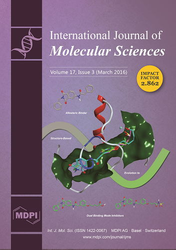
Cover Story (view full-size image): Matrix metalloproteinase 13 (MMP-13) is a validated therapeutic target for a multitude of severe diseases. In this study we performed a structure-based design on a weak allosteric inhibitor of MMP-13, in order to develop dual binding mode inhibitors with improved potency and drug-like properties, by targeting the catalytic zinc (II) ion while still populating the allosteric selectivity loop. The cover picture depicts the design concept of our study. For details concerning the selection of zinc-binding groups, the organic synthesis of the inhibitors and their biological evaluation. View this article.
  • Issues are regarded as officially published after their release is announced to the table of contents alert mailing list.
  • You may sign up for e-mail alerts to receive table of contents of newly released issues.
  • PDF is the official format for papers published in both, html and pdf forms. To view the papers in pdf format, click on the "PDF Full-text" link, and use the free Adobe Reader to open them.
Order results
Result details
Section
Select all
Export citation of selected articles as:
15 pages, 239 KiB  
Review
Mycobacterium tuberculosis: Manipulator of Protective Immunity
by Vanessa C. Korb, Anil A. Chuturgoon * and Devapregasan Moodley
1 Discipline of Medical Biochemistry and Chemical Pathology, University of KwaZulu-Natal, Howard College Campus, King George V Avenue, 4001 Durban, South Africa
Current address: Department of Microbiology and Immunobiology, Harvard Medical School, 77 Avenue Louis Pasteur, Boston, MA 02115, USA.
Int. J. Mol. Sci. 2016, 17(3), 131; https://doi.org/10.3390/ijms17030131 - 25 Feb 2016
Cited by 72 | Viewed by 14736
Abstract
Mycobacterium tuberculosis (MTB) is one of the most successful pathogens in human history and remains a global health challenge. MTB has evolved a plethora of strategies to evade the immune response sufficiently to survive within the macrophage in a bacterial-immunological equilibrium, yet causes [...] Read more.
Mycobacterium tuberculosis (MTB) is one of the most successful pathogens in human history and remains a global health challenge. MTB has evolved a plethora of strategies to evade the immune response sufficiently to survive within the macrophage in a bacterial-immunological equilibrium, yet causes sufficient immunopathology to facilitate its transmission. This review highlights MTB as the driver of disease pathogenesis and presents evidence of the mechanisms by which MTB manipulates the protective immune response into a pathological productive infection. Full article
Show Figures

Graphical abstract

13 pages, 2087 KiB  
Article
Identification of Human UDP-Glucuronosyltransferase 1A4 as the Major Isozyme Responsible for the Glucuronidation of 20(S)-Protopanaxadiol in Human Liver Microsomes
by Jia Li 1, Chunyong He 1, Lianxiang Fang 2, Li Yang 2,3,* and Zhengtao Wang 1,2,3,*
1 Department of Pharmacognosy, China Pharmaceutical University, Nanjing 210038, China
2 The Ministry Of Education Key Laboratory for Standardization of Chinese Medicines and the SATCM Key Laboratory for New Resources and Quality Evaluation of Chinese Medicines, Institute of Chinese Materia Medica, Shanghai University of Traditional Chinese Medicine, Shanghai 201203, China
3 Institute of Chinese Materia Medica, Shanghai University of Traditional Chinese Medicine, Shanghai 201210, China
Int. J. Mol. Sci. 2016, 17(3), 205; https://doi.org/10.3390/ijms17030205 - 9 Mar 2016
Cited by 18 | Viewed by 5750
Abstract
20(S)-protopanaxadiol (PPD), one of the representative aglycones of ginsenosides, has a broad spectrum of pharmacological activities. Although phase I metabolism has been investigated extensively, information regarding phase II metabolism of this compound remains to be elucidated. Here, a glucuronidated metabolite of [...] Read more.
20(S)-protopanaxadiol (PPD), one of the representative aglycones of ginsenosides, has a broad spectrum of pharmacological activities. Although phase I metabolism has been investigated extensively, information regarding phase II metabolism of this compound remains to be elucidated. Here, a glucuronidated metabolite of PPD in human liver microsomes (HLMs) and rat liver microsomes (RLMs) was unambiguously identified as PPD-3-O-β-d-glucuronide by nuclear magnetic resonance spectroscopy and high resolution mass spectrometry. The chemical inhibition and recombinant human UDP-Glucuronosyltransferase (UGT) isoforms assay showed that the PPD glucuronidation was mainly catalyzed by UGT1A4 in HLM, whereas UGT1A3 showed weak catalytic activity. In conclusion, PPD-3-O-β-d-glucuronide was first identified as the principal glucuronidation metabolite of PPD in HLMs, which was catalyzed by UGT1A4. Full article
(This article belongs to the Section Biochemistry)
Show Figures

Graphical abstract

18 pages, 482 KiB  
Review
Xylose Fermentation by Saccharomyces cerevisiae: Challenges and Prospects
by Danuza Nogueira Moysés 1,2, Viviane Castelo Branco Reis 1, João Ricardo Moreira de Almeida 3, Lidia Maria Pepe de Moraes 1 and Fernando Araripe Gonçalves Torres 1,*
1 Departamento de Biologia Celular, Universidade de Brasília, Brasília, DF 70910-900, Brazil
2 Petrobras Research and Development Center, Biotechnology Management, Rio de Janeiro, RJ 21941-915, Brazil
3 Embrapa Agroenergia, Laboratório de Genética e Biotecnologia, Parque Estação Biológica s/n, Av. W3 Norte, Brasília, DF 70770-901, Brazil
Int. J. Mol. Sci. 2016, 17(3), 207; https://doi.org/10.3390/ijms17030207 - 25 Feb 2016
Cited by 240 | Viewed by 18407
Abstract
Many years have passed since the first genetically modified Saccharomyces cerevisiae strains capable of fermenting xylose were obtained with the promise of an environmentally sustainable solution for the conversion of the abundant lignocellulosic biomass to ethanol. Several challenges emerged from these first experiences, [...] Read more.
Many years have passed since the first genetically modified Saccharomyces cerevisiae strains capable of fermenting xylose were obtained with the promise of an environmentally sustainable solution for the conversion of the abundant lignocellulosic biomass to ethanol. Several challenges emerged from these first experiences, most of them related to solving redox imbalances, discovering new pathways for xylose utilization, modulation of the expression of genes of the non-oxidative pentose phosphate pathway, and reduction of xylitol formation. Strategies on evolutionary engineering were used to improve fermentation kinetics, but the resulting strains were still far from industrial application. Lignocellulosic hydrolysates proved to have different inhibitors derived from lignin and sugar degradation, along with significant amounts of acetic acid, intrinsically related with biomass deconstruction. This, associated with pH, temperature, high ethanol, and other stress fluctuations presented on large scale fermentations led the search for yeasts with more robust backgrounds, like industrial strains, as engineering targets. Some promising yeasts were obtained both from studies of stress tolerance genes and adaptation on hydrolysates. Since fermentation times on mixed-substrate hydrolysates were still not cost-effective, the more selective search for new or engineered sugar transporters for xylose are still the focus of many recent studies. These challenges, as well as under-appreciated process strategies, will be discussed in this review. Full article
(This article belongs to the Special Issue Biofuel)
Show Figures

Graphical abstract

14 pages, 3565 KiB  
Article
The Indeterminate Domain Protein ROC1 Regulates Chilling Tolerance via Activation of DREB1B/CBF1 in Rice
by Mingzhu Dou 1,2, Shuai Cheng 2,3, Baotian Zhao 1, Yuanhu Xuan 4,5,* and Minglong Shao 5,*
1 College of Life Sciences, Shandong Normal University, Wenhua East Road 88, Jinan 250014, Shandong, China
2 Northeast Institute of Geography and Agroecology, Chinese Academy of Sciences, Changchun 130102, China
3 University of Chinese Academy of Sciences, Beijing 100049, China
4 College of Plant Protection, Shenyang Agricultural University, Dongling Road 120, Shenyang 110866, Liaoning, China
5 School of Pharmaceutical Science, Wenzhou Medical University, Wenzhou 325000, Zhejiang, China
Int. J. Mol. Sci. 2016, 17(3), 233; https://doi.org/10.3390/ijms17030233 - 25 Feb 2016
Cited by 34 | Viewed by 7049
Abstract
Abiotic stress, including salinity, drought and cold, severely affect diverse aspects of plant development and production. Rice is an important crop that does not acclimate to cold; therefore, it is relatively sensitive to low temperature stress. Dehydration-responsive element-binding protein 1s (DREB1s)/C-repeat binding factors [...] Read more.
Abiotic stress, including salinity, drought and cold, severely affect diverse aspects of plant development and production. Rice is an important crop that does not acclimate to cold; therefore, it is relatively sensitive to low temperature stress. Dehydration-responsive element-binding protein 1s (DREB1s)/C-repeat binding factors (CBFs) are well known for their function in cold tolerance, but the transcriptional regulation of CBFs remains elusive, especially in rice. Here, we performed a yeast one-hybrid assay using the promoter of CBF1, a cold-induced gene, to isolate transcriptional regulators of CBF1. Among the seven candidates identified, an indeterminate domain (IDD) protein named ROC1 (a regulator of CBF1) was further analyzed. The ROC1 transcript was induced by exogenously-treated auxin, while it was not altered by cold or ABA stimuli. ROC1-GFP was localized at the nucleus, and ROC1 showed trans-activation activity in yeast. The electrophoretic mobility shift assay (EMSA) and ChIP analyses revealed that ROC1 directly bound to the promoter of CBF1. Furthermore, ROC1 mutants exhibited chilling-sensitive symptoms and inhibited cold-mediated induction of CBF1 and CBF3, indicating that ROC1 is a positive regulator of cold stress responses. Taken together, this study identified the CBF1 regulator, and the results are important for rice plant adaptation to chilling stress. Full article
(This article belongs to the Special Issue Plant Molecular Biology)
Show Figures

Graphical abstract

20 pages, 2927 KiB  
Article
Identification, Functional Study, and Promoter Analysis of HbMFT1, a Homolog of MFT from Rubber Tree (Hevea brasiliensis)
by Zhenghong Bi 1,2, Xiang Li 3, Huasun Huang 2,* and Yuwei Hua 2,*
1 College of Agronomy, Hainan University, Haikou 570228, China
2 Key Laboratory of Rubber Biology of the Ministry of Agriculture, Rubber Research Institute, Chinese Academy of Tropical Agricultural Sciences, Danzhou 571737, China
3 College of Environment and Plant Protection, Hainan University, Haikou 570228, China
Int. J. Mol. Sci. 2016, 17(3), 247; https://doi.org/10.3390/ijms17030247 - 2 Mar 2016
Cited by 25 | Viewed by 7132
Abstract
A homolog of MOTHER OF FT AND TFL1 (MFT) was isolated from Hevea brasiliensis and its biological function was investigated. Protein multiple sequence alignment and phylogenetic analysis revealed that HbMFT1 conserved critical amino acid residues to distinguish MFT, FLOWERING LOCUS T [...] Read more.
A homolog of MOTHER OF FT AND TFL1 (MFT) was isolated from Hevea brasiliensis and its biological function was investigated. Protein multiple sequence alignment and phylogenetic analysis revealed that HbMFT1 conserved critical amino acid residues to distinguish MFT, FLOWERING LOCUS T (FT) and TERMINAL FLOWER1 (TFL1)-like proteins and showed a closer genetic relationship to the MFT-like group. The accumulation of HbMFT1 was generally detected in various tissues except pericarps, with the highest expression in embryos and relatively higher expression in roots and stems of seedlings, flowering inflorescences, and male and female flowers. HbMFT1 putative promoter analysis showed that tissue-specific, environmental change responsive and hormone-signaling responsive elements were generally present. HbMFT1 was strongly induced under a short-day condition at 28 °C, with the highest expression after the onset of a day. Overexpression of HbMFT1 inhibited seed germination, seedling growth, and flowering in transgenic Arabidopsis. The qRT-PCR further confirmed that APETALA1 (AP1) and FRUITFULL (FUL) were drastically down-regulated in 35S::HbMFT1 plants. A histochemical β-glucuronidase (GUS) assay showed that HbMFT1::GUS activity was mainly detected in stamens and mature seeds coinciding with its original expression and notably induced in rosette leaves and seedlings of transgenic Arabidopsis by exogenous abscisic acid (ABA) due to the presence of ABA cis-elements in HbMFT1 promoter. These results suggested that HbMFT1 was mainly involved in maintenance of seed maturation and stamen development, but negatively controlled germination, growth and development of seedlings and flowering. In addition, the HbMFT1 promoter can be utilized in controlling transgene expression in stamens and seeds of rubber tree or other plant species. Full article
(This article belongs to the Special Issue Plant Molecular Biology)
Show Figures

Figure 1

11 pages, 1996 KiB  
Article
New Strategies and Methods to Study Interactions between Tobacco Mosaic Virus Coat Protein and Its Inhibitors
by Xiangyang Li *, Zhuo Chen, Linhong Jin, Deyu Hu and Song Yang
State Key Laboratory Breeding Base of Green Pesticide and Agricultural Bioengineering, Key Laboratory of Green Pesticide and Agricultural Bioengineering, Ministry of Education, Guizhou University, Guiyang 550025, China
Int. J. Mol. Sci. 2016, 17(3), 252; https://doi.org/10.3390/ijms17030252 - 26 Feb 2016
Cited by 34 | Viewed by 6514
Abstract
Studies of the targets of anti-viral compounds are hot topics in the field of pesticide research. Various efficient anti-TMV (Tobacco Mosaic Virus) compounds, such as Ningnanmycin (NNM), Antofine (ATF), Dufulin (DFL) and Bingqingxiao (BQX) are available. However, the mechanisms of the action of [...] Read more.
Studies of the targets of anti-viral compounds are hot topics in the field of pesticide research. Various efficient anti-TMV (Tobacco Mosaic Virus) compounds, such as Ningnanmycin (NNM), Antofine (ATF), Dufulin (DFL) and Bingqingxiao (BQX) are available. However, the mechanisms of the action of these compounds on targets remain unclear. To further study the mechanism of the action of the anti-TMV inhibitors, the TMV coat protein (TMV CP) was expressed and self-assembled into four-layer aggregate disks in vitro, which could be reassembled into infectious virus particles with TMV RNA. The interactions between the anti-TMV compounds and the TMV CP disk were analyzed by size exclusion chromatography, isothermal titration calorimetry and native-polyacrylamide gel electrophoresis methods. The results revealed that assembly of the four-layer aggregate disk was inhibited by NNM; it changed the four-layer aggregate disk into trimers, and affected the regular assembly of TMV CP and TMV RNA. The four-layer aggregate disk of TMV CP was little inhibited by ATF, DFL and BQX. Our results provide original data, as well as new strategies and methods, for research on the mechanism of action of anti-viral drugs. Full article
(This article belongs to the Section Biochemistry)
Show Figures

Graphical abstract

13 pages, 12037 KiB  
Article
Endogenous Sulfur Dioxide Inhibits Vascular Calcification in Association with the TGF-β/Smad Signaling Pathway
by Zhenzhen Li 1, Yaqian Huang 1, Junbao Du 1,2, Angie Dong Liu 3, Chaoshu Tang 2,4, Yongfen Qi 2,* and Hongfang Jin 1,*
1 Department of Pediatrics, Peking University First Hospital, Beijing 100034, China
2 Key Laboratory of Molecular Cardiology, Ministry of Education, Beijing 100191, China
3 Department of Medical and Health Sciences, Linköping University, Linköping 58183, Sweden
4 Department of Physiology and Pathophysiology, Peking University Health Science Centre, Beijing 100191, China
Int. J. Mol. Sci. 2016, 17(3), 266; https://doi.org/10.3390/ijms17030266 - 23 Feb 2016
Cited by 20 | Viewed by 6889
Abstract
The study was designed to investigate whether endogenous sulfur dioxide (SO2) plays a role in vascular calcification (VC) in rats and its possible mechanisms. In vivo medial vascular calcification was induced in rats by vitamin D3 and nicotine for four weeks. [...] Read more.
The study was designed to investigate whether endogenous sulfur dioxide (SO2) plays a role in vascular calcification (VC) in rats and its possible mechanisms. In vivo medial vascular calcification was induced in rats by vitamin D3 and nicotine for four weeks. In vitro calcification of cultured A7r5 vascular smooth muscle cells (VSMCs) was induced by calcifying media containing 5 mmol/L CaCl2. Aortic smooth muscle (SM) α-actin, runt-related transcription factor 2 (Runx2), transforming growth factor-β (TGF-β) and Smad expression was measured. VC rats showed dispersed calcified nodules among the elastic fibers in calcified aorta with increased aortic calcium content and alkaline phosphatase (ALP) activity. SM α-actin was markedly decreased, but the osteochondrogenic marker Runx2 concomitantly increased and TGF-β/Smad signaling was activated, in association with the downregulated SO2/aspartate aminotransferase (AAT) pathway. However, SO2 supplementation successfully ameliorated vascular calcification, and increased SM α-actin expression, but inhibited Runx2 and TGF-β/Smad expression. In calcified A7r5 VSMCs, the endogenous SO2/AAT pathway was significantly downregulated. SO2 treatment reduced the calcium deposits, calcium content, ALP activity and Runx2 expression and downregulated the TGF-β/Smad pathway in A7r5 cells but increased SM α-actin expression. In brief, SO2 significantly ameliorated vascular calcification in association with downregulation of the TGF-β/Smad pathway. Full article
(This article belongs to the Section Biochemistry)
Show Figures

Graphical abstract

14 pages, 5786 KiB  
Article
The Protective Role of the TOPK/PBK Pathway in Myocardial Ischemia/Reperfusion and H2O2-Induced Injury in H9C2 Cardiomyocytes
by Guozhe Sun 1, Ning Ye 1, Dongxue Dai 1, Yintao Chen 1, Chao Li 2 and Yingxian Sun 1,*
1 Department of Cardiovascular Medicine, the First Hospital of China Medical University, Shenyang 110001, Liaoning, China
2 Department of Biochemistry and Molecular Biology, China Medical University, Shenyang 110122, Liaoning, China
Int. J. Mol. Sci. 2016, 17(3), 267; https://doi.org/10.3390/ijms17030267 - 23 Feb 2016
Cited by 31 | Viewed by 7251
Abstract
T-LAK-cell-originated protein kinase (TOPK) is a PDZ-binding kinase (PBK) that was recently identified as a novel member of the mitogen-activated protein kinase (MAPK) family. It has been shown to play an important role in many cellular functions. However, its role in cardiac function [...] Read more.
T-LAK-cell-originated protein kinase (TOPK) is a PDZ-binding kinase (PBK) that was recently identified as a novel member of the mitogen-activated protein kinase (MAPK) family. It has been shown to play an important role in many cellular functions. However, its role in cardiac function remains unclear. Thus, we have herein explored the biological function of TOPK in myocardial ischemia/reperfusion (I/R) and oxidative stress injury in H9C2 cardiomyocytes. I/R and ischemic preconditioning (IPC) were induced in rats by 3-hour reperfusion after 30-min occlusion of the left anterior descending coronary artery and by 3 cycles of 5-min I/R. Hydrogen peroxide (H2O2) was used to induce oxidative stress in H9C2 cardiomyocytes. TOPK expression was analyzed by western blotting, RT-PCR, immunohistochemical staining, and immunofluorescence imaging studies. The effects of TOPK gene overexpression and its inhibition via its inhibitor HI-TOPK-032 on cell viability and Bcl-2, Bax, ERK1/2, and p-ERK1/2 protein expression were analyzed by MTS assay and western blotting, respectively. The results showed that IPC alleviated myocardial I/R injury and induced TOPK activation. Furthermore, H2O2 induced TOPK phosphorylation in a time-dependent manner. Interestingly, TOPK inhibition aggravated the H2O2-induced oxidative stress injury in myocardiocytes, whereas overexpression relieved it. In addition, the ERK pathway was positively regulated by TOPK signaling. In conclusion, our results indicate that TOPK might mediate a novel survival signal in myocardial I/R, and that its effect on anti-oxidative stress involves the ERK signaling pathway. Full article
(This article belongs to the Special Issue Improvement of Cardiac Function in Heart Failure)
Show Figures

Graphical abstract

30 pages, 6056 KiB  
Article
Expression Patterns of Genes Involved in Ascorbate-Glutathione Cycle in Aphid-Infested Maize (Zea mays L.) Seedlings
by Hubert Sytykiewicz
Department of Biochemistry and Molecular Biology, Siedlce University of Natural Sciences and Humanities, Prusa 12, 08-110 Siedlce, Poland
Int. J. Mol. Sci. 2016, 17(3), 268; https://doi.org/10.3390/ijms17030268 - 23 Feb 2016
Cited by 30 | Viewed by 7347
Abstract
Reduced forms of ascorbate (AsA) and glutathione (GSH) are among the most important non-enzymatic foliar antioxidants in maize (Zea mays L.). The survey was aimed to evaluate impact of bird cherry-oat aphid (Rhopalosiphum padi L.) or grain aphid (Sitobion avenae [...] Read more.
Reduced forms of ascorbate (AsA) and glutathione (GSH) are among the most important non-enzymatic foliar antioxidants in maize (Zea mays L.). The survey was aimed to evaluate impact of bird cherry-oat aphid (Rhopalosiphum padi L.) or grain aphid (Sitobion avenae F.) herbivory on expression of genes related to ascorbate-glutathione (AsA-GSH) cycle in seedlings of six maize varieties (Ambrozja, Nana, Tasty Sweet, Touran, Waza, Złota Karłowa), differing in resistance to the cereal aphids. Relative expression of sixteen maize genes encoding isoenzymes of ascorbate peroxidase (APX1, APX2, APX3, APX4, APX5, APX6, APX7), monodehydroascorbate reductase (MDHAR1, MDHAR2, MDHAR3, MDHAR4), dehydroascorbate reductase (DHAR1, DHAR2, DHAR3) and glutathione reductase (GR1, GR2) was quantified. Furthermore, effect of hemipterans’ attack on activity of APX, MDHAR, DHAR and GR enzymes, and the content of reduced and oxidized ascorbate and glutathione in maize plants were assessed. Seedling leaves of more resistant Z. mays varieties responded higher elevations in abundance of target transcripts. In addition, earlier and stronger aphid-triggered changes in activity of APX, MDHAR, DHAR and GR enzymes, and greater modulations in amount of the analyzed antioxidative metabolites were detected in foliar tissues of highly resistant Ambrozja genotype in relation to susceptible Tasty Sweet plants. Full article
(This article belongs to the Special Issue Plant Innate Immunity)
Show Figures

Figure 1

15 pages, 2237 KiB  
Article
Nutritional Signaling Regulates Vitellogenin Synthesis and Egg Development through Juvenile Hormone in Nilaparvata lugens (Stål)
by Kai Lu 1,2, Xia Chen 1, Wen-Ting Liu 1, Xin-Yu Zhang 2, Ming-Xiao Chen 2 and Qiang Zhou 2,*
1 College of Life Sciences, Fujian Agriculture and Forestry University, Fuzhou 350002, China
2 State Key Laboratory of Biocontrol, School of Life Sciences, Sun Yat-sen University, Guangzhou 510275, China
Int. J. Mol. Sci. 2016, 17(3), 269; https://doi.org/10.3390/ijms17030269 - 26 Feb 2016
Cited by 49 | Viewed by 7128
Abstract
Insect female reproduction which comprises the synthesis of vitellogenein (Vg) in the fat body and its incorporation into developing oocytes, needs a large amount of energy and food resources. Our previous studies found that juvenile hormone (JH) regulates vitellogenesis in the brown planthopper, [...] Read more.
Insect female reproduction which comprises the synthesis of vitellogenein (Vg) in the fat body and its incorporation into developing oocytes, needs a large amount of energy and food resources. Our previous studies found that juvenile hormone (JH) regulates vitellogenesis in the brown planthopper, Nilaparvata lugens. Here, we report on the role of JH in nutrient-regulated Vg synthesis and egg development. We first cloned the genes coding for juvenile hormone acid methyltransferase (JHAMT) which is involved in JH biosynthesis and methoprene-tolerant (Met) for JH action. Amino acids (AAs) induced the expression of jmtN, while showing no effects on the expression of met using an artificial diet culture system. Reduction in JH biosynthesis or its action by RNA interference (RNAi)-mediated silencing of jmtN or met led to a severe inhibition of AAs-induced Vg synthesis and oocyte maturation, together with lower fecundity. Furthermore, exogenous application of JH III partially restored Vg expression levels in jmtN RNAi females. However, JH III application did not rescue Vg synthesis in these met RNAi insects. Our results show that AAs induce Vg synthesis in the fat body and egg development in concert with JH biosynthesis in Nilaparvata lugens (Stål), rather than through JH action. Full article
(This article belongs to the Section Biochemistry)
Show Figures

Graphical abstract

15 pages, 3107 KiB  
Article
Genome-Wide Identification and Transcriptome-Based Expression Profiling of the Sox Gene Family in the Nile Tilapia (Oreochromis niloticus)
by Ling Wei 1,2,†, Chao Yang 1,†, Wenjing Tao 1,† and Deshou Wang 1,*
1 Key Laboratory of Freshwater Fish Reproduction and Development (Ministry of Education), Key Laboratory of Aquatic Science of Chongqing, School of Life Science, Southwest University, Chongqing 400715, China
2 Key Laboratory of Eco-environments in Three Gorges Reservoir Region (Ministry of Education), Chongqing Key Laboratory of Plant Ecology and Resources Research in Three Gorges Reservoir Region, School of Life Science, Southwest University, Chongqing 400715, China
These authors equally contributed to this work.
Int. J. Mol. Sci. 2016, 17(3), 270; https://doi.org/10.3390/ijms17030270 - 23 Feb 2016
Cited by 78 | Viewed by 9644
Abstract
The Sox transcription factor family is characterized with the presence of a Sry-related high-mobility group (HMG) box and plays important roles in various biological processes in animals, including sex determination and differentiation, and the development of multiple organs. In this study, 27 Sox [...] Read more.
The Sox transcription factor family is characterized with the presence of a Sry-related high-mobility group (HMG) box and plays important roles in various biological processes in animals, including sex determination and differentiation, and the development of multiple organs. In this study, 27 Sox genes were identified in the genome of the Nile tilapia (Oreochromis niloticus), and were classified into seven groups. The members of each group of the tilapia Sox genes exhibited a relatively conserved exon-intron structure. Comparative analysis showed that the Sox gene family has undergone an expansion in tilapia and other teleost fishes following their whole genome duplication, and group K only exists in teleosts. Transcriptome-based analysis demonstrated that most of the tilapia Sox genes presented stage-specific and/or sex-dimorphic expressions during gonadal development, and six of the group B Sox genes were specifically expressed in the adult brain. Our results provide a better understanding of gene structure and spatio-temporal expression of the Sox gene family in tilapia, and will be useful for further deciphering the roles of the Sox genes during sex determination and gonadal development in teleosts. Full article
(This article belongs to the Special Issue Fish Molecular Biology)
Show Figures

Graphical abstract

10 pages, 420 KiB  
Communication
Using Copy Number Alterations to Identify New Therapeutic Targets for Bladder Carcinoma
by Donatella Conconi 1, Elena Sala 2, Giorgio Bovo 3, Guido Strada 4, Leda Dalprà 1,2, Marialuisa Lavitrano 1 and Angela Bentivegna 1,*
1 School of Medicine and Surgery, University of Milano-Bicocca, 20900 Monza, Italy
2 Medical Genetics Laboratory, San Gerardo Hospital, 20900 Monza, Italy
3 Department of Pathology, San Gerardo Hospital, 20900 Monza, Italy
4 Urology Division, Bassini Icp Hospital, 20092 Cinisello Balsamo, Italy
Int. J. Mol. Sci. 2016, 17(3), 271; https://doi.org/10.3390/ijms17030271 - 24 Feb 2016
Cited by 11 | Viewed by 5411
Abstract
Bladder cancer represents the ninth most widespread malignancy throughout the world. It is characterized by the presence of two different clinical and prognostic subtypes: non-muscle-invasive bladder cancers (NMIBCs) and muscle-invasive bladder cancers (MIBCs). MIBCs have a poor outcome with a common progression to [...] Read more.
Bladder cancer represents the ninth most widespread malignancy throughout the world. It is characterized by the presence of two different clinical and prognostic subtypes: non-muscle-invasive bladder cancers (NMIBCs) and muscle-invasive bladder cancers (MIBCs). MIBCs have a poor outcome with a common progression to metastasis. Despite improvements in knowledge, treatment has not advanced significantly in recent years, with the absence of new therapeutic targets. Because of the limitations of current therapeutic options, the greater challenge will be to identify biomarkers for clinical application. For this reason, we compared our array comparative genomic hybridization (array-CGH) results with those reported in literature for invasive bladder tumors and, in particular, we focused on the evaluation of copy number alterations (CNAs) present in biopsies and retained in the corresponding cancer stem cell (CSC) subpopulations that should be the main target of therapy. According to our data, CCNE1, MYC, MDM2 and PPARG genes could be interesting therapeutic targets for bladder CSC subpopulations. Surprisingly, HER2 copy number gains are not retained in bladder CSCs, making the gene-targeted therapy less interesting than the others. These results provide precious advice for further study on bladder therapy; however, the clinical importance of these results should be explored. Full article
(This article belongs to the Special Issue Advances in Molecular Oncology)
Show Figures

Graphical abstract

19 pages, 6036 KiB  
Article
Synergetic Effects of PARP Inhibitor AZD2281 and Cisplatin in Oral Squamous Cell Carcinoma in Vitro and in Vivo
by Masaaki Yasukawa 1, Hisako Fujihara 1,2,*, Hiroaki Fujimori 3,4, Koji Kawaguchi 1, Hiroyuki Yamada 5, Ryoko Nakayama 6, Nanami Yamamoto 1, Yuta Kishi 1, Yoshiki Hamada 1 and Mitsuko Masutani 3,4
1 Department of Oral and Maxillofacial Surgery, School of Dental Medicine, Tsurumi University, 2-1-3 Tsurumi, Tsurumi-ku, Yokohama, Kanagawa 230-8501, Japan
2 Department of Oral Hygiene, Tsurumi Junior College, 2-1-3 Tsurumi, Tsurumi-ku, Yokohama, Kanagawa 230-8501, Japan
3 Division of Chemotherapy and Translational Research, National Cancer Center Research Institute, 5-1-1 Tsukiji, Chuo-ku, Tokyo 104-0045, Japan
4 Department of Frontier Life Science, Graduate School of Biochemical Science, Nagasaki University, 1-7-1 Sakamoto, Nagasaki 852-8588, Japan
5 Division of Maxillofacial Surgery, Department of Oral and Maxillofacial Surgery, School of Dentistry, Iwate Medical University 19-1 Uchimaru, Morioka Iwate 020-8050, Japan
6 Department of Pathology, School of Dental Medicine, Tsurumi University, 2-1-3 Tsurumi, Tsurumi-ku, Yokohama, Kanagawa 230-8501, Japan
Int. J. Mol. Sci. 2016, 17(3), 272; https://doi.org/10.3390/ijms17030272 - 24 Feb 2016
Cited by 41 | Viewed by 7452
Abstract
Cisplatin is a commonly used chemotherapeutic drug for treatment of oral carcinoma, and combinatorial effects are expected to exert greater therapeutic efficacy compared with monotherapy. Poly(ADP-ribosyl)ation is reported to be involved in a variety of cellular processes, such as DNA repair, cell death, [...] Read more.
Cisplatin is a commonly used chemotherapeutic drug for treatment of oral carcinoma, and combinatorial effects are expected to exert greater therapeutic efficacy compared with monotherapy. Poly(ADP-ribosyl)ation is reported to be involved in a variety of cellular processes, such as DNA repair, cell death, telomere regulation, and genomic stability. Based on these properties, poly(ADP-ribose) polymerase (PARP) inhibitors are used for treatment of cancers, such as BRCA1/2 mutated breast and ovarian cancers, or certain solid cancers in combination with anti-cancer drugs. However, the effects on oral cancer have not been fully evaluated. In this study, we examined the effects of PARP inhibitor on the survival of human oral cancer cells in vitro and xenografted tumors derived from human oral cancer cells in vivo. In vitro effects were assessed by microculture tetrazolium and survival assays. The PARP inhibitor AZD2281 (olaparib) showed synergetic effects with cisplatin in a dose-dependent manner. Combinatorial treatment with cisplatin and AZD2281 significantly inhibited xenografted tumor growth compared with single treatment of cisplatin or AZD2281. Histopathological analysis revealed that cisplatin and AZD2281 increased TUNEL-positive cells and decreased Ki67- and CD31-positive cells. These results suggest that PARP inhibitors have the potential to improve therapeutic strategies for oral cancer. Full article
Show Figures

Graphical abstract

31 pages, 1941 KiB  
Review
The Immunology of Neuromyelitis Optica—Current Knowledge, Clinical Implications, Controversies and Future Perspectives
by Michalina Jasiak-Zatonska 1,*, Alicja Kalinowska-Lyszczarz 2, Slawomir Michalak 2,3 and Wojciech Kozubski 1
1 Department of Neurology, Poznan University of Medical Sciences, 49 Przybyszewskiego St., 60-355 Poznan, Poland
2 Department of Neurochemistry and Neuropathology, Poznan University of Medical Sciences, 49 Przybyszewskiego St., 60-355 Poznan, Poland
3 Neuroimmunological Unit, Mossakowski Medical Research Centre, Polish Academy of Sciences, 5 Pawinskiego St., 02-106 Warsaw, Poland
Int. J. Mol. Sci. 2016, 17(3), 273; https://doi.org/10.3390/ijms17030273 - 2 Mar 2016
Cited by 112 | Viewed by 18708
Abstract
Neuromyelitis optica (NMO) is an autoimmune, demyelinating disorder of the central nervous system (CNS) with typical clinical manifestations of optic neuritis and acute transverse myelitis attacks. Previously believed to be a variant of multiple sclerosis (MS), it is now considered an independent disorder [...] Read more.
Neuromyelitis optica (NMO) is an autoimmune, demyelinating disorder of the central nervous system (CNS) with typical clinical manifestations of optic neuritis and acute transverse myelitis attacks. Previously believed to be a variant of multiple sclerosis (MS), it is now considered an independent disorder which needs to be differentiated from MS. The discovery of autoantibodies against aquaporin-4 (AQP4-IgGs) changed our understanding of NMO immunopathogenesis and revolutionized the diagnostic process. AQP4-IgG is currently regarded as a specific biomarker of NMO and NMO spectrum disorders (NMOsd) and a key factor in its pathogenesis. Nevertheless, AQP4-IgG seronegativity in 10%–25% of NMO patients suggests that there are several other factors involved in NMO immunopathogenesis, i.e., autoantibodies against aquaporin-1 (AQP1-Abs) and antibodies against myelin oligodendrocyte glycoprotein (MOG-IgGs). This manuscript reviews current knowledge about NMO immunopathogenesis, pointing out the controversial issues and showing potential directions for future research. Further efforts should be made to broaden our knowledge of NMO immunology which could have important implications for clinical practice, including the use of potential novel biomarkers to facilitate an early and accurate diagnosis, and modern treatment strategies improving long-term outcome of NMO patients. Full article
(This article belongs to the Special Issue Advances in Multiple Sclerosis 2016)
Show Figures

Graphical abstract

12 pages, 1411 KiB  
Article
Identification and Characterization of the Gene CYP340W1 from Plutella xylostella and Its Possible Involvement in Resistance to Abamectin
by Xue Gao 1, Jiaqiang Yang 1, Baoyun Xu 1, Wen Xie 1, Shaoli Wang 1, Youjun Zhang 1, Fengshan Yang 2 and Qingjun Wu 1,*
1 Department of Plant Protection, Institute of Vegetables and Flowers, Chinese Academy of Agricultural Sciences, Beijing 100081, China
2 College of Life Science, University of Hei Longjiang, Harbin 150000, China
Int. J. Mol. Sci. 2016, 17(3), 274; https://doi.org/10.3390/ijms17030274 - 18 Mar 2016
Cited by 25 | Viewed by 5734
Abstract
Abamectin has been used to control the diamondback moth, Plutella xylostella (P. xylostella), which is a major agricultural pest that can rapidly develop resistance against insecticides including abamectin. Although cytochrome P450 has been confirmed to play an important role in resistance [...] Read more.
Abamectin has been used to control the diamondback moth, Plutella xylostella (P. xylostella), which is a major agricultural pest that can rapidly develop resistance against insecticides including abamectin. Although cytochrome P450 has been confirmed to play an important role in resistance in P. xylostella, the specific P450 genes associated with the resistance are unclear. The full-length cDNA of the cytochrome P450 gene CYP340W1 was cloned and characterized in the present study. The cDNA assembly yielded a sequence of 1929 bp, containing the open reading frame (ORF) 1491 bp and encodes a 496-amino acid peptide. CYP340W1 was expressed in all P. xylostella developmental stages but its expression level was highest in larvae and especially in the heads of larvae. The expression of CYP340W1 was significantly higher in an abamectin-resistant strain (ABM-R) than in its susceptible counterpart (ABM-S). In addition, expression of CYP340W1 was increased when the ABM-R strain was exposed to abamectin. When injected into third-stage ABM-R larvae, CYP340W1 dsRNA significantly reduced CYP340W1 expression at 6 h and reduced expression by 83% at 12 h. As a consequence of RNAi, the mortality of the injected abamectin-resistant larvae increased after a 48-h exposure to abamectin. The results indicate that the overexpression of CYP340W1 plays an important role in abamectin resistance in P. xylostella. Full article
Show Figures

Figure 1

9 pages, 440 KiB  
Review
The Role of Sialyl-Tn in Cancer
by Jennifer Munkley
Institute of Genetic Medicine, Newcastle University, Newcastle-upon-Tyne NE1 3BZ, UK
Int. J. Mol. Sci. 2016, 17(3), 275; https://doi.org/10.3390/ijms17030275 - 24 Feb 2016
Cited by 157 | Viewed by 12742
Abstract
Activation of an aberrant glycosylation pathway in cancer cells can lead to expression of the onco-foetal sialyl-Tn (sTn) antigen. STn is a truncated O-glycan containing a sialic acid α-2,6 linked to GalNAc α-O-Ser/Thr and is associated with an adverse outcome [...] Read more.
Activation of an aberrant glycosylation pathway in cancer cells can lead to expression of the onco-foetal sialyl-Tn (sTn) antigen. STn is a truncated O-glycan containing a sialic acid α-2,6 linked to GalNAc α-O-Ser/Thr and is associated with an adverse outcome and poor prognosis in cancer patients. The biosynthesis of the sTn antigen has been linked to the expression of the sialytransferase ST6GalNAc1, and also to mutations in and loss of heterozygosity of the COSMC gene. sTn neo- or over-expression occurs in many types of epithelial cancer including gastric, colon, breast, lung, oesophageal, prostate and endometrial cancer. sTn is believed to be carried by a variety of glycoproteins and may influence protein function and be involved in tumour development. This review discusses how the role of sTn in cancer development and tumour cell invasiveness might be organ specific and occur through different mechanisms depending on each cancer type or subtype. As the sTn-antigen is expressed early in carcinogenesis targeting sTn in cancer may enable the targeting of tumours from the earliest stage. Full article
(This article belongs to the Special Issue Glycan–Receptor Interaction)
Show Figures

Figure 1

17 pages, 11606 KiB  
Article
Integrative Analyses of miRNA-mRNA Interactions Reveal let-7b, miR-128 and MAPK Pathway Involvement in Muscle Mass Loss in Sex-Linked Dwarf Chickens
by Wen Luo 1,2,3, Shumao Lin 4, Guihuan Li 1,2,3, Qinghua Nie 1,2,3 and Xiquan Zhang 1,2,3,*
1 Department of Animal Genetics, Breeding and Reproduction, College of Animal Science, South China Agricultural University, Guangzhou 510642, Guangdong, China
2 Guangdong Provincial Key Lab of Agro-Animal Genomics and Molecular Breeding, South China Agricultural University, Guangzhou 510642, Guangdong, China
3 Key Lab of Chicken Genetics, Breeding and Reproduction, Ministry of Agriculture, South China Agricultural University, Guangzhou 510642, Guangdong, China
4 College of Life Science, Foshan University, Foshan 528231, Guangdong, China
Int. J. Mol. Sci. 2016, 17(3), 276; https://doi.org/10.3390/ijms17030276 - 24 Feb 2016
Cited by 33 | Viewed by 7612
Abstract
The sex-linked dwarf (SLD) chicken is an ideal model system for understanding growth hormone (GH)-action and growth hormone receptor (GHR) function because of its recessive mutation in the GHR gene. Skeletal muscle mass is reduced in the SLD chicken with a smaller muscle [...] Read more.
The sex-linked dwarf (SLD) chicken is an ideal model system for understanding growth hormone (GH)-action and growth hormone receptor (GHR) function because of its recessive mutation in the GHR gene. Skeletal muscle mass is reduced in the SLD chicken with a smaller muscle fiber diameter. Our previous study has presented the mRNA and miRNA expression profiles of the SLD chicken and normal chicken between embryo day 14 and seven weeks of age. However, the molecular mechanism of GHR-deficient induced muscle mass loss is still unclear, and the key molecules and pathways underlying the GHR-deficient induced muscle mass loss also remain to be illustrated. Here, by functional network analysis of the differentially expressed miRNAs and mRNAs between the SLD and normal chickens, we revealed that let-7b, miR-128 and the MAPK pathway might play key roles in the GHR-deficient induced muscle mass loss, and that the reduced cell division and growth are potential cellular processes during the SLD chicken skeletal muscle development. Additionally, we also found some genes and miRNAs involved in chicken skeletal muscle development, through the MAPK, PI3K-Akt, Wnt and Insulin signaling pathways. This study provides new insights into the molecular mechanism underlying muscle mass loss in the SLD chickens, and some regulatory networks that are crucial for chicken skeletal muscle development. Full article
(This article belongs to the Special Issue MicroRNA Regulation)
Show Figures

Graphical abstract

12 pages, 1308 KiB  
Article
Multiple Evolutionary Selections Involved in Synonymous Codon Usages in the Streptococcus agalactiae Genome
by Yan-Ping Ma 1,2, Hao Ke 2,*, Zhi-Ling Liang 2, Zhen-Xing Liu 2, Le Hao 2, Jiang-Yao Ma 2 and Yu-Gu Li 1,*
1 College of Veterinary Medicine, South China Agricultural University, Guangzhou 510642, China
2 Guangdong Open Laboratory of Veterinary Public Health, Guangdong Laboratory for Animal Diseases, Institute of Animal Health, Guangdong Academy of Agricultural Sciences, Guangzhou 510640, China
Int. J. Mol. Sci. 2016, 17(3), 277; https://doi.org/10.3390/ijms17030277 - 24 Feb 2016
Cited by 10 | Viewed by 5240
Abstract
Streptococcus agalactiae is an important human and animal pathogen. To better understand the genetic features and evolution of S. agalactiae, multiple factors influencing synonymous codon usage patterns in S. agalactiae were analyzed in this study. A- and U-ending rich codons were used [...] Read more.
Streptococcus agalactiae is an important human and animal pathogen. To better understand the genetic features and evolution of S. agalactiae, multiple factors influencing synonymous codon usage patterns in S. agalactiae were analyzed in this study. A- and U-ending rich codons were used in S. agalactiae function genes through the overall codon usage analysis, indicating that Adenine (A)/Thymine (T) compositional constraints might contribute an important role to the synonymous codon usage pattern. The GC3% against the effective number of codon (ENC) value suggested that translational selection was the important factor for codon bias in the microorganism. Principal component analysis (PCA) showed that (i) mutational pressure was the most important factor in shaping codon usage of all open reading frames (ORFs) in the S. agalactiae genome; (ii) strand specific mutational bias was not capable of influencing the codon usage bias in the leading and lagging strands; and (iii) gene length was not the important factor in synonymous codon usage pattern in this organism. Additionally, the high correlation between tRNA adaptation index (tAI) value and codon adaptation index (CAI), frequency of optimal codons (Fop) value, reinforced the role of natural selection for efficient translation in S. agalactiae. Comparison of synonymous codon usage pattern between S. agalactiae and susceptible hosts (human and tilapia) showed that synonymous codon usage of S. agalactiae was independent of the synonymous codon usage of susceptible hosts. The study of codon usage in S. agalactiae may provide evidence about the molecular evolution of the bacterium and a greater understanding of evolutionary relationships between S. agalactiae and its hosts. Full article
(This article belongs to the Special Issue Microbial Genomics and Metabolomics)
Show Figures

Graphical abstract

10 pages, 210 KiB  
Review
Critical Overview of the Risk Scoring Systems to Predict Non-Responsiveness to Intravenous Immunoglobulin in Kawasaki Syndrome
by Donato Rigante 1,*, Laura Andreozzi 1, Michele Fastiggi 1, Benedetta Bracci 1, Marco Francesco Natale 1 and Susanna Esposito 2
1 Institute of Pediatrics, Università Cattolica Sacro Cuore, Fondazione Policlinico Universitario A. Gemelli, Rome 00168, Italy
2 Pediatric Highly Intensive Care Unit, Department of Pathophysiology and Transplantation, Università degli Studi di Milano, Fondazione IRCCS Ca’ Granda Ospedale Maggiore Policlinico, Milan 20122, Italy
Int. J. Mol. Sci. 2016, 17(3), 278; https://doi.org/10.3390/ijms17030278 - 24 Feb 2016
Cited by 66 | Viewed by 6636
Abstract
Kawasaki syndrome (KS) is the most relevant cause of heart disease in children living in developed countries. Intravenous immunoglobulin (IVIG) has a preventive function in the formation of coronary artery abnormalities and a poor strictly-curative action in established coronary damage. More than two [...] Read more.
Kawasaki syndrome (KS) is the most relevant cause of heart disease in children living in developed countries. Intravenous immunoglobulin (IVIG) has a preventive function in the formation of coronary artery abnormalities and a poor strictly-curative action in established coronary damage. More than two decades ago, the Harada score was set to assess which children with KS should be subject to administration of IVIG, evaluating retrospectively a large cohort of patients with regard to age, sex and laboratory data. Nowadays, high dose IVIG is administered to all children with a confirmed diagnosis of KS, but a tool for predicting non-responsiveness to the initial infusion of IVIG has not been found. The prediction of IVIG resistance is a crucial issue, as recognising these high-risk patients should consent the administration of an intensified initial treatment in combination with IVIG in order to prevent coronary injuries. Few reports have focused on factors, referring to both clinical parameters and laboratory data at the onset of KS, in order to predict which patients might be IVIG non-responsive. We have analysed three different risk scores which were formulated to predict IVIG resistance in Japanese children with typical KS, but their application in non-Japanese patients or in those with incomplete and atypical patterns of the disease has been studied in a fragmentary way. Overall, our analysis showed that early and definite ascertainment of likely IVIG non-responders who require additional therapies reducing the development of coronary artery involvement in children with KS is still a challenge. Full article
12 pages, 4680 KiB  
Article
Identification of Autophagy in the Pine Wood Nematode Bursaphelenchus xylophilus and the Molecular Characterization and Functional Analysis of Two Novel Autophagy-Related Genes, BxATG1 and BxATG8
by Li-Na Deng 1,2,3, Xiao-Qin Wu 1,2,*, Jian-Ren Ye 1,2 and Qi Xue 1,2
1 Co-Innovation Center for Sustainable Forestry in Southern China, College of Forestry, Nanjing Forestry University, Nanjing 210037, Jiangsu, China
2 Jiangsu Key Laboratory for Prevention and Management of Invasive Species, Nanjing Forestry University, Nanjing 210037, Jiangsu, China
3 Yancheng Institute of Technology, School of Ocean and Biological Engineering, Yancheng 224051, Jiangsu, China
Int. J. Mol. Sci. 2016, 17(3), 279; https://doi.org/10.3390/ijms17030279 - 3 Mar 2016
Cited by 21 | Viewed by 6294
Abstract
The pine wood nematode, Bursaphelenchus xylophilus, causes huge economic losses in pine forests, has a complex life cycle, and shows the remarkable ability to survive under unfavorable and changing environmental conditions. This ability may be related to autophagy, which is still poorly [...] Read more.
The pine wood nematode, Bursaphelenchus xylophilus, causes huge economic losses in pine forests, has a complex life cycle, and shows the remarkable ability to survive under unfavorable and changing environmental conditions. This ability may be related to autophagy, which is still poorly understood in B. xylophilus and no autophagy-related genes have been previously characterized. In this study, transmission electron microscopy was used to confirm that autophagy exists in B. xylophilus. The full-length cDNAs of BxATG1 and BxATG8 were first cloned from B. xylophilus, and BxATG1 and BxATG8 were characterized using bioinformatics methods. The expression pattern of the autophagy marker BxATG8 was investigated using in situ hybridization (ISH). BxATG8 was expressed in esophageal gland and hypodermal seam cells. We tested the effects of RNA interference (RNAi) on BxATG1 and BxATG8. The results revealed that BxATG1 and BxATG8 were likely associated with propagation of nematodes on fungal mats. This study confirmed the molecular characterization and functions of BxATG1 and BxATG8 in B. xylophilus and provided fundamental information between autophagy and B. xylophilus. Full article
(This article belongs to the Section Biochemistry)
Show Figures

Graphical abstract

17 pages, 547 KiB  
Review
MicroRNAs as Biomarkers for Liver Disease and Hepatocellular Carcinoma
by C. Nelson Hayes 1,2 and Kazuaki Chayama 1,2,3,*
1 Department of Gastroenterology and Metabolism, Applied Life Sciences, Institute of Biomedical and Health Sciences, Hiroshima University, 1-2-3 Kasumi, Minami-ku, Hiroshima 734-8551, Japan
2 Liver Research Project Center, Hiroshima University, Hiroshima 734-8551, Japan
3 Laboratory for Digestive Diseases, Center for Genomic Medicine, RIKEN, Hiroshima 734-8551, Japan
Int. J. Mol. Sci. 2016, 17(3), 280; https://doi.org/10.3390/ijms17030280 - 24 Feb 2016
Cited by 170 | Viewed by 14563
Abstract
Serum levels of liver enzymes, such as alanine transaminase, aspartate transaminase, and α-fetoprotein, provide insight into liver function and are used during treatment of liver disease, but such information is limited. In the case of hepatocellular carcinoma (HCC), which is often not detected [...] Read more.
Serum levels of liver enzymes, such as alanine transaminase, aspartate transaminase, and α-fetoprotein, provide insight into liver function and are used during treatment of liver disease, but such information is limited. In the case of hepatocellular carcinoma (HCC), which is often not detected until an advanced stage, more sensitive biomarkers may help to achieve earlier detection. Serum also contains microRNAs, a class of small non-coding RNAs that play an important role in regulating gene expression. miR-122 is specific to the liver and correlates strongly with liver enzyme levels and necroinflammatory activity, and other microRNAs are correlated with the degree of fibrosis. miR-122 has also been found to be required for hepatitis C virus (HCV) infection, whereas other microRNAs have been shown to play antiviral roles. miR-125a-5p and miR-1231 have been shown to directly target hepatitis B virus (HBV) transcripts, and others are up- or down-regulated in infected individuals. MicroRNA profiles also differ in the case of HBV and HCV infection as well as between HBeAg-positive and negative patients, and in patients with occult versus active HBV infection. In such patients, monitoring of changes in microRNA profiles might provide earlier warning of neoplastic changes preceding HCC. Full article
(This article belongs to the Special Issue MicroRNA in Various Disease States as Biomarkers)
Show Figures

Graphical abstract

12 pages, 816 KiB  
Review
Mitochondrial Molecular Pathophysiology of Nonalcoholic Fatty Liver Disease: A Proteomics Approach
by Natalia Nuño-Lámbarri 1,†, Varenka J. Barbero-Becerra 1,†, Misael Uribe 2 and Norberto C. Chávez-Tapia 1,2,*
1 Traslational Research Unit, Médica Sur Clinic & Foundation, Mexico City 14050, Mexico
2 Obesity and Digestive Diseases Unit, Médica Sur Clinic & Foundation, Mexico City 14050, Mexico
These authors contributed equally to this work.
Int. J. Mol. Sci. 2016, 17(3), 281; https://doi.org/10.3390/ijms17030281 - 15 Mar 2016
Cited by 27 | Viewed by 10356
Abstract
Nonalcoholic fatty liver disease (NAFLD) is a chronic liver condition that can progress to nonalcoholic steatohepatitis, cirrhosis and cancer. It is considered an emerging health problem due to malnourishment or a high-fat diet (HFD) intake, which is observed worldwide. It is well known [...] Read more.
Nonalcoholic fatty liver disease (NAFLD) is a chronic liver condition that can progress to nonalcoholic steatohepatitis, cirrhosis and cancer. It is considered an emerging health problem due to malnourishment or a high-fat diet (HFD) intake, which is observed worldwide. It is well known that the hepatocytes’ apoptosis phenomenon is one of the most important features of NAFLD. Thus, this review focuses on revealing, through a proteomics approach, the complex network of protein interactions that promote fibrosis, liver cell stress, and apoptosis. According to different types of in vitro and murine models, it has been found that oxidative/nitrative protein stress leads to mitochondrial dysfunction, which plays a major role in stimulating NAFLD damage. Human studies have revealed the importance of novel biomarkers, such as retinol-binding protein 4, lumican, transgelin 2 and hemoglobin, which have a significant role in the disease. The post-genome era has brought proteomics technology, which allows the determination of molecular pathogenesis in NAFLD. This has led to the search for biomarkers which improve early diagnosis and optimal treatment and which may effectively prevent fatal consequences such as cirrhosis or cancer. Full article
(This article belongs to the Special Issue Non-Alcoholic Fatty Liver Disease Research 2016)
Show Figures

Graphical abstract

15 pages, 2193 KiB  
Article
Two Different Approaches for Oral Administration of Voriconazole Loaded Formulations: Electrospun Fibers versus β-Cyclodextrin Complexes
by Panoraia I. Siafaka 1, Neslihan Üstündağ Okur 2, Mariza Mone 1, Spyridoula Giannakopoulou 1, Sevda Er 3, Eleni Pavlidou 4, Evangelos Karavas 5 and Dimitrios N. Bikiaris 1,*
1 Laboratory of Polymer Chemistry and Technology, Department of Chemistry, Aristotle University of Thessaloniki, 54 124 Thessaloniki, Greece
2 School of Pharmacy, Department of Pharmaceutical Technology, Istanbul Medipol University, Beykoz, 34810 Istanbul, Turkey
3 School of Pharmacy, Department of Microbiology, Istanbul Medipol University, Beykoz, 34810 Istanbul, Turkey
4 Department of Physics, Aristotle University of Thessaloniki, 54 124 Thessaloniki, Greece
5 Pharmathen S.A, Dervenakion Str. 6, 153 51 Attiki, Greece
Int. J. Mol. Sci. 2016, 17(3), 282; https://doi.org/10.3390/ijms17030282 - 25 Feb 2016
Cited by 54 | Viewed by 6480
Abstract
In this work, a comparison between two different preparation methods for the improvement of dissolution rate of an antifungal agent is presented. Poly(ε-caprolactone) (PCL) electrospun fibers and β-cyclodextrin (β-CD) complexes, which were produced via an electrospinning process and an inclusion complexation method, respectively, [...] Read more.
In this work, a comparison between two different preparation methods for the improvement of dissolution rate of an antifungal agent is presented. Poly(ε-caprolactone) (PCL) electrospun fibers and β-cyclodextrin (β-CD) complexes, which were produced via an electrospinning process and an inclusion complexation method, respectively, were addressed for the treatment of fungal infections. Voriconazole (VRCZ) drug was selected as a model drug. PCL nanofibers were characterized on the basis of morphology while phase solubility studies for β-CDs complexes were performed. Various concentrations (5, 10, 15 and 20 wt %) of VRCZ were loaded to PCL fibers and β-CD inclusions to study the in vitro release profile as well as in vitro antifungal activity. The results clearly indicated that all formulations showed an improved VRCZ solubility and can inhibit fungi proliferation. Full article
(This article belongs to the Special Issue Drug Delivery and Antimicrobial Agents)
Show Figures

Graphical abstract

18 pages, 2691 KiB  
Article
Physiological Investigation and Transcriptome Analysis of Polyethylene Glycol (PEG)-Induced Dehydration Stress in Cassava
by Lili Fu, Zehong Ding, Bingying Han, Wei Hu, Yajun Li and Jiaming Zhang *
1 Institute of Tropical Bioscience and Biotechnology, Chinese Academy of Tropical Agricultural Sciences, Xueyuan Road 4, Haikou 571101, China
These authors contributed equally to this work.
Int. J. Mol. Sci. 2016, 17(3), 283; https://doi.org/10.3390/ijms17030283 - 25 Feb 2016
Cited by 89 | Viewed by 9523
Abstract
Cassava is an important tropical and sub-tropical root crop that is adapted to drought environment. However, severe drought stress significantly influences biomass accumulation and starchy root production. The mechanism underlying drought-tolerance remains obscure in cassava. In this study, changes of physiological characters and [...] Read more.
Cassava is an important tropical and sub-tropical root crop that is adapted to drought environment. However, severe drought stress significantly influences biomass accumulation and starchy root production. The mechanism underlying drought-tolerance remains obscure in cassava. In this study, changes of physiological characters and gene transcriptome profiles were investigated under dehydration stress simulated by polyethylene glycol (PEG) treatments. Five traits, including peroxidase (POD) activity, proline content, malondialdehyde (MDA), soluble sugar and soluble protein, were all dramatically induced in response to PEG treatment. RNA-seq analysis revealed a gradient decrease of differentially expressed (DE) gene number in tissues from bottom to top of a plant, suggesting that cassava root has a quicker response and more induced/depressed DE genes than leaves in response to drought. Overall, dynamic changes of gene expression profiles in cassava root and leaves were uncovered: genes related to glycolysis, abscisic acid and ethylene biosynthesis, lipid metabolism, protein degradation, and second metabolism of flavonoids were significantly induced, while genes associated with cell cycle/organization, cell wall synthesis and degradation, DNA synthesis and chromatin structure, protein synthesis, light reaction of photosynthesis, gibberelin pathways and abiotic stress were greatly depressed. Finally, novel pathways in ABA-dependent and ABA-independent regulatory networks underlying PEG-induced dehydration response in cassava were detected, and the RNA-Seq results of a subset of fifteen genes were confirmed by real-time PCR. The findings will improve our understanding of the mechanism related to dehydration stress-tolerance in cassava and will provide useful candidate genes for breeding of cassava varieties better adapted to drought environment. Full article
(This article belongs to the Special Issue Gene–Environment Interactions)
Show Figures

Graphical abstract

11 pages, 3440 KiB  
Article
Pressure Combined with Ischemia/Reperfusion Injury Induces Deep Tissue Injury via Endoplasmic Reticulum Stress in a Rat Pressure Ulcer Model
by Fei-Fei Cui 1,2, Ying-Ying Pan 2, Hao-Huang Xie 2, Xiao-Hui Wang 2, Hong-Xue Shi 3, Jian Xiao 3, Hong-Yu Zhang 3, Hao-Teng Chang 4,5,* and Li-Ping Jiang 2,6,*
1 Department of Nursing, the Affiliated Dongyang People’s Hospital of Wenzhou Medical University, Jinhua 322100, China
2 Department of Nursing School, Wenzhou Medical University, Wenzhou 325035, China
3 School of Pharmacy, Key Laboratory of Biotechnology and Pharmaceutical Engineering, Wenzhou Medical University, Wenzhou 325035, China
4 Graduate Institute of Basic Medical Science, China Medical University, Taichung 40402, Taiwan
5 Department of Computer Science and Information Engineering, Asia University, Taichung 41354, Taiwan
6 Department of Nursing, The Affiliated Xinhua Hospital of Shanghai Jiaotong University School of Medicine, Shanghai 200092, China
Int. J. Mol. Sci. 2016, 17(3), 284; https://doi.org/10.3390/ijms17030284 - 25 Feb 2016
Cited by 40 | Viewed by 8350
Abstract
Pressure ulcer is a complex and significant health problem in long-term bedridden patients, and there is currently no effective treatment or efficient prevention method. Furthermore, the molecular mechanisms and pathogenesis contributing to the deep injury of pressure ulcers are unclear. The aim of [...] Read more.
Pressure ulcer is a complex and significant health problem in long-term bedridden patients, and there is currently no effective treatment or efficient prevention method. Furthermore, the molecular mechanisms and pathogenesis contributing to the deep injury of pressure ulcers are unclear. The aim of the study was to explore the role of endoplasmic reticulum (ER) stress and Akt/GSK3β signaling in pressure ulcers. A model of pressure-induced deep tissue injury in adult Sprague-Dawley rats was established. Rats were treated with 2-h compression and subsequent 0.5-h release for various cycles. After recovery, the tissue in the compressed regions was collected for further analysis. The compressed muscle tissues showed clear cellular degenerative features. First, the expression levels of ER stress proteins GRP78, CHOP, and caspase-12 were generally increased compared to those in the control. Phosphorylated Akt and phosphorylated GSK3β were upregulated in the beginning of muscle compression, and immediately significantly decreased at the initiation of ischemia-reperfusion injury in compressed muscles tissue. These data show that ER stress may be involved in the underlying mechanisms of cell degeneration after pressure ulcers and that the Akt/GSK3β signal pathway may play an important role in deep tissue injury induced by pressure and ischemia/reperfusion. Full article
(This article belongs to the Special Issue Inflammatory Skin Conditions)
Show Figures

Graphical abstract

14 pages, 14039 KiB  
Article
Changes in Immunogenicity during the Development of Urinary Bladder Cancer: A Preliminary Study
by Wojciech Jóźwicki 1,2,*, Anna A. Brożyna 1,2, Jerzy Siekiera 3 and Andrzej T. Slominski 4,5
1 Department of Tumour Pathology and Pathomorphology, Faculty of Health Sciences, Nicolaus Copernicus University Collegium Medicum in Bydgoszcz, Romanowska Street 2, Bydgoszcz 85-796, Poland
2 Department of Tumour Pathology and Pathomorphology, Oncology Centre-Prof. Franciszek Łukaszczyk Memorial Hospital, Romanowska Street 2, Bydgoszcz 85-796, Poland
3 Department of Urology, Oncology Centre-Prof. Franciszek Łukaszczyk Memorial Hospital, Romanowska Street 2, Bydgoszcz 85-796, Poland
4 Departments of Dermatology and Pathology, University of Alabama at Birmingham, Birmingham, AL 35294, USA
5 Laboratory Service of the VA Medical Center, 700 South 19th Street, Birmingham, AL 35233, USA
Int. J. Mol. Sci. 2016, 17(3), 285; https://doi.org/10.3390/ijms17030285 - 25 Feb 2016
Cited by 12 | Viewed by 5123
Abstract
In the present study, we evaluated tumor-infiltrating lymphocytes (TILs) and blood regulatory T lymphocyte (Tregs, CD4+/CD25+/FoxP3+) expression in bladder cancer patients. The number of CD4+, CD8+, CD25+, FoxP3+ and CD20+ TILs was analyzed in association with clinico-pathomorphological features. In more advanced metastasizing tumors, [...] Read more.
In the present study, we evaluated tumor-infiltrating lymphocytes (TILs) and blood regulatory T lymphocyte (Tregs, CD4+/CD25+/FoxP3+) expression in bladder cancer patients. The number of CD4+, CD8+, CD25+, FoxP3+ and CD20+ TILs was analyzed in association with clinico-pathomorphological features. In more advanced metastasizing tumors, showing non-classic differentiation (ND) and a more aggressive tissue invasion type (TIT), the number of TILs decreased. A low number of CD4+ TILs was associated with poor prognosis. Similarly, Treg frequency before surgery and after surgical treatment was significantly lower in more advanced tumors. The changes in TILs, as well as of local and systemic Tregs, were accompanied by changes in the histological phenotype of urothelial carcinoma regarding pT stage, NDs, TIT, and clinical outcomes. The number of TILs and the frequency of blood Tregs (indicators of antitumor response) may be essential for choosing an immunotherapy that is adjusted to the immune status according to the phase of tumor growth. Moreover, a significant reduction in the number of CD4+ and CD8+ TILs with the development of NDs in more advanced tumors may be associated with lower tumor immunogenicity, resulting in immune tolerance towards tumor tissue. These observations and the tendency of urothelial bladder carcinoma to undergo NDs in a heterogeneous manner during tumor progression suggest complex interactions between bladder cancer immunogenicity and stages of tumor progression. Full article
(This article belongs to the Section Biochemistry)
Show Figures

Graphical abstract

12 pages, 205 KiB  
Communication
The Utilization of the Immune System in Lung Cancer Treatment: Beyond Chemotherapy
by Carmen W. H. Chan 1, Stephen K. W. Tsui 2, Bernard M. H. Law 1,*, Winnie K. W. So 1, Fiona W. K. Tang 1 and Cho-Lee Wong 1
1 The Nethersole School of Nursing, The Chinese University of Hong Kong, Shatin, the New Territories, Hong Kong, China
2 School of Biomedical Sciences, The Chinese University of Hong Kong, Shatin, the New Territories, Hong Kong, China
Int. J. Mol. Sci. 2016, 17(3), 286; https://doi.org/10.3390/ijms17030286 - 25 Feb 2016
Cited by 7 | Viewed by 5856
Abstract
Lung cancer is ranked first worldwide as one of the main cancers in terms of prevalence and mortality rate. The development of effective treatment strategies against lung cancer is therefore of paramount importance. Traditionally, chemotherapy was employed in the treatment of various cancers. [...] Read more.
Lung cancer is ranked first worldwide as one of the main cancers in terms of prevalence and mortality rate. The development of effective treatment strategies against lung cancer is therefore of paramount importance. Traditionally, chemotherapy was employed in the treatment of various cancers. However, the non-specific nature of the actions of chemotherapeutic drugs and the potential for tumors to develop resistance to these drugs may render chemotherapy a less favorable option for cancer treatment. Immunotherapy provides an alternative strategy for this purpose. It involves the utilization of the immune system and the immune effector cells to elicit an immune response to the tumors, thereby eliminating them. Strategies include the administration of pro-inflammatory cytokines for immune stimulation, the removal of immunological checkpoints using monoclonal antibodies, and the use of cancer vaccines to enhance immunity against tumors. This article summarizes the above strategies, highlights the reasons why immunotherapy is superior to chemotherapy for the purpose of tumor removal, and reviews the recent clinical studies comparing the clinical outcomes of patients undergoing immunotherapy and chemotherapy. The article also describes advances in immunotherapeutic strategies for the treatment of lung cancer. Full article
Show Figures

Graphical abstract

14 pages, 2045 KiB  
Review
Non-Intensive Care Unit Acquired Pneumonia: A New Clinical Entity?
by Marta Di Pasquale 1, Stefano Aliberti 2, Marco Mantero 1, Sonia Bianchini 3 and Francesco Blasi 1,*
1 Department of Pathophysiology and Transplantation, Università degli Studi di Milano, IRCCS Fondazione Ospedale Maggiore Policlinico Cà Granda, Milan 20122, Italy
2 School of Medicine and Surgery, University of Milan Bicocca, AO San Gerardo, Via Pergolesi 33, Monza 20090, Italy
3 Pediatric Highly Intensive Care Unit, Department of Pathophysiology and Transplantation, Università degli Studi di Milano, Fondazione IRCCS Cà Granda Ospedale Maggiore Policlinico, Milan 20122, Italy
Int. J. Mol. Sci. 2016, 17(3), 287; https://doi.org/10.3390/ijms17030287 - 25 Feb 2016
Cited by 28 | Viewed by 11219
Abstract
Hospital-acquired pneumonia (HAP) is a frequent cause of nosocomial infections, responsible for great morbidity and mortality worldwide. The majority of studies on HAP have been conducted in patients hospitalized in the intensive care unit (ICU), as mechanical ventilation represents a major risk factor [...] Read more.
Hospital-acquired pneumonia (HAP) is a frequent cause of nosocomial infections, responsible for great morbidity and mortality worldwide. The majority of studies on HAP have been conducted in patients hospitalized in the intensive care unit (ICU), as mechanical ventilation represents a major risk factor for nosocomial pneumonia and specifically for ventilator-associated pneumonia. However, epidemiological data seem to be different between patients acquiring HAP in the ICU vs. general wards, suggesting the importance of identifying non ICU-acquired pneumonia (NIAP) as a clinical distinct entity in terms of both etiology and management. Early detection of NIAP, along with an individualized management, is needed to reduce antibiotic use and side effects, bacterial resistance and mortality. The present article reviews the pathophysiology, diagnosis, treatment and prevention of NIAP. Full article
Show Figures

Graphical abstract

18 pages, 1799 KiB  
Review
Modulating Astrocyte Transition after Stroke to Promote Brain Rescue and Functional Recovery: Emerging Targets Include Rho Kinase
by Hima Charika S. Abeysinghe 1,2, Ellie L. Phillips 3, Heung Chin-Cheng 3, Philip M. Beart 4 and Carli L. Roulston 1,*
1 Neurotrauma Research, Department of Medicine, St Vincent’s Campus, University of Melbourne, Parkville, VIC 3065, Australia
2 Department of Surgery, St Vincent’s Campus, University of Melbourne, Parkville, VIC 3065, Australia
3 Department of Biochemistry and Molecular Biology, Bio21 Insitute, University of Melbourne, Parkville, VIC 3010, Australia
4 The Florey Institute of Neuroscience and Mental Health, Melbourne Brain Centre, Parkville, VIC 3010, Australia
Int. J. Mol. Sci. 2016, 17(3), 288; https://doi.org/10.3390/ijms17030288 - 26 Feb 2016
Cited by 50 | Viewed by 11193
Abstract
Stroke is a common and serious condition, with few therapies. Whilst previous focus has been directed towards biochemical events within neurons, none have successfully prevented the progression of injury that occurs in the acute phase. New targeted treatments that promote recovery after stroke [...] Read more.
Stroke is a common and serious condition, with few therapies. Whilst previous focus has been directed towards biochemical events within neurons, none have successfully prevented the progression of injury that occurs in the acute phase. New targeted treatments that promote recovery after stroke might be a better strategy and are desperately needed for the majority of stroke survivors. Cells comprising the neurovascular unit, including blood vessels and astrocytes, present an alternative target for supporting brain rescue and recovery in the late phase of stroke, since alteration in the unit also occurs in regions outside of the lesion. One of the major changes in the unit involves extensive morphological transition of astrocytes resulting in altered energy metabolism, decreased glutamate reuptake and recycling, and retraction of astrocyte end feed from both blood vessels and neurons. Whilst globally inhibiting transitional change in astrocytes after stroke is reported to result in further damage and functional loss, we discuss the available evidence to suggest that the transitional activation of astrocytes after stroke can be modulated for improved outcomes. In particular, we review the role of Rho-kinase (ROCK) in reactive gliosis and show that inhibiting ROCK after stroke results in reduced scar formation and improved functional recovery. Full article
(This article belongs to the Special Issue The Immune System and Inflammation in Cerebral Ischemia)
Show Figures

Figure 1

19 pages, 3159 KiB  
Article
Erythropoietin Ameliorates Neonatal Hypoxia-Ischemia-Induced Neurobehavioral Deficits, Neuroinflammation, and Hippocampal Injury in the Juvenile Rat
by Kuo-Mao Lan 1,2,†, Lu-Tai Tien 2,†, Zhengwei Cai 3,*, Shuying Lin 3, Yi Pang 3, Sachiko Tanaka 4, Philip G. Rhodes 3, Abhay J. Bhatt 3, Renate D. Savich 3 and Lir-Wan Fan 3,*
1 Department of Anesthesiology, Chi-Mei General Hospital, Tainan 71004, Taiwan
2 School of Medicine, Fu Jen Catholic University, Xinzhuang District, New Taipei City 24205, Taiwan
3 Department of Pediatrics, Division of Newborn Medicine, University of Mississippi Medical Center, Jackson, MS 39216, USA
4 Department of Pharmacology, Toxicology & Therapeutics, Division of Toxicology, School of Pharmacy, Showa University, Shingawa-ku, Tokyo 142-8555, Japan
These authors contributed equally to this work.
Int. J. Mol. Sci. 2016, 17(3), 289; https://doi.org/10.3390/ijms17030289 - 26 Feb 2016
Cited by 42 | Viewed by 6838
Abstract
The hematopoietic growth factor erythropoietin (EPO) has been shown to be neuroprotective against hypoxia-ischemia (HI) in Postnatal Day 7 (P7)–P10 or adult animal models. The current study was aimed to determine whether EPO also provides long-lasting neuroprotection against HI in P5 rats, which [...] Read more.
The hematopoietic growth factor erythropoietin (EPO) has been shown to be neuroprotective against hypoxia-ischemia (HI) in Postnatal Day 7 (P7)–P10 or adult animal models. The current study was aimed to determine whether EPO also provides long-lasting neuroprotection against HI in P5 rats, which is relevant to immature human infants. Sprague-Dawley rats at P5 were subjected to right common carotid artery ligation followed by an exposure to 6% oxygen with balanced nitrogen for 1.5 h. Human recombinant EPO (rEPO, at a dose of 5 units/g) was administered intraperitoneally one hour before or immediately after insult, followed by additional injections at 24 and 48 h post-insult. The control rats were injected with normal saline following HI. Neurobehavioral tests were performed on P8 and P20, and brain injury was examined on P21. HI insult significantly impaired neurobehavioral performance including sensorimotor, locomotor activity and cognitive ability on the P8 and P20 rats. HI insult also resulted in brain inflammation (as indicated by microglia activation) and neuronal death (as indicated by Jade B positive staining) in the white matter, striatum, cortex, and hippocampal areas of the P21 rat. Both pre- and post-treatment with rEPO significantly improved neurobehavioral performance and protected against the HI-induced neuronal death, microglia activation (OX42+) as well as loss of mature oligodendrocytes (APC-CC1+) and hippocampal neurons (Nissl+). The long-lasting protective effects of rEPO in the neonatal rat HI model suggest that to exert neurotrophic activity in the brain might be an effective approach for therapeutic treatment of neonatal brain injury induced by hypoxia-ischemia. Full article
(This article belongs to the Special Issue The Immune System and Inflammation in Cerebral Ischemia)
Show Figures

Figure 1

22 pages, 819 KiB  
Review
Withaferin-A—A Natural Anticancer Agent with Pleitropic Mechanisms of Action
by In-Chul Lee 1 and Bu Young Choi 2,*
1 Department of Cosmetic science, Seowon University, Cheongju, Chungbuk 361-742, Korea
2 Department of Pharmaceutical Science & Engineering, Seowon University, Cheongju, Chungbuk 361-742, Korea
Int. J. Mol. Sci. 2016, 17(3), 290; https://doi.org/10.3390/ijms17030290 - 4 Mar 2016
Cited by 135 | Viewed by 12698
Abstract
Cancer, being the second leading cause of mortality, exists as a formidable health challenge. In spite of our enormous efforts, the emerging complexities in the molecular nature of disease progression limit the real success in finding an effective cancer cure. It is now [...] Read more.
Cancer, being the second leading cause of mortality, exists as a formidable health challenge. In spite of our enormous efforts, the emerging complexities in the molecular nature of disease progression limit the real success in finding an effective cancer cure. It is now conceivable that cancer is, in fact, a progressive illness, and the morbidity and mortality from cancer can be reduced by interfering with various oncogenic signaling pathways. A wide variety of structurally diverse classes of bioactive phytochemicals have been shown to exert anticancer effects in a large number of preclinical studies. Multiple lines of evidence suggest that withaferin-A can prevent the development of cancers of various histotypes. Accumulating data from different rodent models and cell culture experiments have revealed that withaferin-A suppresses experimentally induced carcinogenesis, largely by virtue of its potent anti-oxidative, anti-inflammatory, anti-proliferative and apoptosis-inducing properties. Moreover, withaferin-A sensitizes resistant cancer cells to existing chemotherapeutic agents. The purpose of this review is to highlight the mechanistic aspects underlying anticancer effects of withaferin-A. Full article
Show Figures

Graphical abstract

15 pages, 395 KiB  
Review
RNA Interference in the Age of CRISPR: Will CRISPR Interfere with RNAi?
by Unnikrishnan Unniyampurath 1, Rajendra Pilankatta 2 and Manoj N. Krishnan 1,*
1 Program on Emerging Infectious Diseases, Duke-NUS Medical School, 8 College Road, Singapore 169857, Singapore
2 Department of Biochemistry and Molecular Biology, School of Biological Sciences, Central University of Kerala, Nileshwar 671328, India
Int. J. Mol. Sci. 2016, 17(3), 291; https://doi.org/10.3390/ijms17030291 - 26 Feb 2016
Cited by 63 | Viewed by 12861
Abstract
The recent emergence of multiple technologies for modifying gene structure has revolutionized mammalian biomedical research and enhanced the promises of gene therapy. Over the past decade, RNA interference (RNAi) based technologies widely dominated various research applications involving experimental modulation of gene expression at [...] Read more.
The recent emergence of multiple technologies for modifying gene structure has revolutionized mammalian biomedical research and enhanced the promises of gene therapy. Over the past decade, RNA interference (RNAi) based technologies widely dominated various research applications involving experimental modulation of gene expression at the post-transcriptional level. Recently, a new gene editing technology, Clustered Regularly Interspaced Short Palindromic Repeats (CRISPR) and the CRISPR-associated protein 9 (Cas9) (CRISPR/Cas9) system, has received unprecedented acceptance in the scientific community for a variety of genetic applications. Unlike RNAi, the CRISPR/Cas9 system is bestowed with the ability to introduce heritable precision insertions and deletions in the eukaryotic genome. The combination of popularity and superior capabilities of CRISPR/Cas9 system raises the possibility that this technology may occupy the roles currently served by RNAi and may even make RNAi obsolete. We performed a comparative analysis of the technical aspects and applications of the CRISPR/Cas9 system and RNAi in mammalian systems, with the purpose of charting out a predictive picture on whether the CRISPR/Cas9 system will eclipse the existence and future of RNAi. The conclusion drawn from this analysis is that RNAi will still occupy specific domains of biomedical research and clinical applications, under the current state of development of these technologies. However, further improvements in CRISPR/Cas9 based technology may ultimately enable it to dominate RNAi in the long term. Full article
(This article belongs to the Special Issue RNA Interference)
Show Figures

Figure 1

12 pages, 1190 KiB  
Article
Silicon-Doped Titanium Dioxide Nanotubes Promoted Bone Formation on Titanium Implants
by Xijiang Zhao 1,2,†, Tao Wang 1,†, Shi Qian 3, Xuanyong Liu 3,*, Junying Sun 1,* and Bin Li 1,*
1 Department of Orthopaedics, the First Affiliated Hospital, Orthopaedic Institute, Soochow University, 188 Shizi St, Suzhou 215006, China
2 Department of Orthopedics, the Affiliated Hospital of Jiangnan University, 200 Huihe Rd, Wuxi 214062, China
3 State Key Laboratory of High Performance Ceramics and Superfine Microstructures, Chinese Academy of Sciences, 1295 Dingxi Rd, Shanghai 200050, China
These authors contributed equally to this work.
Int. J. Mol. Sci. 2016, 17(3), 292; https://doi.org/10.3390/ijms17030292 - 26 Feb 2016
Cited by 52 | Viewed by 6284
Abstract
While titanium (Ti) implants have been extensively used in orthopaedic and dental applications, the intrinsic bioinertness of untreated Ti surface usually results in insufficient osseointegration irrespective of the excellent biocompatibility and mechanical properties of it. In this study, we prepared surface modified Ti [...] Read more.
While titanium (Ti) implants have been extensively used in orthopaedic and dental applications, the intrinsic bioinertness of untreated Ti surface usually results in insufficient osseointegration irrespective of the excellent biocompatibility and mechanical properties of it. In this study, we prepared surface modified Ti substrates in which silicon (Si) was doped into the titanium dioxide (TiO2) nanotubes on Ti surface using plasma immersion ion implantation (PIII) technology. Compared to TiO2 nanotubes and Ti alone, Si-doped TiO2 nanotubes significantly enhanced the expression of genes related to osteogenic differentiation, including Col-I, ALP, Runx2, OCN, and OPN, in mouse pre-osteoblastic MC3T3-E1 cells and deposition of mineral matrix. In vivo, the pull-out mechanical tests after two weeks of implantation in rat femur showed that Si-doped TiO2 nanotubes improved implant fixation strength by 18% and 54% compared to TiO2-NT and Ti implants, respectively. Together, findings from this study indicate that Si-doped TiO2 nanotubes promoted the osteogenic differentiation of osteoblastic cells and improved bone-Ti integration. Therefore, they may have considerable potential for the bioactive surface modification of Ti implants. Full article
(This article belongs to the Section Materials Science)
Show Figures

Graphical abstract

10 pages, 3364 KiB  
Article
How to Study Biofilms after Microbial Colonization of Materials Used in Orthopaedic Implants
by Lorenzo Drago 1,2,*, Serse Agrappi 1, Monica Bortolin 1, Marco Toscano 2, Carlo Luca Romanò 3 and Elena De Vecchi 1
1 Laboratory of Clinical Chemistry and Microbiology, IRCCS Galeazzi Orthopaedic Institute, via R. Galeazzi 4, 20161 Milan, Italy
2 Laboratory of Clinical Microbiology, Department of Biomedical Sciences for Health, University of Milan, via L. Mangiagalli 31, 20133 Milan, Italy
3 Department of Bone and Joint Infections and Reconstructive Surgery, IRCCS Galeazzi Orthopaedic Institute, via R. Galeazzi 4, 20161 Milan, Italy
Int. J. Mol. Sci. 2016, 17(3), 293; https://doi.org/10.3390/ijms17030293 - 26 Feb 2016
Cited by 31 | Viewed by 8790
Abstract
Over the years, various techniques have been proposed for the quantitative evaluation of microbial biofilms. Spectrophotometry after crystal violet staining is a widespread method for biofilm evaluation, but several data indicate that it does not guarantee a good specificity, although it is rather [...] Read more.
Over the years, various techniques have been proposed for the quantitative evaluation of microbial biofilms. Spectrophotometry after crystal violet staining is a widespread method for biofilm evaluation, but several data indicate that it does not guarantee a good specificity, although it is rather easy to use and cost saving. Confocal laser microscopy is one of the most sensitive and specific tools to study biofilms, and it is largely used for research. However, in some cases, no quantitative measurement of the matrix thickness or of the amount of embedded microorganisms has been performed, due to limitation in availability of dedicated software. For this reason, we have developed a protocol to evaluate the microbial biofilm formed on sandblasted titanium used for orthopaedic implants, that allows measurement of biomass volume and the amount of included cells. Results indicate good reproducibility in terms of measurement of biomass and microbial cells. Moreover, this protocol has proved to be applicable for evaluation of the efficacy of different anti-biofilm treatments used in the orthopaedic setting. Summing up, the protocol here described is a valid and inexpensive method for the study of microbial biofilm on prosthetic implant materials. Full article
(This article belongs to the Section Materials Science)
Show Figures

Graphical abstract

11 pages, 1804 KiB  
Article
The Human Host Defense Ribonucleases 1, 3 and 7 Are Elevated in Patients with Sepsis after Major Surgery—A Pilot Study
by Lukas Martin, Patrick Koczera, Nadine Simons, Elisabeth Zechendorf, Janine Hoeger, Gernot Marx and Tobias Schuerholz *
Department of Intensive Care and Intermediate Care, University Hospital Aachen, Rheinisch-Westfälische Technische Hochschule (RWTH) Aachen University, Pauwelsstrasse 30, Aachen 52074, Germany
Int. J. Mol. Sci. 2016, 17(3), 294; https://doi.org/10.3390/ijms17030294 - 26 Feb 2016
Cited by 22 | Viewed by 6035
Abstract
Sepsis is the most common cause of death in intensive care units and associated with widespread activation of host innate immunity responses. Ribonucleases (RNases) are important components of the innate immune system, however the role of RNases in sepsis has not been investigated. [...] Read more.
Sepsis is the most common cause of death in intensive care units and associated with widespread activation of host innate immunity responses. Ribonucleases (RNases) are important components of the innate immune system, however the role of RNases in sepsis has not been investigated. We evaluated serum levels of RNase 1, 3 and 7 in 20 surgical sepsis patients (Sepsis), nine surgical patients (Surgery) and 10 healthy controls (Healthy). RNase 1 and 3 were elevated in Sepsis compared to Surgery (2.2- and 3.1-fold, respectively; both p < 0.0001) or compared to Healthy (3.0- and 15.5-fold, respectively; both p < 0.0001). RNase 1 showed a high predictive value for the development of more than two organ failures (AUC 0.82, p = 0.01). Patients with renal dysfunction revealed higher RNase 1 levels than without renal dysfunction (p = 0.03). RNase 1 and 3 were higher in respiratory failure than without respiratory failure (p < 0.0001 and p = 0.02, respectively). RNase 7 was not detected in Healthy patients and only in two patients of Surgery, however RNase 7 was detected in 10 of 20 Sepsis patients. RNase 7 was higher in renal or metabolic failure than without failure (p = 0.04 and p = 0.02, respectively). In conclusion, RNase 1, 3 and 7 are secreted into serum under conditions with tissue injury, such as major surgery or sepsis. Thus, RNases might serve as laboratory parameters to diagnose and monitor organ failure in sepsis. Full article
(This article belongs to the Special Issue Antimicrobial RNases in Host Defense)
Show Figures

Graphical abstract

13 pages, 736 KiB  
Review
Genome Editing in C. elegans and Other Nematode Species
by Takuma Sugi 1,2
1 PRESTO, Japan Science and Technology Agency, Kawaguchi 332-0012, Japan
2 Institute for Integrated Cell-Material Sciences (WPI-iCeMS), Kyoto University, Yoshida-Honmachi, Sakyo-ku, Kyoto 606-8501, Japan
Int. J. Mol. Sci. 2016, 17(3), 295; https://doi.org/10.3390/ijms17030295 - 26 Feb 2016
Cited by 18 | Viewed by 9274
Abstract
Caenorhabditis elegans, a 1 mm long free-living nematode, is a popular model animal that has been widely utilized for genetic investigations of various biological processes. Characteristic features that make C. elegans a powerful model of choice for eukaryotic genetic studies include its [...] Read more.
Caenorhabditis elegans, a 1 mm long free-living nematode, is a popular model animal that has been widely utilized for genetic investigations of various biological processes. Characteristic features that make C. elegans a powerful model of choice for eukaryotic genetic studies include its rapid life cycle (development from egg to adult in 3.5 days at 20 °C), well-annotated genome, simple morphology (comprising only 959 somatic cells in the hermaphrodite), and transparency (which facilitates non-invasive fluorescence observations). However, early approaches to introducing mutations in the C. elegans genome, such as chemical mutagenesis and imprecise excision of transposons, have required large-scale mutagenesis screens. To avoid this laborious and time-consuming procedure, genome editing technologies have been increasingly used in nematodes including C. briggsae and Pristionchus pacificus, thereby facilitating their genetic analyses. Here, I review the recent progress in genome editing technologies using zinc-finger nucleases (ZFNs), transcriptional activator-like nucleases (TALENs), and clustered regularly interspaced short palindromic repeats (CRISPR)/Cas9 in nematodes and offer perspectives on their use in the future. Full article
(This article belongs to the Special Issue Genome Editing)
Show Figures

Figure 1

14 pages, 3523 KiB  
Article
Biochemical Characterization and Complete Conversion of Coenzyme Specificity of Isocitrate Dehydrogenase from Bifidobacterium longum
by Shi-Ping Huang, Hong-Mei Cheng, Peng Wang and Guo-Ping Zhu *
1 The Research Center of Life Omics and Health and Anhui Provincial Key Laboratory of the Conservation and Exploitation of Biological Resources, Anhui Normal University, Wuhu 241000, Anhui, China
These authors contributed equally to this work.
Int. J. Mol. Sci. 2016, 17(3), 296; https://doi.org/10.3390/ijms17030296 - 26 Feb 2016
Cited by 8 | Viewed by 5514
Abstract
Bifidobacterium longum is a very important gram-positive non-pathogenic bacterium in the human gastrointestinal tract for keeping the digestive and immune system healthy. Isocitrate dehydrogenase (IDH) from B. longum (BlIDH), a novel member in Type II subfamily, was overexpressed, purified and biochemically [...] Read more.
Bifidobacterium longum is a very important gram-positive non-pathogenic bacterium in the human gastrointestinal tract for keeping the digestive and immune system healthy. Isocitrate dehydrogenase (IDH) from B. longum (BlIDH), a novel member in Type II subfamily, was overexpressed, purified and biochemically characterized in detail. The active form of BlIDH was an 83-kDa homodimer. Kinetic analysis showed BlIDH was a NADP+-dependent IDH (NADP-IDH), with a 567- and 193-fold preference for NADP+ over NAD+ in the presence of Mg2+ and Mn2+, respectively. The maximal activity for BlIDH occurred at 60 °C (with Mn2+) and 65 °C (with Mg2+), and pH 7.5 (with Mn2+) and pH 8.0 (with Mg2+). Heat-inactivation profiles revealed that BlIDH retained 50% of maximal activity after incubation at 45 °C for 20 min with either Mn2+ or Mg2+. Furthermore, the coenzyme specificity of BlIDH can be completely reversed from NADP+ to NAD+ by a factor of 2387 by replacing six residues. This current work, the first report on the coenzyme specificity conversion of Type II NADP-IDHs, would provide better insight into the evolution of NADP+ use by the IDH family. Full article
(This article belongs to the Section Biochemistry)
Show Figures

Graphical abstract

6 pages, 183 KiB  
Brief Report
Identification of Human Adenovirus in Respiratory Samples with Luminex Respiratory Virus Panel Fast V2 Assay and Real-Time Polymerase Chain Reaction
by Susanna Esposito 1,*, Alessia Scala 1, Sonia Bianchini 1, Alberto Zampiero 1, Emilio Fossali 2 and Nicola Principi 1
1 Pediatric Highly Intensive Care Unit, Department of Pathophysiology and Transplantation, Università degli Studi di Milano, Fondazione IRCCS Ca’ Granda Ospedale Maggiore Policlinico, Milan 20122, Italy
2 Pediatric Emergency Unit, Fondazione IRCCS Ca’ Granda Ospedale Maggiore Policlinico, Milan 20122, Italy
Int. J. Mol. Sci. 2016, 17(3), 297; https://doi.org/10.3390/ijms17030297 - 26 Feb 2016
Cited by 16 | Viewed by 4632
Abstract
In order to compare the last version of the Respiratory Virus Panel (RVP) Fast assay for human Adenovirus (hAdv) detection with a specific real-time polymerase chain reaction (qPCR), which is considered the gold standard for hAdv detection, nasopharyngeal samples collected from 309 children [...] Read more.
In order to compare the last version of the Respiratory Virus Panel (RVP) Fast assay for human Adenovirus (hAdv) detection with a specific real-time polymerase chain reaction (qPCR), which is considered the gold standard for hAdv detection, nasopharyngeal samples collected from 309 children (age range, four months to eight years) with respiratory tract infection were tested using the RVP Fast v2 assay (Luminex Molecular Diagnostics, Inc., Toronto, ON, Canada) and a specific TaqMan qPCR to identify hAdv DNA. The RVP Fast v2 assay detected 30/61 (49.2%) hAdv infections that had been identified by real-time qPCR, demonstrating a significantly lower detection rate (p < 0.001). The sensitivity of the RVP Fast v2 assay in comparison to qPCR was lower in younger children (42.9% vs. 57.7%; Cohen’s kappa coefficient, 0.53); in samples with co-infections (40.0% vs. 56.7%; Cohen’s kappa coefficient, 0.52); and in samples with hAdv type C (45.9% vs. 57.1%; Cohen’s kappa coefficient, 0.60). Samples with lower viral loads were associated with a significantly lower sensitivity of the RVP Fast v2 assay (35.1% vs. 68.2%, p = 0.01; Cohen’s kappa coefficients, 0.49). The RVP Fast v2 assay has important limitations for the detection of hAdv and cannot be used to evaluate whether hAdvs are the main etiologic agent responsible for an outbreak or when epidemiological studies are performed. Full article
11 pages, 3251 KiB  
Article
Immunohistochemical Analysis of Cerebral Thrombi Retrieved by Mechanical Thrombectomy from Patients with Acute Ischemic Stroke
by Michael K. Schuhmann, Ignaz Gunreben, Christoph Kleinschnitz and Peter Kraft *,†
1 Department of Neurology, University Hospital Würzburg, 97080 Würzburg, Germany
These authors contributed equally to this work.
Int. J. Mol. Sci. 2016, 17(3), 298; https://doi.org/10.3390/ijms17030298 - 26 Feb 2016
Cited by 59 | Viewed by 9325
Abstract
Mechanical thrombectomy is a novel treatment option for patients with acute ischemic stroke (AIS). Only a few studies have previously suggested strategies to categorize retrieved clots according to their histologic composition. However, these reports did not analyze potential biomarkers that are of importance [...] Read more.
Mechanical thrombectomy is a novel treatment option for patients with acute ischemic stroke (AIS). Only a few studies have previously suggested strategies to categorize retrieved clots according to their histologic composition. However, these reports did not analyze potential biomarkers that are of importance in stroke-related inflammation. We therefore histopathologically investigated 37 intracerebral thrombi mechanically retrieved from patients with AIS, and focused on the composition of immune cells and platelets. We also conducted correlation analyses of distinctive morphologic patterns (erythrocytic, serpentine, layered, red, white, mixed appearance) with clinical parameters. Most T cells and monocytes were detected in erythrocytic and red clots, in which the distribution of these cells was random. In contrast, von Willebrand factor (vWF)-positive areas co-localized with regions of fibrin and collagen. While clots with huge amounts of vWF seem to be associated with a high National Institute of Health Stroke Scale score at admission, histologic findings could not predict the clinical outcome at discharge. In summary, we provide the first histologic description of mechanically retrieved intracerebral thrombi regarding biomarkers relevant for inflammation in ischemic stroke. Full article
(This article belongs to the Special Issue The Immune System and Inflammation in Cerebral Ischemia)
Show Figures

Graphical abstract

24 pages, 708 KiB  
Review
Nutrigenetics and Nutrimiromics of the Circadian System: The Time for Human Health
by Víctor Micó, Laura Díez-Ricote and Lidia Daimiel *
Nutritional Genomics of Cardiovascular Disease and Obesity, IMDEA Food CEI UAM + CSIC, 28049 Madrid, Spain
Int. J. Mol. Sci. 2016, 17(3), 299; https://doi.org/10.3390/ijms17030299 - 26 Feb 2016
Cited by 18 | Viewed by 11026
Abstract
Even though the rhythmic oscillations of life have long been known, the precise molecular mechanisms of the biological clock are only recently being explored. Circadian rhythms are found in virtually all organisms and affect our lives. Thus, it is not surprising that the [...] Read more.
Even though the rhythmic oscillations of life have long been known, the precise molecular mechanisms of the biological clock are only recently being explored. Circadian rhythms are found in virtually all organisms and affect our lives. Thus, it is not surprising that the correct running of this clock is essential for cellular functions and health. The circadian system is composed of an intricate network of genes interwined in an intrincated transcriptional/translational feedback loop. The precise oscillation of this clock is controlled by the circadian genes that, in turn, regulate the circadian oscillations of many cellular pathways. Consequently, variations in these genes have been associated with human diseases and metabolic disorders. From a nutrigenetics point of view, some of these variations modify the individual response to the diet and interact with nutrients to modulate such response. This circadian feedback loop is also epigenetically modulated. Among the epigenetic mechanisms that control circadian rhythms, microRNAs are the least studied ones. In this paper, we review the variants of circadian-related genes associated to human disease and nutritional response and discuss the current knowledge about circadian microRNAs. Accumulated evidence on the genetics and epigenetics of the circadian system points to important implications of chronotherapy in the clinical practice, not only in terms of pharmacotherapy, but also for dietary interventions. However, interventional studies (especially nutritional trials) that include chronotherapy are scarce. Given the importance of chronobiology in human health such studies are warranted in the near future. Full article
(This article belongs to the Special Issue Nutrigenetics and Nutrigenomics)
Show Figures

Graphical abstract

16 pages, 509 KiB  
Review
Gut Microbiota and Nonalcoholic Fatty Liver Disease: Insights on Mechanism and Application of Metabolomics
by Xuyun He 1, Guang Ji 2, Wei Jia 1,3,* and Houkai Li 1,*
1 Center for Chinese Medical Therapy and Systems Biology, Shanghai University of Traditional Chinese Medicine, Shanghai 201203, China
2 Institute of Digestive Disease, Longhua Hospital, Shanghai University of Traditional Chinese Medicine, Shanghai 201203, China
3 Center for Translational Medicine, and Shanghai Key Laboratory of Diabetes Mellitus, Department of Endocrinology and Metabolism, Shanghai Jiao Tong University Affiliated Sixth People’s Hospital, Shanghai 200233, China
Int. J. Mol. Sci. 2016, 17(3), 300; https://doi.org/10.3390/ijms17030300 - 15 Mar 2016
Cited by 72 | Viewed by 16166
Abstract
Gut microbiota are intricately involved in the development of obesity-related metabolic diseases such as nonalcoholic fatty liver disease (NAFLD), type 2 diabetes, and insulin resistance. In the current review, we discuss the role of gut microbiota in the development of NAFLD by focusing [...] Read more.
Gut microbiota are intricately involved in the development of obesity-related metabolic diseases such as nonalcoholic fatty liver disease (NAFLD), type 2 diabetes, and insulin resistance. In the current review, we discuss the role of gut microbiota in the development of NAFLD by focusing on the mechanisms of gut microbiota-mediated host energy metabolism, insulin resistance, regulation of bile acids and choline metabolism, as well as gut microbiota-targeted therapy. We also discuss the application of a metabolomic approach to characterize gut microbial metabotypes in NAFLD. Full article
(This article belongs to the Special Issue Metabolomic Technologies in Medicine)
Show Figures

Graphical abstract

13 pages, 4793 KiB  
Article
Reduced Connexin26 in the Mature Cochlea Increases Susceptibility to Noise-Induced Hearing Loss in Mice
by Xing-Xing Zhou 1,†, Sen Chen 1,†, Le Xie 1, Yu-Zi Ji 1, Xia Wu 1, Wen-Wen Wang 1, Qi Yang 1, Jin-Tao Yu 1, Yu Sun 1,*, Xi Lin 2 and Wei-Jia Kong 1,3,*
1 Department of Otorhinolaryngology, Union Hospital, Tongji Medical College, Huazhong University of Science and Technology, Jiefang Avenue 1277, Wuhan 430022, China
2 Department of Otolaryngology Head and Neck Surgery, Emory University School of Medicine, 615 Michael Street, Whitehead Bldg Rm#543, Atlanta, GA 30322, USA
3 Institute of Otorhinolaryngology, Tongji Medical College, Huazhong University of Science and Technology, Wuhan 430022, China
These authors contributed equally to this work.
Int. J. Mol. Sci. 2016, 17(3), 301; https://doi.org/10.3390/ijms17030301 - 26 Feb 2016
Cited by 35 | Viewed by 7033
Abstract
Connexin26 (Cx26, encoded by GJB2) mutations are the most common cause of non-syndromic deafness. GJB2 is thought to be involved in noise-induced hearing loss (NIHL). However, the role of Cx26 in NIHL is still obscure. To explore the association between Cx26 and [...] Read more.
Connexin26 (Cx26, encoded by GJB2) mutations are the most common cause of non-syndromic deafness. GJB2 is thought to be involved in noise-induced hearing loss (NIHL). However, the role of Cx26 in NIHL is still obscure. To explore the association between Cx26 and NIHL, we established a Cx26 knockdown (KD) mouse model by conditional knockdown of Cx26 at postnatal day 18 (P18), and then we observed the auditory threshold and morphologic changes in these mice with or without noise exposure. The Cx26 KD mice did not exhibit substantial hearing loss and hair cell degeneration, while the Cx26 KD mice with acoustic trauma experienced higher hearing loss than simple noise exposure siblings and nearly had no recovery. Additionally, extensive outer hair cell loss and more severe destruction of the basal organ of Corti were observed in Cx26 KD mice after noise exposure. These data indicate that reduced Cx26 expression in the mature mouse cochlea may increase susceptibility to noise-induced hearing loss and facilitate the cell degeneration in the organ of Corti. Full article
(This article belongs to the Section Biochemistry)
Show Figures

Figure 1

12 pages, 2355 KiB  
Article
Co-Production of Fungal Biomass Derived Constituents and Ethanol from Citrus Wastes Free Sugars without Auxiliary Nutrients in Airlift Bioreactor
by Behzad Satari 1,2, Keikhosro Karimi 2, Mohammad J. Taherzadeh 1 and Akram Zamani 1,*
1 Swedish Centre for Resource Recovery, University of Borås, 50190 Borås, Sweden
2 Department of Chemical Engineering, Isfahan University of Technology, Isfahan 84156-83111, Iran
Int. J. Mol. Sci. 2016, 17(3), 302; https://doi.org/10.3390/ijms17030302 - 26 Feb 2016
Cited by 48 | Viewed by 10827
Abstract
The potential of two zygomycetes fungi, Mucor indicus and Rhizopus oryzae, in assimilating citrus waste free sugars (CWFS) and producing fungal chitosan, oil, and protein as well as ethanol was investigated. Extraction of free sugars from citrus waste can reduce its environmental [...] Read more.
The potential of two zygomycetes fungi, Mucor indicus and Rhizopus oryzae, in assimilating citrus waste free sugars (CWFS) and producing fungal chitosan, oil, and protein as well as ethanol was investigated. Extraction of free sugars from citrus waste can reduce its environmental impact by decreasing the possibility of wild microorganisms growth and formation of bad odors, a typical problem facing the citrus industries. A total sugar concentration of 25.1 g/L was obtained by water extraction of citrus waste at room temperature, used for fungal cultivation in shake flasks and airlift bioreactor with no additional nutrients. In shake flasks cultivations, the fungi were only able to assimilate glucose, while fructose remained almost intact. In contrast, the cultivation of M. indicus and R. oryzae in the four-liter airlift bioreactor resulted in the consumption of almost all sugars and production of 250 and 280 g fungal biomass per kg of consumed sugar, respectively. These biomasses correspondingly contained 40% and 51% protein and 9.8% and 4.4% oil. Furthermore, the fungal cell walls, obtained after removing the alkali soluble fraction of the fungi, contained 0.61 and 0.69 g chitin and chitosan per g of cell wall for M. indicus and R. oryzae, respectively. Moreover, the maximum ethanol yield of 36% and 18% was obtained from M. indicus and R. oryzae, respectively. Furthermore, that M. indicus grew as clump mycelia in the airlift bioreactor, while R. oryzae formed spherical suspended pellets, is a promising feature towards industrialization of the process. Full article
(This article belongs to the Special Issue Biofuel)
Show Figures

Graphical abstract

13 pages, 4498 KiB  
Article
AfAP2-1, An Age-Dependent Gene of Aechmea fasciata, Responds to Exogenous Ethylene Treatment
by Ming Lei, Zhi-Ying Li, Jia-Bin Wang, Yun-Liu Fu, Meng-Fei Ao and Li Xu *
Ministry of Agriculture Key Laboratory of Crop Gene Resources and Germplasm Enhancement in Southern China, Institute of Tropical Crop Genetic Resources, Chinese Academy of Tropical Agricultural Sciences, Danzhou 571737, China
Int. J. Mol. Sci. 2016, 17(3), 303; https://doi.org/10.3390/ijms17030303 - 27 Feb 2016
Cited by 6 | Viewed by 6049
Abstract
The Bromeliaceae family is one of the most morphologically diverse families with a pantropical distribution. To schedule an appropriate flowering time for bromeliads, ethylene is commonly used to initiate flower development in adult plants. However, the mechanism by which ethylene induces flowering in [...] Read more.
The Bromeliaceae family is one of the most morphologically diverse families with a pantropical distribution. To schedule an appropriate flowering time for bromeliads, ethylene is commonly used to initiate flower development in adult plants. However, the mechanism by which ethylene induces flowering in adult bromeliads remains unknown. Here, we identified an APETALA2 (AP2)-like gene, AfAP2-1, in Aechmea fasciata. AfAP2-1 contains two AP2 domains and is a nuclear-localized protein. It functions as a transcriptional activator, and the activation domain is located in the C-terminal region. The expression level of AfAP2-1 is higher in juvenile plants than in adult plants, and the AfAP2-1 transcript level was rapidly and transiently reduced in plants treated with exogenous ethylene. Overexpression of AfAP2-1 in Arabidopsis thaliana results in an extremely delayed flowering phenotype. These results suggested that AfAP2-1 responds to ethylene and is a putative age-dependent flowering regulator in A. fasciata. Full article
(This article belongs to the Special Issue Plant Molecular Biology)
Show Figures

Figure 1

15 pages, 573 KiB  
Article
Key Immune Events of the Pathomechanisms of Early Cardioembolic Stroke: Multi-Database Mining and Systems Biology Approach
by Chia-Chou Wu and Bor-Sen Chen *
Laboratory of Control and Systems Biology, Department of Electrical Engineering, National Tsing Hua University, Hsinchu 30013, Taiwan
Int. J. Mol. Sci. 2016, 17(3), 305; https://doi.org/10.3390/ijms17030305 - 27 Feb 2016
Cited by 6 | Viewed by 5496
Abstract
While inflammation has generally been regarded as a negative factor in stroke recovery, this viewpoint has recently been challenged by demonstrating that inflammation is a necessary and sufficient factor for regeneration in the zebrafish brain injury model. This close relationship with inflammation suggests [...] Read more.
While inflammation has generally been regarded as a negative factor in stroke recovery, this viewpoint has recently been challenged by demonstrating that inflammation is a necessary and sufficient factor for regeneration in the zebrafish brain injury model. This close relationship with inflammation suggests that a re-examination of the immune system’s role in strokes is necessary. We used a systems biology approach to investigate the role of immune-related functions via their interactions with other molecular functions in early cardioembolic stroke. Based on protein interaction models and on microarray data from the blood of stroke subjects and healthy controls, networks were constructed to delineate molecular interactions at four early stages (pre-stroke, 3 h, 5 h and 24 h after stroke onset) of cardioembolic stroke. A comparative analysis of functional networks identified interactions of immune-related functions with other molecular functions, including growth factors, neuro/hormone and housekeeping functions. These provide a potential pathomechanism for early stroke pathophysiology. In addition, several potential targets of miRNA and methylation regulations were derived based on basal level changes observed in the core networks and literature. The results provide a more comprehensive understanding of stroke progression mechanisms from an immune perspective and shed light on acute stroke treatments. Full article
(This article belongs to the Special Issue The Immune System and Inflammation in Cerebral Ischemia)
Show Figures

Graphical abstract

8 pages, 1234 KiB  
Article
Potentially Treatable Disorder Diagnosed Post Mortem by Exome Analysis in a Boy with Respiratory Distress
by Valentina Imperatore 1, Maria Antonietta Mencarelli 1,2, Chiara Fallerini 1, Laura Bianciardi 1, Francesca Ariani 1,2, Simone Furini 3, Alessandra Renieri 1,2,*, Francesca Mari 1,2,* and Elisa Frullanti 1
1 Medical Genetics, University of Siena, Policlinico Le Scotte, Viale Bracci 2, 53100 Siena, Italy
2 Genetica Medica, Azienda Ospedaliera Universitaria Senese, 53100 Siena, Italy
3 Department of Medical Biotechnologies, University of Siena, 53100 Siena, Italy
Int. J. Mol. Sci. 2016, 17(3), 306; https://doi.org/10.3390/ijms17030306 - 27 Feb 2016
Cited by 7 | Viewed by 5461
Abstract
We highlight the importance of exome sequencing in solving a clinical case of a child who died at 14 months after a series of respiratory crises. He was the half-brother of a girl diagnosed at 7 years with the early-onset seizure variant of [...] Read more.
We highlight the importance of exome sequencing in solving a clinical case of a child who died at 14 months after a series of respiratory crises. He was the half-brother of a girl diagnosed at 7 years with the early-onset seizure variant of Rett syndrome due to CDKL5 mutation. We performed a test for CDKL5 in the boy, which came back negative. Driven by the mother’s compelling need for a diagnosis, we moved forward performing whole exome sequencing analysis. Surprisingly, two missense mutations in compound heterozygosity were identified in the RAPSN gene encoding a receptor-associated protein with a key role in clustering and anchoring nicotinic acetylcholine receptors at synaptic sites. This gene is responsible for a congenital form of myasthenic syndrome, a disease potentially treatable with cholinesterase inhibitors. Therefore, an earlier diagnosis in this boy would have led to a better clinical management and prognosis. Our study supports the key role of exome sequencing in achieving a definite diagnosis in severe perinatal diseases, an essential step especially when a specific therapy is available. Full article
(This article belongs to the Special Issue Next-Generation Sequencing for Clinical Application)
Show Figures

Graphical abstract

21 pages, 3273 KiB  
Article
Metabolomic Profiling of Plasma from Melioidosis Patients Using UHPLC-QTOF MS Reveals Novel Biomarkers for Diagnosis
by Susanna K. P. Lau 1,2,3,4,*,†, Kim-Chung Lee 4,†, George C. S. Lo 4, Vanessa S. Y. Ding 4, Wang-Ngai Chow 4, Tony Y. H. Ke 4, Shirly O. T. Curreem 4, Kelvin K. W. To 1,2,3,4, Deborah T. Y. Ho 4, Siddharth Sridhar 4, Sally C. Y. Wong 4, Jasper F. W. Chan 4, Ivan F. N. Hung 2,5, Kong-Hung Sze 4, Ching-Wan Lam 6, Kwok-Yung Yuen 1,2,3,4 and Patrick C. Y. Woo 1,2,3,4,*
1 State Key Laboratory of Emerging Infectious Diseases, The University of Hong Kong, Pokfulam, Hong Kong, China
2 Research Centre of Infection and Immunology, The University of Hong Kong, Pokfulam, Hong Kong, China
3 Carol Yu Centre for Infection, The University of Hong Kong, Pokfulam, Hong Kong, China
4 Department of Microbiology, The University of Hong Kong, Pokfulam, Hong Kong, China
5 Department of Medicine, The University of Hong Kong, Pokfulam, Hong Kong, China
6 Department of Pathology, The University of Hong Kong, Pokfulam, Hong Kong, China
These authors contributed equally to this work.
Int. J. Mol. Sci. 2016, 17(3), 307; https://doi.org/10.3390/ijms17030307 - 27 Feb 2016
Cited by 16 | Viewed by 7732
Abstract
To identify potential biomarkers for improving diagnosis of melioidosis, we compared plasma metabolome profiles of melioidosis patients compared to patients with other bacteremia and controls without active infection, using ultra-high-performance liquid chromatography-electrospray ionization-quadruple time-of-flight mass spectrometry. Principal component analysis (PCA) showed that the [...] Read more.
To identify potential biomarkers for improving diagnosis of melioidosis, we compared plasma metabolome profiles of melioidosis patients compared to patients with other bacteremia and controls without active infection, using ultra-high-performance liquid chromatography-electrospray ionization-quadruple time-of-flight mass spectrometry. Principal component analysis (PCA) showed that the metabolomic profiles of melioidosis patients are distinguishable from bacteremia patients and controls. Using multivariate and univariate analysis, 12 significant metabolites from four lipid classes, acylcarnitine (n = 6), lysophosphatidylethanolamine (LysoPE) (n = 3), sphingomyelins (SM) (n = 2) and phosphatidylcholine (PC) (n = 1), with significantly higher levels in melioidosis patients than bacteremia patients and controls, were identified. Ten of the 12 metabolites showed area-under-receiver operating characteristic curve (AUC) >0.80 when compared both between melioidosis and bacteremia patients, and between melioidosis patients and controls. SM(d18:2/16:0) possessed the largest AUC when compared, both between melioidosis and bacteremia patients (AUC 0.998, sensitivity 100% and specificity 91.7%), and between melioidosis patients and controls (AUC 1.000, sensitivity 96.7% and specificity 100%). Our results indicate that metabolome profiling might serve as a promising approach for diagnosis of melioidosis using patient plasma, with SM(d18:2/16:0) representing a potential biomarker. Since the 12 metabolites were related to various pathways for energy and lipid metabolism, further studies may reveal their possible role in the pathogenesis and host response in melioidosis. Full article
(This article belongs to the Special Issue Microbial Genomics and Metabolomics)
Show Figures

Graphical abstract

12 pages, 3958 KiB  
Article
Tamoxifen Treatment of Breast Cancer Cells: Impact on Hedgehog/GLI1 Signaling
by Victoria E. Villegas 1,2,3,*,†, Milena Rondón-Lagos 3,4,†, Laura Annaratone 4, Isabella Castellano 4, Adriana Grismaldo 3, Anna Sapino 4,5 and Peter G. Zaphiropoulos 1
1 Department of Biosciences and Nutrition, Karolinska Institutet, Huddinge 14183, Sweden
2 Doctoral Program in Biomedical Sciences, Universidad del Rosario, Bogotá 11001000, Colombia
3 Faculty of Natural Sciences and Mathematics, Universidad del Rosario, Bogotá 11001000, Colombia
4 Department of Medical Sciences, University of Turin, Via Santena 7, 10126 Turin, Italy
5 Institute for Cancer Research and Treatment (IRCC), Strada Provinciale 142, 10060 Candiolo, Italy
These authors contributed equally to this work.
Int. J. Mol. Sci. 2016, 17(3), 308; https://doi.org/10.3390/ijms17030308 - 27 Feb 2016
Cited by 25 | Viewed by 6958
Abstract
The selective estrogen receptor (ER) modulator tamoxifen (TAM) has become the standard therapy for the treatment of ER+ breast cancer patients. Despite the obvious benefits of TAM, a proportion of patients acquire resistance to treatment, and this is a significant clinical problem. Consequently, [...] Read more.
The selective estrogen receptor (ER) modulator tamoxifen (TAM) has become the standard therapy for the treatment of ER+ breast cancer patients. Despite the obvious benefits of TAM, a proportion of patients acquire resistance to treatment, and this is a significant clinical problem. Consequently, the identification of possible mechanisms involved in TAM-resistance should help the development of new therapeutic targets. In this study, we present in vitro data using a panel of different breast cancer cell lines and demonstrate the modulatory effect of TAM on cellular proliferation and expression of Hedgehog signaling components, including the terminal effector of the pathway, the transcription factor GLI1. A variable pattern of expression following TAM administration was observed, reflecting the distinctive properties of the ER+ and ER− cell lines analyzed. Remarkably, the TAM-induced increase in the proliferation of the ER+ ZR-75-1 and BT474 cells parallels a sustained upregulation of GLI1 expression and its translocation to the nucleus. These findings, implicating a TAM-GLI1 signaling cross-talk, could ultimately be exploited not only as a means for novel prognostication markers but also in efforts to effectively target breast cancer subtypes. Full article
(This article belongs to the Special Issue Advances in Molecular Oncology)
Show Figures

Graphical abstract

10 pages, 4269 KiB  
Article
Alternatives to Outdoor Daylight Illumination for Photodynamic Therapy—Use of Greenhouses and Artificial Light Sources
by Catharina M. Lerche *, Ida M. Heerfordt, Jakob Heydenreich and Hans Christian Wulf
Department of Dermatology, Bispebjerg Hospital, University of Copenhagen, Bispebjerg Bakke 23, DK-2400 Copenhagen, Denmark
Int. J. Mol. Sci. 2016, 17(3), 309; https://doi.org/10.3390/ijms17030309 - 29 Feb 2016
Cited by 73 | Viewed by 9359
Abstract
Daylight-mediated photodynamic therapy (daylight PDT) is a simple and pain free treatment of actinic keratoses. Weather conditions may not always allow daylight PDT outdoors. We compared the spectrum of five different lamp candidates for indoor “daylight PDT” and investigated their ability to photobleach [...] Read more.
Daylight-mediated photodynamic therapy (daylight PDT) is a simple and pain free treatment of actinic keratoses. Weather conditions may not always allow daylight PDT outdoors. We compared the spectrum of five different lamp candidates for indoor “daylight PDT” and investigated their ability to photobleach protoporphyrin IX (PpIX). Furthermore, we measured the amount of PpIX activating daylight available in a glass greenhouse, which can be an alternative when it is uncomfortable for patients to be outdoors. The lamps investigated were: halogen lamps (overhead and slide projector), white light-emitting diode (LED) lamp, red LED panel and lamps used for conventional PDT. Four of the five light sources were able to photobleach PpIX completely. For halogen light and the red LED lamp, 5000 lux could photobleach PpIX whereas 12,000 lux were needed for the white LED lamp. Furthermore, the greenhouse was suitable for daylight PDT since the effect of solar light is lowered only by 25%. In conclusion, we found four of the five light sources and the greenhouse usable for indoor daylight PDT. The greenhouse is beneficial when the weather outside is rainy or windy. Only insignificant ultraviolet B radiation (UVB) radiation passes through the greenhouse glass, so sun protection is not needed. Full article
(This article belongs to the Special Issue Advances in Photodynamic Therapy)
Show Figures

Graphical abstract

9 pages, 711 KiB  
Review
Roles of RNA-Binding Proteins in DNA Damage Response
by Mihoko Kai
Department of Radiation Oncology, Johns Hopkins University, School of Medicine, Baltimore, MD 21231, USA
Int. J. Mol. Sci. 2016, 17(3), 310; https://doi.org/10.3390/ijms17030310 - 27 Feb 2016
Cited by 94 | Viewed by 14932 | Correction
Abstract
Living cells experience DNA damage as a result of replication errors and oxidative metabolism, exposure to environmental agents (e.g., ultraviolet light, ionizing radiation (IR)), and radiation therapies and chemotherapies for cancer treatments. Accumulation of DNA damage can lead to multiple diseases such as [...] Read more.
Living cells experience DNA damage as a result of replication errors and oxidative metabolism, exposure to environmental agents (e.g., ultraviolet light, ionizing radiation (IR)), and radiation therapies and chemotherapies for cancer treatments. Accumulation of DNA damage can lead to multiple diseases such as neurodegenerative disorders, cancers, immune deficiencies, infertility, and also aging. Cells have evolved elaborate mechanisms to deal with DNA damage. Networks of DNA damage response (DDR) pathways are coordinated to detect and repair DNA damage, regulate cell cycle and transcription, and determine the cell fate. Upstream factors of DNA damage checkpoints and repair, “sensor” proteins, detect DNA damage and send the signals to downstream factors in order to maintain genomic integrity. Unexpectedly, we have discovered that an RNA-processing factor is involved in DNA repair processes. We have identified a gene that contributes to glioblastoma multiforme (GBM)’s treatment resistance and recurrence. This gene, RBM14, is known to function in transcription and RNA splicing. RBM14 is also required for maintaining the stem-like state of GBM spheres, and it controls the DNA-PK-dependent non-homologous end-joining (NHEJ) pathway by interacting with KU80. RBM14 is a RNA-binding protein (RBP) with low complexity domains, called intrinsically disordered proteins (IDPs), and it also physically interacts with PARP1. Furthermore, RBM14 is recruited to DNA double-strand breaks (DSBs) in a poly(ADP-ribose) (PAR)-dependent manner (unpublished data). DNA-dependent PARP1 (poly-(ADP) ribose polymerase 1) makes key contributions in the DNA damage response (DDR) network. RBM14 therefore plays an important role in a PARP-dependent DSB repair process. Most recently, it was shown that the other RBPs with intrinsically disordered domains are recruited to DNA damage sites in a PAR-dependent manner, and that these RBPs form liquid compartments (also known as “liquid-demixing”). Among the PAR-associated IDPs are FUS/TLS (fused in sarcoma/translocated in sarcoma), EWS (Ewing sarcoma), TARF15 (TATA box-binding protein-associated factor 68 kDa) (also called FET proteins), a number of heterogeneous nuclear ribonucleoproteins (hnRNPs), and RBM14. Importantly, various point mutations within the FET genes have been implicated in pathological protein aggregation in neurodegenerative diseases, specifically with amyotrophic lateral sclerosis (ALS), and frontotemporal lobe degeneration (FTLD). The FET proteins also frequently exhibit gene translocation in human cancers, and emerging evidence shows their physical interactions with DDR proteins and thus implies their involvement in the maintenance of genome stability. Full article
(This article belongs to the Special Issue DNA Damage and Repair in Degenerative Diseases 2016)
Show Figures

Figure 1

14 pages, 1739 KiB  
Article
De Novo Transcriptome Assembly in Shiraia bambusicola to Investigate Putative Genes Involved in the Biosynthesis of Hypocrellin A
by Ning Zhao, Xi Lin, Shan-Shan Qi, Zhi-Mei Luo, Shuang-Lin Chen and Shu-Zhen Yan *
College of Life Sciences, Nanjing Normal University, Nanjing 210023, China
Int. J. Mol. Sci. 2016, 17(3), 311; https://doi.org/10.3390/ijms17030311 - 27 Feb 2016
Cited by 39 | Viewed by 5918
Abstract
Shiraia bambusicola is a species of the monotypic genus Shiraia in the phylum Ascomycota. In China, it is known for its pharmacological properties that are used to treat rheumatic arthritis, sciatica, pertussis, tracheitis and so forth. Its major medicinal active metabolite is hypocrellin [...] Read more.
Shiraia bambusicola is a species of the monotypic genus Shiraia in the phylum Ascomycota. In China, it is known for its pharmacological properties that are used to treat rheumatic arthritis, sciatica, pertussis, tracheitis and so forth. Its major medicinal active metabolite is hypocrellin A, which exhibits excellent antiviral and antitumor properties. However, the genes involved in the hypocrellin A anabolic pathways were still unknown due to the lack of genomic information for this species. To investigate putative genes that are involved in the biosynthesis of hypocrellin A and determine the pathway, we performed transcriptome sequencing for Shiraia bambusicola S4201-W and the mutant S4201-D1 for the first time. S4201-W has excellent hypocrellin A production, while the mutant S4201-D1 does not. Then, we obtained 38,056,034 and 39,086,896 clean reads from S4201-W and S4201-D1, respectively. In all, 17,923 unigenes were de novo assembled, and the N50 length was 1970 bp. Based on the negative binomial distribution test, 716 unigenes were found to be upregulated, and 188 genes were downregulated in S4201-D1, compared with S4201-W. We have found seven unigenes involved in the biosynthesis of hypocrellin A and proposed a putative hypocrellin A biosynthetic pathway. These data will provide a valuable resource and theoretical basis for future molecular studies of hypocrellin A, help identify the genes involved in the biosynthesis of hypocrellin A and help facilitate functional studies for enhancing hypocrellin A production. Full article
(This article belongs to the Section Biochemistry)
Show Figures

Figure 1

10 pages, 211 KiB  
Article
Low Serum Lysosomal Acid Lipase Activity Correlates with Advanced Liver Disease
by Eyal Shteyer 1,2,*,†, Rivka Villenchik 1,†, Mahmud Mahamid 3,4, Nidaa Nator 1 and Rifaat Safadi 1,4
1 The Liver Unit, Gastroenterology Institute, Hadassah Medical Center, Hadassah Medical School, The Hebrew University, Jerusalem 9112001, Israel
2 Pediatric Gastroenterology Institute, Shaare Zedek Medical Center, Hadassah Medical School, The Hebrew University, Jerusalem 9103102, Israel
3 Liver Unit, Gastroenterology Institute, Shaare Zedek Medical Center, Hadassah Medical School, The Hebrew University, Jerusalem 9112001, Israel
4 Liver Unit, Holy Family Hospital; Safed Medical School, Bar Ilan University, Nazareth 1641110, Israel
These authors contributed equally to this work.
Int. J. Mol. Sci. 2016, 17(3), 312; https://doi.org/10.3390/ijms17030312 - 27 Feb 2016
Cited by 26 | Viewed by 5900
Abstract
Fatty liver has become the most common liver disorder and is recognized as a major health burden in the Western world. The causes for disease progression are not fully elucidated but lysosomal impairment is suggested. Here we evaluate a possible role for lysosomal [...] Read more.
Fatty liver has become the most common liver disorder and is recognized as a major health burden in the Western world. The causes for disease progression are not fully elucidated but lysosomal impairment is suggested. Here we evaluate a possible role for lysosomal acid lipase (LAL) activity in liver disease. To study LAL levels in patients with microvesicular, idiopathic cirrhosis and nonalcoholic fatty liver disease (NAFLD). Medical records of patients with microvesicular steatosis, cryptogenic cirrhosis and NAFLD, diagnosed on the basis of liver biopsies, were included in the study. Measured serum LAL activity was correlated to clinical, laboratory, imaging and pathological data. No patient exhibited LAL activity compatible with genetic LAL deficiency. However, serum LAL activity inversely predicted liver disease severity. A LAL level of 0.5 was the most sensitive for detecting both histologic and noninvasive markers for disease severity, including lower white blood cell count and calcium, and elevated γ-glutamyltransferase, creatinine, glucose, glycated hemoglobin, uric acid and coagulation function. Serum LAL activity <0.5 indicates severe liver injury in patients with fatty liver and cirrhosis. Further studies should define the direct role of LAL in liver disease severity and consider the possibility of replacement therapy. Full article
(This article belongs to the Special Issue Non-Alcoholic Fatty Liver Disease Research 2016)
12 pages, 554 KiB  
Review
The Latin American DILI Registry Experience: A Successful Ongoing Collaborative Strategic Initiative
by Fernando Bessone 1,*,†, Nelia Hernandez 2,†, M. Isabel Lucena 3,*, Raúl J. Andrade 3 and On behalf of the Latin DILI Network (LATINDILIN) and Spanish DILI Registry 1,‡
1 Hospital Provincial del Centenario, University of Rosario School of Medicine, Urquiza 3101, 2000 Rosario, Argentina
2 Hospital de Clínicas, Facultad de Medicina, UdelaR, Av Italia s/n, 11600 Montevideo, Uruguay
3 Instituto de Investigación Biomédica de Málaga (IBIMA), Hospital Universitario Virgen de la Victoria, Universidad de Málaga, CIBERehd, Blvd. L Pasteur 32, 29071 Málaga, Spain
These authors contributed equally to this work.
Membership of the Latin DILI Network and Spanish DILI Registry is provided in the Acknowledgments.
Int. J. Mol. Sci. 2016, 17(3), 313; https://doi.org/10.3390/ijms17030313 - 29 Feb 2016
Cited by 57 | Viewed by 6813
Abstract
Drug induced liver injury (DILI) is a rare but well recognized serious adverse reaction. Pre-marketing studies may not detect liver injury, and DILI becomes very often apparent after the drug is launched to the market. Specific biomarkers for DILI prediction or diagnosis are [...] Read more.
Drug induced liver injury (DILI) is a rare but well recognized serious adverse reaction. Pre-marketing studies may not detect liver injury, and DILI becomes very often apparent after the drug is launched to the market. Specific biomarkers for DILI prediction or diagnosis are not available. Toxic liver reactions present with a wide spectrum of phenotypes and severity, and our knowledge on the mechanisms underlying idiosyncratic reactions and individual susceptibility is still limited. To overcome these limitations, country-based registries and multicenter research networks have been created in Europe and North America. Reliable epidemiological data on DILI in Latin America (LA), a region with a large variety of ethnic groups, were however lacking. Fortunately, a LA network of DILI was set up in 2011, with the support of the Spanish DILI Registry from the University of Malaga. The primary aim of the Latin DILI Network (LATINDILIN) Registry was to prospectively identify bona fide DILI cases and to collect biological samples to study genetic biomarkers. Physicians involved in the project must complete a structured report form describing the DILI case presentation and follow-up which is submitted to a Coordinator Center in each country, where it is further assessed for completeness. During the last four years, several LA countries (Argentina, Uruguay, Chile, Mexico, Paraguay, Brazil, Ecuador, Peru, Venezuela and Colombia) have joined the network and committed with this project. At that point, to identify both our strengths and weaknesses was a very important issue. In this review, we will describe how the LATINDILI Registry was created. The aims and methods to achieve these objectives will be discussed in depth. Additionally, both the difficulties we have faced and the strategies to solve them will be also pinpointed. Finally, we will report on our preliminary results, and discuss ideas to expand and to keep running this network. Full article
(This article belongs to the Special Issue Drug, Herb, and Dietary Supplement Hepatotoxicity)
Show Figures

Graphical abstract

15 pages, 3364 KiB  
Article
Molecular Recognition of the Catalytic Zinc(II) Ion in MMP-13: Structure-Based Evolution of an Allosteric Inhibitor to Dual Binding Mode Inhibitors with Improved Lipophilic Ligand Efficiencies
by Thomas Fischer and Rainer Riedl *
Center for Organic and Medicinal Chemistry, Institute of Chemistry and Biotechnology, Zurich University of Applied Sciences ZHAW, Einsiedlerstrasse 31, 8820 Wädenswil, Switzerland
Int. J. Mol. Sci. 2016, 17(3), 314; https://doi.org/10.3390/ijms17030314 - 1 Mar 2016
Cited by 16 | Viewed by 8140
Abstract
Matrix metalloproteinases (MMPs) are a class of zinc dependent endopeptidases which play a crucial role in a multitude of severe diseases such as cancer and osteoarthritis. We employed MMP-13 as the target enzyme for the structure-based design and synthesis of inhibitors able to [...] Read more.
Matrix metalloproteinases (MMPs) are a class of zinc dependent endopeptidases which play a crucial role in a multitude of severe diseases such as cancer and osteoarthritis. We employed MMP-13 as the target enzyme for the structure-based design and synthesis of inhibitors able to recognize the catalytic zinc ion in addition to an allosteric binding site in order to increase the affinity of the ligand. Guided by molecular modeling, we optimized an initial allosteric inhibitor by addition of linker fragments and weak zinc binders for recognition of the catalytic center. Furthermore we improved the lipophilic ligand efficiency (LLE) of the initial inhibitor by adding appropriate zinc binding fragments to lower the clogP values of the inhibitors, while maintaining their potency. All synthesized inhibitors showed elevated affinity compared to the initial hit, also most of the novel inhibitors displayed better LLE. Derivatives with carboxylic acids as the zinc binding fragments turned out to be the most potent inhibitors (compound 3 (ZHAWOC5077): IC50 = 134 nM) whereas acyl sulfonamides showed the best lipophilic ligand efficiencies (compound 18 (ZHAWOC5135): LLE = 2.91). Full article
(This article belongs to the Special Issue Enzyme-Inhibitor Interaction as Examples of Molecular Recognition)
Show Figures

Graphical abstract

15 pages, 1569 KiB  
Article
5-Methoxyl Aesculetin Abrogates Lipopolysaccharide-Induced Inflammation by Suppressing MAPK and AP-1 Pathways in RAW 264.7 Cells
by Lei Wu 1,2,†, Xueqin Li 3,†, Haifeng Wu 4,†, Wei Long 5, Xiaojian Jiang 2, Ting Shen 2, Qian Qiang 2, Chuanling Si 1,6,*, Xinfeng Wang 2,*, Yunyao Jiang 7 and Weicheng Hu 2,*
1 Tianjin Key Laboratory of Pulp & Paper, Tianjin University of Science & Technology, Tianjin 300457, China
2 Jiangsu Collaborative Innovation Center of Regional Modern Agriculture & Environmental Protection/Jiangsu Key Laboratory for Eco-Agricultural Biotechnology around Hongze Lake, Huaiyin Normal University, Huaian 223300, China
3 Department of Gerontology, Huai’an First People’s Hospital, Nanjing Medical University, Huaian 223300, China
4 Key Laboratory of Bioactive Substances and Resources Utilization of Chinese Herbal Medicine, Ministry of Education, Institute of Medicinal Plant Development, Peking Union Medical College and Chinese Academy of Medical Sciences, Beijing 100193, China
5 Institute of Radiation Medicine, Chinese Academy of Medical Sciences and Peking Union Medical College, Tianjin 300192, China
6 State Key Laboratory of Tree Genetics and Breeding, Northeast Forestry University, Harbin 150040, China
7 Department of Medical Biotechnology, College of Biomedical Science, Kangwon National University, Chuncheon 200-701, Korea
These authors contributed equally to this work.
Int. J. Mol. Sci. 2016, 17(3), 315; https://doi.org/10.3390/ijms17030315 - 1 Mar 2016
Cited by 31 | Viewed by 7586
Abstract
For the first time, a pale amorphous coumarin derivative, 5-methoxyl aesculetin (MOA), was isolated from the dried bark of Fraxinus rhynchophylla Hance (Oleaceae). MOA modulates cytokine expression in lipopolysaccharide (LPS)-treated RAW 264.7 macrophages, but the precise mechanisms are still not fully understood. We [...] Read more.
For the first time, a pale amorphous coumarin derivative, 5-methoxyl aesculetin (MOA), was isolated from the dried bark of Fraxinus rhynchophylla Hance (Oleaceae). MOA modulates cytokine expression in lipopolysaccharide (LPS)-treated RAW 264.7 macrophages, but the precise mechanisms are still not fully understood. We determined the effects of MOA on the production of inflammatory mediators and pro-inflammatory cytokines in the LPS-induced inflammatory responses of RAW 264.7 macrophages. MOA significantly inhibited the LPS-induced production of nitric oxide (NO), prostaglandin E2 (PGE2), tumor necrosis factor-α (TNF-α), interleukin-6, and interleukin-1β. It also effectively attenuated inducible nitric oxide (NO) synthase, cyclooxygenase-2, and TNF-α mRNA expression and significantly decreased the levels of intracellular reactive oxygen species. It inhibited phosphorylation of the extracellular signal-regulated kinase (ERK1/2), thus blocking nuclear translocation of activation protein (AP)-1. In a molecular docking study, MOA was shown to target the binding site of ERK via the formation of three hydrogen bonds with two residues of the kinase, which is sufficient for the inhibition of ERK. These results suggest that the anti-inflammatory effects of MOA in RAW 264.7 macrophages derive from its ability to block both the activation of mitogen-activated protein kinases (MAPKs) and one of their downstream transcription factors, activator protein-1 (AP-1). Our observations support the need for further research into MOA as a promising therapeutic agent in inflammatory diseases. Full article
(This article belongs to the Section Biochemistry)
Show Figures

Graphical abstract

17 pages, 11765 KiB  
Article
Laminin Receptor-Avid Nanotherapeutic EGCg-AuNPs as a Potential Alternative Therapeutic Approach to Prevent Restenosis
by Menka Khoobchandani 1,2, Kavita Katti 1,2, Adam Maxwell 3, William P. Fay 3,4,*,† and Kattesh V. Katti 1,2,4,5,6,7,*,†
1 Department of Radiology, University of Missouri, Columbia, MO 65211, USA
2 Institute of Green Nanotechnology, University of Missouri, Columbia, MO 65211, USA
3 Department of Medicine, University of Missouri, Harry S. Truman Memorial Veterans Hospital, Columbia, MO 65211, USA
4 Department of Medical Pharmacology and Physiology, University of Missouri, Columbia, MO 65211, USA
5 Department of Physics, University of Missouri, Columbia, MO 65211, USA
6 Department of Biological Engineering, University of Missouri, Columbia, MO 65211, USA
7 University of Missouri Research Reactor, University of Missouri, Columbia, MO 65211, USA
These authors contributed equally to this work.
Int. J. Mol. Sci. 2016, 17(3), 316; https://doi.org/10.3390/ijms17030316 - 1 Mar 2016
Cited by 37 | Viewed by 6584
Abstract
In our efforts to develop new approaches to treat and prevent human vascular diseases, we report herein our results on the proliferation and migration of human smooth muscles cells (SMCs) and endothelial cells (ECs) using epigallocatechin-3-gallate conjugated gold nanoparticles (EGCg-AuNPs) as possible alternatives [...] Read more.
In our efforts to develop new approaches to treat and prevent human vascular diseases, we report herein our results on the proliferation and migration of human smooth muscles cells (SMCs) and endothelial cells (ECs) using epigallocatechin-3-gallate conjugated gold nanoparticles (EGCg-AuNPs) as possible alternatives to drug coated stents. Detailed in vitro stability studies of EGCg-AuNPs in various biological fluids, affinity and selectivity towards SMCs and ECs have been investigated. The EGCg-AuNPs showed selective inhibitory efficacy toward the migration of SMCs. However, the endothelial cells remained unaffected under similar experimental conditions. The cellular internalization studies have indicated that EGCg-AuNPs internalize into the SMCs and ECs within short periods of time through laminin receptor mediated endocytosis mode. Favorable toxicity profiles and selective affinity toward SMCs and ECs suggest that EGCg-AuNPs may provide attractive alternatives to drug coated stents and therefore offer new therapeutic approaches in treating cardiovascular diseases. Full article
(This article belongs to the Special Issue Drug Delivery and Antimicrobial Agents)
Show Figures

Graphical abstract

10 pages, 504 KiB  
Review
Molecular Mechanisms of Floral Boundary Formation in Arabidopsis
by Hongyang Yu 1,2 and Tengbo Huang 1,*
1 College of Life Sciences and Oceanography, Shenzhen University, 3688 Nanhai Ave., Shenzhen 518060, China
2 Key Laboratory of Optoelectronic Devices and Systems of Ministry of Education and Guangdong Province, College of Optoelectronic Engineering, Shenzhen University, Shenzhen 518060, China
Int. J. Mol. Sci. 2016, 17(3), 317; https://doi.org/10.3390/ijms17030317 - 2 Mar 2016
Cited by 13 | Viewed by 8135 | Correction
Abstract
Boundary formation is a crucial developmental process in plant organogenesis. Boundaries separate cells with distinct identities and act as organizing centers to control the development of adjacent organs. In flower development, initiation of floral primordia requires the formation of the meristem-to-organ (M–O) boundaries [...] Read more.
Boundary formation is a crucial developmental process in plant organogenesis. Boundaries separate cells with distinct identities and act as organizing centers to control the development of adjacent organs. In flower development, initiation of floral primordia requires the formation of the meristem-to-organ (M–O) boundaries and floral organ development depends on the establishment of organ-to-organ (O–O) boundaries. Studies in this field have revealed a suite of genes and regulatory pathways controlling floral boundary formation. Many of these genes are transcription factors that interact with phytohormone pathways. This review will focus on the functions and interactions of the genes that play important roles in the floral boundaries and discuss the molecular mechanisms that integrate these regulatory pathways to control the floral boundary formation. Full article
(This article belongs to the Special Issue Plant Molecular Biology)
Show Figures

Graphical abstract

13 pages, 2740 KiB  
Article
Identification of Toxic Pyrrolizidine Alkaloids and Their Common Hepatotoxicity Mechanism
by Xinmiao Yan 1, Hong Kang 2, Jun Feng 1, Yiyan Yang 1, Kailin Tang 1, Ruixin Zhu 1, Li Yang 3, Zhengtao Wang 3 and Zhiwei Cao 1,*
1 School of Life Sciences and Technology, Tongji University, Shanghai 200092, China
2 School of Biomedical Informatics, University of Texas Health Science Center, Houston, TX 77030, USA
3 The Ministry of Education (MOE) Key Laboratory for Standardization of Chinese Medicines, Institute of Chinese Materia Medica, Shanghai University of Traditional Chinese Medicine, Shanghai 201203, China
Int. J. Mol. Sci. 2016, 17(3), 318; https://doi.org/10.3390/ijms17030318 - 7 Mar 2016
Cited by 21 | Viewed by 7516
Abstract
Pyrrolizidine Alkaloids (PAs) are currently one of the most important botanical hepatotoxic ingredients. Glutathion (GSH) metabolism is the most reported pathway involved in hepatotoxicity mechanism of PAs. We speculate that, for different PAs, there should be a common mechanism underlying their hepatotoxicity in [...] Read more.
Pyrrolizidine Alkaloids (PAs) are currently one of the most important botanical hepatotoxic ingredients. Glutathion (GSH) metabolism is the most reported pathway involved in hepatotoxicity mechanism of PAs. We speculate that, for different PAs, there should be a common mechanism underlying their hepatotoxicity in GSH metabolism. Computational methods were adopted to test our hypothesis in consideration of the limitations of current experimental approaches. Firstly, the potential targets of 22 PAs (from three major PA types) in GSH metabolism were identified by reverse docking; Secondly, glutathione S-transferase A1 (GSTA1) and glutathione peroxidase 1 (GPX1) targets pattern was found to be a special characteristic of toxic PAs with stepwise multiple linear regressions; Furthermore, the molecular mechanism underlying the interactions within toxic PAs and these two targets was demonstrated with the ligand-protein interaction analysis; Finally, GSTA1 and GPX1 were proved to be significant nodes in GSH metabolism. Overall, toxic PAs could be identified by GSTA1 and GPX1 targets pattern, which suggests their common hepatotoxicity mechanism: the interfering of detoxication in GSH metabolism. In addition, all the strategies developed here could be extended to studies on toxicity mechanism of other toxins. Full article
(This article belongs to the Special Issue Proteins and Protein-Ligand Interactions)
Show Figures

Graphical abstract

14 pages, 2646 KiB  
Article
Molecular Cloning and Expression Analysis of Eight PgWRKY Genes in Panax ginseng Responsive to Salt and Hormones
by Hao Xiu, Mohammed Nuruzzaman, Xiangqian Guo, Hongzhe Cao, Jingjia Huang, Xianghui Chen, Kunlu Wu, Ru Zhang, Yuzhao Huang, Junli Luo and Zhiyong Luo *
1 Molecular Biology Research Center, State Key Laboratory of Medical Genetics, School of Life Sciences, Central South University, Changsha 410078, China
These authors contributed equally to this work.
Int. J. Mol. Sci. 2016, 17(3), 319; https://doi.org/10.3390/ijms17030319 - 4 Mar 2016
Cited by 32 | Viewed by 7416
Abstract
Despite the importance of WRKY genes in plant physiological processes, little is known about their roles in Panax ginseng C.A. Meyer. Forty-eight unigenes on this species were previously reported as WRKY transcripts using the next-generation sequencing (NGS) technology. Subsequently, one gene that encodes [...] Read more.
Despite the importance of WRKY genes in plant physiological processes, little is known about their roles in Panax ginseng C.A. Meyer. Forty-eight unigenes on this species were previously reported as WRKY transcripts using the next-generation sequencing (NGS) technology. Subsequently, one gene that encodes PgWRKY1 protein belonging to subgroup II-d was cloned and functionally characterized. In this study, eight WRKY genes from the NGS-based transcriptome sequencing dataset designated as PgWRKY2-9 have been cloned and characterized. The genes encoding WRKY proteins were assigned to WRKY Group II (one subgroup II-c, four subgroup II-d, and three subgroup II-e) based on phylogenetic analysis. The cDNAs of the cloned PgWRKYs encode putative proteins ranging from 194 to 358 amino acid residues, each of which includes one WRKYGQK sequence motif and one C2H2-type zinc-finger motif. Quantitative real-time PCR (qRT-PCR) analysis demonstrated that the eight analyzed PgWRKY genes were expressed at different levels in various organs including leaves, roots, adventitious roots, stems, and seeds. Importantly, the transcription responses of these PgWRKYs to methyl jasmonate (MeJA) showed that PgWRKY2, PgWRKY3, PgWRKY4, PgWRKY5, PgWRKY6, and PgWRKY7 were downregulated by MeJA treatment, while PgWRKY8 and PgWRKY9 were upregulated to varying degrees. Moreover, the PgWRKY genes increased or decreased by salicylic acid (SA), abscisic acid (ABA), and NaCl treatments. The results suggest that the PgWRKYs may be multiple stress–inducible genes responding to both salt and hormones. Full article
(This article belongs to the Special Issue Gene–Environment Interactions)
Show Figures

Graphical abstract

12 pages, 4154 KiB  
Article
CD86+/CD206+, Diametrically Polarized Tumor-Associated Macrophages, Predict Hepatocellular Carcinoma Patient Prognosis
by Pingping Dong 1,2,3,†, Lijie Ma 1,4,†, Longzi Liu 1,4, Guangxi Zhao 1,2,3, Si Zhang 1, Ling Dong 2,3, Ruyi Xue 2,3,* and She Chen 1,*
1 Key Laboratory of Glycoconjugate Research Ministry of Public Health, Department of Biochemistry and Molecular Biology, Shanghai Medical College, Fudan University, Shanghai 200032, China
2 Department of Gastroenterology and Hepatology, Zhongshan Hospital, Fudan University, Shanghai 200032, China
3 Shanghai Institute of Liver Diseases, Zhongshan Hospital, Fudan University, Shanghai 200032, China
4 Department of Hepatic Surgery, Liver Cancer Institute, and Key Laboratory of Carcinogenesis and Cancer Invasion (Ministry of Education), Zhongshan Hospital, Fudan University, Shanghai 200032, China
These authors contributed equally to this work.
Int. J. Mol. Sci. 2016, 17(3), 320; https://doi.org/10.3390/ijms17030320 - 1 Mar 2016
Cited by 168 | Viewed by 13126
Abstract
Tumor-associated macrophages (TAMs), the most abundant infiltrating immune cells in tumor microenvironment, have distinct functions in hepatocellular carcinoma (HCC) progression. CD68+ TAMs represent multiple polarized immune cells mainly containing CD86+ antitumoral M1 macrophages and CD206+ protumoral M2 macrophages. TAMs expression [...] Read more.
Tumor-associated macrophages (TAMs), the most abundant infiltrating immune cells in tumor microenvironment, have distinct functions in hepatocellular carcinoma (HCC) progression. CD68+ TAMs represent multiple polarized immune cells mainly containing CD86+ antitumoral M1 macrophages and CD206+ protumoral M2 macrophages. TAMs expression and density were assessed by immunohistochemical staining of CD68, CD86, and CD206 in tissue microarrays from 253 HCC patients. Clinicopathologic features and prognostic value of these markers were evaluated. We found that CD68+ TAMs were not associated with clinicopathologic characteristics and prognosis in HCC. Low presence of CD86+ TAMs and high presence of CD206+ TAMs were markedly correlated with aggressive tumor phenotypes, such as multiple tumor number and advanced tumor-node-metastasis (TNM) stage; and were associated with poor overall survival (OS) (p = 0.027 and p = 0.024, respectively) and increased time to recurrence (TTR) (p = 0.037 and p = 0.031, respectively). In addition, combined analysis of CD86 and CD206 provided a better indicator for OS (p = 0.011) and TTR (p = 0.024) in HCC than individual analysis of CD86 and CD206. Moreover, CD86+/CD206+ TAMs predictive model also had significant prognosis value in α-fetoprotein (AFP)-negative patients (OS: p = 0.002, TTR: p = 0.005). Thus, these results suggest that combined analysis of immune biomarkers CD86 and CD206 could be a promising HCC prognostic biomarker. Full article
Show Figures

Graphical abstract

17 pages, 2915 KiB  
Article
Asbestos Induces Oxidative Stress and Activation of Nrf2 Signaling in Murine Macrophages: Chemopreventive Role of the Synthetic Lignan Secoisolariciresinol Diglucoside (LGM2605)
by Ralph A. Pietrofesa, Anastasia Velalopoulou, Steven M. Albelda and Melpo Christofidou-Solomidou *
Division of Pulmonary, Allergy, and Critical Care Medicine and the Department of Medicine, University of Pennsylvania Perelman School of Medicine, 3615 Civic Center Boulevard, Abramson Research Center, Office Suite 1016C, Philadelphia, PA 19104, USA
Int. J. Mol. Sci. 2016, 17(3), 322; https://doi.org/10.3390/ijms17030322 - 1 Mar 2016
Cited by 44 | Viewed by 7680
Abstract
The interaction of asbestos fibers with macrophages generates harmful reactive oxygen species (ROS) and subsequent oxidative cell damage that are key processes linked to malignancy. Secoisolariciresinol diglucoside (SDG) is a non-toxic, flaxseed-derived pluripotent compound that has antioxidant properties and may thus function as [...] Read more.
The interaction of asbestos fibers with macrophages generates harmful reactive oxygen species (ROS) and subsequent oxidative cell damage that are key processes linked to malignancy. Secoisolariciresinol diglucoside (SDG) is a non-toxic, flaxseed-derived pluripotent compound that has antioxidant properties and may thus function as a chemopreventive agent for asbestos-induced mesothelioma. We thus evaluated synthetic SDG (LGM2605) in asbestos-exposed, elicited murine peritoneal macrophages as an in vitro model of tissue phagocytic response to the presence of asbestos in the pleural space. Murine peritoneal macrophages (MFs) were exposed to crocidolite asbestos fibers (20 µg/cm2) and evaluated at various times post exposure for cytotoxicity, ROS generation, malondialdehyde (MDA), and levels of 8-iso Prostaglandin F2α (8-isoP). We then evaluated the ability of LGM2605 to mitigate asbestos-induced oxidative stress by administering LGM2605 (50 µM) 4-h prior to asbestos exposure. We observed a significant (p < 0.0001), time-dependent increase in asbestos-induced cytotoxicity, ROS generation, and the release of MDA and 8-iso Prostaglandin F2α, markers of lipid peroxidation, which increased linearly over time. LGM2605 treatment significantly (p < 0.0001) reduced asbestos-induced cytotoxicity and ROS generation, while decreasing levels of MDA and 8-isoP by 71%–88% and 41%–73%, respectively. Importantly, exposure to asbestos fibers induced cell protective defenses, such as cellular Nrf2 activation and the expression of phase II antioxidant enzymes, HO-1 and Nqo1 that were further enhanced by LGM2605 treatment. LGM2605 boosted antioxidant defenses, as well as reduced asbestos-induced ROS generation and markers of oxidative stress in murine peritoneal macrophages, supporting its possible use as a chemoprevention agent in the development of asbestos-induced malignant mesothelioma. Full article
Show Figures

Figure 1

17 pages, 11219 KiB  
Article
Dietary Apigenin Exerts Immune-Regulatory Activity in Vivo by Reducing NF-κB Activity, Halting Leukocyte Infiltration and Restoring Normal Metabolic Function
by Horacio Cardenas 1,2,†, Daniel Arango 1,2,3,†, Courtney Nicholas 1,2,3,†, Silvia Duarte 1,2,4, Gerard J. Nuovo 5, Wei He 3,5, Oliver H. Voss 2, M. Elba Gonzalez-Mejia 1,2, Denis C. Guttridge 5, Erich Grotewold 2,6 and Andrea I. Doseff 1,2,*
1 Department of Physiology and Cell Biology, the Heart and Lung Research Institute, the Ohio State University, Columbus, OH 43210, USA
2 Department of Molecular Genetics, the Ohio State University, Columbus, OH 43210, USA
3 Molecular Cellular and Developmental Biology Graduate Program, the Ohio State University, Columbus, OH 43210, USA
4 Nutrition Graduate Program, the Ohio State University, Columbus, OH 43210, USA
5 Comprehensive Cancer Center, the Ohio State University, Columbus, OH 43210, USA
6 Center for Applied Plant Sciences, the Ohio State University, Columbus, OH 43210, USA
These authors contributed equally to this work.
Int. J. Mol. Sci. 2016, 17(3), 323; https://doi.org/10.3390/ijms17030323 - 1 Mar 2016
Cited by 80 | Viewed by 8157
Abstract
The increasing prevalence of inflammatory diseases and the adverse effects associated with the long-term use of current anti-inflammatory therapies prompt the identification of alternative approaches to reestablish immune balance. Apigenin, an abundant dietary flavonoid, is emerging as a potential regulator of inflammation. Here, [...] Read more.
The increasing prevalence of inflammatory diseases and the adverse effects associated with the long-term use of current anti-inflammatory therapies prompt the identification of alternative approaches to reestablish immune balance. Apigenin, an abundant dietary flavonoid, is emerging as a potential regulator of inflammation. Here, we show that apigenin has immune-regulatory activity in vivo. Apigenin conferred survival to mice treated with a lethal dose of Lipopolysaccharide (LPS) restoring normal cardiac function and heart mitochondrial Complex I activity. Despite the adverse effects associated with high levels of splenocyte apoptosis in septic models, apigenin had no effect on reducing cell death. However, we found that apigenin decreased LPS-induced apoptosis in lungs, infiltration of inflammatory cells and chemotactic factors’ accumulation, re-establishing normal lung architecture. Using NF-κB luciferase transgenic mice, we found that apigenin effectively modulated NF-κB activity in the lungs, suggesting the ability of dietary compounds to exert immune-regulatory activity in an organ-specific manner. Collectively, these findings provide novel insights into the underlying immune-regulatory mechanisms of dietary nutraceuticals in vivo. Full article
(This article belongs to the Special Issue The Mechanism of Action of Food Components in Disease Prevention)
Show Figures

Figure 1

11 pages, 1357 KiB  
Article
Microarray Study of Pathway Analysis Expression Profile Associated with MicroRNA-29a with Regard to Murine Cholestatic Liver Injuries
by Sung-Chou Li 1,†, Feng-Sheng Wang 1,†, Ya-Ling Yang 2, Mao-Meng Tiao 3, Jiin-Haur Chuang 4 and Ying-Hsien Huang 3,*
1 Genomics and Proteomics Core Laboratory, Department of Medical Research, Kaohsiung Chang Gung Memorial Hospital and Chang Gung University College of Medicine, 833 Kaohsiung, Taiwan
2 Department of Anesthesiology, Kaohsiung Chang Gung Memorial Hospital and Chang Gung University College of Medicine, 833 Kaohsiung, Taiwan
3 Department of Pediatrics, Kaohsiung Chang Gung Memorial Hospital and Chang Gung University College of Medicine, 833 Kaohsiung, Taiwan
4 Department of Surgery, Kaohsiung Chang Gung Memorial Hospital and Chang Gung University College of Medicine, 833 Kaohsiung, Taiwan
These authors contributed equally to this work.
Int. J. Mol. Sci. 2016, 17(3), 324; https://doi.org/10.3390/ijms17030324 - 1 Mar 2016
Cited by 38 | Viewed by 6053
Abstract
Accumulating evidence demonstrates that microRNA-29 (miR-29) expression is prominently decreased in patients with hepatic fibrosis, which consequently stimulates hepatic stellate cells’ (HSCs) activation. We used a cDNA microarray study to gain a more comprehensive understanding of genome-wide gene expressions by adjusting miR-29a expression [...] Read more.
Accumulating evidence demonstrates that microRNA-29 (miR-29) expression is prominently decreased in patients with hepatic fibrosis, which consequently stimulates hepatic stellate cells’ (HSCs) activation. We used a cDNA microarray study to gain a more comprehensive understanding of genome-wide gene expressions by adjusting miR-29a expression in a bile duct-ligation (BDL) animal model. Methods: Using miR-29a transgenic mice and wild-type littermates and applying the BDL mouse model, we characterized the function of miR-29a with regard to cholestatic liver fibrosis. Pathway enrichment analysis and/or specific validation were performed for differentially expressed genes found within the comparisons. Results: Analysis of the microarray data identified a number of differentially expressed genes due to the miR-29a transgene, BDL, or both. Additional pathway enrichment analysis revealed that TGF-β signaling had a significantly differential activated pathway depending on the occurrence of miR-29a overexpression or the lack thereof. Furthermore, overexpression was found to elicit changes in Wnt/β-catenin after BDL. Conclusion: This study verified that an elevated miR-29a level could alleviate liver fibrosis caused by cholestasis. Furthermore, the protective effects of miR-29a correlate with the downregulation of TGF-β and associated with Wnt/β-catenin signal pathway following BDL. Full article
(This article belongs to the Special Issue MicroRNA in Various Disease States as Biomarkers)
Show Figures

Graphical abstract

13 pages, 1210 KiB  
Article
Adjuvant Effect of Quillaja saponaria Saponin (QSS) on Protective Efficacy and IgM Generation in Turbot (Scophthalmus maximus) upon Immersion Vaccination
by Yujuan Wang 1, Xiuhua Wang 1,2,*, Jie Huang 1,2 and Jun Li 2,3,4,*
1 Yellow Sea Fisheries Research Institute, Chinese Academy of Fishery Sciences, Qingdao 266071, China
2 Laboratory for Marine Fisheries Science and Food Production Processes, Qingdao National Laboratory for Marine Science and Technology, Qingdao 266071, China
3 Key Laboratory of Experimental Marine Biology, Institute of Oceanology, Chinese Academy of Sciences, Qingdao 266071, China
4 School of Biological Sciences, Lake Superior State University, Sault Ste. Marie, MI 49783, USA
Int. J. Mol. Sci. 2016, 17(3), 325; https://doi.org/10.3390/ijms17030325 - 2 Mar 2016
Cited by 20 | Viewed by 7198
Abstract
The adjuvant effect of Quillaja saponaria saponin (QSS) on protection of turbot fry was investigated with immersion vaccination of formalin-killed Vibrio anguillarum O1 and various concentrations of QSS (5, 25, 45 and 65 mg/L). Fish were challenged at days 7, 14 and 28 [...] Read more.
The adjuvant effect of Quillaja saponaria saponin (QSS) on protection of turbot fry was investigated with immersion vaccination of formalin-killed Vibrio anguillarum O1 and various concentrations of QSS (5, 25, 45 and 65 mg/L). Fish were challenged at days 7, 14 and 28 post-vaccination. Significantly high relative percent of survival (RPS) ((59.1 ± 13.6)%, (81.7 ± 8.2)%, (77.8 ± 9.6)%) were recorded in the fish that received bacterins immersion with QSS at 45 mg/L, which is comparable to the positive control group vaccinated by intraperitoneal injection (IP). Moreover, a remarkably higher serum antibody titer was also demonstrated after 28 days in the vaccinated fish with QSS (45 mg/L) than those vaccinated fish without QSS (p < 0.05), but lower than the IP immunized fish (p < 0.05). Significant upregulation of IgM gene expression has also been identified in the tissues of skin, gill, spleen and kidney from the immunized fish in comparison to the control fish. Taken together, the present study indicated that QSS was able to dramatically evoke systemic and mucosal immune responses in immunized fish. Therefore, QSS might be a promising adjuvant candidate for fish vaccination via an immersion administering route. Full article
(This article belongs to the Special Issue Fish Molecular Biology)
Show Figures

Graphical abstract

10 pages, 589 KiB  
Review
Natural Killer Cells—An Epigenetic Perspective of Development and Regulation
by Alexander Schenk, Wilhelm Bloch and Philipp Zimmer *
Department of Molecular and Cellular Sports Medicine, Institute for Cardiovascular Research and Sports Medicine, German Sport University Cologne, Am Sportpark Muengersdorf 6, 50933 Cologne, Germany
Int. J. Mol. Sci. 2016, 17(3), 326; https://doi.org/10.3390/ijms17030326 - 1 Mar 2016
Cited by 24 | Viewed by 6499
Abstract
Based on their ability to recognize and eliminate various endo- and exogenous pathogens as well as pathological alterations, Natural Killer (NK) cells represent an important part of the cellular innate immune system. Although the knowledge about their function is growing, little is known [...] Read more.
Based on their ability to recognize and eliminate various endo- and exogenous pathogens as well as pathological alterations, Natural Killer (NK) cells represent an important part of the cellular innate immune system. Although the knowledge about their function is growing, little is known about their development and regulation on the molecular level. Research of the past decade suggests that modifications of the chromatin, which do not affect the base sequence of the DNA, also known as epigenetic alterations, are strongly involved in these processes. Here, the impact of epigenetic modifications on the development as well as the expression of important activating and inhibiting NK-cell receptors and their effector function is reviewed. Furthermore, external stimuli such as physical activity and their influence on the epigenetic level are discussed. Full article
(This article belongs to the Section Biochemistry)
Show Figures

Graphical abstract

20 pages, 1046 KiB  
Review
Endoplasmic Reticulum Stress and Associated ROS
by Hafiz Maher Ali Zeeshan 1, Geum Hwa Lee 1, Hyung-Ryong Kim 2,* and Han-Jung Chae 1,*
1 Department of Pharmacology and New Drug Development Institute, School of Medicine, Chonbuk National University, Jeonju, Chonbuk 561-180, Korea
2 Department of Dental Pharmacology and Wonkwang Biomaterial Implant Research Institute, School of Dentistry, Wonkwang University, Iksan, Chonbuk 570-749, Korea
Int. J. Mol. Sci. 2016, 17(3), 327; https://doi.org/10.3390/ijms17030327 - 2 Mar 2016
Cited by 710 | Viewed by 28558
Abstract
The endoplasmic reticulum (ER) is a fascinating network of tubules through which secretory and transmembrane proteins enter unfolded and exit as either folded or misfolded proteins, after which they are directed either toward other organelles or to degradation, respectively. The ER redox environment [...] Read more.
The endoplasmic reticulum (ER) is a fascinating network of tubules through which secretory and transmembrane proteins enter unfolded and exit as either folded or misfolded proteins, after which they are directed either toward other organelles or to degradation, respectively. The ER redox environment dictates the fate of entering proteins, and the level of redox signaling mediators modulates the level of reactive oxygen species (ROS). Accumulating evidence suggests the interrelation of ER stress and ROS with redox signaling mediators such as protein disulfide isomerase (PDI)-endoplasmic reticulum oxidoreductin (ERO)-1, glutathione (GSH)/glutathione disuphide (GSSG), NADPH oxidase 4 (Nox4), NADPH-P450 reductase (NPR), and calcium. Here, we reviewed persistent ER stress and protein misfolding-initiated ROS cascades and their significant roles in the pathogenesis of multiple human disorders, including neurodegenerative diseases, diabetes mellitus, atherosclerosis, inflammation, ischemia, and kidney and liver diseases. Full article
(This article belongs to the Special Issue Modulators of Endoplasmic Reticulum Stress)
Show Figures

Graphical abstract

10 pages, 5859 KiB  
Article
Gleditsia sinensis Thorn Attenuates the Collagen-Based Migration of PC3 Prostate Cancer Cells through the Suppression of α2β1 Integrin Expression
by Sujin Ryu 1, Ki Moon Park 2 and Seung Ho Lee 1,*
1 Department of Nano-Bioengineering, Incheon National University, 119 Academy-ro, Yeonsu-gu, Incheon 406-772, Korea
2 Department of Food Science and Biotechnology, Sungkyunkwan University, Seoul 440-746, Korea
Int. J. Mol. Sci. 2016, 17(3), 328; https://doi.org/10.3390/ijms17030328 - 2 Mar 2016
Cited by 26 | Viewed by 7265
Abstract
Gleditsia sinensis thorns (GST) have been used as a traditional medicine for carbuncles and skin diseases. The purpose of this study was to decide whether non-toxicological levels of water extract of GST (WEGST) are effective in inhibiting the progress of prostate cancer formation [...] Read more.
Gleditsia sinensis thorns (GST) have been used as a traditional medicine for carbuncles and skin diseases. The purpose of this study was to decide whether non-toxicological levels of water extract of GST (WEGST) are effective in inhibiting the progress of prostate cancer formation and to identify the target molecule involved in the WEGST-mediated inhibitory process of prostate cancer cell migration and in vivo tumor formation. Through the Boyden chamber migration assay, we found that non-toxic levels of WEGST could not attenuate the PC3 migration to the bottom area coated with serum but significantly inhibited PC3 cell migration to the collagen-coated bottom area. We also found that non-toxic levels of WEGST significantly attenuated collagen against adhesion. Interestingly, ectopic administration of WEGST could not affect the expression of α2β1 integrin, which is known as a receptor of collagen. However, when the PC3 cells adhered to a collagen-coated plate, the expression of α2 integrin but not that of β1 integrin was significantly inhibited by the administration of non-toxic levels of WEGST, leading to the inhibition of focal adhesion kinase (FAK) phosphorylation. Furthermore, oral administration of WEGST (25 mg/kg/day) significantly inhibited the size of a PC3 cell-xenografted tumor. Taken together, these results suggest a novel molecular mechanism for WEGST to inhibit prostate cancer progression at particular stages, such as collagen-mediated adhesion and migration, and it might provide further development for the therapeutic use of WEGST in the treatment of prostate cancer progression. Full article
Show Figures

Graphical abstract

11 pages, 1844 KiB  
Article
Evaluation of the Cytotoxic Behavior of Fungal Extracellular Synthesized Ag Nanoparticles Using Confocal Laser Scanning Microscope
by Taher A. Salaheldin 1, Sherif M. Husseiny 2, Abdullah M. Al-Enizi 3, Ahmed Elzatahry 4,* and Alan H. Cowley 5
1 Nanotechnology and Advanced Materials Central Lab, Agriculture Research Center, PO Box 588 Orman, Giza 12619, Egypt
2 Faculty of Women for Art, Science & Education, Ain Shams University, PO Box 11757 Alkurba, Cairo 11341, Egypt
3 Department of Chemistry, College of Science, King Saud University, Riyadh 11451, Saudi Arabia
4 Materials Science and Technology Program, College of Arts and Sciences, Qatar University, PO Box 2713, Doha, Qatar
5 Department of Chemistry, University of Texas at Austin, Austin, TX 78712, USA
Int. J. Mol. Sci. 2016, 17(3), 329; https://doi.org/10.3390/ijms17030329 - 3 Mar 2016
Cited by 24 | Viewed by 6953
Abstract
Silver nanoparticles have been synthesized by subjecting a reaction medium to a Fusarium oxysporum biomass at 28 °C for 96 h. The biosynthesized Ag nanoparticles were characterized on the basis of their anticipated peak at 405 nm using UV-Vis-NIR spectroscopy. Structural confirmation was [...] Read more.
Silver nanoparticles have been synthesized by subjecting a reaction medium to a Fusarium oxysporum biomass at 28 °C for 96 h. The biosynthesized Ag nanoparticles were characterized on the basis of their anticipated peak at 405 nm using UV-Vis-NIR spectroscopy. Structural confirmation was evident from the characteristic X-ray diffraction (XRD) pattern, high-resolution transmission electron Microscopy (HRTEM) and the particle size analyzer. The Ag nanoparticles were of dimension 40 ± 5 nm and spherical in shape. The study mainly focused on using the confocal laser scanning microscope (CLSM) to examine the cytotoxic activities of fungal synthesized Ag nanoparticles on a human breast carcinoma cell line MCF7 cell, which featured remarkable vacuolation, thus indicating a potent cytotoxic activity. Full article
(This article belongs to the Special Issue Bioactive Nanoparticles)
Show Figures

Graphical abstract

15 pages, 6365 KiB  
Review
Biomimetic Membranes for Multi-Redox Center Proteins
by Renate L. C. Naumann *, Andreas F. Geiss, Christoph Steininger and Wolfgang Knoll
Austrian Institute of Technology GmbH, AIT, Donau-City-Str. 1, 1220 Vienna, Austria
Int. J. Mol. Sci. 2016, 17(3), 330; https://doi.org/10.3390/ijms17030330 - 3 Mar 2016
Cited by 6 | Viewed by 5937
Abstract
His-tag technology was applied for biosensing purposes involving multi-redox center proteins (MRPs). An overview is presented on various surfaces ranging from flat to spherical and modified with linker molecules with nitrile-tri-acetic acid (NTA) terminal groups to bind his-tagged proteins in a strict orientation. [...] Read more.
His-tag technology was applied for biosensing purposes involving multi-redox center proteins (MRPs). An overview is presented on various surfaces ranging from flat to spherical and modified with linker molecules with nitrile-tri-acetic acid (NTA) terminal groups to bind his-tagged proteins in a strict orientation. The bound proteins are submitted to in situ dialysis in the presence of lipid micelles to form a so-called protein-tethered bilayer lipid membrane (ptBLM). MRPs, such as the cytochrome c oxidase (CcO) from R. sphaeroides and P. denitrificans, as well as photosynthetic reactions centers (RCs) from R. sphaeroides, were thus investigated. Electrochemical and surface-sensitive optical techniques, such as surface plasmon resonance, surface plasmon-enhanced fluorescence, surface-enhanced infrared absorption spectroscopy (SEIRAS) and surface-enhanced resonance Raman spectroscopy (SERRS), were employed in the case of the ptBLM structure on flat surfaces. Spherical particles ranging from µm size agarose gel beads to nm size nanoparticles modified in a similar fashion were called proteo-lipobeads (PLBs). The particles were investigated by laser-scanning confocal fluorescence microscopy (LSM) and UV/Vis spectroscopy. Electron and proton transfer through the proteins were demonstrated to take place, which was strongly affected by the membrane potential. MRPs can thus be used for biosensing purposes under quasi-physiological conditions. Full article
(This article belongs to the Special Issue Membrane Protein Based Biosensors)
Show Figures

Graphical abstract

30 pages, 317 KiB  
Review
Scientific and Regulatory Perspectives in Herbal and Dietary Supplement Associated Hepatotoxicity in the United States
by Mark I. Avigan 1, Robert P. Mozersky 2 and Leonard B. Seeff 3,*
1 Office of Pharmacovigilance and Epidemiology, Center for Drug Evaluation and Research, Food and Drug Administration, 10903 New Hampshire Avenue, Silver Spring, MD 20993, USA
2 Office of Dietary Supplement Products, Center for Food Safety and Applied Nutrition, 5100 Paint Branch Parkway, College Park, MD 20740, USA
3 6403 Hillmead Rd, Bethesda, MD 20817, USA
Int. J. Mol. Sci. 2016, 17(3), 331; https://doi.org/10.3390/ijms17030331 - 3 Mar 2016
Cited by 96 | Viewed by 15443
Abstract
In the United States (US), the risk of hepatotoxicity linked to the widespread use of certain herbal products has gained increased attention among regulatory scientists. Based on current US law, all dietary supplements sold domestically, including botanical supplements, are regulated by the Food [...] Read more.
In the United States (US), the risk of hepatotoxicity linked to the widespread use of certain herbal products has gained increased attention among regulatory scientists. Based on current US law, all dietary supplements sold domestically, including botanical supplements, are regulated by the Food and Drug Administration (FDA) as a special category of foods. Under this designation, regulatory scientists do not routinely evaluate the efficacy of these products prior to their marketing, despite the content variability and phytochemical complexity that often characterizes them. Nonetheless, there has been notable progress in the development of advanced scientific methods to qualitatively and quantitatively measure ingredients and screen for contaminants and adulterants in botanical products when hepatotoxicity is recognized. Full article
(This article belongs to the Special Issue Drug, Herb, and Dietary Supplement Hepatotoxicity)
Show Figures

Graphical abstract

19 pages, 2009 KiB  
Review
Crosstalk between Autophagy and Apoptosis: Potential and Emerging Therapeutic Targets for Cardiac Diseases
by Meng Li 1, Ping Gao 2 and Junping Zhang 3,*
1 Graduate School, Tianjin University of Traditional Chinese Medicine, Tianjin 300193, China
2 Department of Medical Imaging, Urumqi General Hospital of Lanzhou Military Area Command, Urumqi 830000, China
3 Department of cardiology, First Teaching Hospital of Tianjin University of Traditional Chinese Medcine, Tianjin 300192, China
Int. J. Mol. Sci. 2016, 17(3), 332; https://doi.org/10.3390/ijms17030332 - 3 Mar 2016
Cited by 135 | Viewed by 11320
Abstract
Autophagy is a cell survival process which is related to breaking down and reusing cytoplasm components. Moreover, autophagy regulates cell death under certain conditions. Apoptosis has the characteristics of chromatin agglutination and the shrinking of nuclear and apoptosis body form. Even if the [...] Read more.
Autophagy is a cell survival process which is related to breaking down and reusing cytoplasm components. Moreover, autophagy regulates cell death under certain conditions. Apoptosis has the characteristics of chromatin agglutination and the shrinking of nuclear and apoptosis body form. Even if the mechanisms of autophagy and apoptosis have differences, some proteins modulate both autophagy and apoptosis. Crosstalk between them exists. This review highlights recent advances in the interaction of autophagy and apoptosis and its importance in the development of cardiovascular diseases. Full article
(This article belongs to the Section Biochemistry)
Show Figures

Graphical abstract

16 pages, 1079 KiB  
Article
A New Secondary Structure Assignment Algorithm Using Cα Backbone Fragments
by Chen Cao 1,2,†, Guishen Wang 1,2,†, An Liu 3, Shutan Xu 1,2, Lincong Wang 1,2 and Shuxue Zou 1,2,*
1 College of Computer Science and Technology, Jilin University, Changchun 130012, China
2 Key Laboratory of Symbol Computation and Knowledge Engineering of the Ministry of Education, Jilin University, Changchun 130012, China
3 College of Pharmaceutical Science, Jilin University, Changchun 130012, China
These authors contributed equally to this work.
Int. J. Mol. Sci. 2016, 17(3), 333; https://doi.org/10.3390/ijms17030333 - 11 Mar 2016
Cited by 22 | Viewed by 8118
Abstract
The assignment of secondary structure elements in proteins is a key step in the analysis of their structures and functions. We have developed an algorithm, SACF (secondary structure assignment based on Cα fragments), for secondary structure element (SSE) assignment based on the [...] Read more.
The assignment of secondary structure elements in proteins is a key step in the analysis of their structures and functions. We have developed an algorithm, SACF (secondary structure assignment based on Cα fragments), for secondary structure element (SSE) assignment based on the alignment of Cα backbone fragments with central poses derived by clustering known SSE fragments. The assignment algorithm consists of three steps: First, the outlier fragments on known SSEs are detected. Next, the remaining fragments are clustered to obtain the central fragments for each cluster. Finally, the central fragments are used as a template to make assignments. Following a large-scale comparison of 11 secondary structure assignment methods, SACF, KAKSI and PROSS are found to have similar agreement with DSSP, while PCASSO agrees with DSSP best. SACF and PCASSO show preference to reducing residues in N and C cap regions, whereas KAKSI, P-SEA and SEGNO tend to add residues to the terminals when DSSP assignment is taken as standard. Moreover, our algorithm is able to assign subtle helices (310-helix, π-helix and left-handed helix) and make uniform assignments, as well as to detect rare SSEs in β-sheets or long helices as outlier fragments from other programs. The structural uniformity should be useful for protein structure classification and prediction, while outlier fragments underlie the structure–function relationship. Full article
(This article belongs to the Section Physical Chemistry, Theoretical and Computational Chemistry)
Show Figures

Graphical abstract

18 pages, 1653 KiB  
Review
Biomaterials with Antibacterial and Osteoinductive Properties to Repair Infected Bone Defects
by Haiping Lu 1,†, Yi Liu 2,†, Jing Guo 1,†, Huiling Wu 3,*, Jingxiao Wang 4,* and Gang Wu 2
1 School of Stomatology, Zhejiang Chinese Medical University, Hangzhou 310053, China
2 Department of Oral Implantology and Prosthetic Dentistry, Academic Center for Dentistry Amsterdam (ACTA), University of Amsterdam and VU University Amsterdam, MOVE Research Institute, Amsterdam 1081LA, The Netherlands
3 The First Affiliated Hospital, Medical School, Zhejiang University, Hangzhou 310003, China
4 The First Affiliated Hospital, Wenzhou Medical University, Wenzhou 325000, China
These authors contributed evenly to this work.
Int. J. Mol. Sci. 2016, 17(3), 334; https://doi.org/10.3390/ijms17030334 - 3 Mar 2016
Cited by 153 | Viewed by 13409
Abstract
The repair of infected bone defects is still challenging in the fields of orthopedics, oral implantology and maxillofacial surgery. In these cases, the self-healing capacity of bone tissue can be significantly compromised by the large size of bone defects and the potential/active bacterial [...] Read more.
The repair of infected bone defects is still challenging in the fields of orthopedics, oral implantology and maxillofacial surgery. In these cases, the self-healing capacity of bone tissue can be significantly compromised by the large size of bone defects and the potential/active bacterial activity. Infected bone defects are conventionally treated by a systemic/local administration of antibiotics to control infection and a subsequent implantation of bone grafts, such as autografts and allografts. However, these treatment options are time-consuming and usually yield less optimal efficacy. To approach these problems, novel biomaterials with both antibacterial and osteoinductive properties have been developed. The antibacterial property can be conferred by antibiotics and other novel antibacterial biomaterials, such as silver nanoparticles. Bone morphogenetic proteins are used to functionalize the biomaterials with a potent osteoinductive property. By manipulating the carrying modes and release kinetics, these biomaterials are optimized to maximize their antibacterial and osteoinductive functions with minimized cytotoxicity. The findings, in the past decade, have shown a very promising application potential of the novel biomaterials with the dual functions in treating infected bone defects. In this review, we will summarize the current knowledge of novel biomaterials with both antibacterial and osteoinductive properties. Full article
(This article belongs to the Special Issue Drug Delivery and Antimicrobial Agents)
Show Figures

Graphical abstract

13 pages, 2487 KiB  
Article
Functionalized Multi-Wall Carbon Nanotubes Enhance Transfection and Expression Efficiency of Plasmid DNA in Fish Cells
by Guanglu Liu 1, Yuan Wang 2, Yang Hu 1, Xiaobo Yu 2, Bin Zhu 2 and Gaoxue Wang 2,*
1 College of Science, Northwest A & F University, Xinong Road 22nd, Yangling 712100, China
2 College of Animal Science and Technology, Northwest A & F University, Xinong Road 22nd, Yangling 712100, China
Int. J. Mol. Sci. 2016, 17(3), 335; https://doi.org/10.3390/ijms17030335 - 3 Mar 2016
Cited by 19 | Viewed by 6488
Abstract
DNA vaccines are considered to be the most promising method against infectious diseases in the aquaculture industry. In the present study, we investigated the potency of ammonium group-functionalized multi-walled carbon nanotubes (MWCNTs) in enhancing the transfection and expression efficiency of plasmid DNA (pEGFP- [...] Read more.
DNA vaccines are considered to be the most promising method against infectious diseases in the aquaculture industry. In the present study, we investigated the potency of ammonium group-functionalized multi-walled carbon nanotubes (MWCNTs) in enhancing the transfection and expression efficiency of plasmid DNA (pEGFP-vp5) in Ctenopharyngodon idellus kidney (CIK) cells. Agarose gel shift assay results show that ammonium group-functionalized carbon nanotubes are able to condense DNA in varying degrees. Scanning electron microscope (SEM) images shows that CIK cells show a great affinity for MWCNTs-NH3+ and the CNTs covering the cell surface tend to orient their tips perpendicularly to the cell surface, and appear to be “needle-pricking the cells”. Transmission electron microscope (TEM) images confirmed that MWCNTs-NH3+ penetrate the cell membranes and are widely dispersed in the CIK cell. Real-time PCR was used to detect the transfection efficiency through the expression of the outer capsid protein (VP5). The results showed that the MWCNTs-NH3+:DNA complexes are able to transfect CIK cells effectively at different charge ratio than naked DNA. Subsequent studies confirmed that both functional groups and charge ratio are important factors that determine the transfection efficiency of plasmid DNA. All these results indicated that MWCNTs-NH3+:DNA complexes could be suitable for developing DNA vaccine for the control of virus infection in the aquaculture industry. Full article
(This article belongs to the Special Issue Fish Molecular Biology)
Show Figures

Graphical abstract

22 pages, 3843 KiB  
Review
The Functions of Metallothionein and ZIP and ZnT Transporters: An Overview and Perspective
by Tomoki Kimura 1,* and Taiho Kambe 2,*
1 Department of Life Science, Faculty of Science and Engineering, Setsunan University, Neyagawa, Osaka 572-8508, Japan
2 Division of Integrated Life Science, Graduate School of Biostudies, Kyoto University, Kyoto 606-8502, Japan
Int. J. Mol. Sci. 2016, 17(3), 336; https://doi.org/10.3390/ijms17030336 - 4 Mar 2016
Cited by 342 | Viewed by 24896
Abstract
Around 3000 proteins are thought to bind zinc in vivo, which corresponds to ~10% of the human proteome. Zinc plays a pivotal role as a structural, catalytic, and signaling component that functions in numerous physiological processes. It is more widely used as [...] Read more.
Around 3000 proteins are thought to bind zinc in vivo, which corresponds to ~10% of the human proteome. Zinc plays a pivotal role as a structural, catalytic, and signaling component that functions in numerous physiological processes. It is more widely used as a structural element in proteins than any other transition metal ion, is a catalytic component of many enzymes, and acts as a cellular signaling mediator. Thus, it is expected that zinc metabolism and homeostasis have sophisticated regulation, and elucidating the underlying molecular basis of this is essential to understanding zinc functions in cellular physiology and pathogenesis. In recent decades, an increasing amount of evidence has uncovered critical roles of a number of proteins in zinc metabolism and homeostasis through influxing, chelating, sequestrating, coordinating, releasing, and effluxing zinc. Metallothioneins (MT) and Zrt- and Irt-like proteins (ZIP) and Zn transporters (ZnT) are the proteins primarily involved in these processes, and their malfunction has been implicated in a number of inherited diseases such as acrodermatitis enteropathica. The present review updates our current understanding of the biological functions of MTs and ZIP and ZnT transporters from several new perspectives. Full article
(This article belongs to the Special Issue Metalloproteins)
Show Figures

Figure 1

14 pages, 1153 KiB  
Article
From Olive Fruits to Olive Oil: Phenolic Compound Transfer in Six Different Olive Cultivars Grown under the Same Agronomical Conditions
by Nassima Talhaoui 1,2, Ana María Gómez-Caravaca 1,2,*, Lorenzo León 3, Raúl De la Rosa 3, Alberto Fernández-Gutiérrez 1,2 and Antonio Segura-Carretero 1,2
1 Department of Analytical Chemistry, University of Granada, Avda. Fuentenueva s/n, 18071 Granada, Spain
2 Research and Development of Functional Food Centre (CIDAF), PTS Granada, Avda. del Conocimiento s/n, Edificio Bioregión, 18016 Granada, Spain
3 IFAPA Center of “Alameda del Obispo”, Avda. Menéndez Pidal s/n, E-14004 Córdoba, Spain
Int. J. Mol. Sci. 2016, 17(3), 337; https://doi.org/10.3390/ijms17030337 - 4 Mar 2016
Cited by 82 | Viewed by 7928
Abstract
Phenolic compounds are responsible of the nutritional and sensory quality of extra-virgin olive oil (EVOO). The composition of phenolic compounds in EVOO is related to the initial content of phenolic compounds in the olive-fruit tissues and the activity of enzymes acting on these [...] Read more.
Phenolic compounds are responsible of the nutritional and sensory quality of extra-virgin olive oil (EVOO). The composition of phenolic compounds in EVOO is related to the initial content of phenolic compounds in the olive-fruit tissues and the activity of enzymes acting on these compounds during the industrial process to produce the oil. In this work, the phenolic composition was studied in six major cultivars grown in the same orchard under the same agronomical and environmental conditions in an effort to test the effects of cultivars on phenolic composition in fruits and oils as well as on transfer between matrices. The phenolic fractions were identified and quantified using high-performance liquid chromatography-diode array detector-time-of-flight-mass spectrometry. A total of 33 phenolic compounds were determined in the fruit samples and a total of 20 compounds in their corresponding oils. Qualitative and quantitative differences in phenolic composition were found among cultivars in both matrices, as well as regarding the transfer rate of phenolic compounds from fruits to oil. The results also varied according to the different phenolic groups evaluated, with secoiridoids registering the highest transfer rates from fruits to oils. Moreover, wide-ranging differences have been noticed between cultivars for the transfer rates of secoiridoids (4.36%–65.63% of total transfer rate) and for flavonoids (0.18%–0.67% of total transfer rate). ‘Picual’ was the cultivar that transferred secoiridoids to oil at the highest rate, whereas ‘Changlot Real’ was the cultivar that transferred flavonoids at the highest rates instead. Principal-component analysis confirmed a strong genetic effect on the basis of the phenolic profile both in the olive fruits and in the oils. Full article
(This article belongs to the Special Issue Advances in Molecular Research of Functional and Nutraceutical Food)
Show Figures

Figure 1

19 pages, 262 KiB  
Review
Amyloid-β and Astrocytes Interplay in Amyloid-β Related Disorders
by Yazan S. Batarseh, Quoc-Viet Duong, Youssef M. Mousa, Sweilem B. Al Rihani, Khaled Elfakhri and Amal Kaddoumi *
Department of Basic Pharmaceutical Sciences, School of Pharmacy, University of Louisiana at Monroe, Monroe, LA 70504, USA
Int. J. Mol. Sci. 2016, 17(3), 338; https://doi.org/10.3390/ijms17030338 - 4 Mar 2016
Cited by 72 | Viewed by 14357
Abstract
Amyloid-β (Aβ) pathology is known to promote chronic inflammatory responses in the brain. It was thought previously that Aβ is only associated with Alzheimer’s disease and Down syndrome. However, studies have shown its involvement in many other neurological disorders. The role of astrocytes [...] Read more.
Amyloid-β (Aβ) pathology is known to promote chronic inflammatory responses in the brain. It was thought previously that Aβ is only associated with Alzheimer’s disease and Down syndrome. However, studies have shown its involvement in many other neurological disorders. The role of astrocytes in handling the excess levels of Aβ has been highlighted in the literature. Astrocytes have a distinctive function in both neuronal support and protection, thus its involvement in Aβ pathological process may tip the balance toward chronic inflammation and neuronal death. In this review we describe the involvement of astrocytes in Aβ related disorders including Alzheimer’s disease, Down syndrome, cerebral amyloid angiopathy, and frontotemporal dementia. Full article
(This article belongs to the Special Issue Amyloid-beta and Neurological Diseases)
Show Figures

Graphical abstract

14 pages, 746 KiB  
Review
Local Anesthetic-Induced Neurotoxicity
by Mark Verlinde 1, Markus W. Hollmann 1, Markus F. Stevens 1, Henning Hermanns 1, Robert Werdehausen 2 and Philipp Lirk 1,*
1 Department of Anesthesiology, Academic Medical Center, University of Amsterdam, Meibergdreef 9, Amsterdam 1105AZ, The Netherlands
2 Department of Anesthesiology, Medical Faculty, Heinrich-Heine-University Düsseldorf, Moorenstrasse 5, Düsseldorf 40225, Germany
Int. J. Mol. Sci. 2016, 17(3), 339; https://doi.org/10.3390/ijms17030339 - 4 Mar 2016
Cited by 156 | Viewed by 16504
Abstract
This review summarizes current knowledge concerning incidence, risk factors, and mechanisms of perioperative nerve injury, with focus on local anesthetic-induced neurotoxicity. Perioperative nerve injury is a complex phenomenon and can be caused by a number of clinical factors. Anesthetic risk factors for perioperative [...] Read more.
This review summarizes current knowledge concerning incidence, risk factors, and mechanisms of perioperative nerve injury, with focus on local anesthetic-induced neurotoxicity. Perioperative nerve injury is a complex phenomenon and can be caused by a number of clinical factors. Anesthetic risk factors for perioperative nerve injury include regional block technique, patient risk factors, and local anesthetic-induced neurotoxicity. Surgery can lead to nerve damage by use of tourniquets or by direct mechanical stress on nerves, such as traction, transection, compression, contusion, ischemia, and stretching. Current literature suggests that the majority of perioperative nerve injuries are unrelated to regional anesthesia. Besides the blockade of sodium channels which is responsible for the anesthetic effect, systemic local anesthetics can have a positive influence on the inflammatory response and the hemostatic system in the perioperative period. However, next to these beneficial effects, local anesthetics exhibit time and dose-dependent toxicity to a variety of tissues, including nerves. There is equivocal experimental evidence that the toxicity varies among local anesthetics. Even though the precise order of events during local anesthetic-induced neurotoxicity is not clear, possible cellular mechanisms have been identified. These include the intrinsic caspase-pathway, PI3K-pathway, and MAPK-pathways. Further research will need to determine whether these pathways are non-specifically activated by local anesthetics, or whether there is a single common precipitating factor. Full article
(This article belongs to the Special Issue Molecular Research in Neurotoxicology)
Show Figures

Figure 1

13 pages, 2176 KiB  
Article
Computational Study on New Natural Compound Inhibitors of Pyruvate Dehydrogenase Kinases
by Xiaoli Zhou 1, Shanshan Yu 2,3, Jing Su 1 and Liankun Sun 1,*
1 College of Basic Medical Sciences, Jilin University, Changchun 130021, Jilin, China
2 State Key Laboratory of MicrobialMetabolism, School of Life Sciences and Biotechnology, Shanghai Jiao Tong University, Shanghai 200240, China
3 Jilin Ginseng Academy, Changchun University of Chinese Medicine, Changchun 130000, Jilin, China
Int. J. Mol. Sci. 2016, 17(3), 340; https://doi.org/10.3390/ijms17030340 - 4 Mar 2016
Cited by 45 | Viewed by 10916
Abstract
Pyruvate dehydrogenase kinases (PDKs) are key enzymes in glucose metabolism, negatively regulating pyruvate dehyrogenase complex (PDC) activity through phosphorylation. Inhibiting PDKs could upregulate PDC activity and drive cells into more aerobic metabolism. Therefore, PDKs are potential targets for metabolism related diseases, such as [...] Read more.
Pyruvate dehydrogenase kinases (PDKs) are key enzymes in glucose metabolism, negatively regulating pyruvate dehyrogenase complex (PDC) activity through phosphorylation. Inhibiting PDKs could upregulate PDC activity and drive cells into more aerobic metabolism. Therefore, PDKs are potential targets for metabolism related diseases, such as cancers and diabetes. In this study, a series of computer-aided virtual screening techniques were utilized to discover potential inhibitors of PDKs. Structure-based screening using Libdock was carried out following by ADME (adsorption, distribution, metabolism, excretion) and toxicity prediction. Molecular docking was used to analyze the binding mechanism between these compounds and PDKs. Molecular dynamic simulation was utilized to confirm the stability of potential compound binding. From the computational results, two novel natural coumarins compounds (ZINC12296427 and ZINC12389251) from the ZINC database were found binding to PDKs with favorable interaction energy and predicted to be non-toxic. Our study provide valuable information of PDK-coumarins binding mechanisms in PDK inhibitor-based drug discovery. Full article
Show Figures

Graphical abstract

26 pages, 761 KiB  
Review
Oncostatic-Cytoprotective Effect of Melatonin and Other Bioactive Molecules: A Common Target in Mitochondrial Respiration
by Nicola Pacini * and Fabio Borziani
Laboratorio Privato di Biochimica F. Pacini, via trabocchetto 10, 89126 Reggio Calabria, Italy
Int. J. Mol. Sci. 2016, 17(3), 341; https://doi.org/10.3390/ijms17030341 - 7 Mar 2016
Cited by 30 | Viewed by 8378
Abstract
For several years, oncostatic and antiproliferative properties, as well as thoses of cell death induction through 5-methoxy-N-acetiltryptamine or melatonin treatment, have been known. Paradoxically, its remarkable scavenger, cytoprotective and anti-apoptotic characteristics in neurodegeneration models, such as Alzheimer’s disease and Parkinson’s disease [...] Read more.
For several years, oncostatic and antiproliferative properties, as well as thoses of cell death induction through 5-methoxy-N-acetiltryptamine or melatonin treatment, have been known. Paradoxically, its remarkable scavenger, cytoprotective and anti-apoptotic characteristics in neurodegeneration models, such as Alzheimer’s disease and Parkinson’s disease are known too. Analogous results have been confirmed by a large literature to be associated to the use of many other bioactive molecules such as resveratrol, tocopherol derivatives or vitamin E and others. It is interesting to note that the two opposite situations, namely the neoplastic pathology and the neurodegeneration, are characterized by deep alterations of the metabolome, of mitochondrial function and of oxygen consumption, so that the oncostatic and cytoprotective action can find a potential rationalization because of the different metabolic and mitochondrial situations, and in the effect that these molecules exercise on the mitochondrial function. In this review we discuss historical and general aspects of melatonin, relations between cancers and the metabolome and between neurodegeneration and the metabolome, and the possible effects of melatonin and of other bioactive molecules on metabolic and mitochondrial dynamics. Finally, we suggest a common general mechanism as responsible for the oncostatic/cytoprotective effect of melatonin and of other molecules examined. Full article
(This article belongs to the Special Issue Biochemistry and Mechanisms of Melanogenesis)
Show Figures

Graphical abstract

11 pages, 1684 KiB  
Article
Dried Saliva Spots: A Robust Method for Detecting Streptococcus pneumoniae Carriage by PCR
by Cassandra L. Krone, Anna E. Oja, Kirsten Van de Groep, Elisabeth A. M. Sanders, Debby Bogaert and Krzysztof Trzciński *
1 Pediatric Immunology and Infectious Diseases, Wilhelmina Children’s Hospital, University Medical Center Utrecht, 3508 AB Utrecht, The Netherlands
These authors contributed equally.
Int. J. Mol. Sci. 2016, 17(3), 343; https://doi.org/10.3390/ijms17030343 - 5 Mar 2016
Cited by 16 | Viewed by 6183
Abstract
The earliest studies in the late 19th century on Streptococcus pneumoniae (S. pneumoniae) carriage used saliva as the primary specimen. However, interest in saliva declined after the sensitive mouse inoculation method was replaced by conventional culture, which made isolation of pneumococci [...] Read more.
The earliest studies in the late 19th century on Streptococcus pneumoniae (S. pneumoniae) carriage used saliva as the primary specimen. However, interest in saliva declined after the sensitive mouse inoculation method was replaced by conventional culture, which made isolation of pneumococci from the highly polymicrobial oral cavity virtually impossible. Here, we tested the feasibility of using dried saliva spots (DSS) for studies on pneumococcal carriage. Saliva samples from children and pneumococcus-spiked saliva samples from healthy adults were applied to paper, dried, and stored, with and without desiccant, at temperatures ranging from −20 to 37 °C for up to 35 days. DNA extracted from DSS was tested with quantitative-PCR (qPCR) specifically for S. pneumoniae. When processed immediately after drying, the quantity of pneumococcal DNA detected in spiked DSS from adults matched the levels in freshly spiked raw saliva. Furthermore, pneumococcal DNA was stable in DSS stored with desiccant for up to one month over a broad range of temperatures. There were no differences in the results when spiking saliva with varied pneumococcal strains. The collection of saliva can be a particularly useful in surveillance studies conducted in remote settings, as it does not require trained personnel, and DSS are resilient to various transportation conditions. Full article
Show Figures

Figure 1

14 pages, 1169 KiB  
Article
Effect of Pyridine on the Mesophase of Teraryl Liquid Crystals: A New Series of Nematic Liquid Crystals Named 2-(4-Alkoxybiphen-4′-yl)-5-methylpyridines
by Win-Long Chia * and Yu-Sin Huang
Department of Chemistry, Fu Jen Catholic University, New Taipei City 24205, Taiwan
Int. J. Mol. Sci. 2016, 17(3), 344; https://doi.org/10.3390/ijms17030344 - 7 Mar 2016
Cited by 5 | Viewed by 4667
Abstract
A new series of teraryl 2-(4-alkoxybiphen-4′-yl)-5-methylpyridines (nO-PPPyMe, n = 3–8) nematic liquid crystal compounds, bearing a biphenylene core and a picoline terminus, were synthesized using a short two-step reaction, and overall yields between 34% and 38% were obtained. Spectral analysis results [...] Read more.
A new series of teraryl 2-(4-alkoxybiphen-4′-yl)-5-methylpyridines (nO-PPPyMe, n = 3–8) nematic liquid crystal compounds, bearing a biphenylene core and a picoline terminus, were synthesized using a short two-step reaction, and overall yields between 34% and 38% were obtained. Spectral analysis results were in accordance with the expected structures. The thermotropic behavior of the teraryl liquid crystal compounds was investigated through polarized optical microscopy and differential scanning calorimetry. All compounds exhibited a solely enantiotropic nematic phase at the medium–high temperature range of 162.4–234.2 °C. Furthermore, the results for the nO-PPPyMe series were analyzed relative to three other compound series, mO-PPPyCN (m = 2–8), iO-PPQMe (i = 3–8) and xO-PPyPMe (x = 1–10). Consequently, the effect of pyridine on the mesophase of teraryl liquid crystals was demonstrated. Full article
(This article belongs to the Section Materials Science)
Show Figures

Graphical abstract

26 pages, 9222 KiB  
Article
Molecular Characterization and Expression Analyses of the Complement Component C8α, C8β and C9 Genes in Yellow Catfish (Pelteobagrus fulvidraco) after the Aeromonas hydrophila Challenge
by Huan Zheng 1,2,†, Wei Ji 1,2,†, Gui-Rong Zhang 1,2, Xiao-Ting Zhang 1,2, Ze-Chao Shi 2,3, Kai-Jian Wei 1,2,*, Rui-Bin Yang 1,2 and Jonathan P. A. Gardner 1,2,4
1 Key Laboratory of Freshwater Animal Breeding, Ministry of Agriculture, College of Fisheries, Huazhong Agricultural University, Wuhan 430070, China
2 Freshwater Aquaculture Collaborative Innovation Center of Hubei Province, Wuhan 430070, China
3 Key Laboratory of Freshwater Biodiversity Conservation, Ministry of Agriculture, Yangtze River Fisheries Research Institute, Chinese Academy of Fishery Sciences, Wuhan 430070, China
4 School of Biological Sciences, Victoria University of Wellington, P O Box 600, Wellington 6140, New Zealand
These authors contributed equally to this work.
Int. J. Mol. Sci. 2016, 17(3), 345; https://doi.org/10.3390/ijms17030345 - 8 Mar 2016
Cited by 19 | Viewed by 6164
Abstract
The complement components C8α, C8β and C9 have important roles in the innate immune system against invading microorganisms. Partial cDNA sequences of the Pf_C8α, Pf_C8β and Pf_C9 genes (Pf: abbreviation of Pelteobagrus fulvidraco) were cloned from yellow catfish. The [...] Read more.
The complement components C8α, C8β and C9 have important roles in the innate immune system against invading microorganisms. Partial cDNA sequences of the Pf_C8α, Pf_C8β and Pf_C9 genes (Pf: abbreviation of Pelteobagrus fulvidraco) were cloned from yellow catfish. The Pf_C8α, Pf_C8β and Pf_C9 genes showed the greatest amino acid similarity to C8α (54%) and C8β (62%) of zebrafish and to C9 (52%) of grass carp, respectively. Ontogenetic expression analyses using real-time quantitative PCR suggested that the three genes may play crucial roles during embryonic and early larval development. The mRNA expressions of the three genes were all at the highest levels in liver tissue, and at lower or much lower levels in 16 other tissues, demonstrating that the liver is the primary site for the protein synthesis of Pf_C8α, Pf_C8β and Pf_C9. Injection of Aeromonas hydrophila led to up-regulation of the three genes in the spleen, head kidney, kidney, liver and blood tissues, indicating that the three genes may contribute to the host’s defense against invading pathogenic microbes. An increased understanding of the functions of the Pf_C8α, Pf_C8β and Pf_C9 genes in the innate immunity of yellow catfish will help enhance production of this valuable freshwater species. Full article
(This article belongs to the Special Issue Fish Molecular Biology)
Show Figures

Graphical abstract

26 pages, 17102 KiB  
Article
Proteomic Analysis Reveals the Leaf Color Regulation Mechanism in Chimera Hosta “Gold Standard” Leaves
by Juanjuan Yu 1,2,†, Jinzheng Zhang 3, Qi Zhao 1, Yuelu Liu 3, Sixue Chen 4, Hongliang Guo 5,†, Lei Shi 3 and Shaojun Dai 1,*
1 Development Center of Plant Germplasm Resources, College of Life and Environmental Sciences, Shanghai Normal University, Shanghai 200234, China
2 Alkali Soil Natural Environmental Science Center, Northeast Forestry University, Key Laboratory of Saline-alkali Vegetation Ecology Restoration in Oil Field, Ministry of Education, Harbin 150040, China
3 Institute of Botany, Chinese Academy of Sciences, Beijing 100093, China
4 Department of Biology, Genetics Institute, Plant Molecular and Cellular Biology Program, Interdisciplinary Center for Biotechnology Research, University of Florida, Gainesville, FL 32610, USA
5 Food Engineering College, Harbin University of Commerce, Harbin 150028, China
The authors contributed equally to this work.
Int. J. Mol. Sci. 2016, 17(3), 346; https://doi.org/10.3390/ijms17030346 - 8 Mar 2016
Cited by 11 | Viewed by 9269
Abstract
Leaf color change of variegated leaves from chimera species is regulated by fine-tuned molecular mechanisms. Hosta “Gold Standard” is a typical chimera Hosta species with golden-green variegated leaves, which is an ideal material to investigate the molecular mechanisms of leaf variegation. In this [...] Read more.
Leaf color change of variegated leaves from chimera species is regulated by fine-tuned molecular mechanisms. Hosta “Gold Standard” is a typical chimera Hosta species with golden-green variegated leaves, which is an ideal material to investigate the molecular mechanisms of leaf variegation. In this study, the margin and center regions of young and mature leaves from Hosta “Gold Standard”, as well as the leaves from plants after excess nitrogen fertilization were studied using physiological and comparative proteomic approaches. We identified 31 differentially expressed proteins in various regions and development stages of variegated leaves. Some of them may be related to the leaf color regulation in Hosta “Gold Standard”. For example, cytosolic glutamine synthetase (GS1), heat shock protein 70 (Hsp70), and chloroplastic elongation factor G (cpEF-G) were involved in pigment-related nitrogen synthesis as well as protein synthesis and processing. By integrating the proteomics data with physiological results, we revealed the metabolic patterns of nitrogen metabolism, photosynthesis, energy supply, as well as chloroplast protein synthesis, import and processing in various leaf regions at different development stages. Additionally, chloroplast-localized proteoforms involved in nitrogen metabolism, photosynthesis and protein processing implied that post-translational modifications were crucial for leaf color regulation. These results provide new clues toward understanding the mechanisms of leaf color regulation in variegated leaves. Full article
(This article belongs to the Special Issue Plant Proteomic Research)
Show Figures

Graphical abstract

11 pages, 406 KiB  
Review
Hypertension and Dementia: Epidemiological and Experimental Evidence Revealing a Detrimental Relationship
by Marialuisa Perrotta 1, Giuseppe Lembo 1,2,* and Daniela Carnevale 1,2
1 Department of Angiocardioneurology and Translational Medicine, IRCCS Neuromed, 86077 Pozzilli, Italy
2 Department of Molecular Medicine, “Sapienza” University of Rome, 00161 Rome, Italy
Int. J. Mol. Sci. 2016, 17(3), 347; https://doi.org/10.3390/ijms17030347 - 8 Mar 2016
Cited by 53 | Viewed by 9490
Abstract
Hypertension and dementia represent two major public health challenges worldwide, notably in the elderly population. Although these two conditions have classically been recognized as two distinct diseases, mounting epidemiological, clinical and experimental evidence suggest that hypertension and dementia are strictly intertwined. Here, we [...] Read more.
Hypertension and dementia represent two major public health challenges worldwide, notably in the elderly population. Although these two conditions have classically been recognized as two distinct diseases, mounting epidemiological, clinical and experimental evidence suggest that hypertension and dementia are strictly intertwined. Here, we briefly report how hypertension profoundly affects brain homeostasis, both at the structural and functional level. Chronic high blood pressure modifies the cerebral vasculature, increasing the risk of Aβ clearance impairment. The latter, excluding genetic etiologies, is considered one of the main causes of Aβ deposition in the brain. Studies have shown that hypertension induces cerebral arterial stiffening and microvascular dysfunction, thus contributing to dementia pathophysiology. This review examines the existing and the updated literature which has attempted to explain and clarify the relationship between hypertension and dementia at the pathophysiological level. Full article
(This article belongs to the Special Issue Amyloid-beta and Neurological Diseases)
Show Figures

Graphical abstract

12 pages, 4943 KiB  
Article
Molecular Cloning and Expression Analysis of IgD in Nile Tilapia (Oreochromis niloticus) in Response to Streptococcus agalactiae Stimulus
by Bei Wang 1, Pei Wang 2, Zao-He Wu 1, Yi-Shan Lu 1, Zhong-Liang Wang 1 and Ji-Chang Jian 1,*
1 College of Fishery, Guangdong Ocean University, Guangdong Provincial Key Laboratory of Pathogenic Biology and Epidemiology for Aquatic Economic Animals, Guangdong Key Laboratory of Control for Diseases of Aquatic Economic Animals, Zhanjiang 524088, China
2 Guangxi Key Laboratory of Beibu Gulf Marine Biodiversity Conservation, Qinzhou 535099, China
Int. J. Mol. Sci. 2016, 17(3), 348; https://doi.org/10.3390/ijms17030348 - 8 Mar 2016
Cited by 26 | Viewed by 6321
Abstract
IgD is considered to be a recently-evolved Ig and a puzzling molecule, being previously found in all vertebrate taxa, except for birds. Although IgD likely plays an important role in vertebrate immune responses, the function of IgD in Nile tilapia (Oreochromis niloticus [...] Read more.
IgD is considered to be a recently-evolved Ig and a puzzling molecule, being previously found in all vertebrate taxa, except for birds. Although IgD likely plays an important role in vertebrate immune responses, the function of IgD in Nile tilapia (Oreochromis niloticus) is virtually unknown. In the present study, a membrane form of IgD (mIgD) heavy chains were cloned from the GIFT strain of Nile tilapia (designated On-mIgD). The On-mIgD heavy chain’s cDNA is composed of 3347 bp with a 31 bp of 5′-UTR, 3015 bp open reading frame (ORF) and 301 bp 3′-UTR, encoding a polypeptide of 1004 amino acids (GenBank accession no: KF530821). Phylogenetic analysis revealed that On-mIgD heavy chains showed the highest similarity to Siniperca chuatsi. Quantitative real-time PCR (qRT-PCR) analysis showed that On-mIgD expression occurred predominately in head kidney, thymus, spleen, and kidney. After Streptococcus agalactiae infection, transcripts of On-mIgD increased and reached its peak at 48 h in the head kidney and thymus, and 72 h in the spleen, respectively. Taken together, these results collectively indicated that IgD could possibly have a key role to play in the immune response when bacterial infections in Nile tilapia. Full article
(This article belongs to the Special Issue Fish Molecular Biology)
Show Figures

Graphical abstract

16 pages, 1133 KiB  
Review
MicroRNAs in Osteoclastogenesis and Function: Potential Therapeutic Targets for Osteoporosis
by Xiao Ji, Xiang Chen and Xijie Yu *
1 Laboratory of Endocrinology and Metabolism, Department of Endocrinology, West China Hospital, Sichuan University, 610041 Chengdu, China
These authors contribute equally to this work.
Int. J. Mol. Sci. 2016, 17(3), 349; https://doi.org/10.3390/ijms17030349 - 9 Mar 2016
Cited by 87 | Viewed by 8936
Abstract
Abnormal osteoclast formation and resorption play a fundamental role in osteoporosis pathogenesis. Over the past two decades, much progress has been made to target osteoclasts. The existing therapeutic drugs include bisphosphonates, hormone replacement therapy, selective estrogen receptor modulators, calcitonin and receptor activator of [...] Read more.
Abnormal osteoclast formation and resorption play a fundamental role in osteoporosis pathogenesis. Over the past two decades, much progress has been made to target osteoclasts. The existing therapeutic drugs include bisphosphonates, hormone replacement therapy, selective estrogen receptor modulators, calcitonin and receptor activator of nuclear factor NF-κB ligand (RANKL) inhibitor (denosumab), etc. Among them, bisphosphonates are most widely used due to their low price and high efficiency in reducing the risk of fracture. However, bisphosphonates still have their limitations, such as the gastrointestinal side-effects, osteonecrosis of the jaw, and atypical subtrochanteric fracture. Based on the current situation, research for new drugs to regulate bone resorption remains relevant. MicroRNAs (miRNAs) are a new group of small, noncoding RNAs of 19–25 nucleotides, which negatively regulate gene expression after transcription. Recent studies discovered miRNAs play a considerable function in bone remodeling by regulating osteoblast and osteoclast differentiation and function. An increasing number of miRNAs have been identified to participate in osteoclast formation, differentiation, apoptosis, and resorption. miRNAs show great promise to serve as biomarkers and potential therapeutic targets for osteoporosis. In this review, we will summarize our current understanding of how miRNAs regulate osteoclastogenesis and function. We will further discuss the approach to develop drugs for osteoporosis based on these miRNA networks. Full article
(This article belongs to the Special Issue MicroRNA in Various Disease States as Biomarkers)
Show Figures

Graphical abstract

12 pages, 3642 KiB  
Article
A Protease Inhibitor with Induction Therapy with Natural Interferon-β in Patients with HCV Genotype 1b Infection
by Yutaka Kishida 1,*, Naohiko Imaizumi 1,†, Hirohisa Tanimura 1,†, Shinichiro Kashiwamura 2,† and Toru Kashiwagi 3,†
1 Division of Gastroenterology and Hepatology, Department of Internal Medicine, Osaka Kaisei Hospital, Osaka City, Osaka 532-0003, Japan
2 Laboratory of Host Defenses Institute for Advanced Medical Science, Hyogo College of Medicine, Nishinomiya City, Hyogo 668-8501, Japan
3 Division of Gastroenterology and Hepatology, Department of Internal Medicine, Osaka Hospital of Japan Community Healthcare Organization, Osaka city, Osaka 553-0003, Japan
These authors contributed equally to this work.
Int. J. Mol. Sci. 2016, 17(3), 350; https://doi.org/10.3390/ijms17030350 - 9 Mar 2016
Cited by 2 | Viewed by 4869
Abstract
The restoration of innate immune responses has potential as a novel therapeutic strategy for chronic hepatitis C (CHC). We compared the efficacy and safety of induction therapy (IT) with natural interferon-β (n-IFN-β) followed by pegylated-IFN-α/ribavirin (PR) alone (group A, n = 30) and [...] Read more.
The restoration of innate immune responses has potential as a novel therapeutic strategy for chronic hepatitis C (CHC). We compared the efficacy and safety of induction therapy (IT) with natural interferon-β (n-IFN-β) followed by pegylated-IFN-α/ribavirin (PR) alone (group A, n = 30) and IT with a protease inhibitor (PI) (simeprevir or vaniprevir)/PR (group B, n = 13) in CHC patients with genotype 1b and high viral loads. During IT with nIFN-β, virologic response rates in group A and group B were 10% and 8% (p = 0.6792) at week 4, 30% and 16% (p = 0.6989) at week 12 and 47% and 20% (p = 0.0887) at week 24 respectively. During and after the treatment with PR alone or PI/PR, virologic response rates in groups A and B were 50% and 82% (p = 0.01535) at week 4, 53% and 91% (p = 0.006745) at week 8, 57% and 91% (p = 0.001126) at week 12, 57% and 100% (p < 0.001845) at the end of the treatment and 57% and 80% (p < 0.005166) after treatment cessation. IT with PI/PR linked to the restoration of innate immune response was tolerated well, overcame virological breakthrough, enhanced early virologic responses, and resulted in a sustained virologic response in difficult-to-treat CHC patients. IT with PI/PR is beneficial for treating difficult-to-treat CHC patients. Full article
(This article belongs to the Special Issue Viral Hepatitis Research)
Show Figures

Graphical abstract

13 pages, 1926 KiB  
Review
Chemical Conditioning as an Approach to Ischemic Stroke Tolerance: Mitochondria as the Target
by Zhen Jin, Jinzi Wu and Liang-Jun Yan *
Department of Pharmaceutical Sciences, UNT System College of Pharmacy, University of North Texas Health Science Center, Fort Worth, TX 76107, USA
Int. J. Mol. Sci. 2016, 17(3), 351; https://doi.org/10.3390/ijms17030351 - 8 Mar 2016
Cited by 31 | Viewed by 8641
Abstract
It is well established that the brain can be prepared to resist or tolerate ischemic stroke injury, and mitochondrion is a major target for this tolerance. The preparation of ischemic stroke tolerance can be achieved by three major approaches: ischemic conditioning, hypoxic conditioning [...] Read more.
It is well established that the brain can be prepared to resist or tolerate ischemic stroke injury, and mitochondrion is a major target for this tolerance. The preparation of ischemic stroke tolerance can be achieved by three major approaches: ischemic conditioning, hypoxic conditioning and chemical conditioning. In each conditioning approach, there are often two strategies that can be used to achieve the conditioning effects, namely preconditioning (Pre-C) and postconditioning (Post-C). In this review, we focus on chemical conditioning of mitochondrial proteins as targets for neuroprotection against ischemic stroke injury. Mitochondrial targets covered include complexes I, II, IV, the ATP-sensitive potassium channel (mitoKATP), adenine dinucleotide translocase (ANT) and the mitochondrial permeability transition pore (mPTP). While numerous mitochondrial proteins have not been evaluated in the context of chemical conditioning and ischemic stroke tolerance, the paradigms and approaches reviewed in this article should provide general guidelines on testing those mitochondrial components that have not been investigated. A deep understanding of mitochondria as the target of chemical conditioning for ischemic stroke tolerance should provide valuable insights into strategies for fighting ischemic stroke, a leading cause of death in the world. Full article
(This article belongs to the Special Issue Neuroprotective Strategies 2016)
Show Figures

Graphical abstract

12 pages, 4684 KiB  
Article
Spinal Cord T-Cell Infiltration in the Rat Spared Nerve Injury Model: A Time Course Study
by Christophe Gattlen 1,2,*, Christine B. Clarke 1,2,†, Nicolas Piller 1,2, Guylène Kirschmann 1,2, Marie Pertin 1,2, Isabelle Decosterd 1,2, Romain-Daniel Gosselin 1,2 and Marc R. Suter 1,2,*
1 Pain Center, Department of Anesthesiology, Lausanne University Hospital (CHUV) and University of Lausanne, 1011 Lausanne, Switzerland
2 Department of Fundamental Neurosciences, University of Lausanne, 1005 Lausanne, Switzerland
This author passed away.
Int. J. Mol. Sci. 2016, 17(3), 352; https://doi.org/10.3390/ijms17030352 - 9 Mar 2016
Cited by 37 | Viewed by 9434
Abstract
The immune system is involved in the development of neuropathic pain. In particular, the infiltration of T-lymphocytes into the spinal cord following peripheral nerve injury has been described as a contributor to sensory hypersensitivity. We used the spared nerve injury (SNI) model of [...] Read more.
The immune system is involved in the development of neuropathic pain. In particular, the infiltration of T-lymphocytes into the spinal cord following peripheral nerve injury has been described as a contributor to sensory hypersensitivity. We used the spared nerve injury (SNI) model of neuropathic pain in Sprague Dawley adult male rats to assess proliferation, and/or protein/gene expression levels for microglia (Iba1), T-lymphocytes (CD2) and cytotoxic T-lymphocytes (CD8). In the dorsal horn ipsilateral to SNI, Iba1 and BrdU stainings revealed microglial reactivity and proliferation, respectively, with different durations. Iba1 expression peaked at D4 and D7 at the mRNA and protein level, respectively, and was long-lasting. Proliferation occurred almost exclusively in Iba1 positive cells and peaked at D2. Gene expression observation by RT-qPCR array suggested that T-lymphocytes attracting chemokines were upregulated after SNI in rat spinal cord but only a few CD2/CD8 positive cells were found. A pronounced infiltration of CD2/CD8 positive T-cells was seen in the spinal cord injury (SCI) model used as a positive control for lymphocyte infiltration. Under these experimental conditions, we show early and long-lasting microglia reactivity in the spinal cord after SNI, but no lymphocyte infiltration was found. Full article
(This article belongs to the Special Issue Molecular and Cellular Mechanisms of Pain)
Show Figures

Graphical abstract

12 pages, 1820 KiB  
Article
Transcriptome and Gene Ontology (GO) Enrichment Analysis Reveals Genes Involved in Biotin Metabolism That Affect l-Lysine Production in Corynebacterium glutamicum
by Hong-Il Kim, Jong-Hyeon Kim and Young-Jin Park *
1 Department of Biomedical Chemistry, Konkuk University, Chungju 27478, Korea
These authors contributed equally to this work.
Int. J. Mol. Sci. 2016, 17(3), 353; https://doi.org/10.3390/ijms17030353 - 9 Mar 2016
Cited by 16 | Viewed by 8177
Abstract
Corynebacterium glutamicum is widely used for amino acid production. In the present study, 543 genes showed a significant change in their mRNA expression levels in l-lysine-producing C. glutamicum ATCC21300 than that in the wild-type C. glutamicum ATCC13032. Among these 543 [...] Read more.
Corynebacterium glutamicum is widely used for amino acid production. In the present study, 543 genes showed a significant change in their mRNA expression levels in l-lysine-producing C. glutamicum ATCC21300 than that in the wild-type C. glutamicum ATCC13032. Among these 543 differentially expressed genes (DEGs), 28 genes were up- or downregulated. In addition, 454 DEGs were functionally enriched and categorized based on BLAST sequence homologies and gene ontology (GO) annotations using the Blast2GO software. Interestingly, NCgl0071 (bioB, encoding biotin synthase) was expressed at levels ~20-fold higher in the l-lysine-producing ATCC21300 strain than that in the wild-type ATCC13032 strain. Five other genes involved in biotin metabolism or transport—NCgl2515 (bioA, encoding adenosylmethionine-8-amino-7-oxononanoate aminotransferase), NCgl2516 (bioD, encoding dithiobiotin synthetase), NCgl1883, NCgl1884, and NCgl1885—were also expressed at significantly higher levels in the l-lysine-producing ATCC21300 strain than that in the wild-type ATCC13032 strain, which we determined using both next-generation RNA sequencing and quantitative real-time PCR analysis. When we disrupted the bioB gene in C. glutamicum ATCC21300, l-lysine production decreased by approximately 76%, and the three genes involved in biotin transport (NCgl1883, NCgl1884, and NCgl1885) were significantly downregulated. These results will be helpful to improve our understanding of C. glutamicum for industrial amino acid production. Full article
(This article belongs to the Special Issue Microbial Genomics and Metabolomics)
Show Figures

Graphical abstract

12 pages, 396 KiB  
Article
Effects of Beverages on Alcohol Metabolism: Potential Health Benefits and Harmful Impacts
by Fang Wang 1, Yu-Jie Zhang 1, Yue Zhou 1, Ya Li 1, Tong Zhou 1, Jie Zheng 1, Jiao-Jiao Zhang 1, Sha Li 2, Dong-Ping Xu 1 and Hua-Bin Li 1,3,*
1 Guangdong Provincial Key Laboratory of Food, Nutrition and Health, School of Public Health, Sun Yat-Sen University, Guangzhou 510080, China
2 School of Chinese Medicine, The University of Hong Kong, Hong Kong 999077, China
3 South China Sea Bioresource Exploitation and Utilization Collaborative Innovation Center, Sun Yat-Sen University, Guangzhou 510006, China
Int. J. Mol. Sci. 2016, 17(3), 354; https://doi.org/10.3390/ijms17030354 - 9 Mar 2016
Cited by 38 | Viewed by 14165
Abstract
Nonalcoholic beverages are usually consumed accompanying alcoholic drinks, and their effects on alcohol metabolism are unclear in vivo. In this study, the effects of 20 nonalcoholic beverages on alcohol metabolism and liver injury caused by alcohol were evaluated in mice. Kunming mice [...] Read more.
Nonalcoholic beverages are usually consumed accompanying alcoholic drinks, and their effects on alcohol metabolism are unclear in vivo. In this study, the effects of 20 nonalcoholic beverages on alcohol metabolism and liver injury caused by alcohol were evaluated in mice. Kunming mice were orally fed with alcohol (52%, v/v) and beverages. The concentrations of ethanol and acetaldehyde in blood as well as the activities of alcohol dehydrogenase (ADH) and aldehyde dehydrogenase (ALDH) in liver were assessed to indicate alcohol metabolism. The levels of aspartate aminotransferase (AST) and alanine transaminase (ALT) in serum as well as the levels of malonaldehyde (MDA) and superoxide dismutase (SOD) in liver were measured to reflect the alcohol-induced liver injury. The results showed that the treatment of soda water, green tea and honey chrysanthemum tea could accelerate ethanol metabolism and prevent liver injuries caused by alcohol when companied with excessive alcohol drinking. They might be potential dietary supplements for the alleviation of harmful effects from excessive alcohol consumption. On the contrary, some beverages such as fresh orange juice and red bull are not advised to drink when companied with alcohol consumption due to their adverse effects on ethanol induced liver injury. Full article
(This article belongs to the Section Biochemistry)
Show Figures

Graphical abstract

25 pages, 1751 KiB  
Review
Type 2 Diabetes in Non-Alcoholic Fatty Liver Disease and Hepatitis C Virus Infection—Liver: The “Musketeer” in the Spotlight
by Stefano Ballestri 1, Fabio Nascimbeni 2,3, Dante Romagnoli 2, Enrica Baldelli 3, Giovanni Targher 4 and Amedeo Lonardo 2,*
1 Operating Unit Internal Medicine, Pavullo General Hospital, Azienda USL Modena, ViaSuore di San Giuseppe Benedetto Cottolengo, 5, Pavullo, 41026 Modena, Italy
2 Outpatient Liver Clinic and Operating Unit Internal Medicine, NOCSAE, Azienda USL Modena, Via P. Giardini, 1355, 41126 Modena, Italy
3 Department of Biomedical, Metabolic and Neural Sciences, University of Modena and Reggio Emilia, Via P. Giardini, 1355, 41126 Modena, Italy
4 Section of Endocrinology, Diabetes and Metabolism, Department of Medicine, University and Azienda Ospedaliera Universitaria Integrata of Verona, Piazzale Stefani, 1, 37126 Verona, Italy
Int. J. Mol. Sci. 2016, 17(3), 355; https://doi.org/10.3390/ijms17030355 - 9 Mar 2016
Cited by 45 | Viewed by 13464
Abstract
The pathogenesis of type 2 diabetes (T2D) involves chronic hyperinsulinemia due to systemic and hepatic insulin resistance (IR), which if uncorrected, will lead to progressive pancreatic beta cell failure in predisposed individuals. Non-alcoholic fatty liver disease (NAFLD) encompasses a spectrum of fatty (simple [...] Read more.
The pathogenesis of type 2 diabetes (T2D) involves chronic hyperinsulinemia due to systemic and hepatic insulin resistance (IR), which if uncorrected, will lead to progressive pancreatic beta cell failure in predisposed individuals. Non-alcoholic fatty liver disease (NAFLD) encompasses a spectrum of fatty (simple steatosis and steatohepatitis) and non-fatty liver changes (NASH-cirrhosis with or without hepatocellular carcinoma (HCC)) that are commonly observed among individuals with multiple metabolic derangements, notably including visceral obesity, IR and T2D. Hepatitis C virus (HCV) infection is also often associated with both hepatic steatosis and features of a specific HCV-associated dysmetabolic syndrome. In recent years, the key role of the steatotic liver in the development of IR and T2D has been increasingly recognized. Thus, in this comprehensive review we summarize the rapidly expanding body of evidence that links T2D with NAFLD and HCV infection. For each of these two liver diseases with systemic manifestations, we discuss the epidemiological burden, the pathophysiologic mechanisms and the clinical implications. To date, substantial evidence suggests that NAFLD and HCV play a key role in T2D development and that the interaction of T2D with liver disease may result in a “vicious circle”, eventually leading to an increased risk of all-cause mortality and liver-related and cardiovascular complications. Preliminary evidence also suggests that improvement of NAFLD is associated with a decreased incidence of T2D. Similarly, the prevention of T2D following HCV eradication in the era of direct-acting antiviral agents is a biologically plausible result. However, additional studies are required for further clarification of mechanisms involved. Full article
(This article belongs to the Special Issue Non-Alcoholic Fatty Liver Disease Research 2016)
Show Figures

Figure 1

16 pages, 1118 KiB  
Review
Crosstalk between Long Noncoding RNAs and MicroRNAs in Health and Disease
by Ahmed S. Bayoumi 1, Amer Sayed 2, Zuzana Broskova 1, Jian-Peng Teoh 1, James Wilson 2, Huabo Su 1, Yao-Liang Tang 1 and Il-man Kim 1,3,*
1 Vascular Biology Center, Medical College of Georgia, Augusta University, Augusta, GA 30912, USA
2 Department of Internal Medicine, Medical College of Georgia, Augusta University, Augusta, GA 30912, USA
3 Department of Biochemistry and Molecular Biology, Medical College of Georgia, Augusta University, Augusta, GA 30912, USA
Int. J. Mol. Sci. 2016, 17(3), 356; https://doi.org/10.3390/ijms17030356 - 11 Mar 2016
Cited by 206 | Viewed by 10307
Abstract
Protein-coding genes account for only a small part of the human genome; in fact, the vast majority of transcripts are comprised of non-coding RNAs (ncRNAs) including long ncRNAs (lncRNAs) and small ncRNAs, microRNAs (miRs). Accumulating evidence indicates that ncRNAs could play critical roles [...] Read more.
Protein-coding genes account for only a small part of the human genome; in fact, the vast majority of transcripts are comprised of non-coding RNAs (ncRNAs) including long ncRNAs (lncRNAs) and small ncRNAs, microRNAs (miRs). Accumulating evidence indicates that ncRNAs could play critical roles in regulating many cellular processes which are often implicated in health and disease. For example, ncRNAs are aberrantly expressed in cancers, heart diseases, and many other diseases. LncRNAs and miRs are therefore novel and promising targets to be developed into biomarkers for diagnosis and prognosis as well as treatment options. The interaction between lncRNAs and miRs as well as its pathophysiological significance have recently been reported. Mechanistically, it is believed that lncRNAs exert “sponge-like” effects on various miRs, which subsequently inhibits miR-mediated functions. This crosstalk between two types of ncRNAs frequently contributes to the pathogenesis of the disease. In this review, we provide a summary of the recent studies highlighting the interaction between these ncRNAs and the effects of this interaction on disease pathogenesis and regulation. Full article
(This article belongs to the Collection Regulation by Non-coding RNAs)
Show Figures

Graphical abstract

11 pages, 1200 KiB  
Article
Spinal Glutamate Transporters Are Involved in the Development of Electroacupuncture Tolerance
by Luying Cui, Yi Ding, Jie Zeng, Yan Feng, Meng Li and Mingxing Ding *
College of Veterinary Medicine, Huazhong Agricultural University, Wuhan 430070, China
Int. J. Mol. Sci. 2016, 17(3), 357; https://doi.org/10.3390/ijms17030357 - 10 Mar 2016
Cited by 15 | Viewed by 4718
Abstract
Background: Electroacupuncture (EA) tolerance is a gradual decline in EA antinociception because of its repeated or prolonged use. This study aims to explore the role of spinal glutamate transporters (GTs) in EA tolerance (EAT). Methods: Rats were treated with EA once per day [...] Read more.
Background: Electroacupuncture (EA) tolerance is a gradual decline in EA antinociception because of its repeated or prolonged use. This study aims to explore the role of spinal glutamate transporters (GTs) in EA tolerance (EAT). Methods: Rats were treated with EA once per day for eight consecutive days, and their L4-5 spinal cords were collected at days 0, 2, 4, 6 and 8. The levels of three spinal GTs and their mRNAs were detected with Western blot and pPCR, respectively. Then, riluzole, a positive GT regulator, was administered intrathecally in order to observe its effect on EA analgesia after repeated EA. Results: The expression levels of the spinal GTs increased at days 2 and 4, and gradually decreased as the times of EA increased. At day 8, no difference was observed in the spinal GTs between the sham treatment and the EA treatment. Intrathecal administration of riluzole dose-dependently attenuated the decreased EA analgesia. Conclusion: These results indicated the participation of the spinal GTs in EAT. Full article
(This article belongs to the Section Biochemistry)
Show Figures

Graphical abstract

19 pages, 1482 KiB  
Review
Molecular Selection, Modification and Development of Therapeutic Oligonucleotide Aptamers
by Yuanyuan Yu 1, Chao Liang 1, Quanxia Lv 1, Defang Li 1, Xuegong Xu 2, Baoqin Liu 2,*, Aiping Lu 1,* and Ge Zhang 1,*
1 Institute for Advancing Translational Medicine in Bone & Joint Diseases, School of Chinese Medicine, Hong Kong Baptist University, Hong Kong, China
2 Zhengzhou Hospital of Traditional Chinese Medicine, Zhengzhou 450007, China
Int. J. Mol. Sci. 2016, 17(3), 358; https://doi.org/10.3390/ijms17030358 - 11 Mar 2016
Cited by 54 | Viewed by 8336
Abstract
Monoclonal antibodies are the dominant agents used in inhibition of biological target molecules for disease therapeutics, but there are concerns of immunogenicity, production, cost and stability. Oligonucleotide aptamers have comparable affinity and specificity to targets with monoclonal antibodies whilst they have minimal immunogenicity, [...] Read more.
Monoclonal antibodies are the dominant agents used in inhibition of biological target molecules for disease therapeutics, but there are concerns of immunogenicity, production, cost and stability. Oligonucleotide aptamers have comparable affinity and specificity to targets with monoclonal antibodies whilst they have minimal immunogenicity, high production, low cost and high stability, thus are promising inhibitors to rival antibodies for disease therapy. In this review, we will compare the detailed advantages and disadvantages of antibodies and aptamers in therapeutic applications and summarize recent progress in aptamer selection and modification approaches. We will present therapeutic oligonucleotide aptamers in preclinical studies for skeletal diseases and further discuss oligonucleotide aptamers in different stages of clinical evaluation for various disease therapies including macular degeneration, cancer, inflammation and coagulation to highlight the bright commercial future and potential challenges of therapeutic oligonucleotide aptamers. Full article
Show Figures

Graphical abstract

19 pages, 8036 KiB  
Article
Co-Expression and Co-Localization of Cartilage Glycoproteins CHI3L1 and Lubricin in Osteoarthritic Cartilage: Morphological, Immunohistochemical and Gene Expression Profiles
by Marta Anna Szychlinska 1, Francesca Maria Trovato 2, Michelino Di Rosa 3, Lucia Malaguarnera 3, Lidia Puzzo 4, Rosy Leonardi 5, Paola Castrogiovanni 1 and Giuseppe Musumeci 1,*
1 Department of Biomedical and Biotechnological Sciences, Human Anatomy and Histology Section, School of Medicine, University of Catania, Via S. Sofia 87, 95123 Catania, Italy
2 Departments of Clinical and Experimental Medicine, Internal Medicine Division, School of Medicine, University of Catania, 95123 Catania, Italy
3 Department of Biomedical and Biotechnological Sciences, Pathology Section, School of Medicine, University of Catania, 95125 Catania, Italy
4 Department of Medical and Surgical Sciences and Advanced Technologies, Anatomic Pathology Section, School of Medicine, University of Catania, 95123 Catania, Italy
5 Department of Medical and Surgical Science, Section of Dentistry, University of Catania, 95123 Catania, Italy
Int. J. Mol. Sci. 2016, 17(3), 359; https://doi.org/10.3390/ijms17030359 - 11 Mar 2016
Cited by 68 | Viewed by 9064
Abstract
Osteoarthritis is the most common human arthritis characterized by degeneration of articular cartilage. Several studies reported that levels of human cartilage glycoprotein chitinase 3-like-1 (CHI3L1) are known as a potential marker for the activation of chondrocytes and the progression of Osteoarthritis (OA), whereas [...] Read more.
Osteoarthritis is the most common human arthritis characterized by degeneration of articular cartilage. Several studies reported that levels of human cartilage glycoprotein chitinase 3-like-1 (CHI3L1) are known as a potential marker for the activation of chondrocytes and the progression of Osteoarthritis (OA), whereas lubricin appears to be chondroprotective. The aim of this study was to investigate the co-expression and co-localization of CHI3L1 and lubricin in normal and osteoarthritic rat articular cartilage to correlate their modified expression to a specific grade of OA. Samples of normal and osteoarthritic rat articular cartilage were analyzed by the Kellgren–Lawrence OA severity scores, the Kraus’ modified Mankin score and the Histopathology Osteoarthritis Research Society International (OARSI) system for histomorphometric evaluations, and through CHI3L1 and lubricin gene expression, immunohistochemistry and double immuno-staining analysis. The immunoexpression and the mRNA levels of lubricin increased in normal cartilage and decreased in OA cartilage (normal vs. OA, p < 0.01). By contrast, the immunoexpression and the mRNA levels of CHI3L1 increased in OA cartilage and decreased in normal cartilage (normal vs. OA, p < 0.01). Our findings are consistent with reports suggesting that these two glycoproteins are functionally associated with the development of OA and in particular with grade 2/3 of OA, suggesting that in the future they could be helpful to stage the severity and progression of the disease. Full article
(This article belongs to the Section Biochemistry)
Show Figures

Graphical abstract

1 pages, 135 KiB  
Editorial
Botany in Molecular Era: A Modern Science with Ancient Roots
by Marcello Iriti
Department of Agricultural and Food Sciences, Milan State University, via G. Celoria 2, 20133 Milan, Italy
Int. J. Mol. Sci. 2016, 17(3), 360; https://doi.org/10.3390/ijms17030360 - 10 Mar 2016
Viewed by 4466
Abstract
Botany—the study of plant life—is an ancient science.[...] Full article
(This article belongs to the Section Molecular Plant Sciences)
16 pages, 9336 KiB  
Article
Effects of Two Sublethal Concentrations of Mercury Chloride on the Morphology and Metallothionein Activity in the Liver of Zebrafish (Danio rerio)
by Rachele Macirella 1, Antonello Guardia 1, Daniela Pellegrino 1, Ilaria Bernabò 1, Valentina Tronci 2, Lars O. E. Ebbesson 2,3, Settimio Sesti 1, Sandro Tripepi 1 and Elvira Brunelli 1,*
1 Department of Biology, Ecology and Earth Science, University of Calabria, Via P. Bucci 4/B, Rende (Cosenza) 87036, Italy
2 Uni Research Environment, Uni Research, Bergen 5006, Norway
3 Department of Biology, University of Bergen, Bergen High Technology Center, Bergen 5020, Norway
Int. J. Mol. Sci. 2016, 17(3), 361; https://doi.org/10.3390/ijms17030361 - 11 Mar 2016
Cited by 41 | Viewed by 8250
Abstract
Mercury (Hg) is a highly hazardous pollutant widely used in industrial, pharmaceutical and agricultural fields. Mercury is found in the environment in several forms, elemental, inorganic (iHg) and organic, all of which are toxic. Considering that the liver is the organ primarily involved [...] Read more.
Mercury (Hg) is a highly hazardous pollutant widely used in industrial, pharmaceutical and agricultural fields. Mercury is found in the environment in several forms, elemental, inorganic (iHg) and organic, all of which are toxic. Considering that the liver is the organ primarily involved in the regulation of metabolic pathways, homeostasis and detoxification we investigated the morphological and ultrastructural effects in Danio rerio liver after 96 h exposure to two low HgCl2 concentrations (7.7 and 38.5 μg/L). We showed that a short-term exposure to very low concentrations of iHg severely affects liver morphology and ultrastructure. The main effects recorded in this work were: cytoplasm vacuolization, decrease in both lipid droplets and glycogen granules, increase in number of mitochondria, increase of rough endoplasmic reticulum and pyknotic nuclei. Pathological alterations observed were dose dependent. Trough immunohistochemistry, in situ hybridization and real-time PCR analysis, the induction of metallothionein (MT) under stressor conditions was also evaluated. Some of observed alterations could be considered as a general response of tissue to heavy metals, whereas others (such as increased number of mitochondria and increase of RER) may be considered as an adaptive response to mercury. Full article
Show Figures

Figure 1

10 pages, 621 KiB  
Article
Development and Characterization of Novel Microsatellite Markers for the Peach Fruit Moth Carposina sasakii (Lepidoptera: Carposinidae) Using Next-Generation Sequencing
by You-Zhu Wang 1,2, Li-Jun Cao 1,3, Jia-Ying Zhu 2,* and Shu-Jun Wei 1,*
1 Institute of Plant and Environmental Protection, Beijing Academy of Agriculture and Forestry Sciences, Beijing 100097, China
2 Key Laboratory of Forest Disaster Warning and Control of Yunnan Province, College of Forestry, Southwest Forestry University, Kunming 650224, China
3 Beijing Key Laboratory for Forest Pest Control, College of Forestry, Beijing Forestry University, Beijing 100083, China
Int. J. Mol. Sci. 2016, 17(3), 362; https://doi.org/10.3390/ijms17030362 - 15 Mar 2016
Cited by 21 | Viewed by 5402
Abstract
The peach fruit moth Carposina sasakii is an economically important pest on dozens of fruits from Rosaceae and Rhamnaceae in Northeast Asia. We developed novel microsatellite markers for C. sasakii from randomly sequenced regions of the genome using next-generation sequencing. In total, 95,153 [...] Read more.
The peach fruit moth Carposina sasakii is an economically important pest on dozens of fruits from Rosaceae and Rhamnaceae in Northeast Asia. We developed novel microsatellite markers for C. sasakii from randomly sequenced regions of the genome using next-generation sequencing. In total, 95,153 microsatellite markers were isolated from 4.70 GB genomic sequences. Thirty-five polymorphic markers were developed by assessing in 63 individuals from two geographical populations. The allele numbers ranged from 2 to 9 with an average value of 4.60 per locus, while the polymorphism information content ranged from 0.075 to 0.696 with an average value of 0.407. Furthermore, the observed and expected heterozygosity varied from 0.000 to 0.677 and 0.062 to 0.771, respectively. The microsatellites developed provide abundant molecular markers for investigating genetic structure, genetic diversity, and existence of host-plant associated biotypes of C. sasakii. Full article
(This article belongs to the Special Issue Plant Molecular Biology)
Show Figures

Graphical abstract

18 pages, 1337 KiB  
Review
Arginine Metabolism in Bacterial Pathogenesis and Cancer Therapy
by Lifeng Xiong 1,†, Jade L. L. Teng 1,2,3,†, Michael G. Botelho 4, Regina C. Lo 5,6, Susanna K. P. Lau 1,2,3,7,* and Patrick C. Y. Woo 1,2,3,7,*
1 Department of Microbiology, The University of Hong Kong, Hong Kong, China
2 Research Centre of Infection and Immunology, The University of Hong Kong, Hong Kong, China
3 State Key Laboratory of Emerging Infectious Diseases, The University of Hong Kong, Hong Kong, China
4 Faculty of Dentistry, The University of Hong Kong, Hong Kong, China
5 Department of Pathology, The University of Hong Kong, Hong Kong, China
6 State Key Laboratory of Liver Research, The University of Hong Kong, Hong Kong, China
7 Carol Yu Centre for Infection, The University of Hong Kong, Hong Kong, China
These authors contributed equally to this work.
Int. J. Mol. Sci. 2016, 17(3), 363; https://doi.org/10.3390/ijms17030363 - 11 Mar 2016
Cited by 114 | Viewed by 17852
Abstract
Antibacterial resistance to infectious diseases is a significant global concern for health care organizations; along with aging populations and increasing cancer rates, it represents a great burden for government healthcare systems. Therefore, the development of therapies against bacterial infection and cancer is an [...] Read more.
Antibacterial resistance to infectious diseases is a significant global concern for health care organizations; along with aging populations and increasing cancer rates, it represents a great burden for government healthcare systems. Therefore, the development of therapies against bacterial infection and cancer is an important strategy for healthcare research. Pathogenic bacteria and cancer have developed a broad range of sophisticated strategies to survive or propagate inside a host and cause infection or spread disease. Bacteria can employ their own metabolism pathways to obtain nutrients from the host cells in order to survive. Similarly, cancer cells can dysregulate normal human cell metabolic pathways so that they can grow and spread. One common feature of the adaption and disruption of metabolic pathways observed in bacterial and cancer cell growth is amino acid pathways; these have recently been targeted as a novel approach to manage bacterial infections and cancer therapy. In particular, arginine metabolism has been illustrated to be important not only for bacterial pathogenesis but also for cancer therapy. Therefore, greater insights into arginine metabolism of pathogenic bacteria and cancer cells would provide possible targets for controlling of bacterial infection and cancer treatment. This review will summarize the recent progress on the relationship of arginine metabolism with bacterial pathogenesis and cancer therapy, with a particular focus on arginase and arginine deiminase pathways of arginine catabolism. Full article
(This article belongs to the Collection Feature Annual Reviews in Molecular Sciences)
Show Figures

Graphical abstract

12 pages, 939 KiB  
Article
Geranylgeraniol and Neurological Impairment: Involvement of Apoptosis and Mitochondrial Morphology
by Annalisa Marcuzzi 1,*, Elisa Piscianz 2, Marina Zweyer 1, Roberta Bortul 1, Claudia Loganes 1, Martina Girardelli 2, Gabriele Baj 1, Lorenzo Monasta 2 and Claudio Celeghini 1
1 Piazzale Europa 1, University of Trieste, Trieste 34127, Italy
2 Institute for Maternal and Child Health—IRCCS “Burlo Garofolo”, Via dell’Istria, 65/1, Trieste 34137, Italy
Int. J. Mol. Sci. 2016, 17(3), 365; https://doi.org/10.3390/ijms17030365 - 11 Mar 2016
Cited by 19 | Viewed by 7191
Abstract
Deregulation of the cholesterol pathway is an anomaly observed in human diseases, many of which have in common neurological involvement and unknown pathogenesis. In this study we have used Mevalonate Kinase Deficiency (MKD) as a disease-model in order to investigate the link between [...] Read more.
Deregulation of the cholesterol pathway is an anomaly observed in human diseases, many of which have in common neurological involvement and unknown pathogenesis. In this study we have used Mevalonate Kinase Deficiency (MKD) as a disease-model in order to investigate the link between the deregulation of the mevalonate pathway and the consequent neurodegeneration. The blocking of the mevalonate pathway in a neuronal cell line (Daoy), using statins or mevalonate, induced an increase in the expression of the inflammasome gene (NLRP3) and programmed cell death related to mitochondrial dysfunction. The morphology of the mitochondria changed, clearly showing the damage induced by oxidative stress and the decreased membrane potential associated with the alterations of the mitochondrial function. The co-administration of geranylgeraniol (GGOH) reduced the inflammatory marker and the damage of the mitochondria, maintaining its shape and components. Our data allow us to speculate about the mechanism by which isoprenoids are able to rescue the inflammatory marker in neuronal cells, independently from the block of the mevalonate pathway, and about the fact that cell death is mitochondria-related. Full article
(This article belongs to the Special Issue Neuroprotective Strategies 2016)
Show Figures

Graphical abstract

15 pages, 2963 KiB  
Article
Probing the Ion Binding Site in a DNA Holliday Junction Using Förster Resonance Energy Transfer (FRET)
by Jacob L. Litke, Yan Li, Laura M. Nocka and Ishita Mukerji *
Department of Molecular Biology and Biochemistry and Molecular Biophysics Program, Wesleyan University, Middletown, CT 06459-0175, USA
Int. J. Mol. Sci. 2016, 17(3), 366; https://doi.org/10.3390/ijms17030366 - 10 Mar 2016
Cited by 8 | Viewed by 8947
Abstract
Holliday Junctions are critical DNA intermediates central to double strand break repair and homologous recombination. The junctions can adopt two general forms: open and stacked-X, which are induced by protein or ion binding. In this work, fluorescence spectroscopy, metal ion luminescence and thermodynamic [...] Read more.
Holliday Junctions are critical DNA intermediates central to double strand break repair and homologous recombination. The junctions can adopt two general forms: open and stacked-X, which are induced by protein or ion binding. In this work, fluorescence spectroscopy, metal ion luminescence and thermodynamic measurements are used to elucidate the ion binding site and the mechanism of junction conformational change. Förster resonance energy transfer measurements of end-labeled junctions monitored junction conformation and ion binding affinity, and reported higher affinities for multi-valent ions. Thermodynamic measurements provided evidence for two classes of binding sites. The higher affinity ion-binding interaction is an enthalpy driven process with an apparent stoichiometry of 2.1 ± 0.2. As revealed by Eu3+ luminescence, this binding class is homogeneous, and results in slight dehydration of the ion with one direct coordination site to the junction. Luminescence resonance energy transfer experiments confirmed the presence of two ions and indicated they are 6–7 Å apart. These findings are in good agreement with previous molecular dynamics simulations, which identified two symmetrical regions of high ion density in the center of stacked junctions. These results support a model in which site-specific binding of two ions in close proximity is required for folding of DNA Holliday junctions into the stacked-X conformation. Full article
(This article belongs to the Special Issue Förster Resonance Energy Transfer (FRET) 2015)
Show Figures

Graphical abstract

17 pages, 1033 KiB  
Review
Bidirectional Relationships and Disconnects between NAFLD and Features of the Metabolic Syndrome
by Patrick Wainwright 1 and Christopher D. Byrne 2,3,*
1 Clinical Biochemistry, University Hospital Southampton, Tremona Road, Southampton SO16 6YD, UK
2 Nutrition and Metabolism, Faculty of Medicine, University of Southampton, Tremona Road, Southampton SO16 6YD, UK
3 Southampton National Institute for Health Research Biomedical Research Centre, University Hospital Southampton, Tremona Road, Southampton SO16 6YD, UK
Int. J. Mol. Sci. 2016, 17(3), 367; https://doi.org/10.3390/ijms17030367 - 11 Mar 2016
Cited by 116 | Viewed by 11171
Abstract
Non-alcoholic fatty liver disease (NAFLD) represents a wide spectrum of liver disease from simple steatosis, to steatohepatitis, (both with and without liver fibrosis), cirrhosis and end-stage liver failure. NAFLD also increases the risk of hepatocellular carcinoma (HCC) and both HCC and end stage [...] Read more.
Non-alcoholic fatty liver disease (NAFLD) represents a wide spectrum of liver disease from simple steatosis, to steatohepatitis, (both with and without liver fibrosis), cirrhosis and end-stage liver failure. NAFLD also increases the risk of hepatocellular carcinoma (HCC) and both HCC and end stage liver disease may markedly increase risk of liver-related mortality. NAFLD is increasing in prevalence and is presently the second most frequent indication for liver transplantation. As NAFLD is frequently associated with insulin resistance, central obesity, dyslipidaemia, hypertension and hyperglycaemia, NAFLD is often considered the hepatic manifestation of the metabolic syndrome. There is growing evidence that this relationship between NAFLD and metabolic syndrome is bidirectional, in that NAFLD can predispose to metabolic syndrome features, which can in turn exacerbate NAFLD or increase the risk of its development in those without a pre-existing diagnosis. Although the relationship between NAFLD and metabolic syndrome is frequently bidirectional, recently there has been much interest in genotype/phenotype relationships where there is a disconnect between the liver disease and metabolic syndrome features. Such potential examples of genotypes that are associated with a dissociation between liver disease and metabolic syndrome are patatin-like phospholipase domain-containing protein-3 (PNPLA3) (I148M) and transmembrane 6 superfamily member 2 protein (TM6SF2) (E167K) genotypes. This review will explore the bidirectional relationship between metabolic syndrome and NAFLD, and will also discuss recent insights from studies of PNPLA3 and TM6SF2 genotypes that may give insight into how and why metabolic syndrome features and liver disease are linked in NAFLD. Full article
(This article belongs to the Special Issue Non-Alcoholic Fatty Liver Disease Research 2016)
Show Figures

Graphical abstract

13 pages, 1119 KiB  
Review
Resveratrol Inhibition of Cellular Respiration: New Paradigm for an Old Mechanism
by Luis Alberto Madrigal-Perez 1,2 and Minerva Ramos-Gomez 2,*
1 Laboratorio de Biotecnología Microbiana, Ingeniería Bioquímica, Instituto Tecnológico Superior de Ciudad Hidalgo, Ciudad Hidalgo 61100, Mexico
2 PROPAC, Facultad de Química, Universidad Autónoma de Querétaro, Cerro de las Campanas S/N, Queretaro 76010, Mexico
Int. J. Mol. Sci. 2016, 17(3), 368; https://doi.org/10.3390/ijms17030368 - 17 Mar 2016
Cited by 31 | Viewed by 11001
Abstract
Resveratrol (3,4′,5-trihydroxy-trans-stilbene, RSV) has emerged as an important molecule in the biomedical area. This is due to its antioxidant and health benefits exerted in mammals. Nonetheless, early studies have also demonstrated its toxic properties toward plant-pathogenic fungi of this phytochemical. Both [...] Read more.
Resveratrol (3,4′,5-trihydroxy-trans-stilbene, RSV) has emerged as an important molecule in the biomedical area. This is due to its antioxidant and health benefits exerted in mammals. Nonetheless, early studies have also demonstrated its toxic properties toward plant-pathogenic fungi of this phytochemical. Both effects appear to be opposed and caused by different molecular mechanisms. However, the inhibition of cellular respiration is a hypothesis that might explain both toxic and beneficial properties of resveratrol, since this phytochemical: (1) decreases the production of energy of plant-pathogenic organisms, which prevents their proliferation; (2) increases adenosine monophosphate/adenosine diphosphate (AMP/ADP) ratio that can lead to AMP protein kinase (AMPK) activation, which is related to its health effects, and (3) increases the reactive oxygen species generation by the inhibition of electron transport. This pro-oxidant effect induces expression of antioxidant enzymes as a mechanism to counteract oxidative stress. In this review, evidence is discussed that supports the hypothesis that cellular respiration is the main target of resveratrol. Full article
(This article belongs to the Special Issue Macro- and Micro-nutrient Antioxidants)
Show Figures

Graphical abstract

13 pages, 3060 KiB  
Article
Postnatal High-Fat Diet Increases Liver Steatosis and Apoptosis Threatened by Prenatal Dexamethasone through the Oxidative Effect
by Ying-Hsien Huang, Chih-Jen Chen, Kuo-Shu Tang, Jiunn-Ming Sheen, Mao-Meng Tiao *, You-Lin Tain, Chih-Cheng Chen, En-Wei Chu, Shih-Wen Li, Hong-Ren Yu and Li-Tung Huang
Department of Pediatrics, College of Medicine, Kaohsiung Chang Gung Memorial Hospital and Chang Gung University, No. 123, Ta-Pei road, Niaohsung, Kaohsiung 833, Taiwan
Int. J. Mol. Sci. 2016, 17(3), 369; https://doi.org/10.3390/ijms17030369 - 11 Mar 2016
Cited by 17 | Viewed by 6532
Abstract
The objective of this study was to investigate cellular apoptosis in prenatal glucocorticoid overexposure and a postnatal high fat diet in rats. Pregnant Sprague-Dawley rats at gestational days 14 to 21 were administered saline (vehicle) or dexamethasone and weaned onto either a normal [...] Read more.
The objective of this study was to investigate cellular apoptosis in prenatal glucocorticoid overexposure and a postnatal high fat diet in rats. Pregnant Sprague-Dawley rats at gestational days 14 to 21 were administered saline (vehicle) or dexamethasone and weaned onto either a normal fat diet or a high fat diet for 180 days; in total four experimental groups were designated, i.e., vehicle treated group (VEH), dexamethasone treated group (DEX), vehicle treated plus high-fat diet (VHF), and dexamethasone treated plus high-fat diet (DHF). Chronic effects of prenatal liver programming were assessed at postnatal day 180. The apoptotic pathways involved proteins were analyzed by Western blotting for their expressions. Apoptosis and liver steatosis were also examined by histology. We found that liver steatosis and apoptosis were increased in the DHF, DEX, and VHF treated groups, and that the DHF treated group was increased at higher levels than the DEX and VHF treated groups. The expression of leptin was decreased more in the DHF treated group than in the DEX and VHF treated groups. Decreased peroxisome proliferator-activated receptor-gamma coactivator 1α, phosphoinositide-3-kinase, manganese superoxide dismutase and increased malondialdehyde expression levels were seen in DHF treated group relative to the DEX treated group. The DHF treated group exhibited higher levels of oxidative stress, apoptosis and liver steatosis than the DEX treated group. These results indicate that the environment of high-fat diet plays an important role in the development of liver injury after prenatal stress. Full article
(This article belongs to the Section Molecular Toxicology)
Show Figures

Graphical abstract

18 pages, 1002 KiB  
Article
Genetic Diversity and Population Structure of Broomcorn Millet (Panicum miliaceum L.) Cultivars and Landraces in China Based on Microsatellite Markers
by Minxuan Liu 1, Yue Xu 2, Jihong He 3, Shuang Zhang 1, Yinyue Wang 1,4 and Ping Lu 1,*
1 Institute of Crop Science, Chinese Academy of Agricultural Sciences, Beijing 100081, China
2 School of Life Science, Jilin University, Changchun 130012, China
3 Institute of Crop Science, Gansu Academy of Agricultural Sciences, Lanzhou 030000, China
4 Faculty of Life Science, Jilin Agricultural University, Changchun 130118, China
Int. J. Mol. Sci. 2016, 17(3), 370; https://doi.org/10.3390/ijms17030370 - 14 Mar 2016
Cited by 62 | Viewed by 7542
Abstract
Broomcorn millet (Panicum miliaceum L.), one of the first domesticated crops, has been grown in Northern China for at least 10,000 years. The species is presently a minor crop, and evaluation of its genetic diversity has been very limited. In this study, [...] Read more.
Broomcorn millet (Panicum miliaceum L.), one of the first domesticated crops, has been grown in Northern China for at least 10,000 years. The species is presently a minor crop, and evaluation of its genetic diversity has been very limited. In this study, we analyzed the genetic diversity of 88 accessions of broomcorn millet collected from various provinces of China. Amplification with 67 simple sequence repeat (SSR) primers revealed moderate levels of diversity in the investigated accessions. A total of 179 alleles were detected, with an average of 2.7 alleles per locus. Polymorphism information content and expected heterozygosity ranged from 0.043 to 0.729 (mean = 0.376) and 0.045 to 0.771 (mean = 0.445), respectively. Cluster analysis based on the unweighted pair group method of mathematical averages separated the 88 accessions into four groups at a genetic similarity level of 0.633. A genetic structure assay indicated a close correlation between geographical regions and genetic diversity. The uncovered information will be valuable for defining gene pools and developing breeding programs for broomcorn millet. Furthermore, the millet-specific SSR markers developed in this study should serve as useful tools for assessment of genetic diversity and elucidation of population structure in broomcorn millet. Full article
(This article belongs to the Special Issue Plant Molecular Biology)
Show Figures

Graphical abstract

15 pages, 3536 KiB  
Article
Localization and Spectroscopic Analysis of the Cu(I) Binding Site in Wheat Metallothionein Ec-1
by Katsiaryna Tarasava, Jens Loebus and Eva Freisinger *
Department of Chemistry, University of Zurich, 8057 Zurich, Switzerland
Int. J. Mol. Sci. 2016, 17(3), 371; https://doi.org/10.3390/ijms17030371 - 11 Mar 2016
Cited by 10 | Viewed by 8483
Abstract
The early cysteine-labeled metallothionein (MT) from Triticum aestivum (common wheat), denoted Ec-1, features two structurally well-defined domains, γ and βE, coordinating two and four Zn(II) ions, respectively. While the protein is currently assumed to function mainly in zinc homeostasis, [...] Read more.
The early cysteine-labeled metallothionein (MT) from Triticum aestivum (common wheat), denoted Ec-1, features two structurally well-defined domains, γ and βE, coordinating two and four Zn(II) ions, respectively. While the protein is currently assumed to function mainly in zinc homeostasis, a low amount of copper ions was also recently detected in a native Ec-1 sample. To evaluate the observed copper binding in more detail, the recombinant Zn6Ec-1 form was exposed to different amounts of Cu(I) ions and the resulting species characterized with spectroscopic methods. Data reveal that the first Cu(I) equivalent coordinates exclusively to the N-terminal γ-domain of the protein and replaces one Zn(II) ion. To analyze the ability of the γ-domain for coordination of monovalent metal ions in more detail, the γ-Ec-1 peptide fragment was incubated with increasing amounts of Cu(I) and the process monitored with UV–VIS, circular dichroism, and luminescence spectroscopy. Closely similar spectra are observed regardless if the apo- or the metal ion-loaded and, hence, pre-folded forms, were used for the titration experiments with Cu(I). The results indicate that low amounts of Cu(I) ions displace the two metal ions subsequently and stoichiometrically, despite the different coordination geometry requirements of Cu(I) and Zn(II). Full article
(This article belongs to the Special Issue Applied Bioinorganic Chemistry and Selected Papers from 13th ISABC)
Show Figures

Graphical abstract

10 pages, 610 KiB  
Review
Carbapenems to Treat Multidrug and Extensively Drug-Resistant Tuberculosis: A Systematic Review
by Giovanni Sotgiu 1,†, Lia D’Ambrosio 2,3,†, Rosella Centis 2,†, Simon Tiberi 4, Susanna Esposito 5, Simone Dore 1, Antonio Spanevello 6,7 and Giovanni Battista Migliori 2,*
1 Clinical Epidemiology and Medical Statistics Unit, Department of Biomedical Sciences, University of Sassari—Research, Medical Education and Professional Development Unit, AOU Sassari, Sassari 07100, Italy
2 World Health Organization Collaborating Centre for Tuberculosis and Lung Diseases, Fondazione S. Maugeri, IRCCS (Istituto di Ricovero e Cura a Carattere Sceintifico), Via Roncaccio 16, Tradate 21049, Italy
3 Public Health Consulting Group, Lugano 6900, Switzerland
4 Division of Infection, Royal London Hospital, Barts Health NHS Trust, London E1 2ES, UK
5 Pediatric Highly Intensive Care Unit, Department of Pathophysiology and Transplantation, Università degli Studi di Milano, Fondazione IRCCS (Istituto di Ricovero e Cura a Carattere Sceintifico) Ca’ Granda Ospedale Maggiore Policlinico, Milan 20122, Italy
6 Pneumology Unit, Fondazione Maugeri, IRCCS (Istituto di Ricovero e Cura a Carattere Sceintifico), Tradate 21049, Italy
7 Department of Clinical and Experimental Medicine, University of Insubria, Varese 21100, Italy
These authors contributed equally to this work.
Int. J. Mol. Sci. 2016, 17(3), 373; https://doi.org/10.3390/ijms17030373 - 12 Mar 2016
Cited by 83 | Viewed by 8103
Abstract
Background: Carbapenems (ertapenem, imipenem, meropenem) are used to treat multidrug-resistant (MDR-) and extensively drug-resistant tuberculosis (XDR-TB), even if the published evidence is limited, particularly when it is otherwise difficult to identify the recommended four active drugs to be included in the regimen. No [...] Read more.
Background: Carbapenems (ertapenem, imipenem, meropenem) are used to treat multidrug-resistant (MDR-) and extensively drug-resistant tuberculosis (XDR-TB), even if the published evidence is limited, particularly when it is otherwise difficult to identify the recommended four active drugs to be included in the regimen. No systematic review to date has ever evaluated the efficacy, safety, and tolerability of carbapenems. Methods: A search of peer-reviewed, scientific evidence was carried out, aimed at evaluating the efficacy/effectiveness, safety, and tolerability of carbapenem-containing regimens in individuals with pulmonary/extra-pulmonary disease which was bacteriologically confirmed as M/XDR-TB. We used PubMed to identify relevant full-text, English manuscripts up to the 20 December 2015, excluding editorials and reviews. Results: Seven out of 160 studies satisfied the inclusion criteria: two on ertapenem, one on imipenem, and four on meropenem, all published between 2005 and 2016. Of seven studies, six were retrospective, four were performed in a single center, two enrolled children, two had a control group, and six reported a proportion of XDR-TB cases higher than 20%. Treatment success was higher than 57% in five studies with culture conversion rates between 60% and 94.8%. Conclusions: The safety and tolerability is very good, with the proportion of adverse events attributable to carbapenems below 15%. Full article
Show Figures

Graphical abstract

12 pages, 2607 KiB  
Article
Mesenchymal Stem Cells Increase Neo-Angiogenesis and Albumin Production in a Liver Tissue-Engineered Engraftment
by Amedeo Carraro 1, Maurizio Buggio 2, Chiara Gardin 3,*, Umberto Tedeschi 1, Letizia Ferroni 3 and Barbara Zavan 3,*
1 Department of General Surgery and Odontoiatrics, Liver Transplant Unit, University Hospital of Verona, P.le Aristide Stefani 1, Verona 37126, Italy
2 Nanomedicine Lab, Institute of Inflammation and Repair, Faculty of Medical & Human Sciences, University of Manchester, Manchester M13 9PT, UK
3 Department of Biomedical Sciences, University of Padova, Viale Giuseppe Colombo, 3, Padova 35131, Italy
Int. J. Mol. Sci. 2016, 17(3), 374; https://doi.org/10.3390/ijms17030374 - 12 Mar 2016
Cited by 10 | Viewed by 5925
Abstract
The construction of a three-dimensional (3D) liver tissue is limited by many factors; one of them is the lack of vascularization inside the tissue-engineered construct. An engineered liver pocket-scaffold able to increase neo-angiogenesis in vivo could be a solution to overcome these limitations. [...] Read more.
The construction of a three-dimensional (3D) liver tissue is limited by many factors; one of them is the lack of vascularization inside the tissue-engineered construct. An engineered liver pocket-scaffold able to increase neo-angiogenesis in vivo could be a solution to overcome these limitations. In this work, a hyaluronan (HA)-based scaffold enriched with human mesenchymal stem cells (hMSCs) and rat hepatocytes was pre-conditioned in a bioreactor system, then implanted into the liver of rats. Angiogenesis and hepatocyte metabolic functions were monitored. The formation of a de novo vascular network within the HA-based scaffold, as well as an improvement in albumin production by the implanted hepatocytes, were detected. The presence of hMSCs in the HA-scaffold increased the concentration of growth factors promoting angiogenesis inside the graft. This event ensured a high blood vessel density, coupled with a support to metabolic functions of hepatocytes. All together, these results highlight the important role played by stem cells in liver tissue-engineered engraftment. Full article
(This article belongs to the Special Issue Advances in Cell Transplantation)
Show Figures

Graphical abstract

15 pages, 1020 KiB  
Article
Expression and Sequence Variants of Inflammatory Genes; Effects on Plasma Inflammation Biomarkers Following a 6-Week Supplementation with Fish Oil
by Hubert Cormier 1,2, Iwona Rudkowska 3,4, Simone Lemieux 1,2, Patrick Couture 1,4 and Marie-Claude Vohl 1,2,*
1 Institute of Nutrition and Functional Foods (INAF), Laval University, Quebec City, QC G1V 0A6, Canada
2 School of Nutrition, Laval University, Quebec City, QC G1V 0A6, Canada
3 Endocrinology and Nephrology, Department of Kinesiology, CHU de Québec Research Center, Laval University, Quebec City, QC G1V 0A6, Canada
4 Centre Hospitalier Universitaire (CHU) de Québec Research Center, Laval University, Quebec City, QC G1V 4G2, Canada
Int. J. Mol. Sci. 2016, 17(3), 375; https://doi.org/10.3390/ijms17030375 - 15 Mar 2016
Cited by 17 | Viewed by 14974
Abstract
(1) Background: A growing body of literature suggest that polymorphisms (SNPs) from inflammation-related genes could possibly play a role in cytokine production and then interact with dietary n-3 fatty acids (FAs) to modulate inflammation. The aim of the present study was to [...] Read more.
(1) Background: A growing body of literature suggest that polymorphisms (SNPs) from inflammation-related genes could possibly play a role in cytokine production and then interact with dietary n-3 fatty acids (FAs) to modulate inflammation. The aim of the present study was to test whether gene expression of selected inflammatory genes was altered following an n-3 PUFA supplementation and to test for gene–diet interactions modulating plasma inflammatory biomarker levels. (2) Methods: 191 subjects completed a 6-week n-3 FA supplementation with 5 g/day of fish oil. Gene expression of TNF-α and IL6 was assessed in peripheral blood mononuclear cells (PBMCs) using the TaqMan technology. Genotyping of 20 SNPs from the TNF-LTA gene cluster, IL1β, IL6 and CRP genes was performed. (3) Results: There was no significant reduction of plasma IL-6, TNF-α and C-reactive protein (CRP) levels after the 6-week fish oil supplementation. TNF-α and IL6 were slightly overexpressed in PBMCs after the supplementation (fold changes of 1.05 ± 0.38 and 1.18 ± 0.49, respectively (n = 191)), but relative quantification (RQ) within the −0.5 to 2.0 fold are considered as nonbiologically significant. In a MIXED model for repeated measures adjusted for the effects of age, sex and BMI, gene by supplementation interaction effects were observed for rs1143627, rs16944, rs1800797, and rs2069840 on IL6 levels, for rs2229094 on TNF-α levels and for rs1800629 on CRP levels (p < 0.05 for all). (4) Conclusions: This study shows that a 6-week n-3 FA supplementation with 5 g/day of fish oil did not alter gene expression levels of TNF-α and IL6 in PBMCs and did not have an impact on inflammatory biomarker levels. However, gene–diet interactions were observed between SNPs within inflammation-related genes modulating plasma inflammatory biomarker levels. Full article
(This article belongs to the Special Issue Nutrigenetics and Nutrigenomics)
Show Figures

Graphical abstract

14 pages, 985 KiB  
Review
Extracellular Vesicles: A New Frontier in Biomarker Discovery for Non-Alcoholic Fatty Liver Disease
by Linda A. Ban 1, Nicholas A. Shackel 2 and Susan V. McLennan 1,*
1 Greg Brown Diabetes and Endocrine Laboratory, Charles Perkins Centre, University of Sydney, NSW 2006, Australia
2 Liver Cell Biology Laboratory, Centenary Institute of Cancer Medicine and Cell Biology, Camperdown, NSW 2006, Australia
Int. J. Mol. Sci. 2016, 17(3), 376; https://doi.org/10.3390/ijms17030376 - 14 Mar 2016
Cited by 63 | Viewed by 11431
Abstract
In recent years, the global burden of obesity and diabetes has seen a parallel rise in other metabolic complications, such as non-alcoholic fatty liver disease (NAFLD). This condition, once thought to be a benign accumulation of hepatic fat, is now recognized as a [...] Read more.
In recent years, the global burden of obesity and diabetes has seen a parallel rise in other metabolic complications, such as non-alcoholic fatty liver disease (NAFLD). This condition, once thought to be a benign accumulation of hepatic fat, is now recognized as a serious and prevalent disorder that is conducive to inflammation and fibrosis. Despite the rising incidence of NAFLD, there is currently no reliable method for its diagnosis or staging besides the highly invasive tissue biopsy. This limitation has resulted in the study of novel circulating markers as potential candidates, one of the most popular being extracellular vesicles (EVs). These submicron membrane-bound structures are secreted from stressed and activated cells, or are formed during apoptosis, and are known to be involved in intercellular communication. The cargo of EVs depends upon the parent cell and has been shown to be changed in disease, as is their abundance in the circulation. The role of EVs in immunity and epigenetic regulation is widely attested, and studies showing a correlation with disease severity have made these structures a favorable target for diagnostic as well as therapeutic purposes. This review will highlight the research that is available on EVs in the context of NAFLD, the current limitations, and projections for their future utility in a clinical setting. Full article
(This article belongs to the Special Issue Non-Alcoholic Fatty Liver Disease Research 2016)
Show Figures

Graphical abstract

12 pages, 1420 KiB  
Article
Influence of Long-Distance Bicycle Riding on Serum/Urinary Biomarkers of Prostate Cancer
by Zbynek Heger 1,2, Jaromir Gumulec 3, Ales Ondrak 4, Jan Skoda 4, Zdenek Zitka 4, Natalia Cernei 1,2, Michal Masarik 2,3, Ondrej Zitka 1,2 and Vojtech Adam 1,2,*
1 Department of Chemistry and Biochemistry, Mendel University in Brno, Zemedelska 1, CZ-613 00 Brno, Czech Republic
2 Central European Institute of Technology, Brno University of Technology, Technicka 3058/10, CZ-616 00 Brno, Czech Republic
3 Department of Physiology, Faculty of Medicine, Masaryk University, Kamenice 5, CZ-625 00 Brno, Czech Republic
4 Faculty of Sports Studies, University Sports Centre, Masaryk University, Komenskeho namesti 2, CZ-662 43 Brno, Czech Republic
Int. J. Mol. Sci. 2016, 17(3), 377; https://doi.org/10.3390/ijms17030377 - 17 Mar 2016
Cited by 6 | Viewed by 7475
Abstract
Herein, we present a study focused on the determination of the influence of long-distance (53 km) bicycle riding on levels of chosen biochemical urinary and serum prostate cancer (PCa) biomarkers total prostate-specific antigen (tPSA), free PSA (fPSA) and sarcosine. Fourteen healthy participants with [...] Read more.
Herein, we present a study focused on the determination of the influence of long-distance (53 km) bicycle riding on levels of chosen biochemical urinary and serum prostate cancer (PCa) biomarkers total prostate-specific antigen (tPSA), free PSA (fPSA) and sarcosine. Fourteen healthy participants with no evidence of prostate diseases, in the age range from 49–57 years with a median of 52 years, underwent physical exercise (mean race time of 150 ± 20 min, elevation increase of 472 m) and pre- and post-ride blood/urine sampling. It was found that bicycle riding resulted in elevated serum uric acid (p = 0.001, median 271.76 vs. 308.44 µmol/L pre- and post-ride, respectively), lactate (p = 0.01, median 2.98 vs. 4.8 mmol/L) and C-reactive protein (p = 0.01, 0.0–0.01 mg/L). It is noteworthy that our work supports the studies demonstrating an increased PSA after mechanical manipulation of the prostate. The subjects exhibited either significantly higher post-ride tPSA (p = 0.002, median 0.69 vs. 1.1 ng/mL pre- and post-ride, respectively) and fPSA (p = 0.028, median 0.25 vs. 0.35 ng/mL). Contrary to that, sarcosine levels were not significantly affected by physical exercise (p = 0.20, median 1.64 vs. 1.92 µmol/mL for serum sarcosine, and p = 0.15, median 0.02 µmol/mmol of creatinine vs. 0.01 µmol/mmol of creatinine for urinary sarcosine). Taken together, our pilot study provides the first evidence that the potential biomarker of PCa—sarcosine does not have a drawback by means of a bicycle riding-induced false positivity, as was shown in the case of PSA. Full article
(This article belongs to the Section Molecular Pathology, Diagnostics, and Therapeutics)
Show Figures

Graphical abstract

21 pages, 4068 KiB  
Article
De novo Transcriptome Generation and Annotation for Two Korean Endemic Land Snails, Aegista chejuensis and Aegista quelpartensis, Using Illumina Paired-End Sequencing Technology
by Se Won Kang 1,†, Bharat Bhusan Patnaik 1,2,†, Hee-Ju Hwang 1, So Young Park 1, Tae Hun Wang 1, Eun Bi Park 1, Jong Min Chung 1, Dae Kwon Song 1, Hongray Howrelia Patnaik 1, Jae Bong Lee 3, Changmu Kim 4, Soonok Kim 4, Hong Seog Park 5, Jun Sang Lee 6, Yeon Soo Han 7 and Yong Seok Lee 1,*
1 Department of Life Science and Biotechnology, College of Natural Sciences, Soonchunhyang University, 22 Soonchunhyangro, Shinchang-myeon, Asan, Chungcheongnam-do 31538, Korea
2 Trident School of Biotech Sciences, Trident Academy of Creative Technology (TACT), Chandaka Industrial Estate, Chandrasekharpur, Bhubaneswar, Odisha 751024, India
3 Korea Zoonosis Research Institute (KOZRI), Chonbuk National University, 820-120 Hana-ro, Iksan, Jeollabuk-do 54528, Korea
4 National Institute of Biological Resources, 42, Hwangyeong-ro, Seo-gu, Incheon 22689, Korea
5 Research Institute, GnC BIO Co., LTD. 621-6 Banseok-dong, Yuseong-gu, Daejeon 34069, Korea
6 Institute of Environmental Research, Kangwon National University, 1 Kangwondaehak-gil, Chuncheon-si, Gangwon-do 243341, Korea
7 College of Agriculture and Life Science, Chonnam National University, 77 Yongbong-ro, Buk-gu, Gwangju 61186, Korea
These authors contributed equally to this work.
Int. J. Mol. Sci. 2016, 17(3), 379; https://doi.org/10.3390/ijms17030379 - 15 Mar 2016
Cited by 9 | Viewed by 6817
Abstract
Aegista chejuensis and Aegista quelpartensis (Family-Bradybaenidae) are endemic to Korea, and are considered vulnerable due to declines in their population. The limited genetic resources for these species restricts the ability to prioritize conservation efforts. We sequenced the transcriptomes of these species using Illumina [...] Read more.
Aegista chejuensis and Aegista quelpartensis (Family-Bradybaenidae) are endemic to Korea, and are considered vulnerable due to declines in their population. The limited genetic resources for these species restricts the ability to prioritize conservation efforts. We sequenced the transcriptomes of these species using Illumina paired-end technology. Approximately 257 and 240 million reads were obtained and assembled into 198,531 and 230,497 unigenes for A. chejuensis and A. quelpartensis, respectively. The average and N50 unigene lengths were 735.4 and 1073 bp, respectively, for A. chejuensis, and 705.6 and 1001 bp, respectively, for A. quelpartensis. In total, 68,484 (34.5%) and 77,745 (33.73%) unigenes for A. chejuensis and A. quelpartensis, respectively, were annotated to databases. Gene Ontology terms were assigned to 23,778 (11.98%) and 26,396 (11.45) unigenes, for A. chejuensis and A. quelpartensis, respectively, while 5050 and 5838 unigenes were mapped to 117 and 124 pathways in the Kyoto Encyclopedia of Genes and Genomes database. In addition, we identified and annotated 9542 and 10,395 putative simple sequence repeats (SSRs) in unigenes from A. chejuensis and A. quelpartensis, respectively. We designed a list of PCR primers flanking the putative SSR regions. These microsatellites may be utilized for future phylogenetics and conservation initiatives. Full article
(This article belongs to the Section Biochemistry)
Show Figures

Graphical abstract

11 pages, 2498 KiB  
Article
A RNA-DNA Hybrid Aptamer for Nanoparticle-Based Prostate Tumor Targeted Drug Delivery
by John C. Leach 1, Andrew Wang 2, Kaiming Ye 1,3 and Sha Jin 1,3,*
1 Department of Biomedical Engineering, College of Engineering, University of Arkansas, Fayetteville, AR 72701, USA
2 Ocean Nanotech, 2143 Worth Lane, Springdale, AR 72764, USA
3 Department of Biomedical Engineering, Thomas J. Watson School of Engineering and Applied Sciences, State University of New York in Binghamton, Binghamton, NY 13902, USA
Int. J. Mol. Sci. 2016, 17(3), 380; https://doi.org/10.3390/ijms17030380 - 14 Mar 2016
Cited by 52 | Viewed by 8809
Abstract
The side effects of radio- and chemo-therapy pose long-term challenges on a cancer patient’s health. It is, therefore, highly desirable to develop more effective therapies that can specifically target carcinoma cells without damaging normal and healthy cells. Tremendous efforts have been made in [...] Read more.
The side effects of radio- and chemo-therapy pose long-term challenges on a cancer patient’s health. It is, therefore, highly desirable to develop more effective therapies that can specifically target carcinoma cells without damaging normal and healthy cells. Tremendous efforts have been made in the past to develop targeted drug delivery systems for solid cancer treatment. In this study, a new aptamer, A10-3-J1, which recognizes the extracellular domain of the prostate specific membrane antigen (PSMA), was designed. A super paramagnetic iron oxide nanoparticle-aptamer-doxorubicin (SPIO-Apt-Dox) was fabricated and employed as a targeted drug delivery platform for cancer therapy. This DNA RNA hybridized aptamer antitumor agent was able to enhance the cytotoxicity of targeted cells while minimizing collateral damage to non-targeted cells. This SPIO-Apt-Dox nanoparticle has specificity to PSMA+ prostate cancer cells. Aptamer inhibited nonspecific uptake of membrane-permeable doxorubic to the non-target cells, leading to reduced untargeted cytotoxicity and endocytic uptake while enhancing targeted cytotoxicity and endocytic uptake. The experimental results indicate that the drug delivery platform can yield statistically significant effectiveness being more cytotoxic to the targeted cells as opposed to the non-targeted cells. Full article
(This article belongs to the Special Issue Bioactive Nanoparticles)
Show Figures

Figure 1

17 pages, 1645 KiB  
Article
Molecular Neurobiology and Promising New Treatment in Depression
by Sang Won Jeon and Yong-Ku Kim *
Department of Psychiatry, College of Medicine, Korea University, Ansan Hospital, 123, Jeokgeum-ro, Danwon-gu, Ansan-si, Gyeonggi-do, Seoul 15355, Korea
Int. J. Mol. Sci. 2016, 17(3), 381; https://doi.org/10.3390/ijms17030381 - 15 Mar 2016
Cited by 52 | Viewed by 14639
Abstract
The limited effects of currently available antidepressants are becoming an urgent issue in depression research. It takes a long time to determine treatment effects, and the overall remission rate is low. Although we expect the development of non-monoamine antidepressants in the near future, [...] Read more.
The limited effects of currently available antidepressants are becoming an urgent issue in depression research. It takes a long time to determine treatment effects, and the overall remission rate is low. Although we expect the development of non-monoamine antidepressants in the near future, efforts in this regard over the past several decades have not yet been compensated. Thus, researchers and clinicians should clarify the neurobiological mechanisms of integrated modulators that regulate changes in genes, cells, the brain, and behaviors associated with depression. In this study, we review molecular neurobiological theories and new treatments for depression. Beyond neuroanatomy and monoamine theory, we discuss cells and molecules, neural plasticity, neurotrophisms, endocrine mechanisms, immunological mechanisms, genetics, circadian rhythms, and metabolic regulation in depression. In addition, we introduce the possibility of new antidepressant drug development using protein translation signaling (mTOR) pathways. Full article
Show Figures

Graphical abstract

13 pages, 1027 KiB  
Review
Telomeres, NAFLD and Chronic Liver Disease
by Benedetta Donati and Luca Valenti *
Department of Pathophysiology and Transplantation, Università degli Studi di Milano, Fondazione IRCCS Ca’ Granda Ospedale Policlinico Milano, 20122 Milano, Italy
Int. J. Mol. Sci. 2016, 17(3), 383; https://doi.org/10.3390/ijms17030383 - 15 Mar 2016
Cited by 66 | Viewed by 9457
Abstract
Telomeres consist of repeat DNA sequences located at the terminal portion of chromosomes that shorten during mitosis, protecting the tips of chromosomes. During chronic degenerative conditions associated with high cell replication rate, progressive telomere attrition is accentuated, favoring senescence and genomic instability. Several [...] Read more.
Telomeres consist of repeat DNA sequences located at the terminal portion of chromosomes that shorten during mitosis, protecting the tips of chromosomes. During chronic degenerative conditions associated with high cell replication rate, progressive telomere attrition is accentuated, favoring senescence and genomic instability. Several lines of evidence suggest that this process is involved in liver disease progression: (a) telomere shortening and alterations in the expression of proteins protecting the telomere are associated with cirrhosis and hepatocellular carcinoma; (b) advanced liver damage is a feature of a spectrum of genetic diseases impairing telomere function, and inactivating germline mutations in the telomerase complex (including human Telomerase Reverse Transcriptase (hTERT) and human Telomerase RNA Component (hTERC)) are enriched in cirrhotic patients independently of the etiology; and (c) experimental models suggest that telomerase protects from liver fibrosis progression. Conversely, reactivation of telomerase occurs during hepatocarcinogenesis, allowing the immortalization of the neoplastic clone. The role of telomere attrition may be particularly relevant in the progression of nonalcoholic fatty liver, an emerging cause of advanced liver disease. Modulation of telomerase or shelterins may be exploited to prevent liver disease progression, and to define specific treatments for different stages of liver disease. Full article
(This article belongs to the Special Issue Non-Alcoholic Fatty Liver Disease Research 2016)
Show Figures

Graphical abstract

9 pages, 1118 KiB  
Article
Overexpression of G0/G1 Switch Gene 2 in Adipose Tissue of Transgenic Quail Inhibits Lipolysis Associated with Egg Laying
by Paula Renee Chen 1,†, Sangsu Shin 2,†, Young Min Choi 3, Elizabeth Kim 1, Jae Yong Han 4 and Kichoon Lee 1,*
1 Department of Animal Sciences, The Ohio State University, Columbus, OH 43210, USA
2 Department of Animal Biotechnology, Kyungpook National University, Sangju 742-711, Korea
3 Department of Animal Science and Biotechnology, Kyungpook National University, Sangju 742-711, Korea
4 Department of Agricultural Biotechnology, Seoul National University, Seoul 151-921, Korea
These authors contributed equally to this work.
Int. J. Mol. Sci. 2016, 17(3), 384; https://doi.org/10.3390/ijms17030384 - 15 Mar 2016
Cited by 12 | Viewed by 5406
Abstract
In avians, yolk synthesis is regulated by incorporation of portomicrons from the diet, transport of lipoproteins from the liver, and release of lipids from adipose tissue; however, the extent to which lipolysis in adipose tissue contributes to yolk synthesis and egg production has [...] Read more.
In avians, yolk synthesis is regulated by incorporation of portomicrons from the diet, transport of lipoproteins from the liver, and release of lipids from adipose tissue; however, the extent to which lipolysis in adipose tissue contributes to yolk synthesis and egg production has yet to be elucidated. G0/G1 switch gene 2 (G0S2) is known to bind and inhibit adipose triglyceride lipase (ATGL), the rate-limiting enzyme in lipolysis. The objective of this study was to determine whether overexpression of the G0S2 gene in adipose tissue could successfully inhibit endogenous ATGL activity associated with egg laying. Two independent lines of transgenic quail overexpressing G0S2 had delayed onset of egg production and reduced number of eggs over a six-week period compared to non-transgenic quail. Although no differences in measured parameters were observed at the pre-laying stage (5 weeks of age), G0S2 transgenic quail had significantly larger interclavicular fat pad weights and adipocyte sizes and lower NEFA concentrations in the serum at early (1 week after laying first egg) and active laying (5 weeks after laying first egg) stages. Overexpression of G0S2 inhibited lipolysis during early and active laying, which drastically shifted the balance towards a net accumulation of triacylglycerols and increased adipose tissue mass. Thereby, egg production was negatively affected as less triacylglycerols were catabolized to produce lipids for the yolk. Full article
(This article belongs to the Section Biochemistry)
Show Figures

Graphical abstract

11 pages, 983 KiB  
Article
C/EBP β Mediates Endoplasmic Reticulum Stress Regulated Inflammatory Response and Extracellular Matrix Degradation in LPS-Stimulated Human Periodontal Ligament Cells
by Yudi Bai 1,*, Yi Wei 1, Lian Wu 1, Jianhua Wei 2, Xiaojing Wang 1 and Yuxiang Bai 3,*
1 State Key Laboratory of Military Stomatology, Department of Pediatric Dentistry, School of Stomatology, the Fourth Military Medical University, Xi’an 710032, China
2 Department of Oral and Maxillofacial Surgery, School of Stomatology, the Fourth Military Medical University, Xi’an 710032, China
3 Department of Health Statistics, the Fourth Military Medical University, Xi’an 710033, China
Int. J. Mol. Sci. 2016, 17(3), 385; https://doi.org/10.3390/ijms17030385 - 22 Mar 2016
Cited by 36 | Viewed by 8214
Abstract
Periodontitis is an oral inflammatory disease that not only affects the integrity of local tooth-supporting tissues but also impacts systemic health. A compositional shift in oral microbiota has been considered as the main cause of periodontitis; however, the potential mechanism has not been [...] Read more.
Periodontitis is an oral inflammatory disease that not only affects the integrity of local tooth-supporting tissues but also impacts systemic health. A compositional shift in oral microbiota has been considered as the main cause of periodontitis; however, the potential mechanism has not been fully defined. Herein, we investigated the role of CCAAT/enhancer-binding protein β (C/EBP β), a member of the C/EBP family of transcription factors, in human periodontal ligament cells (hPDLCs) exposed to Porphyromonas gingivalis (P. gingivalis) lipopolysaccharide (LPS). RT-PCR and Western blotting analysis showed that the expression of C/EBP β was significantly increased in hPDLCs stimulated with LPS stimuli. Overexpression of C/EBP β by the recombinant adenoviral vector pAd/C/EBP β markedly increased the expression of the pro-inflammatory cytokines IL-6 and IL-8, and matrix metalloproteinases (MMP)-8 and -9 in hPDLCs in response to LPS. Furthermore, the activation of endoplasmic reticulum (ER) stress was confirmed in LPS-stimulated hPDLCs by measuring the expression of the ER stress marker molecules protein kinase-like ER kinase (PERK), eIF2α, GRP78/Bip, and C/EBP homologous protein (CHOP). The ER stress inhibitor salubrinal repressed, but inducer tunicamycin enhanced, the production of IL-6, IL-8, MMP-8, and MMP-9 in hPDLCs. Additionally, ER stress inducer tunicamycin significantly increased the expression level of C/EBP β in hPDLCs. Blocking of C/EBP β by siRNA resulted in a significant decrease in the secretion of IL-6 and IL-8 and expression of MMP-8 and MMP-9 induced by tunicamycin treatment in hPDLCs. Taken together, ER stress appears to play a regulatory role in the inflammatory response and extracellular matrix (ECM) degradation in hPDLCs in response to LPS stimuli by activating C/EBP β expression. This enhances our understanding of human periodontitis pathology. Full article
(This article belongs to the Special Issue Modulators of Endoplasmic Reticulum Stress)
Show Figures

Graphical abstract

19 pages, 6528 KiB  
Article
TFE3 Alleviates Hepatic Steatosis through Autophagy-Induced Lipophagy and PGC1α-Mediated Fatty Acid β-Oxidation
by Jie Xiong 1,2,†, Kezhou Wang 3,†, Jiangping He 1, Guangya Zhang 1, Dandan Zhang 1 and Fengling Chen 1,*
1 Department of Endocrinology, Shanghai Ninth People’s Hospital, Shanghai Jiao Tong University School of Medicine, Shanghai 201999, China
2 Department of Gastroenterology, Shanghai General Hospital, Shanghai Jiao Tong University School of Medicine, Shanghai 200080, China
3 Department of Pathology and Pathophysiology, Dalian Medical University, Dalian 116044, China
These authors contributed equally to this work.
Int. J. Mol. Sci. 2016, 17(3), 387; https://doi.org/10.3390/ijms17030387 - 18 Mar 2016
Cited by 50 | Viewed by 10073
Abstract
Autophagy flux deficiency is closely related to the development of hepatic steatosis. Transcription factor E3 (TFE3) is reported to be a crucial gene that regulates autophagy flux and lysosome function. Therefore, we investigated the role of TFE3 in a cell model [...] Read more.
Autophagy flux deficiency is closely related to the development of hepatic steatosis. Transcription factor E3 (TFE3) is reported to be a crucial gene that regulates autophagy flux and lysosome function. Therefore, we investigated the role of TFE3 in a cell model of hepatic steatosis. We constructed L02 hepatocyte lines that stably over-expressed or knocked down the expression of TFE3. Subsequently, the effects of TFE3 on hepatocellular lipid metabolism were determined by autophagy flux assay, lipid oil red O (ORO) staining, immunofluorescence staining, and mitochondrial β-oxidation assessment. Finally, we analyzed whether peroxisome proliferative activated receptor gamma coactivator 1α (PGC1α) was the potential target gene of TFE3 in the regulation of hepatic steatosis using a chromatin immunoprecipitation (CHIP) assay and a luciferase reporter system. We found that overexpression of TFE3 markedly alleviated hepatocellular steatosis. On the contrary, downregulation of TFE3 resulted in an aggravated steatosis. The mechanistic studies revealed that the TFE3-manipulated regulatory effects on hepatocellular steatosis are dependent on autophagy-induced lipophagy and PGC1α-mediated fatty acid β-oxidation because blocking these pathways with an Atg5 small interfering RNA (siRNA) or PGC1α siRNA dramatically blunted the TFE3-mediated regulation of steatosis. In conclusion, TFE3 gene provides a novel insight into the treatment of hepatic steatosis and other metabolic disease. Full article
Show Figures

Graphical abstract

18 pages, 1740 KiB  
Article
A Comparative Study of Molecular Structure, pKa, Lipophilicity, Solubility, Absorption and Polar Surface Area of Some Antiplatelet Drugs
by Milan Remko 1,*, Anna Remková 2 and Ria Broer 3
1 Department of Pharmaceutical Chemistry, Faculty of Pharmacy, Comenius University in Bratislava, Odbojarov 10, SK-832 32 Bratislava, Slovakia
2 Department of Internal Medicine, Faculty of Medicine, Slovak Medical University, Limbová 12, SK–833 03 Bratislava, Slovakia
3 Department of Theoretical Chemistry, Zernike Institute for Advanced Materials, University of Groningen, Nijenborgh 4, 9747 AG Groningen, The Netherlands
Int. J. Mol. Sci. 2016, 17(3), 388; https://doi.org/10.3390/ijms17030388 - 19 Mar 2016
Cited by 30 | Viewed by 9717
Abstract
Theoretical chemistry methods have been used to study the molecular properties of antiplatelet agents (ticlopidine, clopidogrel, prasugrel, elinogrel, ticagrelor and cangrelor) and several thiol-containing active metabolites. The geometries and energies of most stable conformers of these drugs have been computed at the Becke3LYP/6-311++G(d,p) [...] Read more.
Theoretical chemistry methods have been used to study the molecular properties of antiplatelet agents (ticlopidine, clopidogrel, prasugrel, elinogrel, ticagrelor and cangrelor) and several thiol-containing active metabolites. The geometries and energies of most stable conformers of these drugs have been computed at the Becke3LYP/6-311++G(d,p) level of density functional theory. Computed dissociation constants show that the active metabolites of prodrugs (ticlopidine, clopidogrel and prasugrel) and drugs elinogrel and cangrelor are completely ionized at pH 7.4. Both ticagrelor and its active metabolite are present at pH = 7.4 in neutral undissociated form. The thienopyridine prodrugs ticlopidine, clopidogrel and prasugrel are lipophilic and insoluble in water. Their lipophilicity is very high (about 2.5–3.5 logP values). The polar surface area, with regard to the structurally-heterogeneous character of these antiplatelet drugs, is from very large interval of values of 3–255 Å2. Thienopyridine prodrugs, like ticlopidine, clopidogrel and prasugrel, with the lowest polar surface area (PSA) values, exhibit the largest absorption. A high value of polar surface area (PSA) of cangrelor (255 Å2) results in substantial worsening of the absorption in comparison with thienopyridine drugs. Full article
(This article belongs to the Section Physical Chemistry, Theoretical and Computational Chemistry)
Show Figures

Graphical abstract

17 pages, 2694 KiB  
Article
Discovery of Dual ETA/ETB Receptor Antagonists from Traditional Chinese Herbs through in Silico and in Vitro Screening
by Xing Wang 1, Yuxin Zhang 2, Qing Liu 2, Zhixin Ai 1, Yanling Zhang 2, Yuhong Xiang 3 and Yanjiang Qiao 2,*
1 Beijing Key Lab of Traditional Chinese Medicine (TCM) Collateral Disease Theory Research, School of Traditional Chinese Medicine, Capital Medical University, Beijing 100069, China
2 Key Laboratory of TCM-Information Engineer of State Administration of TCM, School of Chinese Materia Medica, Beijing University of Chinese Medicine, Beijing 100102, China
3 Department of Chemistry, Capital Normal University, Beijing 100069, China
Int. J. Mol. Sci. 2016, 17(3), 389; https://doi.org/10.3390/ijms17030389 - 16 Mar 2016
Cited by 14 | Viewed by 7315
Abstract
Endothelin-1 receptors (ETAR and ETBR) act as a pivotal regulator in the biological effects of ET-1 and represent a potential drug target for the treatment of multiple cardiovascular diseases. The purpose of the study is to discover dual ETA/ETB receptor antagonists from traditional [...] Read more.
Endothelin-1 receptors (ETAR and ETBR) act as a pivotal regulator in the biological effects of ET-1 and represent a potential drug target for the treatment of multiple cardiovascular diseases. The purpose of the study is to discover dual ETA/ETB receptor antagonists from traditional Chinese herbs. Ligand- and structure-based virtual screening was performed to screen an in-house database of traditional Chinese herbs, followed by a series of in vitro bioassay evaluation. Aristolochic acid A (AAA) was first confirmed to be a dual ETA/ETB receptor antagonist based intracellular calcium influx assay and impedance-based assay. Dose-response curves showed that AAA can block both ETAR and ETBR with IC50 of 7.91 and 7.40 μM, respectively. Target specificity and cytotoxicity bioassay proved that AAA is a selective dual ETA/ETB receptor antagonist and has no significant cytotoxicity on HEK293/ETAR and HEK293/ETBR cells within 24 h. It is a feasible and effective approach to discover bioactive compounds from traditional Chinese herbs using in silico screening combined with in vitro bioassay evaluation. The structural characteristic of AAA for its activity was especially interpreted, which could provide valuable reference for the further structural modification of AAA. Full article
Show Figures

Graphical abstract

11 pages, 912 KiB  
Article
Long-Term Treatment with Citicoline Prevents Cognitive Decline and Predicts a Better Quality of Life after a First Ischemic Stroke
by Jose Alvarez-Sabín 1,*, Estevo Santamarina 1, Olga Maisterra 1, Carlos Jacas 2, Carlos Molina 1 and Manuel Quintana 1
1 Department of Neurology, Hospital Vall d’Hebron, Universitat Autónoma de Barcelona, Barcelona 08035, Spain
2 Department of Psychiatry, Hospital Vall d’Hebron, Universitat Autónoma de Barcelona, Barcelona 08035, Spain
Int. J. Mol. Sci. 2016, 17(3), 390; https://doi.org/10.3390/ijms17030390 - 16 Mar 2016
Cited by 31 | Viewed by 10476
Abstract
Stroke, as the leading cause of physical disability and cognitive impairment, has a very significant impact on patients’ quality of life (QoL). The objective of this study is to know the effect of citicoline treatment in Qol and cognitive performance in the long-term [...] Read more.
Stroke, as the leading cause of physical disability and cognitive impairment, has a very significant impact on patients’ quality of life (QoL). The objective of this study is to know the effect of citicoline treatment in Qol and cognitive performance in the long-term in patients with a first ischemic stroke. This is an open-label, randomized, parallel study of citicoline vs. usual treatment. All subjects were selected 6 weeks after suffering a first ischemic stroke and randomized into parallel arms. Neuropsychological evaluation was performed at 1 month, 6 months, 1 year and 2 years after stroke, and QoL was measured using the EuroQoL-5D questionnaire at 2 years. 163 patients were followed during 2 years. The mean age was 67.5 years-old, and 50.9% were women. Age and absence of citicoline treatment were independent predictors of both utility and poor quality of life. Patients with cognitive impairment had a poorer QoL at 2 years (0.55 vs. 0.66 in utility, p = 0.015). Citicoline treatment improved significantly cognitive status during follow-up (p = 0.005). In conclusion, treatment with long-term citicoline is associated with a better QoL and improves cognitive status 2 years after a first ischemic stroke. Full article
(This article belongs to the Special Issue Neuroprotective Strategies 2016)
Show Figures

Graphical abstract

16 pages, 4153 KiB  
Article
Ionogels Based on Poly(methyl methacrylate) and Metal-Containing Ionic Liquids: Correlation between Structure and Mechanical and Electrical Properties
by Kerstin Zehbe 1, Matthias Kollosche 2, Sebastian Lardong 1, Alexandra Kelling 1, Uwe Schilde 1 and Andreas Taubert 1,*
1 Institute of Chemistry, University Potsdam, Karl-Liebknecht-Straße 24-25, 14476 Potsdam-Golm, Germany
2 Institute of Physics & Astronomy, University Potsdam, Karl-Liebknecht-Straße 24-25, 14476 Potsdam-Golm, Germany
Int. J. Mol. Sci. 2016, 17(3), 391; https://doi.org/10.3390/ijms17030391 - 16 Mar 2016
Cited by 27 | Viewed by 7662
Abstract
Ionogels (IGs) based on poly(methyl methacrylate) (PMMA) and the metal-containing ionic liquids (ILs) bis-1-butyl-3-methlimidazolium tetrachloridocuprate(II), tetrachloride cobaltate(II), and tetrachlorido manganate(II) have been synthesized and their mechanical and electrical properties have been correlated with their microstructure. Unlike many previous examples, the current IGs show [...] Read more.
Ionogels (IGs) based on poly(methyl methacrylate) (PMMA) and the metal-containing ionic liquids (ILs) bis-1-butyl-3-methlimidazolium tetrachloridocuprate(II), tetrachloride cobaltate(II), and tetrachlorido manganate(II) have been synthesized and their mechanical and electrical properties have been correlated with their microstructure. Unlike many previous examples, the current IGs show a decreasing stability in stress-strain experiments on increasing IL fractions. The conductivities of the current IGs are lower than those observed in similar examples in the literature. Both effects are caused by a two-phase structure with micrometer-sized IL-rich domains homogeneously dispersed an IL-deficient continuous PMMA phase. This study demonstrates that the IL-polymer miscibility and the morphology of the IGs are key parameters to control the (macroscopic) properties of IGs. Full article
(This article belongs to the Special Issue Ionic Liquids 2016 and Selected Papers from ILMAT III)
Show Figures

Graphical abstract

10 pages, 8507 KiB  
Article
Spatial Temporal Dynamics and Molecular Evolution of Re-Emerging Rabies Virus in Taiwan
by Yung-Cheng Lin 1,2, Pei-Yu Chu 3, Mei-Yin Chang 4, Kuang-Liang Hsiao 1, Jih-Hui Lin 5,* and Hsin-Fu Liu 1,2,6,*
1 Department of Bioscience and Biotechnology, National Taiwan Ocean University, Keelung 20224, Taiwan
2 Department of Medical Research, Mackay Memorial Hospital, Taipei 10449, Taiwan
3 Department of Medical Laboratory Science and Biotechnology, Kaohsiung Medical University, Kaohsiung 80708, Taiwan
4 Department of Medical Laboratory Science and Biotechnology, Fooyin University, Kaohsiung 83102, Taiwan
5 Center for Diagnostics and Vaccine Development, Centers for Disease Control, Taipei 11561, Taiwan
6 Department of Nursing, National Taipei University of Nursing and Health Sciences, Taipei 11219, Taiwan
Int. J. Mol. Sci. 2016, 17(3), 392; https://doi.org/10.3390/ijms17030392 - 17 Mar 2016
Cited by 12 | Viewed by 6885
Abstract
Taiwan has been recognized by the World Organization for Animal Health as rabies-free since 1961. Surprisingly, rabies virus (RABV) was identified in a dead Formosan ferret badger in July 2013. Later, more infected ferret badgers were reported from different geographic regions of Taiwan. [...] Read more.
Taiwan has been recognized by the World Organization for Animal Health as rabies-free since 1961. Surprisingly, rabies virus (RABV) was identified in a dead Formosan ferret badger in July 2013. Later, more infected ferret badgers were reported from different geographic regions of Taiwan. In order to know its evolutionary history and spatial temporal dynamics of this virus, phylogeny was reconstructed by maximum likelihood and Bayesian methods based on the full-length of glycoprotein (G), matrix protein (M), and nucleoprotein (N) genes. The evolutionary rates and phylogeographic were determined using Beast and SPREAD software. Phylogenetic trees showed a monophyletic group containing all of RABV isolates from Taiwan and it further separated into three sub-groups. The estimated nucleotide substitution rates of G, M, and N genes were between 2.49 × 10⁻⁴–4.75 × 10⁻⁴ substitutions/site/year, and the mean ratio of dN/dS was significantly low. The time of the most recent common ancestor was estimated around 75, 89, and 170 years, respectively. Phylogeographic analysis suggested the origin of the epidemic could be in Eastern Taiwan, then the Formosan ferret badger moved across the Central Range of Taiwan to western regions and separated into two branches. In this study, we illustrated the evolution history and phylogeographic of RABV in Formosan ferret badgers. Full article
(This article belongs to the Special Issue Microbial Genomics and Metabolomics)
Show Figures

Graphical abstract

15 pages, 1072 KiB  
Article
Phenolic Profile and Biological Activities of the Pepino (Solanum muricatum) Fruit and Its Wild Relative S. caripense
by Francisco J. Herraiz 1,†, Débora Villaño 2,†, Mariola Plazas 1, Santiago Vilanova 1, Federico Ferreres 2, Jaime Prohens 1,* and Diego A. Moreno 2
1 Instituto de Conservación y Mejora de la Agrodiversidad Valenciana, Universitat Politècnica de València, Camino de Vera 14, 46022 Valencia, Spain
2 Centro de Edafología y Biología Aplicada del Segura–Consejo Superior de Investigaciones Científicas, Food Science and Technology Department, Research Group on Quality, Safety and Bioactivity of Plant Foods. Campus Universitario de Espinardo–25, Espinardo, 30100 Murcia, Spain
These authors contributed equally to this work.
Int. J. Mol. Sci. 2016, 17(3), 394; https://doi.org/10.3390/ijms17030394 - 16 Mar 2016
Cited by 28 | Viewed by 8043
Abstract
The pepino (Solanum muricatum) is an edible and juicy fruit native to the Andean region which is becoming increasingly important. However, little information is available on its phenolic composition and bioactive properties. Four pepino varieties (37-A, El Camino, Puzol, and Valencia) [...] Read more.
The pepino (Solanum muricatum) is an edible and juicy fruit native to the Andean region which is becoming increasingly important. However, little information is available on its phenolic composition and bioactive properties. Four pepino varieties (37-A, El Camino, Puzol, and Valencia) and one accession (E-7) of its close wild relative S. caripense were characterized by HPLC-DAD-MSn/ESI. Twenty-four hydroxycinnamic acid derivatives were detected (5 to 16 compounds per variety or accession), with differences of more than two-fold for their total content among the materials studied. The major phenolics in the pepino varieties were chlorogenic acids and derivatives, while in S. caripense a caffeoyl-synapoyl-quinic acid was the major compound. The in vitro antioxidant capacity (DPPH (2,2-diphenyl-1-picrylhydrazyl hydrate), ORAC (oxygen radical absorbance capacity), and TRC (total reducing capacity) tests) was higher in S. caripense. Pepino and S. caripense extracts were not toxic for RAW 264.7 macrophage cells, and the raw extracts inhibited NO production of the lipopolysaccharide (LPS)-stimulated macrophages by 36% (El Camino) to 67% (37-A). No single variety ranked high simultaneously for hydroxycinnamic acids content, antioxidant activity and biological activity. We suggest the screening of large collections of germplasm or the use of complementary crosses between Puzol (high for hydroxycinnamic acids and biological activity) and S. caripense E-7 (high for antioxidant activity) to select and breed pepino varieties with enhanced properties. Full article
(This article belongs to the Special Issue Advances in Molecular Research of Functional and Nutraceutical Food)
Show Figures

Graphical abstract

10 pages, 2836 KiB  
Article
Intronic Polymorphisms in the CDKN2B-AS1 Gene Are Strongly Associated with the Risk of Myocardial Infarction and Coronary Artery Disease in the Saudi Population
by Sayed AbdulAzeez 1,*, Awatif N. Al-Nafie 2, Abdullah Al-Shehri 3, J. Francis Borgio 1, Ekaterina V. Baranova 4, Mohammed S. Al-Madan 5, Rudaynah A. Al-Ali 3, Fahad Al-Muhanna 3, Abdullah Al-Ali 6, Mohammed Al-Mansori 3, Mohammed Fakhry Ibrahim 3, Folkert W. Asselbergs 7, Brendan Keating 8, Bobby P. C. Koeleman 9 and Amein K. Al-Ali 10
1 Department of Genetic Research, Institute for Research and Medical Consultation (IRMC), University of Dammam, P.O. Box 1982, 31441 Dammam, Saudi Arabia
2 Department of Pathology, King Fahd Hospital of the University, University of Dammam, 34445 Al-Khobar, Saudi Arabia
3 Department of Internal Medicine, King Fahd Hospital of the University, University of Dammam, 34445 Al-Khobar, Saudi Arabia
4 Department of Pharmaceutical Sciences, Division of Pharmacoepidemiology and Clinical Pharmacology, Utrecht University, 3508 Utrecht, The Netherlands
5 Department of Pediatrics, King Fahd Hospital of the University, University of Dammam, 34445 Al-Khobar, Saudi Arabia
6 Department of Cardiology, King Fahd Hospital, Al-Hafof, 36441 Al-Ahssa, Saudi Arabia
7 Division of Heart & Lungs, Department of Cardiology, University Medical Center Utrecht, 3508 GA Utrecht, The Netherlands
8 Penn Transplant Institute, Hospital of the University of Pennsylvania, Philadelphia, 19104 PA, USA
9 Department of Medical Genetics, University Medical Center Utrecht, 3508 Utrecht, The Netherlands
10 Institute for Research and Medical Consultation (IRMC), University of Dammam, 31441 Dammam, Saudi Arabia
Int. J. Mol. Sci. 2016, 17(3), 395; https://doi.org/10.3390/ijms17030395 - 17 Mar 2016
Cited by 38 | Viewed by 8791
Abstract
Recent genome-wide association studies identified single nucleotide polymorphisms (SNPs) on the chromosome 9p21.3 conferring the risk for CAD (coronary artery disease) in individuals of Caucasian ancestry. We performed a genetic association study to investigate the effect of 12 candidate SNPs within 9p21.3 locus [...] Read more.
Recent genome-wide association studies identified single nucleotide polymorphisms (SNPs) on the chromosome 9p21.3 conferring the risk for CAD (coronary artery disease) in individuals of Caucasian ancestry. We performed a genetic association study to investigate the effect of 12 candidate SNPs within 9p21.3 locus on the risk of CAD in the Saudi population of the Eastern Province of Saudi Arabia. A total of 250 Saudi CAD patients who had experienced an myocardial infarction (MI) and 252 Saudi age-matched healthy controls were genotyped using TaqMan assay. Controls with evidenced lack of CAD provided 90% of statistical power at the type I error rate of 0.05. Five percent of the results were rechecked for quality control using Sanger sequencing, the results of which concurred with the TaqMan genotyping results. Association analysis of 12 SNPs indicated a significant difference in the genotype distribution for four SNPs between cases and controls (rs564398 p = 0.0315, χ2 = 4.6, odds ratio (OD) = 1.5; rs4977574 p = 0.0336, χ2 = 4.5, OD = 1.4; rs2891168 p = 1.85 × 10 − 10, χ2 = 40.6, OD = 2.1 and rs1333042 p = 5.14 × 10 − 9, χ2 = 34.1, OD = 2.2). The study identified three protective haplotypes (TAAG p = 1.00 × 10 − 4; AGTA p = 0.022 and GGGCC p = 0.0175) and a risk haplotype (TGGA p = 2.86 × 10 − 10) for the development of CAD. This study is in line with others that indicated that the SNPs located in the intronic region of the CDKN2B-AS1 gene are associated with CAD. Full article
(This article belongs to the Special Issue Human Single Nucleotide Polymorphisms and Disease Diagnostics)
Show Figures

Graphical abstract

14 pages, 1551 KiB  
Article
MicroRNA Expression during Bovine Oocyte Maturation and Fertilization
by Graham C. Gilchrist 1, Allison Tscherner 1, Thomas Nalpathamkalam 2, Daniele Merico 2 and Jonathan LaMarre 1,*
1 Department of Biomedical Sciences, University of Guelph, Guelph, ON N1G 2W1, Canada
2 Program in Genetics and Genome Biology, The Centre for Applied Genomics, The Hospital for Sick Children, Toronto, ON M5G 0A4, Canada
Int. J. Mol. Sci. 2016, 17(3), 396; https://doi.org/10.3390/ijms17030396 - 18 Mar 2016
Cited by 85 | Viewed by 8193
Abstract
Successful fertilization and subsequent embryo development rely on complex molecular processes starting with the development of oocyte competence through maturation. MicroRNAs (miRNAs) are small non-coding RNA molecules that function as gene regulators in many biological systems, including the oocyte and embryo. In order [...] Read more.
Successful fertilization and subsequent embryo development rely on complex molecular processes starting with the development of oocyte competence through maturation. MicroRNAs (miRNAs) are small non-coding RNA molecules that function as gene regulators in many biological systems, including the oocyte and embryo. In order to further explore the roles of miRNAs in oocyte maturation, we employed small RNA sequencing as a screening tool to identify and characterize miRNA populations present in pools of bovine germinal vesicle (GV) oocytes, metaphase II (MII) oocytes, and presumptive zygotes (PZ). Each stage contained a defined miRNA population, some of which showed stable expression while others showed progressive changes between stages that were subsequently confirmed by quantitative reverse transcription polymerase chain reaction (RT-PCR). Bta-miR-155, bta-miR-222, bta-miR-21, bta-let-7d, bta-let-7i, and bta-miR-190a were among the statistically significant differentially expressed miRNAs (p < 0.05). To determine whether changes in specific primary miRNA (pri-miRNA) transcripts were responsible for the observed miRNA changes, we evaluated pri-miR-155, -222 and let-7d expression. Pri-miR-155 and -222 were not detected in GV oocytes but pri-miR-155 was present in MII oocytes, indicating transcription during maturation. In contrast, levels of pri-let-7d decreased during maturation, suggesting that the observed increase in let-7d expression was likely due to processing of the primary transcript. This study demonstrates that both dynamic and stable populations of miRNAs are present in bovine oocytes and zygotes and extend previous studies supporting the importance of the small RNA landscape in the maturing bovine oocyte and early embryo. Full article
(This article belongs to the Special Issue MicroRNA Regulation)
Show Figures

Figure 1

9 pages, 446 KiB  
Article
Impact of Lipoprotein Lipase Gene Polymorphism, S447X, on Postprandial Triacylglycerol and Glucose Response to Sequential Meal Ingestion
by Israa M. Shatwan 1,2, Anne-Marie Minihane 1,3, Christine M. Williams 4, Julie A. Lovegrove 1,4, Kim G. Jackson 1,4 and Karani S. Vimaleswaran 1,4,*
1 Hugh Sinclair Unit of Human Nutrition, Department of Food and Nutritional Sciences, University of Reading, Reading RG6 6AP, UK
2 Food and Nutrition Department, Faculty of Home Economics, King Abdulaziz University, Jeddah 21589, Saudi Arabia
3 Department of Nutrition and Preventive Medicine, Norwich Medical School, University of East Anglia, Norwich NR4 7TJ, UK
4 Institute for Cardiovascular and Metabolic Research (ICMR), University of Reading, Reading RG6 6AS, UK
Int. J. Mol. Sci. 2016, 17(3), 397; https://doi.org/10.3390/ijms17030397 - 18 Mar 2016
Cited by 12 | Viewed by 6954
Abstract
Lipoprotein lipase (LPL) is a key rate-limiting enzyme for the hydrolysis of triacylglycerol (TAG) in chylomicrons and very low-density lipoprotein. Given that postprandial assessment of lipoprotein metabolism may provide a more physiological perspective of disturbances in lipoprotein homeostasis compared to assessment in the [...] Read more.
Lipoprotein lipase (LPL) is a key rate-limiting enzyme for the hydrolysis of triacylglycerol (TAG) in chylomicrons and very low-density lipoprotein. Given that postprandial assessment of lipoprotein metabolism may provide a more physiological perspective of disturbances in lipoprotein homeostasis compared to assessment in the fasting state, we have investigated the influence of two commonly studied LPL polymorphisms (rs320, HindIII; rs328, S447X) on postprandial lipaemia, in 261 participants using a standard sequential meal challenge. S447 homozygotes had lower fasting HDL-C (p = 0.015) and a trend for higher fasting TAG (p = 0.057) concentrations relative to the 447X allele carriers. In the postprandial state, there was an association of the S447X polymorphism with postprandial TAG and glucose, where S447 homozygotes had 12% higher TAG area under the curve (AUC) (p = 0.037), 8.4% higher glucose-AUC (p = 0.006) and 22% higher glucose-incremental area under the curve (IAUC) (p = 0.042). A significant gene–gender interaction was observed for fasting TAG (p = 0.004), TAG-AUC (Pinteraction = 0.004) and TAG-IAUC (Pinteraction = 0.016), where associations were only evident in men. In conclusion, our study provides novel findings of an effect of LPL S447X polymorphism on the postprandial glucose and gender-specific impact of the polymorphism on fasting and postprandial TAG concentrations in response to sequential meal challenge in healthy participants. Full article
(This article belongs to the Special Issue Nutrigenetics and Nutrigenomics)
Show Figures

Graphical abstract

13 pages, 1037 KiB  
Article
Antiallergic Phorbol Ester from the Seeds of Aquilaria malaccensis
by Michal Korinek 1,2,†, Vitthal D. Wagh 1,†, I-Wen Lo 1, Yu-Ming Hsu 1, Hsue-Yin Hsu 3, Tsong-Long Hwang 4,5,6, Yang-Chang Wu 1,7,8,9, Yuan-Bin Cheng 1,10, Bing-Hung Chen 2,11,* and Fang-Rong Chang 1,10,12,13,*
1 Graduate Institute of Natural Products, College of Pharmacy, Kaohsiung Medical University, Kaohsiung 807, Taiwan
2 Department of Biotechnology, College of Life Science, Kaohsiung Medical University, Kaohsiung 807, Taiwan
3 Department of Life Sciences, Tzu Chi University, Hualien 970, Taiwan
4 Graduate Institute of Natural Products, College of Medicine, Chang Gung University, Taoyuan 333, Taiwan
5 Research Center for Industry of Human Ecology and Graduate Institute of Health Industry Technology, Chang Gung University of Science and Technology, Taoyuan 333, Taiwan
6 Department of Anesthesiology, Chang Gung Memorial Hospital, Taoyuan 333, Taiwan
7 School of Pharmacy, College of Pharmacy, China Medical University, Taichung 404, Taiwan
8 Chinese Medicine Research and Development Center, China Medical University Hospital, Taichung 404, Taiwan
9 Center for Molecular Medicine, China Medical University Hospital, Taichung 404, Taiwan
10 Center for Infectious Disease and Cancer Research, Kaohsiung Medical University, Kaohsiung 807, Taiwan
11 The Institute of Biomedical Sciences, National Sun Yat-Sen University, Kaohsiung 804, Taiwan
12 Cancer Center, Kaohsiung Medical University Hospital, Kaohsiung 807, Taiwan
13 Department of Marine Biotechnology and Resources, National Sun Yat-sen University, Kaohsiung 804, Taiwan
These authors contributed equally to this work.
add Show full affiliation list remove Hide full affiliation list
Int. J. Mol. Sci. 2016, 17(3), 398; https://doi.org/10.3390/ijms17030398 - 21 Mar 2016
Cited by 25 | Viewed by 8374
Abstract
The Aquilaria malaccensis (Thymelaeaceae) tree is a source of precious fragrant resin, called agarwood, which is widely used in traditional medicines in East Asia against diseases such as asthma. In our continuous search for active natural products, A. malaccensis seeds ethanolic extract demonstrated [...] Read more.
The Aquilaria malaccensis (Thymelaeaceae) tree is a source of precious fragrant resin, called agarwood, which is widely used in traditional medicines in East Asia against diseases such as asthma. In our continuous search for active natural products, A. malaccensis seeds ethanolic extract demonstrated antiallergic effect with an IC50 value less than 1 µg/mL. Therefore, the present research aimed to purify and identify the antiallergic principle of A. malaccensis through a bioactivity-guided fractionation approach. We found that phorbol ester-rich fraction was responsible for the antiallergic activity of A. malaccensis seeds. One new active phorbol ester, 12-O-(2Z,4E,6E)-tetradeca-2,4,6-trienoylphorbol-13-acetate, aquimavitalin (1) was isolated. The structure of 1 was assigned by means of 1D and 2D NMR data and high-resolution mass spectrometry (HR-MS). Aquimavitalin (1) showed strong inhibitory activity in A23187- and antigen-induced degranulation assay with IC50 values of 1.7 and 11 nM, respectively, with a therapeutic index up to 71,000. The antiallergic activities of A. malaccensis seeds and aquimavitalin (1) have never been revealed before. The results indicated that A. malaccensis seeds and the pure compound have the potential for use in the treatment of allergy. Full article
(This article belongs to the Special Issue The Mechanism of Action of Food Components in Disease Prevention)
Show Figures

Graphical abstract

13 pages, 7823 KiB  
Article
Chemical Elicitor-Induced Modulation of Antioxidant Metabolism and Enhancement of Secondary Metabolite Accumulation in Cell Suspension Cultures of Scrophularia kakudensis Franch
by Abinaya Manivannan 1, Prabhakaran Soundararajan 1, Yoo Gyeong Park 2 and Byoung Ryong Jeong 1,2,3,*
1 Division of Applied Life Science (BK21 Plus), Graduate School, Gyeongsang National University, Jinju 660-701, Korea
2 Institute of Agriculture and Life Science, Gyeongsang National University, Jinju 660-701, Korea
3 Research Institute of Life Science, Gyeongsang National University, Jinju 660-701, Korea
Int. J. Mol. Sci. 2016, 17(3), 399; https://doi.org/10.3390/ijms17030399 - 18 Mar 2016
Cited by 49 | Viewed by 8605
Abstract
Scrophularia kakudensis is an important medicinal plant with pharmaceutically valuable secondary metabolites. To develop a sustainable source of naturaceuticals with vital therapeutic importance, a cell suspension culture was established in S. kakudensis for the first time. Friable calli were induced from the leaf [...] Read more.
Scrophularia kakudensis is an important medicinal plant with pharmaceutically valuable secondary metabolites. To develop a sustainable source of naturaceuticals with vital therapeutic importance, a cell suspension culture was established in S. kakudensis for the first time. Friable calli were induced from the leaf explants cultured on a Murashige and Skoog (MS) medium containing 3.0 mg·L−1 6-benzyladenine (BA) in a combination with 2 mg·L−1 2,4-dichlorophenoxy acetic acid (2,4-D). From the callus cultures, a cell suspension culture was initiated and the cellular differentiation was investigated. In addition, the effect of biotic elicitors such as methyl jasmonate (MeJa), salicylic acid (SA), and sodium nitroprusside (SNP) on the accumulation of secondary metabolites and antioxidant properties was demonstrated. Among the elicitors, the MeJa elicited the accumulation of total phenols, flavonoids, and acacetin, a flavonoid compound with multiple pharmaceutical values. Similarly, the higher concentrations of the MeJa significantly modulated the activities of antioxidant enzymes and enhanced the scavenging potentials of free radicals of cell suspension extracts. Overall, the outcomes of this study can be utilized for the large scale production of pharmaceutically important secondary metabolites from S. kakudensis through cell suspension cultures. Full article
(This article belongs to the Special Issue Plant-Derived Pharmaceuticals by Molecular Farming 2016)
Show Figures

Graphical abstract

12 pages, 701 KiB  
Article
Retrospective Study of Metastatic Melanoma and Renal Cell Carcinoma to the Brain with Multivariate Analysis of Prognostic Pre-Treatment Clinical Factors
by Ethan A. Ferrel 1,2,3, Andrew T. Roehrig 1,2,3, Erin A. Kaya 1,2, Jonathan D. Carlson 1,4, Benjamin C. Ling 4, Aaron Wagner 1,2, Alexander R. MacKay 1,2, Jason A. Call 2, John J. Demakas 1,5, Wayne T. Lamoreaux 1,2, Robert K. Fairbanks 1,2, Barton S. Cooke 1, Ben Peressini 6 and Christopher M. Lee 1,2,*
1 Gamma Knife of Spokane, 910 W 5th Ave, Suite 102, Spokane, WA 99204, USA
2 Cancer Care Northwest, 910 W 5th Ave, Suite 102, Spokane, WA 99204, USA
3 University of Washington School of Medicine, 1959 NE Pacific St, Seattle, WA 98195, USA
4 Inland Neurosurgery & Spine Associates, 105 W 8th Ave, Suite 200, Spokane, WA 99204, USA
5 Rockwood Clinic, 801 W. 5th Ave, Suite 525, Spokane, WA 99204, USA
6 DataWorks Northwest, LLC, 3952 N Magnuson St, Coeur D’Alene, ID 83815, USA
Int. J. Mol. Sci. 2016, 17(3), 400; https://doi.org/10.3390/ijms17030400 - 18 Mar 2016
Cited by 12 | Viewed by 6030
Abstract
Patients with brain metastasis from renal cell carcinoma (RCC) or melanoma have historically had very poor prognoses of less than one year. Stereotactic radiosurgery (SRS) can be an effective treatment for patients with these tumors. This study analyzes the effect of pretreatment prognostic [...] Read more.
Patients with brain metastasis from renal cell carcinoma (RCC) or melanoma have historically had very poor prognoses of less than one year. Stereotactic radiosurgery (SRS) can be an effective treatment for patients with these tumors. This study analyzes the effect of pretreatment prognostic factors on overall survival (OS) for RCC and melanoma patients with metastasis to the brain treated with SRS. A total of 122 patients with brain metastases from either RCC or melanoma were grouped by age at brain metastasis diagnosis, whether they received whole brain radiation therapy (WBRT) in addition to SRS, or they underwent surgical resection, Karnofsky Performance Score (KPS), number of brain metastases, and primary tumor. Median survival times for melanoma patients and RCC patients were 8.20 ± 3.06 and 12.70 ± 2.63 months, respectively. Patients with >5 metastases had a significantly shorter median survival time (6.60 ± 2.45 months) than the reference group (1 metastasis, 10.70 ± 13.40 months, p = 0.024). Patients with KPS ≤ 60 experienced significantly shorter survival than the reference group (KPS = 90–100), with median survival times of 5.80 ± 2.46 months (p < 0.001) and 45.20 ± 43.52 months, respectively. We found a median overall survival time of 12.7 and 8.2 months for RCC and melanoma, respectively. Our study determined that a higher number of brain metastases (>5) and lower KPS were statistically significant predictors of a lower OS prognosis. Full article
(This article belongs to the Special Issue Brain Metastasis 2016)
Show Figures

Figure 1

31 pages, 3431 KiB  
Review
Biosurfactants: Multifunctional Biomolecules of the 21st Century
by Danyelle Khadydja F. Santos 1,2, Raquel D. Rufino 2,3, Juliana M. Luna 2,3, Valdemir A. Santos 2,3 and Leonie A. Sarubbo 1,2,3,*
1 Northeast Botechnology Network (RENORBIO), Federal Rural University of Pernambuco, Rua Dom Manoel de Medeiros, s/n, Dois Irmãos, 52171-900 Recife-PE, Brazil
2 Center of Sciences and Technology, Catholic University of Pernambuco (UNICAP), Rua do Príncipe, 526, Boa Vista, 50050-900 Recife-PE, Brazil
3 Advanced Institute of Technology and Innovation (IATI), Rua Carlos Porto Carreiro, 70, Derby, 50070-090 Recife-PE, Brazil
Int. J. Mol. Sci. 2016, 17(3), 401; https://doi.org/10.3390/ijms17030401 - 18 Mar 2016
Cited by 855 | Viewed by 30479
Abstract
In the era of global industrialisation, the exploration of natural resources has served as a source of experimentation for science and advanced technologies, giving rise to the manufacturing of products with high aggregate value in the world market, such as biosurfactants. Biosurfactants are [...] Read more.
In the era of global industrialisation, the exploration of natural resources has served as a source of experimentation for science and advanced technologies, giving rise to the manufacturing of products with high aggregate value in the world market, such as biosurfactants. Biosurfactants are amphiphilic microbial molecules with hydrophilic and hydrophobic moieties that partition at liquid/liquid, liquid/gas or liquid/solid interfaces. Such characteristics allow these biomolecules to play a key role in emulsification, foam formation, detergency and dispersal, which are desirable qualities in different industries. Biosurfactant production is considered one of the key technologies for development in the 21st century. Besides exerting a strong positive impact on the main global problems, biosurfactant production has considerable importance to the implantation of sustainable industrial processes, such as the use of renewable resources and “green” products. Biodegradability and low toxicity have led to the intensification of scientific studies on a wide range of industrial applications for biosurfactants in the field of bioremediation as well as the petroleum, food processing, health, chemical, agricultural and cosmetic industries. In this paper, we offer an extensive review regarding knowledge accumulated over the years and advances achieved in the incorporation of biomolecules in different industries. Full article
(This article belongs to the Section Green Chemistry)
Show Figures

Graphical abstract

15 pages, 1952 KiB  
Article
A Novel Pretreatment-Free Duplex Chamber Digital PCR Detection System for the Absolute Quantitation of GMO Samples
by Pengyu Zhu 1, Chenguang Wang 1, Kunlun Huang 1,2, Yunbo Luo 1,2 and Wentao Xu 1,2,*
1 Beijing Advanced Innovation Center for Food Nutrition and Human Health, College of Food Science & Nutritional Engineering, China Agricultural University, Beijing 100083, China
2 Beijing Laboratory for Food Quality and Safety, College of Food Science and Nutritional Engineering, China Agricultural University, Beijing 100083, China
Int. J. Mol. Sci. 2016, 17(3), 402; https://doi.org/10.3390/ijms17030402 - 18 Mar 2016
Cited by 19 | Viewed by 6821
Abstract
Digital polymerase chain reaction (PCR) has developed rapidly since it was first reported in the 1990s. However, pretreatments are often required during preparation for digital PCR, which can increase operation error. The single-plex amplification of both the target and reference genes may cause [...] Read more.
Digital polymerase chain reaction (PCR) has developed rapidly since it was first reported in the 1990s. However, pretreatments are often required during preparation for digital PCR, which can increase operation error. The single-plex amplification of both the target and reference genes may cause uncertainties due to the different reaction volumes and the matrix effect. In the current study, a quantitative detection system based on the pretreatment-free duplex chamber digital PCR was developed. The dynamic range, limit of quantitation (LOQ), sensitivity and specificity were evaluated taking the GA21 event as the experimental object. Moreover, to determine the factors that may influence the stability of the duplex system, we evaluated whether the pretreatments, the primary and secondary structures of the probes and the SNP effect influence the detection. The results showed that the LOQ was 0.5% and the sensitivity was 0.1%. We also found that genome digestion and single nucleotide polymorphism (SNP) sites affect the detection results, whereas the unspecific hybridization within different probes had little side effect. This indicated that the detection system was suited for both chamber-based and droplet-based digital PCR. In conclusion, we have provided a simple and flexible way of achieving absolute quantitation for genetically modified organism (GMO) genome samples using commercial digital PCR detection systems. Full article
(This article belongs to the Section Molecular Pathology, Diagnostics, and Therapeutics)
Show Figures

Graphical abstract

15 pages, 2074 KiB  
Article
Evolving Diversity of Hepatitis C Viruses in Yunnan Honghe, China
by Lanhui Yang 1,2,*, Chenyan Jiang 3, Song Hu 4, Qiongni Diao 3, Jia Li 4, Wei Si 1, Mei Chen 4 and Richard Y. Zhao 2,*
1 Department of Clinical Laboratory, The First People’s Hospital of Honghe, Mengzi 661100, China
2 Division of Molecular Pathology, Department of Pathology, University of Maryland School of Medicine, Baltimore, 21201 MD, USA
3 Division of Life Science, College of Life Science and Technology, Honghe University, Mengzi 661100, China
4 Department of Infectious Diseases, The First People’s Hospital of Honghe, Mengzi 661100, China
Int. J. Mol. Sci. 2016, 17(3), 403; https://doi.org/10.3390/ijms17030403 - 18 Mar 2016
Cited by 6 | Viewed by 5830
Abstract
The Chinese Honghe Autonomous Prefecture (Honghe) in Yunnan Province is a unique ethnic area because it is inhabited by more than ten different minority ethnic groups. Geographically, Honghe directly shares a border with Vietnam. The objective of this study was to investigate genetic [...] Read more.
The Chinese Honghe Autonomous Prefecture (Honghe) in Yunnan Province is a unique ethnic area because it is inhabited by more than ten different minority ethnic groups. Geographically, Honghe directly shares a border with Vietnam. The objective of this study was to investigate genetic diversity and distribution of the Hepatitis C virus (HCV) in Honghe. Ninety nine subjects who were infected with HCV or HCV/HIV (Human Immunodeficiency Virus Type 1) were recruited into this study. HCV genotypes and subtypes were determined based on the sequences of the core/envelope 1 (C/E1) and the nonstructural protein 5B (NS5B) genomic regions. The viral diversity and origins of dissemination were examined by phylogenetic analyses. Three HCV genotypes (1, 3 and 6) with six subtypes (1b, 3b, 3a, 6a, 6n and 6v) were identified. The most predominant form was genotype 3 (54.6%) followed by 6 (34.3%), and 1 (9.1%). The HCV subtype 3b appeared to be the most frequent form (38.4%) followed by 6n (20.2%) and 3a (16.2%). Statistical analyses suggested a possible rise of the genotype 6a in Honghe among intravenous drug users with HCV/HIV co-infections. Further phylogenetic analyses suggested that similar HCV-6a viruses might have been circulating in the Honghe area for more than a decade, which likely originated from Vietnam or vice versa. Two HCV samples with single HCV infection (SC34 and SC45) were isolated that could represent new recombinant variants. Although the genetic prevalence of HCV in Honghe is in general agreement with that of Southwest China and Yunnan Province, the diversity of HCV genotypes and subtypes in Honghe is somewhat unique and evolving. Information presented here should provide useful information for future health surveillance and prevention of HCV infection in this area. Full article
Show Figures

Graphical abstract

11 pages, 1433 KiB  
Article
First-Generation Antipsychotic Haloperidol Alters the Functionality of the Late Endosomal/Lysosomal Compartment in Vitro
by Alberto Canfrán-Duque 1, Luis C. Barrio 2, Milagros Lerma 1, Gema De la Peña 1, Jorge Serna 3, Oscar Pastor 3, Miguel A. Lasunción 1,4,*,† and Rebeca Busto 1,4,*,†
1 Servicio de Bioquímica-Investigación, Hospital Universitario Ramón y Cajal, IRyCIS, 28034 Madrid, Spain
2 Unidad de Neurología Experimental, Hospital Universitario Ramón y Cajal, IRyCIS, 28034 Madrid, Spain
3 Servicio de Bioquímica-Clínica, Hospital Universitario Ramón y Cajal, IRyCIS, 28034 Madrid, Spain
4 CIBER Fisiopatología de la Obesidad y Nutrición (CIBEROBN), Instituto de Salud Carlos III (ISCIII), 28029 Madrid, Spain
These authors contributed equally to this work.
Int. J. Mol. Sci. 2016, 17(3), 404; https://doi.org/10.3390/ijms17030404 - 18 Mar 2016
Cited by 18 | Viewed by 6287
Abstract
First- and second-generation antipsychotics (FGAs and SGAs, respectively), have the ability to inhibit cholesterol biosynthesis and also to interrupt the intracellular cholesterol trafficking, interfering with low-density lipoprotein (LDL)-derived cholesterol egress from late endosomes/lysosomes. In the present work, we examined the effects of FGA [...] Read more.
First- and second-generation antipsychotics (FGAs and SGAs, respectively), have the ability to inhibit cholesterol biosynthesis and also to interrupt the intracellular cholesterol trafficking, interfering with low-density lipoprotein (LDL)-derived cholesterol egress from late endosomes/lysosomes. In the present work, we examined the effects of FGA haloperidol on the functionality of late endosomes/lysosomes in vitro. In HepG2 hepatocarcinoma cells incubated in the presence of 1,1′-dioctadecyl-3,3,3,3′-tetramethylindocarbocyanineperchlorate (DiI)-LDL, treatment with haloperidol caused the enlargement of organelles positive for late endosome markers lysosome-associated membrane protein 2 (LAMP-2) and LBPA (lysobisphosphatidic acid), which also showed increased content of both free-cholesterol and DiI derived from LDL. This indicates the accumulation of LDL-lipids in the late endosomal/lysosomal compartment caused by haloperidol. In contrast, LDL traffic through early endosomes and the Golgi apparatus appeared to be unaffected by the antipsychotic as the distribution of both early endosome antigen 1 (EEA1) and coatomer subunit β (β-COP) were not perturbed. Notably, treatment with haloperidol significantly increased the lysosomal pH and decreased the activities of lysosomal protease and β-d-galactosidase in a dose-dependent manner. We conclude that the alkalinization of the lysosomes’ internal milieu induced by haloperidol affects lysosomal functionality. Full article
(This article belongs to the Special Issue Antipsychotics)
Show Figures

Graphical abstract

4 pages, 1116 KiB  
Editorial
Announcing the International Journal of Molecular Sciences Junior Scientists Travel Awards 2016
by International Journal of Molecular Sciences Editorial Office
Klybeckstrasse 64, 4057 Basel, Switzerland
Int. J. Mol. Sci. 2016, 17(3), 405; https://doi.org/10.3390/ijms17030405 - 19 Mar 2016
Viewed by 4037
Abstract
With the goal of recognizing outstanding contributions to the field of molecular sciences by early-career investigators, including assistant professors, postdoctoral students and PhD students, [...] Full article
18 pages, 3285 KiB  
Article
Isolation and Expression Analysis of STAT Members from Synechogobius hasta and Their Roles in Leptin Affecting Lipid Metabolism
by Kun Wu 1, Xiao-Ying Tan 1,2,*, Chuan-Chuan Wei 1, Wen-Jing You 1, Mei-Qin Zhuo 1 and Yu-Feng Song 1
1 Key Laboratory of Freshwater Animal Breeding, Ministry of Agriculture of P.R.C., Fishery College, Huazhong Agricultural University, Wuhan 430070, China
2 Collaborative Innovation Center for Efficient and Health Production of Fisheries in Hunan Province, Changde 415000, China
Int. J. Mol. Sci. 2016, 17(3), 406; https://doi.org/10.3390/ijms17030406 - 22 Mar 2016
Cited by 13 | Viewed by 6824
Abstract
Signal transducers and activators of transcription proteins (STATs) act as important mediators in multiple biological processes induced by a large number of cytokines. In the present study, full-length cDNA sequences of seven STAT members, including some splicing variants different from those in mammals, [...] Read more.
Signal transducers and activators of transcription proteins (STATs) act as important mediators in multiple biological processes induced by a large number of cytokines. In the present study, full-length cDNA sequences of seven STAT members, including some splicing variants different from those in mammals, were obtained from Synechogobius hasta. The phylogenetic analysis revealed that the seven STAT members were derived from paralogous genes that might have arisen by whole genome duplication (WGD) events during vertebrate evolution. All of these members share similar domain structure compared with those of mammals, and were widely expressed across the tested tissues (brain, gill, heart, intestine, liver, muscle and spleen), but at variable levels. Incubation in vitro of recombinant human leptin changed the intracellular triglyceride (TG) content and mRNA levels of several STATs members, as well as expressions and activities of genes involved in lipid metabolism. Furthermore, Tyrphostin B42 (AG490), a specific inhibitor of the Janus Kinase 2(JAK2)-STAT pathway, partially reversed leptin-induced change on STAT3 and its two spliced isoforms expression, as well as expressions and activities of genes involved in lipid metabolism. As a consequence, the decrease of TG content was also reversed. Thus, our study suggests that STAT3 is the requisite for the leptin signal and the activation of the STAT3 member may account for the leptin-induced changes in lipid metabolism in S. hasta. Full article
(This article belongs to the Special Issue Fish Molecular Biology)
Show Figures

Graphical abstract

25 pages, 9487 KiB  
Article
The Role of KV7.3 in Regulating Osteoblast Maturation and Mineralization
by Ji Eun Yang, Min Seok Song, Yiming Shen, Pan Dong Ryu and So Yeong Lee *
Laboratory of Veterinary Pharmacology, College of Veterinary Medicine and Research Institute for Veterinary Science, Seoul National University, 1 Gwanak-ro, Gwanak-gu, Seoul 151-742, Korea
Int. J. Mol. Sci. 2016, 17(3), 407; https://doi.org/10.3390/ijms17030407 - 18 Mar 2016
Cited by 10 | Viewed by 6782
Abstract
KCNQ (KV7) channels are voltage-gated potassium (KV) channels, and the function of KV7 channels in muscles, neurons, and sensory cells is well established. We confirmed that overall blockade of KV channels with tetraethylammonium augmented the mineralization [...] Read more.
KCNQ (KV7) channels are voltage-gated potassium (KV) channels, and the function of KV7 channels in muscles, neurons, and sensory cells is well established. We confirmed that overall blockade of KV channels with tetraethylammonium augmented the mineralization of bone-marrow-derived human mesenchymal stem cells during osteogenic differentiation, and we determined that KV7.3 was expressed in MG-63 and Saos-2 cells at the mRNA and protein levels. In addition, functional KV7 currents were detected in MG-63 cells. Inhibition of KV7.3 by linopirdine or XE991 increased the matrix mineralization during osteoblast differentiation. This was confirmed by alkaline phosphatase, osteocalcin, and osterix in MG-63 cells, whereas the expression of Runx2 showed no significant change. The extracellular glutamate secreted by osteoblasts was also measured to investigate its effect on MG-63 osteoblast differentiation. Blockade of KV7.3 promoted the release of glutamate via the phosphorylation of extracellular signal-regulated kinase 1/2-mediated upregulation of synapsin, and induced the deposition of type 1 collagen. However, activation of KV7.3 by flupirtine did not produce notable changes in matrix mineralization during osteoblast differentiation. These results suggest that KV7.3 could be a novel regulator in osteoblast differentiation. Full article
(This article belongs to the Section Biochemistry)
Show Figures

Figure 1

16 pages, 5777 KiB  
Article
Activin A-Smad Signaling Mediates Connective Tissue Growth Factor Synthesis in Liver Progenitor Cells
by Ze-Yang Ding 1, Guan-Nan Jin 1,2, Wei Wang 1, Yi-Min Sun 1, Wei-Xun Chen 1, Lin Chen 1, Hui-Fang Liang 1, Pran K. Datta 3, Ming-Zhi Zhang 4, Bixiang Zhang 1,* and Xiao-Ping Chen 1,*
1 Hepatic Surgery Center, Tongji Hospital, Tongji Medical College, Huazhong University of Science and Technology, Wuhan 430030, China
2 Department of Nephrology, Union Hospital, Tongji Medical College, Huazhong University of Science and Technology, Wuhan 430030, China
3 Division of Hematology and Oncology, Department of Medicine, University of Alabama at Birmingham, Birmingham, AL 35294, USA
4 Division of Nephrology and Hypertension, Department of Medicine, Vanderbilt University, Nashville, TN 37235, USA
Int. J. Mol. Sci. 2016, 17(3), 408; https://doi.org/10.3390/ijms17030408 - 22 Mar 2016
Cited by 15 | Viewed by 8428
Abstract
Liver progenitor cells (LPCs) are activated in chronic liver damage and may contribute to liver fibrosis. Our previous investigation reported that LPCs produced connective tissue growth factor (CTGF/CCN2), an inducer of liver fibrosis, yet the regulatory mechanism of the production of CTGF/CCN2 in [...] Read more.
Liver progenitor cells (LPCs) are activated in chronic liver damage and may contribute to liver fibrosis. Our previous investigation reported that LPCs produced connective tissue growth factor (CTGF/CCN2), an inducer of liver fibrosis, yet the regulatory mechanism of the production of CTGF/CCN2 in LPCs remains elusive. In this study, we report that Activin A is an inducer of CTGF/CCN2 in LPCs. Here we show that expression of both Activin A and CTGF/CCN2 were upregulated in the cirrhotic liver, and the expression of Activin A positively correlates with that of CTGF/CCN2 in liver tissues. We go on to show that Activin A induced de novo synthesis of CTGF/CCN2 in LPC cell lines LE/6 and WB-F344. Furthermore, Activin A contributed to autonomous production of CTGF/CCN2 in liver progenitor cells (LPCs) via activation of the Smad signaling pathway. Smad2, 3 and 4 were all required for this induction. Collectively, these results provide evidence for the fibrotic role of LPCs in the liver and suggest that the Activin A-Smad-CTGF/CCN2 signaling in LPCs may be a therapeutic target of liver fibrosis. Full article
(This article belongs to the Special Issue Molecular Mechanisms of Human Liver Diseases)
Show Figures

Graphical abstract

18 pages, 6041 KiB  
Article
Biodegradation of Single-Walled Carbon Nanotubes in Macrophages through Respiratory Burst Modulation
by Jie Hou 1, Bin Wan 1,*, Yu Yang 1, Xiao-Min Ren 1, Liang-Hong Guo 1,2,* and Jing-Fu Liu 1
1 State Key Laboratory of Environmental Chemistry and Ecotoxicology, Research Center for Eco-Environmental Sciences, Chinese Academy of Sciences, Beijing 100085, China
2 Institute of Environment and Health, Jianghan University, Wuhan 430056, China
Int. J. Mol. Sci. 2016, 17(3), 409; https://doi.org/10.3390/ijms17030409 - 22 Mar 2016
Cited by 34 | Viewed by 7654
Abstract
The biodegradation of carbon nanotubes (CNTs) may be one of major determinants of the toxic outcomes in exposed individuals. In this study, we employed a macrophage/monocyte model, Raw264.7, to investigate the feasibility of regulating the biodegradation of three types of single-walled carbon nanotubes [...] Read more.
The biodegradation of carbon nanotubes (CNTs) may be one of major determinants of the toxic outcomes in exposed individuals. In this study, we employed a macrophage/monocyte model, Raw264.7, to investigate the feasibility of regulating the biodegradation of three types of single-walled carbon nanotubes (SWCNTs) (pristine, ox-, and OH-SWCNTs) by respiratory burst modulation. An artificial fluid mimicking the enzymatic reactions of respiratory burst was constituted to reveal the role of respiratory burst played in SWCNT biodegradation. The biodegradation of SWCNTs were characterized by Raman, ultraviolet-visible-near-infrared spectroscopy, and transmission electron microscopy. Our results showed significantly accelerated biodegradation of ox-SWCNTs and OH-SWCNTs in macrophages activated by phorbol myristate acetate (PMA), which could be prevented by N-acetyl-l-cysteine (NAC), whereas p-SWCNTs were resistant to biodegradation. Similar tendencies were observed by using the in vitro enzymatic system, and the degradation rates of these SWCNTs are in the order of OH-SWCNTs > ox-SWCNTs >> p-SWCNTs, suggesting a pivotal role of respiratory burst in accelerating the biodegradation of SWCNTs and that defect sites on SWCNTs might be a prerequisite for the biodegradation to occur. Our findings might provide invaluable clues on the development of intervention measurements for relieving the side effects of SWCNTs and would help to design safer SWCNT products with higher biodegradability and less toxicity. Full article
(This article belongs to the Special Issue Inorganic Nanostructures in Biological Systems)
Show Figures

Graphical abstract

10 pages, 2740 KiB  
Article
Disrupted Homeostatic Cytokines Expression in Secondary Lymph Organs during HIV Infection
by Lintao Zhao 1,†, Jianbao Gao 1,†, Yan Li 2,†, Lina Liu 1, Yang Yang 1, Bo Guo 3,* and Bo Zhu 1,*
1 Department of Oncology, Xinqiao Hospital, Third Military Medical University, Chongqing 400037, China
2 Department of Nephrology, Xinqiao Hospital, Third Military Medical University, Chongqing 400037, China
3 Department of Pathogenic Biology, Third Military Medical University, Chongqing 400037, China
These authors contributed equally to this work.
Int. J. Mol. Sci. 2016, 17(3), 413; https://doi.org/10.3390/ijms17030413 - 22 Mar 2016
Cited by 6 | Viewed by 5244
Abstract
Research has firmly established that infection by human immunodeficiency virus (HIV) leads to structural disruption in secondary lymph organs (SLOs) and that IL-7 expression by SLOs is downregulated in simian immunodeficiency virus (SIV)-infected rhesus macaques. However, the foregoing has not been demonstrated in [...] Read more.
Research has firmly established that infection by human immunodeficiency virus (HIV) leads to structural disruption in secondary lymph organs (SLOs) and that IL-7 expression by SLOs is downregulated in simian immunodeficiency virus (SIV)-infected rhesus macaques. However, the foregoing has not been demonstrated in HIV-infected patients. As well, SLO-produced chemokines and cytokines, other than IL-7, have not been tested. In this study, SLOs in HIV-infected patients exhibit decreased levels of lymphoid cytokines, such as IL-7 and C–C motif chemokine ligand 21 (CCL21), due to lower expression of lymphotoxin (LT)-β. Previous research has shown that LT-β is produced mainly by CD4+T cells in rhesus macaques, while our study found the same level of LT-β expressed by CD4+T and CD8+T cells in humans. CD8+T cells substitute for depleted CD4+T cells LT-β production. Only the total number of CD3+T cells can account for the majority of LT-β in human SLOs. This study indicates a possible mechanism and a potential target for improvement of SLO function in HIV-infected patients, a novel adjuvant therapy for AIDS. Full article
Show Figures

Graphical abstract

15 pages, 995 KiB  
Conference Report
Ocular Stem Cell Research from Basic Science to Clinical Application: A Report from Zhongshan Ophthalmic Center Ocular Stem Cell Symposium
by Hong Ouyang 1, Jeffrey L. Goldberg 2, Shuyi Chen 1, Wei Li 3, Guo-Tong Xu 4, Wei Li 5, Kang Zhang 6, Robert B. Nussenblatt 7, Yizhi Liu 1, Ting Xie 8,*, Chi-Chao Chan 1,7,* and Donald J. Zack 9,*
1 State Key Laboratory of Ophthalmology, Zhongshan Ophthalmic Center, Sun Yat-sen University, Guangzhou 510060, China
2 Department of Ophthalmology, Stanford University, Palo Alto, CA 94303, USA
3 Unit on Retinal Neurophysiology, National Eye Institute, National Institutes of Health, Bethesda, MD 20892, USA
4 Department of Ophthalmology, Tongji University, Shanghai 200092, China
5 Department of Ophthalmology, Xiamen University, Xiamen 361005, China
6 Department of Ophthalmology, University of California San Diego, San Diego, CA 92093, USA
7 Laboratory of Immunology, National Eye Institute, National Institutes of Health, Bethesda, MD 20892, USA
8 Stowers Institute for Medical Research, Kansas City, MO 64110, USA
9 Wilmer Ophthalmological Institute, Johns Hopkins University, Baltimore, MD 21231, USA
Int. J. Mol. Sci. 2016, 17(3), 415; https://doi.org/10.3390/ijms17030415 - 22 Mar 2016
Cited by 14 | Viewed by 12626
Abstract
Stem cells hold promise for treating a wide variety of diseases, including degenerative disorders of the eye. The eye is an ideal organ for stem cell therapy because of its relative immunological privilege, surgical accessibility, and its being a self-contained system. The eye [...] Read more.
Stem cells hold promise for treating a wide variety of diseases, including degenerative disorders of the eye. The eye is an ideal organ for stem cell therapy because of its relative immunological privilege, surgical accessibility, and its being a self-contained system. The eye also has many potential target diseases amenable to stem cell-based treatment, such as corneal limbal stem cell deficiency, glaucoma, age-related macular degeneration (AMD), and retinitis pigmentosa (RP). Among them, AMD and glaucoma are the two most common diseases, affecting over 200 million people worldwide. Recent results on the clinical trial of retinal pigment epithelial (RPE) cells from human embryonic stem cells (hESCs) and induced pluripotent stem cells (iPSCs) in treating dry AMD and Stargardt’s disease in the US, Japan, England, and China have generated great excitement and hope. This marks the beginning of the ocular stem cell therapy era. The recent Zhongshan Ophthalmic Center Ocular Stem Cell Symposium discussed the potential applications of various stem cell types in stem cell-based therapies, drug discoveries and tissue engineering for treating ocular diseases. Full article
Show Figures

Graphical abstract

16 pages, 5000 KiB  
Article
Immunogenicity and Cross-Protective Efficacy Induced by Outer Membrane Proteins from Salmonella Typhimurium Mutants with Truncated LPS in Mice
by Qiong Liu 1,†, Qing Liu 2,*,†, Xinxin Zhao 1, Tian Liu 1, Jie Yi 1, Kang Liang 1 and Qingke Kong 1,*
1 Institute of Preventive Veterinary Medicine, Sichuan Agricultural University, Chengdu 611130, China
2 Department of Bioengineering, College of Veterinary Medicine, Sichuan Agricultural University, Chengdu 611130, China
These authors contributed equally to this work.
Int. J. Mol. Sci. 2016, 17(3), 416; https://doi.org/10.3390/ijms17030416 - 22 Mar 2016
Cited by 34 | Viewed by 11541
Abstract
Lipopolysaccharide (LPS) is a major virulence factor present in the outer membrane of Salmonella enterica serovar Typhimurium (S. Typhimurium). Outer membrane proteins (OMPs) from Salmonella show high immunogenicity and provide protection against Salmonella infection, and truncated LPS alters the outer membrane composition [...] Read more.
Lipopolysaccharide (LPS) is a major virulence factor present in the outer membrane of Salmonella enterica serovar Typhimurium (S. Typhimurium). Outer membrane proteins (OMPs) from Salmonella show high immunogenicity and provide protection against Salmonella infection, and truncated LPS alters the outer membrane composition of the cell wall. In our previous study, we demonstrated that Salmonella mutants carrying truncated LPS failed to induce strong immune responses and cross-reaction to other enteric bacteria, due to their high attenuation and low colonization in the host. Therefore, we plan to investigate whether outer membrane proteins from Salmonella mutants with truncated LPS resulting from a series of nonpolar mutations, including ∆waaC12, ∆waaF15, ∆waaG42, ∆rfaH49, ∆waaI43, ∆waaJ44, ∆waaL46, ∆wbaP45 and ∆wzy-48, affect immunogenicity and provide protection against diverse Salmonella challenge. In this study, the immunogenicity and cross-protection efficiency of purified OMPs from all mutants were investigated to explore a potential OMP vaccine to protect against homologous or heterologous serotype Salmonella challenge. The results demonstrated that OMPs from three Salmonella mutants (∆waaC12, ∆waaJ44 and ∆waaL46) induced higher immune responses and provided good protection against homologous S. Typhimurium. The OMPs from these three mutants were also selected to determine the cross-protective efficacy against homologous and heterologous serotype Salmonella. Our results indicated that the mutant ∆waaC12 can elicit higher cross-reactivity and can provide good protection against S. Choleraesuis and S. Enteritidis infection and that the cross-reactivity may be ascribed to an antigen of approximately 18.4–30 kDa. Full article
Show Figures

Graphical abstract

12 pages, 7941 KiB  
Article
Alcohol Consumption during Pregnancy: Analysis of Two Direct Metabolites of Ethanol in Meconium
by Arantza Sanvisens 1,†, Neus Robert 2,†, José María Hernández 3,†, Paola Zuluaga 1,†, Magí Farré 4,†, Wifredo Coroleu 5,†, Montserrat Serra 6,†, Jordi Tor 1,† and Robert Muga 1,*,†
1 Departments of Internal Medicine, Hospital Universitari Germans Trias i Pujol, Universitat Autònoma de Barcelona, 08916 Badalona, Spain
2 Emergency Medicine, Hospital Universitari Germans Trias i Pujol, Universitat Autònoma de Barcelona, 08916 Badalona, Spain
3 Proteomic and Metabolomic Unit, Fundació Institut d’Investigació Germans Trias i Pujol, 08916 Badalona, Spain
4 Clinical Pharmacology, Hospital Universitari Germans Trias i Pujol, Universitat Autònoma de Barcelona, 08916 Badalona, Spain
5 Paediatrics, Hospital Universitari Germans Trias i Pujol, Universitat Autònoma de Barcelona, 08916 Badalona, Spain
6 Obstetrics and Gyneacology, Hospital Universitari Germans Trias i Pujol, Universitat Autònoma de Barcelona, 08916 Badalona, Spain
These authors contributed equally to this work.
Int. J. Mol. Sci. 2016, 17(3), 417; https://doi.org/10.3390/ijms17030417 - 22 Mar 2016
Cited by 10 | Viewed by 7753
Abstract
Alcohol consumption in young women is a widespread habit that may continue during pregnancy and induce alterations in the fetus. We aimed to characterize prevalence of alcohol consumption in parturient women and to assess fetal ethanol exposure in their newborns by analyzing two [...] Read more.
Alcohol consumption in young women is a widespread habit that may continue during pregnancy and induce alterations in the fetus. We aimed to characterize prevalence of alcohol consumption in parturient women and to assess fetal ethanol exposure in their newborns by analyzing two direct metabolites of ethanol in meconium. This is a cross-sectional study performed in September 2011 and March 2012 in a series of women admitted to an obstetric unit following childbirth. During admission, socio-demographic and substance use (alcohol, tobacco, cannabis, cocaine, and opiates) during pregnancy were assessed using a structured questionnaire and clinical charts. We also recorded the characteristics of pregnancy, childbirth, and neonates. The meconium analysis was performed by liquid chromatography—tandem mass spectrometry (LC-MS/MS) to detect the presence of ethyl glucuronide (EtG) and ethyl sulfate (EtS). Fifty-one parturient and 52 neonates were included and 48 meconium samples were suitable for EtG and EtS detection. The median age of women was 30 years (interquartile range (IQR): 26–34 years); EtG was present in all meconium samples and median concentration of EtG was 67.9 ng/g (IQR: 36.0–110.6 ng/g). With respect to EtS, it was undetectable (<0.01 ng/g) in the majority of samples (79.1%). Only three (6%) women reported alcohol consumption during pregnancy in face-to-face interviews. However, prevalence of fetal exposure to alcohol through the detection of EtG and EtS was 4.2% and 16.7%, respectively. Prevention of alcohol consumption during pregnancy and the detection of substance use with markers of fetal exposure are essential components of maternal and child health. Full article
(This article belongs to the Special Issue Alcoholism: Molecular Mechanisms and Treatment Strategies)
Show Figures

Figure 1

13 pages, 3108 KiB  
Article
Melanocytes Affect Nodal Expression and Signaling in Melanoma Cells: A Lesson from Pediatric Large Congenital Melanocytic Nevi
by Naira V. Margaryan, Alina Gilgur, Elisabeth A. Seftor, Chad Purnell, Nicoleta C. Arva, Arun K. Gosain, Mary J. C. Hendrix and Luigi Strizzi *
Stanley Manne Children’s Research Institute, Ann & Robert H. Lurie Children’s Hospital of Chicago, Northwestern University Feinberg School of Medicine, Chicago, IL 60611, USA
Int. J. Mol. Sci. 2016, 17(3), 418; https://doi.org/10.3390/ijms17030418 - 22 Mar 2016
Cited by 6 | Viewed by 6850
Abstract
Expression of Nodal, a Transforming Growth Factor-beta (TGF-β) related growth factor, is associated with aggressive melanoma. Nodal expression in adult dysplastic nevi may predict the development of aggressive melanoma in some patients. A subset of pediatric patients diagnosed with giant or large congenital [...] Read more.
Expression of Nodal, a Transforming Growth Factor-beta (TGF-β) related growth factor, is associated with aggressive melanoma. Nodal expression in adult dysplastic nevi may predict the development of aggressive melanoma in some patients. A subset of pediatric patients diagnosed with giant or large congenital melanocytic nevi (LCMN) has shown increased risk for development of melanoma. Here, we investigate whether Nodal expression can help identify the rare cases of LCMN that develop melanoma and shed light on why the majority of these patients do not. Immunohistochemistry (IHC) staining results show varying degree of Nodal expression in pediatric dysplastic nevi and LCMN. Moreover, median scores from Nodal IHC expression analysis were not significantly different between these two groups. Additionally, none of the LCMN patients in this study developed melanoma, regardless of Nodal IHC levels. Co-culture experiments revealed reduced tumor growth and lower levels of Nodal and its signaling molecules P-SMAD2 and P-ERK1/2 when melanoma cells were grown in vivo or in vitro with normal melanocytes. The same was observed in melanoma cells cultured with melanocyte conditioned media containing pigmented melanocyte derived melanosomes (MDM). Since MDM contain molecules capable of inactivating radical oxygen species, to investigate potential anti-oxidant effect of MDM on Nodal expression and signaling in melanoma, melanoma cells were treated with either N-acetyl-l-cysteine (NAC), a component of the anti-oxidant glutathione or synthetic melanin, which in addition to providing pigmentation can also exert free radical scavenging activity. Melanoma cells treated with NAC or synthetic melanin showed reduced levels of Nodal, P-SMAD2 and P-ERK1/2 compared to untreated melanoma cells. Thus, the potential role for Nodal in melanoma development in LCMN is less evident than in adult dysplastic nevi possibly due to melanocyte cross-talk in LCMN capable of offsetting or delaying the pro-melanoma effects of Nodal via anti-oxidant effects of MDM. Full article
(This article belongs to the Special Issue Biochemistry and Mechanisms of Melanogenesis)
Show Figures

Graphical abstract

16 pages, 1279 KiB  
Review
LARP6 Meets Collagen mRNA: Specific Regulation of Type I Collagen Expression
by Yujie Zhang and Branko Stefanovic *
Department of Biomedical Sciences, College of Medicine, Florida State University, Tallahassee, FL 32306, USA
Int. J. Mol. Sci. 2016, 17(3), 419; https://doi.org/10.3390/ijms17030419 - 22 Mar 2016
Cited by 66 | Viewed by 10042
Abstract
Type I collagen is the most abundant structural protein in all vertebrates, but its constitutive rate of synthesis is low due to long half-life of the protein (60–70 days). However, several hundred fold increased production of type I collagen is often seen in [...] Read more.
Type I collagen is the most abundant structural protein in all vertebrates, but its constitutive rate of synthesis is low due to long half-life of the protein (60–70 days). However, several hundred fold increased production of type I collagen is often seen in reparative or reactive fibrosis. The mechanism which is responsible for this dramatic upregulation is complex, including multiple levels of regulation. However, posttranscriptional regulation evidently plays a predominant role. Posttranscriptional regulation comprises processing, transport, stabilization and translation of mRNAs and is executed by RNA binding proteins. There are about 800 RNA binding proteins, but only one, La ribonucleoprotein domain family member 6 (LARP6), is specifically involved in type I collagen regulation. In the 5′untranslated region (5’UTR) of mRNAs encoding for type I and type III collagens there is an evolutionally conserved stem-loop (SL) structure; this structure is not found in any other mRNA, including any other collagen mRNA. LARP6 binds to the 5′SL in sequence specific manner to regulate stability of collagen mRNAs and their translatability. Here, we will review current understanding of how is LARP6 involved in posttranscriptional regulation of collagen mRNAs. We will also discuss how other proteins recruited by LARP6, including nonmuscle myosin, vimentin, serine threonine kinase receptor associated protein (STRAP), 25 kD FK506 binding protein (FKBP25) and RNA helicase A (RHA), contribute to this process. Full article
(This article belongs to the Special Issue Post-Transcriptional Gene Regulation by Ribonucleoprotein Complexes)
Show Figures

Graphical abstract

10 pages, 512 KiB  
Article
Transcranial Magnetic Stimulation of the Supplementary Motor Area in the Treatment of Obsessive-Compulsive Disorder: A Multi-Site Study
by Emily R. Hawken 1,2,†, Dancho Dilkov 3,†, Emil Kaludiev 3, Selcuk Simek 4, Felicia Zhang 1 and Roumen Milev 1,*
1 Departments of Psychiatry, Queen’s University, Kingston, ON K7L 4X3, Canada
2 Biomedical and Molecular Sciences, Queen’s University, Kingston, ON K7L 4X3, Canada
3 Department of Psychiatry, Military Medical Academy, Sofia 1000, Bulgaria
4 Department of Psychiatry, Sincan State Hospital, Istanbul 34400, Turkey
These authors contributed equally to this work.
Int. J. Mol. Sci. 2016, 17(3), 420; https://doi.org/10.3390/ijms17030420 - 22 Mar 2016
Cited by 74 | Viewed by 8537
Abstract
Recently, strategies beyond pharmacological and psychological treatments have been developed for the management of obsessive-compulsive disorder (OCD). Specifically, repetitive transcranial magnetic stimulation (rTMS) has been employed as an adjunctive treatment in cases of treatment-refractory OCD. Here, we investigate six weeks of low frequency [...] Read more.
Recently, strategies beyond pharmacological and psychological treatments have been developed for the management of obsessive-compulsive disorder (OCD). Specifically, repetitive transcranial magnetic stimulation (rTMS) has been employed as an adjunctive treatment in cases of treatment-refractory OCD. Here, we investigate six weeks of low frequency rTMS, applied bilaterally and simultaneously over the sensory motor area, in OCD patients in a randomized, double-blind placebo-controlled clinical trial. Twenty-two participants were randomly enrolled into the treatment (ACTIVE = 10) or placebo (SHAM = 12) groups. At each of seven visits (baseline; day 1 and weeks 2, 4, and 6 of treatment; and two and six weeks after treatment) the Yale-Brown Obsessive Compulsive Scale (Y-BOCS) was administered. At the end of the six weeks of rTMS, patients in the ACTIVE group showed a clinically significant decrease in Y-BOCS scores compared to both the baseline and the SHAM group. This effect was maintained six weeks following the end of rTMS treatment. Therefore, in this sample, rTMS appeared to significantly improve the OCD symptoms of the treated patients beyond the treatment window. More studies need to be conducted to determine the generalizability of these findings and to define the duration of rTMS’ clinical effect on the Y-BOCS. Clinical Trial Registration Number (NCT) at www.clinicaltrials.gov: NCT00616486. Full article
Show Figures

Figure 1

21 pages, 1468 KiB  
Review
MicroRNAs as Biomarkers for Diagnosis, Prognosis and Theranostics in Prostate Cancer
by Gloria Bertoli *, Claudia Cava and Isabella Castiglioni
Institute of Molecular Bioimaging and Physiology, National Research Council (IBFM-CNR), Segrate 20090, Segrate (Mi), Italy
Int. J. Mol. Sci. 2016, 17(3), 421; https://doi.org/10.3390/ijms17030421 - 22 Mar 2016
Cited by 131 | Viewed by 11303
Abstract
Prostate cancer (PC) includes several phenotypes, from indolent to highly aggressive cancer. Actual diagnostic and prognostic tools have several limitations, and there is a need for new biomarkers to stratify patients and assign them optimal therapies by taking into account potential genetic and [...] Read more.
Prostate cancer (PC) includes several phenotypes, from indolent to highly aggressive cancer. Actual diagnostic and prognostic tools have several limitations, and there is a need for new biomarkers to stratify patients and assign them optimal therapies by taking into account potential genetic and epigenetic differences. MicroRNAs (miRNAs) are small sequences of non-coding RNA regulating specific genes involved in the onset and development of PC. Stable miRNAs have been found in biofluids, such as serum and plasma; thus, the measurement of PC-associated miRNAs is emerging as a non-invasive tool for PC detection and monitoring. In this study, we conduct an in-depth literature review focusing on miRNAs that may contribute to the diagnosis and prognosis of PC. The role of miRNAs as a potential theranostic tool in PC is discussed. Using a meta-analysis approach, we found a group of 29 miRNAs with diagnostic properties and a group of seven miRNAs with prognostic properties, which were found already expressed in both biofluids and PC tissues. We tested the two miRNA groups on The Cancer Genome Atlas dataset of PC tissue samples with a machine-learning approach. Our results suggest that these 29 miRNAs should be considered as potential panel of biomarkers for the diagnosis of PC, both as in vivo non-invasive test and ex vivo confirmation test. Full article
(This article belongs to the Special Issue MicroRNA in Various Disease States as Biomarkers)
Show Figures

Graphical abstract

13 pages, 8243 KiB  
Article
Myricetin Prevents Alveolar Bone Loss in an Experimental Ovariectomized Mouse Model of Periodontitis
by Jialiang Huang 1,†, Chuanlong Wu 2,†, Bo Tian 2,†, Xiao Zhou 1, Nian Ma 1 and Yufen Qian 1,*
1 Department of Orthodontics, Shanghai Ninth People’s Hospital, Shanghai Jiao Tong University, School of Medicine, 639 Zhizaoju Road, Shanghai 200011, China
2 Shanghai Key Laboratory of Orthopaedic Implants, Department of Orthopaedics, Shanghai Ninth People’s Hospital, Shanghai Jiao Tong University, School of Medicine, Shanghai 200011, China
These authors contributed equally to this work.
Int. J. Mol. Sci. 2016, 17(3), 422; https://doi.org/10.3390/ijms17030422 - 22 Mar 2016
Cited by 30 | Viewed by 8700
Abstract
Periodontitis is a common chronic inflammatory disease, which leads to alveolar bone resorption. Healthy and functional alveolar bone, which can support the teeth and enable their movement, is very important for orthodontic treatment. Myricetin inhibited osteoclastogenesis by suppressing the expression of some genes, [...] Read more.
Periodontitis is a common chronic inflammatory disease, which leads to alveolar bone resorption. Healthy and functional alveolar bone, which can support the teeth and enable their movement, is very important for orthodontic treatment. Myricetin inhibited osteoclastogenesis by suppressing the expression of some genes, signaling pathways, and cytokines. This study aimed to investigate the effects of myricetin on alveolar bone loss in an ovariectomized (OVX) mouse model of periodontitis as well as in vitro osteoclast formation and bone resorption. Twenty-four healthy eight-week-old C57BL/J6 female mice were assigned randomly to four groups: phosphate-buffered saline (PBS) control (sham) OVX + ligature + PBS (vehicle), and OVX + ligature + low or high (2 or 5 mg∙kg−1∙day−1, respectively) doses of myricetin. Myricetin or PBS was injected intraperitoneally (i.p.) every other day for 30 days. The maxillae were collected and subjected to further examination, including micro-computed tomography (micro-CT), hematoxylin and eosin (H&E) staining, and tartrate-resistant acid phosphatase (TRAP) staining; a resorption pit assay was also performed in vitro to evaluate the effects of myricetin on receptor activator of nuclear factor κ-B ligand (RANKL)-induced osteoclastogenesis. Myricetin, at both high and low doses, prevented alveolar bone resorption and increased alveolar crest height in the mouse model and inhibited osteoclast formation and bone resorption in vitro. However, myricetin was more effective at high dose than at low dose. Our study demonstrated that myricetin had a positive effect on alveolar bone resorption in an OVX mouse model of periodontitis and, therefore, may be a potential agent for the treatment of periodontitis and osteoporosis. Full article
(This article belongs to the Section Biochemistry)
Show Figures

Graphical abstract

10 pages, 1308 KiB  
Review
A Review of Ribonuclease 7’s Structure, Regulation, and Contributions to Host Defense
by Brian Becknell 1,2 and John David Spencer 1,2,*
1 Center for Clinical and Translational Research, The Research Institute at Nationwide Children’s, Columbus, OH 43205, USA
2 Division of Pediatric Nephrology, Nationwide Children’s, Columbus, OH 43205, USA
Int. J. Mol. Sci. 2016, 17(3), 423; https://doi.org/10.3390/ijms17030423 - 22 Mar 2016
Cited by 40 | Viewed by 7148
Abstract
The Ribonuclease A Superfamily is composed of a group of structurally similar peptides that are secreted by immune cells and epithelial tissues. Several members of the Ribonuclease A Superfamily demonstrate antimicrobial activity, and it has been suggested that some of these ribonucleases play [...] Read more.
The Ribonuclease A Superfamily is composed of a group of structurally similar peptides that are secreted by immune cells and epithelial tissues. Several members of the Ribonuclease A Superfamily demonstrate antimicrobial activity, and it has been suggested that some of these ribonucleases play an essential role in host defense. Ribonuclease 7 (RNase 7) is an epithelial-derived secreted peptide with potent broad-spectrum antimicrobial activity. This review summarizes the published literature on RNase 7’s antimicrobial properties, structure, regulation, and contributions to host defense. In doing so, we conclude by highlighting key knowledge gaps that must be investigated to completely understand the potential of developing RNase 7 as a novel therapeutic for human infectious diseases. Full article
(This article belongs to the Special Issue Antimicrobial RNases in Host Defense)
Show Figures

Graphical abstract

18 pages, 854 KiB  
Review
Emerging Role of miRNAs in the Drug Resistance of Gastric Cancer
by Ismael Riquelme 1,†, Pablo Letelier 2,†, Angela L. Riffo-Campos 1, Priscilla Brebi 1 and Juan Carlos Roa 3,*
1 Molecular Pathology Laboratory, Department of Pathology, CEGIN-BIOREN, Universidad de La Frontera, Avenida Alemania 0458, 3th Floor, Temuco 4810296, Chile
2 School of Health Sciences, Universidad Católica de Temuco, Manuel Montt 56, Temuco 4813302, Chile
3 Department of Pathology, UC Centre for Investigational Oncology (CITO), Advanced Centre for Chronic Diseases (ACCDiS), Pontificia Universidad Católica de Chile, Marcoleta 377, 7th Floor, Santiago 8330024, Chile
These authors contributed equally to this work.
Int. J. Mol. Sci. 2016, 17(3), 424; https://doi.org/10.3390/ijms17030424 - 22 Mar 2016
Cited by 89 | Viewed by 9408
Abstract
Gastric cancer is the third leading cause of cancer mortality worldwide. Unfortunately, most gastric cancer cases are diagnosed in an advanced, non-curable stage and with a limited response to chemotherapy. Drug resistance is one of the most important causes of therapy failure in [...] Read more.
Gastric cancer is the third leading cause of cancer mortality worldwide. Unfortunately, most gastric cancer cases are diagnosed in an advanced, non-curable stage and with a limited response to chemotherapy. Drug resistance is one of the most important causes of therapy failure in gastric cancer patients. Although the mechanisms of drug resistance have been broadly studied, the regulation of these mechanisms has not been completely understood. Accumulating evidence has recently highlighted the role of microRNAs in the development and maintenance of drug resistance due to their regulatory features in specific genes involved in the chemoresistant phenotype of malignancies, including gastric cancer. This review summarizes the current knowledge about the miRNAs’ characteristics, their regulation of the genes involved in chemoresistance and their potential as targeted therapies for personalized treatment in resistant gastric cancer. Full article
(This article belongs to the Special Issue MicroRNA in Various Disease States as Biomarkers)
Show Figures

Graphical abstract

12 pages, 568 KiB  
Review
Nocturnal Pruritus: The Battle for a Peaceful Night’s Sleep
by Michael Joseph Lavery 1, Carolyn Stull 1, Michael Owen Kinney 2 and Gil Yosipovitch 1,*
1 Department of Dermatology/Temple Itch Center, Lewis Katz School of Medicine, Temple University, 3322 North Broad Street-Suite 212, Philadelphia, PA 19140, USA
2 Department of Neurosciences, Royal Victoria Hospital, 274 Grosvenor Road, Belfast, Northern Ireland BT12 6BA, UK
Int. J. Mol. Sci. 2016, 17(3), 425; https://doi.org/10.3390/ijms17030425 - 22 Mar 2016
Cited by 99 | Viewed by 15244
Abstract
Chronic pruritus is a debilitating condition with numerous etiologies. Many patients suffer from nocturnal pruritus, which can decrease quality of life and affect mortality in hemodialysis patients. Nocturnal pruritus may occur in all sleep stages but is most prevalent in stages N1 and [...] Read more.
Chronic pruritus is a debilitating condition with numerous etiologies. Many patients suffer from nocturnal pruritus, which can decrease quality of life and affect mortality in hemodialysis patients. Nocturnal pruritus may occur in all sleep stages but is most prevalent in stages N1 and N2. Further research is needed to elucidate the pathophysiology of nocturnal itch, which will aid in the development of tailored management strategies. Full article
(This article belongs to the Special Issue Sleep, Circadian Rhythm and Skin)
Show Figures

Graphical abstract

13 pages, 2211 KiB  
Article
Arsenite Regulates Prolongation of Glycan Residues of Membrane Glycoprotein: A Pivotal Study via Wax Physisorption Kinetics and FTIR Imaging
by Chih-Hung Lee 1, Chia-Yen Hsu 2, Pei-Yu Huang 2, Ching-Iue Chen 3, Yao-Chang Lee 2,4,*,† and Hsin-Su Yu 5,6,*,†
1 Department of Dermatology, Kaohsiung Chang Gung Memorial Hospital and Chang Gung University College of Medicine, Kaohsiung 83301, Taiwan
2 Biomedical and Molecular Imaging Lab, X-ray and IR Imaging Group, National Synchrotron Radiation Research Center, 101 Hsin-Ann Road, Hsinchu Science Park, Hsinchu 30076, Taiwan
3 Beamline Group, National Synchrotron Radiation Research Center, Hsinchu 30076, Taiwan
4 Department of Optics and Photonics, National Central University, Chung-Li 32001, Taiwan
5 National Institute of Environmental Health Sciences, National Health Research Institutes, No. 35, Keyan Road, Zhunan Town, Zhunan 35053, Taiwan
6 Department of Dermatology, Kaohsiung Medical University, Kaohsiung, No. 100, Shih-Chuan 1st Road, Kaohsiung 807, Taiwan
These authors contributed equally to this work.
Int. J. Mol. Sci. 2016, 17(3), 427; https://doi.org/10.3390/ijms17030427 - 22 Mar 2016
Cited by 7 | Viewed by 6517
Abstract
Arsenic exposure results in several human cancers, including those of the skin, lung, and bladder. As skin cancers are the most common form, epidermal keratinocytes (KC) are the main target of arsenic exposure. The mechanisms by which arsenic induces carcinogenesis remains unclear, but [...] Read more.
Arsenic exposure results in several human cancers, including those of the skin, lung, and bladder. As skin cancers are the most common form, epidermal keratinocytes (KC) are the main target of arsenic exposure. The mechanisms by which arsenic induces carcinogenesis remains unclear, but aberrant cell proliferation and dysregulated energy homeostasis play a significant role. Protein glycosylation is involved in many key physiological processes, including cell proliferation and differentiation. To evaluate whether arsenite exposure affected protein glycosylation, the alteration of chain length of glycan residues in arsenite treated skin cells was estimated. Herein we demonstrated that the protein glycosylation was adenosine triphosphate (ATP)-dependent and regulated by arsenite exposure by using Fourier transform infrared (FTIR) reflectance spectroscopy, synchrotron-radiation-based FTIR (SR-FTIR) microspectroscopy, and wax physisorption kinetics coupled with focal-plane-array-based FTIR (WPK-FPA-FTIR) imaging. We were able to estimate the relative length of surface protein-linked glycan residues on arsenite-treated skin cells, including primary KC and two skin cancer cell lines, HSC-1 and HaCaT cells. Differential physisorption of wax adsorbents adhered to long-chain (elongated type) and short-chain (regular type) glycan residues of glycoprotein of skin cell samples treated with various concentration of arsenite was measured. The physisorption ratio of beeswax remain/n-pentacosane remain for KC cells was increased during arsenite exposure. Interestingly, this increase was reversed after oligomycin (an ATP synthase inhibitor) pretreatment, suggesting the chain length of protein-linked glycan residues is likely ATP-dependent. This is the first study to demonstrate the elongation and termination of surface protein-linked glycan residues using WPK-FPA-FTIR imaging in eukaryotes. Herein the result may provide a scientific basis to target surface protein-linked glycan residues in the process of arsenic carcinogenesis. Full article
Show Figures

Graphical abstract

15 pages, 576 KiB  
Review
Targeted Delivery Systems for Molecular Therapy in Skeletal Disorders
by Lei Dang, Jin Liu, Fangfei Li, Luyao Wang, Defang Li, Baosheng Guo, Xiaojuan He, Feng Jiang, Chao Liang, Biao Liu, Shaikh Atik Badshah, Bing He, Jun Lu, Cheng Lu, Aiping Lu * and Ge Zhang *
1 Institute for Advancing Translational Medicine in Bone & Joint Diseases, School of Chinese Medicine, Hong Kong Baptist University, Hong Kong, China
These authors contributed equally to this work.
Int. J. Mol. Sci. 2016, 17(3), 428; https://doi.org/10.3390/ijms17030428 - 22 Mar 2016
Cited by 36 | Viewed by 7397
Abstract
Abnormalities in the integral components of bone, including bone matrix, bone mineral and bone cells, give rise to complex disturbances of skeletal development, growth and homeostasis. Non-specific drug delivery using high-dose systemic administration may decrease therapeutic efficacy of drugs and increase the risk [...] Read more.
Abnormalities in the integral components of bone, including bone matrix, bone mineral and bone cells, give rise to complex disturbances of skeletal development, growth and homeostasis. Non-specific drug delivery using high-dose systemic administration may decrease therapeutic efficacy of drugs and increase the risk of toxic effects in non-skeletal tissues, which remain clinical challenges in the treatment of skeletal disorders. Thus, targeted delivery systems are urgently needed to achieve higher drug delivery efficiency, improve therapeutic efficacy in the targeted cells/tissues, and minimize toxicities in non-targeted cells/tissues. In this review, we summarize recent progress in the application of different targeting moieties and nanoparticles for targeted drug delivery in skeletal disorders, and also discuss the advantages, challenges and perspectives in their clinical translation. Full article
Show Figures

Graphical abstract

17 pages, 538 KiB  
Review
Caveolae and Caveolin-1 Integrate Reverse Cholesterol Transport and Inflammation in Atherosclerosis
by Li Qin 1, Neng Zhu 2, Bao-Xue Ao 1, Chan Liu 1, Ya-Ning Shi 1, Ke Du 1, Jian-Xiong Chen 1,3, Xi-Long Zheng 1,4 and Duan-Fang Liao 1,*
1 School of Pharmacy, Hunan University of Chinese Medicine, Changsha 410208, China
2 Department of Urology, The First Hospital of Hunan University of Chinese Medicine, Changsha 410208, China
3 Department of Pharmacology and Toxicology, University of Mississippi Medical Center, School of Medicine, Jackson, MS 39216, USA
4 Department of Biochemistry & Molecular Biology, the Libin Cardiovascular Institute of Alberta, University of Calgary, 3330 Hospital Drive NW, Calgary, AB T2N 4N1, Canada
Int. J. Mol. Sci. 2016, 17(3), 429; https://doi.org/10.3390/ijms17030429 - 22 Mar 2016
Cited by 46 | Viewed by 15074
Abstract
Lipid disorder and inflammation play critical roles in the development of atherosclerosis. Reverse cholesterol transport is a key event in lipid metabolism. Caveolae and caveolin-1 are in the center stage of cholesterol transportation and inflammation in macrophages. Here, we propose that reverse cholesterol [...] Read more.
Lipid disorder and inflammation play critical roles in the development of atherosclerosis. Reverse cholesterol transport is a key event in lipid metabolism. Caveolae and caveolin-1 are in the center stage of cholesterol transportation and inflammation in macrophages. Here, we propose that reverse cholesterol transport and inflammation in atherosclerosis can be integrated by caveolae and caveolin-1. Full article
(This article belongs to the Section Biochemistry)
Show Figures

Graphical abstract

11 pages, 5098 KiB  
Article
Koumine Attenuates Lipopolysaccaride-Stimulated Inflammation in RAW264.7 Macrophages, Coincidentally Associated with Inhibition of NF-κB, ERK and p38 Pathways
by Zhihang Yuan 1,2,†, Froilan Bernard Matias 3,†, Jing Wu 1,2, Zengenni Liang 4 and Zhiliang Sun 1,2,*
1 Department of Clinical Veterinary Medicine, College of Veterinary Medicine, Hunan Agricultural University, Changsha 410128, China
2 Hunan Co-Innovation Center for Utilization of Botanical Functional Ingredients, Changsha 410128, China
3 Department of Animal Management, College of Veterinary Science and Medicine, Central Luzon State University, Science City of Muñoz, Nueva Ecija 3120, Philippines
4 Department of Hunan Agricultural Product Processing Institute, Changsha 410128, China
These authors contributed equally to this work.
Int. J. Mol. Sci. 2016, 17(3), 430; https://doi.org/10.3390/ijms17030430 - 22 Mar 2016
Cited by 45 | Viewed by 6759
Abstract
Medicinal herbal plants have been commonly used for intervention of different diseases and health enhancement worldwide. Koumine, an alkaloid monomer found abundantly in Gelsemium plants, can be effectively used as an anti-inflammatory medication. In this study, the mechanisms associated with the preventative effect [...] Read more.
Medicinal herbal plants have been commonly used for intervention of different diseases and health enhancement worldwide. Koumine, an alkaloid monomer found abundantly in Gelsemium plants, can be effectively used as an anti-inflammatory medication. In this study, the mechanisms associated with the preventative effect of koumine on lipopolysaccharide (LPS)-mediated inflammation in RAW264.7 macrophages were investigated. Koumine induced a decrease in the level of inducible nitric oxide synthase (iNOS) protein, concomitant reduction in the production of nitric oxide (NO) and reduction of the levels of interleukin (IL)-6, tumor necrosis factor-α (TNF-α) and IL-1β. Furthermore, koumine decreased the phosphorylation of p65 and inhibited nuclear factor κ Bα (IκBα) proteins, resulting in lower production of nuclear factor (NF)-κB transactivation. Koumine also induced a decrease in the phosphorylation of extracellular-signal-regulated kinases (ERK) and p38 in RAW264 cells. In conclusion, these findings reveal that koumine decreases the productions of pro-inflammatory mediators though the suppression of p38 and ERK MAPK phosphorylation and the inhibition of NF-κB activation in RAW264.7 cells. Full article
(This article belongs to the Section Molecular Pathology, Diagnostics, and Therapeutics)
Show Figures

Figure 1

11 pages, 734 KiB  
Review
Molecular Insight into Gut Microbiota and Rheumatoid Arthritis
by Xiaohao Wu 1,2,†, Bing He 1,2,†, Jin Liu 1,2,†, Hui Feng 3,4,†, Yinghui Ma 3,4, Defang Li 1,2,5, Baosheng Guo 1,2,5, Chao Liang 1,2,5, Lei Dang 1,2, Luyao Wang 1,2, Jing Tian 4, Hailong Zhu 1,2,*, Lianbo Xiao 3,4,*, Cheng Lu 1,2,6,*, Aiping Lu 1,2,3,4,5,6,* and Ge Zhang 1,2,4,5,*
1 Institute for Advancing Translational Medicine in Bone & Joint Diseases, School of Chinese Medicine, Hong Kong Baptist University, Hong Kong SAR, China
2 Institute of Integrated Bioinformedicine and Translational Science, Hong Kong Baptist University, Hong Kong SAR, China
3 Guanghua Integrative Hospital, Shanghai 200000, China
4 Institute of Arthritis Research, Shanghai Academy of Chinese Medical Sciences, Shanghai 200000, China
5 Research Group of Bone and Joint Diseases, HKBU Institute of Science & Technology, Haimen 226100, China
6 Institute of Basic Research in Clinical Medicine, China Academy of Chinese Medical Sciences, Beijing 100000, China
These authors contributed equally to this work.
Int. J. Mol. Sci. 2016, 17(3), 431; https://doi.org/10.3390/ijms17030431 - 22 Mar 2016
Cited by 74 | Viewed by 14038
Abstract
Rheumatoid arthritis (RA) is a systemic, inflammatory, and autoimmune disorder. Gut microbiota play an important role in the etiology of RA. With the considerable progress made in next-generation sequencing techniques, the identified gut microbiota difference between RA patients and healthy individuals provides an [...] Read more.
Rheumatoid arthritis (RA) is a systemic, inflammatory, and autoimmune disorder. Gut microbiota play an important role in the etiology of RA. With the considerable progress made in next-generation sequencing techniques, the identified gut microbiota difference between RA patients and healthy individuals provides an updated overview of the association between gut microbiota and RA. We reviewed the reported correlation and underlying molecular mechanisms among gut microbiota, the immune system, and RA. It has become known that gut microbiota contribute to the pathogenesis of RA via multiple molecular mechanisms. The progressive understanding of the dynamic interaction between gut microbiota and their host will help in establishing a highly individualized management for each RA patient, and achieve a better efficacy in clinical practice, or even discovering new drugs for RA. Full article
(This article belongs to the Special Issue Translational Molecular Medicine & Molecular Drug Discovery)
Show Figures

Graphical abstract

Previous Issue
Next Issue
Back to TopTop