Next Issue
Volume 14, April-1
Previous Issue
Volume 14, March-1


Cells, Volume 14, Issue 6 (March-2 2025) – 79 articles

Cover Story (view full-size image): 
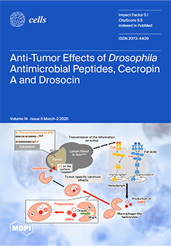
Two Drosophila antimicrobial peptides (AMPs), Cecropin A and Drosocin, are induced in the body fat of multiple sex combs (mxc) mutant larvae with tumors in hematopoietic tissues depending on the Toll- and Imd-mediated innate immune pathways. Overexpression of the AMPs suppressed tumor growth via apoptosis induction in the mutant but not in normal tissues, indicating anti-tumor properties. The injection of synthetic Cecropin also induced apoptosis in the tumors. The AMPs were incorporated into the macrophage-like cells in mutant but not normal larvae. Another AMP, Drosomycin, was taken up via phagocytosis factors, Draper and Shark. Enhanced phosphatidylserine signals were observed on the tumor surface. Inhibition of the exposed signals enhanced tumor growth. AMPs may target the signals to induce apoptosis to execute their tumor-specific effects. View this paper
  • Issues are regarded as officially published after their release is announced to the table of contents alert mailing list.
  • You may sign up for e-mail alerts to receive table of contents of newly released issues.
  • PDF is the official format for papers published in both, html and pdf forms. To view the papers in pdf format, click on the "PDF Full-text" link, and use the free Adobe Reader to open them.
Order results
Result details
Section
Select all
Export citation of selected articles as:
68 pages, 19995 KiB  
Review
Sexual Dimorphism in Cardiometabolic Diseases: From Development to Senescence and Therapeutic Approaches
by Thea Chevalley, Marion Dübi, Laurent Fumeaux, Maria Serena Merli, Alexandre Sarre, Natacha Schaer, Umberto Simeoni and Catherine Yzydorczyk
Cells 2025, 14(6), 467; https://doi.org/10.3390/cells14060467 - 20 Mar 2025
Viewed by 631
Abstract
The global incidence and prevalence of cardiometabolic disorders have risen significantly in recent years. Although lifestyle choices in adulthood play a crucial role in the development of these conditions, it is well established that events occurring early in life can have an important [...] Read more.
The global incidence and prevalence of cardiometabolic disorders have risen significantly in recent years. Although lifestyle choices in adulthood play a crucial role in the development of these conditions, it is well established that events occurring early in life can have an important effect. Recent research on cardiometabolic diseases has highlighted the influence of sexual dimorphism on risk factors, underlying mechanisms, and response to therapies. In this narrative review, we summarize the current understanding of sexual dimorphism in cardiovascular and metabolic diseases in the general population and within the framework of the Developmental Origins of Health and Disease (DOHaD) concept. We explore key risk factors and mechanisms, including the influence of genetic and epigenetic factors, placental and embryonic development, maternal nutrition, sex hormones, energy metabolism, microbiota, oxidative stress, cell death, inflammation, endothelial dysfunction, circadian rhythm, and lifestyle factors. Finally, we discuss some of the main therapeutic approaches, responses to which may be influenced by sexual dimorphism, such as antihypertensive and cardiovascular treatments, oxidative stress management, nutrition, cell therapies, and hormone replacement therapy. Full article
Show Figures

Graphical abstract

32 pages, 2353 KiB  
Review
Sirtuins and Gut Microbiota: Dynamics in Health and a Journey from Metabolic Dysfunction to Hepatocellular Carcinoma
by Mahmoud Zhra, Muhammad Affan Elahi, Aamira Tariq, Ahmed Abu-Zaid and Ahmed Yaqinuddin
Cells 2025, 14(6), 466; https://doi.org/10.3390/cells14060466 - 20 Mar 2025
Viewed by 495
Abstract
Metabolic dysfunction leading to non-alcoholic fatty liver disease (NAFLD) exhibits distinct molecular and immune signatures that are influenced by factors like gut microbiota. The gut microbiome interacts with the liver via a bidirectional relationship with the gut–liver axis. Microbial metabolites, sirtuins, and immune [...] Read more.
Metabolic dysfunction leading to non-alcoholic fatty liver disease (NAFLD) exhibits distinct molecular and immune signatures that are influenced by factors like gut microbiota. The gut microbiome interacts with the liver via a bidirectional relationship with the gut–liver axis. Microbial metabolites, sirtuins, and immune responses are pivotal in different metabolic diseases. This extensive review explores the complex and multifaceted interrelationship between sirtuins and gut microbiota, highlighting their importance in health and disease, particularly metabolic dysfunction and hepatocellular carcinoma (HCC). Sirtuins (SIRTs), classified as a group of NAD+-dependent deacetylases, serve as crucial modulators of a wide spectrum of cellular functions, including metabolic pathways, the inflammatory response, and the process of senescence. Their subcellular localization and diverse functions link them to various health conditions, including NAFLD and cancer. Concurrently, the gut microbiota, comprising diverse microorganisms, significantly influences host metabolism and immune responses. Recent findings indicate that sirtuins modulate gut microbiota composition and function, while the microbiota can affect sirtuin activity. This bidirectional relationship is particularly relevant in metabolic disorders, where dysbiosis contributes to disease progression. The review highlights recent findings on the roles of specific sirtuins in maintaining gut health and their implications in metabolic dysfunction and HCC development. Understanding these interactions offers potential therapeutic avenues for managing diseases linked to metabolic dysregulation and liver pathology. Full article
Show Figures

Figure 1

30 pages, 7720 KiB  
Article
Juglone-Bearing Thiopyrano[2,3-d]thiazoles Induce Apoptosis in Colorectal Adenocarcinoma Cells
by Yuliia Kozak, Nataliya Finiuk, Robert Czarnomysy, Agnieszka Gornowicz, Roman Pinyazhko, Andrii Lozynskyi, Serhii Holota, Olga Klyuchivska, Andriy Karkhut, Svyatoslav Polovkovych, Mykola Klishch, Rostyslav Stoika, Roman Lesyk, Krzysztof Bielawski and Anna Bielawska
Cells 2025, 14(6), 465; https://doi.org/10.3390/cells14060465 - 20 Mar 2025
Viewed by 351
Abstract
Colorectal cancer is a major global health challenge, with current treatments limited by toxicity and resistance. Thiazole derivatives, known for their bioactivity, are emerging as promising alternatives. Juglone (5-hydroxy-1,4-naphthoquinone) is a naturally occurring compound with known anticancer properties, and its incorporation into thiopyrano[2,3-d]thiazole [...] Read more.
Colorectal cancer is a major global health challenge, with current treatments limited by toxicity and resistance. Thiazole derivatives, known for their bioactivity, are emerging as promising alternatives. Juglone (5-hydroxy-1,4-naphthoquinone) is a naturally occurring compound with known anticancer properties, and its incorporation into thiopyrano[2,3-d]thiazole scaffolds may enhance their therapeutic potential. This study examined the cytotoxicity of thiopyrano[2,3-d]thiazoles and their effects on apoptosis in colorectal cancer cells. Les-6547 and Les-6557 increased the population of ROS-positive HT-29 cancer cells approximately 10-fold compared with control cells (36.3% and 38.5% vs. 3.8%, respectively), potentially contributing to various downstream effects. Elevated ROS levels were associated with cell cycle arrest, inhibition of DNA biosynthesis, and reduced cell proliferation. A significant shift in the cell cycle distribution was observed, with an increase in S-phase (from 17.3% in the control to 34.7% to 51.3% for Les-6547 and Les-6557, respectively) and G2/M phase (from 24.3% to 39.9% and 28.8%). Additionally, Les-6547 and Les-6557 inhibited DNA biosynthesis in HT-29 cells, with IC50 values of 2.21 µM and 2.91 µM, respectively. Additionally, ROS generation may initiate the intrinsic apoptotic pathway. Les-6547 and Les-6557 activated both intrinsic and extrinsic apoptotic pathways, demonstrated by notable increases in the activity of caspase 3/7, 8, 9, and 10. This study provides a robust basis for investigating the detailed molecular mechanisms of action and therapeutic potential of Les-6547 and Les-6557. Full article
(This article belongs to the Section Cell Proliferation and Division)
Show Figures

Figure 1

14 pages, 3819 KiB  
Article
Extracellular Vesicles from Regenerating Skeletal Muscle Mitigate Muscle Atrophy in an Amyotrophic Lateral Sclerosis Mouse Model
by Jinghui Gao, Aria Sikal, Rachel Hankin, Yaochao Zheng, Elijah Sterling, Kenny Chan and Yao Yao
Cells 2025, 14(6), 464; https://doi.org/10.3390/cells14060464 - 20 Mar 2025
Viewed by 440
Abstract
Amyotrophic lateral sclerosis (ALS) is a devastating neuromuscular disease characterized by progressive motor neuron degeneration and muscle atrophy, with no effective treatments available. Chronic inflammation, which impairs muscle regeneration and promotes proteolysis, is a key contributor to ALS-related muscle atrophy and a promising [...] Read more.
Amyotrophic lateral sclerosis (ALS) is a devastating neuromuscular disease characterized by progressive motor neuron degeneration and muscle atrophy, with no effective treatments available. Chronic inflammation, which impairs muscle regeneration and promotes proteolysis, is a key contributor to ALS-related muscle atrophy and a promising therapeutic target. Here, we applied extracellular vesicles (EVs) derived from regenerating skeletal muscles 14 days post-acute injury (CTXD14SkM-EVs), which possess a unique anti-inflammatory profile, to target muscle defects in ALS. We found that CTXD14SkM-EVs enhanced myoblast differentiation and fusion in a cellular muscle-wasting model induced by pro-inflammatory cytokine tumor necrosis factor alpha. Intramuscular administration of these EVs into an ALS mouse model mitigated muscle atrophy by promoting muscle regeneration, shifting macrophage polarization from pro-inflammatory M1 to anti-inflammatory M2 state, and suppressing the aberrant Nuclear Factor Kappa B (NF-κB) signaling, a key driver of muscle protein degradation. These results underscore the therapeutic potential of regenerating muscle-derived EVs for combating muscle atrophy in ALS. Full article
Show Figures

Graphical abstract

16 pages, 3864 KiB  
Article
Impact of a High-Fat Diet on the Gut Microbiome: A Comprehensive Study of Microbial and Metabolite Shifts During Obesity
by Md Abdullah Al Mamun, Ahmed Rakib, Mousumi Mandal and Udai P. Singh
Cells 2025, 14(6), 463; https://doi.org/10.3390/cells14060463 - 20 Mar 2025
Viewed by 897
Abstract
Over the last few decades, the prevalence of metabolic diseases such as obesity, diabetes, non-alcoholic fatty liver disease, hypertension, and hyperuricemia has surged, primarily due to high-fat diet (HFD). The pathologies of these metabolic diseases show disease-specific alterations in the composition and function [...] Read more.
Over the last few decades, the prevalence of metabolic diseases such as obesity, diabetes, non-alcoholic fatty liver disease, hypertension, and hyperuricemia has surged, primarily due to high-fat diet (HFD). The pathologies of these metabolic diseases show disease-specific alterations in the composition and function of their gut microbiome. How HFD alters the microbiome and its metabolite to mediate adipose tissue (AT) inflammation and obesity is not well known. Thus, this study aimed to identify the changes in the gut microbiome and metabolomic signatures induced by an HFD to alter obesity. To explore the changes in the gut microbiota and metabolites, 16S rRNA gene amplicon sequencing and metabolomic analyses were performed after HFD and normal diet (ND) feeding. We noticed that, at taxonomic levels, the number of operational taxonomic units (OTUs), along with the Chao and Shannon indexes, significantly shifted in HFD-fed mice compared to those fed a ND. Similarly, at the phylum level, an increase in Firmicutes and a decrease in Bacteroidetes were noticed in HFD-fed mice. At the genus level, an increase in Lactobacillus and Ruminococcus was observed, while Allobaculum, Clostridium, and Akkermansia were markedly reduced in the HFD group. Many bacteria from the Ruminococcus genus impair bile acid metabolism and restrict weight loss. Firmicutes are efficient in breaking down complex carbohydrates into short-chain fatty acids (SCFAs) and other metabolites, whereas Bacteroidetes are involved in a more balanced or efficient energy extraction. Thus, an increase in Firmicutes over Bacteroidetes enhances the absorption of more calories from food, which may contribute to obesity. Taken together, the altered gut microbiota and metabolites trigger AT inflammation, which contributes to metabolic dysregulation and disease progression. Thus, this study highlights the potential of the gut microbiome in the development of therapeutic strategies for obesity and related metabolic disorders. Full article
(This article belongs to the Section Cellular Pathology)
Show Figures

Graphical abstract

17 pages, 2340 KiB  
Review
How Do Peripheral Neurons and Glial Cells Participate in Pain Alleviation by Physical Activity?
by Menachem Hanani
Cells 2025, 14(6), 462; https://doi.org/10.3390/cells14060462 - 20 Mar 2025
Viewed by 495
Abstract
Chronic pain is a global health problem with major socioeconomic implications. Drug therapy for chronic pain is limited, prompting search for non-pharmacological treatments. One such approach is physical exercise, which has been found to be beneficial for numerous health issues. Research in recent [...] Read more.
Chronic pain is a global health problem with major socioeconomic implications. Drug therapy for chronic pain is limited, prompting search for non-pharmacological treatments. One such approach is physical exercise, which has been found to be beneficial for numerous health issues. Research in recent years has yielded considerable evidence for the analgesic actions of exercise in humans and experimental animals, but the underlying mechanisms are far from clear. It was proposed that exercise influences the pain pathways by interacting with the immune system, mainly by reducing inflammatory responses, but the release of endogenous analgesic mediators is another possibility. Exercise acts on neurons and glial cells in both the central and peripheral nervous systems. This review focuses on the periphery, with emphasis on possible glia–neuron interactions. Key topics include interactions of Schwann cells with axons (myelinated and unmyelinated), satellite glial cells in sensory ganglia, enteric glial cells, and the sympathetic nervous system. An attempt is made to highlight several neurological diseases that are associated with pain and the roles that glial cells may play in exercise-induced pain alleviation. Among the diseases are fibromyalgia and Charcot–Marie–Tooth disease. The hypothesis that active skeletal muscles exert their effects on the nervous system by releasing myokines is discussed. Full article
(This article belongs to the Section Cells of the Nervous System)
Show Figures

Figure 1

24 pages, 6066 KiB  
Article
Efficacy of Deferoxamine Mesylate in Serum and Serum-Free Media: Adult Ventral Root Schwann Cell Survival Following Hydrogen Peroxide-Induced Cell Death
by Yee Hang Ethan Ma, Abhinay R. Putta, Cyrus H. H. Chan, Stephen R. Vidman, Paula Monje and Giles W. Plant
Cells 2025, 14(6), 461; https://doi.org/10.3390/cells14060461 - 20 Mar 2025
Viewed by 401
Abstract
Schwann cell (SC) transplantation shows promise in treating spinal cord injury as a pro-regenerative agent to allow host endogenous neurons to bridge over the lesion. However, SC transplants face significant oxidative stress facilitated by ROS in the lesion, leading to poor survival. deferoxamine [...] Read more.
Schwann cell (SC) transplantation shows promise in treating spinal cord injury as a pro-regenerative agent to allow host endogenous neurons to bridge over the lesion. However, SC transplants face significant oxidative stress facilitated by ROS in the lesion, leading to poor survival. deferoxamine mesylate (DFO) is a neuroprotective agent shown to reduce H2O2-induced cell death in serum-containing conditions. Here we show that DFO is not necessary to induce neuroprotection under serum-free conditions by cell survival quantification and phenotypic analysis via immunohistochemistry, Hif1α and collagen IV quantification via whole cell corrected total cell fluorescence, and cell death transcript changes via RT-qPCR. Our results indicate survival of SC regardless of DFO pretreatment in serum-free conditions and an increased survival facilitated by DFO in serum-containing conditions. Furthermore, our results showed strong nuclear expression of Hif1α in serum-free conditions regardless of DFO pre-treatment and a nuclear expression of Hif1α in DFO-treated SCs in serum conditions. Transcriptomic analysis reveals upregulation of autophagy transcripts in SCs grown in serum-free media relative to SCs in serum conditions, with and without DFO and H2O2. Thus, indicating a pro-repair and regenerative state of the SCs in serum-free conditions. Overall, results indicate the protectiveness of CDM in enhancing SC survival against ROS-induced cell death in vitro. Full article
(This article belongs to the Section Cells of the Nervous System)
Show Figures

Figure 1

28 pages, 3566 KiB  
Review
Role of PDE4 Family in Cardiomyocyte Physiology and Heart Failure
by Ivan Sherstnev, Aleksandra Judina, Giovanni Battista Luciani, Alessandra Ghigo, Emilio Hirsch and Julia Gorelik
Cells 2025, 14(6), 460; https://doi.org/10.3390/cells14060460 - 20 Mar 2025
Viewed by 596
Abstract
Phosphodiesterase 4 (PDE4) is a key regulator of cyclic adenosine monophosphate (cAMP) signalling in cardiomyocytes, controlling contractility, calcium handling, and hypertrophic responses. PDE4 provides spatial and temporal precision to cAMP signalling, particularly under β-adrenergic stimulation, through its compartmentalised activity in subcellular nanodomains, including [...] Read more.
Phosphodiesterase 4 (PDE4) is a key regulator of cyclic adenosine monophosphate (cAMP) signalling in cardiomyocytes, controlling contractility, calcium handling, and hypertrophic responses. PDE4 provides spatial and temporal precision to cAMP signalling, particularly under β-adrenergic stimulation, through its compartmentalised activity in subcellular nanodomains, including the sarcoplasmic reticulum, plasma membrane and nuclear envelope. This review highlights the cardiac PDE4 isoforms PDE4A, PDE4B and PDE4D, focusing on their distinct localisation and contributions to cardiac physiology and pathophysiology, particularly in heart failure and arrhythmias. Although PDE4 plays a smaller role in overall cAMP hydrolysis in human hearts than in rodents, its compartmentalised function remains critical. Recent therapeutic advances have shifted from pan-PDE4 inhibitors to isoform-specific approaches to enhance efficacy while minimising systemic toxicity. We discuss the potential of selective PDE4 modulators, gene therapies and combination strategies in restoring cAMP compartmentation and preventing maladaptive cardiac remodelling. By integrating rodent and human studies, this review underscores the translational challenges and therapeutic opportunities surrounding PDE4, positioning it as both a key regulator of cardiac signalling and a promising target for heart failure therapies. Full article
Show Figures

Figure 1

19 pages, 1304 KiB  
Review
Ciliary Ion Channels in Polycystic Kidney Disease
by Lubna A. Alshriem, Raghad Buqaileh, Qasim Alorjani and Wissam AbouAlaiwi
Cells 2025, 14(6), 459; https://doi.org/10.3390/cells14060459 - 19 Mar 2025
Viewed by 540
Abstract
Polycystic kidney disease (PKD) is the most common hereditary disorder that disrupts renal function and frequently progresses to end-stage renal disease. Recent advances have elucidated the critical role of primary cilia and ciliary ion channels, including transient receptor potential (TRP) channels, cystic fibrosis [...] Read more.
Polycystic kidney disease (PKD) is the most common hereditary disorder that disrupts renal function and frequently progresses to end-stage renal disease. Recent advances have elucidated the critical role of primary cilia and ciliary ion channels, including transient receptor potential (TRP) channels, cystic fibrosis transmembrane conductance regulator (CFTR), and polycystin channels, in the pathogenesis of PKD. While some channels primarily function as chloride conductance channels (e.g., CFTR), others primarily regulate calcium (Ca+2) homeostasis. These ion channels are essential for cellular signaling and maintaining the normal kidney architecture. Dysregulation of these pathways due to genetic mutations in PKD1 and PKD2 leads to disrupted Ca+2 and cAMP signaling, aberrant fluid secretion, and uncontrolled cellular proliferation, resulting in tubular cystogenesis. Understanding the molecular mechanisms underlying these dysfunctions has opened the door for innovative therapeutic strategies, including TRPV4 activators, CFTR inhibitors, and calcimimetics, to mitigate cyst growth and preserve renal function. This review summarizes the current knowledge on the roles of ciliary ion channels in PKD pathophysiology, highlights therapeutic interventions targeting these channels, and identifies future research directions for improving patient outcomes. Full article
(This article belongs to the Special Issue The Role of Cilia in Health and Diseases—2nd Edition)
Show Figures

Figure 1

24 pages, 1111 KiB  
Review
A Comprehensive Exploration of the Biological Effects of Adipose-Derived Stem Cells in the Treatment of Systemic Sclerosis
by Gabriele Storti, Riccardo Foti, Roberta Foti, Marco Palmesano, Martina Patacchiola, Dalila Incognito, Giulio Cervelli, Benedetto Longo, Maria Giovanna Scioli, Elena Fiorelli, Sonia Terriaca, Andrea Lisa, Bong Sung Kim, Augusto Orlandi and Valerio Cervelli
Cells 2025, 14(6), 458; https://doi.org/10.3390/cells14060458 - 19 Mar 2025
Viewed by 401
Abstract
Systemic sclerosis (SSc) is a complex autoimmune disease characterized by vasculopathy and tissue fibrosis affecting the skin and internal organs. Genetic and environmental factors influence susceptibility, severity, and onset. Current treatments are limited and not always effective, leading researchers to investigate new approaches, [...] Read more.
Systemic sclerosis (SSc) is a complex autoimmune disease characterized by vasculopathy and tissue fibrosis affecting the skin and internal organs. Genetic and environmental factors influence susceptibility, severity, and onset. Current treatments are limited and not always effective, leading researchers to investigate new approaches, such as the use of adipose-derived mesenchymal stem cells (ADSCs) through fat grafting. This review seeks to understand how ADSCs may impact the development and progression of SSc, with a particular focus on how these cells could alter immune responses and reduce fibrosis. ADSCs have been found to affect various immune cells, including T cells, B cells, macrophages, and dendritic cells, by releasing cytokines, chemokines, and growth factors. These interactions generally suppress inflammation and promote a regulatory immune environment. Additionally, ADSCs can influence the extracellular matrix, helping to prevent fibrosis through signaling molecules like exosomes. ADSCs show promise as a treatment for SSc due to their ability to modulate the immune system and reduce fibrosis. Early clinical studies are encouraging, but more research is needed to fully understand how they work and to develop effective treatment protocols. Full article
Show Figures

Figure 1

50 pages, 4089 KiB  
Review
Colorectal Organoids: Models, Imaging, Omics, Therapy, Immunology, and Ethics
by Martina Taglieri, Linda Di Gregorio, Serena Matis, Chiara Rosa Maria Uras, Massimo Ardy, Sara Casati, Monica Marchese, Alessandro Poggi, Lizzia Raffaghello and Roberto Benelli
Cells 2025, 14(6), 457; https://doi.org/10.3390/cells14060457 - 19 Mar 2025
Viewed by 399
Abstract
Colorectal epithelium was the first long-term 3D organoid culture established in vitro. Identification of the key components essential for the long-term survival of the stem cell niche allowed an indefinite propagation of these cultures and the modulation of their differentiation into various lineages [...] Read more.
Colorectal epithelium was the first long-term 3D organoid culture established in vitro. Identification of the key components essential for the long-term survival of the stem cell niche allowed an indefinite propagation of these cultures and the modulation of their differentiation into various lineages of mature intestinal epithelial cells. While these methods were eventually adapted to establish organoids from different organs, colorectal organoids remain a pioneering model for the development of new applications in health and disease. Several basic and applicative aspects of organoid culture, modeling, monitoring and testing are analyzed in this review. We also tackle the ethical problems of biobanking and distribution of these precious research tools, frequently confined in the laboratory of origin or condemned to destruction at the end of the project. Full article
(This article belongs to the Special Issue Organoids as an Experimental Tool)
Show Figures

Figure 1

19 pages, 4640 KiB  
Article
A Cost-Effective and Easy to Assemble 3D Human Microchannel Blood–Brain Barrier Model and Its Application in Tumor Cell Adhesion Under Flow
by Yunfei Li and Bingmei M. Fu
Cells 2025, 14(6), 456; https://doi.org/10.3390/cells14060456 - 19 Mar 2025
Viewed by 451
Abstract
By utilizing polydimethylsiloxane (PDMS), collagen hydrogel, and a cell line for human cerebral microvascular endothelial cells, we produced a 3D microchannel blood–brain barrier (BBB) model under physiological flow. This 3D BBB has a circular-shaped cross-section and a diameter of ~100 μm, which can [...] Read more.
By utilizing polydimethylsiloxane (PDMS), collagen hydrogel, and a cell line for human cerebral microvascular endothelial cells, we produced a 3D microchannel blood–brain barrier (BBB) model under physiological flow. This 3D BBB has a circular-shaped cross-section and a diameter of ~100 μm, which can properly mimic the cerebral microvessel responsible for material exchange between the circulating blood and brain tissue. The permeability of the 3D microchannel BBB to a small molecule (sodium fluorescein with a molecular weight of 376) and that to a large molecule (Dex-70k) are the same as those of rat cerebral microvessels. This 3D BBB model can replicate the effects of a plasma protein, orosomucoid, a cytokine, vascular endothelial growth factor (VEGF), and an enzyme, heparinase III, on either rat cerebral or mesenteric microvessesels in terms of permeability and the modulation of glycocalyx (heparan sulfate). It can also replicate the adhesion of a breast cancer cell, MDA-MB-231, in rat mesenteric microvessels under no treatment or treatments with VEGF, orosomucoid, and heparinase III. Because of difficulties in accessing human cerebral microvessels, this inexpensive and easy to assemble 3D human BBB model can be applied to investigate BBB-modulating mechanisms in health and in disease and to develop therapeutic interventions targeting tumor metastasis to the brain. Full article
(This article belongs to the Special Issue Cellular and Molecular Mechanisms of Vascular-Related Diseases)
Show Figures

Figure 1

15 pages, 10679 KiB  
Article
Bovine Meat and Milk Factor (BMMF) Protein Is Expressed in Macrophages Spread Widely over the Mucosa of Colorectal Cancer Patients
by Sumen Siqin, Ekaterina Nikitina, Mohammad Rahbari, Claudia Ernst, Damir Krunic, Emrullah Birgin, Claudia Tessmer, Ilse Hofmann, Nuh Rahbari and Timo Bund
Cells 2025, 14(6), 455; https://doi.org/10.3390/cells14060455 - 19 Mar 2025
Viewed by 369
Abstract
Red meat consumption is considered a risk factor for colorectal cancer (CRC) development and stimulated isolation of plasmid-like DNA molecules from bovine serum and milk, termed bovine meat and milk factors (BMMFs). BMMFs encode a conserved replication protein (Rep). Increased populations of Rep-expressing [...] Read more.
Red meat consumption is considered a risk factor for colorectal cancer (CRC) development and stimulated isolation of plasmid-like DNA molecules from bovine serum and milk, termed bovine meat and milk factors (BMMFs). BMMFs encode a conserved replication protein (Rep). Increased populations of Rep-expressing macrophages have been identified in the peritumor of CRC patients and pre-cancerous tissues when compared to the tissues of healthy individuals. This supports the concept that BMMFs increase cancer risk by indirect carcinogenesis, upon induction of chronic inflammation. However, the spread of Rep+ immune cells in tissues at greater distances from primary tumors has not yet been assessed. Here, we immunohistologically analyzed the presence of Rep+ immune cells in sets of tumor, peritumor and, additionally, distant tissues of CRC patients (n = 13). We identified consistently high numbers of BMMF-positive macrophages in mucosal tissues at distances of as much as 25 cm away from the primary tumors, at levels comparable to peritumors and associated with M2-like macrophage polarization. The broad distribution of BMMFs suggests that BMMF+ macrophages might already exist at stages of pre-cancerous dysplasia or before. Quantification of BMMF tissue expression during colonoscopy might help to preventively stratify individuals at risk of developing polyps/CRC and recommend them for enhanced surveillance and/or changes in dietary lifestyle. Full article
Show Figures

Graphical abstract

9 pages, 6690 KiB  
Brief Report
Dysregulation of T Follicular Helper and Regulatory Cells in IRF5-SLE Homozygous Risk Carriers and Systemic Lupus Erythematosus Patients
by Bharati Matta, Lydia Thomas, Vinay Sharma and Betsy J. Barnes
Cells 2025, 14(6), 454; https://doi.org/10.3390/cells14060454 - 19 Mar 2025
Viewed by 367
Abstract
T follicular helper (Tfh) and T follicular regulatory cells (Tfr) are required for antibody production and are dysregulated in SLE. Genetic variants within or near interferon regulatory factor 5 (IRF5) are associated with SLE risk. We previously reported higher plasma cells [...] Read more.
T follicular helper (Tfh) and T follicular regulatory cells (Tfr) are required for antibody production and are dysregulated in SLE. Genetic variants within or near interferon regulatory factor 5 (IRF5) are associated with SLE risk. We previously reported higher plasma cells and autoantibodies in healthy IRF5-SLE homozygous risk carriers. Here, we report the dysregulation of circulating Tfh and Tfr in both SLE patients and presymptomatic IRF5-SLE homozygous risk carriers. Full article
(This article belongs to the Special Issue Genetic and Cellular Basis of Autoimmune Diseases)
Show Figures

Figure 1

10 pages, 2266 KiB  
Communication
Impact of Secondhand Smoke and E-Cigarette Exposure on Placental Apoptotic and Growth-Regulatory Proteins in Mouse Pregnancy
by Logan Beck, Madison N. Kirkham, Marley Shin, Benjamin T. Bikman, Paul R. Reynolds and Juan A. Arroyo
Cells 2025, 14(6), 453; https://doi.org/10.3390/cells14060453 - 19 Mar 2025
Viewed by 387
Abstract
Apoptosis is critical in placental development, and its dysregulation is linked to pregnancy complications such as intrauterine growth restriction (IUGR) and preeclampsia (PE). Environmental exposures, particularly secondhand smoke (SHS) and e-cigarettes (eCigs), may contribute to placental dysfunction through apoptotic pathways. This study examined [...] Read more.
Apoptosis is critical in placental development, and its dysregulation is linked to pregnancy complications such as intrauterine growth restriction (IUGR) and preeclampsia (PE). Environmental exposures, particularly secondhand smoke (SHS) and e-cigarettes (eCigs), may contribute to placental dysfunction through apoptotic pathways. This study examined the effects of SHS and eCig exposure on placental apoptosis and growth-regulatory proteins in a murine model. C57BL/6 pregnant mice were exposed to SHS or eCigs at two critical gestational time points: early trophoblast invasion (E12.5 to E18.5) and established invasion (E14.5 to E18.5). Placental tissues were collected and analyzed for pro-apoptotic and anti-apoptotic markers, heat shock proteins, insulin-like growth factor-binding proteins (IGFBPs), and growth regulators. SHS exposure increased pro-apoptotic markers (BAD, Fas/FasL) and decreased mitochondrial function markers (cytochrome c), indicating compromised cellular survival. Both SHS and eCig exposure reduced anti-apoptotic markers (BCL-2, HSP27, survivin) and growth regulators (IGF-1, IGFBPs). SHS and eCig exposure create a pro-apoptotic environment in the placenta, potentially impairing fetal development through altered apoptotic and growth-regulatory pathways. These findings underscore the risks of environmental exposures during pregnancy, highlighting the need for strategies to minimize maternal exposure to SHS and eCigs. Full article
Show Figures

Figure 1

25 pages, 1841 KiB  
Review
Biomaterial Properties and Differentiation Strategies for Tenogenic Differentiation of Mesenchymal Stem Cells
by Brendon Roets, Heidi Abrahamse and Anine Crous
Cells 2025, 14(6), 452; https://doi.org/10.3390/cells14060452 - 18 Mar 2025
Viewed by 314
Abstract
Tendinopathy is a prevalent musculoskeletal condition that affects both aging populations and individuals involved in repetitive, high-intensity activities, such as athletes. Current treatment options primarily address symptom management or involve surgery, which carries a significant risk of complications and re-injury. This highlights the [...] Read more.
Tendinopathy is a prevalent musculoskeletal condition that affects both aging populations and individuals involved in repetitive, high-intensity activities, such as athletes. Current treatment options primarily address symptom management or involve surgery, which carries a significant risk of complications and re-injury. This highlights the need for regenerative medicine approaches that combine stem cells, biomaterials, and growth factors. However, achieving effective tenogenic differentiation remains challenging due to the absence of standardized differentiation protocols. Consequently, a review of existing research has been conducted to identify optimal biomaterial properties and growth factor protocols. Findings suggest that the ideal biomaterial for tenogenic differentiation should feature a 3D structure to preserve tenogenic expression, incorporate a combination of aligned micro- and nanofibers to promote differentiation, and require further investigation into optimal stiffness. Additionally, growth factor protocols should include an induction phase to initiate tenogenic lineage commitment, followed by a maintenance phase to support matrix production and maturation. Full article
(This article belongs to the Special Issue Stem Cell, Differentiation, Regeneration and Diseases)
Show Figures

Figure 1

13 pages, 4959 KiB  
Communication
Characterisation of Cytotoxicity-Related Receptors on γδ T Cells in Chronic Lymphocytic Leukaemia
by Michał Zarobkiewicz, Natalia Lehman, Izabela Morawska-Michalska, Adam Michalski, Wioleta Kowalska, Agata Szymańska, Waldemar Tomczak and Agnieszka Bojarska-Junak
Cells 2025, 14(6), 451; https://doi.org/10.3390/cells14060451 - 18 Mar 2025
Viewed by 318
Abstract
Chronic lymphocytic leukaemia (CLL) is a haematological malignancy primarily affecting older adults, characterised by the proliferation of functionally impaired B lymphocytes with abnormal expression of CD5, a typical T cell marker. The current study investigates the expression of cytotoxicity-related receptors (CD16, CD56, CD57, [...] Read more.
Chronic lymphocytic leukaemia (CLL) is a haematological malignancy primarily affecting older adults, characterised by the proliferation of functionally impaired B lymphocytes with abnormal expression of CD5, a typical T cell marker. The current study investigates the expression of cytotoxicity-related receptors (CD16, CD56, CD57, CD69) and a checkpoint (LAG-3) on γδ T cells in CLL patients. Sixty-nine treatment-naive CLL patients and fourteen healthy controls were recruited. Flow cytometry analysis revealed that the CLL patients had higher expressions of CD56 and LAG-3 and lower CD16 on their γδ T cells compared to the healthy controls. Subgroup analysis showed that ZAP-70-negative patients exhibited increased CD69, while CD38-negative patients showed higher CD16 expression. Additionally, CD16 expression was inversely correlated with serum LDH levels, a marker of disease progression. Bioinformatic analysis of the LAG-3 ligand mRNA in a CLL dataset indicated higher expression of HLA-DQA2 and HLA-DRB5 in patients with unmutated IGVH. Our findings highlight the altered expression of key cytotoxicity markers on γδ T cells in CLL, suggesting their potential role in disease progression and as a therapeutic target. In particular, the use of anti-LAG-3 antibodies seems promising. Full article
(This article belongs to the Special Issue The Role of T-Cells in Lymphomas and Leukemias)
Show Figures

Figure 1

18 pages, 4408 KiB  
Article
Capturing the Heterogeneity of the PDAC Tumor Microenvironment: Novel Triple Co-Culture Spheroids for Drug Screening and Angiogenic Evaluation
by Ruben Verloy, Angela Privat-Maldonado, Jonas Van Audenaerde, Sophie Rovers, Hannah Zaryouh, Jorrit De Waele, Delphine Quatannens, Dieter Peeters, Geert Roeyen, Christophe Deben, Evelien Smits and Annemie Bogaerts
Cells 2025, 14(6), 450; https://doi.org/10.3390/cells14060450 - 18 Mar 2025
Viewed by 556
Abstract
Pancreatic ductal adenocarcinoma (PDAC) presents significant treatment challenges due to its desmoplastic reaction, which impedes therapeutic effectiveness, highlighting the need for advanced vitro models to better mimic the complex tumor environment. The current three-dimensional co-culture models of fibroblasts and endothelial cells are lacking, [...] Read more.
Pancreatic ductal adenocarcinoma (PDAC) presents significant treatment challenges due to its desmoplastic reaction, which impedes therapeutic effectiveness, highlighting the need for advanced vitro models to better mimic the complex tumor environment. The current three-dimensional co-culture models of fibroblasts and endothelial cells are lacking, which presents a challenge for performing more comprehensive in vitro research. Our study developed triple co-culture spheroid models using MiaPaCa-2 and BxPC-3 cancer cell lines, with RLT-PSC and hPSC21 pancreatic stellate cell lines and the endothelial cell line HMEC-1. These models were assessed through growth assays, multicolor flow cytometry to optimize cell ratios, cell viability assays to evaluate drug responses, and a tube formation assay with a spheroid-conditioned medium to examine angiogenesis. Our triple co-culture spheroids effectively replicate the PDAC microenvironment, showing significant variations in drug responses influenced by cellular composition, density, and spatial arrangement. The tube formation assay showcased the potential of our models to quantitatively assess a treatment-induced angiogenic response. These cost-effective triple-co-culture in vitro spheroid models provide vital insights into the PDAC microenvironment, significantly improving the quality of the in vitro evaluation of treatment responses. Full article
Show Figures

Graphical abstract

25 pages, 5063 KiB  
Article
Cell Homeostasis or Cell Death—The Balancing Act Between Autophagy and Apoptosis Caused by Steatosis-Induced Endoplasmic Reticulum (ER) Stress
by Anna Stilkerich, Gerda Schicht, Lena Seidemann, René Hänsel, Adrian Friebel, Stefan Hoehme, Daniel Seehofer and Georg Damm
Cells 2025, 14(6), 449; https://doi.org/10.3390/cells14060449 - 18 Mar 2025
Viewed by 473
Abstract
Metabolic-dysfunction-associated steatotic liver disease (MASLD) is a prevalent liver condition with potential progression to cirrhosis and impaired regeneration post-resection. A key mechanism underlying lipotoxicity is endoplasmic reticulum (ER) stress, particularly the activation of the unfolded protein response (UPR). This study investigates the interplay [...] Read more.
Metabolic-dysfunction-associated steatotic liver disease (MASLD) is a prevalent liver condition with potential progression to cirrhosis and impaired regeneration post-resection. A key mechanism underlying lipotoxicity is endoplasmic reticulum (ER) stress, particularly the activation of the unfolded protein response (UPR). This study investigates the interplay between lipid accumulation, endoplasmic reticulum (ER) stress, and cellular outcomes, focusing on the balance between autophagy and apoptosis. We cultured primary human hepatocytes (PHH) in a free fatty acid (FFA)-enriched medium for 120 h, assessing lipid accumulation, metabolism, and the expression of selected UPR markers. Additionally, we investigated the effects of lipid load on cell activity and growth in proliferating HepG2 cells. We observed that FFA uptake consistently induced ER stress, shifting cellular responses toward apoptosis under high lipid loads. Donor-specific differences were evident, particularly in lipid storage, excretion, and sensitivity to lipotoxicity. Some donors exhibited limited triglyceride (TAG) storage and excretion, leading to an excess of FFA whose metabolic fate remains unclear. Proliferation was more sensitive to lipid accumulation than overall cell activity, with even low FFA concentrations impairing growth, highlighting the vulnerability of regenerative processes to steatosis. The study elucidates how ER stress pathways, such as PERK-CHOP and IRE1α-JNK, are differentially activated in response to lipid overload, tipping the balance toward apoptosis in severe cases. The limited activation of repair mechanisms, such as autophagy, further emphasizes the critical role of ER stress in determining hepatocyte fate. The donor-dependent variability highlights the need for personalized strategies to mitigate lipotoxic effects and enhance liver regeneration in steatosis-related conditions. Full article
Show Figures

Figure 1

32 pages, 24604 KiB  
Article
The Recovery of Epidermal Proliferation Pattern in Human Skin Xenograft
by Olga Cherkashina, Alexandra Tsitrina, Danila Abolin, Elena Morgun, Anastasiya Kosykh, Marat Sabirov, Ekaterina Vorotelyak and Ekaterina Kalabusheva
Cells 2025, 14(6), 448; https://doi.org/10.3390/cells14060448 - 17 Mar 2025
Viewed by 400
Abstract
Abnormalities in epidermal keratinocyte proliferation are a characteristic feature of a range of dermatological conditions. These include hyperproliferative states in psoriasis and dermatitis as well as hypoproliferative states in chronic wounds. This emphasises the importance of investigating the proliferation kinetics under conditions of [...] Read more.
Abnormalities in epidermal keratinocyte proliferation are a characteristic feature of a range of dermatological conditions. These include hyperproliferative states in psoriasis and dermatitis as well as hypoproliferative states in chronic wounds. This emphasises the importance of investigating the proliferation kinetics under conditions of healthy skin and identifying the key regulators of epidermal homeostasis, maintenance, and recovery following wound healing. Animal models contribute to our understanding of human epidermal self-renewal. Human skin xenografting overcomes the ethical limitations of studying human skin during regeneration. The application of this approach has allowed for the identification of a single population of stem cells and both slowly and rapidly cycling progenitors within the epidermal basal layer and the mapping of their location in relation to rete ridges and hair follicles. Furthermore, we have traced the dynamics of the proliferation pattern reorganization that occurs during epidermal regeneration, underlining the role of YAP activity in epidermal relief formation. Full article
(This article belongs to the Collection Feature Papers in 'Cell Proliferation and Division')
Show Figures

Graphical abstract

19 pages, 2883 KiB  
Article
Chromosomal Instability Is Associated with cGAS–STING Activation in EGFR-TKI Refractory Non-Small-Cell Lung Cancer
by Kimio Yonesaka, Takashi Kurosaki, Junko Tanizaki, Hisato Kawakami, Kaoru Tanaka, Osamu Maenishi, Shiki Takamura, Kazuko Sakai, Yasutaka Chiba, Takeshi Teramura, Hiroki Goto, Eri Otsuka, Hiroaki Okida, Masanori Funabashi, Yuuri Hashimoto, Kenji Hirotani, Yasuki Kamai, Takashi Kagari, Kazuto Nishio, Kazuhiro Kakimi and Hidetoshi Hayashiadd Show full author list remove Hide full author list
Cells 2025, 14(6), 447; https://doi.org/10.3390/cells14060447 - 17 Mar 2025
Viewed by 626
Abstract
Epidermal growth factor receptor tyrosine kinase inhibitors (EGFR-TKIs) are standard therapies for EGFR-mutated non-small-cell lung cancer (NSCLC); however, their efficacy is inconsistent. Secondary mutations in the EGFR or other genes that lead to resistance have been identified, but resistance mechanisms have not [...] Read more.
Epidermal growth factor receptor tyrosine kinase inhibitors (EGFR-TKIs) are standard therapies for EGFR-mutated non-small-cell lung cancer (NSCLC); however, their efficacy is inconsistent. Secondary mutations in the EGFR or other genes that lead to resistance have been identified, but resistance mechanisms have not been fully identified. Chromosomal instability (CIN) is a hallmark of cancer and results in genetic diversity. In this study, we demonstrated by transcriptomic analysis that CIN activates the cGAS–STING signaling pathway, which leads to EGFR-TKI refractoriness in a subset of EGFR-mutated NSCLC patients. Furthermore, EGFR-mutated H1975dnMCAK cells, which frequently underwent chromosomal mis-segregation, demonstrated refractoriness to the EGFR-TKI osimertinib compared to control cells. Second, H1975dnMCAK cells exhibited activation of cGAS–STING signaling and its downstream signaling, including tumor-promoting cytokine IL-6. Finally, chromosomally unstable EGFR-mutated NSCLC exhibited enhanced epithelial–mesenchymal transition (EMT). Blockade of cGAS–STING-TBK1 signaling reversed EMT, resulting in restored susceptibility to EGFR-TKIs in vitro and in vivo. These results suggest that CIN may lead to the activation of cGAS–STING signaling in some EGFR-mutated NSCLC, resulting in EMT-associated EGFR-TKI resistance. Full article
(This article belongs to the Special Issue Unlocking the Secrets Behind Drug Resistance at the Cellular Level)
Show Figures

Graphical abstract

35 pages, 31411 KiB  
Article
The Role of Integrin β1D Mislocalization in the Pathophysiology of Calpain 3-Related Limb–Girdle Muscular Dystrophy
by Andrea Valls, Cristina Ruiz-Roldán, Jenita Immanuel, Sonia Alonso-Martín, Eduard Gallardo, Roberto Fernández-Torrón, Mario Bonilla, Ana Lersundi, Aurelio Hernández-Laín, Cristina Domínguez-González, Juan Jesús Vílchez, Pablo Iruzubieta, Adolfo López de Munain and Amets Sáenz
Cells 2025, 14(6), 446; https://doi.org/10.3390/cells14060446 - 17 Mar 2025
Viewed by 464
Abstract
Limb–girdle muscular dystrophy R1 (LGMDR1) is characterized by progressive proximal muscle weakness due to mutations in the CAPN3 gene. Little is known about CAPN3’s function in muscle, but its loss results in aberrant sarcomere formation. Human muscle structure was analyzed in this study, [...] Read more.
Limb–girdle muscular dystrophy R1 (LGMDR1) is characterized by progressive proximal muscle weakness due to mutations in the CAPN3 gene. Little is known about CAPN3’s function in muscle, but its loss results in aberrant sarcomere formation. Human muscle structure was analyzed in this study, with observations including integrin β1D isoform (ITGβ1D) mislocalization, a lack of Talin-1 (TLN1) in the sarcolemma and the irregular expression of focal adhesion kinase (FAK) in LGMDR1 muscles, suggesting a lack of integrin activation with an altered sarcolemma, extracellular matrix (ECM) assembly and signaling pathway deregulation, which may cause frailty in LGMDR1 muscle fibers. Additionally, altered nuclear morphology, centrosome distribution and microtubule organization have been found in muscle cells derived from LGMDR1 patients. Full article
(This article belongs to the Special Issue Muscle Structure and Function in Health and Disease)
Show Figures

Figure 1

20 pages, 3749 KiB  
Review
EFR3A, an Intriguing Gene, and Protein with a Scaffolding Function
by Magdalena Trybus, Anita Hryniewicz-Jankowska, Aleksander Czogalla and Aleksander F. Sikorski
Cells 2025, 14(6), 445; https://doi.org/10.3390/cells14060445 - 17 Mar 2025
Viewed by 458
Abstract
The EFR3 (Eighty-Five Requiring 3) protein and its homologs are rather poorly understood eukaryotic plasma membrane peripheral proteins. They belong to the armadillo-like family of superhelical proteins. In higher vertebrates two paralog genes, A and B were found, each expressing at least 2–3 [...] Read more.
The EFR3 (Eighty-Five Requiring 3) protein and its homologs are rather poorly understood eukaryotic plasma membrane peripheral proteins. They belong to the armadillo-like family of superhelical proteins. In higher vertebrates two paralog genes, A and B were found, each expressing at least 2–3 protein isoforms. EFR3s are involved in several physiological functions, mostly including phosphatidyl inositide phosphates, e.g., phototransduction (insects), GPCRs, and insulin receptors regulated processes (mammals). Mutations in the EFR3A were linked to several types of human disorders, i.e., neurological, cardiovascular, and several tumors. Structural data on the atomic level indicate the extended superhelical rod-like structure of the first two-thirds of the molecule with a typical armadillo repeat motif (ARM) in the N-terminal part and a triple helical motif in its C-terminal part. EFR3s’ best-known molecular function is anchoring the giant phosphatidylinositol 4-kinase A complex to the plasma membrane crucial for cell signaling, also linked directly to the KRAS mutant oncogenic function. Another function connected to the newly uncovered interaction of EFR3A with flotillin-2 may be the participation of the former in the organization and regulation of the membrane raft domain. This review presents EFR3A as an intriguing subject of future studies. Full article
Show Figures

Figure 1

17 pages, 4793 KiB  
Article
Pyruvate Regulates the Expression of DLAT to Promote Follicular Growth
by Liuhong Zhang, Yixuan Guo, Enyuan Huang, Jianing Lu, Tiantian Wang, Yonghua Shi, Meng Lv, Yongcai Chen, Shuo Li, Xiaolong Yuan and Jiaqi Li
Cells 2025, 14(6), 444; https://doi.org/10.3390/cells14060444 - 17 Mar 2025
Viewed by 371
Abstract
Increasing evidence has suggested that dihydrolipoamide S-acetyltransferase (DLAT), a subunit of the pyruvate dehydrogenase complex, is crucial for pyruvate metabolism and the regulation of cell death. The excessive death of granulosa cells (GCs) hinders the progression of follicular growth. However, the [...] Read more.
Increasing evidence has suggested that dihydrolipoamide S-acetyltransferase (DLAT), a subunit of the pyruvate dehydrogenase complex, is crucial for pyruvate metabolism and the regulation of cell death. The excessive death of granulosa cells (GCs) hinders the progression of follicular growth. However, the relationship between DLAT and follicular growth is poorly understood. Here, we found that pyruvate significantly shortened the age of pubertal initiation in mice and promoted follicular growth by promoting the proliferation of GCs. In addition, pyruvate up-regulated the expression of DLAT and the high level of DLAT was observed in large follicles, which were associated with follicular growth. Mechanistically, DLAT increased the mRNA and protein levels of proliferation pathways such as PCNA and MCL1 to promote GC proliferation. Additionally, DLAT bound to CASP3 and CASP9 proteins to inhibit the apoptosis of GCs. Taken together, these results reveal a mechanism that pyruvate regulated DLAT to promote follicular growth, and DLAT represents a promising target that supports new strategies for improving the growth of follicles. Full article
Show Figures

Figure 1

21 pages, 5521 KiB  
Article
A Novel Model for Simultaneous Evaluation of Hyperoxia-Mediated Brain and Lung Injury in Neonatal Rats
by Stefanie Obst, Meray Serdar, Josephine Herz, Karina Kempe, Meriem Assili, Mandana Rizazad, Dharmesh Hirani, Miguel A. Alejandre Alcazar, Stefanie Endesfelder, Marius A. Möbius, Mario Rüdiger, Ursula Felderhoff-Müser and Ivo Bendix
Cells 2025, 14(6), 443; https://doi.org/10.3390/cells14060443 - 16 Mar 2025
Viewed by 478
Abstract
Despite improved neonatal intensive care, the risk of premature-born infants developing bronchopulmonary dysplasia (BPD) and encephalopathy of prematurity (EoP) remains high. With hyperoxia being a major underlying factor, both preterm-birth-related complications are suggested to be closely interrelated. However, experimental models are lacking for [...] Read more.
Despite improved neonatal intensive care, the risk of premature-born infants developing bronchopulmonary dysplasia (BPD) and encephalopathy of prematurity (EoP) remains high. With hyperoxia being a major underlying factor, both preterm-birth-related complications are suggested to be closely interrelated. However, experimental models are lacking for the assessment of the potentially close interplay between both organs. To establish a model, suitable for the assessment of both affected organs, Wistar rats were exposed to 80% oxygen from postnatal day 2 (P2) for seven days. Brain and lung tissues were analysed via histomorphometry, immunohistochemistry, real-time PCR, and western blot at term P11. In the brain, hyperoxia induced significant hypomyelination accompanied by a reduction in oligodendrocytes and CD68 expression on microglia cells. These changes correlate with arrested alveolarisation and an increased number of macrophages in the lung. Interestingly, in contrast to the reduced formation of pulmonary microvessels, an increased vascular density was detected in the brain. Seven days of hyperoxia induces typical characteristics of BPD and EoP in neonatal rats, thereby linking impaired alveolarisation with disturbed myelination in the brain and providing an experimental model for understanding pathophysiological mechanisms and identifying organ-spanning novel therapeutic interventions targeting both diseases. Full article
(This article belongs to the Special Issue Perinatal Brain Injury—from Pathophysiology to Therapy)
Show Figures

Figure 1

25 pages, 1636 KiB  
Review
Control of Replication Stress Response by Cytosolic Fe-S Cluster Assembly (CIA) Machinery
by Chiara Frigerio, Michela Galli, Sara Castelli, Aurora Da Prada and Michela Clerici
Cells 2025, 14(6), 442; https://doi.org/10.3390/cells14060442 - 16 Mar 2025
Viewed by 411
Abstract
Accurate DNA replication is essential for the maintenance of genome stability and the generation of healthy offspring. When DNA replication is challenged, signals accumulate at blocked replication forks that elicit a multifaceted cellular response, orchestrating DNA replication, DNA repair and cell cycle progression. [...] Read more.
Accurate DNA replication is essential for the maintenance of genome stability and the generation of healthy offspring. When DNA replication is challenged, signals accumulate at blocked replication forks that elicit a multifaceted cellular response, orchestrating DNA replication, DNA repair and cell cycle progression. This replication stress response promotes the recovery of DNA replication, maintaining chromosome integrity and preventing mutations. Defects in this response are linked to heightened genetic instability, which contributes to tumorigenesis and genetic disorders. Iron–sulfur (Fe-S) clusters are emerging as important cofactors in supporting the response to replication stress. These clusters are assembled and delivered to target proteins that function in the cytosol and nucleus via the conserved cytosolic Fe-S cluster assembly (CIA) machinery and the CIA targeting complex. This review summarizes recent advances in understanding the structure and function of the CIA machinery in yeast and mammals, emphasizing the critical role of Fe-S clusters in the replication stress response. Full article
Show Figures

Graphical abstract

10 pages, 545 KiB  
Article
Clinical Value of Tocilizumab in Reducing Mortality in Refractory Septic Shock in Children with Hematologic and Non-Hematologic Diseases
by En-Pei Lee, Jainn-Jim Lin, Shih-Hsiang Chen, Oi-Wa Chan, Ya-Ting Su, Man-Ru Hsiao, Shao-Hsuan Hsia and Han-Ping Wu
Cells 2025, 14(6), 441; https://doi.org/10.3390/cells14060441 - 16 Mar 2025
Viewed by 524
Abstract
Background: Pediatric sepsis remains the main cause of morbidity and mortality among children. Interleukin (IL)-6 is usually produced after infection, and elevated IL-6 levels may cause multisystemic damage. This study aimed to evaluate the effect of tocilizumab, an IL-6 receptor antibody, on children [...] Read more.
Background: Pediatric sepsis remains the main cause of morbidity and mortality among children. Interleukin (IL)-6 is usually produced after infection, and elevated IL-6 levels may cause multisystemic damage. This study aimed to evaluate the effect of tocilizumab, an IL-6 receptor antibody, on children with septic shock. Methods: We conducted a retrospective cohort study of children diagnosed with septic shock and admitted to the pediatric intensive care unit (PICU) between 2018 and 2024. Tocilizumab was administered within 24 h to patients with high IL-6 levels who developed refractory septic shock. Outcomes, including 28-day mortality, morbidity, length of PICU stay, and shock duration, were analyzed between septic children with different etiologies and differed treatments. Results: Fifty-four children with refractory septic shock were included. Patients treated with tocilizumab (n = 21) showed improved outcomes compared to those without tocilizumab (n = 33), including shorter PICU stays and lower mortality rates (14.2% vs. 54.5%, p = 0.03). Subgroup analysis revealed that in the non-hematologic group, tocilizumab-treated patients had a 0% mortality rate compared to 50% in untreated patients (p = 0.006). In the hematologic group, tocilizumab-treated patients exhibited a 27.2% mortality rate compared to 61.5% in untreated patients (p = 0.09). Trends in IL-6 levels (D1 to D7) were significantly higher in non-survivors compared to survivors and in patients with hematological malignancies compared to those without. No adverse events, including secondary infections or long-term liver impairment, were observed. Conclusions: Tocilizumab appears to mitigate systemic inflammation and improve outcomes in children with refractory septic shock and elevated IL-6 levels. Further prospective studies are warranted to confirm these findings and establish treatment guidelines. Full article
Show Figures

Figure 1

10 pages, 799 KiB  
Review
Molecular Signaling Pathways in Wound-Induced Hair-Follicle Neogenesis
by Soung-Hoon Lee
Cells 2025, 14(6), 440; https://doi.org/10.3390/cells14060440 - 16 Mar 2025
Viewed by 598
Abstract
Wound-induced hair-follicle neogenesis (WIHN) is the phenomenon of regenerating new hair follicles from wounds in mammals. The WIHN involves both developmental and adult wound-healing processes. Moreover, the WIHN is regulated by a variety of factors, particularly multiple molecular signaling pathways produced in several [...] Read more.
Wound-induced hair-follicle neogenesis (WIHN) is the phenomenon of regenerating new hair follicles from wounds in mammals. The WIHN involves both developmental and adult wound-healing processes. Moreover, the WIHN is regulated by a variety of factors, particularly multiple molecular signaling pathways produced in several types of cells. Here, the role of multiple signaling in different types of cells in WIHN is comprehensively described. Furthermore, the lack of dermal γδ T cells in the human scalp has hindered the clinical application of WIHN, but the development of drugs such as Wnt signaling activators is increasing the effectiveness of WIHN in humans. Overall, understanding the underlying mechanisms that regulate WIHN may help treat skin diseases, including alopecia. Full article
(This article belongs to the Special Issue Molecular Signalings in Hair Regeneration)
Show Figures

Graphical abstract

16 pages, 1964 KiB  
Review
GOLPH3-mTOR Crosstalk and Glycosylation: A Molecular Driver of Cancer Progression
by Anna Frappaolo, Gianluca Zaccagnini and Maria Grazia Giansanti
Cells 2025, 14(6), 439; https://doi.org/10.3390/cells14060439 - 14 Mar 2025
Viewed by 553
Abstract
Originally identified in proteomic-based studies of the Golgi, Golgi phosphoprotein 3 (GOLPH3) is a highly conserved protein from yeast to humans. GOLPH3 localizes to the Golgi through the interaction with phosphatidylinositol-4-phosphate and is required for Golgi architecture and vesicular trafficking. Many studies revealed [...] Read more.
Originally identified in proteomic-based studies of the Golgi, Golgi phosphoprotein 3 (GOLPH3) is a highly conserved protein from yeast to humans. GOLPH3 localizes to the Golgi through the interaction with phosphatidylinositol-4-phosphate and is required for Golgi architecture and vesicular trafficking. Many studies revealed that the overexpression of GOLPH3 is associated with tumor metastasis and a poor prognosis in several cancer types, including breast cancer, glioblastoma multiforme, and colon cancer. The purpose of this review article is to provide the current progress of our understanding of GOLPH3 molecular and cellular functions, which may potentially reveal therapeutic avenues to inhibit its activity. Specifically, recent papers have demonstrated that GOLPH3 protein functions as a cargo adaptor for COP I-coated intra Golgi vesicles and impinges on Golgi glycosylation pathways. In turn, GOLPH3-dependent defects have been associated with malignant phenotypes in cancer cells. Additionally, the oncogenic activity of GOLPH3 has been linked with enhanced signaling downstream of mechanistic target of rapamycin (mTOR) in several cancer types. Consistent with these data, GOLPH3 controls organ growth in Drosophila by associating with mTOR signaling proteins. Finally, compelling evidence demonstrates that GOLPH3 is essential for cytokinesis, a process required for the maintenance of genomic stability. Full article
(This article belongs to the Section Cell Proliferation and Division)
Show Figures

Figure 1

13 pages, 7776 KiB  
Article
Long-Term Regulation of IL-17 Expression in Pacific Oyster Hemocytes by mGluR5 Through the Phosphoinositide Pathway
by Yiran Si, Deliang Li, Wenjing Ren, Xueshu Zhang, Lingling Wang and Linsheng Song
Cells 2025, 14(6), 438; https://doi.org/10.3390/cells14060438 - 14 Mar 2025
Viewed by 456
Abstract
Metabotropic glutamate receptor 5 (mGluR5) is a critical regulator of immune responses within the neuroimmune system, influencing cytokine secretion and immune cell function. Although extensively studied in mammals, its role in regulating IL-17 in invertebrate immunity is poorly understood. This study examines CgmGluR5 [...] Read more.
Metabotropic glutamate receptor 5 (mGluR5) is a critical regulator of immune responses within the neuroimmune system, influencing cytokine secretion and immune cell function. Although extensively studied in mammals, its role in regulating IL-17 in invertebrate immunity is poorly understood. This study examines CgmGluR5 expression and downstream signaling activation in Pacific oyster (Crassostrea gigas) hemocytes following glutamate (Glu) and Vibrio splendidus treatment. Glu treatment significantly induced the expression of CgmGluR5 and key signaling molecules, including PLC, DAG, IP3, Ca²⁺, and PKC, while enhancing mRNA levels of CgIL17-1, CgIL17-5, and CgCaspase3. Elevated Ca²⁺ content and CgIL17 expression in hemocytes were observed at 12 h post-Glu exposure, indicating CgmGluR5-mediated immune regulation through the phosphoinositide pathway. A 1.14-fold increase in the apoptosis rate was found in the Glu treatment group compared to the control group. Knockdown of CgmGluR5 suppressed CgIL17-1 and CgIL17-5 expression and reduced granulocyte proportions, reflecting its role in immune regulation. This study shows that CgmGluR5 mediates long-term immune regulation in oysters through the phosphoinositide pathway, providing new theoretical insights for aquaculture immune management. Full article
Show Figures

Figure 1

Previous Issue
Next Issue
Back to TopTop