Previous Issue
Volume 15, August


J. Funct. Biomater., Volume 15, Issue 9 (September 2024) – 9 articles

  • Issues are regarded as officially published after their release is announced to the table of contents alert mailing list.
  • You may sign up for e-mail alerts to receive table of contents of newly released issues.
  • PDF is the official format for papers published in both, html and pdf forms. To view the papers in pdf format, click on the "PDF Full-text" link, and use the free Adobe Reader to open them.
Order results
Result details
Section
Select all
Export citation of selected articles as:
14 pages, 4138 KiB  
Article
Comparison of Biomechanical and Microstructural Properties of Aortic Graft Materials in Aortic Repair Surgeries
by Haoliang Sun, Zirui Cheng, Xiaoya Guo, Hongcheng Gu, Dalin Tang and Liang Wang
J. Funct. Biomater. 2024, 15(9), 248; https://doi.org/10.3390/jfb15090248 - 28 Aug 2024
Abstract
Mechanical mismatch between native aortas and aortic grafts can induce graft failure. This study aims to compare the mechanical and microstructural properties of different graft materials used in aortic repair surgeries with those of normal and dissected human ascending aortas. Five types of [...] Read more.
Mechanical mismatch between native aortas and aortic grafts can induce graft failure. This study aims to compare the mechanical and microstructural properties of different graft materials used in aortic repair surgeries with those of normal and dissected human ascending aortas. Five types of materials including normal aorta (n = 10), dissected aorta (n = 6), human pericardium (n = 8), bovine pericardium (n = 8) and Dacron graft (n = 5) were collected to perform uniaxial tensile testing to determine their material stiffness, and ultimate strength/stretch. The elastin and collagen contents in four tissue groups except for Dacron were quantified by histological examinations, while the material ultrastructure of five material groups was visualized by scanning electron microscope. Statistical results showed that three graft materials including Dacron, human pericardium and bovine pericardium had significantly higher ultimate strength and stiffness than both normal and dissected aortas. Human and bovine pericardia had significantly lower ultimate stretch than native aortas. Histological examinations revealed that normal and diseased aortic tissues had a significantly higher content of elastic fiber than two pericardial tissues, but less collagen fiber content. All four tissue groups exhibited lamellar fiber ultrastructure, with aortic tissues possessing thinner lamella. Dacron was composed of densely coalesced polyethylene terephthalate fibers in thick bundles. Aortic graft materials with denser fiber ultrastructure and/or higher content of collagen fiber than native aortic tissues, exhibited higher ultimate strength and stiffness. This information provides a basis to understand the mechanical failure of aortic grafts, and inspire the design of biomimetic aortic grafts. Full article
(This article belongs to the Special Issue Functional Composite Biomaterials for Tissue Repair)
Show Figures

Figure 1

15 pages, 1247 KiB  
Systematic Review
The Effectiveness of Curcumin Nanoparticle-Coated Titanium Surfaces in Osteogenesis: A Systematic Review
by Nandita Suresh, Matti Mauramo, Tuomas Waltimo, Timo Sorsa and Sukumaran Anil
J. Funct. Biomater. 2024, 15(9), 247; https://doi.org/10.3390/jfb15090247 - 27 Aug 2024
Viewed by 226
Abstract
(1) Background: This systematic review critically appraises and synthesizes evidence from in vitro studies investigating the effects of curcumin nanoparticles on titanium surface modification, focusing on cell adhesion, proliferation, osteogenic differentiation, and mineralization. (2) Methods: A comprehensive electronic search was conducted in PubMed, [...] Read more.
(1) Background: This systematic review critically appraises and synthesizes evidence from in vitro studies investigating the effects of curcumin nanoparticles on titanium surface modification, focusing on cell adhesion, proliferation, osteogenic differentiation, and mineralization. (2) Methods: A comprehensive electronic search was conducted in PubMed, Cochrane Central Register of Controlled Trials, and Google Scholar databases, yielding six in vitro studies that met the inclusion criteria. The search strategy and study selection process followed PRISMA (Preferred Reporting Items for Systematic Reviews and Meta-Analyses) guidelines. A qualitative methodological assessment was performed using the SciRAP (Science in Risk Assessment and Policy) method, which evaluated the reporting and methodological quality of the included studies. (3) Results: All six studies consistently demonstrated that curcumin-coated titanium surfaces inhibited osteoclastogenesis and promoted osteogenic activity, evidenced by enhanced cell adhesion, proliferation, osteogenic differentiation, and mineralization. The mean reporting quality score was 91.8 (SD = 5.7), and the mean methodological quality score was 85.8 (SD = 10.50), as assessed by the SciRAP method. Half of the studies used hydroxyapatite-coated titanium as a control, while the other half used uncoated titanium, introducing potential variability in baseline comparisons. (4) Conclusions: This systematic review provides compelling in vitro evidence supporting the osteogenic potential of curcumin nanoparticle-coated titanium surfaces. The findings suggest that this surface modification strategy may enhance titanium implants’ biocompatibility and osteogenic properties, potentially improving dental and orthopedic implant outcomes. However, the review highlights significant heterogeneity in experimental designs and a concentration of studies from a single research group. Further research, particularly in vivo studies and clinical trials from diverse research teams, is essential to validate these findings and comprehensively understand the translational potential of this promising surface modification approach. Full article
(This article belongs to the Section Synthesis of Biomaterials via Advanced Technologies)
Show Figures

Figure 1

13 pages, 5612 KiB  
Article
Investigating the Mechanisms of Discoloration in Modern Dental Materials: A Comprehensive Characterization Approach
by Maria Gawriołek, Naisargi Varma, Amadeusz Hernik, Wojciech Eliasz, Marta Strykowska, Elżbieta Paszyńska, Beata Czarnecka and Marek Sikorski
J. Funct. Biomater. 2024, 15(9), 246; https://doi.org/10.3390/jfb15090246 - 27 Aug 2024
Viewed by 310
Abstract
In general, patients’ opinions on reaching ideal esthetics while restoring dental tissues is one of the most important part of the oral treatment. Unfortunately, discoloration of dental materials may occur due to intrinsic and extrinsic factors. The aim of the study was to [...] Read more.
In general, patients’ opinions on reaching ideal esthetics while restoring dental tissues is one of the most important part of the oral treatment. Unfortunately, discoloration of dental materials may occur due to intrinsic and extrinsic factors. The aim of the study was to evaluate the color stability of frequently used dental resin materials and determine the mechanism of their discoloration. The study used various characterization techniques (optical microscopy, Fourier-transform infrared spectroscopy, low-temperature N2 adsorption, diffuse reflectance spectroscopy, and luminescence) to understand the effect of surface defects on discoloration. The adsorption of model liquids on the surface was confirmed to be related to the increase in BET surface area. The study found that the adsorption of discolorants, such as coffee, tea, and wine, on the surface of the dental material follows the multilayer BET model. When the surface is smooth, the discoloration is usually within acceptable limits, with a maximum of ∆E = 3.3. The discoloration made by tea and demineralized water was within acceptable limits even after 7 days of exposure. Full article
(This article belongs to the Special Issue Recent Advances in Dental Resin Composites)
Show Figures

Figure 1

18 pages, 3237 KiB  
Article
Effect of Photoaging on the Structure, Optical Properties and Roughness of One-Shade Composite Restoratives
by Aikaterini Petropoulou, Maria Dimitriadi, Spiros Zinelis, Maria Antoniadou and George Eliades
J. Funct. Biomater. 2024, 15(9), 245; https://doi.org/10.3390/jfb15090245 - 26 Aug 2024
Viewed by 238
Abstract
The aim of the study was to evaluate changes in the degree of C=C conversion (DC%), chemical structure, optical properties and roughness of one-shade composites before/after photoaging. Τhe one-shade materials tested were Charisma Topaz One (CHT), Clearfil Majesty ES-2 Universal (MES), Essentia Universal [...] Read more.
The aim of the study was to evaluate changes in the degree of C=C conversion (DC%), chemical structure, optical properties and roughness of one-shade composites before/after photoaging. Τhe one-shade materials tested were Charisma Topaz One (CHT), Clearfil Majesty ES-2 Universal (MES), Essentia Universal (ESU) and Omnichroma (OMN), with G-aenial Anterior (CNA) serving as control. Specimens (2 mm thickness) were prepared and tested for DC% and chemical structure (ATR–FTIR spectroscopy), optical properties (L*a*b*–ΔΕ, translucency parameter–TP, opalescence parameter–OP, contrast ratio–CR and total transmittance–TT by UV–Vis spectroscopy) and roughness (Sa, Sz, Sdr, Sds and Sc by optical profilometry) before and after photoaging (Xe-arc weatherometer). Significant differences were found in DC% between top–bottom surfaces (ESU, OMN before; ESU, CNA after). Photoaging improved DC%, reduced ester peaks implying photodegradation, reduced L* (CHT, OMN, CNA), a* (CHT, CNA), b* (OMN, CNA), TP (all, except for MES), OP (only MES), CR (only MES, but an increase in CNA) and TT (CHT, OMN). OMN, CNA and MES demonstrated ΔΕ > 3.3. Photoaging significantly increased all roughness parameters in all materials, except for MES (Sz, Sdr, Sc) and OMN (Sdr). Although listed in the same group, significant differences were found in one-shade composites before and after photoaging. Several products were strongly affected by photoaging, demonstrating evidence of photodegradation, an increased roughness and color changes exceeding the clinically acceptable levels. Full article
(This article belongs to the Special Issue Biomaterials in Conservative Dentistry and Prosthodontics)
Show Figures

Figure 1

13 pages, 4588 KiB  
Article
Surface Analysis of Orthodontic Mini-Implants after Their Clinical Use
by Tamara Rahela Ioana, Filip George Boeru, Iulian Antoniac, Ioana Mitruț, Ionela Elisabeta Staicu, Anne Marie Rauten, Willi Andrei Uriciuc and Horia Octavian Manolea
J. Funct. Biomater. 2024, 15(9), 244; https://doi.org/10.3390/jfb15090244 - 24 Aug 2024
Viewed by 293
Abstract
Temporary anchorage devices (TADs) are orthodontic mini-implants with remarkable characteristics that, once inserted, present mechanical retention (primary stability) without the process of bone osseointegration. However, interaction with the biological environment may cause changes in the morphology of the external surface of dental TADs. [...] Read more.
Temporary anchorage devices (TADs) are orthodontic mini-implants with remarkable characteristics that, once inserted, present mechanical retention (primary stability) without the process of bone osseointegration. However, interaction with the biological environment may cause changes in the morphology of the external surface of dental TADs. In this study, we used 17 TADs made of aluminum–vanadium titanium alloy, produced by two companies, which were analyzed through optical microscopy after being removed from the patients during orthodontic treatment. We evaluated the changes that appeared on the TADs’ surfaces after their use in the biological environment, depending on the morphological area in which they were inserted. In our study, we found changes in the morphology of the implant surface, and especially deposits of biological material in all study groups. On all samples examined after clinical use, regardless of the period of use, corrosion surfaces in different locations were observed. Our obtained results support the idea that the biological environment is aggressive for mini-implant structures, always producing changes to their surface during their clinical use. Full article
(This article belongs to the Section Dental Biomaterials)
Show Figures

Figure 1

18 pages, 606 KiB  
Review
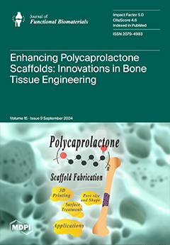
Polycaprolactone in Bone Tissue Engineering: A Comprehensive Review of Innovations in Scaffold Fabrication and Surface Modifications
by Hsin-Yu Liang, Wei-Keung Lee, Jui-Tsen Hsu, Jie-Yu Shih, Tien-Li Ma, Thi Thuy Tien Vo, Chiang-Wen Lee, Ming-Te Cheng and I-Ta Lee
J. Funct. Biomater. 2024, 15(9), 243; https://doi.org/10.3390/jfb15090243 - 24 Aug 2024
Viewed by 526
Abstract
Bone tissue engineering has seen significant advancements with innovative scaffold fabrication techniques such as 3D printing. This review focuses on enhancing polycaprolactone (PCL) scaffold properties through structural modifications, including surface treatments, pore architecture adjustments, and the incorporation of biomaterials like hydroxyapatite (HA). These [...] Read more.
Bone tissue engineering has seen significant advancements with innovative scaffold fabrication techniques such as 3D printing. This review focuses on enhancing polycaprolactone (PCL) scaffold properties through structural modifications, including surface treatments, pore architecture adjustments, and the incorporation of biomaterials like hydroxyapatite (HA). These modifications aim to improve scaffold conformation, cellular behavior, and mechanical performance, with particular emphasis on the role of mesenchymal stem cells (MSCs) in bone regeneration. The review also explores the potential of integrating nanomaterials and graphene oxide (GO) to further enhance the mechanical and biological properties of PCL scaffolds. Future directions involve optimizing scaffold structures and compositions for improved bone tissue regeneration outcomes. Full article
Show Figures

Figure 1

14 pages, 3747 KiB  
Article
The Exponential Shapeshifting Response of N-Vinylcaprolactam Hydrogel Bilayers Due to Temperature Change for Potential Minimally Invasive Surgery
by Billy Shu Hieng Tie, Mark Daly, Shuo Zhuo, Elaine Halligan, Gavin Keane, Joseph Geever and Luke Geever
J. Funct. Biomater. 2024, 15(9), 242; https://doi.org/10.3390/jfb15090242 - 24 Aug 2024
Viewed by 359
Abstract
Poly (N-vinylcaprolactam) (PNVCL) and poly (N-isopropylacrylamide) (PNIPAm) are two popular negatively temperature-responsive hydrogels, due to their biocompatibility, softness, hydrophilicity, superabsorbency, viscoelasticity, and near-physiological lower critical solution temperature (LCST). These characteristics make them ideal for biomedical applications. When combined with other materials, hydrogel expansion [...] Read more.
Poly (N-vinylcaprolactam) (PNVCL) and poly (N-isopropylacrylamide) (PNIPAm) are two popular negatively temperature-responsive hydrogels, due to their biocompatibility, softness, hydrophilicity, superabsorbency, viscoelasticity, and near-physiological lower critical solution temperature (LCST). These characteristics make them ideal for biomedical applications. When combined with other materials, hydrogel expansion induces the morphing of the assembly due to internal stress differences. Our recent developments in NVCL hydrogel, enhanced by nanoclay incorporation, have driven us to the creation of a bilayer structure to study its shapeshifting response across various temperatures. This study focused on the bending behaviour of bilayer samples composed of an active hydrogel layer and a passive non-swellable layer. Using photopolymerisation, circular discs and rectangular bilayer samples of varying sizes were fabricated. Homogeneous circular samples demonstrated that hydrogel density increased proportionally with temperature, with the swelling ratio exhibiting two distinct rates of change below and above its LCST. In bilayer samples, the volume of the passive layer influenced bending, and its optimal volume was identified. The investigation revealed that geometry affected the overall bending effect due to changes in the passive layer stiffness. Lastly, a temperature-responsive gripper capable of picking up objects several times its own weight was demonstrated, highlighting the potential of NVCL hydrogels as bioactuators for minimally invasive surgery. Full article
(This article belongs to the Special Issue Design and Synthesis Composites for Biomedical Application)
Show Figures

Graphical abstract

33 pages, 20993 KiB  
Review
Nanoparticles in Bone Regeneration: A Narrative Review of Current Advances and Future Directions in Tissue Engineering
by Samira Farjaminejad, Rosana Farjaminejad and Franklin Garcia-Godoy
J. Funct. Biomater. 2024, 15(9), 241; https://doi.org/10.3390/jfb15090241 - 23 Aug 2024
Viewed by 331
Abstract
The rising demand for effective bone regeneration has underscored the limitations of traditional methods like autografts and allografts, including donor site morbidity and insufficient biological signaling. This review examines nanoparticles (NPs) in tissue engineering (TE) to address these challenges, evaluating polymers, metals, ceramics, [...] Read more.
The rising demand for effective bone regeneration has underscored the limitations of traditional methods like autografts and allografts, including donor site morbidity and insufficient biological signaling. This review examines nanoparticles (NPs) in tissue engineering (TE) to address these challenges, evaluating polymers, metals, ceramics, and composites for their potential to enhance osteogenesis and angiogenesis by mimicking the extracellular matrix (ECM) nanostructure. The methods involved synthesizing and characterizing nanoparticle-based scaffoldsand integrating hydroxyapatite (HAp) with polymers to enhance mechanical properties and osteogenic potential. The results showed that these NPs significantly promote cell growth, differentiation, and bone formation, with carbon-based NPs like graphene and carbon nanotubes showing promise. NPs offer versatile, biocompatible, and customizable scaffolds that enhance drug delivery and support bone repair. Despite promising results, challenges with cytotoxicity, biodistribution, and immune responses remain. Addressing these issues through surface modifications and biocompatible molecules can improve the biocompatibility and efficacy of nanomaterials. Future research should focus on long-term in vivo studies to assess the safety and efficacy of NP-based scaffolds and explore synergistic effects with other bioactive molecules or growth factors. This review underscores the transformative potential of NPs in advancing BTE and calls for further research to optimize these technologies for clinical applications. Full article
(This article belongs to the Special Issue Biomaterials in Bone Reconstruction)
Show Figures

Figure 1

18 pages, 4562 KiB  
Article
Insights into the Dual Anticancer and Antibacterial Activities of Composites Based on Silver Camphorimine Complexes
by Joana P. Costa, Sílvia A. Sousa, Jorge H. Leitão, Fernanda Marques, Marta M. Alves and M. Fernanda N. N. Carvalho
J. Funct. Biomater. 2024, 15(9), 240; https://doi.org/10.3390/jfb15090240 - 23 Aug 2024
Viewed by 377
Abstract
Hydroxyapatite (HAp) is a widely used biocompatible material in orthopedic composite preparations. However, HAp composites that exhibit both anticancer and antibacterial activities through bioactive coordination complexes are relatively rare. To explore orthopedic applications, we blended several silver camphorimine compounds with HAp to create [...] Read more.
Hydroxyapatite (HAp) is a widely used biocompatible material in orthopedic composite preparations. However, HAp composites that exhibit both anticancer and antibacterial activities through bioactive coordination complexes are relatively rare. To explore orthopedic applications, we blended several silver camphorimine compounds with HAp to create [Ag(I)] composites. All compounds [Ag(NO3)(L)n] (n = 1,2) based on camphorimine (LA), camphor sulfonimine (LB) or imine bi-camphor (LC) ligands demonstrated significant cytotoxic activity (IC50 = 0.30–2.6 μgAg/mL) against osteosarcoma cancer cells (HOS). Based on their structural and electronic characteristics, four complexes (14) were selected for antibacterial evaluation against Escherichia coli, Burkholderia contaminans, Pseudomonas aeruginosa, and Staphylococcus aureus. All complexes (14) revealed combined anticancer and antibacterial activities; therefore, they were used to prepare [Ag(I)]:HAp composites of 50:50% and 20:80% weight compositions and the activities of the composites were assessed. Results showed that they retain the dual anticancer and antibacterial characteristics of their precursor complexes. To replicate the clinical context of bone-filling applications, hand-pressed surfaces (pellets) were prepared. It is worth highlighting that no agglutination agent was necessary for the pellet’s consistency. The biological properties of the so-prepared pellets were assessed, and the HOS cells and bacteria spreading on the pellet’s surface were analyzed by SEM. Notably, composite 4B, derived from the bicamphor (LC) complex [Ag(NO3)(OC10H14N(C6H4)2NC10H14O)], exhibited significant anticancer activity against HOS cells and antibacterial activity against P. aeruginosa, fostering potential clinical applications on post-surgical OS treatment. Full article
(This article belongs to the Special Issue Design and Synthesis Composites for Biomedical Application)
Show Figures

Graphical abstract

Previous Issue
Back to TopTop