Next Issue
Volume 14, January
Previous Issue
Volume 13, November


Biosensors, Volume 13, Issue 12 (December 2023) – 47 articles

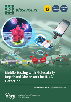
Cover Story (view full-size image): Periodontal disease, characterized by persistent inflammation impacting the gingival tissues supporting the teeth, can result in tooth loss attributed to the continual accumulation of dental plaque. Here, a biosensing platform employing the molecularly imprinted polymer (MIP) technique was devised through protein imprinting with the specific biomarker IL-1β, recognized as a pivotal indicator in diagnosing periodontitis. This MIP-based biosensor was integrated with a smartphone interface, thereby constituting a mobile point-of-care testing (POCT) platform designed for expeditious diagnostic purposes. The proposed scheme, featuring artificial antibody-based biosensors, exhibited a robust electrochemical performance that aligns with established periodontitis detection guidelines. View this paper
  • Issues are regarded as officially published after their release is announced to the table of contents alert mailing list.
  • You may sign up for e-mail alerts to receive table of contents of newly released issues.
  • PDF is the official format for papers published in both, html and pdf forms. To view the papers in pdf format, click on the "PDF Full-text" link, and use the free Adobe Reader to open them.
Order results
Result details
Section
Select all
Export citation of selected articles as:
9 pages, 681 KiB  
Communication
Impact of Optical Cavity on Refractive Index Sensitivity of Gold Nanohole Arrays
by Maria A. Shokova and Vladimir E. Bochenkov
Biosensors 2023, 13(12), 1038; https://doi.org/10.3390/bios13121038 - 18 Dec 2023
Viewed by 1698
Abstract
Refractive index sensing based on surface plasmon resonance (SPR) is a highly efficient label-free technique for biomolecular detection. The performance of this method is defined by the dielectric properties of a sensing layer and its structure. Nanohole arrays in thin metal films provide [...] Read more.
Refractive index sensing based on surface plasmon resonance (SPR) is a highly efficient label-free technique for biomolecular detection. The performance of this method is defined by the dielectric properties of a sensing layer and its structure. Nanohole arrays in thin metal films provide good refractive index sensitivity but often suffer from a large resonance linewidth, which limits their broad practical application in biosensorics. Coupling the broad plasmon modes to sharp resonances can reduce the peak widths, but at the same time it can also degrade the sensitivity. Here, we use Finite-Difference Time Domain simulations to study the factors affecting the sensing performance of gold-silica-gold optical cavities with nanohole arrays in the dielectric and top metal layers. We demonstrate that by tuning resonator size and inter-hole spacing, the performance of the biosensor can be optimized and the figure of merit of the order of 5–7 is reached. Full article
Show Figures

Figure 1

15 pages, 3973 KiB  
Article
Green Synthesis of Gold Nanoparticles Using Peach Extract Incorporated in Graphene for the Electrochemical Determination of Antioxidant Butylated Hydroxyanisole in Food Matrices
by Emanuela Grechi Döll, Edson Roberto Santana, João Paulo Winiarski, Luan Gabriel Baumgarten and Iolanda Cruz Vieira
Biosensors 2023, 13(12), 1037; https://doi.org/10.3390/bios13121037 - 18 Dec 2023
Cited by 16 | Viewed by 2513
Abstract
Butylated hydroxyanisole (BHA) is a synthetic phenolic antioxidant widely used in various food matrices to prevent oxidative rancidity. However, its presence has been associated with liver damage and carcinogenesis in animals. Thus, an electrochemical sensor was built using a composite of gold nanoparticles [...] Read more.
Butylated hydroxyanisole (BHA) is a synthetic phenolic antioxidant widely used in various food matrices to prevent oxidative rancidity. However, its presence has been associated with liver damage and carcinogenesis in animals. Thus, an electrochemical sensor was built using a composite of gold nanoparticles synthesized in peach extract (Prunus persica (L.) Batsch) and graphene. Peach extract served as a reducing and stabilizing agent for gold nanoparticles, as a dispersing agent for graphene, and as a film former to immobilize the composite on the surface of a glassy carbon electrode. The gold nanoparticles were characterized using spectroscopic and microscopic techniques, and the electrodes were electrochemically characterized using electrochemical impedance spectroscopy and cyclic voltammetry. The sensor provided higher current responses and lower charge transfer resistances compared to the unmodified glassy carbon electrode. Under the established optimized working conditions (0.1 mol L−1 Britton–Robinson buffer, pH 4.0, and differential pulse voltammetry), the calibration curve exhibited a linear range from 0.2 to 9.8 µmol L–1, with a detection limit of 70 nmol L−1. The proposed sensor represented a sensitive and practical analytical tool for the accurate determination of BHA in mayonnaise samples. Full article
Show Figures

Figure 1

13 pages, 2317 KiB  
Article
Phenotypic Characterization of 2D and 3D Prostate Cancer Cell Systems Using Electrical Impedance Spectroscopy
by Lexi L. Crowell, Juan Sebastian Yakisich, Brian Aufderheide and Tayloria N. G. Adams
Biosensors 2023, 13(12), 1036; https://doi.org/10.3390/bios13121036 - 18 Dec 2023
Cited by 3 | Viewed by 2249
Abstract
Prostate cancer is the second leading cause of death in men. A challenge in treating prostate cancer is overcoming cell plasticity, which links cell phenotype changes and chemoresistance. In this work, a microfluidic device coupled with electrical impedance spectroscopy (EIS), an electrode-based cell [...] Read more.
Prostate cancer is the second leading cause of death in men. A challenge in treating prostate cancer is overcoming cell plasticity, which links cell phenotype changes and chemoresistance. In this work, a microfluidic device coupled with electrical impedance spectroscopy (EIS), an electrode-based cell characterization technique, was used to study the electrical characteristics of phenotype changes for (1) prostate cancer cell lines (PC3, DU145, and LNCaP cells), (2) cells grown in 2D monolayer and 3D suspension cell culture conditions, and (3) cells in the presence (or absence) of the anti-cancer drug nigericin. To validate observations of phenotypic change, we measured the gene expression of two epithelial markers, E-cadherin (CDH1) and Tight Junction Protein 1 (ZO-1). Our results showed that PC3, DU145, and LNCaP cells were discernible with EIS. Secondly, moderate phenotype changes based on differences in cell culture conditions were detected with EIS and supported by the gene expression of CDH1. Lastly, we showed that EIS can detect chemoresistant-related cell phenotypes with nigericin drug treatment. EIS is a promising label-free tool for detecting cell phenotype changes associated with chemoresistance. Further development will enable the detection and characterization of many other types of cancer cells. Full article
(This article belongs to the Section Biosensors and Healthcare)
Show Figures

Graphical abstract

13 pages, 306 KiB  
Article
Rapid On-Site Detection of Arboviruses by a Direct RT-qPCR Assay
by Moufid Mhamadi, Giulia Mencattelli, Alioune Gaye, El Hadji Ndiaye, Aïssatou Aïcha Sow, Martin Faye, Marie Henriette Dior Ndione, Moussa Moïse Diagne, Moundhir Mhamadi, Ousmane Faye, Manfred Weidmann, Oumar Faye, Mawlouth Diallo and Cheikh Tidiane Diagne
Biosensors 2023, 13(12), 1035; https://doi.org/10.3390/bios13121035 - 16 Dec 2023
Viewed by 2392
Abstract
Arthropod-borne diseases currently constitute a source of major health concerns worldwide. They account for about 50% of global infectious diseases and cause nearly 700,000 deaths every year. Their rapid increase and spread constitute a huge challenge for public health, highlighting the need for [...] Read more.
Arthropod-borne diseases currently constitute a source of major health concerns worldwide. They account for about 50% of global infectious diseases and cause nearly 700,000 deaths every year. Their rapid increase and spread constitute a huge challenge for public health, highlighting the need for early detection during epidemics, to curtail the virus spread, and to enhance outbreak management. Here, we compared a standard quantitative polymerase chain reaction (RT-qPCR) and a direct RT-qPCR assay for the detection of Zika (ZIKV), Chikungunya (CHIKV), and Rift Valley Fever (RVFV) viruses from experimentally infected-mosquitoes. The direct RT-qPCR could be completed within 1.5 h and required 1 µL of viral supernatant from homogenized mosquito body pools. Results showed that the direct RT-qPCR can detect 85.71%, 89%, and 100% of CHIKV, RVFV, and ZIKV samples by direct amplifications compared to the standard method. The use of 1:10 diluted supernatant is suggested for CHIKV and RVFV direct RT-qPCR. Despite a slight drop in sensitivity for direct PCR, our technique is more affordable, less time-consuming, and provides a better option for qualitative field diagnosis during outbreak management. It represents an alternative when extraction and purification steps are not possible because of insufficient sample volume or biosecurity issues. Full article
18 pages, 3788 KiB  
Article
DNA Probes for Cas12a-Based Assay with Fluorescence Anisotropy Enhanced Due to Anchors and Salts
by Irina V. Safenkova, Alexey V. Samokhvalov, Kseniya V. Serebrennikova, Sergei A. Eremin, Anatoly V. Zherdev and Boris B. Dzantiev
Biosensors 2023, 13(12), 1034; https://doi.org/10.3390/bios13121034 - 16 Dec 2023
Cited by 4 | Viewed by 2480
Abstract
CRISPR/Cas12a is a potent biosensing tool known for its high specificity in DNA analysis. Cas12a recognizes the target DNA and acquires nuclease activity toward single-stranded DNA (ssDNA) probes. We present a straightforward and versatile approach to transforming common Cas12a-cleavable DNA probes into enhancing [...] Read more.
CRISPR/Cas12a is a potent biosensing tool known for its high specificity in DNA analysis. Cas12a recognizes the target DNA and acquires nuclease activity toward single-stranded DNA (ssDNA) probes. We present a straightforward and versatile approach to transforming common Cas12a-cleavable DNA probes into enhancing tools for fluorescence anisotropy (FA) measurements. Our study involved investigating 13 ssDNA probes with linear and hairpin structures, each featuring fluorescein at one end and a rotation-slowing tool (anchor) at the other. All anchors induced FA changes compared to fluorescein, ranging from 24 to 110 mr. Significant FA increases (up to 180 mr) were obtained by adding divalent metal salts (Mg2+, Ca2+, Ba2+), which influenced the rigidity and compactness of the DNA probes. The specific Cas12a-based recognition of double-stranded DNA (dsDNA) fragments of the bacterial phytopathogen Erwinia amylovora allowed us to determine the optimal set (probe structure, anchor, concentration of divalent ion) for FA-based detection. The best sensitivity was obtained using a hairpin structure with dC10 in the loop and streptavidin located near the fluorescein at the stem in the presence of 100 mM Mg2+. The detection limit of the dsDNA target was equal to 0.8 pM, which was eight times more sensitive compared to the common fluorescence-based method. The enhancing set ensured detection of single cells of E. amylovora per reaction in an analysis based on CRISPR/Cas12a with recombinase polymerase amplification. Our approach is universal and easy to implement. Combining FA with Cas12a offers enhanced sensitivity and signal reliability and could be applied to different DNA and RNA analytes. Full article
(This article belongs to the Special Issue CRISPR/Cas-Based Biosensing Systems: Development and Applications)
Show Figures

Graphical abstract

14 pages, 6259 KiB  
Article
Enhancing the Study of Quantal Exocytotic Events: Combining Diamond Multi-Electrode Arrays with Amperometric PEak Analysis (APE) an Automated Analysis Code
by Giulia Tomagra, Alice Re, Veronica Varzi, Pietro Aprà, Adam Britel, Claudio Franchino, Sofia Sturari, Nour-Hanne Amine, Remco H. S. Westerink, Valentina Carabelli and Federico Picollo
Biosensors 2023, 13(12), 1033; https://doi.org/10.3390/bios13121033 - 16 Dec 2023
Viewed by 2063
Abstract
MicroGraphited-Diamond-Multi Electrode Arrays (μG-D-MEAs) can be successfully used to reveal, in real time, quantal exocytotic events occurring from many individual neurosecretory cells and/or from many neurons within a network. As μG-D-MEAs arrays are patterned with up to 16 sensing microelectrodes, each of them [...] Read more.
MicroGraphited-Diamond-Multi Electrode Arrays (μG-D-MEAs) can be successfully used to reveal, in real time, quantal exocytotic events occurring from many individual neurosecretory cells and/or from many neurons within a network. As μG-D-MEAs arrays are patterned with up to 16 sensing microelectrodes, each of them recording large amounts of data revealing the exocytotic activity, the aim of this work was to support an adequate analysis code to speed up the signal detection. The cutting-edge technology of microGraphited-Diamond-Multi Electrode Arrays (μG-D-MEAs) has been implemented with an automated analysis code (APE, Amperometric Peak Analysis) developed using Matlab R2022a software to provide easy and accurate detection of amperometric spike parameters, including the analysis of the pre-spike foot that sometimes precedes the complete fusion pore dilatation. Data have been acquired from cultured PC12 cells, either collecting events during spontaneous exocytosis or after L-DOPA incubation. Validation of the APE code was performed by comparing the acquired spike parameters with those obtained using Quanta Analysis (Igor macro) by Mosharov et al. Full article
(This article belongs to the Special Issue Novel Biosensors for Cell Analysis)
Show Figures

Figure 1

15 pages, 3260 KiB  
Article
Optimized Copper-Based Microfeathers for Glucose Detection
by Carlota Guati, Lucía Gómez-Coma, Marcos Fallanza and Inmaculada Ortiz
Biosensors 2023, 13(12), 1032; https://doi.org/10.3390/bios13121032 - 15 Dec 2023
Cited by 1 | Viewed by 1948
Abstract
Diabetes is expected to rise substantially by 2045, prompting extensive research into accessible glucose electrochemical sensors, especially those based on non-enzymatic materials. In this context, advancing the knowledge of stable metal-based compounds as alternatives to non-enzymatic sensors becomes a scientific challenge. Nonetheless, these [...] Read more.
Diabetes is expected to rise substantially by 2045, prompting extensive research into accessible glucose electrochemical sensors, especially those based on non-enzymatic materials. In this context, advancing the knowledge of stable metal-based compounds as alternatives to non-enzymatic sensors becomes a scientific challenge. Nonetheless, these materials have encountered difficulties in maintaining stable responses under physiological conditions. This work aims to advance knowledge related to the synthesis and characterization of copper-based electrodes for glucose detection. The microelectrode presented here exhibits a wide linear range and a sensitivity of 1009 µA∙cm−2∙mM−1, overperfoming the results reported in literature so far. This electrode material has also demonstrated outstanding results in terms of reproducibility, repeatability, and stability, thereby meeting ISO 15197:2015 standards. Our study guides future research on next-generation sensors that combine copper with other materials to enhance activity in neutral media. Full article
Show Figures

Graphical abstract

23 pages, 4321 KiB  
Review
Low-Cost Plant-Based Metal and Metal Oxide Nanoparticle Synthesis and Their Use in Optical and Electrochemical (Bio)Sensors
by Iulia Corina Ciobotaru, Daniela Oprea, Constantin Claudiu Ciobotaru and Teodor Adrian Enache
Biosensors 2023, 13(12), 1031; https://doi.org/10.3390/bios13121031 - 15 Dec 2023
Cited by 1 | Viewed by 2186
Abstract
Technological progress has led to the development of analytical tools that promise a huge socio-economic impact on our daily lives and an improved quality of life for all. The use of plant extract synthesized nanoparticles in the development and fabrication of optical or [...] Read more.
Technological progress has led to the development of analytical tools that promise a huge socio-economic impact on our daily lives and an improved quality of life for all. The use of plant extract synthesized nanoparticles in the development and fabrication of optical or electrochemical (bio)sensors presents major advantages. Besides their low-cost fabrication and scalability, these nanoparticles may have a dual role, serving as a transducer component and as a recognition element, the latter requiring their functionalization with specific components. Different approaches, such as surface modification techniques to facilitate precise biomolecule attachment, thereby augmenting recognition capabilities, or fine tuning functional groups on nanoparticle surfaces are preferred for ensuring stable biomolecule conjugation while preserving bioactivity. Size optimization, maximizing surface area, and tailored nanoparticle shapes increase the potential for robust interactions and enhance the transduction. This article specifically aims to illustrate the adaptability and effectiveness of these biosensing platforms in identifying precise biological targets along with their far-reaching implications across various domains, spanning healthcare diagnostics, environmental monitoring, and diverse bioanalytical fields. By exploring these applications, the article highlights the significance of prioritizing the use of natural resources for nanoparticle synthesis. This emphasis aligns with the worldwide goal of envisioning sustainable and customized biosensing solutions, emphasizing heightened sensitivity and selectivity. Full article
(This article belongs to the Special Issue Nanocomposite-Based Biosensors: Recent Advances and Perspectives)
Show Figures

Figure 1

19 pages, 3252 KiB  
Article
Antibody Profiling: Kinetics with Native Biomarkers for Diagnostic Assay and Drug Developments
by Ute Jucknischke, Sebastian Friebe, Markus Rehle, Laura Quast and Sven H. Schmidt
Biosensors 2023, 13(12), 1030; https://doi.org/10.3390/bios13121030 - 14 Dec 2023
Viewed by 1857
Abstract
Despite remarkable progress in applied Surface Plasmon Resonance (SPR)-based methods, concise monitoring of kinetic properties for native biomarkers from patient samples is still lacking. Not only are low concentrations of native targets in patient samples, often in the pM range, a limiting and [...] Read more.
Despite remarkable progress in applied Surface Plasmon Resonance (SPR)-based methods, concise monitoring of kinetic properties for native biomarkers from patient samples is still lacking. Not only are low concentrations of native targets in patient samples, often in the pM range, a limiting and challenging factor, but body fluids as complex matrices furthermore complicate measurements. The here-described method enables the determination of kinetic constants and resulting affinities for native antigens from patients’ cerebrospinal fluid (CSF) and sera binding to antibodies. Using a significantly extended target-enrichment step, we modified a common sandwich-assay protocol, based on a primary and secondary antibody. We successfully analyze antibody kinetics of native targets from a variety of origins, with consistent results, independent of their source. Moreover, native neurofilament light chain (NFL) was investigated as an exemplary biomarker. Obtained data reveal antibodies recognizing recombinant NFL with high affinities, while showing no, or only significantly weakened binding to native NFL. The indicated differences for recombinant vs. native material demonstrate another beneficial application. Our assay is highly suitable for gaining valuable insights into characteristics of native biomarkers, thus impacting on the binder development of diagnostic reagents or pharmaceutical drugs. Full article
(This article belongs to the Section Biosensors and Healthcare)
Show Figures

Figure 1

12 pages, 3557 KiB  
Article
A Point-of-Care Testing Device Utilizing Graphene-Enhanced Fiber Optic SPR Sensor for Real-Time Detection of Infectious Pathogens
by Shiyu Jiang, Siyu Qian, Shunning Zhu, Jinxin Lu, Yunxin Hu, Cheng Zhang, Yikai Geng, Xuefeng Chen, Ying Guo, Zhaoliang Chen, Jie Pu, Zhendong Guo and Shengchun Liu
Biosensors 2023, 13(12), 1029; https://doi.org/10.3390/bios13121029 - 14 Dec 2023
Cited by 3 | Viewed by 2023
Abstract
Timely detection of highly infectious pathogens is essential for preventing and controlling public health risks. However, most traditional testing instruments require multiple tedious steps and ultimately testing in hospitals and third-party laboratories. The sample transfer process significantly prolongs the time to obtain test [...] Read more.
Timely detection of highly infectious pathogens is essential for preventing and controlling public health risks. However, most traditional testing instruments require multiple tedious steps and ultimately testing in hospitals and third-party laboratories. The sample transfer process significantly prolongs the time to obtain test results. To tackle this aspect, a portable fiber optic surface plasmon resonance (FO-SPR) device was developed for the real-time detection of infectious pathogens. The portable device innovatively integrated a compact FO-SPR sensing component, a signal acquisition and processing system, and an embedded power supply unit. A gold-plated fiber is used as the FO-SPR sensing probe. Compared with traditional SPR sensing systems, the device is smaller size, lighter weight, and higher convenience. To enhance the detection capacity of pathogens, a monolayer graphene was coated on the sensing region of the FO-SPR sensing probe. Severe acute respiratory syndrome coronavirus 2 (SARS-CoV-2) was used to evaluate the performance of the portable device. The device can accurately detect the SARS-CoV-2 spike S1 protein in phosphate-buffered saline (PBS) and artificial saliva within just 20 min, and the device successfully detected cultured SARS-CoV-2 virus. Furthermore, the FO-SPR probe has long-term stability, remaining stable for up to 8 days. It could distinguish between the SARS-CoV-2 spike protein and the MERS-CoV spike protein. Hence, this FO-SPR device provides reliable, rapid, and portable access to test results. It provides a promising point-of-care testing (POCT) tool for on-site screening of infectious pathogens. Full article
(This article belongs to the Section Biosensors and Healthcare)
Show Figures

Figure 1

15 pages, 3158 KiB  
Article
Cost-Efficient Micro-Well Array-Based Colorimetric Antibiotic Susceptibility Testing (MacAST) for Bacteria from Culture or Community
by Huilin Zhang, Lei Wang, Zhiguo Zhang, Jianhan Lin and Feng Ju
Biosensors 2023, 13(12), 1028; https://doi.org/10.3390/bios13121028 - 14 Dec 2023
Viewed by 2288
Abstract
Rapid and cost-efficient antibiotic susceptibility testing (AST) is key to timely prescription-oriented diagnosis and precision treatment. However, current AST methods have limitations in throughput or cost effectiveness, and are impractical for microbial communities. Here, we developed a high-throughput micro-well array-based colorimetric AST (macAST) [...] Read more.
Rapid and cost-efficient antibiotic susceptibility testing (AST) is key to timely prescription-oriented diagnosis and precision treatment. However, current AST methods have limitations in throughput or cost effectiveness, and are impractical for microbial communities. Here, we developed a high-throughput micro-well array-based colorimetric AST (macAST) system equipped with a self-developed smartphone application that could efficiently test sixteen combinations of bacteria strains and antibiotics, achieving comparable AST results based on resazurin metabolism assay. For community samples, we integrated immunomagnetic separation into the macAST (imacAST) system to specifically enrich the target cells before testing, which shortened bacterial isolation time from days to only 45 min and achieved AST of the target bacteria with a low concentration (~103 CFU/mL). This proof-of-concept study developed a high-throughput AST system with an at least ten-fold reduction in cost compared with a system equipped with a microscope or Raman spectrum. Based on colorimetric readout, the antimicrobial susceptibility of the bacteria from microbial communities can be delivered within 6 h, compared to days being required based on standard procedures, bypassing the need for precise instrumentation in therapy to combat bacterial antibiotic resistance in resource-limited settings. Full article
(This article belongs to the Special Issue Biosensors for Monitoring Pathogenic Agents (Volume II))
Show Figures

Figure 1

13 pages, 6128 KiB  
Article
A Novel Aptamer Biosensor Based on a Localized Surface Plasmon Resonance Sensing Chip for High-Sensitivity and Rapid Enrofloxacin Detection
by Pan Wang, Liyun Ding, Yumei Zhang and Xingdong Jiang
Biosensors 2023, 13(12), 1027; https://doi.org/10.3390/bios13121027 - 13 Dec 2023
Cited by 4 | Viewed by 1890
Abstract
Enrofloxacin, a fluoroquinolone widely used in animal husbandry, presents environmental and human health hazards due to its stability and incomplete hydrolysis leading to residue accumulation. To address this concern, a highly sensitive aptamer biosensor utilizing a localized surface plasmon resonance (LSPR) sensing chip [...] Read more.
Enrofloxacin, a fluoroquinolone widely used in animal husbandry, presents environmental and human health hazards due to its stability and incomplete hydrolysis leading to residue accumulation. To address this concern, a highly sensitive aptamer biosensor utilizing a localized surface plasmon resonance (LSPR) sensing chip and microfluidic technology was developed for rapid enrofloxacin residue detection. AuNPs were prepared by the seed method and the AuNPs-Apt complexes were immobilized on the chip by the sulfhydryl groups modified on the end of the aptamer. The properties and morphologies of the sensing chip and AuNPs-Apt complexes were characterized by Fourier transform infrared spectroscopy (FTIR), UV-Vis spectrophotometer, and scanning electron microscope (SEM), respectively. The sensing chip was able to detect enrofloxacin in the range of 0.01–100 ng/mL with good linearity, and the relationship between the response of the sensing chip and the concentration was Δλ (nm) = 1.288log ConENR (ng/mL) + 5.245 (R2 = 0.99), with the limit of detection being 0.001 ng/mL. The anti-interference, repeatability, and selectivity of this sensing chip were studied in detail. Compared with other sensors, this novel aptamer biosensor based on AuNPs-Apt complexes is expected to achieve simple, stable, and economical application in the field of enrofloxacin detection. Full article
(This article belongs to the Special Issue Recent Progress in Bioplasmonics Technologies)
Show Figures

Figure 1

13 pages, 2831 KiB  
Article
Tamm Plasmon Polariton Biosensors Based on Porous Silicon: Design, Validation and Analysis
by Guoguang Rong and Mohamad Sawan
Biosensors 2023, 13(12), 1026; https://doi.org/10.3390/bios13121026 - 11 Dec 2023
Cited by 1 | Viewed by 2164
Abstract
Tamm Plasmon Polariton (TPP) is a nanophotonic phenomenon that has attracted much attention due to its spatial strong field confinement, ease of mode excitation, and polarization independence. TPP has applications in sensing, storage, lasing, perfect absorber, solar cell, nonlinear optics, and many others. [...] Read more.
Tamm Plasmon Polariton (TPP) is a nanophotonic phenomenon that has attracted much attention due to its spatial strong field confinement, ease of mode excitation, and polarization independence. TPP has applications in sensing, storage, lasing, perfect absorber, solar cell, nonlinear optics, and many others. In this work, we demonstrate a biosensing platform based on TPP resonant mode. Both theoretical analyses based on the transfer matrix method and experimental validation through nonspecific detection of liquids of different refractive indices and specific detection of SARS-CoV-2 nucleocapsid protein (N-protein) are presented. Results show that the TPP biosensor has high sensitivity and good specificity. For N-protein detection, the sensitivity can be up to 1.5 nm/(µg/mL), and the limit of detection can reach down to 7 ng/mL with a spectrometer of 0.01 nm resolution in wavelength shift. Both nonspecific detection of R.I. liquids and specific detection of N-protein have been simulated and compared with experimental results to demonstrate consistency. This work paves the way for design, optimization, fabrication, characterization, and performance analysis of TPP based biosensors. Full article
(This article belongs to the Special Issue New Biosensors and Nanosensors)
Show Figures

Figure 1

30 pages, 9926 KiB  
Review
Recent Advances in Nanowire-Based Wearable Physical Sensors
by Junlin Gu, Yunfei Shen, Shijia Tian, Zhaoguo Xue and Xianhong Meng
Biosensors 2023, 13(12), 1025; https://doi.org/10.3390/bios13121025 - 11 Dec 2023
Cited by 6 | Viewed by 3105
Abstract
Wearable electronics is a technology that closely integrates electronic devices with the human body or clothing, which can realize human–computer interaction, health monitoring, smart medical, and other functions. Wearable physical sensors are an important part of wearable electronics. They can sense various physical [...] Read more.
Wearable electronics is a technology that closely integrates electronic devices with the human body or clothing, which can realize human–computer interaction, health monitoring, smart medical, and other functions. Wearable physical sensors are an important part of wearable electronics. They can sense various physical signals from the human body or the surrounding environment and convert them into electrical signals for processing and analysis. Nanowires (NW) have unique properties such as a high surface-to-volume ratio, high flexibility, high carrier mobility, a tunable bandgap, a large piezoresistive coefficient, and a strong light–matter interaction. They are one of the ideal candidates for the fabrication of wearable physical sensors with high sensitivity, fast response, and low power consumption. In this review, we summarize recent advances in various types of NW-based wearable physical sensors, specifically including mechanical, photoelectric, temperature, and multifunctional sensors. The discussion revolves around the structural design, sensing mechanisms, manufacture, and practical applications of these sensors, highlighting the positive role that NWs play in the sensing process. Finally, we present the conclusions with perspectives on current challenges and future opportunities in this field. Full article
(This article belongs to the Special Issue Advances in Wearable Biosensors for Healthcare Monitoring)
Show Figures

Figure 1

14 pages, 2040 KiB  
Article
C-undecylcalix[4]resorcinarene Langmuir–Blodgett/Porous Reduced Graphene Oxide Composite Film as a Electrochemical Sensor for the Determination of Tryptophan
by Yanju Wu, Keyu Chen and Fei Wang
Biosensors 2023, 13(12), 1024; https://doi.org/10.3390/bios13121024 - 10 Dec 2023
Cited by 2 | Viewed by 1593
Abstract
In this study, a composite film was developed for the electrochemical sensing of tryptophan (Trp). Porous reduced graphene oxide (PrGO) was utilized as the electron transfer layer, and a C-undecylcalix[4]resorcinarene Langmuir–Blodgett (CUCR-LB) film served as the molecular recognition layer. Atomic force microscopy (AFM), [...] Read more.
In this study, a composite film was developed for the electrochemical sensing of tryptophan (Trp). Porous reduced graphene oxide (PrGO) was utilized as the electron transfer layer, and a C-undecylcalix[4]resorcinarene Langmuir–Blodgett (CUCR-LB) film served as the molecular recognition layer. Atomic force microscopy (AFM), transmission electron microscopy (TEM), Raman spectroscopy, scanning electron microscopy (SEM), and electrochemical experiments were employed to analyze the characteristics of the CUCR-LB/PrGO composite film. The electrochemical behavior of Trp on the CUCR-LB/PrGO composite film was investigated, revealing a Trp linear response range of 1.0 × 10⁻⁷ to 3.0 × 10⁻⁵ mol L−1 and a detection limit of 3.0 × 10⁻⁸ mol L−1. Furthermore, the developed electroanalytical method successfully determined Trp content in an amino acid injection sample. This study not only introduces a rapid and reliable electrochemical method for the determination of Trp but also presents a new strategy for constructing high-performance electrochemical sensing platforms. Full article
Show Figures

Figure 1

13 pages, 3676 KiB  
Article
Integration of Glutamate Dehydrogenase and Nanoporous Gold for Electrochemical Detection of Glutamate
by Ting Cai, Keshuai Shang, Xiaolei Wang, Xiaoyan Qi, Ruijun Liu and Xia Wang
Biosensors 2023, 13(12), 1023; https://doi.org/10.3390/bios13121023 - 10 Dec 2023
Cited by 1 | Viewed by 1952
Abstract
Glutamate, a non-essential amino acid produced by fermentation, plays a significant role in disease diagnosis and food safety. It is important to enable the real-time monitoring of glutamate concentration for human health and nutrition. Due to the challenges in directly performing electrochemical oxidation–reduction [...] Read more.
Glutamate, a non-essential amino acid produced by fermentation, plays a significant role in disease diagnosis and food safety. It is important to enable the real-time monitoring of glutamate concentration for human health and nutrition. Due to the challenges in directly performing electrochemical oxidation–reduction reactions of glutamate, this study leverages the synergistic effect of glutamate dehydrogenase (GLDH) and nanoporous gold (NPG) to achieve the indirect and accurate detection of glutamate within the range of 50 to 700 μM by measuring the generated quantity of NADH during the enzymatic reaction. The proposed biosensor demonstrates remarkable performance characteristics, including a detection sensitivity of 1.95 μA mM−1 and a limit of detection (LOD) of 6.82 μM. The anti-interference tests indicate an average recognition error ranging from −3.85% to +2.60%, spiked sample recovery rates between 95% and 105%, and a relative standard deviation (RSD) of less than 4.97% for three replicate experiments. Therefore, the GLDH-NPG/GCE biosensor presented in this work exhibits excellent accuracy and repeatability, providing a novel alternative for rapid glutamate detection. This research contributes significantly to enhancing the precise monitoring of glutamate concentration, thereby offering more effective guidance and control for human health and nutrition. Full article
(This article belongs to the Section Biosensors and Healthcare)
Show Figures

Figure 1

14 pages, 3422 KiB  
Communication
Towards the Application of a Label-Free Approach for Anti-CD47/PD-L1 Bispecific Antibody Discovery
by Artem S. Grevtsev, Alexandra D. Azarian, Alexey K. Misorin, Daria O. Chernyshova, Pavel A. Iakovlev and Mikhail S. Karbyshev
Biosensors 2023, 13(12), 1022; https://doi.org/10.3390/bios13121022 - 9 Dec 2023
Cited by 1 | Viewed by 1922
Abstract
The engineering of bispecific antibodies that exhibit optimal affinity and functional activity presents a significant scientific challenge. To tackle this, investigators employ an assortment of protein assay techniques, such as label-free interaction methodologies, which offer rapidity and convenience for the evaluation of extensive [...] Read more.
The engineering of bispecific antibodies that exhibit optimal affinity and functional activity presents a significant scientific challenge. To tackle this, investigators employ an assortment of protein assay techniques, such as label-free interaction methodologies, which offer rapidity and convenience for the evaluation of extensive sample sets. These assays yield intricate data pertaining to the affinity towards target antigens and Fc-receptors, instrumental in predicting cellular test outcomes. Nevertheless, the fine-tuning of affinity is of paramount importance to mitigate potential adverse effects while maintaining efficient obstruction of ligand–receptor interactions. In this research, biolayer interferometry (BLI) was utilized to probe the functional characteristics of bispecific antibodies targeting cluster of differentiation 47 (CD47) and programmed death-ligand 1 (PD-L1) antigens, encompassing affinity, concurrent binding to two disparate antigens, and the inhibition of ligand–receptor interactions. The findings derived from BLI were juxtaposed with data from in vitro signal regulatory protein-α (SIRP-α)/CD47 blockade reporter bioassays for two leading bispecific antibody candidates, each demonstrating distinct affinity to CD47. Full article
(This article belongs to the Special Issue Application of Biosensors in Cell or Tissue Analysis)
Show Figures

Figure 1

16 pages, 2695 KiB  
Article
CRISPR/Cas12a-Based Detection Platform for Early and Rapid Diagnosis of Scrub Typhus
by Pooja Bhardwaj, Nikita Shrikant Nanaware, Sthita Pragnya Behera, Smita Kulkarni, Hirawati Deval, Rajesh Kumar, Gaurav Raj Dwivedi, Rajni Kant and Rajeev Singh
Biosensors 2023, 13(12), 1021; https://doi.org/10.3390/bios13121021 - 8 Dec 2023
Cited by 2 | Viewed by 3181
Abstract
Orientia tsutsugamushi is responsible for causing scrub typhus (ST) and is the leading cause of acute encephalitis syndrome (AES) in AES patients. A rapid and sensitive method to detect scrub typhus on-site is essential for the timely deployment of control measures. In the [...] Read more.
Orientia tsutsugamushi is responsible for causing scrub typhus (ST) and is the leading cause of acute encephalitis syndrome (AES) in AES patients. A rapid and sensitive method to detect scrub typhus on-site is essential for the timely deployment of control measures. In the current study, we developed a rapid, sensitive, and instrument-free lateral flow assay (LFA) detection method based on CRISPR/Cas12a technology for diagnosing ST (named LoCIST). The method is completed in three steps: first, harnessing the ability of recombinase polymerase for isothermal amplification of the target gene; second, CRISPR/Cas12a-based recognition of the target; and third, end-point detection by LFA. The detection limit of LoCIST was found to be one gene copy of ST genomic DNA per reaction, and the process was complete within an hour. In 81 clinical samples, the assay showed no cross-reactivity with other rickettsial DNA and was 100% consistent with PCR detection of ST. LoCIST demonstrated 97.6% sensitivity and 100% specificity. Overall, the LoCIST offers a novel alternative for the portable, simple, sensitive, and specific detection of ST, and it may help prevent and control AES outbreaks due to ST. In conclusion, LoCIST does not require specialized equipment and poses a potential for future applications as a point-of-care diagnostic. Full article
(This article belongs to the Special Issue CRISPR/Cas-Based Biosensing Systems: Development and Applications)
Show Figures

Figure 1

16 pages, 4524 KiB  
Article
Reduced Graphene Oxide/Organic Dye Composites for Bioelectroconversion of Saccharides: Application for Detection of Saccharides and α-Amylase Assessments
by Marius Butkevicius, Justina Gaidukevic, Vidute Gureviciene and Julija Razumiene
Biosensors 2023, 13(12), 1020; https://doi.org/10.3390/bios13121020 - 8 Dec 2023
Viewed by 1730
Abstract
In this study, PQQ-dependent glucose dehydrogenase (PQQ-GDH) was immobilized onto reduced graphene oxide (rGO) modified with organic dyes from three different classes (acridine, arylmethane, and diazo); namely, neutral red (NR), malachite green (MG), and congo red (CR) formed three types of biosensors. All [...] Read more.
In this study, PQQ-dependent glucose dehydrogenase (PQQ-GDH) was immobilized onto reduced graphene oxide (rGO) modified with organic dyes from three different classes (acridine, arylmethane, and diazo); namely, neutral red (NR), malachite green (MG), and congo red (CR) formed three types of biosensors. All three rGO/organic dye composites were characterized by scanning electron microscopy, X-ray photoelectron spectroscopy, and Raman spectroscopy. The impact of three rGO/organic dye modifications employed in bioelectrocatalytic systems on changes in enzyme activity and substrate selectivity was investigated. The highest sensitivity of 39 µA/cm2 was obtained for 1 mM of glucose when a rGO_MG/PQQ-GDH biosensor was used. A significant improvement in the electrochemical response of biosensors was attributed to the higher amount of pyrrolic nitrogen groups on the surface of the rGO/organic dye composites. Modifications of rGO by NR and MG not only improved the surfaces for efficient direct electron transfer (DET) but also influenced the enzyme selectivity through proper binding and orientation of the enzyme. The accuracy of the biosensor’s action was confirmed by the spectrophotometric analysis. Perspectives for using the proposed bioelectrocatalytic systems operating on DET principles for total or single monosaccharide and/or disaccharide determination/bioconversion systems or for diagnoses have been presented through examples of bioconversion of D-glucose, D-xylose, and maltose. Full article
(This article belongs to the Special Issue Nanocomposite-Based Biosensors: Recent Advances and Perspectives)
Show Figures

Graphical abstract

28 pages, 4653 KiB  
Review
Advancements in Wearable EEG Technology for Improved Home-Based Sleep Monitoring and Assessment: A Review
by Manal Mohamed, Nourelhuda Mohamed and Jae Gwan Kim
Biosensors 2023, 13(12), 1019; https://doi.org/10.3390/bios13121019 - 7 Dec 2023
Cited by 4 | Viewed by 5302
Abstract
Sleep is a fundamental aspect of daily life, profoundly impacting mental and emotional well-being. Optimal sleep quality is vital for overall health and quality of life, yet many individuals struggle with sleep-related difficulties. In the past, polysomnography (PSG) has served as the gold [...] Read more.
Sleep is a fundamental aspect of daily life, profoundly impacting mental and emotional well-being. Optimal sleep quality is vital for overall health and quality of life, yet many individuals struggle with sleep-related difficulties. In the past, polysomnography (PSG) has served as the gold standard for assessing sleep, but its bulky nature, cost, and the need for expertise has made it cumbersome for widespread use. By recognizing the need for a more accessible and user-friendly approach, wearable home monitoring systems have emerged. EEG technology plays a pivotal role in sleep monitoring, as it captures crucial brain activity data during sleep and serves as a primary indicator of sleep stages and disorders. This review provides an overview of the most recent advancements in wearable sleep monitoring leveraging EEG technology. We summarize the latest EEG devices and systems available in the scientific literature, highlighting their design, form factors, materials, and methods of sleep assessment. By exploring these developments, we aim to offer insights into cutting-edge technologies, shedding light on wearable EEG sensors for advanced at-home sleep monitoring and assessment. This comprehensive review contributes to a broader perspective on enhancing sleep quality and overall health using wearable EEG sensors. Full article
(This article belongs to the Special Issue Wearable Devices for Biosensors and Healthcare)
Show Figures

Figure 1

26 pages, 6680 KiB  
Review
Enzyme Cascade Electrode Reactions with Nanomaterials and Their Applicability towards Biosensor and Biofuel Cells
by Shalini devi Kalyana Sundaram, Md. Motaher Hossain, Muhammad Rezki, Kotoko Ariga and Seiya Tsujimura
Biosensors 2023, 13(12), 1018; https://doi.org/10.3390/bios13121018 - 7 Dec 2023
Cited by 21 | Viewed by 2796
Abstract
Nanomaterials, including carbon nanotubes, graphene oxide, metal–organic frameworks, metal nanoparticles, and porous carbon, play a crucial role as efficient carriers to enhance enzyme activity through substrate channeling while improving enzyme stability and reusability. However, there are significant debates surrounding aspects such as enzyme [...] Read more.
Nanomaterials, including carbon nanotubes, graphene oxide, metal–organic frameworks, metal nanoparticles, and porous carbon, play a crucial role as efficient carriers to enhance enzyme activity through substrate channeling while improving enzyme stability and reusability. However, there are significant debates surrounding aspects such as enzyme orientation, enzyme loading, retention of enzyme activity, and immobilization techniques. Consequently, these subjects have become the focus of intensive research in the realm of multi-enzyme cascade reactions. Researchers have undertaken the challenge of creating functional in vitro multi-enzyme systems, drawing inspiration from natural multi-enzyme processes within living organisms. Substantial progress has been achieved in designing multi-step reactions that harness the synthetic capabilities of various enzymes, particularly in applications such as biomarker detection (e.g., biosensors) and the development of biofuel cells. This review provides an overview of recent developments in concurrent and sequential approaches involving two or more enzymes in sequence. It delves into the intricacies of multi-enzyme cascade reactions conducted on nanostructured electrodes, addressing both the challenges encountered and the innovative solutions devised in this field. Full article
(This article belongs to the Section Biosensor and Bioelectronic Devices)
Show Figures

Graphical abstract

21 pages, 2706 KiB  
Review
Biohybrid Nanoparticle-Based In Situ Monitoring of In Vivo Drug Delivery
by Sohee Ju and Hyeon-Yeol Cho
Biosensors 2023, 13(12), 1017; https://doi.org/10.3390/bios13121017 - 6 Dec 2023
Viewed by 2339
Abstract
Nanomaterials have gained huge attention worldwide owing to their unique physicochemical characteristics which enable their applications in the field of biomedicine and drug delivery systems. Although nanodrug delivery systems (NDDSs) have better target specificity and bioavailability than traditional drug delivery systems, their behavior [...] Read more.
Nanomaterials have gained huge attention worldwide owing to their unique physicochemical characteristics which enable their applications in the field of biomedicine and drug delivery systems. Although nanodrug delivery systems (NDDSs) have better target specificity and bioavailability than traditional drug delivery systems, their behavior and clearance mechanisms in living subjects remain unclear. In this regard, the importance of bioimaging methods has come to the forefront for investigating the biodistribution of nanocarriers and discovering drug release mechanisms in vivo. In this review, we introduce several examples of biohybrid nanoparticles and their clinical applications, focusing on their advantages and limitations. The various bioimaging methods for monitoring the fate of nanodrugs in biological systems and the future perspectives of NDDSs have also been discussed. Full article
(This article belongs to the Special Issue Nanoparticle-Based Biosensors for Detection)
Show Figures

Figure 1

15 pages, 4067 KiB  
Article
Controlling the Nucleation and Growth of Salt from Bodily Fluid for Enhanced Biosensing Applications
by Siddharth Srivastava, Yusuke Terai, Jun Liu, Giovanni Capellini and Ya-Hong Xie
Biosensors 2023, 13(12), 1016; https://doi.org/10.3390/bios13121016 - 6 Dec 2023
Viewed by 1937
Abstract
Surface-enhanced Raman spectroscopy (SERS) represents a transformative tool in medical diagnostics, particularly for the early detection of key biomarkers such as small extracellular vesicles (sEVs). Its unparalleled sensitivity and compatibility with intricate biological samples make it an ideal candidate for revolutionizing noninvasive diagnostic [...] Read more.
Surface-enhanced Raman spectroscopy (SERS) represents a transformative tool in medical diagnostics, particularly for the early detection of key biomarkers such as small extracellular vesicles (sEVs). Its unparalleled sensitivity and compatibility with intricate biological samples make it an ideal candidate for revolutionizing noninvasive diagnostic methods. However, a significant challenge that mars its efficacy is the throughput limitation, primarily anchored in the prerequisite of hotspot and sEV colocalization within a minuscule range. This paper delves deep into this issue, introducing a never-attempted-before approach which harnesses the principles of crystallization—nucleation and growth. By synergistically coupling lasers with plasmonic resonances, we navigate the challenges associated with the analyte droplet drying method and the notorious coffee ring effect. Our method, rooted in a profound understanding of crystallization’s materials science, exhibits the potential to significantly increase the areal density of accessible plasmonic hotspots and efficiently guide exosomes to defined regions. In doing so, we not only overcome the throughput challenge but also promise a paradigm shift in the arena of minimally invasive biosensing, ushering in advanced diagnostic capabilities for life-threatening diseases. Full article
(This article belongs to the Special Issue Feature Issue of Optical and Photonic Biosensors Section - Volume II)
Show Figures

Figure 1

13 pages, 2205 KiB  
Article
Highly Conductive Peroxidase-like Ce-MoS2 Nanoflowers for the Simultaneous Electrochemical Detection of Dopamine and Epinephrine
by Annadurai Thamilselvan, Thinh Viet Dang and Moon Il Kim
Biosensors 2023, 13(12), 1015; https://doi.org/10.3390/bios13121015 - 6 Dec 2023
Viewed by 1937
Abstract
The accurate and simultaneous detection of neurotransmitters, such as dopamine (DA) and epinephrine (EP), is of paramount importance in clinical diagnostic fields. Herein, we developed cerium–molybdenum disulfide nanoflowers (Ce-MoS2 NFs) using a simple one-pot hydrothermal method and demonstrated that they are highly [...] Read more.
The accurate and simultaneous detection of neurotransmitters, such as dopamine (DA) and epinephrine (EP), is of paramount importance in clinical diagnostic fields. Herein, we developed cerium–molybdenum disulfide nanoflowers (Ce-MoS2 NFs) using a simple one-pot hydrothermal method and demonstrated that they are highly conductive and exhibit significant peroxidase-mimicking activity, which was applied for the simultaneous electrochemical detection of DA and EP. Ce-MoS2 NFs showed a unique structure, comprising MoS2 NFs with divalent Ce ions. This structural design imparted a significantly enlarged surface area of 220.5 m2 g−1 with abundant active sites as well as enhanced redox properties, facilitating electron transfer and peroxidase-like catalytic action compared with bare MoS2 NFs without Ce incorporation. Based on these beneficial features, Ce-MoS2 NFs were incorporated onto a screen-printed electrode (Ce-MoS2 NFs/SPE), enabling the electrochemical detection of H2O2 based on their peroxidase-like activity. Ce-MoS2 NFs/SPE biosensors also showed distinct electrocatalytic oxidation characteristics for DA and EP, consequently yielding the highly selective, sensitive, and simultaneous detection of target DA and EP. Dynamic linear ranges for both DA and EP were determined to be 0.05~100 μM, with detection limits (S/N = 3) of 28 nM and 44 nM, respectively. This study shows the potential of hierarchically structured Ce-incorporated MoS2 NFs to enhance the detection performances of electrochemical biosensors, thus enabling extensive applications in healthcare, diagnostics, and environmental monitoring. Full article
Show Figures

Graphical abstract

13 pages, 5145 KiB  
Article
The Label-Free Detection and Identification of SARS-CoV-2 Using Surface-Enhanced Raman Spectroscopy and Principal Component Analysis
by Lu Zhou, Ambra Vestri, Valentina Marchesano, Massimo Rippa, Domenico Sagnelli, Gerardo Picazio, Giovanna Fusco, Jiaguang Han, Jun Zhou and Lucia Petti
Biosensors 2023, 13(12), 1014; https://doi.org/10.3390/bios13121014 - 5 Dec 2023
Cited by 5 | Viewed by 2205
Abstract
The World Health Organization (WHO) declared in a May 2023 announcement that the COVID-19 illness is no longer categorized as a Public Health Emergency of International Concern (PHEIC); nevertheless, it is still considered an actual threat to world health, social welfare and economic [...] Read more.
The World Health Organization (WHO) declared in a May 2023 announcement that the COVID-19 illness is no longer categorized as a Public Health Emergency of International Concern (PHEIC); nevertheless, it is still considered an actual threat to world health, social welfare and economic stability. Consequently, the development of a convenient, reliable and affordable approach for detecting and identifying SARS-CoV-2 and its emerging new variants is crucial. The fingerprint and signal amplification characteristics of surface-enhanced Raman spectroscopy (SERS) could serve as an assay scheme for SARS-CoV-2. Here, we report a machine learning-based label-free SERS technique for the rapid and accurate detection and identification of SARS-CoV-2. The SERS spectra collected from samples of four types of coronaviruses on gold nanoparticles film, fabricated using a Langmuir–Blodgett self-assembly, can provide more spectroscopic signatures of the viruses and exhibit low limits of detection (<100 TCID50/mL or even <10 TCID50/mL). Furthermore, the key Raman bands of the SERS spectra were systematically captured by principal component analysis (PCA), which effectively distinguished SARS-CoV-2 and its variant from other coronaviruses. These results demonstrate that the combined use of SERS technology and PCA analysis has great potential for the rapid analysis and discrimination of multiple viruses and even newly emerging viruses without the need for a virus-specific probe. Full article
(This article belongs to the Special Issue High-Efficiency Surface-Enhanced Raman Scattering Biosensing)
Show Figures

Figure 1

26 pages, 3410 KiB  
Article
Mobile Point-of-Care Device Using Molecularly Imprinted Polymer-Based Chemosensors Targeting Interleukin-1β Biomarker
by Rowoon Park, Sangheon Jeon, Jae Won Lee, Jeonghwa Jeong, Young Woo Kwon, Sung Hyun Kim, Joonkyung Jang, Dong-Wook Han and Suck Won Hong
Biosensors 2023, 13(12), 1013; https://doi.org/10.3390/bios13121013 - 5 Dec 2023
Cited by 3 | Viewed by 2656
Abstract
Molecularly imprinted polymers (MIPs) have garnered significant attention as a promising material for engineering specific biological receptors with superior chemical complementarity to target molecules. In this study, we present an electrochemical biosensing platform incorporating MIP films for the selective detection of the interleukin-1β [...] Read more.
Molecularly imprinted polymers (MIPs) have garnered significant attention as a promising material for engineering specific biological receptors with superior chemical complementarity to target molecules. In this study, we present an electrochemical biosensing platform incorporating MIP films for the selective detection of the interleukin-1β (IL-1β) biomarker, particularly suitable for mobile point-of-care testing (POCT) applications. The IL-1β-imprinted biosensors were composed of poly(eriochrome black T (EBT)), including an interlayer of poly(3,4-ethylene dioxythiophene) and a 4-aminothiophenol monolayer, which were electrochemically polymerized simultaneously with template proteins (i.e., IL-1β) on custom flexible screen-printed carbon electrodes (SPCEs). The architecture of the MIP films was designed to enhance the sensor sensitivity and signal stability. This approach involved a straightforward sequential-electropolymerization process and extraction for leaving behind cavities (i.e., rebinding sites), resulting in the efficient production of MIP-based biosensors capable of molecular recognition for selective IL-1β detection. The electrochemical behaviors were comprehensively investigated using cyclic voltammograms and electrochemical impedance spectroscopy responses to assess the imprinting effect on the MIP films formed on the SPCEs. In line with the current trend in in vitro diagnostic medical devices, our simple and effective MIP-based analytical system integrated with mobile POCT devices offers a promising route to the rapid detection of biomarkers, with particular potential for periodontitis screening. Full article
(This article belongs to the Special Issue Nanomaterial Based Biosensors for Biomedical Applications)
Show Figures

Figure 1

13 pages, 2986 KiB  
Article
Early Blood Clot Detection Using Forward Scattering Light Measurements Is Not Superior to Delta Pressure Measurements
by Anna Fischbach, Michael Lamberti, Julia Alexandra Simons, Erik Wrede, Alexander Theißen, Patrick Winnersbach, Rolf Rossaint, André Stollenwerk and Christian Bleilevens
Biosensors 2023, 13(12), 1012; https://doi.org/10.3390/bios13121012 - 4 Dec 2023
Viewed by 2447
Abstract
The occurrence of thrombus formation within an extracorporeal membrane oxygenator is a common complication during extracorporeal membrane oxygenation therapy and can rapidly result in a life-threatening situation due to arterial thromboembolism, causing stroke, pulmonary embolism, and limb ischemia in the patient. The standard [...] Read more.
The occurrence of thrombus formation within an extracorporeal membrane oxygenator is a common complication during extracorporeal membrane oxygenation therapy and can rapidly result in a life-threatening situation due to arterial thromboembolism, causing stroke, pulmonary embolism, and limb ischemia in the patient. The standard clinical practice is to monitor the pressure at the inlet and outlet of oxygenators, indicating fulminant, obstructive clot formation indicated by an increasing pressure difference (ΔP). However, smaller blood clots at early stages are not detectable. Therefore, there is an unmet need for sensors that can detect blood clots at an early stage to minimize the associated thromboembolic risks for patients. This study aimed to evaluate if forward scattered light (FSL) measurements can be used for early blood clot detection and if it is superior to the current clinical gold standard (pressure measurements). A miniaturized in vitro test circuit, including a custom-made test chamber, was used. Heparinized human whole blood was circulated through the test circuit until clot formation occurred. Four LEDs and four photodiodes were placed along the sidewall of the test chamber in different positions for FSL measurements. The pressure monitor was connected to the inlet and the outlet to detect changes in ΔP across the test chamber. Despite several modifications in the LED positions on the test chamber, the FSL measurements could not reliably detect a blood clot within the in vitro test circuit, although the pressure measurements used as the current clinical gold standard detected fulminant clot formation in 11 independent experiments. Full article
(This article belongs to the Special Issue Biophysical Sensors for Biomedical/Health Monitoring Applications)
Show Figures

Figure 1

16 pages, 7900 KiB  
Article
Electrochemical Biosensors for Express Analysis of the Integral Toxicity of Polymer Materials
by Natalia Yu. Yudina, Tatyana N. Kozlova, Daniil A. Bogachikhin, Maria M. Kosarenina, Vyacheslav A. Arlyapov and Sergey V. Alferov
Biosensors 2023, 13(12), 1011; https://doi.org/10.3390/bios13121011 - 4 Dec 2023
Cited by 1 | Viewed by 1742
Abstract
Biosensors based on an oxygen electrode, a mediator electrode, and a mediator microbial biofuel cell (MFC) using the bacteria Gluconobacter oxydans B-1280 were formed and tested to determine the integral toxicity. G. oxydans bacteria exhibited high sensitivity to the toxic effects of phenol, [...] Read more.
Biosensors based on an oxygen electrode, a mediator electrode, and a mediator microbial biofuel cell (MFC) using the bacteria Gluconobacter oxydans B-1280 were formed and tested to determine the integral toxicity. G. oxydans bacteria exhibited high sensitivity to the toxic effects of phenol, 2,4-dinitrophenol, salicylic and trichloroacetic acid, and a number of heavy metal ions. The system “G. oxydans bacteria–ferrocene–graphite-paste electrode” was superior in sensitivity to biosensors formed using an oxygen electrode and MFC, in particular regarding heavy metal ions (EC50 of Cr3+, Mn2+, and Cd2+ was 0.8 mg/dm3, 0.3 mg/dm3 and 1.6 mg/dm3, respectively). It was determined that the period of stable functioning of electrochemical systems during measurements was reduced by half (from 30 to 15 days) due to changes in the enzyme system of microbial cells when exposed to toxicants. Samples of the products made from polymeric materials were analyzed using developed biosensor systems and standard biotesting methods based on inhibiting the growth of duckweed Lemna minor, reducing the motility of bull sperm, and quenching the luminescence of the commercial test system “Ecolum”. The developed bioelectrocatalytic systems were comparable in sensitivity to commercial biosensors, which made it possible to correlate the results and identify, by all methods, a highly toxic sample containing diphenylmethane-4,4′-diisocyanate according to GC-MS data. Full article
Show Figures

Figure 1

15 pages, 6344 KiB  
Article
A Fiber-Optical Dosimetry Sensor for Gamma-Ray Irradiation Measurement in Biological Applications
by Adel Shaaban Awad Elsharkawi, Huda A. Alazab, Mahmoud Sayed, Mostafa A. Askar, Ibrahim Y. Abdelrahman, Amany A. Arafa, Hassan I. Saleh, Lotfy R. Gomaa and Yi-Chun Du
Biosensors 2023, 13(12), 1010; https://doi.org/10.3390/bios13121010 - 3 Dec 2023
Cited by 1 | Viewed by 1830
Abstract
In this paper, we propose a novel fiber-optical dosimetry sensor for radiation measurement in biological applications. A two-dimensional (2D) fiber-optical dosimeter (FOD) for radiation measurement is considered. The sensors are arranged as a 2D array in a tailored holder. This FOD targets accurate [...] Read more.
In this paper, we propose a novel fiber-optical dosimetry sensor for radiation measurement in biological applications. A two-dimensional (2D) fiber-optical dosimeter (FOD) for radiation measurement is considered. The sensors are arranged as a 2D array in a tailored holder. This FOD targets accurate industrial and medical applications which seek more tolerant radiation dosimeters. In this paper, the FOD sensors are subjected to gamma-ray radiation facilities from the 137Cs gamma-ray irradiator type for low doses and 60Co gamma-ray irradiator for high doses. For better evaluation of radiation effects on the FOD sample, the measurements are performed using eight sensors (hollow cylinder shape) with two samples in each dose. The sensors were measured before and after each irradiation. To the author’s knowledge, the measurements of FOD transplanted inside animals are presented for the first time in this paper. A 2D simulation program has been implemented for numerical simulation based on the attenuation factors from the absorbed dose inside the in vivo models. A comparison between the FOD and the standard thermo-luminescence detector is presented based on the test of in vivo animal models. The results indicate that the proposed FOD sensor is more stable and has higher sensitivity. Full article
(This article belongs to the Section Optical and Photonic Biosensors)
Show Figures

Graphical abstract

15 pages, 1371 KiB  
Article
Patient Stratification for Antibiotic Prescriptions Based on the Bound-Free Phase Detection Immunoassay of C-Reactive Protein in Serum Samples
by Benita Johannsen, Desirée Baumgartner, Michal Karpíšek, David Stejskal, Noémie Boillat-Blanco, José Knüsli, Marcus Panning, Nils Paust, Roland Zengerle and Konstantinos Mitsakakis
Biosensors 2023, 13(12), 1009; https://doi.org/10.3390/bios13121009 - 3 Dec 2023
Cited by 2 | Viewed by 1930
Abstract
C-reactive protein is a well-studied host response biomarker, whose diagnostic performance depends on its accurate classification into concentration zones defined by clinical scenario-specific cutoff values. We validated a newly developed, bead-based, bound-free phase detection immunoassay (BFPD-IA) versus a commercial CE-IVD enzyme-linked immunosorbent assay [...] Read more.
C-reactive protein is a well-studied host response biomarker, whose diagnostic performance depends on its accurate classification into concentration zones defined by clinical scenario-specific cutoff values. We validated a newly developed, bead-based, bound-free phase detection immunoassay (BFPD-IA) versus a commercial CE-IVD enzyme-linked immunosorbent assay (ELISA) kit and a commercial CE-IVD immunoturbidimetric assay (ITA) kit. The latter was performed on a fully automated DPC Konelab 60i clinical analyzer used in routine diagnosis. We classified 53 samples into concentration zones derived from four different sets of cutoff values that are related to antibiotic prescription scenarios in the case of respiratory tract infections. The agreements between the methods were ELISA/ITA at 87.7%, ELISA/BFPD-IA at 87.3%, and ITA/-BFPD-IA at 93.9%, reaching 98–99% in all cases when considering the calculated relative combined uncertainty of the single measurement of each sample. In a subgroup of 37 samples, which were analyzed for absolute concentration quantification, the scatter plot slopes’ correlations were as follows: ELISA/ITA 1.15, R2 = 0.97; BFPD-IA/ELISA 1.12, R2 = 0.95; BFPD-IA/ITA 0.95, R2 = 0.93. These very good performances and the agreement between BFPD-IA and ITA (routine diagnostic), combined with BFPD-IA’s functional advantages over ITA (and ELISA)—such as quick time to result (~20 min), reduced consumed reagents (only one assay buffer and no washing), few and easy steps, and compatibility with nucleic-acid-amplification instruments—render it a potential approach for a reliable, cost-efficient, evidence-based point-of-care diagnostic test for guiding antibiotic prescriptions. Full article
(This article belongs to the Special Issue Advances in Magnetic Particle-Based Bioassays)
Show Figures

Figure 1

Previous Issue
Next Issue
Back to TopTop