Next Issue
Volume 14, August
Previous Issue
Volume 14, June


Biosensors, Volume 14, Issue 7 (July 2024) – 45 articles

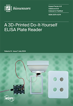
Cover Story (view full-size image): Simple analytical devices suitable for the analysis of various biochemical and immunochemical markers are highly desirable and can provide laboratory diagnoses outside standard hospitals. This study focuses on constructing an easily reproducible do-it-yourself ELISA plate reader biosensor device, assembled from generally available and inexpensive parts such as 3D printed objects and a common smartphone camera. The final colorimetric biosensor was able to process standard 96-well microplates and was verified on the TNF-alpha assay. The results of this study will inform the development of simple analytical devices easily reproducible by 3D printing and found on generally available electronics. View this paper
  • Issues are regarded as officially published after their release is announced to the table of contents alert mailing list.
  • You may sign up for e-mail alerts to receive table of contents of newly released issues.
  • PDF is the official format for papers published in both, html and pdf forms. To view the papers in pdf format, click on the "PDF Full-text" link, and use the free Adobe Reader to open them.
Order results
Result details
Section
Select all
Export citation of selected articles as:
35 pages, 3384 KiB  
Review
Boosting Electrochemical Sensing Performances Using Molecularly Imprinted Nanoparticles
by Francesco Gagliani, Tiziano Di Giulio, Muhammad Ibrar Asif, Cosimino Malitesta and Elisabetta Mazzotta
Biosensors 2024, 14(7), 358; https://doi.org/10.3390/bios14070358 - 22 Jul 2024
Viewed by 808
Abstract
Nanoparticles of molecularly imprinted polymers (nanoMIPs) combine the excellent recognition ability of imprinted polymers with specific properties related to the nanosize, such as a high surface-to-volume ratio, resulting in highly performing recognition elements with surface-exposed binding sites that promote the interaction with the [...] Read more.
Nanoparticles of molecularly imprinted polymers (nanoMIPs) combine the excellent recognition ability of imprinted polymers with specific properties related to the nanosize, such as a high surface-to-volume ratio, resulting in highly performing recognition elements with surface-exposed binding sites that promote the interaction with the target and, in turn, binding kinetics. Different synthetic strategies are currently available to produce nanoMIPs, with the possibility to select specific conditions in relation to the nature of monomers/templates and, importantly, to tune the nanoparticle size. The excellent sensing properties, combined with the size, tunability, and flexibility of synthetic protocols applicable to different targets, have enabled the widespread use of nanoMIPs in several applications, including sensors, imaging, and drug delivery. The present review summarizes nanoMIPs applications in sensors, specifically focusing on electrochemical detection, for which nanoMIPs have been mostly applied. After a general survey of the most widely adopted nanoMIP synthetic approaches, the integration of imprinted nanoparticles with electrochemical transducers will be discussed, representing a key step for enabling a reliable and stable sensor response. The mechanisms for electrochemical signal generation will also be compared, followed by an illustration of nanoMIP-based electrochemical sensor employment in several application fields. The high potentialities of nanoMIP-based electrochemical sensors are presented, and possible reasons that still limit their commercialization and issues to be resolved for coupling electrochemical sensing and nanoMIPs in an increasingly widespread daily-use technology are discussed. Full article
(This article belongs to the Special Issue Nanotechnology-Enabled Biosensors)
Show Figures

Figure 1

15 pages, 3553 KiB  
Article
Electrochemical and Fluorescence MnO2-Polymer Dot Electrode Sensor for Osteoarthritis-Based Peroxisomal β-Oxidation Knockout Model
by Akhmad Irhas Robby, Songling Jiang, Eun-Jung Jin and Sung Young Park
Biosensors 2024, 14(7), 357; https://doi.org/10.3390/bios14070357 - 22 Jul 2024
Viewed by 568
Abstract
A coenzyme A (CoA-SH)-responsive dual electrochemical and fluorescence-based sensor was designed utilizing an MnO2-immobilized-polymer-dot (MnO2@D-PD)-coated electrode for the sensitive detection of osteoarthritis (OA) in a peroxisomal β-oxidation knockout model. The CoA-SH-responsive MnO2@D-PD-coated electrode interacted sensitively with CoA-SH [...] Read more.
A coenzyme A (CoA-SH)-responsive dual electrochemical and fluorescence-based sensor was designed utilizing an MnO2-immobilized-polymer-dot (MnO2@D-PD)-coated electrode for the sensitive detection of osteoarthritis (OA) in a peroxisomal β-oxidation knockout model. The CoA-SH-responsive MnO2@D-PD-coated electrode interacted sensitively with CoA-SH in OA chondrocytes, triggering electroconductivity and fluorescence changes due to cleavage of the MnO2 nanosheet on the electrode. The MnO2@D-PD-coated electrode can detect CoA-SH in immature articular chondrocyte primary cells, as indicated by the significant increase in resistance in the control medium (R24h = 2.17 MΩ). This sensor also sensitively monitored the increase in resistance in chondrocyte cells in the presence of acetyl-CoA inducers, such as phytol (Phy) and sodium acetate (SA), in the medium (R24h = 2.67, 3.08 MΩ, respectively), compared to that in the control medium, demonstrating the detection efficiency of the sensor towards the increase in the CoA-SH concentration. Furthermore, fluorescence recovery was observed owing to MnO2 cleavage, particularly in the Phy- and SA-supplemented media. The transcription levels of OA-related anabolic (Acan) and catabolic factors (Adamts5) in chondrocytes also confirmed the interaction between CoA-SH and the MnO2@D-PD-coated electrode. Additionally, electrode integration with a wireless sensing system provides inline monitoring via a smartphone, which can potentially be used for rapid and sensitive OA diagnosis. Full article
(This article belongs to the Special Issue Electrochemical Biosensors for Disease Detection)
Show Figures

Figure 1

39 pages, 7541 KiB  
Review
AI-Assisted Detection of Biomarkers by Sensors and Biosensors for Early Diagnosis and Monitoring
by Tomasz Wasilewski, Wojciech Kamysz and Jacek Gębicki
Biosensors 2024, 14(7), 356; https://doi.org/10.3390/bios14070356 - 22 Jul 2024
Viewed by 674
Abstract
The steady progress in consumer electronics, together with improvement in microflow techniques, nanotechnology, and data processing, has led to implementation of cost-effective, user-friendly portable devices, which play the role of not only gadgets but also diagnostic tools. Moreover, numerous smart devices monitor patients’ [...] Read more.
The steady progress in consumer electronics, together with improvement in microflow techniques, nanotechnology, and data processing, has led to implementation of cost-effective, user-friendly portable devices, which play the role of not only gadgets but also diagnostic tools. Moreover, numerous smart devices monitor patients’ health, and some of them are applied in point-of-care (PoC) tests as a reliable source of evaluation of a patient’s condition. Current diagnostic practices are still based on laboratory tests, preceded by the collection of biological samples, which are then tested in clinical conditions by trained personnel with specialistic equipment. In practice, collecting passive/active physiological and behavioral data from patients in real time and feeding them to artificial intelligence (AI) models can significantly improve the decision process regarding diagnosis and treatment procedures via the omission of conventional sampling and diagnostic procedures while also excluding the role of pathologists. A combination of conventional and novel methods of digital and traditional biomarker detection with portable, autonomous, and miniaturized devices can revolutionize medical diagnostics in the coming years. This article focuses on a comparison of traditional clinical practices with modern diagnostic techniques based on AI and machine learning (ML). The presented technologies will bypass laboratories and start being commercialized, which should lead to improvement or substitution of current diagnostic tools. Their application in PoC settings or as a consumer technology accessible to every patient appears to be a real possibility. Research in this field is expected to intensify in the coming years. Technological advancements in sensors and biosensors are anticipated to enable the continuous real-time analysis of various omics fields, fostering early disease detection and intervention strategies. The integration of AI with digital health platforms would enable predictive analysis and personalized healthcare, emphasizing the importance of interdisciplinary collaboration in related scientific fields. Full article
(This article belongs to the Special Issue Microfluidic Biosensing Technologies for Point-of-Care Applications)
Show Figures

Graphical abstract

11 pages, 1601 KiB  
Article
CLICK-FLISA Based on Metal–Organic Frameworks for Simultaneous Detection of Fumonisin B1 (FB1) and Zearalenone (ZEN) in Maize
by Jingyang Zhang, Banglei Zhu, Xiaoyu Zhang, Yuan Peng, Shuang Li, Dianpeng Han, Shuyue Ren, Kang Qin, Yu Wang, Huanying Zhou and Zhixian Gao
Biosensors 2024, 14(7), 355; https://doi.org/10.3390/bios14070355 - 21 Jul 2024
Viewed by 601
Abstract
Mycotoxins are secondary products produced primarily by fungi and are pathogens of animals and cereals, not only affecting agriculture and the food industry but also causing great economic losses. The development of rapid and sensitive methods for the detection of mycotoxins in food [...] Read more.
Mycotoxins are secondary products produced primarily by fungi and are pathogens of animals and cereals, not only affecting agriculture and the food industry but also causing great economic losses. The development of rapid and sensitive methods for the detection of mycotoxins in food is of great significance for livelihood issues. This study employed an amino-functionalized zirconium luminescent metal–organic framework (LOF) (i.e., UiO-66-NH2). Click chemistry was utilized to assemble UiO-66-NH2 in a controlled manner, generating LOF assemblies to serve as probes for fluorescence-linked immunoassays. The proposed fluoroimmunoassay method for Zearalenone (ZEN) and Fumonisin B1 (FB1) detection based on the UiO-66-NH2 assembled probe (CLICK-FLISA) afforded a linear response range of 1–20 μmol/L for ZEN, 20 μmol/L for FB1, and a very low detection limit (0.048–0.065 μmol/L for ZEN; 0.048–0.065 μmol/L for FB1). These satisfying results demonstrate promising applications for on-site quick testing in practical sample analysis. Moreover, the amino functionalization may also serve as a modification strategy to design luminescent sensors for other food contaminants. Full article
(This article belongs to the Section Environmental Biosensors and Biosensing)
Show Figures

Figure 1

19 pages, 3124 KiB  
Article
Capacitive Neuromodulation via Material-Based Passive Interaction: Efficacy in Motor Function Improvement in Parkinson Disease
by Fabrizio D’Errico, Francesco Serio and Gianluigi Carioni
Biosensors 2024, 14(7), 354; https://doi.org/10.3390/bios14070354 - 20 Jul 2024
Viewed by 710
Abstract
A non-invasive and non-pharmacological approach is evaluated for the proprioceptive and postural improvement of PD subjects. The authors evaluated the effectiveness of a class I medical device according to EU regulation 745/2017 designed to develop the mechanism of action based on the modulation [...] Read more.
A non-invasive and non-pharmacological approach is evaluated for the proprioceptive and postural improvement of PD subjects. The authors evaluated the effectiveness of a class I medical device according to EU regulation 745/2017 designed to develop the mechanism of action based on the modulation of action potentials, which occurs in prevalent pathways of the afferent peripheral nervous system efferent in subjects with spasticity. The present observational study, structured in a double-blind randomized manner, therefore, had the main aim of evaluating the ability of the device to improve on the motor and proprioceptive function of PD patients. This study was based on the instrumented gait analysis performed according to the Timed Up and Go (TUG) test procedure, as well as using a fall risk assessment in accordance with the Berg Balance Scale (BBS) procedures. This study involved 25 participants in the active group (no placebo) and 25 in the non-active group (placebo), the latter to whom non-functional devices were applied, but in every respect identical to the functional devices applied to the 25 patients in the no placebo group. Data analysis was conducted using statistical methodologies for statistics, the statistical significance of the results for the observed samples and the interdependence between the measured variables. The study of the mechanism of action based on the remodulation of action potentials was preliminary conducted through numerical modeling of the Hodgkin–Huxley axon, modified by introducing the influence of the capacitive device applied in clinical tests into the validated model to target the dielectric properties of materials constituting the passive sensor. The use of the neuromodulation device promises observable improvements in motor function among PD patients, including increased limb mobility and greater postural stability. Full article
(This article belongs to the Special Issue Wearable Devices for Biosensors and Healthcare)
Show Figures

Figure 1

22 pages, 1159 KiB  
Review
Allergen Microarrays and New Physical Approaches to More Sensitive and Specific Detection of Allergen-Specific Antibodies
by Pavel Sokolov, Irina Evsegneeva, Alexander Karaulov, Alyona Sukhanova and Igor Nabiev
Biosensors 2024, 14(7), 353; https://doi.org/10.3390/bios14070353 - 20 Jul 2024
Viewed by 465
Abstract
The prevalence of allergic diseases has increased tremendously in recent decades, which can be attributed to growing exposure to environmental triggers, changes in dietary habits, comorbidity, and the increased use of medications. In this context, the multiplexed diagnosis of sensitization to various allergens [...] Read more.
The prevalence of allergic diseases has increased tremendously in recent decades, which can be attributed to growing exposure to environmental triggers, changes in dietary habits, comorbidity, and the increased use of medications. In this context, the multiplexed diagnosis of sensitization to various allergens and the monitoring of the effectiveness of treatments for allergic diseases become particularly urgent issues. The detection of allergen-specific antibodies, in particular, sIgE and sIgG, is a modern alternative to skin tests due to the safety and efficiency of this method. The use of allergen microarrays to detect tens to hundreds of allergen-specific antibodies in less than 0.1 mL of blood serum enables the transition to a deeply personalized approach in the diagnosis of these diseases while reducing the invasiveness and increasing the informativeness of analysis. This review discusses the technological approaches underlying the development of allergen microarrays and other protein microarrays, including the methods of selection of the microarray substrates and matrices for protein molecule immobilization, the obtainment of allergens, and the use of different types of optical labels for increasing the sensitivity and specificity of the detection of allergen-specific antibodies. Full article
Show Figures

Figure 1

17 pages, 3796 KiB  
Article
Evaluation of In Vitro Serotonin-Induced Electrochemical Fouling Performance of Boron Doped Diamond Microelectrode Using Fast-Scan Cyclic Voltammetry
by Mason L. Perillo, Bhavna Gupta, James R. Siegenthaler, Isabelle E. Christensen, Brandon Kepros, Abu Mitul, Ming Han, Robert Rechenberg, Michael F. Becker, Wen Li and Erin K. Purcell
Biosensors 2024, 14(7), 352; https://doi.org/10.3390/bios14070352 - 19 Jul 2024
Viewed by 525
Abstract
Fast-scan cyclic voltammetry (FSCV) is an electrochemical sensing technique that can be used for neurochemical sensing with high spatiotemporal resolution. Carbon fiber microelectrodes (CFMEs) are traditionally used as FSCV sensors. However, CFMEs are prone to electrochemical fouling caused by oxidative byproducts of repeated [...] Read more.
Fast-scan cyclic voltammetry (FSCV) is an electrochemical sensing technique that can be used for neurochemical sensing with high spatiotemporal resolution. Carbon fiber microelectrodes (CFMEs) are traditionally used as FSCV sensors. However, CFMEs are prone to electrochemical fouling caused by oxidative byproducts of repeated serotonin (5-HT) exposure, which makes them less suitable as chronic 5-HT sensors. Our team is developing a boron-doped diamond microelectrode (BDDME) that has previously been shown to be relatively resistant to fouling caused by protein adsorption (biofouling). We sought to determine if this BDDME exhibits resistance to electrochemical fouling, which we explored on electrodes fabricated with either femtosecond laser cutting or physical cleaving. We recorded the oxidation current response after 25 repeated injections of 5-HT in a flow-injection cell and compared the current drop from the first with the last injection. The 5-HT responses were compared with dopamine (DA), a neurochemical that is known to produce minimal fouling oxidative byproducts and has a stable repeated response. Physical cleaving of the BDDME yielded a reduction in fouling due to 5-HT compared with the CFME and the femtosecond laser cut BDDME. However, the femtosecond laser cut BDDME exhibited a large increase in sensitivity over the cleaved BDDME. An extended stability analysis was conducted for all device types following 5-HT fouling tests. This analysis demonstrated an improvement in the long-term stability of boron-doped diamond over CFMEs, as well as a diminishing sensitivity of the laser-cut BDDME over time. This work reports the electrochemical fouling performance of the BDDME when it is repeatedly exposed to DA or 5-HT, which informs the development of a chronic, diamond-based electrochemical sensor for long-term neurotransmitter measurements in vivo. Full article
(This article belongs to the Special Issue Diamond Technology for Biosensing and Quantum Sensing)
Show Figures

Figure 1

12 pages, 2991 KiB  
Article
An Efficient Bio-Receptor Layer Combined with a Plasmonic Plastic Optical Fiber Probe for Cortisol Detection in Saliva
by Francesco Arcadio, Mimimorena Seggio, Rosalba Pitruzzella, Luigi Zeni, Alessandra Maria Bossi and Nunzio Cennamo
Biosensors 2024, 14(7), 351; https://doi.org/10.3390/bios14070351 - 19 Jul 2024
Viewed by 541
Abstract
Cortisol is a clinically validated stress biomarker that takes part in many physiological and psychological functions related to the body’s response to stress factors. In particular, it has emerged as a pivotal tool for understanding stress levels and overall well-being. Usually, in clinics, [...] Read more.
Cortisol is a clinically validated stress biomarker that takes part in many physiological and psychological functions related to the body’s response to stress factors. In particular, it has emerged as a pivotal tool for understanding stress levels and overall well-being. Usually, in clinics, cortisol levels are monitored in blood or urine, but significant changes are also registered in sweat and saliva. In this work, a surface plasmon resonance probe based on a D-shaped plastic optical fiber was functionalized with a glucocorticoid receptor exploited as a highly efficient bioreceptor specific to cortisol. The developed plastic optical fiber biosensor was tested for cortisol detection in buffer and artificial saliva. The biosensor response showed very good selectivity towards other hormones and a detection limit of about 59 fM and 96 fM in phosphate saline buffer and artificial saliva, respectively. The obtained detection limit, with a rapid detection time (about 5 min) and a low-cost sensor system, paved the way for determining the cortisol concentration in saliva samples without any extraction process or sample pretreatment via a point-of-care test. Full article
(This article belongs to the Special Issue Plasmonic Biosensors for Biomedical Applications)
Show Figures

Figure 1

19 pages, 5361 KiB  
Review
Aptamer Screening: Current Methods and Future Trend towards Non-SELEX Approach
by Zhihui Fang, Xiaorui Feng, Fan Tang, Han Jiang, Shuyuan Han, Ran Tao and Chenze Lu
Biosensors 2024, 14(7), 350; https://doi.org/10.3390/bios14070350 - 18 Jul 2024
Viewed by 650
Abstract
Aptamers are nucleic acid sequences that specifically bind with target molecules and are vital to applications such as biosensing, drug development, disease diagnostics, etc. The traditional selection procedure of aptamers is based on the Systematic Evolution of Ligands by an Exponential Enrichment (SELEX) [...] Read more.
Aptamers are nucleic acid sequences that specifically bind with target molecules and are vital to applications such as biosensing, drug development, disease diagnostics, etc. The traditional selection procedure of aptamers is based on the Systematic Evolution of Ligands by an Exponential Enrichment (SELEX) process, which relies on repeating cycles of screening and amplification. With the rapid development of aptamer applications, RNA and XNA aptamers draw more attention than before. But their selection is troublesome due to the necessary reverse transcription and transcription process (RNA) or low efficiency and accuracy of enzymes for amplification (XNA). In light of this, we review the recent advances in aptamer selection methods and give an outlook on future development in a non-SELEX approach, which simplifies the procedure and reduces the experimental costs. We first provide an overview of the traditional SELEX methods mostly designed for screening DNA aptamers to introduce the common tools and methods. Then a section on the current screening methods for RNA and XNA is prepared to demonstrate the efforts put into screening these aptamers and the current difficulties. We further predict that the future trend of aptamer selection lies in non-SELEX methods that do not require nucleic acid amplification. We divide non-SELEX methods into an immobilized format and non-immobilized format and discuss how high-resolution partitioning methods could facilitate the further improvement of selection efficiency and accuracy. Full article
Show Figures

Graphical abstract

18 pages, 10963 KiB  
Article
Label-Free Electrochemical Dopamine Biosensor Based on Electrospun Nanofibers of Polyaniline/Carbon Nanotube Composites
by Chanaporn Kaewda and Saengrawee Sriwichai
Biosensors 2024, 14(7), 349; https://doi.org/10.3390/bios14070349 - 18 Jul 2024
Viewed by 557
Abstract
The development of conducting polymer incorporated with carbon materials-based electrochemical biosensors has been intensively studied due to their excellent electrical, optical, thermal, physical and chemical properties. In this work, a label-free electrochemical dopamine (DA) biosensor based on polyaniline (PANI) and its aminated derivative, [...] Read more.
The development of conducting polymer incorporated with carbon materials-based electrochemical biosensors has been intensively studied due to their excellent electrical, optical, thermal, physical and chemical properties. In this work, a label-free electrochemical dopamine (DA) biosensor based on polyaniline (PANI) and its aminated derivative, i.e., poly(3-aminobenzylamine) (PABA), composited with functionalized multi-walled carbon nanotubes (f-CNTs), was developed to utilize a conducting polymer as a transducing material. The electrospun nanofibers of the composites were fabricated on the surface of fluorine-doped tin oxide (FTO)-coated glass substrate under the optimized condition. The PANI/f-CNTs and PABA/f-CNTs electrospun nanofibers were characterized by attenuated total reflectance–Fourier transform infrared (ATR-FTIR) spectroscopy, X-ray photoelectron spectroscopy (XPS), scanning electron microscopy (SEM) and transmission electron microscopy (TEM), which confirmed the existence of f-CNTs in the composites. The electroactivity of the electrospun nanofibers was investigated in phosphate buffer saline solution using cyclic voltammetry (CV) before being employed for label-free electrochemical detection of DA using differential pulse voltammetry (DPV). The sensing performances including sensitivity, selectivity, stability, repeatability and reproducibility of the fabricated electrospun nanofiber films were also electrochemically evaluated. The electrochemical DA biosensor based on PANI/f-CNTs and PABA/f-CNTs electrospun nanofibers exhibited a sensitivity of 6.88 µA·cm−2·µM−1 and 7.27 µA·cm−2·µM−1 in the linear range of 50–500 nM (R2 = 0.98) with a limit of detection (LOD) of 0.0974 µM and 0.1554 µM, respectively. The obtained DA biosensor showed great stability, repeatability and reproducibility with precious selectivity under the common interferences, i.e., glucose, ascorbic acid and uric acid. Moreover, the developed electrochemical DA biosensor also showed the good reliability under detection of DA in artificial urine. Full article
(This article belongs to the Special Issue Biomaterials for Biosensing Applications)
Show Figures

Graphical abstract

15 pages, 3010 KiB  
Article
Enhanced Point-of-Care SARS-CoV-2 Detection: Integrating RT-LAMP with Microscanning
by Minkyeong Choi, Eunji Lee, Seoyeon Park, Chae-Seung Lim and Woong-Sik Jang
Biosensors 2024, 14(7), 348; https://doi.org/10.3390/bios14070348 - 17 Jul 2024
Viewed by 495
Abstract
The COVID-19 pandemic has highlighted the urgent need for rapid and accurate diagnostic methods for various infectious diseases, including SARS-CoV-2. Traditional RT-PCR methods, while highly sensitive and specific, require complex equipment and skilled personnel. In response, we developed an integrated RT-LAMP-MS assay, which [...] Read more.
The COVID-19 pandemic has highlighted the urgent need for rapid and accurate diagnostic methods for various infectious diseases, including SARS-CoV-2. Traditional RT-PCR methods, while highly sensitive and specific, require complex equipment and skilled personnel. In response, we developed an integrated RT-LAMP-MS assay, which combines rapid reverse transcription loop-mediated isothermal amplification (RT-LAMP) with microscanning (MS) technology for detecting SARS-CoV-2. The assay uses magnesium pyrophosphate formed during LAMP amplification as a visual marker, allowing direct observation via microscopy without the need for additional chemical indicators or probes. For the SARS-CoV-2/IC RT-LAMP-MS assay, the sample-LAMP reagent mixture was added to a microchip with SARS-CoV-2 primers and internal controls, then incubated at 62 °C for 30 min in a heat block, followed by amplification analysis using a microscanner. In clinical tests, the RT-LAMP-MS assay showed 99% sensitivity and 100% specificity, which is identical to the RT-LAMP results and comparable to the commercial AllplexTM SARS-CoV-2 assay results. Additionally, the limit of detection (LOD) was determined to be 10−1 PFU mL−1 (dynamic range: 103~10−1 PFU mL−1). The assay delivers results in 30 min, uses low-cost equipment, and demonstrates 100% reproducibility in repeated tests, making it suitable for point-of-care use in resource-limited settings. Full article
Show Figures

Figure 1

12 pages, 3346 KiB  
Article
Development of a “Signal-On” Fluorescent Aptasensor for Highly Selective and Sensitive Detection of ZEN in Cereal Products Using Nitrogen-Doped Carbon Dots Based on the Inner Filter Effect
by Qi Sun, Yuting Zhou, Miaomiao Ma, Fuyan Zhang, Shuang Li, Zhuoer Chen, Yu Fang, Tao Le and Fuguo Xing
Biosensors 2024, 14(7), 347; https://doi.org/10.3390/bios14070347 - 17 Jul 2024
Viewed by 451
Abstract
This study aimed to develop a novel fluorescent aptasensor for the quantitative detection of zearalenone (ZEN), addressing the limitations of conventional detection techniques in terms of speed, sensitivity, and ease of use. Nitrogen-doped carbon dots (N-CDs) were synthesized via the hydrothermal method, resulting [...] Read more.
This study aimed to develop a novel fluorescent aptasensor for the quantitative detection of zearalenone (ZEN), addressing the limitations of conventional detection techniques in terms of speed, sensitivity, and ease of use. Nitrogen-doped carbon dots (N-CDs) were synthesized via the hydrothermal method, resulting in spherical particles with a diameter of 3.25 nm. These N-CDs demonstrated high water solubility and emitted a bright blue light at 440 nm when excited at 355 nm. The fluorescence of N-CDs was quenched by dispersed gold nanoparticles (AuNPs) through the inner filter effect, while aggregated AuNPs induced by NaCl did not affect the fluorescence of N-CDs. The aptamer could protect AuNPs from NaCl-induced aggregation, but the presence of ZEN weakened this protective effect. Based on this principle, optimal conditions for ZEN detection included 57 mM NaCl, 12.5 nM aptamer concentration, incubation of AuNPs with NaCl for 15 min in Tris-EDTA(TE) buffer, and incubation of aptamer with ZEN and NaCl for 30 min. Under these optimized conditions, the “signal-on” fluorescent aptasensor for ZEN detection showed a linear range of 0.25 to 200 ng/mL with a low detection limit of 0.0875 ng/mL. Furthermore, the developed aptasensor exhibited excellent specificity and could rapidly detect ZEN in corn flour samples or corn oil, achieving satisfactory recovery rates ranging from 84.7% to 108.6%. Therefore, this study presents an economical, convenient, sensitive, and rapid method for accurately quantifying ZEN in cereal products. Full article
(This article belongs to the Special Issue Noble Metal Nanoparticle-Based Nanoplatforms for Biosensors)
Show Figures

Graphical abstract

17 pages, 4041 KiB  
Article
Ultrasonic Sensor: A Fast and Non-Destructive System to Measure the Viscosity and Density of Molecular Fluids
by Romina Muñoz, Juan-Francisco Fuentealba, Sebastián Michea, Paula A. Santana, Juan Ignacio Martinez, Nathalie Casanova-Morales and Vicente Salinas-Barrera
Biosensors 2024, 14(7), 346; https://doi.org/10.3390/bios14070346 - 16 Jul 2024
Viewed by 523
Abstract
This study presents the design and development of an ultrasonic sensor as a fundamental tool for characterizing the properties of fluids and biofluids. The analysis primarily focuses on measuring the electrical parameters of the system, which correlate with the density and viscosity of [...] Read more.
This study presents the design and development of an ultrasonic sensor as a fundamental tool for characterizing the properties of fluids and biofluids. The analysis primarily focuses on measuring the electrical parameters of the system, which correlate with the density and viscosity of the solutions, in sample volumes of microliters and with high temporal resolution (up to 1 data point per second). The use of this sensor allows the fast and non-destructive evaluation of the viscosity and density of fluids deposited on its free surface. The measurements are based on obtaining the impedance versus frequency curve and the phase difference curve (between current and voltage) versus frequency. In this way, characteristic parameters of the transducer, such as the resonance frequency, phase, minimum impedance, and the quality factor of the resonant system, can characterize variations in density and viscosity in the fluid under study. The results obtained revealed the sensor’s ability to identify two parameters sensitive to viscosity and two parameters sensitive to density. As a proof of concept, the unfolding of the bovine albumin protein was studied, resulting in a curve that reflects its unfolding kinetics in the presence of urea. Full article
(This article belongs to the Section Biosensor and Bioelectronic Devices)
Show Figures

Figure 1

22 pages, 6502 KiB  
Review
Beta-Barrel Nanopores as Diagnostic Sensors: An Engineering Perspective
by Rani Wiswedel, Anh Thi Ngoc Bui, Jinhyung Kim and Mi-Kyung Lee
Biosensors 2024, 14(7), 345; https://doi.org/10.3390/bios14070345 - 16 Jul 2024
Viewed by 613
Abstract
Biological nanopores are ultrasensitive and highly attractive platforms for disease diagnostics, including the sequencing of viral and microbial genes and the detection of biomarkers and pathogens. To utilize biological nanopores as diagnostic sensors, they have been engineered through various methods resulting in the [...] Read more.
Biological nanopores are ultrasensitive and highly attractive platforms for disease diagnostics, including the sequencing of viral and microbial genes and the detection of biomarkers and pathogens. To utilize biological nanopores as diagnostic sensors, they have been engineered through various methods resulting in the accurate and highly sensitive detection of biomarkers and disease-related biomolecules. Among diverse biological nanopores, the β-barrel-containing nanopores have advantages in nanopore engineering because of their robust structure, making them well-suited for modifications. In this review, we highlight the engineering approaches for β-barrel-containing nanopores used in single-molecule sensing for applications in early diagnosis and prognosis. In the highlighted studies, β-barrel nanopores can be modified by genetic mutation to change the structure; alter charge distributions; or add enzymes, aptamers, and protein probes to enhance sensitivity and accuracy. Furthermore, this review discusses challenges and future perspectives for advancing nanopore-based diagnostic sensors. Full article
(This article belongs to the Section Biosensor and Bioelectronic Devices)
Show Figures

Graphical abstract

15 pages, 4370 KiB  
Article
Aptamer Based on Silver Nanoparticle-Modified Flexible Carbon Ink Printed Electrode for the Electrochemical Detection of Chikungunya Virus
by Pradakshina Sharma, Mohd. Rahil Hasan, Ubaid Mushtaq Naikoo, Shaheen Khatoon, Roberto Pilloton and Jagriti Narang
Biosensors 2024, 14(7), 344; https://doi.org/10.3390/bios14070344 - 16 Jul 2024
Viewed by 744
Abstract
Medical devices have progressed from their initial bulky forms to smart devices. However, their rigidity hampers their seamless integration into everyday life. The fields of stretchable, textile, and flexible electronics are emerging research areas with the potential to drive significant technological progress. This [...] Read more.
Medical devices have progressed from their initial bulky forms to smart devices. However, their rigidity hampers their seamless integration into everyday life. The fields of stretchable, textile, and flexible electronics are emerging research areas with the potential to drive significant technological progress. This research presents a laboratory-based technique to produce highly sensitive and flexible biosensors for detecting the chikungunya virus. These biosensors are based on 0D nanomaterials and demonstrate significant advancements in voltammetry. The electrochemical platform was created utilizing the stencil printing (StPE) technique. Adapting the biosensor setup involved the selection of aptamer as the biorecognition element bound with silver nanoparticles (AgNPs). This biosensor was employed in the voltammetric identification of the Chikungunya virus antigen (CHIKV-Ag) within a solution containing 0.5 mM potassium ferro/ferri cyanide, a redox pair. The biosensor was employed to evaluate CHIKV-Ag within a human serum sample. It demonstrated a linear detection span ranging from 0.1 ng/mL to 1 μg/mL, with a detection limit of 0.1 ng/mL for CHIKV-Ag. The proposed approach, due to its flexibility in production and the electrocatalytic attributes displayed by the zero-dimensional nanostructure, presents innovative opportunities for cost-effective and tailored aptamer-based bioelectronics, thereby broadening the scope of this domain. Full article
(This article belongs to the Special Issue Advanced Microfluidic Devices and Lab-on-Chip (Bio)sensors)
Show Figures

Graphical abstract

18 pages, 2271 KiB  
Article
Modeling and Analysis of Environmental Electromagnetic Interference in Multiple-Channel Neural Recording Systems for High Common-Mode Interference Rejection Performance
by Gang Wang, Changhua You, Chengcong Feng, Wenliang Yao, Zhengtuo Zhao, Ning Xue and Lei Yao
Biosensors 2024, 14(7), 343; https://doi.org/10.3390/bios14070343 - 15 Jul 2024
Viewed by 558
Abstract
Environmental electromagnetic interference (EMI) has always been a major interference source for multiple-channel neural recording systems, and little theoretical work has been attempted to address it. In this paper, equivalent circuit models are proposed to model both electromagnetic interference sources and neural signals [...] Read more.
Environmental electromagnetic interference (EMI) has always been a major interference source for multiple-channel neural recording systems, and little theoretical work has been attempted to address it. In this paper, equivalent circuit models are proposed to model both electromagnetic interference sources and neural signals in such systems, and analysis has been performed to generate the design guidelines for neural probes and the subsequent recording circuit towards higher common-mode interference (CMI) rejection performance while maintaining the recorded neural action potential (AP) signal quality. In vivo animal experiments with a configurable 32-channel neural recording system are carried out to validate the proposed models and design guidelines. The results show the power spectral density (PSD) of environmental 50 Hz EMI interference is reduced by three orders from 4.43 × 10⁻³ V2/Hz to 4.04 × 10⁻⁶ V2/Hz without affecting the recorded AP signal quality in an unshielded experiment environment. Full article
(This article belongs to the Section Biosensor and Bioelectronic Devices)
Show Figures

Figure 1

14 pages, 3754 KiB  
Article
Magnetic Immunoassay Based on Au Pt Bimetallic Nanoparticles/Carbon Nanotube Hybrids for Sensitive Detection of Tetracycline Antibiotics
by Jianxia Lv, Rui Huang, Kun Zeng and Zhen Zhang
Biosensors 2024, 14(7), 342; https://doi.org/10.3390/bios14070342 - 15 Jul 2024
Viewed by 546
Abstract
Misusage of tetracycline (TC) antibiotics residue in animal food has posed a significant threat to human health. Therefore, there is an urgent need to develop highly sensitive and robust assays for detecting TC. In the current study, gold and platinum nanoparticles were deposited [...] Read more.
Misusage of tetracycline (TC) antibiotics residue in animal food has posed a significant threat to human health. Therefore, there is an urgent need to develop highly sensitive and robust assays for detecting TC. In the current study, gold and platinum nanoparticles were deposited on carbon nanotubes (CNTs) through the superposition method (Au@Pt/CNTs-s) and one-pot method (Au@Pt/CNTs-o). Au@Pt/CNTs-s displayed higher enzyme-like activity than Au@Pt/CNTs-o, which were utilized for the development of sensitive magnetic immunoassays. Under the optimized conditions, the limits of detection (LODs) of magnetic immunoassays assisted by Au@Pt/CNTs-s and Au@Pt/CNTs-o against TCs could reach 0.74 ng/mL and 1.74 ng/m, respectively, which were improved 6-fold and 2.5-fold in comparison with conventional magnetic immunoassay. In addition, the measurement of TC-family antibiotics was implemented by this assay, and ascribed to the antibody used that could recognize TC, oxytetracycline, chlortetracycline, and doxycycline with high cross-reactivity. Furthermore, the method showed good accuracy (recoveries, 92.1–114.5% for milk; 88.6–92.4% for pork samples), which also were applied for determination of the targets in real samples. This study provides novel insights into the rapid detection of targets based on high-performance nanocatalysts. Full article
(This article belongs to the Special Issue Nano-Biosensors for Detection and Monitoring (2nd Edition))
Show Figures

Graphical abstract

18 pages, 3868 KiB  
Article
Evaluation of Transducer Elements Based on Different Material Configurations for Aptamer-Based Electrochemical Biosensors
by Ivan Lopez Carrasco, Gianaurelio Cuniberti, Jörg Opitz and Natalia Beshchasna
Biosensors 2024, 14(7), 341; https://doi.org/10.3390/bios14070341 - 13 Jul 2024
Viewed by 809
Abstract
The selection of an appropriate transducer is a key element in biosensor development. Currently, a wide variety of substrates and working electrode materials utilizing different fabrication techniques are used in the field of biosensors. In the frame of this study, the following three [...] Read more.
The selection of an appropriate transducer is a key element in biosensor development. Currently, a wide variety of substrates and working electrode materials utilizing different fabrication techniques are used in the field of biosensors. In the frame of this study, the following three specific material configurations with gold-finish layers were investigated regarding their efficacy to be used as electrochemical (EC) biosensors: (I) a silicone-based sensor substrate with a layer configuration of 50 nm SiO/50 nm SiN/100 nm Au/30–50 nm WTi/140 nm SiO/bulk Si); (II) polyethylene naphthalate (PEN) with a gold inkjet-printed layer; and (III) polyethylene terephthalate (PET) with a screen-printed gold layer. Electrodes were characterized using electrochemical impedance spectroscopy (EIS) and cyclic voltammetry (CV) to evaluate their performance as electrochemical transducers in an aptamer-based biosensor for the detection of cardiac troponin I using the redox molecule hexacyanoferrade/hexacyaniferrade (K3[Fe (CN)6]/K4[Fe (CN)6]. Baseline signals were obtained from clean electrodes after a specific cleaning procedure and after functionalization with the thiolate cardiac troponin I aptamers “Tro4” and “Tro6”. With the goal of improving the PEN-based and PET-based performance, sintered PEN-based samples and PET-based samples with a carbon or silver layer under the gold were studied. The effect of a high number of immobilized aptamers will be tested in further work using the PEN-based sample. In this study, the charge-transfer resistance (Rct), anodic peak height (Ipa), cathodic peak height (Ipc) and peak separation (∆E) were determined. The PEN-based electrodes demonstrated better biosensor properties such as lower initial Rct values, a greater change in Rct after the immobilization of the Tro4 aptamer on its surface, higher Ipc and Ipa values and lower ∆E, which correlated with a higher number of immobilized aptamers compared with the other two types of samples functionalized using the same procedure. Full article
Show Figures

Figure 1

17 pages, 4143 KiB  
Review
Advances of Fluorescent Nanodiamond Platforms for Intracellular and On-Chip Biosensing
by Taisuke Shimada, Yasuyuki Ueda, Yoshinobu Baba and Hiroshi Yukawa
Biosensors 2024, 14(7), 340; https://doi.org/10.3390/bios14070340 - 12 Jul 2024
Viewed by 708
Abstract
Intracellular and extracellular sensing of physical and chemical variables is important for disease diagnosis and the understanding of cellular biology. Optical sensing utilizing fluorescent nanodiamonds (FNDs) is promising for probing intracellular and extracellular variables owing to their biocompatibility, photostability, and sensitivity to physicochemical [...] Read more.
Intracellular and extracellular sensing of physical and chemical variables is important for disease diagnosis and the understanding of cellular biology. Optical sensing utilizing fluorescent nanodiamonds (FNDs) is promising for probing intracellular and extracellular variables owing to their biocompatibility, photostability, and sensitivity to physicochemical quantities. Based on the potential of FNDs, we outlined the optical properties, biocompatibility, surface chemistry of FNDs and their applications in intracellular biosensing. This review also introduces biosensing platforms that combine FNDs and lab-on-a-chip approaches to control the extracellular environment and improve sample/reagent handling and sensing performance. Full article
Show Figures

Figure 1

26 pages, 2477 KiB  
Review
Metal Nanoparticle-Based Biosensors for the Early Diagnosis of Infectious Diseases Caused by ESKAPE Pathogens in the Fight against the Antimicrobial-Resistance Crisis
by Juan Carlos Gutiérrez-Santana, Viridiana Rosas-Espinosa, Evelin Martinez, Esther Casiano-García and Victor Rafael Coria-Jiménez
Biosensors 2024, 14(7), 339; https://doi.org/10.3390/bios14070339 - 11 Jul 2024
Viewed by 980
Abstract
The species included in the ESKAPE group (Enterococcus faecium, Staphylococcus aureus, Klebsiella pneumoniae, Acinetobacter baumannii, Pseudomonas aeruginosa and the genus Enterobacter) have a high capacity to develop antimicrobial resistance (AMR), a health problem that is already among [...] Read more.
The species included in the ESKAPE group (Enterococcus faecium, Staphylococcus aureus, Klebsiella pneumoniae, Acinetobacter baumannii, Pseudomonas aeruginosa and the genus Enterobacter) have a high capacity to develop antimicrobial resistance (AMR), a health problem that is already among the leading causes of death and could kill 10 million people a year by 2050. The generation of new potentially therapeutic molecules has been insufficient to combat the AMR “crisis”, and the World Health Organization (WHO) has stated that it will seek to promote the development of rapid diagnostic strategies. The physicochemical properties of metallic nanoparticles (MNPs) have made it possible to design biosensors capable of identifying low concentrations of ESKAPE bacteria in the short term; other systems identify antimicrobial susceptibility, and some have been designed with dual activity in situ (bacterial detection and antimicrobial activity), which suggests that, in the near future, multifunctional biosensors could exist based on MNPs capable of quickly identifying bacterial pathogens in clinical niches might become commercially available. This review focuses on the use of MNP-based systems for the rapid and accurate identification of clinically important bacterial pathogens, exhibiting the necessity for exhaustive research to achieve these objectives. This review focuses on the use of metal nanoparticle-based systems for the rapid and accurate identification of clinically important bacterial pathogens. Full article
(This article belongs to the Special Issue Nanoparticle-Based Biosensors and Their Applications)
Show Figures

Figure 1

10 pages, 2198 KiB  
Article
Exploring the Application of Terahertz Metamaterials Based on Metallic Strip Structures in Detection of Reverse Micelles
by Ziqin Fu, Jin Chen, Xiangxue Chen, Yu Sun, Fengchao Wang and Jing Yang
Biosensors 2024, 14(7), 338; https://doi.org/10.3390/bios14070338 - 11 Jul 2024
Viewed by 533
Abstract
Terahertz spectroscopy has unique advantages in the study of biological molecules in aqueous solutions. However, water has a strong absorption capability in the terahertz region. Reducing the amount of liquid could decrease interference with the terahertz wave, which may, however, affect the measurement [...] Read more.
Terahertz spectroscopy has unique advantages in the study of biological molecules in aqueous solutions. However, water has a strong absorption capability in the terahertz region. Reducing the amount of liquid could decrease interference with the terahertz wave, which may, however, affect the measurement accuracy. Therefore, it is particularly important to balance the amount and water content of liquid samples. In this work, a terahertz metamaterial sensor based on metallic strips is designed, fabricated, and used to detect reverse micelles. An aqueous confinement environment in reverse micelles can improve the signal-to-noise ratio of the terahertz response. Due to “water pool” trapped in reverse micelles, the DOPC (1,2-dioleoyl-sn-glycero-3-phosphocholine) solution and DOPC emulsion can successfully be identified in intensity by terahertz spectroscopy. Combined with the metamaterial sensor, an obvious frequency shift of 30 GHz can be achieved to distinguish the DOPC emulsion (5%) from the DOPC solution. This approach may provide a potential way for improving the sensitivity of detecting trace elements in a buffer solution, thus offering a valuable toolkit toward bioanalytical applications. Full article
(This article belongs to the Section Biosensor Materials)
Show Figures

Figure 1

14 pages, 16004 KiB  
Article
Research on the Human Motion Recognition Method Based on Wearable
by Zhao Wang, Xing Jin, Yixuan Huang and Yawen Wang
Biosensors 2024, 14(7), 337; https://doi.org/10.3390/bios14070337 - 10 Jul 2024
Viewed by 662
Abstract
The accurate analysis of human dynamic behavior is very important for overcoming the limitations of movement diversity and behavioral adaptability. In this paper, a wearable device-based human dynamic behavior recognition method is proposed. The method collects acceleration and angular velocity data through a [...] Read more.
The accurate analysis of human dynamic behavior is very important for overcoming the limitations of movement diversity and behavioral adaptability. In this paper, a wearable device-based human dynamic behavior recognition method is proposed. The method collects acceleration and angular velocity data through a six-axis sensor to identify information containing specific behavior characteristics in a time series. A human movement data acquisition platform, the DMP attitude solution algorithm, and the threshold algorithm are used for processing. In this experiment, ten volunteers wore wearable sensors on their bilateral forearms, upper arms, thighs, calves, and waist, and movement data for standing, walking, and jumping were collected in school corridors and laboratory environments to verify the effectiveness of this wearable human movement recognition method. The results show that the recognition accuracy for standing, walking, and jumping reaches 98.33%, 96.67%, and 94.60%, respectively, and the average recognition rate is 96.53%. Compared with similar methods, this method not only improves the recognition accuracy but also simplifies the recognition algorithm and effectively saves computing resources. This research is expected to provide a new perspective for the recognition of human dynamic behavior and promote the wider application of wearable technology in the field of daily living assistance and health management. Full article
(This article belongs to the Section Wearable Biosensors)
Show Figures

Figure 1

23 pages, 15840 KiB  
Review
Advances of 3D Cell Co-Culture Technology Based on Microfluidic Chips
by Can Li, Wei He, Yihua Song, Xia Zhang, Jianfei Sun and Zuojian Zhou
Biosensors 2024, 14(7), 336; https://doi.org/10.3390/bios14070336 - 10 Jul 2024
Viewed by 638
Abstract
Cell co-culture technology aims to study the communication mechanism between cells and to better reveal the interactions and regulatory mechanisms involved in processes such as cell growth, differentiation, apoptosis, and other cellular activities. This is achieved by simulating the complex organismic environment. Such [...] Read more.
Cell co-culture technology aims to study the communication mechanism between cells and to better reveal the interactions and regulatory mechanisms involved in processes such as cell growth, differentiation, apoptosis, and other cellular activities. This is achieved by simulating the complex organismic environment. Such studies are of great significance for understanding the physiological and pathological processes of multicellular organisms. As an emerging cell cultivation technology, 3D cell co-culture technology, based on microfluidic chips, can efficiently, rapidly, and accurately achieve cell co-culture. This is accomplished by leveraging the unique microchannel structures and flow characteristics of microfluidic chips. The technology can simulate the native microenvironment of cell growth, providing a new technical platform for studying intercellular communication. It has been widely used in the research of oncology, immunology, neuroscience, and other fields. In this review, we summarize and provide insights into the design of cell co-culture systems on microfluidic chips, the detection methods employed in co-culture systems, and the applications of these models. Full article
(This article belongs to the Special Issue Microfluidics for Biomedical Applications (Volume II))
Show Figures

Figure 1

23 pages, 1753 KiB  
Review
Non-Invasive Brain Sensing Technologies for Modulation of Neurological Disorders
by Salman Alfihed, Majed Majrashi, Muhammad Ansary, Naif Alshamrani, Shahad H. Albrahim, Abdulrahman Alsolami, Hala A. Alamari, Adnan Zaman, Dhaifallah Almutairi, Abdulaziz Kurdi, Mai M. Alzaydi, Thamer Tabbakh and Faisal Al-Otaibi
Biosensors 2024, 14(7), 335; https://doi.org/10.3390/bios14070335 - 9 Jul 2024
Viewed by 954
Abstract
The non-invasive brain sensing modulation technology field is experiencing rapid development, with new techniques constantly emerging. This study delves into the field of non-invasive brain neuromodulation, a safer and potentially effective approach for treating a spectrum of neurological and psychiatric disorders. Unlike traditional [...] Read more.
The non-invasive brain sensing modulation technology field is experiencing rapid development, with new techniques constantly emerging. This study delves into the field of non-invasive brain neuromodulation, a safer and potentially effective approach for treating a spectrum of neurological and psychiatric disorders. Unlike traditional deep brain stimulation (DBS) surgery, non-invasive techniques employ ultrasound, electrical currents, and electromagnetic field stimulation to stimulate the brain from outside the skull, thereby eliminating surgery risks and enhancing patient comfort. This study explores the mechanisms of various modalities, including transcranial direct current stimulation (tDCS) and transcranial magnetic stimulation (TMS), highlighting their potential to address chronic pain, anxiety, Parkinson’s disease, and depression. We also probe into the concept of closed-loop neuromodulation, which personalizes stimulation based on real-time brain activity. While we acknowledge the limitations of current technologies, our study concludes by proposing future research avenues to advance this rapidly evolving field with its immense potential to revolutionize neurological and psychiatric care and lay the foundation for the continuing advancement of innovative non-invasive brain sensing technologies. Full article
Show Figures

Figure 1

12 pages, 2043 KiB  
Article
Integrated Droplet-Based Digital Loop-Mediated Isothermal Amplification Microfluidic Chip with Droplet Generation, Incubation, and Continuous Fluorescence Detection
by Yen-Heng Lin, Yuan-Ting Hung, Wei Chang and Chiuan-Chian Chiou
Biosensors 2024, 14(7), 334; https://doi.org/10.3390/bios14070334 - 8 Jul 2024
Viewed by 650
Abstract
This study integrated sample partition, incubation, and continuous fluorescence detection on a single microfluidic chip for droplet-based digital Loop-Mediated Isothermal Amplification (LAMP) of nucleic acids. This integration eliminated the need to transfer reactions between different platforms, avoiding sample contamination and loss. Prior to [...] Read more.
This study integrated sample partition, incubation, and continuous fluorescence detection on a single microfluidic chip for droplet-based digital Loop-Mediated Isothermal Amplification (LAMP) of nucleic acids. This integration eliminated the need to transfer reactions between different platforms, avoiding sample contamination and loss. Prior to the reaction, filling the channels with an oil phase and adding a glass cover slip on top of the chip overcame the problem of bubble generation in the channels during the LAMP reaction due to heating. Additionally, using two fluorescence intensity thresholds enabled simultaneous detection and counting of positive and negative droplets within a single fluorescence detection channel. The chip can partition approximately 6000 droplets from a 5 µL sample within 10 min, with a droplet diameter of around 110 µm and a coefficient of variation (CV) value of 0.82%. Staphylococcus aureus was quantified via the proposed platform. The results demonstrated a highly accurate correlation coefficient (R = 0.9998), and the detection limit reached a concentration of 1.7 × 10² copies/µL. The entire process of the droplet digital LAMP reaction, from droplet generation to incubation to quantitative results, took a maximum of 70 min. Full article
(This article belongs to the Special Issue Biosensors Based on Microfluidic Devices-2nd Edition)
Show Figures

Figure 1

35 pages, 7069 KiB  
Review
Luminescence Probes in Bio-Applications: From Principle to Practice
by Tao Yan, Fan Weng, Yang Ming, Shijie Zhu, Miao Zhu, Chunsheng Wang, Changfa Guo and Kai Zhu
Biosensors 2024, 14(7), 333; https://doi.org/10.3390/bios14070333 - 8 Jul 2024
Viewed by 819
Abstract
Bioanalysis based on optical imaging has gained significant progress in the last few decades. Luminescence probes are capable of detecting, monitoring, and tracing particular biomolecules in complex biological systems to figure out the roles of these molecules in organisms. Considering the rapid development [...] Read more.
Bioanalysis based on optical imaging has gained significant progress in the last few decades. Luminescence probes are capable of detecting, monitoring, and tracing particular biomolecules in complex biological systems to figure out the roles of these molecules in organisms. Considering the rapid development of luminescence probes for bio-applications and their promising future, we have attempted to explore the working principles and recent advances in bio-applications of luminescence probes, in the hope of helping readers gain a detailed understanding of luminescence probes developed in recent years. In this review, we first focus on the current widely used luminescence probes, including fluorescence probes, bioluminescence probes, chemiluminescence probes, afterglow probes, photoacoustic probes, and Cerenkov luminescence probes. The working principles for each type of luminescence probe are concisely described and the bio-application of the luminescence probes is summarized by category, including metal ions detection, secretion detection, imaging, and therapy. Full article
(This article belongs to the Special Issue Biochips and Biosensors for Health-Care and Diagnostics)
Show Figures

Figure 1

12 pages, 4903 KiB  
Communication
Weak Value Amplification Based Optical Sensor for High Throughput Real-Time Immunoassay of SARS-CoV-2 Spike Protein
by Xiaonan Zhang, Lizhong Zhang, Han Li, Yang Xu, Lingqin Meng, Gengyu Liang, Bei Wang, Le Liu, Tian Guan, Cuixia Guo and Yonghong He
Biosensors 2024, 14(7), 332; https://doi.org/10.3390/bios14070332 - 8 Jul 2024
Viewed by 617
Abstract
The demand for accurate and efficient immunoassays calls for the development of precise, high-throughput analysis methods. This paper introduces a novel approach utilizing a weak measurement interface sensor for immunoassays, offering a solution for high throughput analysis. Weak measurement is a precise quantum [...] Read more.
The demand for accurate and efficient immunoassays calls for the development of precise, high-throughput analysis methods. This paper introduces a novel approach utilizing a weak measurement interface sensor for immunoassays, offering a solution for high throughput analysis. Weak measurement is a precise quantum measurement method that amplifies the weak value of a system in the weak interaction through appropriate pre- and post-selection states. To facilitate the simultaneous analysis of multiple samples, we have developed a chip with six flow channels capable of conducting six immunoassays concurrently. We can perform real-time immunoassay to determine the binding characteristics of spike protein and antibody through real-time analysis of the flow channel images and calculating the relative intensity. The proposed method boasts a simple structure, eliminating the need for intricate nano processes. The spike protein concentration and relative intensity curve were fitted using the Log-Log fitting regression equation, and R2 was 0.91. Utilizing a pre-transformation approach to account for slight variations in detection sensitivity across different flow channels, the present method achieves an impressive limit of detection(LOD) of 0.85 ng/mL for the SARS-CoV-2 the severe acute respiratory syndrome coronavirus 2 (SARS-CoV-2) spike protein, with a system standard deviation of 5.61. Furthermore, this method has been successfully verified for monitoring molecular-specific binding processes and differentiating binding capacities. Full article
(This article belongs to the Section Optical and Photonic Biosensors)
Show Figures

Figure 1

10 pages, 2573 KiB  
Article
A 3D-Printed Do-It-Yourself ELISA Plate Reader as a Biosensor Tested on TNFα Assay
by Miroslav Pohanka, Ondřej Keresteš and Jitka Žáková
Biosensors 2024, 14(7), 331; https://doi.org/10.3390/bios14070331 - 6 Jul 2024
Viewed by 707
Abstract
Simple analytical devices suitable for the analysis of various biochemical and immunechemical markers are highly desirable and can provide laboratory diagnoses outside standard hospitals. This study focuses on constructing an easily reproducible do-it-yourself ELISA plate reader biosensor device, assembled from generally available and [...] Read more.
Simple analytical devices suitable for the analysis of various biochemical and immunechemical markers are highly desirable and can provide laboratory diagnoses outside standard hospitals. This study focuses on constructing an easily reproducible do-it-yourself ELISA plate reader biosensor device, assembled from generally available and inexpensive parts. The colorimetric biosensor was based on standard 96-well microplates, 3D-printed parts, and a smartphone camera as a detector was utilized here as a tool to replace the ELISA method, and its function was illustrated in the assay of TNFα as a model immunochemical marker. The assay provided a limit of detection of 19 pg/mL when the B channel of the RGB color model was used for calibration. The assay was well correlated with the ELISA method, and no significant matrix effect was observed for standard biological samples or interference of proteins expected in a sample. The results of this study will inform the development of simple analytical devices easily reproducible by 3D printing and found on generally available electronics. Full article
(This article belongs to the Special Issue Feature Paper in Biosensor and Bioelectronic Devices 2024)
Show Figures

Figure 1

23 pages, 3403 KiB  
Review
Recent Progress in Organic Electrochemical Transistor-Structured Biosensors
by Zhuotao Hu, Yingchao Hu, Lu Huang, Wei Zhong, Jianfeng Zhang, Dengyun Lei, Yayi Chen, Yao Ni and Yuan Liu
Biosensors 2024, 14(7), 330; https://doi.org/10.3390/bios14070330 - 4 Jul 2024
Viewed by 662
Abstract
The continued advancement of organic electronic technology will establish organic electrochemical transistors as pivotal instruments in the field of biological detection. Here, we present a comprehensive review of the state-of-the-art technology and advancements in the use of organic electrochemical transistors as biosensors. This [...] Read more.
The continued advancement of organic electronic technology will establish organic electrochemical transistors as pivotal instruments in the field of biological detection. Here, we present a comprehensive review of the state-of-the-art technology and advancements in the use of organic electrochemical transistors as biosensors. This review provides an in-depth analysis of the diverse modification materials, methods, and mechanisms utilized in organic electrochemical transistor-structured biosensors (OETBs) for the selective detection of a wide range of target analyte encompassing electroactive species, electro-inactive species, and cancer cells. Recent advances in OETBs for use in sensing systems and wearable and implantable applications are also briefly introduced. Finally, challenges and opportunities in the field are discussed. Full article
Show Figures

Figure 1

12 pages, 2575 KiB  
Communication
Thiram Determination in Milk Samples by Surface Plasmon Resonance Based on Molecularly Imprinted Polymers and Sulphur-Doped Titanium Dioxide
by Sezen Harmankaya, Hacı Ahmet Deveci, Ahmet Harmankaya, Fatma Hazan Gül, Necip Atar and Mehmet Lütfi Yola
Biosensors 2024, 14(7), 329; https://doi.org/10.3390/bios14070329 - 3 Jul 2024
Viewed by 616
Abstract
In this work, a new surface plasmon resonance (SPR) sensor based on sulphur-doped titanium dioxide (S-TiO2) nanostructures and molecularly imprinted polymer (MIP) was presented for thiram (THI) determination in milk samples. Firstly, the S-TiO2 nanomaterial with a high product yield [...] Read more.
In this work, a new surface plasmon resonance (SPR) sensor based on sulphur-doped titanium dioxide (S-TiO2) nanostructures and molecularly imprinted polymer (MIP) was presented for thiram (THI) determination in milk samples. Firstly, the S-TiO2 nanomaterial with a high product yield was prepared by using a facile sol-gel hydrolysis technique with a high product yield. After that, UV polymerization was carried out for the preparation of the THI-imprinted SPR chip based on S-TiO2 using a mixture including ethylene glycol dimethacrylate (EGDMA) as the cross-linker, N,N′-azobisisobutyronitrile (AIBN) as the initiator, and methacryloylamidoglutamicacid (MAGA) as the monomer. The reliability of the sensor preparation procedure has been successfully proven by characterization studies of the prepared nanomaterials and SPR chip surfaces through spectroscopic, microscopic, and electrochemical methods. As a result, the prepared SPR sensor showed linearity in the range of 1.0 × 10⁻⁹–1.0 × 10⁻⁷ M with a detection limit (LOD) of 3.3 × 10⁻¹⁰ M in the real samples, and a sensor technique for THI determination with high sensitivity, repeatability, and selectivity can be included in the literature. Full article
(This article belongs to the Special Issue Advances in Plasmonic Biosensing Technology)
Show Figures

Figure 1

Previous Issue
Next Issue
Back to TopTop