Next Issue
Volume 19, September
Previous Issue
Volume 19, July
 
 
ijms-logo

Journal Browser

Journal Browser

Int. J. Mol. Sci., Volume 19, Issue 8 (August 2018) – 316 articles

Cover Story (view full-size image): 
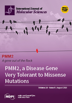
The data from the Exome Aggregation Consortium (ExAC) reveal which human genes are tolerant to mutations and which are not. A surprisingly high number of missense mutations are found in the general population for the gene PMM2, which is primarily responsible for PMM2-CDG, a life-threatening disorder of glycosylation. This finding suggests that lower activity of PMM2 can be beneficial under certain circumstances and that the co-occurrence of variants in modifier genes can worsen the diagnosis of PMM2-CDG. View the paper here.
  • Issues are regarded as officially published after their release is announced to the table of contents alert mailing list.
  • You may sign up for e-mail alerts to receive table of contents of newly released issues.
  • PDF is the official format for papers published in both, html and pdf forms. To view the papers in pdf format, click on the "PDF Full-text" link, and use the free Adobe Reader to open them.
Order results
Result details
Section
Select all
Export citation of selected articles as:
17 pages, 1526 KiB  
Article
One Way to Achieve Germination: Common Molecular Mechanism Induced by Ethylene and After-Ripening in Sunflower Seeds
by Qiong Xia, Marine Saux, Maharajah Ponnaiah, Françoise Gilard, François Perreau, Stéphanie Huguet, Sandrine Balzergue, Nicolas Langlade, Christophe Bailly, Patrice Meimoun, Françoise Corbineau and Hayat El-Maarouf-Bouteau
Int. J. Mol. Sci. 2018, 19(8), 2464; https://doi.org/10.3390/ijms19082464 - 20 Aug 2018
Cited by 14 | Viewed by 5445
Abstract
Dormancy is an adaptive trait that blocks seed germination until the environmental conditions become favorable for subsequent vegetative plant growth. Seed dormancy is defined as the inability to germinate in favorable conditions. Dormancy is alleviated during after-ripening, a dry storage period, during which [...] Read more.
Dormancy is an adaptive trait that blocks seed germination until the environmental conditions become favorable for subsequent vegetative plant growth. Seed dormancy is defined as the inability to germinate in favorable conditions. Dormancy is alleviated during after-ripening, a dry storage period, during which dormant (D) seeds unable to germinate become non-dormant (ND), able to germinate in a wide range of environmental conditions. The treatment of dormant seeds with ethylene (D/ET) promotes seed germination, and abscisic acid (ABA) treatment reduces non-dormant (ND/ABA) seed germination in sunflowers (Helianthus annuus). Metabolomic and transcriptomic studies have been performed during imbibition to compare germinating seeds (ND and D/ET) and low-germinating seeds (D and ND/ABA). A PCA analysis of the metabolites content showed that imbibition did not trigger a significant change during the first hours (3 and 15 h). The metabolic changes associated with germination capacity occurred at 24 h and were related to hexoses, as their content was higher in ND and D/ET and was reduced by ABA treatment. At the transcriptional level, a large number of genes were altered oppositely in germinating, compared to the low-germinating seeds. The metabolomic and transcriptomic results were integrated in the interpretation of the processes involved in germination. Our results show that ethylene treatment triggers molecular changes comparable to that of after-ripening treatment, concerning sugar metabolism and ABA signaling inhibition. Full article
(This article belongs to the Special Issue Seed Development, Dormancy and Germination)
Show Figures

Figure 1

14 pages, 1831 KiB  
Article
ceRNA Cross-Talk in Paulownia Witches’ Broom Disease
by Guoqiang Fan, Zhe Wang, Xiaoqiao Zhai and Yabing Cao
Int. J. Mol. Sci. 2018, 19(8), 2463; https://doi.org/10.3390/ijms19082463 - 20 Aug 2018
Cited by 12 | Viewed by 4857
Abstract
Long noncoding RNA (lncRNA), circular RNA (circRNA), and microRNA (miRNA) are important in the regulation of life activities. However, their function is unclear in Paulownia fortunei. To identify lncRNAs, circRNAs, and miRNA, and investigate their roles in the infection progress of Paulownia [...] Read more.
Long noncoding RNA (lncRNA), circular RNA (circRNA), and microRNA (miRNA) are important in the regulation of life activities. However, their function is unclear in Paulownia fortunei. To identify lncRNAs, circRNAs, and miRNA, and investigate their roles in the infection progress of Paulownia witches’ broom (PaWB) disease, we performed RNA sequencing of healthy and infected P. fortunei. A total of 3126 lncRNAs, 1634 circRNAs, and 550 miRNAs were identified. Among them, 229 lncRNAs, 65 circRNAs, and 65 miRNAs were differentially expressed in a significant manner. We constructed a competing endogenous RNA (ceRNA) network, which contains 5 miRNAs, 4 circRNAs, 5 lncRNAs, and 15 mRNAs, all of which were differentially expressed between healthy and infected P. fortunei. This study provides the first catalog of candidate ceRNAs in Paulownia and gives a revealing insight into the molecular mechanism responsible for PaWB. Full article
(This article belongs to the Section Molecular Plant Sciences)
Show Figures

Graphical abstract

12 pages, 1357 KiB  
Article
Rescue of 2-Deoxyglucose Side Effects by Ketogenic Diet
by Martin Voss, Nadja I. Lorenz, Anna-Luisa Luger, Joachim P. Steinbach, Johannes Rieger and Michael W. Ronellenfitsch
Int. J. Mol. Sci. 2018, 19(8), 2462; https://doi.org/10.3390/ijms19082462 - 20 Aug 2018
Cited by 21 | Viewed by 7780
Abstract
Cancer metabolism is characterized by extensive glucose consumption through aerobic glycolysis. No effective therapy exploiting this cancer trait has emerged so far, in part, due to the substantial side effects of the investigated drugs. In this study, we examined the side effects of [...] Read more.
Cancer metabolism is characterized by extensive glucose consumption through aerobic glycolysis. No effective therapy exploiting this cancer trait has emerged so far, in part, due to the substantial side effects of the investigated drugs. In this study, we examined the side effects of a combination of isocaloric ketogenic diet (KD) with the glycolysis inhibitor 2-deoxyglucose (2-DG). Two groups of eight athymic nude mice were either fed a standard diet (SD) or a caloric unrestricted KD with a ratio of 4 g fat to 1 g protein/carbohydrate. 2-DG was investigated in commonly employed doses of 0.5 to 4 g/kg and up to 8 g/kg. Ketosis was achieved under KD (ketone bodies: SD 0.5 ± 0.14 mmol/L, KD 1.38 ± 0.28 mmol/L, p < 0.01). The intraperitoneal application of 4 g/kg of 2-DG caused a significant increase in blood glucose, which was not prevented by KD. Sedation after the 2-DG treatment was observed and a behavioral test of spontaneous motion showed that KD reduced the sedation by 2-DG (p < 0.001). A 2-DG dose escalation to 8 g/kg was lethal for 50% of the mice in the SD and for 0% of the mice in the KD group (p < 0.01). A long-term combination of KD and an oral 1 or 2 g 2-DG/kg was well-tolerated. In conclusion, KD reduces the sedative effects of 2-DG and dramatically increases the maximum tolerated dose of 2-DG. A continued combination of KD and anti-glycolytic therapy is feasible. This is, to our knowledge, the first demonstration of increased tolerance to glycolysis inhibition by KD. Full article
(This article belongs to the Section Biochemistry)
Show Figures

Graphical abstract

17 pages, 2270 KiB  
Article
The Effect of Agomelatine Treatment on Diabetes-Induced Cognitive Impairments in Rats: Concomitant Alterations in the Hippocampal Neuron Numbers
by Özgür Devrim Can, Umut İrfan Üçel, Ümide Demir Özkay and Emel Ulupınar
Int. J. Mol. Sci. 2018, 19(8), 2461; https://doi.org/10.3390/ijms19082461 - 20 Aug 2018
Cited by 16 | Viewed by 5484
Abstract
Researches that are related to the central nervous system complications of diabetes have indicated higher incidence of cognitive disorders in patients. Since the variety of nootropic drugs used in clinics is limited and none of them consistently improves the outcomes, new and effective [...] Read more.
Researches that are related to the central nervous system complications of diabetes have indicated higher incidence of cognitive disorders in patients. Since the variety of nootropic drugs used in clinics is limited and none of them consistently improves the outcomes, new and effective drug alternatives are needed for the treatment of diabetes-induced cognitive disorders. Based on the nootropic potential of agomelatine, the promising efficacy of this drug on cognitive impairments of diabetic rats was investigated in the current study. Experimental diabetes model was induced by streptozotocin. After development of diabetes-related cognitive impairments in rats, agomelatine (40 and 80 mg/kg) was administrated orally for two weeks. Cognitive performance was assessed by Morris water-maze and passive avoidance tests. Then, the total numbers of neurons in both dentate gyrus and Cornu Ammonis (CA) 1–3 subfields of the hippocampus were estimated by the optical fractionator method. Agomelatine treatment induced notable enhancement in the learning and memory performance of diabetic rats. Moreover, it reversed the neuronal loss in the hippocampal subregions of diabetic animals. Obtained results suggest that agomelatine has a significant potential for the treatment of diabetes-induced cognitive impairments. However, therapeutic efficacy of this drug in diabetic patients suffering from cognitive dysfunctions needs to be confirmed by further clinical trials. Full article
(This article belongs to the Section Molecular Pathology, Diagnostics, and Therapeutics)
Show Figures

Figure 1

18 pages, 1036 KiB  
Review
TGF-β Signaling in Lung Health and Disease
by Akira Saito, Masafumi Horie and Takahide Nagase
Int. J. Mol. Sci. 2018, 19(8), 2460; https://doi.org/10.3390/ijms19082460 - 20 Aug 2018
Cited by 274 | Viewed by 13932
Abstract
Transforming growth factor (TGF)-β is an evolutionarily conserved pleiotropic factor that regulates a myriad of biological processes including development, tissue regeneration, immune responses, and tumorigenesis. TGF-β is necessary for lung organogenesis and homeostasis as evidenced by genetically engineered mouse models. TGF-β is crucial [...] Read more.
Transforming growth factor (TGF)-β is an evolutionarily conserved pleiotropic factor that regulates a myriad of biological processes including development, tissue regeneration, immune responses, and tumorigenesis. TGF-β is necessary for lung organogenesis and homeostasis as evidenced by genetically engineered mouse models. TGF-β is crucial for epithelial-mesenchymal interactions during lung branching morphogenesis and alveolarization. Expression and activation of the three TGF-β ligand isoforms in the lungs are temporally and spatially regulated by multiple mechanisms. The lungs are structurally exposed to extrinsic stimuli and pathogens, and are susceptible to inflammation, allergic reactions, and carcinogenesis. Upregulation of TGF-β ligands is observed in major pulmonary diseases, including pulmonary fibrosis, emphysema, bronchial asthma, and lung cancer. TGF-β regulates multiple cellular processes such as growth suppression of epithelial cells, alveolar epithelial cell differentiation, fibroblast activation, and extracellular matrix organization. These effects are closely associated with tissue remodeling in pulmonary fibrosis and emphysema. TGF-β is also central to T cell homeostasis and is deeply involved in asthmatic airway inflammation. TGF-β is the most potent inducer of epithelial-mesenchymal transition in non-small cell lung cancer cells and is pivotal to the development of tumor-promoting microenvironment in the lung cancer tissue. This review summarizes and integrates the current knowledge of TGF-β signaling relevant to lung health and disease. Full article
(This article belongs to the Special Issue TGF-Beta Super Family Signaling)
Show Figures

Graphical abstract

12 pages, 1720 KiB  
Article
Maternal Resveratrol Therapy Protects Male Rat Offspring against Programmed Hypertension Induced by TCDD and Dexamethasone Exposures: Is It Relevant to Aryl Hydrocarbon Receptor?
by Chien-Ning Hsu, Yu-Ju Lin, Pei-Chen Lu and You-Lin Tain
Int. J. Mol. Sci. 2018, 19(8), 2459; https://doi.org/10.3390/ijms19082459 - 20 Aug 2018
Cited by 36 | Viewed by 4681
Abstract
Hypertension can originate from early-life adverse environmental in utero exposure to dexamethasone (DEX) or 2,3,7,8-tetrachlorodibenzo-p-dioxin (TCDD). Since DEX and TCDD are related to the aryl hydrocarbon receptor (AHR) signaling pathway, we examined whether resveratrol, an AHR modulator and antioxidant, could prevent programmed hypertension [...] Read more.
Hypertension can originate from early-life adverse environmental in utero exposure to dexamethasone (DEX) or 2,3,7,8-tetrachlorodibenzo-p-dioxin (TCDD). Since DEX and TCDD are related to the aryl hydrocarbon receptor (AHR) signaling pathway, we examined whether resveratrol, an AHR modulator and antioxidant, could prevent programmed hypertension via regulating AHR signaling and oxidative stress. Groups of four-month-old male rat offspring were studied (n = 7–8 per group): control, DEX (0.1 mg/kg i.p. from a gestational age of 16 to 22 days), TCDD (200 ng/kg in four once-weekly oral doses), DEX + TCDD, and DEX + TCDD + R (resveratrol 0.05% in drinking water throughout pregnancy and lactation). Maternal TCDD exposure aggravated prenatal DEX-induced hypertension in adult male offspring, which maternal resveratrol therapy prevented. Maternal TCDD exposure aggravated DEX-induced oxidative damage in offspring kidneys, which was prevented by resveratrol therapy. Maternal resveratrol therapy decreased asymmetric and symmetric dimethylarginine (ADMA and SDMA) levels, thereby preventing combined DEX and TCDD exposure-induced programmed hypertension. Increases in renal Ahrr and Cyp1a1 expression induced by DEX + TCDD exposure were restored by resveratrol therapy. The beneficial effects of resveratrol on DEX + TCDD-induced hypertension relate to reduced renal mRNA expression of Ren, Ace, and Agtr1a expression. Thus, the beneficial effects of resveratrol on DEX + TCDD-induced hypertension include reduction of oxidative stress, restoration of nitric oxide (NO) bioavailability, blockade of the renin–angiotensin system (RAS), and antagonizing AHR signaling pathway. Full article
(This article belongs to the Special Issue Aryl Hydrocarbon Receptor in Biology and Toxicology)
Show Figures

Graphical abstract

15 pages, 4259 KiB  
Article
Effects of Liposomal Formulation of Citicoline in Experimental Diabetes-Induced Retinal Neurodegeneration
by Patricia Bogdanov, Joel Sampedro, Cristina Solà-Adell, Olga Simó-Servat, Carla Russo, Luisa Varela-Sende, Rafael Simó and Cristina Hernández
Int. J. Mol. Sci. 2018, 19(8), 2458; https://doi.org/10.3390/ijms19082458 - 20 Aug 2018
Cited by 25 | Viewed by 6324
Abstract
Diabetic retinopathy (DR) has been classically considered a microcirculatory disease of the retina. However, there is growing evidence to suggest that retinal neurodegeneration is also an early event in the pathogenesis of DR. Citicoline has been successfully used as a neuroprotective agent in [...] Read more.
Diabetic retinopathy (DR) has been classically considered a microcirculatory disease of the retina. However, there is growing evidence to suggest that retinal neurodegeneration is also an early event in the pathogenesis of DR. Citicoline has been successfully used as a neuroprotective agent in the treatment of glaucoma but their effects on DR remain to be elucidated. On this basis, the main aim of the present study was to evaluate the effect of topical administration of citicoline in liposomal formulation on retinal neurodegeneration in db/db mouse and to investigate the underlying mechanisms of action. The treatment (citicoline or vehicle) was topically administered twice daily for 15 days. Retinal analyses were performed in vivo by electroretinography and ex vivo by using Western blot and immunofluorescence measurements. We found that the liposomal formulation of citicoline prevented glial activation and neural apoptosis in the diabetic retina. The main mechanism implicated in these beneficial effects were the inhibition of the downregulation of synaptophysin and its anti-inflammatory properties by means of preventing the upregulation of NF-κB and TNF-α (Tumor Necrosis Factor α) induced by diabetes. Overall, these results suggest that topical administration of citicoline in liposomal formulation could be considered as a new strategy for treating the early stages of DR. Full article
Show Figures

Graphical abstract

11 pages, 2723 KiB  
Article
In Vitro Inhibitory Mechanism Effect of TRAIP on the Function of TRAF2 Revealed by Characterization of Interaction Domains
by Eijaz Ahmed Bhat, Chang Min Kim, Sunghwan Kim and Hyun Ho Park
Int. J. Mol. Sci. 2018, 19(8), 2457; https://doi.org/10.3390/ijms19082457 - 20 Aug 2018
Cited by 8 | Viewed by 4299
Abstract
TRAF-interacting protein (TRAIP), a negative regulator of TNF-induced-nuclear factor kappa-light-chain-enhancer of activated B cells (NF-κB) activation, inhibits adaptor protein TRAF2 by direct interaction and is critical in apoptosis, cell proliferation, antiviral response, and embryonic development. Although the critical function of TRAIP in NF-κB [...] Read more.
TRAF-interacting protein (TRAIP), a negative regulator of TNF-induced-nuclear factor kappa-light-chain-enhancer of activated B cells (NF-κB) activation, inhibits adaptor protein TRAF2 by direct interaction and is critical in apoptosis, cell proliferation, antiviral response, and embryonic development. Although the critical function of TRAIP in NF-κB signaling is well-known, the molecular inhibitory mechanism of TRAIP remains unclear. We found that the TRAIP coiled-coil domain altered its stoichiometry between dimer and trimer in a concentration-dependent manner. Additionally, the TRAIP RING domain induced even higher-ordered assembly, which was necessary for interacting with the TRAF-N domain of TRAF2 but not TRAF1. Characterization of the TRAF-N domains of TRAF1 and TRAF2, the tentative TRAIP-binding region of TRAFs, suggested the molecular basis of the inhibitory effect of TRAIP on TRAF2 in NF-κB signaling. Full article
(This article belongs to the Section Biochemistry)
Show Figures

Figure 1

18 pages, 2670 KiB  
Article
Toll-Like Receptor Agonists Modulate Wound Regeneration in Airway Epithelial Cells
by Anna Lewandowska-Polak, Małgorzata Brauncajs, Marzanna Jarzębska, Małgorzata Pawełczyk, Marcin Kurowski, Maciej Chałubiński, Joanna Makowska and Marek L. Kowalski
Int. J. Mol. Sci. 2018, 19(8), 2456; https://doi.org/10.3390/ijms19082456 - 20 Aug 2018
Cited by 11 | Viewed by 4585
Abstract
Background: Impaired regeneration of airway epithelium may lead to persistence of inflammation and remodelling. Regeneration of injured epithelium is a complex phenomenon and the role of toll-like receptors (TLRs) in the stimulation of respiratory virus products in this process has not been established. [...] Read more.
Background: Impaired regeneration of airway epithelium may lead to persistence of inflammation and remodelling. Regeneration of injured epithelium is a complex phenomenon and the role of toll-like receptors (TLRs) in the stimulation of respiratory virus products in this process has not been established. Objective: This study was undertaken to test the hypothesis that the wound repair process in airway epithelium is modulated by microbial products via toll-like receptors. Methods: Injured and not-injured bronchial epithelial cells (ECs) (BEAS-2B line) were incubated with the TLR agonists poly(I:C), lipopolisacharide (LPS), allergen Der p1, and supernatants from virus-infected epithelial cells, either alone or in combination with TLR inhibitors. Regeneration and immune response in injured and not-injured cells were studied. Results: Addition of either poly(I:C) or LPS to ECs induced a marked inhibition of wound repair. Supernatants from RV1b-infected cells also decreased regeneration. Preincubation of injured and not-injured ECs with TLR inhibitors decreased LPS and poly(I:C)-induced repair inhibition. TGF-β and RANTES mRNA expression was higher in injured ECs and IFN-α, IFN-β, IL-8, and VEGF mRNA expression was lower in damaged epithelium as compared to not-injured. Stimulation with poly(I:C) increased IFN-α and IFN-β mRNA expression in injured cells, and LPS stimulation decreased interferons mRNA expression both in not-injured and injured ECs. Conclusion: Regeneration of the airway epithelium is modulated by microbial products via toll-like receptors. Full article
(This article belongs to the Special Issue New Innovations in Wound Healing and Repair)
Show Figures

Figure 1

12 pages, 2438 KiB  
Review
The Dead Can Nurture: Novel Insights into the Function of Dead Organs Enclosing Embryos
by Buzi Raviv, James Godwin, Gila Granot and Gideon Grafi
Int. J. Mol. Sci. 2018, 19(8), 2455; https://doi.org/10.3390/ijms19082455 - 19 Aug 2018
Cited by 17 | Viewed by 5111
Abstract
Plants have evolved a variety of dispersal units whereby the embryo is enclosed by various dead protective layers derived from maternal organs of the reproductive system including seed coats (integuments), pericarps (ovary wall, e.g., indehiscent dry fruits) as well as floral bracts (e.g., [...] Read more.
Plants have evolved a variety of dispersal units whereby the embryo is enclosed by various dead protective layers derived from maternal organs of the reproductive system including seed coats (integuments), pericarps (ovary wall, e.g., indehiscent dry fruits) as well as floral bracts (e.g., glumes) in grasses. Commonly, dead organs enclosing embryos (DOEEs) are assumed to provide a physical shield for embryo protection and means for dispersal in the ecosystem. In this review article, we highlight recent studies showing that DOEEs of various species across families also have the capability for long-term storage of various substances including active proteins (hydrolases and ROS detoxifying enzymes), nutrients and metabolites that have the potential to support the embryo during storage in the soil and assist in germination and seedling establishment. We discuss a possible role for DOEEs as natural coatings capable of “engineering” the seed microenvironment for the benefit of the embryo, the seedling and the growing plant. Full article
(This article belongs to the Special Issue Seed Development, Dormancy and Germination)
Show Figures

Graphical abstract

15 pages, 1303 KiB  
Article
Upregulation of Nrf2 and Decreased Redox Signaling Contribute to Renoprotective Effects of Chemerin Receptor Blockade in Diabetic Mice
by Karla Bianca Neves, Augusto Cesar Montezano, Rheure Alves-Lopes, Thiago Bruder-Nascimento, Rafael Menezes Costa, Roberto S Costa, Rhian M Touyz and Rita C Tostes
Int. J. Mol. Sci. 2018, 19(8), 2454; https://doi.org/10.3390/ijms19082454 - 19 Aug 2018
Cited by 18 | Viewed by 5364
Abstract
Chemerin, acting through its receptor ChemR23, is an adipokine associated with inflammatory response, glucose and lipid metabolism and vascular function. Although this adipokine has been associated with the development and progression of kidney disease, it is not clear whether the chemerin/ChemR23 system plays [...] Read more.
Chemerin, acting through its receptor ChemR23, is an adipokine associated with inflammatory response, glucose and lipid metabolism and vascular function. Although this adipokine has been associated with the development and progression of kidney disease, it is not clear whether the chemerin/ChemR23 system plays a role in renal function in the context of diabetes. Therefore, we sought to determine whether ChemR23 receptor blockade prevents the development and/or progression of diabetic nephropathy and questioned the role of oxidative stress and Nrf2 in this process. Renal redox state and function were assessed in non-diabetic lean db/m and diabetic obese db/db mice treated with vehicle or CCX832 (ChemR23 antagonist). Renal reactive oxygen species (ROS) production, which was increased in diabetic mice, was attenuated by CCX832. This was associated with an increase in Nox 4 expression. Augmented protein oxidation in db/db mice was not observed when mice were treated with CCX832. CCX832 also abrogated impaired Nrf2 nuclear activity and associated downregulation in antioxidants expression in kidneys from db/db mice. Our in vivo findings highlight the role of the redox signaling and Nrf2 system as renoprotective players during chemerin receptor blockade in diabetic mice. The chemerin/ChemR23 system may be an important target to limit renal dysfunction associated with obesity-related diabetes. Full article
Show Figures

Graphical abstract

19 pages, 1163 KiB  
Review
Role of mTOR Signaling in Tumor Microenvironment: An Overview
by Fabiana Conciatori, Chiara Bazzichetto, Italia Falcone, Sara Pilotto, Emilio Bria, Francesco Cognetti, Michele Milella and Ludovica Ciuffreda
Int. J. Mol. Sci. 2018, 19(8), 2453; https://doi.org/10.3390/ijms19082453 - 19 Aug 2018
Cited by 103 | Viewed by 7209
Abstract
The mammalian target of rapamycin (mTOR) pathway regulates major processes by integrating a variety of exogenous cues, including diverse environmental inputs in the tumor microenvironment (TME). In recent years, it has been well recognized that cancer cells co-exist and co-evolve with their TME, [...] Read more.
The mammalian target of rapamycin (mTOR) pathway regulates major processes by integrating a variety of exogenous cues, including diverse environmental inputs in the tumor microenvironment (TME). In recent years, it has been well recognized that cancer cells co-exist and co-evolve with their TME, which is often involved in drug resistance. The mTOR pathway modulates the interactions between the stroma and the tumor, thereby affecting both the tumor immunity and angiogenesis. The activation of mTOR signaling is associated with these pro-oncogenic cellular processes, making mTOR a promising target for new combination therapies. This review highlights the role of mTOR signaling in the characterization and the activity of the TME’s elements and their implications in cancer immunotherapy. Full article
(This article belongs to the Special Issue mTOR in Human Diseases)
Show Figures

Graphical abstract

13 pages, 2546 KiB  
Article
Expression, Functional Characterization, and Preliminary Crystallization of the Cochaperone Prefoldin from the Thermophilic Fungus Chaetomium thermophilum
by Kento Morita, Yohei Y. Yamamoto, Ayaka Hori, Tomohiro Obata, Yuko Uno, Kyosuke Shinohara, Keiichi Noguchi, Kentaro Noi, Teru Ogura, Kentaro Ishii, Koichi Kato, Mahito Kikumoto, Rocio Arranz, Jose M. Valpuesta and Masafumi Yohda
Int. J. Mol. Sci. 2018, 19(8), 2452; https://doi.org/10.3390/ijms19082452 - 19 Aug 2018
Cited by 4 | Viewed by 4867
Abstract
Prefoldin is a hexameric molecular chaperone found in the cytosol of archaea and eukaryotes. Its hexameric complex is built from two related classes of subunits, and has the appearance of a jellyfish: Its body consists of a double β-barrel assembly with six long [...] Read more.
Prefoldin is a hexameric molecular chaperone found in the cytosol of archaea and eukaryotes. Its hexameric complex is built from two related classes of subunits, and has the appearance of a jellyfish: Its body consists of a double β-barrel assembly with six long tentacle-like coiled coils protruding from it. Using the tentacles, prefoldin captures an unfolded protein substrate and transfers it to a group II chaperonin. Based on structural information from archaeal prefoldins, mechanisms of substrate recognition and prefoldin-chaperonin cooperation have been investigated. In contrast, the structure and mechanisms of eukaryotic prefoldins remain unknown. In this study, we succeeded in obtaining recombinant prefoldin from a thermophilic fungus, Chaetomium thermophilum (CtPFD). The recombinant CtPFD could not protect citrate synthase from thermal aggregation. However, CtPFD formed a complex with actin from chicken muscle and tubulin from porcine brain, suggesting substrate specificity. We succeeded in observing the complex formation of CtPFD and the group II chaperonin of C. thermophilum (CtCCT) by atomic force microscopy and electron microscopy. These interaction kinetics were analyzed by surface plasmon resonance using Biacore. Finally, we have shown the transfer of actin from CtPFD to CtCCT. The study of the folding pathway formed by CtPFD and CtCCT should provide important information on mechanisms of the eukaryotic prefoldin–chaperonin system. Full article
(This article belongs to the Special Issue Molecular Chaperones)
Show Figures

Graphical abstract

21 pages, 5201 KiB  
Article
Cellular Pharmacology of Palladinum(III) Hematoporphyrin IX Complexes: Solution Stability, Antineoplastic and Apoptogenic Activity, DNA Binding, and Processing of DNA-Adducts
by Georgi Momekov, Iva Ugrinova, Evdokia Pasheva, Daniela Tsekova and Galina Gencheva
Int. J. Mol. Sci. 2018, 19(8), 2451; https://doi.org/10.3390/ijms19082451 - 19 Aug 2018
Cited by 4 | Viewed by 3769
Abstract
Two paramagnetic PdIII complexes of hematoporphyrin IX ((7,12-bis(1-hydroxyethyl)-3,8,13,17-tetramethyl-21H-23H-porphyn-2,18-dipropionic acid), Hp), namely a dinuclear one [PdIII2(Hp-3H)Cl3(H2O)5]·2PdCl2, Pd1 and a mononuclear metalloporphyrin type [PdIII(Hp-2H)Cl(H2O)]·H2 [...] Read more.
Two paramagnetic PdIII complexes of hematoporphyrin IX ((7,12-bis(1-hydroxyethyl)-3,8,13,17-tetramethyl-21H-23H-porphyn-2,18-dipropionic acid), Hp), namely a dinuclear one [PdIII2(Hp-3H)Cl3(H2O)5]·2PdCl2, Pd1 and a mononuclear metalloporphyrin type [PdIII(Hp-2H)Cl(H2O)]·H2O, Pd2 have been synthesized reproducibly and isolated as neutral compounds at different reaction conditions. Their structure and solution stability have been assayed by UV/Vis and EPR spectroscopy. The compounds researched have shown in vitro cell growth inhibitory effects at micromolar concentration against a panel of human tumor cell lines. A DNA fragmentation test in the HL-60 cell line has indicated that Pd1 causes comparable proapoptotic effects with regard to cisplatin but at substantially higher concentrations. Pd1 and cisplatin form intra-strand guanine bis-adducts as the palladium complex is less capable of forming DNA adducts. This demonstrates its cisplatin-dissimilar pharmacological profile. The test for efficient removal of DNA-adducts by the NER synthesis after modification of pBS plasmids with either cisplatin or Pd1 has manifested that the lesions induced by cisplatin are far better recognized and repaired compared those of Pd1. The study on the recognition and binding of the HMGB-1 protein to cisplatin or Pd1 modified DNA probes have shown that HMG proteins are less involved in the palladium agent cytotoxicity. Full article
Show Figures

Graphical abstract

36 pages, 9132 KiB  
Review
Cytokinin at the Crossroads of Abiotic Stress Signalling Pathways
by Jaroslav Pavlů, Jan Novák, Vladěna Koukalová, Markéta Luklová, Břetislav Brzobohatý and Martin Černý
Int. J. Mol. Sci. 2018, 19(8), 2450; https://doi.org/10.3390/ijms19082450 - 19 Aug 2018
Cited by 111 | Viewed by 11833
Abstract
Cytokinin is a multifaceted plant hormone that plays major roles not only in diverse plant growth and development processes, but also stress responses. We summarize knowledge of the roles of its metabolism, transport, and signalling in responses to changes in levels of both [...] Read more.
Cytokinin is a multifaceted plant hormone that plays major roles not only in diverse plant growth and development processes, but also stress responses. We summarize knowledge of the roles of its metabolism, transport, and signalling in responses to changes in levels of both macronutrients (nitrogen, phosphorus, potassium, sulphur) and micronutrients (boron, iron, silicon, selenium). We comment on cytokinin’s effects on plants’ xenobiotic resistance, and its interactions with light, temperature, drought, and salinity signals. Further, we have compiled a list of abiotic stress-related genes and demonstrate that their expression patterns overlap with those of cytokinin metabolism and signalling genes. Full article
Show Figures

Graphical abstract

23 pages, 4316 KiB  
Review
Implications of Metal Binding and Asparagine Deamidation for Amyloid Formation
by Yutaka Sadakane and Masahiro Kawahara
Int. J. Mol. Sci. 2018, 19(8), 2449; https://doi.org/10.3390/ijms19082449 - 19 Aug 2018
Cited by 16 | Viewed by 5361
Abstract
Increasing evidence suggests that amyloid formation, i.e., self-assembly of proteins and the resulting conformational changes, is linked with the pathogenesis of various neurodegenerative disorders such as Alzheimer’s disease, prion diseases, and Lewy body diseases. Among the factors that accelerate or inhibit oligomerization, we [...] Read more.
Increasing evidence suggests that amyloid formation, i.e., self-assembly of proteins and the resulting conformational changes, is linked with the pathogenesis of various neurodegenerative disorders such as Alzheimer’s disease, prion diseases, and Lewy body diseases. Among the factors that accelerate or inhibit oligomerization, we focus here on two non-genetic and common characteristics of many amyloidogenic proteins: metal binding and asparagine deamidation. Both reflect the aging process and occur in most amyloidogenic proteins. All of the amyloidogenic proteins, such as Alzheimer’s β-amyloid protein, prion protein, and α-synuclein, are metal-binding proteins and are involved in the regulation of metal homeostasis. It is widely accepted that these proteins are susceptible to non-enzymatic posttranslational modifications, and many asparagine residues of these proteins are deamidated. Moreover, these two factors can combine because asparagine residues can bind metals. We review the current understanding of these two common properties and their implications in the pathogenesis of these neurodegenerative diseases. Full article
(This article belongs to the Special Issue Amyloid Fibrils and Methods for Their Study)
Show Figures

Graphical abstract

12 pages, 1575 KiB  
Article
The First Dose of Fingolimod Affects Circulating Extracellular Vesicles in Multiple Sclerosis Patients
by Matías Sáenz-Cuesta, Ainhoa Alberro, Maider Muñoz-Culla, Iñaki Osorio-Querejeta, Marta Fernandez-Mercado, Itziar Lopetegui, Mikel Tainta, Álvaro Prada, Tamara Castillo-Triviño, Juan Manuel Falcón-Pérez, Javier Olascoaga and David Otaegui
Int. J. Mol. Sci. 2018, 19(8), 2448; https://doi.org/10.3390/ijms19082448 - 19 Aug 2018
Cited by 25 | Viewed by 8524
Abstract
Extracellular vesicles (EVs) are membrane-bound particles involved in intercellular communication. They carry proteins, lipids, and nucleotides such as microRNAs (miRNAs) from the secreting cell that can modulate target cells. We and others have previously described the presence of EVs in peripheral blood of [...] Read more.
Extracellular vesicles (EVs) are membrane-bound particles involved in intercellular communication. They carry proteins, lipids, and nucleotides such as microRNAs (miRNAs) from the secreting cell that can modulate target cells. We and others have previously described the presence of EVs in peripheral blood of multiple sclerosis (MS) patients and postulated them as novel biomarkers. However, their immune function in MS pathogenesis and the effect during the onset of new immunomodulatory therapies on EVs remain elusive. Here, we isolated plasma EVs from fingolimod-treated MS patients in order to assess whether EVs are affected by the first dose of the treatment. We quantified EVs, analyzed their miRNA cargo, and checked their immune regulatory function. Results showed an elevated EV concentration with a dramatic change in their miRNA cargo 5 h after the first dose of fingolimod. Besides, EVs obtained prior to fingolimod treatment showed an increased immune regulatory activity compared to EVs obtained 5 h post-treatment. This work suggests that EVs are implicated in the mechanism of action of immunomodulatory treatments from the initial hours and opens a new avenue to explore a potential use of EVs for early treatment monitoring. Full article
(This article belongs to the Special Issue Focus on Exosome-Based Cell-Cell Communication in Health and Disease)
Show Figures

Graphical abstract

13 pages, 2306 KiB  
Article
Effect of N- and C-Terminal Amino Acids on the Interfacial Binding Properties of Phospholipase D from Vibrio parahaemolyticus
by Fanghua Wang, Ruixia Wei, Abdelkarim Abousalham, Wuchong Chen, Bo Yang and Yonghua Wang
Int. J. Mol. Sci. 2018, 19(8), 2447; https://doi.org/10.3390/ijms19082447 - 19 Aug 2018
Cited by 5 | Viewed by 4435
Abstract
The effects of N-terminal (1–34 amino acids) and C-terminal (434–487 amino acids) amino acid sequences on the interfacial binding properties of Phospholipase D from Vibrio parahaemolyticus (VpPLD) were characterized by using monomolecular film technology. Online tools allowed the prediction of the secondary structure [...] Read more.
The effects of N-terminal (1–34 amino acids) and C-terminal (434–487 amino acids) amino acid sequences on the interfacial binding properties of Phospholipase D from Vibrio parahaemolyticus (VpPLD) were characterized by using monomolecular film technology. Online tools allowed the prediction of the secondary structure of the target N- and C-terminal VpPLD sequences. Various truncated forms of VpPLD with different N- or C-terminal deletions were designed, based on their secondary structure, and their membrane binding properties were examined. The analysis of the maximum insertion pressure (MIP) and synergy factor “a” indicated that the loop structure (1–25 amino acids) in the N-terminal segment of VpPLD had a positive effect on the binding of VpPLD to phospholipid monolayers, especially to 1,2-dimyristoyl-sn-glycero-3-phosphoserine and 1,2-dimyristoyl-sn-glycero-3-phosphocholine. The deletion affecting the N-terminus loop structure caused a significant decrease of the MIP and synergy factor a of the protein for these phospholipid monolayers. Conversely, the deletion of the helix structure (26–34 amino acids) basically had no influence on the binding of VpPLD to phospholipid monolayers. The deletion of the C-terminal amino acids 434–487 did not significantly change the binding selectivity of VpPLD for the various phospholipid monolayer tested here. However, a significant increase of the MIP value for all the phospholipid monolayers strongly indicated that the three-strand segment (434–469 amino acids) had a great negative effect on the interfacial binding to these phospholipid monolayers. The deletion of this peptide caused a significantly greater insertion of the protein into the phospholipid monolayers examined. The present study provides detailed information on the effect of the N- and C-terminal segments of VpPLD on the interfacial binding properties of the enzyme and improves our understanding of the interactions between this enzyme and cell membranes. Full article
(This article belongs to the Special Issue Microbial Enzymes)
Show Figures

Graphical abstract

5 pages, 200 KiB  
Editorial
Plant Defense Genes against Biotic Stresses
by Isabel Diaz
Int. J. Mol. Sci. 2018, 19(8), 2446; https://doi.org/10.3390/ijms19082446 - 19 Aug 2018
Cited by 10 | Viewed by 3905
(This article belongs to the Special Issue Plant Defense Genes Against Biotic Stresses)
11 pages, 1955 KiB  
Review
The Pathophysiology of Post-Traumatic Glioma
by Donata Simińska, Klaudyna Kojder, Dariusz Jeżewski, Ireneusz Kojder, Marta Skórka, Izabela Gutowska, Dariusz Chlubek and Irena Baranowska-Bosiacka
Int. J. Mol. Sci. 2018, 19(8), 2445; https://doi.org/10.3390/ijms19082445 - 19 Aug 2018
Cited by 12 | Viewed by 5212
Abstract
Malignant glioma is a brain tumor with a very high mortality rate resulting from the specific morphology of its infiltrative growth and poor early detection rates. The causes of one of its very specific types, i.e., post-traumatic glioma, have been discussed for many [...] Read more.
Malignant glioma is a brain tumor with a very high mortality rate resulting from the specific morphology of its infiltrative growth and poor early detection rates. The causes of one of its very specific types, i.e., post-traumatic glioma, have been discussed for many years, with some studies providing evidence for mechanisms where the reaction to an injury may in some cases lead to the onset of carcinogenesis in the brain. In this review of the available literature, we discuss the consequences of breaking the blood–brain barrier and consequences of the influx of immune-system cells to the site of injury. We also analyze the influence of inflammatory mediators on the expression of genes controlling the process of apoptosis and the effect of chemical mutagenic factors on glial cells in the brain. We present the results of experimental studies indicating a relationship between injury and glioma development. However, epidemiological studies on post-traumatic glioma, of which only a few confirm the conclusions of experimental research, indicate that any potential relationship between injury and glioma, if any, is indirect. Full article
(This article belongs to the Special Issue Molecular and Cellular Mechanisms of Neurotoxicity)
Show Figures

Figure 1

16 pages, 2494 KiB  
Article
Comparative Proteomic Analysis of Two Ralstonia solanacearum Isolates Differing in Aggressiveness
by Guoping Wang, Jie Kong, Dandan Cui, Hongbo Zhao, Puyan Zhao, Shujie Feng, Yahua Zhao and Wenyi Wang
Int. J. Mol. Sci. 2018, 19(8), 2444; https://doi.org/10.3390/ijms19082444 - 18 Aug 2018
Cited by 6 | Viewed by 4136
Abstract
Ralstonia solanacearum is a soil-borne, plant xylem-infecting pathogen that causes the devastating bacterial wilt (BW) disease in a number of plant species. In the present study, two R. solanacearum strains with different degrees of aggressiveness―namely RsH (pathogenic to Hawaii 7996, a tomato cultivar [...] Read more.
Ralstonia solanacearum is a soil-borne, plant xylem-infecting pathogen that causes the devastating bacterial wilt (BW) disease in a number of plant species. In the present study, two R. solanacearum strains with different degrees of aggressiveness―namely RsH (pathogenic to Hawaii 7996, a tomato cultivar resistant against most strains) and RsM (non-pathogenic to Hawaii 7996) were identified. Phylogenetic analysis revealed that both RsM and RsH belonged to phylotype I. To further elucidate the underlying mechanism of the different pathotypes between the two strains, we performed a comparative proteomics study on RsM and RsH in rich and minimal media to identify the change in the level of protein abundance. In total, 24 differential proteins were identified, with four clusters in terms of protein abundance. Further bioinformatics exploration allowed us to classify these proteins into five functional groups. Notably, the pathogenesis of RsM and RsH was particularly characterized by a pronounced difference in the abundance of virulence- and metabolism-related proteins, such as UDP-N-acetylglucosamine 2-epimerase (epsC) and isocitrate lyase (ICL), which were more abundant in the high pathogenicity strain RsH. Thus, we propose that the differences in pathogenicity between RsM and RsH can possibly be partially explained by differences in extracellular polysaccharide (EPS) and glyoxylate metabolism-related proteins. Full article
(This article belongs to the Section Biochemistry)
Show Figures

Figure 1

17 pages, 5580 KiB  
Article
Complete Chloroplast Genome Sequence and Phylogenetic Analysis of Quercus acutissima
by Xuan Li, Yongfu Li, Mingyue Zang, Mingzhi Li and Yanming Fang
Int. J. Mol. Sci. 2018, 19(8), 2443; https://doi.org/10.3390/ijms19082443 - 18 Aug 2018
Cited by 76 | Viewed by 7565
Abstract
Quercus acutissima, an important endemic and ecological plant of the Quercus genus, is widely distributed throughout China. However, there have been few studies on its chloroplast genome. In this study, the complete chloroplast (cp) genome of Q. acutissima was sequenced, analyzed, and [...] Read more.
Quercus acutissima, an important endemic and ecological plant of the Quercus genus, is widely distributed throughout China. However, there have been few studies on its chloroplast genome. In this study, the complete chloroplast (cp) genome of Q. acutissima was sequenced, analyzed, and compared to four species in the Fagaceae family. The size of the Q. acutissima chloroplast genome is 161,124 bp, including one large single copy (LSC) region of 90,423 bp and one small single copy (SSC) region of 19,068 bp, separated by two inverted repeat (IR) regions of 51,632 bp. The GC content of the whole genome is 36.08%, while those of LSC, SSC, and IR are 34.62%, 30.84%, and 42.78%, respectively. The Q. acutissima chloroplast genome encodes 136 genes, including 88 protein-coding genes, four ribosomal RNA genes, and 40 transfer RNA genes. In the repeat structure analysis, 31 forward and 22 inverted long repeats and 65 simple-sequence repeat loci were detected in the Q. acutissima cp genome. The existence of abundant simple-sequence repeat loci in the genome suggests the potential for future population genetic work. The genome comparison revealed that the LSC region is more divergent than the SSC and IR regions, and there is higher divergence in noncoding regions than in coding regions. The phylogenetic relationships of 25 species inferred that members of the Quercus genus do not form a clade and that Q. acutissima is closely related to Q. variabilis. This study identified the unique characteristics of the Q. acutissima cp genome, which will provide a theoretical basis for species identification and biological research. Full article
(This article belongs to the Special Issue Chloroplast)
Show Figures

Graphical abstract

19 pages, 1859 KiB  
Article
Cell Cycle Regulation by Ca2+-Activated K+ (BK) Channels Modulators in SH-SY5Y Neuroblastoma Cells
by Fatima Maqoud, Angela Curci, Rosa Scala, Alessandra Pannunzio, Federica Campanella, Mauro Coluccia, Giuseppe Passantino, Nicola Zizzo and Domenico Tricarico
Int. J. Mol. Sci. 2018, 19(8), 2442; https://doi.org/10.3390/ijms19082442 - 18 Aug 2018
Cited by 20 | Viewed by 4946
Abstract
The effects of Ca2+-activated K+ (BK) channel modulation by Paxilline (PAX) (10−7–10−4 M), Iberiotoxin (IbTX) (0.1–1 × 10⁻⁶ M) and Resveratrol (RESV) (1–2 × 10⁻⁴ M) on cell cycle and proliferation, AKT1pSer473 phosphorylation, cell [...] Read more.
The effects of Ca2+-activated K+ (BK) channel modulation by Paxilline (PAX) (10−7–10−4 M), Iberiotoxin (IbTX) (0.1–1 × 10⁻⁶ M) and Resveratrol (RESV) (1–2 × 10⁻⁴ M) on cell cycle and proliferation, AKT1pSer473 phosphorylation, cell diameter, and BK currents were investigated in SH-SY5Y cells using Operetta-high-content-Imaging-System, ELISA-assay, impedentiometric counting method and patch-clamp technique, respectively. IbTX (4 × 10⁻⁷ M), PAX (5 × 10⁻⁵ M) and RESV (10−4 M) caused a maximal decrease of the outward K+ current at +30 mV (Vm) of −38.3 ± 10%, −31.9 ± 9% and −43 ± 8%, respectively, which was not reversible following washout and cell depolarization. After 6h of incubation, the drugs concentration dependently reduced proliferation. A maximal reduction of cell proliferation, respectively of −60 ± 8% for RESV (2 × 10⁻⁴ M) (IC50 = 1.50 × 10⁻⁴ M), −65 ± 6% for IbTX (10−6 M) (IC50 = 5 × 10⁻⁷ M), −97 ± 6% for PAX (1 × 10⁻⁴ M) (IC50 = 1.06 × 10⁻⁵ M) and AKT1pser473 dephosphorylation was observed. PAX induced a G1/G2 accumulation and contraction of the S-phase, reducing the nuclear area and cell diameter. IbTX induced G1 contraction and G2 accumulation reducing diameter. RESV induced G2 accumulation and S contraction reducing diameter. These drugs share common actions leading to a block of the surface membrane BK channels with cell depolarization and calcium influx, AKT1pser473 dephosphorylation by calcium-dependent phosphatase, accumulation in the G2 phase, and a reduction of diameter and proliferation. In addition, the PAX action against nuclear membrane BK channels potentiates its antiproliferative effects with early apoptosis. Full article
(This article belongs to the Special Issue Calcium Signaling in Human Health and Diseases)
Show Figures

Graphical abstract

12 pages, 1833 KiB  
Article
Stress-Induced Phosphorylation of Nuclear YB-1 Depends on Nuclear Trafficking of p90 Ribosomal S6 Kinase
by Aadhya Tiwari, Simone Rebholz, Eva Maier, Mozhgan Dehghan Harati, Daniel Zips, Christine Sers, H. Peter Rodemann and Mahmoud Toulany
Int. J. Mol. Sci. 2018, 19(8), 2441; https://doi.org/10.3390/ijms19082441 - 18 Aug 2018
Cited by 21 | Viewed by 5979
Abstract
Ionizing radiation (IR) and epidermal growth factor (EGF) stimulate Y-box binding protein-1 (YB-1) phosphorylation at Ser-102 in KRAS wild-type (KRASwt) cells, whereas in KRAS mutated (KRASmut) cells, YB-1 is constitutively phosphorylated, independent of IR or EGF. YB-1 activity stimulates the repair of IR-induced [...] Read more.
Ionizing radiation (IR) and epidermal growth factor (EGF) stimulate Y-box binding protein-1 (YB-1) phosphorylation at Ser-102 in KRAS wild-type (KRASwt) cells, whereas in KRAS mutated (KRASmut) cells, YB-1 is constitutively phosphorylated, independent of IR or EGF. YB-1 activity stimulates the repair of IR-induced DNA double-strand breaks (DSBs) in the nucleus. Thus far, the YB-1 nuclear translocation pattern after cell exposure to various cellular stressors is not clear. In the present study, we investigated the pattern of YB-1 phosphorylation and its possible translocation to the nucleus in KRASwt cells after exposure to IR, EGF treatment, and conditional expression of mutated KRAS(G12V). IR, EGF, and conditional KRAS(G12V) expression induced YB-1 phosphorylation in both the cytoplasmic and nuclear fractions of KRASwt cells. None of the stimuli induced YB-1 nuclear translocation, while p90 ribosomal s6 kinase (RSK) translocation was enhanced in KRASwt cells after any of the stimuli. EGF-induced RSK translocation to the nucleus and nuclear YB-1 phosphorylation were completely blocked by the EGF receptor kinase inhibitor erlotinib. Likewise, RSK inhibition blocked RSK nuclear translocation and nuclear YB-1 phosphorylation after irradiation and KRAS(G12V) overexpression. In summary, acute stimulation of YB-1 phosphorylation does not lead to YB-1 translocation from the cytoplasm to the nucleus. Rather, irradiation, EGF treatment, or KRAS(G12V) overexpression induces RSK activation, leading to its translocation to the nucleus, where it activates already-existing nuclear YB-1. Our novel finding illuminates the signaling pathways involved in nuclear YB-1 phosphorylation and provides a rationale for designing appropriate targeting strategies to block YB-1 in oncology as well as in radiation oncology. Full article
(This article belongs to the Special Issue Advances and Challenges in Biomolecular Radiation Research)
Show Figures

Graphical abstract

15 pages, 4844 KiB  
Article
Functional Characterization of An Allene Oxide Synthase Involved in Biosynthesis of Jasmonic Acid and Its Influence on Metabolite Profiles and Ethylene Formation in Tea (Camellia sinensis) Flowers
by Qiyuan Peng, Ying Zhou, Yinyin Liao, Lanting Zeng, Xinlan Xu, Yongxia Jia, Fang Dong, Jianlong Li, Jinchi Tang and Ziyin Yang
Int. J. Mol. Sci. 2018, 19(8), 2440; https://doi.org/10.3390/ijms19082440 - 18 Aug 2018
Cited by 19 | Viewed by 4315
Abstract
Jasmonic acid (JA) is reportedly involved in the interaction between insects and the vegetative parts of horticultural crops; less attention has, however, been paid to its involvement in the interaction between insects and the floral parts of horticultural crops. Previously, we investigated the [...] Read more.
Jasmonic acid (JA) is reportedly involved in the interaction between insects and the vegetative parts of horticultural crops; less attention has, however, been paid to its involvement in the interaction between insects and the floral parts of horticultural crops. Previously, we investigated the allene oxide synthase 2 (AOS2) gene that was found to be the only JA synthesis gene upregulated in tea (Camellia sinensis) flowers exposed to insect (Thrips hawaiiensis (Morgan)) attacks. In our present study, transient expression analysis in Nicotiana benthamiana plants confirmed that CsAOS2 functioned in JA synthesis and was located in the chloroplast membrane. In contrast to tea leaves, the metabolite profiles of tea flowers were not significantly affected by 10 h JA (2.5 mM) treatment as determined using ultra-performance liquid chromatography/quadrupole time-of-flight mass spectrometry, and gas chromatography-mass spectrometry. Moreover, JA treatment did not significantly influence ethylene formation in tea flowers. These results suggest that JA in tea flowers may have different functions from JA in tea leaves and other flowers. Full article
(This article belongs to the Special Issue Plant-Insect Interactions 2018)
Show Figures

Graphical abstract

28 pages, 3623 KiB  
Review
Melatonin Mitigates Mitochondrial Meltdown: Interactions with SIRT3
by Russel J. Reiter, Dun Xian Tan, Sergio Rosales-Corral, Annia Galano, Mei-Jie Jou and Dario Acuna-Castroviejo
Int. J. Mol. Sci. 2018, 19(8), 2439; https://doi.org/10.3390/ijms19082439 - 18 Aug 2018
Cited by 80 | Viewed by 9477
Abstract
Melatonin exhibits extraordinary diversity in terms of its functions and distribution. When discovered, it was thought to be uniquely of pineal gland origin. Subsequently, melatonin synthesis was identified in a variety of organs and recently it was shown to be produced in the [...] Read more.
Melatonin exhibits extraordinary diversity in terms of its functions and distribution. When discovered, it was thought to be uniquely of pineal gland origin. Subsequently, melatonin synthesis was identified in a variety of organs and recently it was shown to be produced in the mitochondria. Since mitochondria exist in every cell, with a few exceptions, it means that every vertebrate, invertebrate, and plant cell produces melatonin. The mitochondrial synthesis of melatonin is not photoperiod-dependent, but it may be inducible under conditions of stress. Mitochondria-produced melatonin is not released into the systemic circulation, but rather is used primarily in its cell of origin. Melatonin’s functions in the mitochondria are highly diverse, not unlike those of sirtuin 3 (SIRT3). SIRT3 is an NAD+-dependent deacetylase which regulates, among many functions, the redox state of the mitochondria. Recent data proves that melatonin and SIRT3 post-translationally collaborate in regulating free radical generation and removal from mitochondria. Since melatonin and SIRT3 have cohabitated in the mitochondria for many eons, we predict that these molecules interact in many other ways to control mitochondrial physiology. It is predicted that these mutual functions will be intensely investigated in the next decade and importantly, we assume that the findings will have significant applications for preventing/delaying some age-related diseases and aging itself. Full article
Show Figures

Graphical abstract

12 pages, 1195 KiB  
Article
Relaxin 2/RXFP1 Signaling Induces Cell Invasion via the β-Catenin Pathway in Endometrial Cancer
by Misaki Fue, Yasuhiro Miki, Kiyoshi Takagi, Chiaki Hashimoto, Nobuo Yaegashi, Takashi Suzuki and Kiyoshi Ito
Int. J. Mol. Sci. 2018, 19(8), 2438; https://doi.org/10.3390/ijms19082438 - 18 Aug 2018
Cited by 18 | Viewed by 4284
Abstract
Relaxin is known to play an important role in animal pregnancies, including those of humans. It is suggested that relaxin induces aggressive cell growth and invasiveness in several types of cancer, including endometrial cancer. However, the mechanisms of relaxin remain largely unclear. In [...] Read more.
Relaxin is known to play an important role in animal pregnancies, including those of humans. It is suggested that relaxin induces aggressive cell growth and invasiveness in several types of cancer, including endometrial cancer. However, the mechanisms of relaxin remain largely unclear. In this study, we examined the effects of relaxin 2 (RLN2), the major circulating relaxin in humans, on human endometrial carcinoma cell lines. RLN2 treatment induced invasion in HEC-1B and Ishikawa cells. RLN2-induced cell invasion was significantly decreased by transfection of relaxin receptor 1 (RXFP1) siRNAs. The β-catenin inhibitor, XAV939, also significantly inhibited the RLN2-induced cell invasions. Both a decrease of cadherin expression and an increase of β-catenin phosphorylation were observed in response to the RLN2 treatment in HEC-1B and Ishikawa cells. We then examined RLN2 and RXFP1 expression in 80 human endometrioid endometrial carcinoma tissues. RLN2 immunoreactivity was detected in the human endometrial carcinoma cells and had a correlative tendency with histological grade and RXFP1. These results suggest that adherens junctions in cancer cells are weakened by the breakdown of the cadherin/catenin complex, which is induced by β-catenin phosphorylation via RLN2/RXFP1 signaling. Full article
Show Figures

Figure 1

14 pages, 3013 KiB  
Article
The Insensitivity of TASK-3 K2P Channels to External Tetraethylammonium (TEA) Partially Depends on the Cap Structure
by Guierdy Concha, Daniel Bustos, Rafael Zúñiga, Marcelo A. Catalán and Leandro Zúñiga
Int. J. Mol. Sci. 2018, 19(8), 2437; https://doi.org/10.3390/ijms19082437 - 18 Aug 2018
Cited by 10 | Viewed by 3916 | Correction
Abstract
Two-pore domain K+ channels (K2P) display a characteristic extracellular cap structure formed by two M1-P1 linkers, the functional role of which is poorly understood. It has been proposed that the presence of the cap explains the insensitivity of K2 [...] Read more.
Two-pore domain K+ channels (K2P) display a characteristic extracellular cap structure formed by two M1-P1 linkers, the functional role of which is poorly understood. It has been proposed that the presence of the cap explains the insensitivity of K2P channels to several K+ channel blockers including tetraethylammonium (TEA). We have explored this hypothesis using mutagenesis and functional analysis, followed by molecular simulations. Our results show that the deletion of the cap structure of TASK-3 (TWIK-related acid-sensitive K+ channel) generates a TEA-sensitive channel with an IC50 of 11.8 ± 0.4 mM. The enhanced sensitivity to TEA displayed by the cap-less channel is also explained by the presence of an extra tyrosine residue at position 99. These results were corroborated by molecular simulation analysis, which shows an increased stability in the binding of TEA to the cap-less channel when a ring of four tyrosine is present at the external entrance of the permeation pathway. Consistently, Y99A or Y205A single-residue mutants generated in a cap-less channel backbone resulted in TASK-3 channels with low affinity to external TEA. Full article
(This article belongs to the Section Molecular Biophysics)
Show Figures

Figure 1

15 pages, 3682 KiB  
Article
β-adrenergic Receptor Stimulation Revealed a Novel Regulatory Pathway via Suppressing Histone Deacetylase 3 to Induce Uncoupling Protein 1 Expression in Mice Beige Adipocyte
by Ana Yuliana, Huei-Fen Jheng, Satoko Kawarasaki, Wataru Nomura, Haruya Takahashi, Takeshi Ara, Teruo Kawada and Tsuyoshi Goto
Int. J. Mol. Sci. 2018, 19(8), 2436; https://doi.org/10.3390/ijms19082436 - 17 Aug 2018
Cited by 10 | Viewed by 5672
Abstract
Browning of adipose tissue has been prescribed as a potential way to treat obesity, marked by the upregulation of uncoupling protein 1 (Ucp1). Several reports have suggested that histone deacetylase (HDAC) might regulate Ucp1 by remodelling chromatin structure, although the mechanism [...] Read more.
Browning of adipose tissue has been prescribed as a potential way to treat obesity, marked by the upregulation of uncoupling protein 1 (Ucp1). Several reports have suggested that histone deacetylase (HDAC) might regulate Ucp1 by remodelling chromatin structure, although the mechanism remains unclear. Herein, we investigate the effect of β-adrenergic receptor (β-AR) activation on the chromatin state of beige adipocyte. β-AR-stimulated Ucp1 expression via cold (in vivo) and isoproterenol (in vitro) resulted in acetylation of histone activation mark H3K27. H3K27 acetylation was also seen within Ucp1 promoter upon isoproterenol addition, favouring open chromatin for Ucp1 transcriptional activation. This result was found to be associated with the downregulation of class I HDAC mRNA, particularly Hdac3 and Hdac8. Further investigation showed that although HDAC8 activity decreased, Ucp1 expression was not altered when HDAC8 was activated or inhibited. In contrast, HDAC3 mRNA and protein levels were simultaneously downregulated upon isoproterenol addition, resulting in reduced recruitment of HDAC3 to the Ucp1 enhancer region, causing an increased H3K27 acetylation for Ucp1 upregulation. The importance of HDAC3 inhibition was confirmed through the enhanced Ucp1 expression when the cells were treated with HDAC3 inhibitor. This study highlights the novel mechanism of HDAC3-regulated Ucp1 expression during β-AR stimulation. Full article
(This article belongs to the Section Biochemistry)
Show Figures

Graphical abstract

21 pages, 989 KiB  
Review
Selected Molecular Mechanisms Involved in the Parasite–Host System Hymenolepis diminuta–Rattus norvegicus
by Patrycja Kapczuk, Danuta Kosik-Bogacka, Natalia Łanocha-Arendarczyk, Izabela Gutowska, Patrycja Kupnicka, Dariusz Chlubek and Irena Baranowska-Bosiacka
Int. J. Mol. Sci. 2018, 19(8), 2435; https://doi.org/10.3390/ijms19082435 - 17 Aug 2018
Cited by 11 | Viewed by 6128
Abstract
The rat tapeworm Hymenolepis diminuta is a parasite of the small intestine of rodents (mainly mice and rats), and accidentally humans. It is classified as a non-invasive tapeworm due to the lack of hooks on the tapeworm’s scolex, which could cause mechanical damage [...] Read more.
The rat tapeworm Hymenolepis diminuta is a parasite of the small intestine of rodents (mainly mice and rats), and accidentally humans. It is classified as a non-invasive tapeworm due to the lack of hooks on the tapeworm’s scolex, which could cause mechanical damage to host tissues. However, many studies have shown that metabolites secreted by H. diminuta interfere with the functioning of the host’s gastrointestinal tract, causing an increase in salivary secretion, suppression of gastric acid secretion, and an increase in the trypsin activity in the duodenum chyme. Our work presents the biochemical and molecular mechanisms of a parasite-host interaction, including the influence on ion transport and host intestinal microflora, morphology and biochemical parameters of blood, secretion of antioxidant enzymes, expression of Toll-like receptors, mechanisms of immune response, as well as the expression and activity of cyclooxygenases. We emphasize the interrelations between the parasite and the host at the cellular level resulting from the direct impact of the parasite as well as host defense reactions that lead to changes in the host’s tissues and organs. Full article
(This article belongs to the Section Biochemistry)
Show Figures

Figure 1

Previous Issue
Back to TopTop